Embed Size (px)
Citation preview
Eur Respir J 1988. 1, 510-516
Different patterns of gas exchange response to exercise in asbestosis and idiopathic pulmonary
fibrosis
A. GN. Agusti , J. Roca, R. Rodriguez-Roisin, A. Xaubet, A. Agusti-Vidal
Different pal/erns of gas exchange response to exercise in asbestosis and idiopathic pulmonary fibrosis. A. GN. Agusti, J. Roca, R. Rodriguez-Roisin. A. Xaubet. A. Agusti-Vidal. ABSTRACT: To analyse the pattern of pulmonary gas exchange during maximal exercise (Emax) in asbestosis, we compared nine subjects with this disease (1 female/8 male), aged 54 ± 11 yrs (mean ±so), to nine patients (1 female/ 8 male) with idiopathic pulmonary fibrosis (IPF) of a similar age, height, weight and smoking history, both at rest and during Emax. No differences were observed in dynamic and static lung volumes between the groups. However, patients with IPF had a lower DLco sb and Kco (p < O.OOS and 0.05, respectively). At rest, both groups showed mild arterial hypoxaemia (76 ± 11, asbestosis, Ys 11 ± ll mmHg, IPF), widened AaPo 1 (32 ± 14 vs 31±13 mmHg) and slight increases in VO/ VT (47 ± 12 vs 46±11%), respectively. During Emax, Pao1 fell to 51 ± 7 mmHg in patients with IPF whereas those with asbestosis had Pao 1 of 73 ± 21 mmHg (p < 0.05). Conversely, those with asbestosis were able to reduce Vo/VT (from 47±12 to 39± 10%, p=O.Ol ) as opposed to those with IPF (from 46± 11 to 47 ± 13%). Furthermore, DLcosb and AaPo2 during Emax were highly correlated only in IPF (r: - 0.84, p < 0.01). Despite the finding that both diseases represent a diffuse pulmonary fibrosis with a similar degree of resting ventilatory impairment, the pattern of gas exchange during exercise is different in each. These differences may be related to the underlying morphology of each process, which probably includes more airway disease and less pulmonary vascular involvement and/or a different degree of interstitial fibrotic change in asbestosis. Eur Respir J. 1988. l. 510-516.
Servei de Pneumologia, Hospital Clinic, Facultat de Medicina, Universitat de Barcelona, Barcelona 08036, Spain.
Correspondence: R. Rodriguez-Roisin, Servei de Pneumologia, Hospital Clinic, C/ Yillarrocl 170, Barcelona 08036, Spain.
Keywords: Asbestos; D LCO; exercise test; interstitial lung disease; ventilation-perfusion relationships.
Respiratory functional abnormalities in idiopathic pulmonary fibrosis (IPF) classically include a restrictive ventila.tory impairment, a reduction in singlebreath CO diffusing capacity (DLcosb), a rightward and downward shift of the pressure-volume curve and a mild to moderate resting arterial hypoxaemia which deteriorates during exercise [1]. At rest, asbestosis generally displays a similar functional picture [2-5]. It has therefore been inferred that patients with asbestosis also present impaired gas exchange during exercise [2, 4, 5].
airway disease (15], and airflow obstruction may be observed in this condition [ 16-18].
However, previous studies have mostly concerned asbestos-exposed workers (without evidence of asbestosis) [6-ll] and have used non-invasive techniques to evaluate gas exchange [12], or have focused on other aspects of exercise testing [I 0, 13, 14]. T hus, a comparative analysis of gas exchange during exercise between patients with asbestosis and IPF, matched for anthropometric data, smoking habits and the degree of resting ventilatory impairment is lacking. Asbestosis also involves several pathological and functional peculiarities which differ in part from those observed in IPF. The early peribronchiolar fibrosis seen in asbestosis [4, 8] supports the development of
Given that the arterial oxygen tension (Pao2)
response to exercise is less predictable in the face of chronic airways obstruction [19- 21], we wondered whether gas exchange during exercise in asbestosis might differ from that in IPF [1]. T he present study was undertaken to specifically address this question. A better understanding of the gas exchange response to exercise in asbestosis is potentially useful since the legal, social and physiological issues concerning occupational lung diseases are rapidly becoming complex [22].
M ethods
Population sudied
We analysed the results of nine consecutive patients with asbestosis (one female/eight males) who were matched with nine non-consecutive subjects with IPF (one female/eight males). A case-by-case matching was not possible because of the relative rarity of both diseases. However, it was deliberately intended that
EXERCISE IN ASBESTOSIS AND IDIOPATHIC P U LMONARY FIBROSIS 511
Table 1. - Anthropometric, clinical, radiographic, bronchoalveolar lavage (BAL) and pulmonary function test data (mean±so) for patients with asbestosis and idiopathic pulmonary fibrosis
Age yrs Height cm Weight kg Tobacco pack-yr Course § months
BAL
%PMN %Lymph
PVC l FEVI l FEVI/FVC % V so l·s·1
v75 /-s-1 TLC I RV/I'LC % sGaw cmH
20·1·s·1
DLCosb ml·min·1·mmHg·1
Kco min"1·mmHg·1
Asbestosis
54±11 165± 9
75± 9 30±20 57±45
9±2 7±4
2.6±1.1 1.8±0.8 71±11 2.4±1.6 0.5±0.4 4.7±1.7 42±6
0.18±0.10 18.8±2.6 5.2±1.9
(60±20%) (56±23%)
(60±40%) (52±45%) (68±20%)
(70±11%) (97±32%)
Idiopathic pulmonary fibrosis
52±11 164± 6 74±11 26±23
9±14
8±3 7±5
2.6±0.3 2.1±0.3 80±6
4.2±2.0 0.9±0.4 4.6±1.1 39±8
0.24±0.18 12.5±3.9 3.8±1.5
(63±11 %) (66±11 %)
(108±51%) (87±45%) (72±16%)
(45±14%) (69±23%)
p<
0.05
0.005 0.05
§ Course: period of time elapsed from the first objective evidence of disease to testing; %PMN and %Lymph: percentage of neutrophils andlymphocytes, respectively, in BAL fluid (normal values: %PMN:;S;3%; %Lymph: ~12%) [21]. Values between brackets correspond to percentage of predicted [27, 28]. DLCosb: Single breath diffusing capacity for eo.
both groups were matched as closely as possible for sex, age, height, weight and smoking habits (table 1). The whole study was performed as part of the full diagnostic assessment and outpatient care protocol for interstitial lung disease in our institution. Therefore, all the patients were informed of the characteristics and nature of the lung function tests and gave oral consent. All of them were in a stable clinical condition.
Asbestosis
This group included nine subjects exposed to asbestos for an average of 11 ± 8 yrs (range, 3- 25 yrs), whilst 20± 10 yrs (range, 7-33 yrs) had elapsed since the first known exposure; six of them were fibrocement asbestos workers (1, 3, 6- 9), two were textile asbestos workers (4 and 5) and the remaining subject (2) had been working as a loader in a harbour. Chrysotile and crocidolite fibres were the main fibres used in the fibro-cement factory , whereas chrysotile and amosite fibres were used in the textile factory. The diagnosis of asbestosis was made according to the following criteria [23]: a) a history of asbestos exposure; b) radiographic evidence of bilateral diffuse irregular interstitial opacities (coded l / 1 or higher, ILO/UC International Classification 1980) [24]; and c) absence of cardiopulmonary disease resulting in these abnormalities. Supportive, but not compulsory criteria were the presence of end-inspiratory (' late') crackles, as well as forced vital capacity (FVC) and/or
DLcosb below 80% of predicted values [23]. Bronchoalveolar lavage (BAL) was carried out in six patients, four of whom {I , 3, 5 and 6) have been reported elsewhere [23]. BAL showed the presence of asbestos bodies in all the patients tested. An open lung biopsy in patient 3 gave evidence of a diffused interstitial fibrosis with asbestos bodies (25]. None of the patients was receiving systemic steroids and bronchodilators, if any, were withdrawn 12 h before the study.
Idiopathic lung fibrosis
The diagnosis of 'lone' IPF was established, in part, according to the criteria proposed by FULMER et al. [26]: a) presence of progressive exertional breathlessness, without history of hypersensitivity lung disease, chronic pulmonary infection, left ventricular failure and/or exposure to any agent known to cause pulmonary fibrosis; b) existence of a diffuse interstitial pattern on chest X-ray film (coded 1/ 1 or higher, ILO/ UC International Classification 1980), with no left ventricular enlargement; and c) presence of reduced lung volumes and/or single-breath CO diffusing capacity (DLcosb). BAL was carried out in all patients other than 14. Transbronchial biopsy showed morphologic changes suggestive of IPF and ruled out any granulomatous process in three patients ( 11, 12 and 17). In patient 16 an open lung biopsy revealed diffuse interstitial fibrosis without associated granulomata or vasculitis. Patients 10, 14 and 18 had a familial form of IPF. At the time of the study, three

512 A. GN. A GUST! ET AL.
patients (13, 15 and 16) were on regular steroid therapy (15- 20 mg per day prednisolone).
General assessment
All patients answered a medical questionnaire about respiratory symptoms and were submitted to a detailed physical examination. In order to evaluate the course of the disease, we elected to pinpoint its onset from the first objective evidence, according to CARRINGTON et al. [27]; namely, the first abnormal chest roentgenogram, the first documented physical abnormality and/or the first measured pulmonary physiologic deficit. The latter was significantly longer in patients with asbestosis (table I), suggesting therefore a more benign clinical course. It is of note, however, that this evaluation probably underestimates the actual length of the evolution of the disease (27]. All subjects had routine posteroanterior and lateral chest X-ray films and standard ECG records. Chest X-ray films were evaluated according to the ILO/UC 1980 Classification for Pneumoconioses (24] by three independent readers without knowledge of the clinkal and functional findings. The final assessment of each roentgenogram represented the median interpretation of these three observers. Pulmonary function tests included forced vital capacity (FVC), forced expiratory volume in one second (FEY 1) and static lung volumes (Hewlett-Packard 47804A, Pulmonary System Desk, Palo Alto, CA), plethysmographic functional residual capacity and airway resistance (Body test, E. Jaeger, Wiirzburg, W. Germany) and single-breath carbon monoxide diffusing capacity (DLcosb) (Respirometer Model A, PK Morgan Ltd, Chatham, Kent, UK). All reference values for forced spirometry, static lung volumes and DLcosb correspond to those of our own laboratory [28, 29].
Exercise study
All the patients performed an incremental exercise test on a cycle ergometer whilst lying semi-recumbent (E. Jaeger, Wiirzburg, W. Germany). Exercise began at a power output of 0 Watts and increased by 20 Watts each minute [30]. Patients were encouraged to continue for as long as possible, both cardiac rhythm (Hewlett-Packard 7830A, Palo Alto, CA) and ear oximetry (BIOX Oximeter JI A, Ohmeda, Boulder, CO) being continuously monitored. A low resistance, low dead-space (21 ml), non-rebreathing valve (E. Jaeger, Wi.irzburg, W. Germany) was used, and minute-by-minute values for ventilation (YE) and mixed expired oxygen (FE02) and carbon dioxide (FEC02) fractions were obtained. From the latter, oxygen uptake (Vo2) and carbon dioxide output (Vco2) were calculated (11-Dataspir, E. Jaeger, Wlirzburg, W. Germany). Reference values for the incremental exercise test were those of JONES et al. (31 ]. Variations of less than ± 5 beats in heart rate and of less than ±0.1% in FEC02 and FE02 were
taken as indicative of an adequate steady state (30]. After ensuring local collateral circulation, arterial
bJood samples were anaerobically drawn from an indwelling radial artery catheter (Seldicath 1.3 mn1, Plastimed), both at rest and during maximal exercise (Emax), and were assayed for Pao2 , Paco2 and pH (IL 1302 pH Blood Gas Analyser, Milano, Italy) within I 0 min. The alveolar-arterial oxygen pressure difference (AaPo2) was calculated using the standard alveolar air equation [30]: Alveolar Po2 = PI02 -
Paco2 • (Fr02 + (I - F10 2/RQ)), where Pr02 is the inspired Po2, Fr02 the fraction of inspired oxygen (room air), and RQ the measured respiratory exchange ratio (Vco2;Yo2). The dead spaceftidaJ volume ratio (VD/VT) was derived by means of the Bohr equation [30]: VojVT= (Paco2 - PEC02)/Paco2 ,
where PEC02 is mixed expired C02 pressure.
Statistical analysis
Data are expressed as mean ±so. Non-parametric tests were used to analyse differences between groups (Mann-Whitney test) or those intra-group changes induced by exercise (Wilcoxon signed rank test). Linear regression was used when appropriate. Probability values lower than 0.05 were considered significant in all cases.
Results
General findings
Because of the matching process, sex (one female/ eight males), age, height, weight and smoking habits were very similar in both groups (table I). The clinical course was significantly longer in asbestosis (57 ±45 vs 9 ± 14 months, p < 0.05), probably suggesting a more rapid and aggressive progression in IPF. Parenchyma! radiographic involvement was more prominent in patients with IPF. All but one of them had scores equal to or greater than 2/2, whilst four patients with asbestosis showed scores lower than 2/2 [24]. However, the four asbestosis patients showed mild pleural thickening, which was bilateral in two subjects and unilateral in the two others. BAL fluid included a comparable proportion of neutrophils in both groups (table l ), suggesting a similar degree of mild to moderate alveolitis [1]. The observed values agree with previous reports in each group (23, 32, 33]. The mean percentage of lymphocytes in BAL fluid was within normal limits [1].
Routine lung function data
Five patients with asbestosis had a restnctive ventilatory pattern (two mild, one moderate, two severe), two showed an obstructive ventilatory impairment (one mild, one severe), and the two others a mixed ventilatory defect (one mild, one severe). In contrast, six of the patients with IPF disclosed a moderate and one a mild restrictive pattern. Of the remaining two subjects, one showed a normal

EXERC ISE IN ASBESTOSIS AND IDIOPATHIC PULMONARY FIBROSIS 513
ventilatory capacity and the other a mild mixed defect. As a group, no significant differences were observed in dynamic and static lung volumes, either expressed as absolute or as percentage of predicted values (table 1). However, although differences just failed to reach statistical significance, the asbestosis group tended to show more airflow limitation than the IPF group (table 1). As for DLcosb, one subject with asbestosis showed normal values, five had a mild defect and two had moderate reductions. Most of them normalized their values when the alveolar volume was taken into account (Kco). In contrast, six patients with IPF showed a severely impaired DLCOsb, whilst only two had a moderate reduction and one a mild defect DLcosb did not improve when it was divided by the alveolar volume (Kco) in IPF. As a consequence, patients with IPF presented significantly lower DLCOsb and Kco values than those with asbestosis (table 1).
Exercise test
Results at rest. Heart rate, breathing frequency, minute ventilation ('YE), oxygen uptake ('Vo2) and carbon dioxide production (Vco2) were similar in both groups (table 2). Although some patients had normal resting arterial blood gas vaJues, most of them showed mild to moderate hypoxaemia (below 80 mmHg), widened AaPo2 (above 20 mmHg) and slight increases in Vo /VT (above 35%).
Results at symptom-limited exercise. Exercise was stopped whenever the patient complained of dyspnoea, weakness, leg pain and/or showed marked (lower than 80%) ear oximetry desaturation. As expected, heart rate, breathing frequency, YE, Vo2 and Vco2 increased in each group with respect to resting conditions (p < 0.005), although both respira-
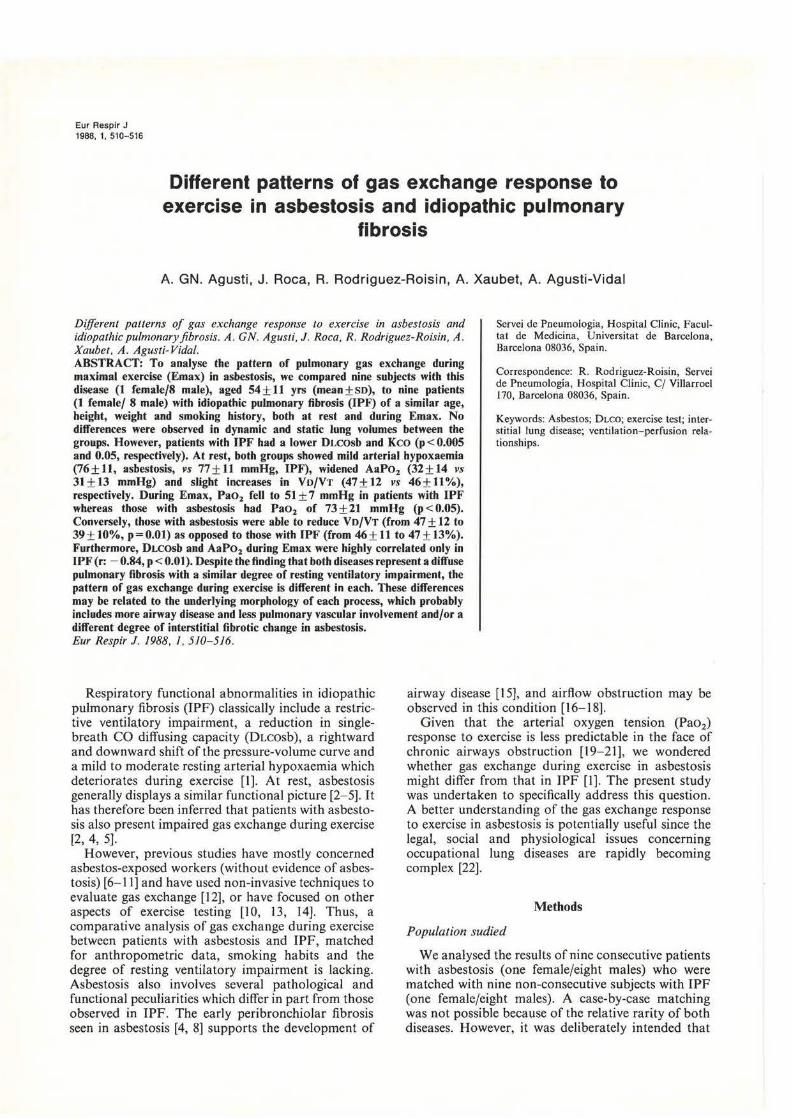
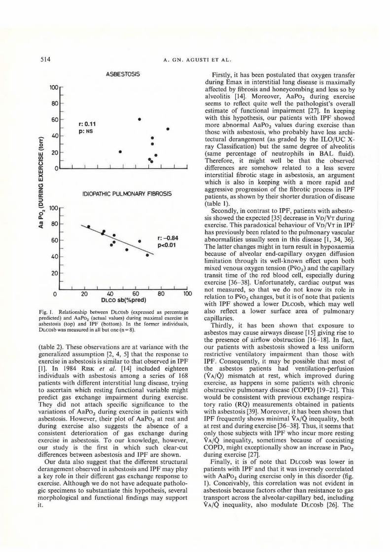
tory rate and YE during exercise were greater in IPF (p < 0.025 and < 0.05, respectively). It is of interest that Vo2 during Emax was similar in both groups, expressed either as absolute values or percentage of predicted (table 2). In other words, each group performed the same amount of exercise (representing almost 60% of their maximal predicted [3 I]). However, Pao2 fell dramaticaJly (from 77 ± ll to 51± 7 mmHg, p<O.OOl) and AaPo2 rose (from 31 ± 13 to 60± 11 mmHg, p<O.OOl) during exercise in the IPF group but remained essentially unchanged in the asbestosis group (from 76 ± 11 to 73 ± 2 I mmHg and from 32± 14 to 35±23 mmHg, respectively). Analysis of individual data in patients with asbestosis showed that AaPo2 decreased during exercise in two, remained unchanged in four and deteriorated in three. In contrast, exercise induced a deleterious effect in all patients with IPF, in keeping with former studies [1, 14, 26, 34). On the other hand, subjects with asbestosis were able to lower Vo/VT with exercise (p=O.Ol) unlike those with IPF (table 2), a finding also previously reported [I , 14, 26, 34). Moreover, DLcosb expressed as percentage of predicted was inversely correlated with AaPo2 during Emax only in IPF (r: - 0.84, p < 0.01) (fig. I).
Discussion
Our study highlights the fact that the pattern of gas exchange during exercise is different in asbestosis and idiopathic pulmonary fibrosis (IPF), even though both groups were matched for age, sex, height, weight, smoking habits and the severity of resting ventilatory impairment. Patients with IPF showed a dramatic fall in Pao2 during exercise, whilst those with asbestosis did not. In addition, Vo /VT fell with exercise only in patients with asbestosis, who also presented a lower increase in minute ventilation
Table 2. -Mean (±so) metabolic, circulatory and gas exchange data for patients with asbestosis and idiopathic pulmonary fibrosis (IPF), at rest and during exercise.
Rest
Asbestosis TPF
Heart rate bpm 78±14 83±17 Resp. rate bpm 22±6 21±6 VE l·min 11±2 13±3 yo2 ml·min-1 373±48 302±59 Vco2 ml·min'1 240±40 250±51 arterial pH 7.42±0.02 7.42±0.03 Pao2 mmHg 76±11 77±11 Pao2 mmHg 37±3 35±4 AaPo
2 mmHg 32±14 31±13
VoNT % 47±12 46±11
p< Asbestosis
121±14 (74±6%) 34±11 37±8 (68±34%)
1074±373 (56±20%) 986±348
7.39±0.04 73±21 38±3 35±23 39±10
Exercise
IPF
123±7 (75±11%) 46±11 49±14 (67±17%)
1089±266 (59±14%) 1054±277 7.37±0.06
51±7 37±5
60±11 47±13
p<
0.025 0.05
0.05
0.05
P values denote the significance of differences between patients with asbestosis and IPF in each condition (rest/exercise). The significance of changes induced by exercise within each group is not shown. Values between brackets correspond to percentage of maximum predicted during exercise [20].
514 A. GN. AGUSTI ET AL .
~ .. .. 0 e UJ en 0 a: 0 w >< w CJ z a: ::;)
e. .. 0 Q. as <
0
r: 0.11 p:NS
ASBESTOSIS
•
• • • • ••
•
IDIOPATHIC PULMONARY FIBROSIS
20 40 60 DLCO sb(%pred)
r: ~.84
P<0.01
100
Fig. I. Relationship between DLCOsb (expressed as percentage predicted) and AaPo2 (actual values) during maximal exercise in asbestosis (top) and IPF (bottom). In the former individuals, DLCOsb was measured in all but one (n = 8).
(table 2). These observations are at variance with the generalized assumption [2, 4, 5] that the response to exercise in asbestosis is similar to that observed in IPF [1). In 1984 R ISK et al. [14] included eighteen individuals with asbestosis among a series of 168 patients with different interstitial lung disease, trying to ascertain which resting functional variable might predict gas exchange impairment during exercise. They did not attach specific significance to the variations of AaPo2 during exercise in patients with asbestosis. However, their plot of AaPo2 at rest and during exercise also suggests the absence of a consistent deterioration of gas exchange during exercise in asbestosis. To our knowledge, however, our study is the first in which such clear-cut differences between asbestosis and IPF are shown.
Our data also suggest that the different structural derangement observed in asbestosis and IPF may play a key role in their different gas exchange response to exercise. Although we do not have adequate pathologic specimens to substantiate this hypothesis, several morphological and functional findings may support it.
Firstly, it has been postulated that oxygen transfer during Emax in interstitial lung disease is maximally affected by fibrosis and honeycombing and less so by alveolitis [14]. Moreover, AaPo2 during exercise seems to reflect quite well the pathologist's overaiJ estimate of functional impairment [27]. In keeping with this hypothesis, our patients with IPF showed more abnormal AaPo2 values during exercise than those with asbestosis, who probably have less architectural derangement (as graded by the ILO/UC Xray Classification) but the same degree of alveolitis (same percentage of neutrophils in BAL fluid). Therefore, it might well be that the observed differences are somehow related to a less severe interstitial fibrotic stage in asbestosis, an argument which is also in keeping with a more rapid and aggressive progression of the fibrotic process in IPF patients, as shown by their shorter duration of disease (table 1).
Secondly, in contrast to IPF, patients with asbestosis showed the expected [35] decrease in Vo/VT during exercise. This paradoxical behaviour ofVo/VT in IPF has previously been related to the pulmonary vascular abnormalities usually seen in this disease [I, 34, 36). The latter changes might in turn result in hypoxaemia because of alveolar end-capillary oxygen diffusion limitation through its well-known effect upon both mixed venous oxygen tension (Pvo2) and the capillary transit time of the red blood cell, especially during exercise (36-38). Unfortunately, cardiac output was not measured, so that we do not know its role in relation to Pvo2 changes, but it is of note that patients with IPF showed a lower DLcosb, which may well also reflect a lower surface area of pulmonary capillaries.
Thirdly, it has been shown that exposure to asbestos may cause airways disease [15) giving rise to the presence of airflow obstruction [ 16-18). In fact, our patients with asbestosis showed a less unifonn restrictive ventilatory impairment than those with IPF. Consequently, it may be possible that most of the asbestos patients had ventilation-perfusion (VA/Q.) mismatch at rest, which improved during exercise, as happens in some patients with chronic obstructive pulmonary disease (COPD) [ 19-21 ]. This would be consistent with previous exchange respiratory ratio (RQ) measurements obtained in patients with asbestosis [39]. Moreover, it has been shown that IPF frequently shows minimal VA/Q inequality, both at rest and during exercise [36- 38). Thus, it seems that only those subjects with IPF who incur more resting VA/Q inequality, sometimes because of coexisting COPD, might exceptionally show an increase in Pao2 during exercise [27).
Finally, it is of note that DLcosb was lower in patients with IPF and that it was inversely correlated with AaPo2 during exercise only in this disorder (fig. 1). Conceivably, this correlation was not evident in asbestosis because factors other than resistance to gas transport across the alveolar-capillary bed, including VA/Q inequality, also modulate DLcosb [26]. The

EXERCISE I N ASBESTOS IS AND IDIOPATHIC PULMONARY FIBROSIS 515
possibility that it might also have emerged in patients with asbestosis, if more subjects with lower DLcosb had been included, cannot be ruled out. H owever, an analysis of our individual data shows that only two patients with asbestosis had a OLCosb lower than 65% of predicted (63 and 48%, respectively) and that Pao2 fell markedly during exercise in the former (from 70 to 49 mmHg) whereas it remained unchanged in the latter (84 to 84 mmHg). In addition, despite the fact that mean DLcosb was different in each group (table 1), mean AaPo2 was quite close at rest but clearly differed during exercise (table 2). These findings suggest that the mechanisms underlying pulmonary gas exchange in these two disorders may not necessarily be coincidental and that a marked reduction in resting DLcosb does not need to be a functiona l marker of arterial desaturation during exercise, at least in asbestosis.
To summarize, the present investigation shows that, in contrast to TPF, pulmonary gas exchange in asbestosis is not systematically impaired during exercise. Conceivably, this may be related to the presence of more airways disease and less pulmonary vascular involvement and/or a lower degree of interstitial fibrotic changes in asbestosis, which in turn may result in a different pattern of VA/Q mismatching.
Acknowledgemenls: This paper was supported, in part, by Grants from the US-Spain Joint Committee (CCA 8309185), CAICYTPR 82-1787 & PR84-0770, and SEPAR-1983. The authors are grateful to P.D. Wagner, for his helpful comments and criticisms; to F. Burgos, C. Gistau, T. Lecha, M. Simo, C. Argaiia and F.A. L6pez for their invaluable technical assistance, and to the Medical Staff of our Service for their cooperation in the care of the patients.
References
I. Crystal RG, Fulmer JD, Robcrts WC, Moss ML, Line BR, Reynolds HY. - Idiopathic pulmonary fibrosis. Clinical, histologic, radiographic, physiologic, scintigrafic, cytologic and biochemical aspects. Ann Intern Med, 1976, 85, 769- 788. 2. Bader ME, Bader RA, Sclikoff IJ. - Pulmonary function in asbestosis of the lung. Am J Med, I 96 I, 30, 235- 242. 3. Becklake M R. - Asbestos-related diseases of the lungs and pleura. Current clinical issues. Am Rev Respir Dis, 1982, 126, 187-194. 4. Beck lake MR. - Asbestos-related fibrosis of the lung (asbestosis) and pleura. In: Pulmonary diseases and disorders, Update 1. A.P. Fishman ed., McGraw Hill, Philadelphia, 1982, pp. 167- 192. 5. Selikoff 11, Lee DHK eds. - Asbestos and disease. Academic Press, New York, 1978. 6. Epler GR, Saber FA, Gaensler EA. - Determination of severe impairment (disability) in interstitial lung disease. Am Rev Respir Dis, 1980, 121, 647-659. 7. Pearle J.- Exercise performance and functional impairment in asbestos-exposed workers. Chest, 1981, 80, 701-705. 8. Howard J, Mohsenifar Z, Brown HV, Koerner SK. - Role of exercise testing in assesing functional respiratory impairment due to asbestos-exposure. J Occup Med, 1982, 24, 685- 689. 9. Sue DY, Oren A, Hansen JE, Wasscrman K. - Lung function and exercise performance in smoking and nonsmoking asbestosexposed workers. Am Rev Respir Dis, 1985, 132, 612- 618. 10. Oren A, Sue DY, Hansen JE, Torrance DJ, Wasserman K. -
The role of exercise testing in impairment evaluation. Am Rev Respir Dis, 19!l7, 135, 230- 235. I I. Sue DY, Oren A, Hansen JE, Wasserman K. - Diffusing capacity for carbon monoxide as a predictor of gas exchange during exercise. N Engl J Med, 1987, 316, 1301-1306. 12. Apps MC, Brittoo MG, Maxwell DL, Hughes DTD, Hanson A. - Brcathlessnel;S and hypoxia on exercise in patients with asbestos related disease (Abstract). Am Rev Respir Dis, 1983, 127, A180. 13. Eriksson L, Jonson B, Wollmer Pet al. - Lung function and working capacity in workers exposed to asbestos-cement. Eur J Respir Dis, 1981, 62 (Suppl.), 56-59. 14. Risk C. Epler GR, Gaensler EA. - Exercise alveolar-arterial oxygen pressure difference in interstitial lung disease. Chest, 1984, 85, 69- 74. 15. Becklake MR. Ernst P. - Asbestos exposure and airway responses. In: Occupational lung disease. J.B.L Gee ed., Churchill Livingstone, New York, 1984, pp. 25-50. 16. Muldoon BC, Turner-Warwick M. - Lung function studies in asbestos workers. Brit J Dis Chest, 1972, 66, 121 - 132. 17. Rodriguez-Roisin R, Merchant JEM, Cochrane GM, Hickey BPH, Turner-Warwick M, Clark TJH. - Maxima.! expiratory flow volume curves in workers exposed to asbestos. Respiration, 1980, 39, 158-165. 18. Segarra F, Baselga Monte M, Lopez lbaiiez P, Perez Nicolas J. - Asbestosis in a Barcelona fibre-cement factory. Environ Res, 1980, 23, 292-300. 19. Simonsson B, Malmberg R, Berglund E. - Pulmonary gas exchange at rest and during exercise in chronic bronchitis. Scand J Respir Dis, 1969, 50, 245-258. 20. Minh VD, Lee HM, Dolan GF, Light RW, Bell J, Vasquez P. - Hypoxemia during exercise in patients with chronic obstructive pulmonary disease. Am Rev Respir Dis, 1979, 120, 787- 794. 21. Raffestin B, Escourrou P, Legrand A, Duroux P, Lockhart A. - Circulatory transport of oxygen in patients with chronic airflow obstruction exercising maximally. Am Rev Respir Dis, 1982, 125, 426-431 . 22. Wiedemann HP, Gee JBL, Balmes JR, Loke J . - Exercise testing in occupational lung diseases. Clin Chest Med, 1985, 5, 157-171. 23. Xaubet A, Rodriguez-Roisin R, Bombi JA, MarinA, Roca J, Agusli-Vidal A. - Correlation of bronchoalveolar lavage and clinical and functional findings in asbestosis. Am Rev Respir Dis, I 986, I 33, 848- 854. 24. ILO/ UC International Classification of Radiographs of Pneumoconiosis, 1980. Occupational Safety and Health Series No. 22, revised. Geneva: International Labour Office, 1980. 25. Abraham JL, Burnett B, Rodriguez-Roisin R. - Correlated environmental, radiologic, physiologic, pathologic and mineralogic analysis in asbestos workers (Abstract). Am Rev Respir Dis, 1982, 125, Al54. 26. Fulmer JD, Roberts WC, Von Gal ER, Crystal RG. -Morphologic-physiologic correlates of the severity of fibrosis and degree of cellularity in idiopathic pulmonary fibrosis. J Clin Invest, 1979, 63, 665-676. 27. Carrington CB, Gaensler EA, Coutu RE, Fitzgerald MX, Gupta RG. - Natural history and treated course of usual and desquamative interstitial pneumonia. N Engl J Med, 1978, 298, 801- 809. 28. Roca J , Sanchis J, Agusti-Vidal A, et al. - Spirometric reference values for a Mediterranean population. Bull Eur Plrysiopathol Respir, 1986, 22, 217- 224. 29. Roca J, Segarra F, Rodriguez-Roisin R, Cobo E, Martinez J, Agusti-Vidal A. - Static lung volumes and single-breath diffusion capacity reference values from a Latin population (Abstract). Am Rev Respir Dis, 1985, 131, A352. 30. Jones NL, Campbell EJM eds. - Clinical exercise testing. Saunders, Philadelphia. 1982. 31 . Jones NL, Makrides L, Hitchcock C, Chypchar T, McCartney N. - Normal standards for an incremental progressive cycle ergometer test. Am Rev Respir Dis, 1985, 131, 700- 708. 32. Watters LC, King TE, Chemiack RE, Waldron JA, Stanford RE, Willcox ML, Christopher KL, Schwarz Ml. - Bronchoalveolar lavage fluid neutrophils increase after corticosteroid

516 A. GN. A GUST! ET AL.
therapy in smokers with idiopathic pulmonary fibrosis. Am Rev Respir Dis, 1986, 133, 104-109. 33. Haslam PL, Dewar A, Butchers P, Primett ZS, NewmanTaylor A, Turner-Warwick M. - Mast cells, atypicallymphocytes and neutrophils in bronchoalvcolar lavage in extrinsic allergic alveolitis. Comparison with other interstitial lung diseases. Am Rev Respir Dis, 1987, 135, 35-47. 34. Keogh BA, Lakatos E, Price D, Crystal RG. -Importance of the lower respiratory tract in oxygen transfer. Am Rev Respir Dis. 1984, 129, 576-880. 35. Wasserman K, Whipp JB. - Exercise physiology in health and disease. Am Rev Respir Dis, 1975, 112, 219-249. 36. Agusti AGN, Roca J, Rodriguez-Roisin R, Gea J, Xaubet A, Wagner PD. - Role of 0 2 diffusion limitation in idiopathic pulmonary fibrosis (Abstract). Am Rev Respir Dis, 1987, 135, A307. 37. Wagner PD. - Ventilation-perfusion inequality and gas exchange during exercise in lung disease. In : Muscular exercise and the lung. J.A. Dempsey, C.E. Reed eds., Univ Wisconin Press, Madison, 1977, pp. 345- 356. 38. Jemudd-Wilhelmsson Y, Hornblad Y, Hedenstierna G. -Ventilation-perfusion relationships in interstitial lung disease. Eur J Respir Dis, 1986, 68, 39-49. 39. Bates DV, Macklem PT, Christie RV. - Response to chemical and physical irritants. In: Respiratory function in disease. D.V. Bates. P.T. Macklcm and R .V. Christie eds., Saunders, Philadelphia, 1971, pp. 361-400.
RESUME: Pour !'etude des echanges gazeux pulmonaires au cours de !'effort maximal (Emax) dans l'asbestose, nous avons
compare 9 sujets atteints de cette affection (I femme et 8 hommes) ages de 54± 11 ans (x±so) a 9 patients (I femme et 8 hommes) atteints de fibrose pulmonaire idiopathique, d'un age, d'une taiUe, d'un poids, et d'unc anamnese de tabagisme similaires, a la fois au repos et au cours de !'effort maJtimal. L'on n'a observe aucune difference dans lcs volumes dynamiques et statiques entre lcs deux groupcs. Toutefois, les patients atteints de fibrose pulmonaire idiopathique avaient une DLcosb et une Kco plus basses (p <0.005 et 0.05, respectivement). Au repos, les deux groupes ont une hypoJtemie arterielle legere (asbcstose: 76± 11, vs fibrose pulmonaire 77± 11, mmHg), unc augmentation de la 6aPo2 (32± 14 vs 31 ± 13 mmHg) et de legeres augmentations du rapport Vo/VT (47± 12 VS 46± 11%). Pendant !'effort maximal, la Pao2 tombc a 51 ± 7 mmHg chez les patients attcints de fibrose idiopathique, alors que ceux atteints d'asbestose ont une Pao2 de 73±21 mmHg (p < 0.05). D'autre part, lcs sujets atteints d'asbestosc peuvent reduire !cur rapport VojVT (de 47 ± 12 a 39± 10%, p=O.Ol), alors que ccux atteints de fibrose idiopathique ne le font pas (46± 11 vcrs 47 ± 13%). D'autre part , la DLcosb et la 6aPo2 au cours de !'effort maximal ne sont en excellente correlation que dans la fibrose pulmonaire idiopathique (r:= - 0.84, p < O.OI). Quoique les deux affections representent des fibroses pulmonaires diffuses entrainant un degre similaire d'insuffisance ventilatoire au repos, les modifications des echanges gazeux au cours de !'effort y sont differentes. Cette difference pourrait etre en relation avec la morphologic sousjaccntc a chacun des deux processus, qui entraine probablement plus de maladies bronchiques et moins d'atteinL~ vasculaires pulmonaires, ainsi quefou des degres differents de modifications interstitielles fibreuscs dans l'asbestose.