Embed Size (px)
Citation preview

Differential effects of alendronate and losartantherapy on osteopenia and aortic aneurysm in micewith severe Marfan syndrome
Harikiran Nistala1,{, Sui Lee-Arteaga1,{, Luca Carta1,{, Jason R. Cook1,{, Silvia Smaldone1,
Gabriella Siciliano1, Aaron N. Rifkin1,2, Harry C. Dietz3, Daniel B. Rifkin2 and
Francesco Ramirez1,∗
1Department of Pharmacology and Systems Therapeutics at the Mount Sinai School of Medicine, New York, NY
10029, USA, 2Department of Cell Biology, New York University School of Medicine, New York, NY 10016, USA and3Departments of Pediatrics, Medicine and Molecular Biology and Genetics, and the Howard Hughes Medical Institute,
Johns Hopkins University School of Medicine, Baltimore, MA 21205, USA
Received July 30, 2010; Revised and Accepted September 13, 2010
Reduced bone mineral density (osteopenia) is a poorly characterized manifestation of pediatric and adultpatients afflicted with Marfan syndrome (MFS), a multisystem disorder caused by structural or quantitativedefects in fibrillin-1 that perturb tissue integrity and TGFb bioavailability. Here we report that mice with pro-gressively severe MFS (Fbn1mgR/mgR mice) develop osteopenia associated with normal osteoblast differen-tiation and bone formation. In vivo and ex vivo experiments, respectively, revealed that adult Fbn1mgR/mgR
mice respond more strongly to locally induced osteolysis and that Fbn1mgR/mgR osteoblasts stimulate pre-osteoclast differentiation more than wild-type cells. Greater osteoclastogenic potential of mutant osteoblastswas largely attributed to Rankl up-regulation secondary to improper TGFb activation and signaling. Losartantreatment, which lowers TGFb signaling and restores aortic wall integrity in mice with mild MFS, did not miti-gate bone loss in Fbn1mgR/mgR mice even though it ameliorated vascular disease. Conversely, alendronatetreatment, which restricts osteoclast activity, improved bone quality but not aneurysm progression inFbn1mgR/mgR mice. Taken together, our findings shed new light on the pathogenesis of osteopenia in MFS,in addition to arguing for a multifaceted treatment strategy in this congenital disorder of the connectivetissue.
INTRODUCTION
Mutations that affect the structure or expression of theextracellular matrix (ECM) glycoprotein fibrillin-1 causepleiotropic manifestations in Marfan syndrome (MFS;OMIM-154700) by impairing connective tissue integrity andby promoting improper latent TGFb activation (1). Consistentwith the involvement of promiscuous TGFb signaling in MFSpathogenesis, systemic administration of TGFb-neutralizingantibodies to mouse models of MFS improves cardiovascular,skeletal muscle and lung abnormalities (2–5). Similarly, treat-ment with losartan, an angiotensin II receptor 1 (AT1R) blocker
(ARB) that lowers TGFb signaling (6,7), restores aortic wallarchitecture in mice with mild MFS (Fbn1C1039G/+ mice)and mitigates aortic root dilation in children with severeMFS (4,8). Although these findings are encouraging, thereare reasons to believe that some MFS patients may notrespond to losartan and/or that this treatment may not curtailother morbid manifestations of MFS, such as those affectingthe skeleton (9–11).
Osteopenia is a controversial finding in MFS, especially inpediatric patients (12–19). Factors that contribute to ambigu-ity include the lack of standardized protocols to comparebone mineral density (BMD) between affected and healthy
†These authors have contributed equally to the study.
∗To whom correspondence should be addressed at: One Gustave L. Levy Place, Box 1603, New York, NY 10029, USA. Tel: +1 2122417237;Fax: +1 2129967214; Email: [email protected]
# The Author 2010. Published by Oxford University Press. All rights reserved.For Permissions, please email: [email protected]
Human Molecular Genetics, 2010, Vol. 19, No. 24 4790–4798doi:10.1093/hmg/ddq409Advance Access published on September 24, 2010
individuals, and the absence of robust normative data for chil-dren (11,20). While preliminary analyses suggest thatmutations in fibrillin-1 cause osteopenia in mice, the under-lying mechanism is similarly controversial due to inherentlimitations of the mutant mouse lines employed in thesestudies (21–23). Progressive bone loss and reduced BMD inTight skin (Tsk/+) mice were correlated with a decreasedrate of osteoblast maturation in vitro and implicitly, withreduced bone formation in vivo (21). The Tsk mutation is alarge internal duplication of fibrillin-1 that in heterozygosityinterferes with microfibril biogenesis and leads to a phenotypethat combines pulmonary and skeletal manifestations of MFSwith cutaneous fibrosis of Stiff Skin syndrome (SSS;OMIM-184900) (24–27). The unusual nature of the Tskmutation, together with the unique phenotype of Tsk/+mice, however raises questions as to whether these animalstruly replicate MFS pathogenesis. In contrast to Tsk/+ osteo-blasts, recent analyses showed that cultured osteoblasts fromFbn12/2 mice mature slightly faster than wild-type cellsand stimulate osteoclast activity in vitro more than controlcells, suggesting a probable defect of bone resorption ratherthan bone formation in these mutant mice (22,23). Neonatallethality of Fbn12/2 mice (28) has unfortunately precludedvalidating this conclusion through the analysis of bone remo-deling in adult mice.
The present study therefore interrogated the role offibrillin-1 in bone remodeling using a combination of in vivoand ex vivo experiments and a mouse model of progressivelysevere adult lethal MFS [Fbn1mgR/mgR mice (29)]. The resultsof our analyses indicate that osteopenia in Fbn1mgR/mgR miceis largely driven by increased bone resorption due toTGFb-dependent dysregulation of the local couplingbetween osteoblast and osteoclast activities. Consistent withthis observation, bisphosphonate restriction of osteoclastactivity mitigated progressive bone loss in Fbn1mgR/mgR
mice. In contrast, losartan treatment was only effective inimproving vascular disease in this adult lethal model ofMFS. Collectively, these findings suggest that a multifacetedtreatment strategy will probably be required to target distinctcellular events that are responsible for organ-specific manifes-tations in MFS.
RESULTS
Fbn1mgR/mgR mice display normal bone formation andincreased bone resorption
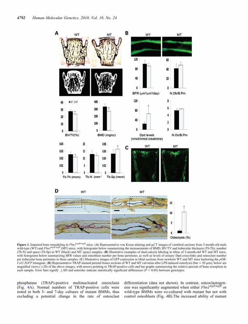
Fbn1mgR/mgR mice produce �15% of the normal amount offibrillin-1 and have a reduced average lifespan of 2–4months due to dissecting aortic aneurysm and pulmonaryinsufficiency (29). Bone histomorphometry and micro-computed tomography (mCT) scanning of lumbar vertebrasfrom 3-month-old Fbn1mgR/mgR mice revealed 15% less bonecontent (bone volume over total volume; BV/TV), 19.7%reduction in apparent BMD (bone mineral content over TV)and decreased trabecular thickness and greater trabecularspace compared with the wild-type counterparts (Fig. 1A).Trabecular number, however, was equivalent to controls(Fig. 1A). Osteopenia in Fbn1mgR/mgR mice was further corre-lated with increased bone resorption, as evidenced by the
greater amount of urinary deoxypyridinoline (Dpd) collagencross-links, a normal rate of bone formation (BFR), as inferredby the unremarkable pattern of dual calcein label incorpor-ation, and an apparently normal complement of both surfaceosteoblasts and osteoclasts (Fig. 1B). Comparable levels ofGFP fluorescence were also noted in neonatal bones ofFbn1mgR/mgR mice and wild-type littermates that harbor thepOBCol2.3GFP transgene, a marker of differentiating osteo-blasts (Fig. 1C) (30). This last finding contrasts with thedecrease in number of mature (GFP-positive) osteoblasts pre-viously observed in the bones of Tsk/+;pOBCol2.3GFP mice(21).
Fbn1mgR/mgR mice were subject to acute stress, namely cal-varial overlay of lipolysaccharide (LPS)-coated titanium par-ticles (31), in order to resolve the apparent discrepancybetween the high levels of urinary Dpd cross-links and thenormal number of surface osteoclasts. Histological analysesof parietal bone sections revealed substantially moreLPS-induced local osteolysis in Fbn1mgR/mgR than in wild-typemice, a finding in agreement with the collagen cross-link datareflecting an unbalanced skeletal turnover in adult Fbn1mgR/
mgR mice (Fig. 1D). This acute injury test also implied thepresence of an osteoclastogenic defect that was undetectedby the single time-point assessment of osteoclast numberduring the chronic process of bone remodeling.
Fbn1mgR/mgR osteoblasts have greater TGFb-dependentosteoclastogenic potential
Local coupling of osteoblast and osteoclast activities is centralto a physiologically balanced skeletal turnover (32). Ex vivoexperiments were therefore performed to examine mutantosteoblast and osteoclast differentiation and thus unravel mech-anisms of defective bone resorption in Fbn1mgR/mgR mice. Con-sistent with the BFR data (Fig. 1B), cultured osteoblasts fromwild-type and Fbn1mgR/mgR mice yielded comparable numbersof mineralized nodules and expressed similar levels of tran-scripts coding for key regulators of cell cycle progression andosteoblast differentiation (Fig. 2A and B). Additionally,normal Col1a2 expression in mutant osteoblasts was in linewith normal osteogenic differentiation in Fbn1mgR/mgR; pOB-Col2.3GFP bones (Fig. 1C), whereas normal Fbn2 expressionruled out a compensatory up-regulation of fibrillin-2 productionin these mutant cells (Fig. 2B). Cell-based assays furthermoreshowed that, compared with controls, conditioned media fromFbn1mgR/mgR osteoblast cultures display more TGFb and BMPactivity, which were respectively associated with normallevels of total TGFb and Bmp transcripts (Fig. 3). These lastfindings supported the emerging notion that mutations infibrillin-1 perturb ECM sequestration of both TGFb and BMPcomplexes, and that enhanced BMP signaling counteracts thenegative effect of increased TGFb signaling on osteoblastmaturation and bone formation (22).
Next, wild-type or Fbn1mgR/mgR bone marrow monocytes(BMMs) were grown in the presence of osteoclastogenicfactors that were either administered exogenously or providedby co-cultured osteoblasts. Consistent with the lack of fibrillinexpression in progenitor and differentiated osteoclasts(http://biogps.gnf.org/#goto), control and mutant BMM cul-tures yielded identical numbers of tartrate-resistant acid
Human Molecular Genetics, 2010, Vol. 19, No. 24 4791
phosphatase (TRAP)-positive multinucleated osteoclasts(Fig. 4A). Normal numbers of TRAP-positive cells werenoted in both 5- and 7-day cultures of mutant BMMs, thusexcluding a potential change in the rate of osteoclast
differentiation (data not shown). In contrast, osteoclastogen-esis was significantly augmented when either Fbn1mgR/mgR orwild-type BMMs were co-cultured with mutant but not withcontrol osteoblasts (Fig. 4B).The increased ability of mutant
Figure 1. Impaired bone remodeling in Fbn1mgR/mgR mice. (A) Representative von Kossa staining and mCT images of vertebral sections from 3-month-old malewild-type (WT) and Fbn1mgR/mgR (MT) mice, with histograms below summarizing the measurements of BMD, BV/TV and trabecular thickness (Tb.Th), number(Tb.N) and space (Tb.Sp) in WT (black) and MT (gray) samples. (B) Illustrative examples of dual-calcein labeling in tibias of 3-month-old WT and MT mice,with histograms below summarizing BFR values and osteoblast number per bone perimeter, as well as levels of urinary Dpd cross-links and osteoclast numberper trabecular bone perimeter in these samples. (C) Illustrative images of GFP expression in tibial sections from newborn WT and MT mice harboring the pOB-Col2.3GFP transgene. (D) Representative TRAP-stained parietal bones sections of WT and MT calvarias after LPS-induced osteolysis (bar ¼ 50 mm); below aremagnified views (×20) of the above images, with arrows pointing to TRAP-positive cells and bar graphs summarizing the relative percent of bone resorption ineach sample. Error bars signify +SD and asterisks indicate statistically significant differences (P , 0.05) between genotypes.
4792 Human Molecular Genetics, 2010, Vol. 19, No. 24
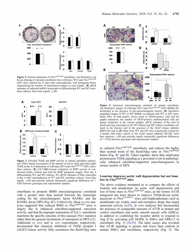
osteoblasts to promote BMM osteoclastogenesis correlatedwith a greater ratio than normal between the transcriptscoding for the osteoclastogenic factor RANKL and theRANKL decoy OPG (Fig. 4C). Collectively, these ex vivo ana-lyses suggested that reduced BMD in Fbn1mgR/mgR mice islargely due to enhanced osteoblast-supported osteoclastactivity, and that the improper maturation of Tsk/+ osteoblastsmanifests the specific outcome of this unusual Fbn1 mutationrather than the general mechanism of osteopenia in MFS (21).Additional ex vivo and in vivo experiments, respectively,documented that chemical inhibition of TGFb receptor I(ALK5) kinase activity fully normalizes the Rankl/Opg ratio
in cultured Fbn1mgR/mgR osteoblasts, and reduces the higherthan normal trend of the Rankl/Opg ratio in Fbn1mgR/mgR
bones (Fig. 4C and D). Taken together, these data implicatedpromiscuous TGFb signaling as a proximal event in pathologi-cally enhanced osteoblast-supported osteoclastogenesis inmouse models of MFS.
Losartan improves aortic wall degeneration but not boneloss in Fbn1mgR/mgR mice
The above evidence prompted us to compare the effects oflosartan and alendronate on aortic wall degeneration andloss of bone mass in Fbn1mgR/mgR mice, as the former AT1Rantagonist has been shown to improve TGFb-driven aorticaneurysm in Fbn1C1039G/+ mice and bisphosphonates such asalendronate are widely used anti-resorptive drugs that targetosteoclast activity (4,33). In vitro analyses first documentedthe presence of comparable amounts of AT1R in mutant andcontrol osteoblasts and aortic smooth muscle cells (SMCs),in addition to confirming the receptor ability to respond toAng II by activating p38 MAPK in SMCs and ERK1/2 inosteoblasts (Fig. 5) (34,35). The latter assays also revealedthat AT1R signaling is greater and lower than controls inmutant SMCs and osteoblasts, respectively (Fig. 5). The
Figure 2. Normal maturation of Fbn1mgR/mgR osteoblasts. (A) Illustrative vonKossa staining of calvarial osteoblasts from wild-type (WT) and Fbn1mgR/mgR
(MT) mice cultured for 21 days after osteoinduction, with histograms belowsummarizing the number of mineralized nodules in each sample. (B) qPCRestimates of indicated mRNA transcripts in differentiating WT and MT osteo-blast cultures. Error bars signify +SD.
Figure 3. Elevated TGFb and BMP activity in mutant osteoblast cultures.(A) TMLC-based assessments of the amount of active (left) and total (right)TGFb at day 4 of maturation of wild-type (WT) and Fbn1mgR/mgR (MT) osteo-blast cultures. (B) C2C12BRA-based evaluation of BMP activity in con-ditioned media, without and with the BMP antagonist noggin, from day 4differentiating WT and MT cultures. (C) qPCR estimates of Bmp transcriptsat day 4 after osteoinduction of WT and MT osteoblast cultures. Error barssignify +SD and asterisks indicate statistically significant differences (P ,
0.05) between genotypes and experimental samples.
Figure 4. Increased osteoclastogenic potential of mutant osteoblasts.(A) Illustrative images of wild-type (WT) and Fbn1mgR/mgR (MT) BMMs dif-ferentiated in the presence of pro-osteoclastogenic factors; (B) illustrativemagnified images of WT or MT BMMs co-cultured with WT or MT osteo-blasts (Ob). In both panels, arrows point to TRAP-positive cells and bargraphs summarize the number of TRAP-positive multinucleated cells persquare centimeter in the various samples. qPCR estimates of the ratio ofRankl and Opg transcripts in (C) WT (black) and MT (white) osteoblasts cul-tured in the absence and in the presence of the ALK5 kinase inhibitorSB431542 and in (D) tibias from WT and MT mice systemically treated for2 months with either vehicle or the ALK5 kinase inhibitor SD-208. Errorbars represent +SD and asterisks signify statistically significant differences(P , 0.05) between genotypes and experimental samples.
Human Molecular Genetics, 2010, Vol. 19, No. 24 4793
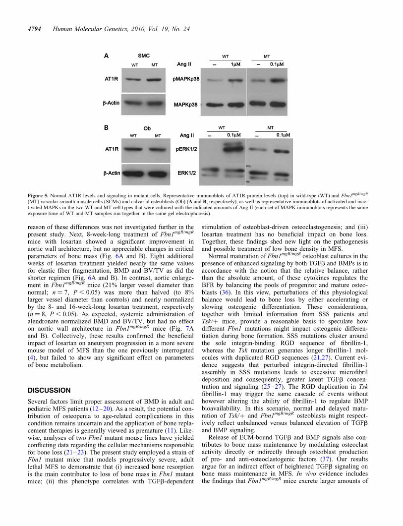
reason of these differences was not investigated further in thepresent study. Next, 8-week-long treatment of Fbn1mgR/mgR
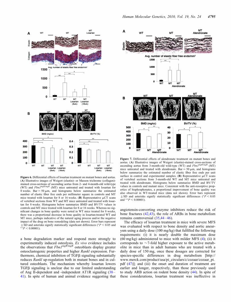
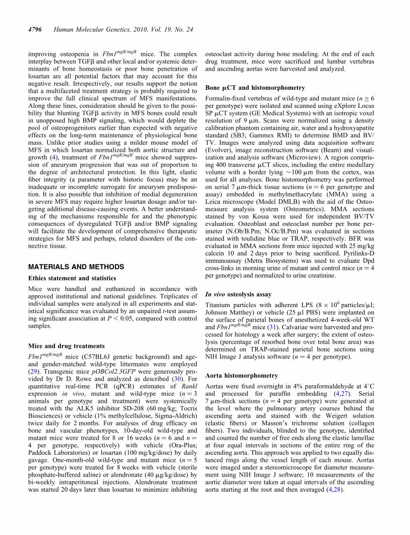
mice with losartan showed a significant improvement inaortic wall architecture, but no appreciable changes in criticalparameters of bone mass (Fig. 6A and B). Eight additionalweeks of losartan treatment yielded nearly the same valuesfor elastic fiber fragmentation, BMD and BV/TV as did theshorter regimen (Fig. 6A and B). In contrast, aortic enlarge-ment in Fbn1mgR/mgR mice (21% larger vessel diameter thannormal; n ¼ 7, P , 0.05) was more than halved (to 8%larger vessel diameter than controls) and nearly normalizedby the 8- and 16-week-long losartan treatment, respectively(n ¼ 8, P , 0.05). As expected, systemic administration ofalendronate normalized BMD and BV/TV, but had no effecton aortic wall architecture in Fbn1mgR/mgR mice (Fig. 7Aand B). Collectively, these results confirmed the beneficialimpact of losartan on aneurysm progression in a more severemouse model of MFS than the one previously interrogated(4), but failed to show any significant effect on parametersof bone metabolism.
DISCUSSION
Several factors limit proper assessment of BMD in adult andpediatric MFS patients (12–20). As a result, the potential con-tribution of osteopenia to age-related complications in thiscondition remains uncertain and the application of bone repla-cement therapies is generally viewed as premature (11). Like-wise, analyses of two Fbn1 mutant mouse lines have yieldedconflicting data regarding the cellular mechanisms responsiblefor bone loss (21–23). The present study employed a strain ofFbn1 mutant mice that models progressively severe, adultlethal MFS to demonstrate that (i) increased bone resorptionis the main contributor to loss of bone mass in Fbn1 mutantmice; (ii) this phenotype correlates with TGFb-dependent
stimulation of osteoblast-driven osteoclastogenesis; and (iii)losartan treatment has no beneficial impact on bone loss.Together, these findings shed new light on the pathogenesisand possible treatment of low bone density in MFS.
Normal maturation of Fbn1mgR/mgR osteoblast cultures in thepresence of enhanced signaling by both TGFb and BMPs is inaccordance with the notion that the relative balance, ratherthan the absolute amount, of these cytokines regulates theBFR by balancing the pools of progenitor and mature osteo-blasts (36). In this view, perturbations of this physiologicalbalance would lead to bone loss by either accelerating orslowing osteogenic differentiation. These considerations,together with limited information from SSS patients andTsk/+ mice, provide a reasonable basis to speculate howdifferent Fbn1 mutations might impact osteogenic differen-tiation during bone formation. SSS mutations cluster aroundthe sole integrin-binding RGD sequence of fibrillin-1,whereas the Tsk mutation generates longer fibrillin-1 mol-ecules with duplicated RGD sequences (21,27). Current evi-dence suggests that perturbed integrin-directed fibrillin-1assembly in SSS mutations leads to excessive microfibrildeposition and consequently, greater latent TGFb concen-tration and signaling (25–27). The RGD duplication in Tskfibrillin-1 may trigger the same cascade of events withouthowever altering the ability of fibrillin-1 to regulate BMPbioavailability. In this scenario, normal and delayed matu-ration of Tsk/+ and Fbn1mgR/mgR osteoblasts might respect-ively reflect unbalanced versus balanced elevation of TGFband BMP signaling.
Release of ECM-bound TGFb and BMP signals also con-tributes to bone mass maintenance by modulating osteoclastactivity directly or indirectly through osteoblast productionof pro- and anti-osteoclastogenic factors (37). Our resultsargue for an indirect effect of heightened TGFb signaling onbone mass maintenance in MFS. In vivo evidence includesthe findings that Fbn1mgR/mgR mice excrete larger amounts of
Figure 5. Normal AT1R levels and signaling in mutant cells. Representative immunoblots of AT1R protein levels (top) in wild-type (WT) and Fbn1mgR/mgR
(MT) vascular smooth muscle cells (SCMs) and calvarial osteoblasts (Ob) (A and B, respectively), as well as representative immunoblots of activated and inac-tivated MAPKs in the two WT and MT cell types that were cultured with the indicated amounts of Ang II (each set of MAPK immunoblots represents the sameexposure time of WT and MT samples run together in the same gel electrophoresis).
4794 Human Molecular Genetics, 2010, Vol. 19, No. 24
a bone degradation marker and respond more strongly toexperimentally induced osteolysis. Ex vivo evidence includesthe observations that Fbn1mgR/mgR osteoblasts display greaterosteoclastogenic properties and higher Rankl expression. Fur-thermore, chemical inhibition of TGFb signaling substantiallyreduces Rankl up-regulation both in mutant bones and in cul-tured osteoblasts. The mechanism whereby losartan lowersTGFb signaling is unclear due to our limited understandingof Ang II-dependent and -independent AT1R signaling (38–41). In spite of human and animal evidence suggesting that
angiotensin-converting enzyme inhibitors reduce the risk ofbone fractures (42,43), the role of ARBs in bone metabolismremains controversial (35,44–46).
The efficacy of losartan treatment in mice with severe MFSwas evaluated with respect to bone density and aortic aneur-ysm using a daily dose (100 mg/kg) that fulfilled the followingrequirements: (i) it is nearly double the maximum dose(60 mg/kg) administered to mice with milder MFS (4); (ii) itcorresponds to �7-fold higher exposure to the active metab-olite in mice than in adult humans who are treated with adaily dose of 150 mg, once these dosages are corrected forspecies-specific differences in drug metabolism [http://www.merck.com/product/usa/pi_circulars/c/cozaar/cozaar_pi.pdf (47)]; and (iii) the onset and duration of treatment areearlier and longer, respectively, than those previously usedto study ARB action on rodent bone density (44). In spite ofthese considerations, losartan treatment was ineffective in
Figure 6. Differential effects of losartan treatment on mutant bones and aortas.(A) Illustrative images of Weigert (elastin)- or Masson trichrome (collagen)-stained cross-sections of ascending aortas from 2- and 4-month-old wild-type(WT) and Fbn1mgR/mgR (MT) mice untreated and treated with losartan for8 weeks. Bar ¼ 50 mm, and histograms below summarize the estimatednumber of elastic fiber free ends per millimeter square in controls and MTmice treated with losartan for 8 or 16 weeks. (B) Representative mCT scansof vertebral sections from WT and MT mice untreated and treated with losar-tan for 8 weeks. Histograms below summarize BMD and BV/TV values incontrols and MT mice treated with losartan for 8 or 16 weeks. Whereas no sig-nificant changes in bone quality were noted in WT mice treated for 8 weeks,there was a proportional decrease in bone quality in losartan-treated WT andMT mice, perhaps indicative of the natural aging process and/or the negativeimpact of the drug on bone remodeling (data not shown). Error bars represent+SD and asterisks signify statistically significant differences (∗P , 0.05 and∗∗P , 0.00001).
Figure 7. Differential effects of alendronate treatment on mutant bones andaortas. (A) Illustrative images of Weigert (elastin)-stained cross-sections ofascending aortas from 3-month-old wild-type (WT) and Fbn1mgR/mgR (MT)mice untreated and treated with alendronate. Bar ¼ 50 mm, and histogramsbelow summarize the estimated number of elastic fiber free ends per unitsurface in control and experimental samples. (B) Representative mCT scansof vertebral sections from 3-month-old WT and MT mice untreated andtreated with alendronate. Histograms below summarize BMD and BV/TVvalues in controls and mutant mice. Consistent with the anti-resorptive prop-erties of bisphosphonates, a proportional improvement of bone quality wasalso observed in WT-treated mice (data not shown). Error bars represent+SD and asterisks signify statistically significant differences (∗P , 0.05and ∗∗P , 0.00001).
Human Molecular Genetics, 2010, Vol. 19, No. 24 4795
improving osteopenia in Fbn1mgR/mgR mice. The complexinterplay between TGFb and other local and/or systemic deter-minants of bone homeostasis or poor bone penetration oflosartan are all potential factors that may account for thisnegative result. Irrespectively, our results support the notionthat a multifaceted treatment strategy is probably required toimprove the full clinical spectrum of MFS manifestations.Along these lines, consideration should be given to the possi-bility that blunting TGFb activity in MFS bones could resultin unopposed high BMP signaling, which would deplete thepool of osteoprogenitors earlier than expected with negativeeffects on the long-term maintenance of physiological bonemass. Unlike prior studies using a milder mouse model ofMFS in which losartan normalized both aortic structure andgrowth (4), treatment of Fbn1mgR/mgR mice showed suppres-sion of aneurysm progression that was out of proportion tothe degree of architectural protection. In this light, elasticfiber integrity (a parameter with historic focus) may be aninadequate or incomplete surrogate for aneurysm predisposi-tion. It is also possible that inhibition of medial degenerationin severe MFS may require higher losartan dosage and/or tar-geting additional disease-causing events. A better understand-ing of the mechanisms responsible for and the phenotypicconsequences of dysregulated TGFb and/or BMP signalingwill facilitate the development of comprehensive therapeuticstrategies for MFS and perhaps, related disorders of the con-nective tissue.
MATERIALS AND METHODS
Ethics statement and statistics
Mice were handled and euthanized in accordance withapproved institutional and national guidelines. Triplicates ofindividual samples were analyzed in all experiments and stat-istical significance was evaluated by an unpaired t-test assum-ing significant association at P , 0.05, compared with controlsamples.
Mice and drug treatments
Fbn1mgR/mgR mice (C57BL6J genetic background) and age-and gender-matched wild-type littermates were employed(29). Transgenic mice pOBCol2.3GFP were generously pro-vided by Dr D. Rowe and analyzed as described (30). Forquantitative real-time PCR (qPCR) estimates of Ranklexpression in vivo, mutant and wild-type mice (n ¼ 3animals per genotype and treatment) were systemicallytreated with the ALK5 inhibitor SD-208 (60 mg/kg; TocrisBiosciences) or vehicle (1% methylcellulose, Sigma-Aldrich)twice daily for 2 months. For analyses of drug efficacy onbone and vascular phenotypes, 10-day-old wild-type andmutant mice were treated for 8 or 16 weeks (n ¼ 6 and n ¼4 per genotype, respectively) with vehicle (Ora-Plus;Paddock Laboratories) or losartan (100 mg/kg/dose) by dailygavage. One-month-old wild-type and mutant mice (n ¼ 5per genotype) were treated for 8 weeks with vehicle (sterilephosphate-buffered saline) or alendronate (40 mg/kg/dose) bybi-weekly intraperitoneal injections. Alendronate treatmentwas started 20 days later than losartan to minimize inhibiting
osteoclast activity during bone modeling. At the end of eachdrug treatment, mice were sacrificed and lumbar vertebrasand ascending aortas were harvested and analyzed.
Bone mCT and histomorphometry
Formalin-fixed vertebras of wild-type and mutant mice (n ≥ 6per genotype) were isolated and scanned using eXplore LocusSP mCT system (GE Medical Systems) with an isotropic voxelresolution of 9 mm. Scans were normalized using a densitycalibration phantom containing air, water and a hydroxyapatitestandard (SB3; Gammex RMI) to determine BMD and BV/TV. Images were analyzed using data acquisition software(Evolver), image reconstruction software (Beam) and visual-ization and analysis software (Microview). A region compris-ing 400 transverse mCT slices, including the entire medullaryvolume with a border lying �100 mm from the cortex, wasused for all analyses. Bone histomorphometry was performedon serial 7 mm-thick tissue sections (n ¼ 6 per genotype andassay) embedded in methylmethacrylate (MMA) using aLeica microscope (Model DMLB) with the aid of the Osteo-measure analysis system (Osteometrics). MMA sectionsstained by von Kossa were used for independent BV/TVevaluation. Osteoblast and osteoclast number per bone per-imeter (N.Ob/B.Pm; N.Oc/B.Pm) was evaluated in sectionsstained with toulidine blue or TRAP, respectively. BFR wasevaluated in MMA sections from mice injected with 25 mg/kgcalcein 10 and 2 days prior to being sacrificed. Pyrilinks-Dimmunoassay (Metra Biosystems) was used to evaluate Dpdcross-links in morning urine of mutant and control mice (n ¼ 4per genotype) and normalized to urine creatinine.
In vivo osteolysis assay
Titanium particles with adherent LPS (8 × 10⁶ particles/ml;Johnson Matthey) or vehicle (25 ml PBS) were implanted onthe surface of parietal bones of anesthetized 4-week-old WTand Fbn1mgR/mgR mice (31). Calvariae were harvested and pro-cessed for histology a week after surgery; the extent of osteo-lysis (percentage of resorbed bone over total bone area) wasdetermined on TRAP-stained parietal bone sections usingNIH Image J analysis software (n ¼ 4 per genotype).
Aorta histomorphometry
Aortas were fixed overnight in 4% paraformaldehyde at 48Cand processed for paraffin embedding (4,27). Serial7 mm-thick sections (n ¼ 4 per genotype) were generated atthe level where the pulmonary artery courses behind theascending aorta and stained with the Weigert solution(elastic fibers) or Masson’s trichrome solution (collagenfibers). Two individuals, blinded to the genotype, identifiedand counted the number of free ends along the elastic lamellaeat four equal intervals in sections of the entire ring of theascending aorta. This approach was applied to two equally dis-tanced rings along the vessel length of each mouse. Aortaswere imaged under a stereomicroscope for diameter measure-ment using NIH Image J software; 10 measurements of theaortic diameter were taken at equal intervals of the ascendingaorta starting at the root and then averaged (4,28).
4796 Human Molecular Genetics, 2010, Vol. 19, No. 24
Cell culture assays
Osteogenic differentiation assays employed primary osteo-blasts isolated from the calvarias of 2–4-day-old mutant andwild-type mice (n ≥ 6 per genotype) and cultured as described(31). Mineral deposits were visualized at day 21 by von Kossastaining/van Geison counter-staining and quantified usingMetaMorph imaging software (Molecular Devices). For osteo-clast differentiation (n ¼ 3 per genotype), BMMs isolatedfrom the long bones of 6–8-week-old wild-type and mutantmice were seeded on 48-well plates alone and cultured inthe presence of 30 ng/ml macrophage colony-stimulatingfactor (R&D Systems) and 50 ng/ml recombinant RANKL(Sigma-Aldrich), or together with primary calvarial osteo-blasts under described conditions (31). Multinucleated TRAP-positive cells were counted using NIH Image J software. Totalprotein extracts from primary aortic SMCs and calvarial osteo-blasts (n ¼ 3 per genotype and cell type) cultured without andwith 0.1 or 1 mM Ang II (Tocris Biosciences) were preparedand analyzed by western blot analysis using antibodiesagainst AT1R (Santa Cruz Biotechnology), b-actin(Sigma-Aldrich) and phosphorylated and unphosphorylatedMAPKs p38 and ERK1/2 (Cell Signaling Technology) asdescribed previously (48).
RNA analyses
qPCR employed 1 mg of total RNA purified from differentsamples or experimental conditions (n ¼ 4 per genotype andassay) and SYBR Green Supermix with ROX (6-carboxy-X-rhodamine; Fermentas) on a Mastercycler ep Realplexinstrument (Eppendorf). All qPCR primer sets were purchasedfrom SuperArray Biosciences Corporation (b-Actin,PPM02945A; Bmp2, PPM03753A; Bmp4, PPM02998E;Bmp7, PPM03001B; Ccnd1, PPM02903E; Cmyc,PPM02924E; Col1a2, PPM04448E; Fbn2, PPM26052A;Opg, PPM03404E; Osx, PPM35999A; Rankl, PPM03047E).Thermal cycling conditions were 958C for 10 min followedby 40 cycles consisting of 958C for 15 s denaturation, 608Cfor 30 s annealing and 728C for 30 s extension. Some qPCRassays (n ¼ 3 independent assays per genotype) were also per-formed on RNA extracted from osteoblasts cultured for 4 daysin the presence of the ALK5 inhibitor SB431542 (1 mM;Sigma-Aldrich). All qPCR analyses were performed in tripli-cate, normalized against b-Actin mRNA and expressed rela-tive to the indicated controls arbitrarily averaged as 1 unit.
TGFb and BMP bioassays
Active TGFb levels were measured in calvarial osteoblastsco-cultured with TMLC cells, whereas total TGFb levelswere measured by incubating TMLC with heat-activated con-ditioned media from the same osteoblasts (n ¼ 7 per genotypeand assay) (49). Both tests were carried out in reduced serumconditions (DMEM containing 0.1% BSA). Absolute amountsof TGFb (pg/ml) were evaluated by comparing reporter genevalues with luciferase units (RLU) of TMLC treated withincreasing doses of rhTGFb1 (R&D Systems). BMP bioassayswere similarly carried out by measuring RLU of C2C12BRAcells incubated with osteoblast-conditioned media with or
without noggin (100 ng/ml; Sigma-Aldrich) (n ¼ 3 per geno-type and assay) (50). BMP activity was expressed as relativefold of luciferase induction compared with wild-type levelsarbitrarily averaged as 1 unit.
ACKNOWLEDGEMENTS
We are indebted to Dr David Rowe for supplying Col1a1transgenic mice; we also thank Ms Maria del Solar for excel-lent technical assistance and Ms Karen Johnson for organizingthe manuscript.
Conflict of Interest statement. None declared.
FUNDING
This work was supported by grants from the National Insti-tutes of Health (AR42044 and AR49698 to F.R.) and theNational Marfan Foundation. J.R.C. is a trainee in the Inte-grated Pharmacological Sciences Training Program supportedby grant T32GM062754 from the National Institute of GeneralMedical Studies.
REFERENCES
1. Ramirez, F. and Dietz, H.C. (2007) Marfan syndrome: from molecularpathogenesis to clinical treatment. Curr. Opin. Genet. Dev., 17, 252–258.
2. Neptune, E.R., Frischmeyer, P.A., Arking, D.E., Myers, L., Bunton, T.E.,Gayraud, B., Ramirez, F., Sakai, L.Y. and Dietz, H.C. (2003)Dysregulation of TGF-b activation contributes to pathogenesis in Marfansyndrome. Nat. Genet., 33, 407–411.
3. Ng, C.M., Cheng, A., Myers, L.A., Martinez-Murillo, F., Jie, C., Bedja, D.,Gabrielson, K.L., Hausladen, J.M., Mecham, R.P., Judge, D.P. et al. (2004)TGF-b activation contributes to pathogenesis of mitral valve prolapsed in amouse model of Marfan syndrome. J. Clin. Invest., 114, 1586–1592.
4. Habashi, J.P., Judge, D.P., Holm, T.M., Cohn, R.D., Loeys, B.L.,Cooper, T.K., Myers, L., Klein, E.C., Liu, G., Calvi, C. et al. (2006)Losartan, an AT1 antagonist, prevents aortic aneurysm in a mouse modelof Marfan syndrome. Science, 312, 117–121.
5. Cohn, R.D., van Erp, C., Habashi, J.P., Loeys, B.L., Klein, E.C.,Holm, T.M., Judge, D.P., Ramirez, F. and Dietz, H.C. (2007) AngiotensinII type 1 receptor blockade attenuates TGFb induced failure of muscleregeneration in multiple myopathic states. Nat. Med., 13, 204–210.
6. Lim, D.-S., Lutucuta, S., Bachireddy, P., Youker, K., Evans, A.,Entman, M., Roberts, R. and Marian, A.J. (2001) Angiotensin II blockadereverses myocardial fibrosis in a transgenic mouse model of humanhypertrophic cardiomyopathy. Circulation, 103, 789–791.
7. Lavoie, P., Robitaille, G., Agharazii, M., Ledbetter, S., Lebel, M. andLariviere, R. (2005) Neutralization of transforming growth factor-battenuates hypertension and prevents renal injury in uremic rats.Hypertens., 23, 1895–1903.
8. Brooke, B.S., Habashi, J.P., Judge, D., Patel, N., Loeys, B. andDietz, H.C. (2008) Angiotensin II blockade and aortic-root dilation inMarfan’s syndrome. N. Engl. J. Med., 358, 2787–2795.
9. Miller, J.A., Thai, K. and Scholey, J.W. (1999) Angiotensin II receptortype I gene polymorphism predicts response to losartan and angiotensin II.Kidney Int., 56, 2173–2180.
10. Arsenault, J., Lehoux, J., Lanthier, L., Cabana, J., Guillemette, G.,Lavigne, P., Leduc, R. and Escher, E. (2010) A single-nucleotidepolymorphism of alanine to threonine at position 163 of the humanangiotensin II type 1 receptor impairs Losartan affinity. Pharmacogenet.Genomics, 20, 377–388.
11. Giampietro, P.F., Raggio, C. and Davis, J.G. (2002) Marfan syndrome:orthopedic and genetic review. Curr. Opin. Pediatr., 14, 35–41.
12. Gray, J.R., Bridges, A.B., Moie, P.A., Pringle, T., Boxer, M. and Paterson,C.R. (1993) Osteoporosis and the Marfan syndrome. Postgrad. Med. J.,69, 373–375.
Human Molecular Genetics, 2010, Vol. 19, No. 24 4797
13. Kohlmeier, L., Gasner, C. and Marcus, R. (1993) Bone mineral status ofwomen with Marfan syndrome. Am. J. Med., 95, 568–572.
14. Kohlmeier, L., Gasner, C., Bachrach, L.K. and Marcus, R. (1995) Thebone mineral status of patients with Marfan syndrome. J. Bone Min. Res.,10, 1550–1555.
15. Tobias, J.H., Dalzell, N. and Child, A.H. (1995) Assessment of bonemineral density in women with Marfan syndrome. Br. J. Rheumatol., 34,516–519.
16. Le Parc, J.M., Plantin, P., Jondeau, G., Goldschild, M., Albert, M. andBoileau, C. (1999) Bone mineral density in sixty adult patients withMarfan syndrome. Osteoporos. Int., 10, 475–479.
17. Carter, N., Duncan, E. and Wordsworth, P. (2000) Bone mineral density inadults with Marfan syndrome. Rheumatology (Oxford), 39, 307–309.
18. Giampietro, P.F., Peterson, M., Schneider, R., Davis, J.G., Raggio, C.,Myers, E., Burke, S.W., Boachie-Adjei, O. and Mueller, C.M. (2003)Assessment of bone mineral density in adults and children with Marfansyndrome. Osteoporos. Int., 14, 559–563.
19. Moura, B., Tubach, F., Sulpice, M., Boileau, C., Jondeau, G., Muti, C.,Chevallier, B., Ounnoughene, Y. and Le Parc, J.M. (2006) Bone mineraldensity in Marfan syndrome. A large case–control study. Joint Bone
Spine, 73, 733–735.20. Giampietro, P.F., Peterson, M.G., Schneider, R., Davis, J.G., Burke, S.W.,
Boachie-Adjei, O., Mueller, C.M. and Raggio, C.L. (2007) Bone mineraldensity determinations by dual-energy X-ray absorptiometry in themanagement of patients with Marfan syndrome-some factors which affectthe measurement. HSS J., 3, 89–92.
21. Barisic-Dujmovic, T., Boban, I., Adams, D.J. and Clark, S.H. (2007)Marfan-like skeletal phenotype in the tight skin (Tsk) mouse. Calcif.
Tissue Int., 81, 305–315.22. Nistala, H., Lee-Arteaga, S., Smaldone, S., Siciliano, G., Carta, L.,
Ono, R., Sengle, G., Arteaga-Solis, E., Levasseur, R., Ducy, P. et al.
(2010) Fibrillin-1 and -2 differentially modulate endogenous TGFb andBMP bioavailability during bone formation. J. Cell Biol., 190, 1107–1121.
23. Nistala, H., Lee-Arteaga, S., Smaldone, S., Siciliano, G. and Ramirez, F.(2010) Extracellular microfibrils control osteoblast-supportedosteoclastogenesis by restricting TGFb stimulation of RANKLproduction. J. Biol. Chem., 21 August 2010 [Epub ahead of print].
24. Siracusa, L.D., McGrath, R., Ma, Q., Moskow, J.J., Manne, J., Christner,P.J., Buchberg, A.M. and Jimenez, S.A. (1996) A tandem duplicationwithin the fibrillin 1 gene is associated with the mouse tight skin mutation.Genome Res., 6, 300–313.
25. Kielty, C., Rughunath, M., Siracusa, L.D., Sherratt, J.J., Peters, R.,Shuttleworth, C.A. and Jimenez, S.A. (1998) The tight skin mouse:demonstration of mutant fibrillin-1 production and assembly intoabnormal microfibrils. J. Cell Biol., 140, 1159–1166.
26. Gayraud, B., Keene, D.R., Sakai, L.Y. and Ramirez, F. (2000) Newinsights into the assembly of extracellular microfibrils from the analysis ofthe fibrillin-1 mutation in the tight skin mouse. J. Cell Biol., 150, 667–680.
27. Loeys, B.L., Gerber, E.E., Riegert-Johnson, D., Iqbal, S., Whiteman, P.,McConnell, V., Chillakuri, C.R., Macaya, D., Coucke, P.J., De Paepe, A.et al. (2010) Mutations in fibrillin-1 cause congenital scleroderma: stiffskin syndrome. Sci. Transl. Med., 2, 23ra20.
28. Carta, L., Pereira, L., Arteaga-Solis, E., Lee-Arteaga, S.Y., Lenart, B.,Starcher, B., Merkel, C.A., Sukoyan, M., Kerkis, A., Hazeki, N. et al.
(2006) Fibrillins 1 and 2 perform partially overlapping functions duringaortic development. J. Biol. Chem., 281, 8016–8023.
29. Pereira, L., Lee, S.Y., Gayraud, B., Andrikopoulos, K., Shapiro, S.D.,Bunton, T., Jensen Biery, N., Dietz, H.C., Sakai, L.Y. and Ramirez, F.(1999) Pathogenetic sequence for aneurysm revealed in mice fibrillin 1.Proc. Natl Acad. Sci. USA, 96, 3819–3823.
30. Kalajzic, I., Staal, A., Yang, W.P., Wu, Y., Johnson, S.E., Feyen, J.H.,Krueger, W., Maye, P., Yu, F., Zhao, Y. et al. (2005) Expression profile ofosteoblast lineage at defined stages of differentiation. J. Biol. Chem., 280,24618–24626.
31. Bi, Y., Nielsen, K.L., Kilts, T.M., Yoon, A., Karsdal, A.M., Wimer, H.F.,Greenfield, E.M., Heegaard, A.M. and Young, M.F. (2006) Biglycan
deficiency increases osteoclast differentiation and activity due to defectiveosteoblasts. Bone, 38, 778–786.
32. Teitelbaum, S.L. (2000) Bone resorption by osteoclasts. Science, 289,1504–1508.
33. Reszka, A.A. and Rodan, G.A. (2003) Mechanism of action ofbisphosphonates. Curr. Osteopor. Res., 1, 45–52.
34. Touyz, R.M., He, G., El Mabrouk, M. and Schiffrin, E.L. (2001) p38 Mapkinase regulates vascular smooth muscle cell collagen synthesis byangiotensin II in SHR but not in WKY. Hypertension, 37, 574–580.
35. Shimizu, H., Nakagami, H., Osako, M.K., Hanayama, R., Kunugiza, Y.,Kizawa, T., Tomita, T., Yoshikawa, H., Ogihara, T. and Morishita, R.(2008) Antiotensin II accelerates osteoporosis by activating osteoclasts.FASEB J., 22, 2465–2475.
36. Maeda, S., Hayashi, M., Komiya, S., Imamura, T. and Miyazono, K.(2004) Endogenous TGFb signaling suppresses maturation of osteoblasticmesenchymal cell. EMBO J., 23, 552–563.
37. Alliston, T., Piek, E. and Derynck, R. (2008) TGF-b family signaling inskeletal development, maintenance and disease. In Derynck, R. andMiyazono, K. (eds), The TGFb Family, Cold Spring Harbor LaboratoryPress, Cold Spring Harbor, NY, pp. 667–723.
38. Rodriguez-Vita, J., Sanchez-Lopez, E., Esteban, V., Ruperez, M., Egido,J. and Ruiz-Ortega, M. (2005) Angiotensin II activates the Smad pathwayin vascular smooth muscle cells by a transforming growthfactor-b-independent mechanism. Circulation, 111, 2509–2417.
39. Yang, F., Chung, A.C., Huang, X.R. and Lan, H.Y. (2009) Angiotensin IIinduces connective tissue growth factor and collagen I expression viatransforming growth factor-b-dependent and -independent Smadpathways: the role of Smad3. Hypertension, 54, 877–884.
40. Zou, Y., Akazawa, H., Qin, Y., Sano, M,, Takano, H., Minamino, T.,Makita, N., Iwanaga, K., Zhu, W., Kudoh, S. et al. (2004) Mechanicalstress activates angiotensin II type 1 receptor without the involvement ofangiotensin II. Nat. Cell Biol., 6, 499–506.
41. Rakesh, K., Yoo, B., Kim, I.M., Salazar, N., Kim, K.S. and Rockman,H.A. (2010) b-arrestin-biased agonism of the angiotensin receptorinduced by mechanical stress. Sci. Signal., 3, ra46.
42. Lynn, H., Kwok, T., Wong, S.Y., Woo, J. and Leung, P.C. (2006)Angiotensin converting enzyme inhibitor use is associated with higherbone mineral density in elderly Chinese. Bone, 38, 584–588.
43. Shimizu, H., Nakagami, H., Osako, M.K., Nakagami, F., Kunugiza, Y.,Tomita, T., Yoshikawaka, H., Rakugi, H., Ogihara, T. and Morishita, R.(2009) Prevention of osteoporosis by angiotensin-converting enzymeinhibitor in spontaneous hypertensive rats. Hypertens. Res, 32, 786–790.
44. Broulik, P.D., Tesar, V., Zima, T. and Jirsa, M. (2001) Impact ofantihypertensive therapy on the skeleton: effects of enalapril and AT1receptor antagonist losartan in female rats. Physiol. Res., 50, 355–358.
45. Izu, Y., Mizoguchi, F., Kawamata, A., Hayata, T., Nakamoto, T.,Nakashima, K., Inagami, T., Ezura, Y. and Noda, M. (2009) AngiotensinII type 2 receptor blockade increases bone mass. J. Biol. Chem., 284,4857–4864.
46. Li, Y.Q., Ji, H., Shen, Y., Ding, L.J., Zhuang, P., Yang, Y.L. and Huang,Q.J. (2009) Chronic treatment with angiotensin AT1 receptor antagonistsreduced serum but not bone TGFb1 levels in ovariectomized rats.Can. J. Physiol. Pharmacol., 87, 51–55.
47. Konstam, M.A., Neaton, J.D., Dickstein, H., Drexel, H., Komajada, M.,Martinez, F.A., Riegger, G.A.J., Malbecq, W., Smith, R.D., Guptha, S.et al. (2009) Effects of high-dose versus low-dose losartan on criticaloutcomes in patients with heart failure (HEAAL study): a randomized,double-blind trial. Lancet, 374, 1840–1848.
48. Carta, L., Smaldone, S., Zilberberg, L., Loch, D., Dietz, H.C., Rifkin, D.B.and Ramirez, F. (2009) p38 MAPK is an early determinant ofpromiscuous Smad2/3 signaling in the aortas of fibrillin-1 (Fbn1)-nullmice. J. Biol. Chem., 284, 5630–5636.
49. Abe, M., Harpel, J.G., Metz, C.N., Nunes, I., Loskutoff, D.J. and Rifkin,D.B. (1994) An assay for transforming growth factor-b using cellstransfected with a plasminogen activator inhibitor-1 promoter-luciferaseconstruct. Anal. Biochem., 216, 276–284.
50. Zilberberg, L., ten Dijke, P., Sakai, L.Y. and Rifkin, D.B. (2007) A rapidand sensitive bioassay to measure bone morphogenetic protein activity.BMC Cell Biol., 8, 41.
4798 Human Molecular Genetics, 2010, Vol. 19, No. 24