Embed Size (px)
Citation preview
Effects of Alendronate on Bone Metabolismin Glucocorticoid-Induced OsteoporosisMeasured by 18F-Fluoride PET: AProspective Study
Kenzo Uchida1, Hideaki Nakajima1, Tsuyoshi Miyazaki1, Takafumi Yayama1, Hideo Kawahara1, Shigeru Kobayashi1,Tatsuro Tsuchida2, Hidehiko Okazawa3, Yasuhisa Fujibayashi3, and Hisatoshi Baba1
1Department of Orthopaedics and Rehabilitation Medicine, Faculty of Medical Sciences, University of Fukui, Fukui, Japan;2Department of Radiology, Faculty of Medical Sciences, University of Fukui, Fukui, Japan; and 3Biomedical Imaging ResearchCenter, University of Fukui, Fukui, Japan
Osteoporosis represents a significant side effect of glucocorti-coid therapy, and alendronate has been reported to preventthis glucocorticoid-induced osteoporosis. Functional imagingwith 18F-fluoride PET allows quantitative analysis of bone metab-olism in specific skeletal regions. However, only a few studieshave quantitatively determined bone turnover and metabolismin glucocorticoid-induced osteoporosis by radiologic imagingtechniques including PET. The aim of this study was to examinechanges in regional bone remodeling and turnover as measuredby 18F-fluoride PET, the relationship between these measuredchanges and conventional bone metabolism parameters, andthe effect of alendronate treatment. Methods: The study groupconsisted of 24 postmenopausal women (mean age, 59.7 y)who had various diseases, excluding rheumatoid arthritis, andhad been treated with 10 mg or more of oral glucocorticoids(prednisolone equivalent) per day for more than 6 mo. Treatmentwith 5 mg of alendronate per day began at the time of study entryand continued for 12 mo. 18F-fluoride PET was performed atbaseline, 3 mo, and 12 mo to determine localized bone turnover,and the results were compared with other bone metabolism pa-rameters. Results: Lumbar spine standardized uptake values(SUVs) were significantly lower (P , 0.05) in the osteoporoticgroup (T-score # 22.5) than in the group that was healthy orosteopenic (T-score . 22.5). Patients treated with alendronatefor 12 mo exhibited significant decreases in serum bone-specificalkaline phosphate (P , 0.05), urinary N-telopeptide for type Icollagen (P , 0.01), lumbar spine SUV (P , 0.01), and femoralneck SUV (P , 0.01) in association with a gradual increase inbone mineral density (BMD) of the lumbar spine relative to thebaseline value (P , 0.05). Although there was a significant corre-lation between BMD and SUV in the lumbar spine at baseline (P ,
0.05), there was no correlation between the 2 variables at 12 moof treatment with alendronate. Conclusion: Alendronate treat-ment resulted in significant decreases in bone metabolism andturnover in the lumbar spine. It also led to an increase in BMD
of the lumbar spine in patients with glucocorticoid-induced oste-oporosis. Our findings suggest that antiresorptive therapy has adirect bone-metabolism effect on skeletal kinetics in glucocorti-coid-induced osteoporosis at the clinically important site of thelumbar spine.
Key Words: glucocorticoid; osteoporosis; 18F-fluoride positronemission tomography (PET); bone metabolism; alendronate
J Nucl Med 2009; 50:1808–1814DOI: 10.2967/jnumed.109.062570
The use of glucocorticoids in the treatment of patientswith various diseases is associated with increased bone lossand the risk of bone fractures. Glucocorticoid-induced oste-oporosis is the result of a combination of systemic effectson mineral metabolism and local effects on bone quality.Glucocorticoids decrease intestinal absorption of calciumand increase renal calcium excretion (1,2). Another impor-tant effect of glucocorticoids on bone is inhibition of boneformation by a decrease in the number of osteoblasts andhampering of their function (3). Glucocorticoids also in-crease the rate of bone resorption by stimulating the for-mation and action of osteoclasts. Although a daily dose of7.5 mg or more of prednisone for at least 6 mo can induceosteoporosis (4,5), lower doses of the drug have also beenlinked to such changes (6). Several international guidelinesfor the prevention and treatment of glucocorticoid-inducedosteoporosis have been developed (7–10). In general, theseguidelines recommend the use of bisphosphonate supple-mentation, in addition to supplementation with calcium andvitamin D3, especially in patients at high risk of fractures.Alendronate is effective in preventing and treating gluco-corticoid-induced osteoporosis (11–13).
Functional imaging with 18F-fluoride PET allows quan-titative analysis of bone metabolism in specific skeletal re-gions (14). The preferential rapid uptake of 18F-fluoride
Received Jan. 26, 2008; revision accepted Mar. 18, 2009.For correspondence or reprints contact: Kenzo Uchida, Department of
Orthopaedics and Rehabilitation Medicine, Faculty of Medical Sciences,University of Fukui, Matsuoka Shimoaizuki 23, Eiheiji, Fukui 910-1193,Japan.
E-mail: [email protected] ª 2009 by the Society of Nuclear Medicine, Inc.
1808 THE JOURNAL OF NUCLEAR MEDICINE • Vol. 50 • No. 11 • November 2009
by on February 9, 2020. For personal use only. jnm.snmjournals.org Downloaded from
reflects sites of high osteoblastic activity related to boneremodeling (15,16). Furthermore, 18F-fluoride has beenused to measure bone blood flow, and a significant corre-lation was reported between 18F-fluoride uptake and oste-oblastic activity, as determined by bone morphometry (17).Extension of these studies to regional bone metabolismshowed significant relationships between regional skeletalkinetic parameters measured by 18F-fluoride PET (18) andthe number and activity of osteoblasts, as well as bone for-mation and mineral apposition rate (19,20). The plasmaclearance technique used in these studies has also been usedclinically to correlate changes in bone metabolism with thetype and severity of metabolic bone disease, such as oste-oporosis (21), renal osteodystrophy (19), and Paget disease(22,23). However, only a few studies have quantitatively de-termined bone turnover and metabolism in glucocorticoid-induced osteoporosis by radiologic imaging techniquesincluding PET (24). To our knowledge, there is no infor-mation on regional changes in bone metabolic activity (e.g.,lumbar spine) in patients treated with alendronate forglucocorticoid-induced osteoporosis.
The present prospective study was designed to determinethe effects of alendronate treatment on regional bone turn-over, measured by 18F-fluoride PET and by global bio-chemical markers and bone mineral density (BMD), inpostmenopausal women with glucocorticoid-induced oste-oporosis.
MATERIALS AND METHODS
SubjectsThe study population consisted of 24 Japanese postmenopausal
women (mean age, 59.7y; range, 50–69 y) free of rheumatoidarthritis, who had been treated with at least 10 mg of oral glu-cocorticoids (prednisolone equivalent) per day for more than6 mo. The underlying conditions included systemic lupus eryth-ematosus in 5 patients, pemphigus in 4, pemphigoid in 4, poly-myositis or dermatomyositis in 3, asthma in 3, multiple sclerosisin 2, malignant lymphoma in 2, and Behcxet disease in 1. None hada history of fractures. Excluding these diseases, none of the pa-tients had any other disease or took any medications, includingcalcium, that affected bone metabolism before baseline measure-ments.
Treatment with 5 mg of oral alendronate once daily was initiatedon the day after the first 18F-fluoride PET scan and continued for theduration of the study (12 mo). All examinations, including 18F-fluoride PET, strictly followed the Ethics Review CommitteeGuidelines of Fukui University, and written informed consent wasobtained from all patients. The 18F-fluoride PET study was under-taken as an Advanced Medical Technology Development Project atFukui University.
Measurement of BMDBMD was measured at the time of study entry (baseline), 6 mo,
and 12 mo. BMD of the lumbar spine (L1–L4) in the posteroan-terior projection and femoral neck (left side), expressed in g/cm2,was measured with dual-energy x-ray absorptiometry (QDR 1000;Hologic). In our university, the coefficient of variance at these
sites was less than 2%. The BMD scan was performed within 2 wkof the 18F-PET scan and measurement of biochemical markers.
Measurement of Biochemical MarkersSerum bone-specific alkaline phosphate (BSALP), a marker of
bone formation, was measured in nonfasting patients at baselineand at 3, 6, and 12 mo. Urinary N-telopeptide for type I collagen(NTx), a marker of bone resorption, was measured in fasting pa-tients (morning, second urine) at baseline and at 3, 6, and 12 mo.Blood and urine specimens were collected on the same day as thePET examination and stored frozen (220�C) until measurement.BSALP and NTx were measured quantitatively using Metra BAP(Quidel Corp.) and Osteomark NTx (Inverness Medical Innova-tions), respectively, in a fully automated enzyme immunoassayapparatus (plate enzyme immunoassay multisystem EMS-01;Nippon Advanced Technology), and the serum and urinary valueswere estimated from the respective optical absorption rate.
18F-Fluoride PET18F-fluoride PET was performed using the Advance system
(GE Healthcare). This system allows simultaneous acquisition of35 transverse slices with interslice spacing of 4.25 mm, with septa(2-dimensional mode). Performance tests showed that the intrinsicresolution of the scanner was 4.0–5.3 mm in the axial directionand 4.6–5.7 mm in the transaxial direction. The field of view andpixel size of the reconstructed images were 512 and 4 mm,respectively. A dose of 185 MBq of 18F ions was injected into theanterocubital vein over a period of 10 s. Fifty minutes after thetracer injection, the patient was positioned supine in the PETscanner, and an emission scan was started at a rate of 2 min perbed position from the skull to the mid thigh (7–8 bed positions).After the emission scan, postinjection transmission scanning wasperformed for 1 min per bed position at the same position as forthe emission scan, using a standard 68Ge/68Ga rod source forcorrection of attenuation. The acquired data were reconstructed byan iterative method with selection of 14 subsets and 2 iterations.The reconstructed tissue-activity images were converted intostandardized uptake value (SUV) images corrected for the injecteddose and patient’s body weight using the following equation:
SUV 5 tissue activity ðkBq=mLÞ · body weight ðkgÞ=injected18F ion dose ðMBqÞ:
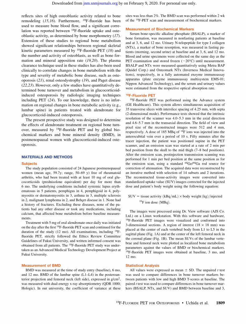
The images were processed using Dr. View software (AJS Co.Ltd.) on a Linux workstation. With this software and hardware,18F-fluoride PET images were visualized and conformed into3-dimensional sections. A region of interest (18 · 18 mm) wasplaced at the center of each vertebral body from L1 to L5 in thesagittal plane (Fig. 1A) and at the center of the left femoral neck inthe coronal plane (Fig. 1B). The mean SUVs of the lumbar verte-brae and femoral neck were plotted as localized bone metabolismparameters against the values of BMD or biochemical markers.18F-fluoride PET images were obtained at baseline, 3 mo, and12 mo.
Statistical AnalysisAll values were expressed as mean 6 SD. The unpaired t test
was used to compare differences in bone turnover markers be-tween patients with low and high BMD T-scores at baseline. Thepaired t test was used to compare differences in bone turnover mar-kers (BSALP, NTx, and SUV) and BMD between baseline and 3,
18F-FLUORIDE PET FOR OSTEOPOROSIS • Uchida et al. 1809
by on February 9, 2020. For personal use only. jnm.snmjournals.org Downloaded from
6, or 12 mo of treatment. The correlations between BSALP, NTx,lumbar spine BMD, and lumbar spine SUV at baseline and 12 moof treatment were examined by Pearson correlation coefficients. AP value of less than 0.05 was considered to represent statisticalsignificance. All statistical analyses were conducted using SPSSsoftware (version 15.0).
RESULTS
Table 1 summarizes the baseline characteristics of thestudy group. The mean time since menopause was 9.8 y(range, 3–19 y). The mean T-scores of the lumbar spine andthe femoral neck were 22.2 (range, 24.43 to 20.16) and22.9 (range, 24.8 to 20.6), respectively. The mean dose oforal glucocorticoids (prednisolone equivalent) before and
during the study was 13.7 6 2.3 mg/d. The values of bothBSALP and NTx showed marked variability among the sub-jects, and the mean NTx tended to be higher than the nor-mal value in our institution. The SUVs of the lumbar spineand the femoral neck were 5.2 6 0.72 and 2.5 6 0.47,respectively, and the former was significantly higher thanthe latter.
According to the baseline BMD T-score of the lumbarspine, based on the World Health Organization criteria (25)for the diagnosis of osteoporosis, patients were categorizedinto a healthy/osteopenic group (T-score . 22.5) or anosteoporotic group (T-score # 22.5) (Table 2). The meanvalues of BSALP, NTx, and femoral neck SUV at baselinetended to be higher in the osteoporotic group, but the dif-ferences were not significant. On the other hand, the lumbarspine SUV was significantly lower in the osteoporotic group(P , 0.05).
Table 3 shows the serial changes in BSALP, NTx, SUV,BMD, and T-score at 3, 6, and 12 mo of alendronate treat-ment. Treatment for 12 mo tended to reduce BSALP, NTx,lumbar spine SUV, and femoral neck SUV but graduallyincreased BMD and the T-score of the lumbar spine andfemoral neck, relative to baseline values. Although alen-dronate treatment over a span of 12 mo significantlyincreased the level of BMD of the lumbar spine (P ,
0.05), such treatment significantly reduced BSALP (P ,
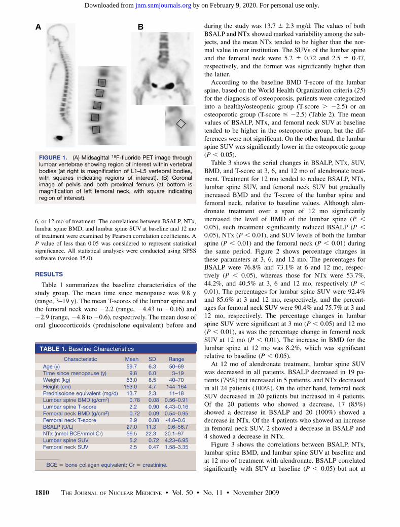
0.05), NTx (P , 0.01), and SUV levels of both the lumbarspine (P , 0.01) and the femoral neck (P , 0.01) duringthe same period. Figure 2 shows percentage changes inthese parameters at 3, 6, and 12 mo. The percentages forBSALP were 76.8% and 73.1% at 6 and 12 mo, respec-tively (P , 0.05), whereas those for NTx were 53.7%,44.2%, and 40.5% at 3, 6 and 12 mo, respectively (P ,
0.01). The percentages for lumbar spine SUV were 92.4%and 85.6% at 3 and 12 mo, respectively, and the percent-ages for femoral neck SUV were 90.4% and 75.7% at 3 and12 mo, respectively. The percentage changes in lumbarspine SUV were significant at 3 mo (P , 0.05) and 12 mo(P , 0.01), as was the percentage change in femoral neckSUV at 12 mo (P , 0.01). The increase in BMD for thelumbar spine at 12 mo was 8.2%, which was significantrelative to baseline (P , 0.05).
At 12 mo of alendronate treatment, lumbar spine SUVwas decreased in all patients. BSALP decreased in 19 pa-tients (79%) but increased in 5 patients, and NTx decreasedin all 24 patients (100%). On the other hand, femoral neckSUV decreased in 20 patients but increased in 4 patients.Of the 20 patients who showed a decrease, 17 (85%)showed a decrease in BSALP and 20 (100%) showed adecrease in NTx. Of the 4 patients who showed an increasein femoral neck SUV, 2 showed a decrease in BSALP and4 showed a decrease in NTx.
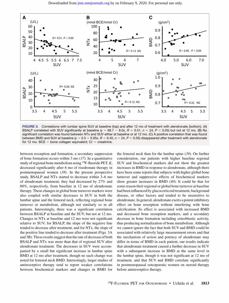
Figure 3 shows the correlations between BSALP, NTx,lumbar spine BMD, and lumbar spine SUV at baseline andat 12 mo of treatment with alendronate. BSALP correlatedsignificantly with SUV at baseline (P , 0.05) but not at
FIGURE 1. (A) Midsagittal 18F-fluoride PET image throughlumbar vertebrae showing region of interest within vertebralbodies (at right is magnification of L1–L5 vertebral bodies,with squares indicating regions of interest). (B) Coronalimage of pelvis and both proximal femurs (at bottom ismagnification of left femoral neck, with square indicatingregion of interest).
TABLE 1. Baseline Characteristics
Characteristic Mean SD Range
Age (y) 59.7 6.3 50–69Time since menopause (y) 9.8 6.0 3–19
Weight (kg) 53.0 8.5 40–70
Height (cm) 153.0 4.7 144–164
Prednisolone equivalent (mg/d) 13.7 2.3 11–18Lumbar spine BMD (g/cm2) 0.78 0.08 0.56–0.91
Lumbar spine T-score 2.2 0.90 4.43–0.16
Femoral neck BMD (g/cm2) 0.72 0.09 0.54–0.95Femoral neck T-score 2.9 0.88 -4.8–0.6
BSALP (U/L) 27.0 11.3 9.6–56.7
NTx (nmol BCE/nmol Cr) 56.5 22.3 20.1–97
Lumbar spine SUV 5.2 0.72 4.23–6.95Femoral neck SUV 2.5 0.47 1.58–3.35
BCE 5 bone collagen equivalent; Cr 5 creatinine.
1810 THE JOURNAL OF NUCLEAR MEDICINE • Vol. 50 • No. 11 • November 2009
by on February 9, 2020. For personal use only. jnm.snmjournals.org Downloaded from
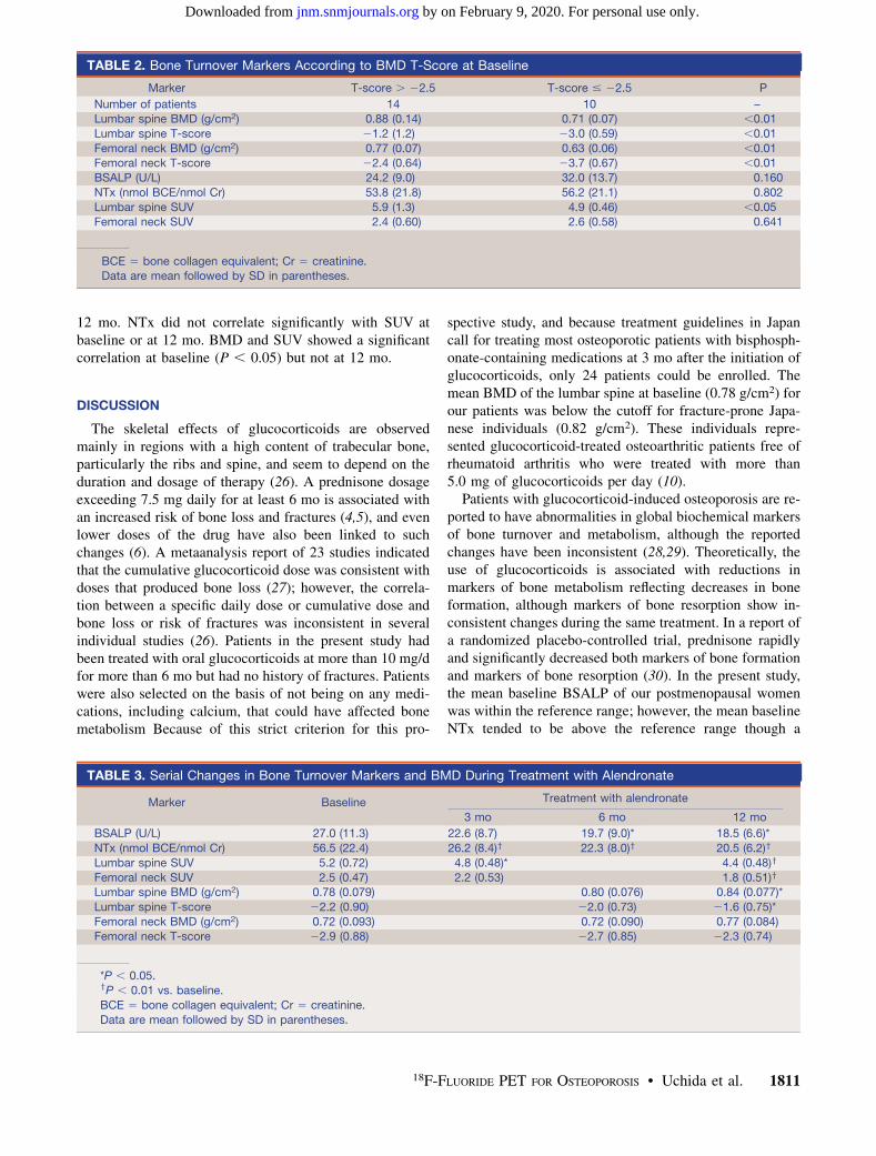
12 mo. NTx did not correlate significantly with SUV atbaseline or at 12 mo. BMD and SUV showed a significantcorrelation at baseline (P , 0.05) but not at 12 mo.
DISCUSSION
The skeletal effects of glucocorticoids are observedmainly in regions with a high content of trabecular bone,particularly the ribs and spine, and seem to depend on theduration and dosage of therapy (26). A prednisone dosageexceeding 7.5 mg daily for at least 6 mo is associated withan increased risk of bone loss and fractures (4,5), and evenlower doses of the drug have also been linked to suchchanges (6). A metaanalysis report of 23 studies indicatedthat the cumulative glucocorticoid dose was consistent withdoses that produced bone loss (27); however, the correla-tion between a specific daily dose or cumulative dose andbone loss or risk of fractures was inconsistent in severalindividual studies (26). Patients in the present study hadbeen treated with oral glucocorticoids at more than 10 mg/dfor more than 6 mo but had no history of fractures. Patientswere also selected on the basis of not being on any medi-cations, including calcium, that could have affected bonemetabolism Because of this strict criterion for this pro-
spective study, and because treatment guidelines in Japancall for treating most osteoporotic patients with bisphosph-onate-containing medications at 3 mo after the initiation ofglucocorticoids, only 24 patients could be enrolled. Themean BMD of the lumbar spine at baseline (0.78 g/cm2) forour patients was below the cutoff for fracture-prone Japa-nese individuals (0.82 g/cm2). These individuals repre-sented glucocorticoid-treated osteoarthritic patients free ofrheumatoid arthritis who were treated with more than5.0 mg of glucocorticoids per day (10).
Patients with glucocorticoid-induced osteoporosis are re-ported to have abnormalities in global biochemical markersof bone turnover and metabolism, although the reportedchanges have been inconsistent (28,29). Theoretically, theuse of glucocorticoids is associated with reductions inmarkers of bone metabolism reflecting decreases in boneformation, although markers of bone resorption show in-consistent changes during the same treatment. In a report ofa randomized placebo-controlled trial, prednisone rapidlyand significantly decreased both markers of bone formationand markers of bone resorption (30). In the present study,the mean baseline BSALP of our postmenopausal womenwas within the reference range; however, the mean baselineNTx tended to be above the reference range though a
TABLE 2. Bone Turnover Markers According to BMD T-Score at Baseline
Marker T-score . 22.5 T-score # 22.5 P
Number of patients 14 10 –Lumbar spine BMD (g/cm2) 0.88 (0.14) 0.71 (0.07) ,0.01
Lumbar spine T-score 21.2 (1.2) 23.0 (0.59) ,0.01
Femoral neck BMD (g/cm2) 0.77 (0.07) 0.63 (0.06) ,0.01Femoral neck T-score 22.4 (0.64) 23.7 (0.67) ,0.01
BSALP (U/L) 24.2 (9.0) 32.0 (13.7) 0.160
NTx (nmol BCE/nmol Cr) 53.8 (21.8) 56.2 (21.1) 0.802
Lumbar spine SUV 5.9 (1.3) 4.9 (0.46) ,0.05Femoral neck SUV 2.4 (0.60) 2.6 (0.58) 0.641
BCE 5 bone collagen equivalent; Cr 5 creatinine.Data are mean followed by SD in parentheses.
TABLE 3. Serial Changes in Bone Turnover Markers and BMD During Treatment with Alendronate
Marker Baseline Treatment with alendronate
3 mo 6 mo 12 mo
BSALP (U/L) 27.0 (11.3) 22.6 (8.7) 19.7 (9.0)* 18.5 (6.6)*
NTx (nmol BCE/nmol Cr) 56.5 (22.4) 26.2 (8.4)y 22.3 (8.0)y 20.5 (6.2)y
Lumbar spine SUV 5.2 (0.72) 4.8 (0.48)* 4.4 (0.48)y
Femoral neck SUV 2.5 (0.47) 2.2 (0.53) 1.8 (0.51)y
Lumbar spine BMD (g/cm2) 0.78 (0.079) 0.80 (0.076) 0.84 (0.077)*
Lumbar spine T-score 22.2 (0.90) 22.0 (0.73) 21.6 (0.75)*
Femoral neck BMD (g/cm2) 0.72 (0.093) 0.72 (0.090) 0.77 (0.084)Femoral neck T-score 22.9 (0.88) 22.7 (0.85) 22.3 (0.74)
*P , 0.05.yP , 0.01 vs. baseline.
BCE 5 bone collagen equivalent; Cr 5 creatinine.
Data are mean followed by SD in parentheses.
18F-FLUORIDE PET FOR OSTEOPOROSIS • Uchida et al. 1811
by on February 9, 2020. For personal use only. jnm.snmjournals.org Downloaded from
statistical comparison of our patients and controls withoutglucocorticoid therapy was not performed. It is possiblethat certain factors such as age, sex, time since menopause,and background disease may affect the level of boneresorption.
Bisphosphonates induce apoptosis of osteoclasts andinhibit bone resorption (13,31). Randomized clinical trialsshowed that treatment with bisphosphonates prevents cor-ticosteroid-induced bone loss (32). Alendronate, a memberof the bisphosphonate family, is effective in the preventionand treatment of glucocorticoid-induced osteoporosis (11–13,32) and has been reported to prevent bone loss and im-prove the BMD of lumbar vertebrae by reducing both boneformation and resorption and suppressing bone metabolism(13). Similarly, alendronate treatment in the present studysignificantly reduced the levels of both biochemical mar-kers (bone formation and resorption), and the decrease inurinary NTx (a marker of bone resorption) was significant;the suppression effect was approximately 60% after 12 mo.Furthermore, dual-energy x-ray absorptiometry measure-ment of the lumbar spine showed that 12 mo of alendronatetreatment increased BMD by 8.2%. A previous study inpostmenopausal women with osteoporosis indicated that thealendronate-induced change in BMD was primarily due tochanges in bone resorption (33). Considered together, theseresults suggest that alendronate treatment prevented furtherreduction in lumbar BMD by reducing bone resorption.
Several studies have examined the feasibility of 18F-fluoride PET for direct assessment of bone turnover inclinically important skeletal sites such as the lumbar spine.Previous studies of patients having disease with high boneturnover showed a significant relationship between boneturnover and biochemical markers (19,22). Brenner et al.(34) reported that the SUV of 18F-fluoride PET correlatedwell with markers of bone metabolism; the net uptake offluoride into the mineral compartment (Ki) was measuredusing arterial blood sampling and kinetic analysis. We alsoobtained similar results with 8 postmenopausal women, whoshowed a significant correlation between Ki and SUV (datanot shown). On the other hand, there are some reports of a
significant correlation between Ki and the histomorpho-metric parameters (19,20). It is possible that tubular re-absorption of fluoride is affected by patient hydration,which could theoretically affect SUVs. None of our patientspresented with renal dysfunction during the course of thisstudy. Therefore, SUVs were substituted for markers of re-gional bone turnover or bone metabolism because of thesimplicity of data acquisition and calculation. However,further research is required to investigate the relationshipbetween regional SUV and histomorphometric parameters.Previous studies of osteoporosis in postmenopausal womenwith 18F-fluoride PET indicated that such patients exhibitlow regional bone formation activity, a good relationbetween bone turnover and changes in BMD, and arisedronate-related decrease in levels of global markers ofbone formation, compared with untreated groups (21,35).Although our osteoporotic patients on glucocorticoid treat-ment did not show a significant change in BSALP and NTx(used as global markers of bone turnover) as measured atbaseline, such treatment significantly reduced lumbar spineSUV in the osteoporotic patients, compared with healthy orosteopenic patients. These findings indicate that the decreasein bone turnover in the lumbar spine measured by SUVreflects the degree of osteoporosis. In this regard, it wasreported previously that fluoride clearance relative to boneminerals depends not only on the rate of bone metabolismbut also on the area available for tracer clearance (36). It ispossible that a reduction in bone mass, as is often seen inpatients with osteoporosis, also reduces the number of sitesavailable for bone remodeling activity, which could indi-rectly influence the measured SUV in our study (22).Accordingly, further studies involving measurements ofPET parameters (such as Ki and SUV) and various bonehistomorphometric parameters, particularly in trabecularbones, are necessary to determine the relationships betweenthe measured PET values and the bone surface area andvolume of such bones.
Follow-up studies have demonstrated that bone resorptionsignificantly decreases within 1 mo of the commencementof antiresorptive therapy, and consequent to the coupling
FIGURE 2. Percentage changes inBSALP and NTx (A), SUV of lumbar spineand femoral neck (B), and BMD of lumbarspine and femoral neck (C) after alendro-nate treatment relative to baseline values.Percentage change in BSALP was signif-icant at 6 and 12 mo; that of NTx wassignificant at 3, 6, and 12 mo. Percentagechange in lumbar spine SUV was signifi-cant at 3 and 12 mo; that of femoral neckSUV was significant at 12 mo. Percentagechange in BMD of lumbar spine wassignificantly different only between base-line and 12 mo of treatment. *P , 0.05.**P , 0.01. FN 5 femoral neck; L 5 lumbarspine; Pre 5 pretreatment.
1812 THE JOURNAL OF NUCLEAR MEDICINE • Vol. 50 • No. 11 • November 2009
by on February 9, 2020. For personal use only. jnm.snmjournals.org Downloaded from
between resorption and formation, a secondary suppressionof bone formation occurs within 3 mo (37). In a quantitativestudy of regional bone metabolism using 18F-fluoride PET, Ki
decreased significantly after 6 mo of risedronate therapy inpostmenopausal women (38). In the present prospectivestudy, BSALP and NTx started to decrease within 3–6 moof alendronate treatment; they had decreased by 27% and60%, respectively, from baseline at 12 mo of alendronatetherapy. These changes in global bone turnover markers werealso coupled with similar decreases in SUV in both thelumbar spine and the femoral neck, reflecting regional boneturnover or metabolism, although not similarly so in allpatients. Interestingly, there was a significant correlationbetween BSALP at baseline and the SUV, but not at 12 mo.Changes in NTx at baseline and 12 mo were not significantrelative to SUV; for BSALP, the slope of the negative linetended to decrease after treatment, and for NTx, the slope ofthe positive line tended to decrease after treatment (Figs. 3Aand 3B). These results suggest that the rate of decrease of bothBSALP and NTx was more than that of regional SUV afteralendronate treatment. The decreases in SUV were accom-panied by a small but significant increase in lumbar spineBMD at 12 mo after treatment, though no such change wasnoted for femoral neck BMD. Interestingly, larger studies ofantiresorptive therapy tend to report weaker correlationsbetween biochemical markers and changes in BMD for
the femoral neck than for the lumbar spine (39). On furtherconsideration, our patients with higher baseline regionalSUV and biochemical markers did not show the greatestincreases in BMD in response to alendronate, although therehave been some reports that subjects with higher global boneturnover and suppressive effects of biochemical markersshow greater increases in BMD (40). It could be that forsome reason their regional or global bone turnover at baselinehad been influenced by glucocorticoid treatment, backgrounddisease, or other factors and tended to be insensitive toalendronate. In general, alendronate exerts a potent inhibitoryeffect on bone resorption without interfering with bonecalcification. Its effect is associated with increased BMDand decreased bone resorption markers, and a secondarydecrease in bone formation including osteoblastic activity,thus producing normalization of bone metabolism. Althoughwe cannot ignore the fact that both SUV and BMD could beassociated with relatively large measurement errors and thatthe mechanism of action and potency of alendronate maydiffer in terms of BMD in each patient, our results indicatethat alendronate treatment caused a further decrease in SUVwith a subsequent increase in BMD at the same level inthe lumbar spine, though it was not significant at 12 mo oftreatment, and that SUV and BMD correlate significantlyin postmenopausal osteoporotic women on steroid therapybefore antiresorptive therapy.
FIGURE 3. Correlations with lumbar spine SUV at baseline (top) and after 12 mo of treatment with alendronate (bottom). (A)BSALP correlated with SUV significantly at baseline (y 5 68.7 2 8.0x, R 5 0.51, n 5 24, P , 0.05) but not at 12 mo. (B) Nosignificant correlation was found between NTx and SUV either at baseline or at 12 mo. (C) A positive correlation that was foundbetween BMD and SUV at baseline (y 5 0.5 1 0.05x, R 5 0.45, n 5 24, P , 0.05) disappeared after treatment with alendronatefor 12 mo. BCE 5 bone collagen equivalent; Cr 5 creatinine.
18F-FLUORIDE PET FOR OSTEOPOROSIS • Uchida et al. 1813
by on February 9, 2020. For personal use only. jnm.snmjournals.org Downloaded from
CONCLUSION
To our knowledge, this was the first study of regional bonemetabolism in the lumbar spine measured using 18F-fluoridePET in patients with glucocorticoid-induced osteoporosis.The study also examined various bone metabolism markersbefore and after alendronate treatment. The results demon-strated decreased bone turnover in the lumbar spine, repre-sented by SUVs, and a correlation between these changes andthe severity of osteoporosis. Furthermore, the results showeda significant decrease in bone metabolism associated withincreased BMD in the lumbar spine after 12 mo of treatmentwith alendronate. These results suggest that antiresorptivetherapy has a direct bone-metabolism effect on skeletalkinetics in glucocorticoid-induced osteoporosis at the clini-cally important site of the lumbar spine.
ACKNOWLEDGMENTS
This work was supported in part by grants from theJapanese Osteoporosis Foundation (2007). We are indebtedto Tomohiro Ohta (Bone and Joint Research Laboratories,Teijin Institute for Biomedical Research) for reviewing themanuscript.
REFERENCES
1. Klein RG, Arnaud SB, Gallangher JC, DeLuca HF, Riggs BL. Intestinal calcium
absorption in exogenous hypercortisonism: role of 25-hydroxyvitamin D and
corticosteroid dose. J Clin Invest. 1977;60:253–259.
2. Rubin MR, Bilezikian JP. Clinical review 151: the role of parathyroid hormone
in the pathogenesis of glucocorticoid-induced osteoporosis—a re-examination of
the evidence. J Clin Endocrinol Metab. 2002;87:4033–4041.
3. Canalis E, Delany AM. Mechanisms of glucocorticoid action in bone. Ann N Y
Acad Sci. 2002;966:73–81.
4. Michel BA, Bloch DA, Wolfe F, Fries JF. Fractures in rheumatoid arthritis: an
evaluation of associated risk factors. J Rheumatol. 1993;20:1666–1669.
5. Verstraeten A, Dequeker J. Vertebral and peripheral bone mineral content and
fracture incidence in postmenopausal patients with rheumatoid arthritis: effect of
low dose corticosteroids. Ann Rheum Dis. 1986;45:852–857.
6. Van Staa TP, Leufkens HG, Abenhaim L, Zhang B, Cooper C. Use of oral
corticosteroids and risk of fractures. J Bone Miner Res. 2000;15:993–1000.
7. Adachi JD, Olszynski WP, Hanley DA, et al. Management of corticosteroid-
induced osteoporosis. Semin Arthritis Rheum. 2000;29:228–251.
8. American College of Rheumatology Ad Hoc Committee on Glucocorticoid-
Induced Osteoporosis. Recommendations for the prevention and treatment of
glucocorticoid-induced osteoporosis: 2001 update. Arthritis Rheum. 2001;44:
1496–1503.
9. Bone and Tooth Society, National Osteoporosis Society, Royal College of
Physicians. Glucocorticoid-Induced Osteoporosis: Guidelines for Prevention
and Treatment. London, U.K.: Royal College of Physicians; 2002.
10. Nawata H, Soen S, Takayanagi R, et al. Guidelines on the management and
treatment of glucocorticoid-induced osteoporosis of the Japanese Society for
Bone and Mineral Research (2004). J Bone Miner Metab. 2005;23:105–109.
11. Adachi JD, Saag KG, Delmas PD, et al. Two-year effects of alendronate on bone
mineral density and vertebral fracture in patients receiving glucocorticoids: a
randomized, double-blind, placebo-controlled extension trial. Arthritis Rheum.
2001;44:202–211.
12. Sambrook PN, Kotowicz M, Nash P, et al. Prevention and treatment of
glucocorticoid osteoporosis: a comparison of calcitriol, vitamin D plus calcium,
and alendronate plus calcium. J Bone Miner Res. 2003;18:919–924.
13. de Nijs RN, Jacobs JW, Lems WF, et al. Alendronate or alfacalcidol in
glucocorticoid-induced osteoporosis. N Engl J Med. 2006;355:675–684.
14. Blau M, Ganatra R, Bender MA. 18F-fluoride for bone imaging. Semin Nucl Med.
1972;2:31–37.
15. Ishiguro K, Nakagaki H, Tsuboi S, et al. Distribution of fluoride in cortical bone
of human rib. Calcif Tissue Int. 1993;52:278–282.
16. Narita N, Kato K, Nakagaki H, et al. Distribution of fluoride concentration in the
rat’s bone. Calcif Tissue Int. 1990;46:200–204.
17. Reeve J, Arlot M, Wootton R, et al. Skeletal blood flow, iliac histomorphometry,
and strontium kinetics in osteoporosis: a relationship between blood flow and
corrected apposition rate. J Clin Endocrinol Metab. 1988;66:1124–1131.
18. Hawkins RA, Choi Y, Huang SC, et al. Evaluation of the skeletal kinetics of
fluorine-18-fluoride ion with PET. J Nucl Med. 1992;33:633–642.
19. Messa C, Goodman WG, Hoh CK, et al. Bone metabolic activity measured with
positron emission tomography and [18F]fluoride ion in renal osteodystrophy:
correlation with bone histomorphometry. J Clin Endocrinol Metab. 1993;77:949–955.
20. Piert M, Zittel TT, Becker GA, et al. Assessment of porcine bone metabolism by
dynamic. J Nucl Med. 2001;42:1091–1100.
21. Frost ML, Fogelman I, Blake GM, Marsden PK, Cook G Jr. Dissociation
between global markers of bone formation and direct measurement of spinal
bone formation in osteoporosis. J Bone Miner Res. 2004;19:1797–1804.
22. Cook GJ, Blake GM, Marsden PK, Cronin B, Fogelman I. Quantification of
skeletal kinetic indices in Paget’s disease using dynamic 18F-fluoride positron
emission tomography. J Bone Miner Res. 2002;17:854–859.
23. Installe J, Nzeusseu A, Bol A, et al. 18F-fluoride PET for monitoring therapeutic
response in Paget’s disease of bone. J Nucl Med. 2005;46:1650–1658.
24. Berding G, Kirchhoff TD, Burchert W, et al. [18F]fluoride PET indicates reduced
bone formation in severe glucocorticoid-induced osteoporosis. Nuklearmedizin.
1998;37:76–79.
25. Assessment of fracture risk and its application to screening for postmenopausal
osteoporosis. Report of a WHO Study Group. World Health Organ Tech Rep Ser.
1994;843:1–129.
26. Dykman TR, Gluck OS, Murphy WA, Hahn TJ, Hahn BH. Evaluation of factors
associated with glucocorticoid-induced osteopenia in patients with rheumatic
disease. Arthritis Rheum. 1985;28:361–368.
27. van Staa TP, Leufkens HG, Cooper C. The epidemiology of corticosteroid-
induced osteoporosis: a meta-analysis. Osteoporos Int. 2002;13:777–787.
28. Conti A, Sartorio A, Ferrero S, Ferrario S, Ambrosi B. Modifications of
biochemical markers of bone and collagen turnover during corticosteroid
therapy. J Endocrinol Invest. 1996;19:127–130.
29. Kollerup G, Hansen M, Horslev-Petersen K. Urinary hydrooxypyridinium cross-
links of collagen in rheumatoid arthritis: relation to disease activity and effects of
methylprednisolone. Br J Rheumatol. 1994;33:816–820.
30. Ton FN, Gunawardene SC, Lee H, Neer RM. Effects of low-dose prednisone on
bone metabolism. J Bone Miner Res. 2005;20:464–470.
31. Rogers MJ. New insights into the molecular mechanisms of action of
bisphosphonates. Curr Pharm Des. 2003;9:2643–2658.
32. Saag KG, Emkey R, Schnitzer TJ, et al. Alendronate for the prevention and
treatment of glucocorticoid-induced osteoporosis. Glucocorticoid-Induced Oste-
oporosis Intervention Study Group. N Engl J Med. 1998;339:292–299.
33. Balena R, Toolan BC, Shea M, et al. The effect of 2-year treatment with the
aminobisphosphonate alendronate on bone metabolism, bone histomorphometry,
and bone strength in ovariectomized nonhuman primates. J Clin Invest. 1993;
92:2577–2586.
34. Brenner W, Vernon C, Muzi M, et al. Comparison of different quantitative
approaches to 18F-fluoride PET scans. J Nucl Med. 2004;45:1493–1500.
35. Frost ML, Cook GJ, Blake GM, Marsden PK, Fogelman I. The relationship
between regional bone turnover measured using 18F-fluoride positron emission
tomography and changes in BMD is equivalent to that seen for biochemical
markers of bone turnover. J Clin Densitom. 2007;10:46–54.
36. Peters AM, Myers MJ. Body fluids, electrolytes and bone. In: Peters AM, Myers
MJ, eds. Physiological Measurements with Radionuclides in Clinical Practice.
New York, NY: Oxford University Press; 1998:262–278.
37. Mortensen L, Charles P, Bekker PJ, Digennaro J, Johnston CC. Risedronate
increases bone mass in an early postmenopausal population: two years of
treatment plus one year of follow-up. J Clin Endocrinol Metab. 1998;83:396–402.
38. Frost ML, Cook GJ, Blake GM, et al. A prospective study of risedronate on
regional bone metabolism and blood flow at the lumbar spine measured by18F-fluoride positron emission tomography. J Bone Miner Res. 2003;18:
2215–2222.
39. Chen P, Satterwhite JH, Licata AA, et al. Early changes in biochemical markers
of bone formation predict BMD response to teriparatide in postmenopausal
women with osteoporosis. J Bone Miner Res. 2005;20:962–970.
40. Greenspan SL, Rosen HN, Parker RA. Early changes in serum N-telopeptide and
C-telopeptide cross-linked collagen type 1 predict long-term response to
alendronate therapy in elderly women. J Clin Endocrinol Metab. 2000;85:
3537–3540.
1814 THE JOURNAL OF NUCLEAR MEDICINE • Vol. 50 • No. 11 • November 2009
by on February 9, 2020. For personal use only. jnm.snmjournals.org Downloaded from
Doi: 10.2967/jnumed.109.062570Published online: October 16, 2009.
2009;50:1808-1814.J Nucl Med. Tatsuro Tsuchida, Hidehiko Okazawa, Yasuhisa Fujibayashi and Hisatoshi BabaKenzo Uchida, Hideaki Nakajima, Tsuyoshi Miyazaki, Takafumi Yayama, Hideo Kawahara, Shigeru Kobayashi,
F-Fluoride PET: A Prospective Study18Measured by Effects of Alendronate on Bone Metabolism in Glucocorticoid-Induced Osteoporosis
http://jnm.snmjournals.org/content/50/11/1808This article and updated information are available at:
http://jnm.snmjournals.org/site/subscriptions/online.xhtml
Information about subscriptions to JNM can be found at:
http://jnm.snmjournals.org/site/misc/permission.xhtmlInformation about reproducing figures, tables, or other portions of this article can be found online at:
(Print ISSN: 0161-5505, Online ISSN: 2159-662X)1850 Samuel Morse Drive, Reston, VA 20190.SNMMI | Society of Nuclear Medicine and Molecular Imaging
is published monthly.The Journal of Nuclear Medicine
© Copyright 2009 SNMMI; all rights reserved.
by on February 9, 2020. For personal use only. jnm.snmjournals.org Downloaded from


![Alendronate treatment alters bone tissues at multiple ...€¦ · trabecular bone where bone turnover is higher compared to cortical bone [14]. The novelty of this study lies in the](https://img.pdfslide.net/doc/110x75/6066b6f076f57e3ead6e765d/alendronate-treatment-alters-bone-tissues-at-multiple-trabecular-bone-where.jpg)