Embed Size (px)
Citation preview
Methods 52 (2010) 316–321
Contents lists available at ScienceDirect
Methods
journal homepage: www.elsevier .com/locate /ymeth
Differential zinc accumulation and expression of human zinc transporter 1 (hZIP1)in prostate glands
Leslie A. Johnson a, Mazhar A. Kanak a, Andre’ Kajdacsy-Balla b, Joseph P. Pestaner c, Omar Bagasra a,⇑a South Carolina Center for Biotechnology, Claflin University, Orangeburg, SC 29115, USAb Department of Pathology, University of Illinois–Chicago, School of Medicine, Chicago, IL 60612, USAc Chief Medical Examiner’s Office, Washington, DC 20003, USA
a r t i c l e i n f o a b s t r a c t
Article history:Accepted 6 August 2010Available online 10 August 2010
Keywords:African American maleDifferential expression of hZIP1Differential ZN stainingEuropean American maleGleason scoreIn situ hybridizationIn situ RT-PCR/hybridizationNewport Green (NPG)Prostate cancerRacial groups
1046-2023/$ - see front matter � 2010 Elsevier Inc. Adoi:10.1016/j.ymeth.2010.08.004
⇑ Corresponding author. Address: Department of Bfor Biotechnology, Claflin University, 400 Magnolia SUSA.
E-mail address: [email protected] (O. Bagasra).
Zinc (Zn) is essential for a very large number and variety of cellular functions but is also potentially toxic.Zn homeostasis is therefore dynamically maintained by a variety of transporters and other proteins dis-tributed in distinct cellular and subcellular compartments. Zn transport is mediated by two major proteinfamilies: the Zip family, which mediates Zn influx, and the ZnTs which are primarily linked to Zn seques-tration into intracellular compartments and are, thereby, involved in lowering cytoplasmic Zn free ionconcentrations. In the prostate epithelial cell, the accumulation of high cellular zinc is a specialized func-tion that is necessary for these cells to carry out the major physiological functions of production andsecretion of prostatic fluids. The loss of Zn accumulation is the most consistent and persistent character-istic of prostate malignancy. Currently, there are no direct methods to determine the relative Zn levels invarious cell types of prostate gland (i.e. stroma, glandular epithelia, acini, and muscular) and no reliableways to compare the Zn in normal versus malignant areas of the gland. Here we report a new method toshow a differential Zn staining method that correlates with various stages of prostate cancer develop-ment in situ and expression of a human Zn transporter1-hZIP1 – in situ by in situ reverse transcriptase-polymerase chain reaction hybridization (ISRTPCR) that correlate with the relative Zn levels determinedby the differential Zn staining method. By utilizing these methods, we show for the first time that: (1) therelative Zn levels are very low to absent in the malignant glands, (2) normal glands show high Zn levels inboth glandular epithelia as well as in stromal tissues, (3) the Zn levels begin to decrease in pre-malignantglands and precedes the development of malignancy, and (4) the expression of human Zn transporter1(hZIP1) appears to correlate with the Zn levels in the prostate glands and may be the major Zn regulatorin this organ.
� 2010 Elsevier Inc. All rights reserved.
1. Introduction
The role of Zn, its underlying active function in the developmentand progression of prostate malignancy and its potential applica-tion in the prevention and treatment of prostate cancer are criticalissues for the medical/scientific community and the public-at-large[1]. Prostate cancer (PC) is the second most common form of cancerdiagnosed in men [1,2] and it is the most prevalent type of cancerobserved in African American (AA) men [3–5]. Very early detectionis the key to effective treatment of PC and to the prevention ofdeaths due to the progression of untreatable advanced stages ofcancer. Mitigating factors, especially benign prostatic hyperplasia(BPH), result in a low accuracy (about 60%) of prostate-specific
ll rights reserved.
iology, South Carolina Centertreet, Orangeburg, SC 29115,
antigen (PSA) testing. Thus, there is an urgent need for a more reli-able biomarker to identify PC at a very early stage and to identify‘at-risk’ individuals. AAs are at significantly higher risk of develop-ing PC particularly at an earlier age and have a more aggressiveform of PC than European American (EA) men [1,2]. Currently,there are no satisfactory ways to differentiate between Stages I/IIindolent and lethal (aggressive) PC at diagnosis. Both early identi-fication and indolent/lethal differentiation are critical because PC,if identified while confined to the prostate, (a) is ‘‘curable” foraggressive tumors by surgery and subsequent treatment and (b)‘‘watchful waiting” would be appropriate for indolent tumors[1–3].
Numerous studies consistently have shown that the Zn levels ofmalignant prostate tissue are significantly decreased as comparedto the normal prostate tissue (reviewed in [6,7]). This consistencypersists in different reports by different investigators employingdifferent populations and tissue samples and involving variousstages of malignancy. The studies of Zaichick et al. [8], Vartsky

L.A. Johnson et al. / Methods 52 (2010) 316–321 317
et al. [9], and Franklin et al. [7] further reveal the critically impor-tant relationship that, in individual analyses, malignant prostatetissue always exhibits relatively low Zn levels as compared to thenormal tissues. In addition, Habib [11] reported that the decreasein Zn occurs early in malignancy. These persistent results, andthe additional corroborating evidence presented previously (re-viewed in [6,7]), has firmly established that the unique zinc-accu-mulating capability of the normal peripheral zone secretoryepithelial cells is lost in the neoplastic transformation to malignantcells [6,7,12–14].
This study was done in order to determine if Zn molecules aredifferentially accumulated in various cell types and hZIP1 is themajor Zn transporter that regulates Zn in prostate glands, as previ-ously proposed [7,15]. In order to determine if Zn accumulation inthe glandular portions of the prostate is significantly different thanthe Zn in the stromal and other cell types, we evaluated prostaticresections from 19 men with prostate cancer and four normal pros-tatic tissues and evaluated their tissues for the differential Zn accu-mulation by utilizing two Zn indicator dyes: New Port Green DCF(NPG) and N-(6-methoxy-8-quinolyl)-p-toluenesulfonamide (TSQ)to determine the relative Zn concentrations in various histologicalcell types, in situ. In addition, we also explore the differentialexpression of hZIP1 by in situ RT-PCR method (ISRTPCR) using thesame group of patients.
2. Methods and materials
2.1. Human subjects and study protocol
Fresh frozen specimens of primary prostate carcinoma were re-ceived from the University of Illinois, Department of Pathology (Dr.Balla) on dry ice, according to the approved protocol of the institu-tional review board as Claflin University IRB. Normal prostate tis-sues were autopsy specimens obtained from the Department ofPathology, Brody School of Medicine, East Carolina University,Greenville, NC (Dr. Pestaner).
2.2. In situ RT-PCR of human tissue sections
Fresh frozen sections from 19 male prostate biopsies with aclinical history of prostate cancer, and 6 from autopsy specimenswith normal glands who died from automobile accidents, wereprocessed for RT-in situ-PCR [16,17]. All reagents for the continua-tion of the experiment were prepared in RNase-free H2O. Briefly,fresh frozen tissue sections were received from each of our collab-orators in a blinded fashion. All slides were fixed in 2% paraformal-dehyde solution, overnight for a 24 h period. After fixation, slideswere washed in 3� PBS once, and then twice in 1� PBS. Theseslides were then treated with proteinase K (6 lg/ml) at room tem-perature for 23 min. Proteinase K was inactivated by incubatingslides on a heat block at 95 �C for 2 min. To perform the amplifica-tion of mRNA sequences for hZIP1, we used multiple spliced se-quences that flank the junctions of two exon splice sites. Becausethese RNA-specific primers will not amplify the genomic DNA tem-plate, one can perform the amplification of multiple mRNAs simul-taneously. The following primer pairs were used for amplification:sense 50-TCAGAGCCTCCAGTGCCTGT-30 and antisense 50-TTGTCTCCTGGACCTGCTGC-30 for hZIP1 that gave a 189-bp product. Toform cDNA copy of the template and to amplify, we used one-stepin situ RT-PCR enzyme – rTth enzyme, which has both the RT andpolymerase function. The amplification cocktail contained the pairof primers at 100 pM each in 50 mM Tris, pH 8.3, 8.5 mM MgCl,10 mM MnCl2, 40 mM KCL, 1 mM dithiothreitol, 10� transcriptionbuffer, 10� chelating buffer, 5 U rTth (recombinant thermostableDNA polymerase) enzyme. After amplification, slides were washed
in 2� SSC buffer, three times and then amplicons were detected byin situ hybridization method [16,17].
Hybridizations were performed with 41 bp FITC (fluoresceinisothiocynate) oligonucleotide probe for hZIP1 (oligonucleotide:50-FITC-CCTGGTGCCCATCTGTGTGCTGCGC-CGGCCAGGAGCTAAC-30). Hybridizations were performed in a buffer containing 50%formaldehyde, 10 mM dithiothreitol, 2� sodium chloride/sodiumcitrate solution, 100 lg/mL fragmented salmon sperm DNA, 2% bo-vine serum albumin, 1 mg/mL Escherichia coli tRNA, and 20 pmolprobes at 95 �C for 2 min, then 45 �C for 18 h. These tissue sectionswere then washed to remove unbound probes and viewed underUV-epifluorescence microscope after the cells were washed. Topreserve the intensity of the hybridized probes, the tissues werenot counter-stained. Parallel hematoxylin and eosin-stained slideswere used to identify various histologic cell types in the tissue sec-tions. Since we utilized frozen section the H&E parallel sections arenot as crisp and sometimes difficult to identify due to tissue dam-age during cutting. Microscopic examination usually reveals cyto-plasmic staining for mRNA versus nuclear staining for DNA. Cellenumeration was performed on coded slides by at least twopathologists. Twenty microliters of RT (reverse transcriptase en-zyme) cocktail was added to each slide. The slides were then sealedwith slide frame sealer and inserted into the slide slots of a ther-mocycler specially designed for in situ PCR (MJR/Bio-Rod – TwinTower, PTC 200, Waltham, MA). The two cycles were programmedfor 30 min at 50 �C, then 95 �C for 5 min (for cDNA step), and thencDNAs were amplified for 30 cycles at 95 �C denaturing, 61.7 �Cannealing, and 72 �C extension.
2.3. Determination of intracellular zinc content
The relative intracellular Zn content in situ was determined byutilizing fresh frozen tissues. For this purpose the cells must bebiochemically active. The relative concentrations of Zn in variouscell types of the prostatic tissues were determined according tothe manufacturer’s instructions (Molecular Probes, Inc., Eugene,Oregon, USA). Briefly, the frozen tissues were incubated with equalmolar concentrations of two zinc-indicator dyes: NPG and TSQ[18,19]. The frozen tissues were incubated in 20 ll/section of theZn indicator cocktail over night and washed in PBS, gently, withoutdisturbing the tissues. The slides were heat-fixed for 10 s at 104 �Cto immobilize the signals. These slides were mounted withsolution containing 50% glycerol in PBS and observed under afluorescent microscope. TSQ has a selective high affinity for Zn(Kd �10 nM) and a detection limit of �0.1 nM. The ZN-TSQ positivecells stain red [18]. NPG has moderate zinc-binding affinity(Kd �1 lM: 19). The ZN-NPG positive cells appear yellowish green.Together, TSQ and NPG provide a relative difference in Zn concen-trations in various cell types of the prostate. TSQ has about 2–3-loghigher affinity for Zn than NPG, but has detection limit of about3-log lower than NPG. Therefore, the cells that contain very lowconcentrations of intracellular Zn appear red and the ones withhigher concentrations appear green. The cells that fall in betweenappear yellowish or yellow-green.
3. Results
Fresh frozen tissues were utilized to determine the relativeintracellular Zn levels in various histological areas of the prostateglands. All prostate sections were from the peripheral zones ofthe glands. In this study, Fig. 1A shows the high level of cellularZn that characterizes the normal glandular epithelial cells (greencolor in Fig. 1A). In contrast, the stroma exhibits relatively lowerlevels of zinc. Therefore, the in situ Zn staining utilizing twodifferent color indicators with different affinity and intracellular

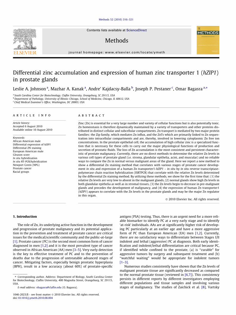
Fig. 1. Zinc levels in prostate tissue frozen sections. Representative zinc levels in prostate sections. Fresh frozen tissues were utilized to determine the relative intracellular Znlevels in various histological areas of the prostate glands. All prostate sections were from the peripheral zones of the glands. High Zn is represented by Newport Green, yellow/green stain and low Zn is represented by TSQ red stain. (A) Normal prostate gland from a 42 year old subject. Of note, the high level of cellular Zn indicated by dark greenstaining with New Port Green (NPG) Zn indicator dye that characterizes the normal glandular epithelial cells (black arrows, in A). In contrast, the stroma exhibits relativelylower levels of zinc, indicated by less intense green color in the stroma (white arrows). Therefore, the in situ Zn staining utilizing two different color indicators with differentaffinity and intracellular threshold provides the differential Zn accumulation between normal glandular epithelium and stroma [18,19]. The marked reductions of cellular Znin the epithelium of the two high grade intraepithelial neoplasia are shown in (B) and (C). The malignant region of the peripheral zone shows a significant depletion of Zn inthe malignant glandular epithelium as exhibited by the red staining (white arrows) in three patient’s resected tissues. Here, one notes relative depletion of Zn indicated byTSQ red Zn indicator dye and relatively higher levels of Zn in the stromal areas. Similar relative depletion of Zn is also observed in (D) pattern is also seen in patient withadenocarcinoma Gleason score 3 + 3 (moderately differentiated) in (D). H&E sections from parallel sections are shown on right side of the slide (final magnification 100�).
318 L.A. Johnson et al. / Methods 52 (2010) 316–321
threshold provides the differential Zn accumulation between nor-mal glandular epithelium and stroma [18,19]. The marked reduc-tion of cellular Zn in the epithelium of the two high gradeintraepithelial neoplasia are apparent in Fig. 1B and C. Similar pat-tern is also seen in patient with adenocarcinoma Gleason score3 + 3 (moderately differentiated) in Fig. 1D. Like the expression ofhZIP1, the loss of Zn occurs early in malignancy. Due to the deple-tion of Zn in the malignant glands, the stromal Zn level gives theappearance of relatively higher Zn levels. Many studies have ob-served that Zn levels are greatly decreased in extracts of resectedmalignant tissue preparations [20]. However, our present studyprovides the first in situ detection of the depleted cellular Zn levelsin adenocarcinomatous glands as compared to the high Zn levels innormal glandular epithelium. Of note, the decrease in Zn level in
the malignant glands is due to a decrease in the cellular accumula-tion of zinc. This suggests that the decrease in intracellular zinc,and not impaired secretion of Zn into the lumen (prostatic fluid),is principally responsible for the decrease in malignant tissue Znlevel. Thus, the results of our study are consistent with previousstudies [6–10,21].
Correspondingly, Fig. 2 the relative expression of mRNA expres-sion for hZIP1 were determined in the 19 prostate resections. Thetypical results represented in Fig. 2 were consistently observed inthe frozen sections of all 19 prostate resections. The results showthat hZIP1 gene expression is evident uniformly in the epitheliumof the normal peripheral zone glands and is relatively low in thestroma (Fig. 2A). hZIP1 expression is markedly down-regulatedto the extent of not being demonstrable in the two high grade
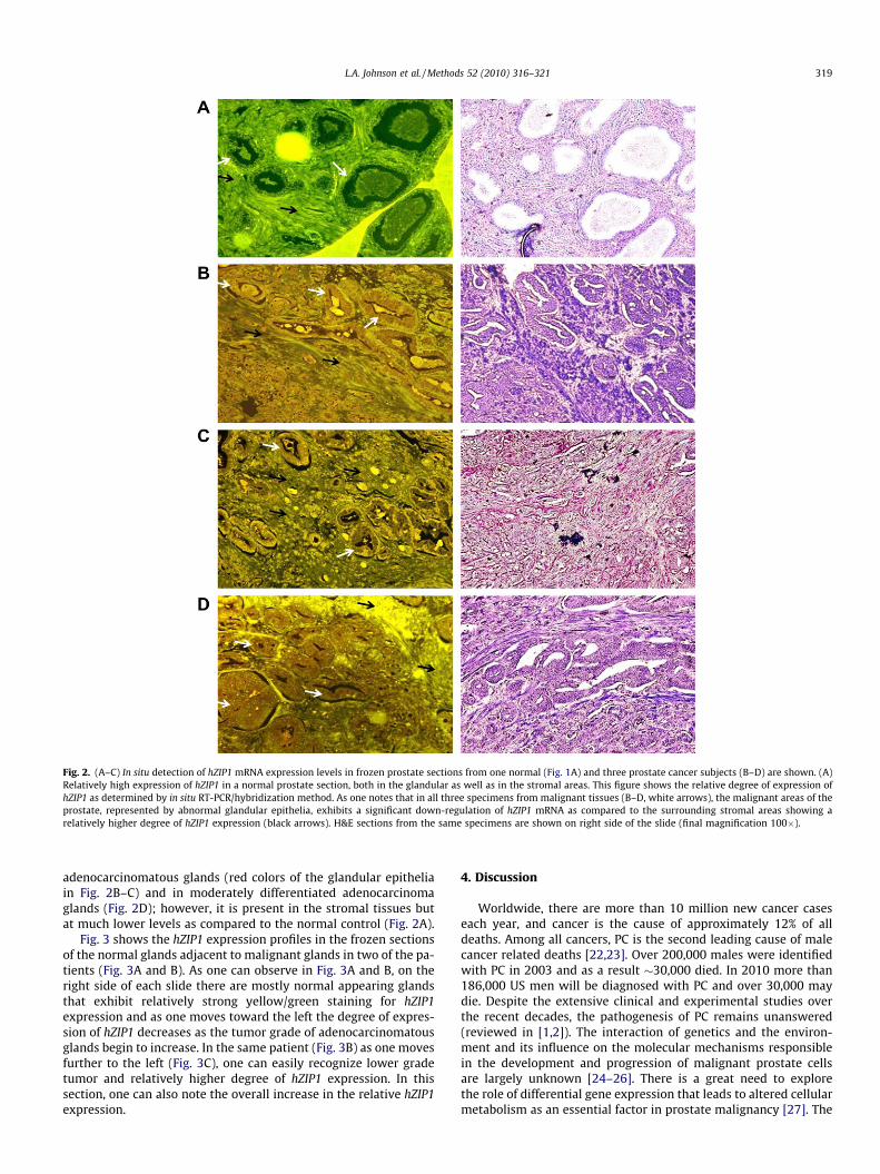
Fig. 2. (A–C) In situ detection of hZIP1 mRNA expression levels in frozen prostate sections from one normal (Fig. 1A) and three prostate cancer subjects (B–D) are shown. (A)Relatively high expression of hZIP1 in a normal prostate section, both in the glandular as well as in the stromal areas. This figure shows the relative degree of expression ofhZIP1 as determined by in situ RT-PCR/hybridization method. As one notes that in all three specimens from malignant tissues (B–D, white arrows), the malignant areas of theprostate, represented by abnormal glandular epithelia, exhibits a significant down-regulation of hZIP1 mRNA as compared to the surrounding stromal areas showing arelatively higher degree of hZIP1 expression (black arrows). H&E sections from the same specimens are shown on right side of the slide (final magnification 100�).
L.A. Johnson et al. / Methods 52 (2010) 316–321 319
adenocarcinomatous glands (red colors of the glandular epitheliain Fig. 2B–C) and in moderately differentiated adenocarcinomaglands (Fig. 2D); however, it is present in the stromal tissues butat much lower levels as compared to the normal control (Fig. 2A).
Fig. 3 shows the hZIP1 expression profiles in the frozen sectionsof the normal glands adjacent to malignant glands in two of the pa-tients (Fig. 3A and B). As one can observe in Fig. 3A and B, on theright side of each slide there are mostly normal appearing glandsthat exhibit relatively strong yellow/green staining for hZIP1expression and as one moves toward the left the degree of expres-sion of hZIP1 decreases as the tumor grade of adenocarcinomatousglands begin to increase. In the same patient (Fig. 3B) as one movesfurther to the left (Fig. 3C), one can easily recognize lower gradetumor and relatively higher degree of hZIP1 expression. In thissection, one can also note the overall increase in the relative hZIP1expression.
4. Discussion
Worldwide, there are more than 10 million new cancer caseseach year, and cancer is the cause of approximately 12% of alldeaths. Among all cancers, PC is the second leading cause of malecancer related deaths [22,23]. Over 200,000 males were identifiedwith PC in 2003 and as a result �30,000 died. In 2010 more than186,000 US men will be diagnosed with PC and over 30,000 maydie. Despite the extensive clinical and experimental studies overthe recent decades, the pathogenesis of PC remains unanswered(reviewed in [1,2]). The interaction of genetics and the environ-ment and its influence on the molecular mechanisms responsiblein the development and progression of malignant prostate cellsare largely unknown [24–26]. There is a great need to explorethe role of differential gene expression that leads to altered cellularmetabolism as an essential factor in prostate malignancy [27]. The

Fig. 3. In situ detection of hZIP1 mRNA and the frozen sections of normal and malignant glands in the same tissue sections. (A and B) Analyses of hZIP1 expression by in situ RT-PCR/hybridization are shown from two patients. Relatively high levels of hZIP1 expressions in normal appearing glands can be seen as a yellowish/green color on the rightportions (white arrows) of the slides whereas low or absent expression can be seen as red colors in malignant glands on the left sides of the slides (black arrows). Of note, thegreenish color in the stoma in (A) and (B) are absent, suggesting low expression of hZIP1. In the same patient (B) as one further moves to the left (C) towards relatively normalappearing area, one can easily recognize higher expression of hZIP1 (shown in greenish color and more normal appearing glands; black arrows). H&E sections from the samespecimens are shown on right side of the slide (final magnification 100�).
320 L.A. Johnson et al. / Methods 52 (2010) 316–321
combination of genetic/molecular/environmental factors and theirrelationships are required to identify the critical events in the pros-tate malignancy process. Such studies are proving to be very usefulin the understanding of the molecular pathogenesis of prostatecancer [6,7,28].
Zinc and Zn transporters play an important role in the molecu-lar pathogenesis of PC [6,7,20,21]. PC afflicts one out of nine menover the age of 65 years. Prostatic intraepithelial neoplasia (PINs)is relatively common and occurs early in life [1,2]. However, pro-gression to invasive carcinoma is significantly less common. Whatare the factors that cause PIN to become invasive? It appears thatrace and ethnicity is also an important factor! PC disproportion-ately affects AA men, who, along with black Jamaican men, havethe highest PC incidence rates in the world [22,23,29–34]. In addi-tion, AA men develop PC significantly earlier and at the time ofdiagnosis they are present with the higher-grade adenocarcinomathan the age-matched EA men [31–35].
At the global level, rates of incidence are low in Asian and Afri-can men, low-to-moderate in EA men, and highest in AA men[22,23,30,32]. Using data collected between 1988 and 1992, Wingoet al. [36] reported that AAs have a 35% higher incidence rate and a223% higher mortality rate from PC as compared with EAs. Similardata has been shown by others [22,23]. The differences in inci-dence and mortality between AAs and EAs have been attributedto both environmental and biological factors [6,7,27–29], Whencompared with EA men, AA men present at a younger age, with
higher grade (Gleason Score), and stage of disease at the onset ofage, and with a greater delay in diagnosis [37–39]. Whether thepathogenesis of PC is different in AA men as compared to EAmen remains unanswered. Whittemore et al. [40] have noted thatAA men appear to have a larger volume of ‘‘latent” PC load. Theseinvestigators believe that larger-volume latent carcinomas arethose that progress to become clinically evident at a faster rate,suggesting that events that account for racial differences in PC inci-dence may occur very early in cell transformation and thus may begenetically controlled [33,35].
4.1. Role of zinc in the pathogenesis of PC
The normal human prostate gland has an unusual capability ofaccumulating high levels of zinc; generally about 10-fold higherthan other soft tissues. This capability resides within the mito-chondrial organelles of glandular secretory epithelial cells of theperipheral zone (PZ). PZ is the main region where PC first appears.Conversely, the central and transitional zones contain relativelyvery low levels of zinc, except in benign prostatic hyperplasia(BPH, reviewed in [6,7,8,10]). Over five decades of clinical studieshave consistently demonstrated that prostate cancer tissuesamples consistently contain about 65% less Zn than normal pros-tate tissue. More precisely, the Zn concentration (nmol/g wetweight) of a normal peripheral zone tissue approximates 3000–4500; malignant peripheral zone tissue approximates one-tenth

L.A. Johnson et al. / Methods 52 (2010) 316–321 321
of that level (400–800); and other soft tissues approximate 200–400. Consequently, malignant prostate tissue Zn levels are de-creased by �70–85% compared to normal peripheral zone, andthe decrease is observed in the glandular epithelial cells. Mostimportantly, one rarely, if ever, finds malignant glands that haveretained the high Zn levels that characterize the normal gland. Inaddition, the decrease in Zn occurs early in the development ofprostate malignancy [6,7]. These established clinical relationshipshave raised important issues that relate to the role and mecha-nisms of Zn accumulation in the normal functioning of the prostategland and the loss of Zn accumulation as a requirement in thedevelopment of prostate malignancy.
It has been shown by Costello and Franklin groups that thefunctional role of Zn accumulation is to inhibit citrate oxidationof the highly specialized secretory epithelial cells, which permitsthe production and secretion of unusually high levels of citrateas a major component of prostatic fluid [7,10]. In addition, highZn levels in the mitochondria inhibits terminal oxidation, truncat-ing the Krebs cycle, hence decreasing the ATP-based energy pro-duction and resulting in growth/proliferation and inducingmitochondrial apoptogenesis [12,15]. And this process subse-quently inhibits tumor invasion [15]. The combination of such ef-fects can be characterized as anti-tumor effects, which lead us topropose that Zn is a tumor-suppressor agent against prostate can-cer. This provides the explanation for the requirement that malig-nant cells lose the capability to accumulate Zn and the basis for theabsence of malignant glands that retain high levels of zinc.
This has led us to pursue the critical issues regarding the mech-anism of Zn accumulation in the normal epithelial cells along withthe mechanism for the lost ability of the malignant cells to accu-mulate zinc [7]. The members of the Zip family of Zn transportershave been identified as important Zn transporters for the cellularuptake and accumulation of Zn in mammalian cells. More specifi-cally, we have identified three hZIPs (hZIP1, 2, and 3) that aredown-regulated [29]. However, hZIP1 has shown to be the mostimportant Zn uptake transporter in prostate cells [6,7,15].
In our present report, by utilizing two different methods: onethat can differentiate the relative low versus high amounts ofintracellular Zn by utilizing specific Zn binding molecules in situand another one that can differentiate the relative degree of hZIP1in situ by ISRTPCR, we demonstrate that Zn is depleted from theneoplastic as well as pre-neoplastic prostatic glandular epithelialcells. Correspondingly, hZIP1 is expressed in human normal andhyperplastic prostate glandular epithelium; and is down-regulatedin adenocarcinomatous glands. Previously, our group has identifiedthe down-regulation of hZIP1 expression in the high prostate can-cer at-risk African American male population as compared withEuropean American males in a small number of patients we tested[29]. In this report, for the first time, we show down-regulation ofhZIP1 in a much larger group of patients and also show that Znaccumulation is very low in the adenocarcinomatous glands.
4.2. Significance of the methods
In the present studies, we present a new method to observe therelative Zn levels in situ in tissues. This method, either alone or inconjunction with in situ hybridization method, can be used inmany different fields of science including: geomedicine (the sci-ence dealing with the influence of natural factors on the geograph-ical distribution of problems in human and veterinary medicine:[41]), nutrition and human health research [42], in phytoremedia-tion [43], toxicology [44], in ecological research [45], in nanotech-nology [46], in environmental science to detect and to determinethe remediation efforts [43,45,47,48], and in many other situationswhere differential detection of Zn may be important. When com-bined with in situ hybridization, the researchers can potentially un-
cover the molecular mechanisms of certain diseases where therelative Zn accumulation may be an important factor in the patho-genesis of many diseases and disorders (i.e. diabetes, and breastcancer) [49,50].
Acknowledgments
We thank Katrina Knight for her editorial assistance. This studywas supported by a Grant from DoD-CDMRP #PC074307.
References
[1] R.A. Smith, V. Cokkinides, D. Brooks, D. Saslow, O.W. Brawley, CA Cancer J. Clin.60 (2010) 99–119.
[2] D.D. Brooks, A. Wolf, R.A. Smith, C. Dash, I. Guessous, J. Natl. Med. Assoc. 102(2010) 423–429.
[3] V.L. Shavers, W. Underwood, R.P. Moser, Am. J. Prev. Med. 37 (2009) 64–67.[4] D. Reynolds, Am. J. Mens Health 2 (2008) 172–177.[5] C.C. Ragin, S. Langevin, S. Rubin, E. Taioli, Genet. Med. 12 (2010) 12–18.[6] R.B. Franklin, L.C. Costello, Arch. Biochem. Biophys. 463 (2) (2007) 211–217.[7] R.B. Franklin, P. Feng, B.C. Milon, M.M. Desouki, K.K. Singh, A. Kajdacsy-Balla, O.
Bagasra, L.C. Costell, Mol. Cancer 4 (32) (2005), doi:10.1186/1476-4598-4-32.[8] V.Y. Zaichick, T.V. Sviridova, S.V. Zaichick, Int. Urol. Nephr. 29 (1997) 565–574.[9] D. Vartsky, S. Shilstein, A. Bercovich, M. Huszar, A. Breskin, R. Chechik, S.
Korotinsky, S.D. Malnick, E. Moriel, J. Urol. 170 (2003) 2258–2262.[10] L.C. Costello, R.B. Franklin, Oncology 59 (2000) 269–282.[11] F.K. Habib, M.K. Mason, P.H. Smith, S.R. Stitch, Br. J. Cancer 39 (1979) 700–704.[12] L.C. Costello, Z. Guan, B. Kukoui, P. Feng, R.B. Franklin, Mitochondrion 4 (2004)
331–338.[13] N.K. Dhar, T.C. Goel, P.C. Dube, A.R. Chowdhury, A.B. Kar, Exp. Mol. Pathol. 19
(1973) 139–142.[14] P. Gonic, D. Oberleas, T. Knechtges, A.S. Prasad, Atomic absorption
determination of zinc in the prostate, Invest. Urol. 6 (1969) 345–347.[15] R.B. Franklin, L.C. Costello, Arch. Biochem. Biophys. 463 (2007) 211–217.[16] O. Bagasra, S.P. Hauptman, H.W. Lischner, M. Sachs, R.J. Pomerantz, N. Engl. J.
Med. 326 (1992) 1385–1391.[17] O. Bagasra, Protocols for the in situ PCR-amplification and detection of mRNA
and DNA sequences, Nat. Protoc. 2 (2007) 2782–2795.[18] G.K. Walkup, S.C. Burdette, S.J. Lippard, R.Y. Tsien, J. Am. Chem. Soc. 122 (2002)
5644.[19] S.L. Sensi, H.Z. Yin, S.G. Carriedo, S.S. Rao, J.H. Weiss, Proc. Natl. Acad. Sci. USA
96 (1999) 2414–2419.[20] L.C. Costello, Y. Liu, J. Zou, R.B. Franklin, J. Biol. Chem. 274 (1999) 17499–
17504.[21] L.C. Costello, R.B. Franklin, Prostate 38 (1998) 285–296.[22] C.M. Zeigler-Johnson, E. Spangler, M. Jalloh, S.M. Gueye, H. Rennert, T.R.
Rebbeck, Can. J. Urol. 15 (2008) 3872–3882.[23] F.T. Odedina, J.O. Ogunbiyi, F.A. Ukoli, J. Natl. Med. Assoc. 98 (2006) 539–543.[24] E.A. Ostrander, J.L. Stanford, Am. J. Hum. Genet. 67 (2000) 1367–1375.[25] K.P. Porkka, T. Visakorpi, Eur. Urol. 45 (2004) 683–691.[26] C.L. Bartels, G.J. Tsongalis, Clin. Chem. 55 (2009) 623–631.[27] M. Muller, Antioxid. Redox Signal. 11 (2009) 59–98.[28] L.C. Costello, P. Feng, B. Milon, M. Tan, R.B. Franklin, Prostate Cancer Prostatic
Dis. 7 (2004) 111–117.[29] I. Rishi, H. Baidouri, J.A. Abbasi, A. Kajdacsy-Balla, J.P. Pestaner, M. Skacel, R.
Tubbs, O. Bagasra, Appl. Immunohistochem. Mol. Morph. 11 (2003) 253–260.[30] R.A. Morton Jr., Urology 44 (1994) 637–645.[31] R.M. Hoffman et al., J. Natl. Cancer Inst. 93 (2001) 388–395.[32] J. Jones, J. Natl. Cancer. Inst. 93 (2001) 342–344.[33] I. Thompson et al., J. Natl. Cancer Inst. 93 (2001) 219–225.[34] W.A. Sakr, C. Ward, D.J. Grignon, G.P. Haas, Mol. Urol. 4 (2000) 109–113.[35] S.O. Asbell et al., J. Natl. Med. Assoc. 92 (2000) 445–449.[36] P.A. Wingo, S. Bolden, T. Tong, S.L. Parker, L.M. Martin, C.W. Heath Jr., CA
Cancer J. Clin. 46 (1996) 113–126.[37] A.S. Robbins, A.S. Whittemore, D.H. Thom, Am. J. Epidemiol. 151 (2000) 409–
416.[38] I.J. Powell et al., Urology 55 (2000) 246–251.[39] G.J. Tarman et al., Urology 56 (2000) 1016–1020.[40] A.S. Whittemore et al., J. Natl. Cancer Inst. 87 (1995) 652–661.[41] E. Steinnes, Environ. Geochem. Health 31 (5) (2009) 523–535.[42] I. Romieu, Int. J. Tuberc. Lung Dis. 9 (2005) 362–374.[43] U. Krämer, Curr. Opin. Biotechnol. 16 (2005) 133–141.[44] S.R. Kleeberger, Exp. Toxicol. Pathol. 57 (Suppl. 1) (2005) 147–153.[45] M.H. Wong, Chemosphere 50 (2003) 775–780.[46] W. Yang, A.T. Kan, W. Chen, M.B. Tomson, Water Res. (2010) (Epub ahead of
print).[47] M.I. Kabir, H. Lee, G. Kim, T. Jun, Water Sci. Technol. 61 (2010) 3156–3161.[48] A.E. Shiel, D. Weis, K.J. Orians, Sci. Total Environ. 408 (2010) 2357–2368.[49] I. Quraishi, S. Collins, J.P. Pestaner, T. Harris, O. Bagasra, Med. Hypotheses 65
(2005) 887–892.[50] K.M. Taylor, H.E. Morgan, K. Smart, N.M. Zahari, S. Pumford, I.O. Ellis, J.F.
Robertson, R.I. Nicholson, Mol. Med. 13 (2007) 396–406.
![Zinc Transporter YiiP Escherichia coli · A second E. coli CDF protein, ZitB, confers zinc resistance in a zinc-sensitive E. coli strain [3]. Purified ZitB catalyzes a 1:1 stoichiometric](https://img.pdfslide.net/doc/110x75/5f6d41f3db601f62eb22aa03/zinc-transporter-yiip-escherichia-coli-a-second-e-coli-cdf-protein-zitb-confers.jpg)