Embed Size (px)
Citation preview
RESEARCH Open Access
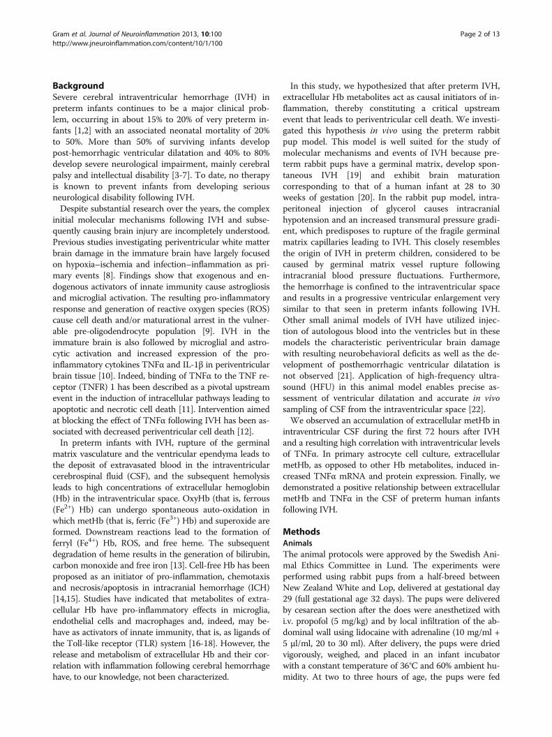
Hemoglobin induces inflammation after pretermintraventricular hemorrhage by methemoglobinformationMagnus Gram1*†, Snjolaug Sveinsdottir2†, Karsten Ruscher3, Stefan R Hansson4, Magnus Cinthio5,Bo Åkerström1 and David Ley2
Abstract
Background: Cerebral intraventricular hemorrhage (IVH) is a major cause of severe neurodevelopmentalimpairment in preterm infants. To date, no therapy is available that prevents infants from developing seriousneurological disability following IVH. Thus, to develop treatment strategies for IVH, it is essential to characterize theinitial sequence of molecular events that leads to brain damage. In this study, we investigated extracellularhemoglobin (Hb) as a causal initiator of inflammation in preterm IVH.
Methods: Using a preterm rabbit pup model, we investigated the molecular mechanisms and events following IVH.We also characterized the concentrations of cell-free Hb metabolites and pro-inflammatory mediators in thecerebrospinal fluid (CSF) of preterm human infants and rabbit pups. Finally, Hb metabolites were evaluated ascausal initiators of inflammation in primary rabbit astrocyte cell cultures.
Results: Following IVH in preterm rabbit pups, the intraventricular CSF concentration of cell-free methemoglobin(metHb) increased from 24 to 72 hours and was strongly correlated with the concentration of TNFα at 72 hours(r2 = 0.896, P <0.001). Also, the mRNA expression of TNFα, IL-1β, and Toll-like receptor-4 and TNFα protein levelswere significantly increased in periventricular tissue at 72 hours, which was accompanied by extensive astrocyteactivation (that is, glial fibrillary acidic protein (GFAP)staining). Furthermore, exposure of primary rabbit astrocyte cellcultures to metHb caused a dose-dependent increase in TNFα mRNA and protein levels, which was not observedfollowing exposure to oxyhemoglobin (oxyHb) or hemin. Finally, a positive correlation (r2 = 0.237, P <0.03) betweenmetHb and TNFα concentrations was observed in the CSF of preterm human infants following IVH.
Conclusions: Following preterm IVH, increased metHb formation in the intraventricular space induces expression ofpro-inflammatory cytokines. Thus, the formation of metHb might be a crucial initial event in the development ofbrain damage following preterm IVH. Accordingly, removal, scavenging, or neutralization of Hb could present atherapeutic opportunity and plausible approach to decreasing the damage in the immature brain followingpreterm IVH.
Keywords: Hemoglobin, Intraventricular hemorrhage, Preterm birth, Perinatal brain damage, Astrocyte,Inflammation, Cerebrospinal fluid, Periventricular brain tissue
* Correspondence: [email protected]†Equal contributors1Division of Infection Medicine, Lund University, S-221 84 Lund, SwedenFull list of author information is available at the end of the article
JOURNAL OF NEUROINFLAMMATION
© 2013 Gram et al.; licensee BioMed Central Ltd. This is an Open Access article distributed under the terms of the CreativeCommons Attribution License (http://creativecommons.org/licenses/by/2.0), which permits unrestricted use, distribution, andreproduction in any medium, provided the original work is properly cited.
Gram et al. Journal of Neuroinflammation 2013, 10:100http://www.jneuroinflammation.com/content/10/1/100
BackgroundSevere cerebral intraventricular hemorrhage (IVH) inpreterm infants continues to be a major clinical prob-lem, occurring in about 15% to 20% of very preterm in-fants [1,2] with an associated neonatal mortality of 20%to 50%. More than 50% of surviving infants developpost-hemorrhagic ventricular dilatation and 40% to 80%develop severe neurological impairment, mainly cerebralpalsy and intellectual disability [3-7]. To date, no therapyis known to prevent infants from developing seriousneurological disability following IVH.
Despite substantial research over the years, the complexinitial molecular mechanisms following IVH and subse-quently causing brain injury are incompletely understood.Previous studies investigating periventricular white matterbrain damage in the immature brain have largely focusedon hypoxia–ischemia and infection–inflammation as pri-mary events [8]. Findings show that exogenous and en-dogenous activators of innate immunity cause astrogliosisand microglial activation. The resulting pro-inflammatoryresponse and generation of reactive oxygen species (ROS)cause cell death and/or maturational arrest in the vulner-able pre-oligodendrocyte population [9]. IVH in theimmature brain is also followed by microglial and astro-cytic activation and increased expression of the pro-inflammatory cytokines TNFα and IL-1β in periventricularbrain tissue [10]. Indeed, binding of TNFα to the TNF re-ceptor (TNFR) 1 has been described as a pivotal upstreamevent in the induction of intracellular pathways leading toapoptotic and necrotic cell death [11]. Intervention aimedat blocking the effect of TNFα following IVH has been as-sociated with decreased periventricular cell death [12].
In preterm infants with IVH, rupture of the germinalmatrix vasculature and the ventricular ependyma leads tothe deposit of extravasated blood in the intraventricularcerebrospinal fluid (CSF), and the subsequent hemolysisleads to high concentrations of extracellular hemoglobin(Hb) in the intraventricular space. OxyHb (that is, ferrous(Fe2+) Hb) can undergo spontaneous auto-oxidation inwhich metHb (that is, ferric (Fe3+) Hb) and superoxide areformed. Downstream reactions lead to the formation offerryl (Fe4+) Hb, ROS, and free heme. The subsequentdegradation of heme results in the generation of bilirubin,carbon monoxide and free iron [13]. Cell-free Hb has beenproposed as an initiator of pro-inflammation, chemotaxisand necrosis/apoptosis in intracranial hemorrhage (ICH)[14,15]. Studies have indicated that metabolites of extra-cellular Hb have pro-inflammatory effects in microglia,endothelial cells and macrophages and, indeed, may be-have as activators of innate immunity, that is, as ligands ofthe Toll-like receptor (TLR) system [16-18]. However, therelease and metabolism of extracellular Hb and their cor-relation with inflammation following cerebral hemorrhagehave, to our knowledge, not been characterized.
In this study, we hypothesized that after preterm IVH,extracellular Hb metabolites act as causal initiators of in-flammation, thereby constituting a critical upstreamevent that leads to periventricular cell death. We investi-gated this hypothesis in vivo using the preterm rabbitpup model. This model is well suited for the study ofmolecular mechanisms and events of IVH because pre-term rabbit pups have a germinal matrix, develop spon-taneous IVH [19] and exhibit brain maturationcorresponding to that of a human infant at 28 to 30weeks of gestation [20]. In the rabbit pup model, intra-peritoneal injection of glycerol causes intracranialhypotension and an increased transmural pressure gradi-ent, which predisposes to rupture of the fragile germinalmatrix capillaries leading to IVH. This closely resemblesthe origin of IVH in preterm children, considered to becaused by germinal matrix vessel rupture followingintracranial blood pressure fluctuations. Furthermore,the hemorrhage is confined to the intraventricular spaceand results in a progressive ventricular enlargement verysimilar to that seen in preterm infants following IVH.Other small animal models of IVH have utilized injec-tion of autologous blood into the ventricles but in thesemodels the characteristic periventricular brain damagewith resulting neurobehavioral deficits as well as the de-velopment of posthemorrhagic ventricular dilatation isnot observed [21]. Application of high-frequency ultra-sound (HFU) in this animal model enables precise as-sessment of ventricular dilatation and accurate in vivosampling of CSF from the intraventricular space [22].
We observed an accumulation of extracellular metHb inintraventricular CSF during the first 72 hours after IVHand a resulting high correlation with intraventricular levelsof TNFα. In primary astrocyte cell culture, extracellularmetHb, as opposed to other Hb metabolites, induced in-creased TNFα mRNA and protein expression. Finally, wedemonstrated a positive relationship between extracellularmetHb and TNFα in the CSF of preterm human infantsfollowing IVH.
MethodsAnimalsThe animal protocols were approved by the Swedish Ani-mal Ethics Committee in Lund. The experiments wereperformed using rabbit pups from a half-breed betweenNew Zealand White and Lop, delivered at gestational day29 (full gestational age 32 days). The pups were deliveredby cesarean section after the does were anesthetized withi.v. propofol (5 mg/kg) and by local infiltration of the ab-dominal wall using lidocaine with adrenaline (10 mg/ml +5 μl/ml, 20 to 30 ml). After delivery, the pups were driedvigorously, weighed, and placed in an infant incubatorwith a constant temperature of 36°C and 60% ambient hu-midity. At two to three hours of age, the pups were fed
Gram et al. Journal of Neuroinflammation 2013, 10:100 Page 2 of 13http://www.jneuroinflammation.com/content/10/1/100
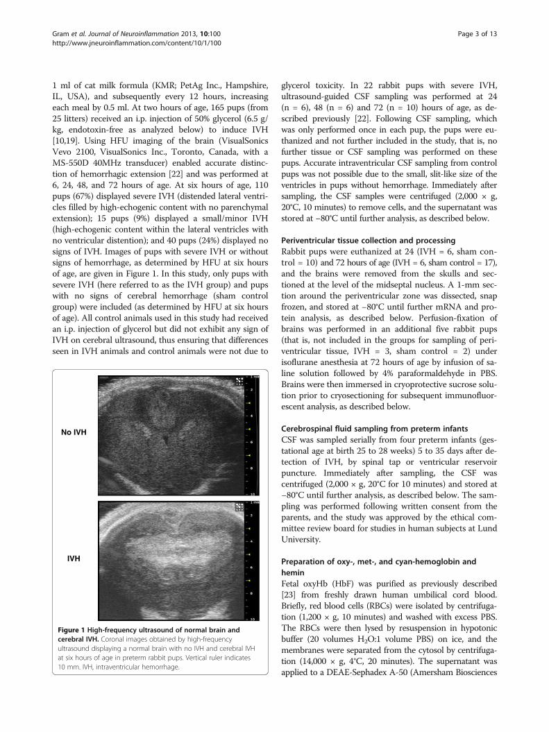
1 ml of cat milk formula (KMR; PetAg Inc., Hampshire,IL, USA), and subsequently every 12 hours, increasingeach meal by 0.5 ml. At two hours of age, 165 pups (from25 litters) received an i.p. injection of 50% glycerol (6.5 g/kg, endotoxin-free as analyzed below) to induce IVH[10,19]. Using HFU imaging of the brain (VisualSonicsVevo 2100, VisualSonics Inc., Toronto, Canada, with aMS-550D 40MHz transducer) enabled accurate distinc-tion of hemorrhagic extension [22] and was performed at6, 24, 48, and 72 hours of age. At six hours of age, 110pups (67%) displayed severe IVH (distended lateral ventri-cles filled by high-echogenic content with no parenchymalextension); 15 pups (9%) displayed a small/minor IVH(high-echogenic content within the lateral ventricles withno ventricular distention); and 40 pups (24%) displayed nosigns of IVH. Images of pups with severe IVH or withoutsigns of hemorrhage, as determined by HFU at six hoursof age, are given in Figure 1. In this study, only pups withsevere IVH (here referred to as the IVH group) and pupswith no signs of cerebral hemorrhage (sham controlgroup) were included (as determined by HFU at six hoursof age). All control animals used in this study had receivedan i.p. injection of glycerol but did not exhibit any sign ofIVH on cerebral ultrasound, thus ensuring that differencesseen in IVH animals and control animals were not due to
glycerol toxicity. In 22 rabbit pups with severe IVH,ultrasound-guided CSF sampling was performed at 24(n = 6), 48 (n = 6) and 72 (n = 10) hours of age, as de-scribed previously [22]. Following CSF sampling, whichwas only performed once in each pup, the pups were eu-thanized and not further included in the study, that is, nofurther tissue or CSF sampling was performed on thesepups. Accurate intraventricular CSF sampling from controlpups was not possible due to the small, slit-like size of theventricles in pups without hemorrhage. Immediately aftersampling, the CSF samples were centrifuged (2,000 × g,20°C, 10 minutes) to remove cells, and the supernatant wasstored at −80°C until further analysis, as described below.
Periventricular tissue collection and processingRabbit pups were euthanized at 24 (IVH = 6, sham con-trol = 10) and 72 hours of age (IVH = 6, sham control = 17),and the brains were removed from the skulls and sec-tioned at the level of the midseptal nucleus. A 1-mm sec-tion around the periventricular zone was dissected, snapfrozen, and stored at −80°C until further mRNA and pro-tein analysis, as described below. Perfusion-fixation ofbrains was performed in an additional five rabbit pups(that is, not included in the groups for sampling of peri-ventricular tissue, IVH = 3, sham control = 2) underisoflurane anesthesia at 72 hours of age by infusion of sa-line solution followed by 4% paraformaldehyde in PBS.Brains were then immersed in cryoprotective sucrose solu-tion prior to cryosectioning for subsequent immunofluor-escent analysis, as described below.
Cerebrospinal fluid sampling from preterm infantsCSF was sampled serially from four preterm infants (ges-tational age at birth 25 to 28 weeks) 5 to 35 days after de-tection of IVH, by spinal tap or ventricular reservoirpuncture. Immediately after sampling, the CSF wascentrifuged (2,000 × g, 20°C for 10 minutes) and stored at−80°C until further analysis, as described below. The sam-pling was performed following written consent from theparents, and the study was approved by the ethical com-mittee review board for studies in human subjects at LundUniversity.
Preparation of oxy-, met-, and cyan-hemoglobin andheminFetal oxyHb (HbF) was purified as previously described[23] from freshly drawn human umbilical cord blood.Briefly, red blood cells (RBCs) were isolated by centrifuga-tion (1,200 × g, 10 minutes) and washed with excess PBS.The RBCs were then lysed by resuspension in hypotonicbuffer (20 volumes H2O:1 volume PBS) on ice, and themembranes were separated from the cytosol by centrifuga-tion (14,000 × g, 4°C, 20 minutes). The supernatant wasapplied to a DEAE-Sephadex A-50 (Amersham Biosciences
IVH
No IVH
Figure 1 High-frequency ultrasound of normal brain andcerebral IVH. Coronal images obtained by high-frequencyultrasound displaying a normal brain with no IVH and cerebral IVHat six hours of age in preterm rabbit pups. Vertical ruler indicates10 mm. IVH, intraventricular hemorrhage.
Gram et al. Journal of Neuroinflammation 2013, 10:100 Page 3 of 13http://www.jneuroinflammation.com/content/10/1/100
AB, Uppsala, Sweden) column and separated using an in-creasing ion gradient. Fractions were collected and the ab-sorbance measured to identify and determine theconcentration of oxyHb. Human Hb was from SigmaChemical Co. (St. Louis, MO, USA) and shown spectro-photometrically [23] to contain at least 70% to 80%metHb. This preparation is referred to here as metHb.Furthermore, metHb was also prepared by incubating theabove described oxyHb solution at 37°C for 72 hours. ThemetHb concentration was quantified as described previ-ously [23]. Hemin (ferriprotoporphyrin IX chloride) waspurchased from Porphyrin Products Inc. (Logan, UT,USA), and a 10 mM stock solution was prepared using di-methyl sulfoxide (DMSO) (Sigma).
Cyan-Hb was prepared as described previously [24], bymixing HbF (1 mM) with KCN (10 mM) and incubatingthe mix for 10 minutes at 20°C. The solution wasdesalted on a Sephadex G-25 column (Amersham).Addition of cyanide to oxyHb locks the Fe2+ atom in itsferrous form, which disables spontaneous oxidation tometHb. All Hb derivatives (oxy-, met- (purchased andin-house prepared) and cyan-Hb) were purified fromendotoxin contamination using the endotoxin removingproduct EndoTrap (Hyglos GmbH, Bernried, Germany)as described by the manufacturer. The absolute purity ofall Hb derivatives (oxy-, met- (purchased and in-houseprepared), cyanHb, and hemin) and the glycerol solution(used for i.p. injections) from contamination with endo-toxin (0 EU/mg Hb/hemin) was determined using theQCL-1000™ Endpoint Chromogenic LAL Assay (Lonza,Basel, Switzerland ) and the lipopolysaccharide (LPS)ELISA Assay kit (Uscn Life Science Inc, Wuhan, China)as described by the manufacturers (Table 1).
Measurement of Hb metabolites in CSFHb metabolite concentrations were determined in CSFfrom preterm infants and rabbit pups using a spectro-photometric method described previously [23]. Briefly,samples were analyzed in a wavelength scan (250 to 700nm) using a Beckman DU-800 spectrophotometer, and ab-sorbance was specifically recorded for total protein (280nm), oxyHb (540 and 577 nm), and metHb (630 nm). The
concentration of oxyHb and metHb was calculated as de-scribed [23] and is displayed as Hb-tetramer concentration.
Primary rabbit pup astrocyte culturesRabbit astroglial cell cultures were prepared from three-day-old healthy rabbit pups (not i.p. injected with glyceroland not used for any other sampling) according to a modi-fied method described previously [25]. Briefly, after de-capitation, the brain regions of interest were mechanicallydissected and digested in trypsin/ethylenediaminete-traacetic acid (EDTA) solution. The tissue then was disso-ciated using a glass pasteur pipette and centrifuged; cellswere resuspended in fresh culture medium and seeded in75 cm2 flasks (cells from one brain/flask). Cells weregrown in complete culture medium, which was changedevery third day. After 10 days, cultures were shaken forone hour (250 rpm) to remove microglial cells, and astro-cytes were resown in subcultures into appropriate culturedishes. When cells reached confluence (cultivation day sixto eight), oxyHb, metHb, cyan-Hb and hemin (preparedimmediately prior to the experiment, as described above)and a mixture of (NH4)Fe(SO4)2, hydrogen peroxide andascorbate (the Fenton reaction) were added to the astro-cyte cultures, and cells were incubated for one to fourhours, as indicated in the figure legends. After incubation,culture medium was collected and cells harvested usingQiazol™ Lysis reagent (Qiagen Sciences, Germantown,MD, USA). Culture medium was analyzed for cell viability,total protein and TNFα protein concentration, and totalRNA was extracted from cells to evaluate TNFα, IL-1β,TLR-4 and heme oxygenase (HO)-1 mRNA expression, asdescribed below.
Cell viability assayThe levels of lactate dehydrogenase (LDH) in astrocyte cellculture media were measured using the CytoTox 96W Non-Radioactive Cytotoxicity Assay (Promega, Madison, WI,USA) according to the instructions from the manufacturer.
Total protein analysisTotal protein concentration in astrocyte cell culturemedia was determined using the Bradford protein assay,as described previously [24]. Albumin was used as a
Table 1 Endotoxin determination of Hb/hemin preparations
Sample Source LAL assay before EndoTrap (EU/mg) LAL assay after EndoTrap (EU/mg) LPS ELISA after EndoTrap
OxyHb In-house purified 2.48 Not detectable Not detectable
MetHb In-house purified Not tested Not detectable Not detectable
MetHb Sigma 10.29 Not detectable Not detectable
CyanHb In-house purified 3.64 Not detectable Not detectable
Hemin Porphyrin Products Not detectable Not detectable Not detectable
Determination of endotoxin levels in the Hb- and hemin preparations using an Endpoint Chromogenic LAL assay and a LPS ELISA assay as described in theMethods section. The endotoxin levels were determined both before and after purification of endotoxins using EndoTrap, as described in the Methods section.The endotoxin levels are determined as EU endotoxin per mg of Hb/hemin. LPS, lipopolysaccharide; MetHb, methemoglobin; OxyHb, oxyhemoglobin.
Gram et al. Journal of Neuroinflammation 2013, 10:100 Page 4 of 13http://www.jneuroinflammation.com/content/10/1/100
standard, and plotting the absorbance at 595 nm versusprotein concentration generated a standard curve.
RNA isolation and real-time PCRTotal RNA was isolated from the periventricular tissueand primary astrocytes using the acid guanidinium phe-nol–chloroform method supplied by Qiagen Sciences. Theoptical density (OD) ratio (260 nm/280 nm) of RNA wasalways higher than 1.8. Reverse transcription wasperformed according to the manufacturer’s instructionson 1 μg total RNA using an iScriptTM cDNA Synthesis Kit(Bio-Rad, Hercules, CA, USA). Real-time PCR was thenused to quantify the TNFα, IL-1β, TLR-4 and HO-1mRNA expression. Data were normalized to humanglyceraldehyde-3-phosphate dehydrogenase (GAPDH).The fold-change values were calculated by normalizingagainst control samples from untreated animals or cells.Primers were designed accordingly: TNFα forward primer5′-CTCCTACCCGAACAAGGTCA-3′, reverse primer5′-CGGTCACCCTTCTCCAACT-3′; IL-1β forwardprimer 5′-AAGAAGAACCCGTCCTCTGCAACA-3′, re-verse primer 5′-TCAGCTCATACGTGCCAGACAACA-3′; TLR-4 forward primer 5′-GAGCACCTGGACCTTTCAAATAAC-3′, reverse primer 5′-GAACTTCTAAACCACTCAGCCCTTG-′3; HO-1 forward primer 5′-GAGATTGAGCGCAACAAGGA-3′, reverse primer 5′-AGCGGTAGAGCTGCTTGAACT-′3; and GAPDHforward primer 5′-GAATCCACTGGCGTCTTCAC-3′,reverse primer 5′-CGTTGCTGACAATCTTGAGAGA-3′. Expression was analyzed using the Maxima SYBRGreen qPCR Master Mix (Thermo Scientific Fermentas,Göteborg, Sweden). Amplification was performed at therespective adequate temperature for 40 cycles in aniCycler Thermal Cycler (Bio-Rad) and data analyzed usingiCycler iQ Optical System Software (Bio-Rad).
TNFα ELISAThe concentrations of TNFα in CSF (preterm infants andrabbit pups), in periventricular tissue from rabbit pupsand in astrocyte cell culture media were determined usingthe Human (CSF preterm infants) and Rabbit TNFαDuoSet ELISA Development kits from R&D Systems,Abingdon, UK. The analysis was performed according tothe instructions from the manufacturer.
ImmunofluorescenceBrain sections (40 μm) from paraformaldehyde-perfusedanimals were washed in PBS, blocked with 2% normalhorse serum and incubated with a polyclonal goat anti-TNFα (1:50, Santa Cruz Biotechnology, Santa Cruz, CA,USA). After overnight incubation at 4°C, sections were in-cubated with a donkey anti-goat biotinylated secondaryantibody (diluted at 1:200, Jackson ImmunoResearch La-boratories, West Grove, PA, USA). Sections were further
incubated with an Alexa-488 streptavidin conjugate (1:200,Invitrogen, Stockholm, Sweden) and a Cy3-conjugatedmonoclonal anti-glial fibrillary acidic protein (GFAP) anti-body (1:500, Sigma-Aldrich, Stockholm, Sweden). Fluores-cent signals were visualized using a confocal microscopysystem (LSM510, Zeiss, Oberkochen, Germany ).
StatisticsPair-wise comparisons between unrelated groups wereperformed with the Students t-test or the Mann–WhitneyU test as appropriate. Comparisons between multiplegroups were performed by analysis of variance (ANOVA)with post hoc Bonferroni correction. Correlations wereassessed by linear regression analysis. P values <0.05 wereconsidered significant.
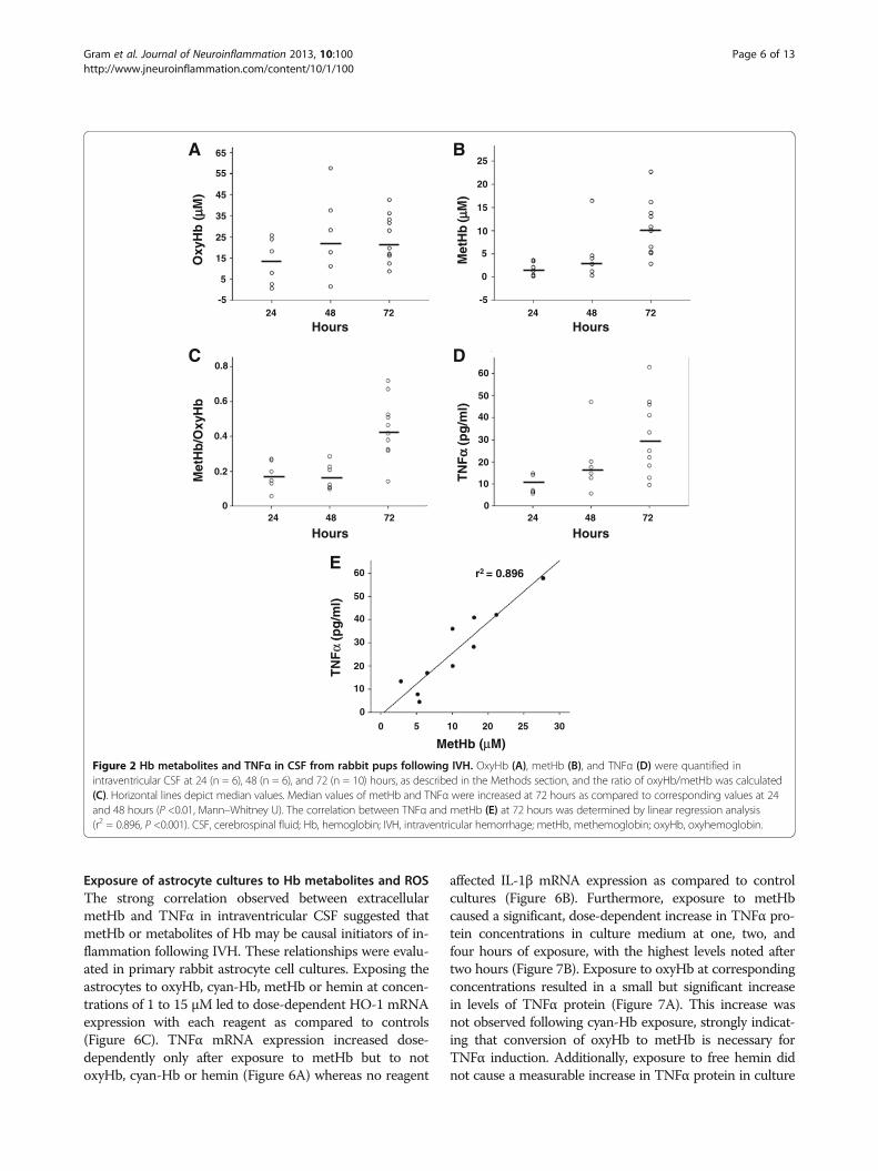
ResultsExtracellular Hb metabolites and TNFα in CSF followingIVHIntraventricular CSF concentrations of extracellular oxyHb,metHb, and TNFα in rabbit pups with IVH were assessedat 24, 48, and 72 hours of age (Figure 2). The median con-centration of oxyHb did not change significantly over time(Figure 2A) whereas that of metHb was significantly in-creased at 72 hours compared to values at 24 and 48 hours(Figure 2B), illustrated by the increasing ratio of metHb/oxyHb over time (Figure 2C). Similarly to metHb, the con-centration of TNFα increased significantly over time andwas highest at 72 hours as compared to 24 and 48 hours(Figure 2D). Furthermore, concentrations of metHb andTNFα exhibited a strong positive correlation at 72 hours(r2 = 0.896, P <0.001, Figure 2E) whereas no correlationwas observed between oxyHb and TNFα (not shown).
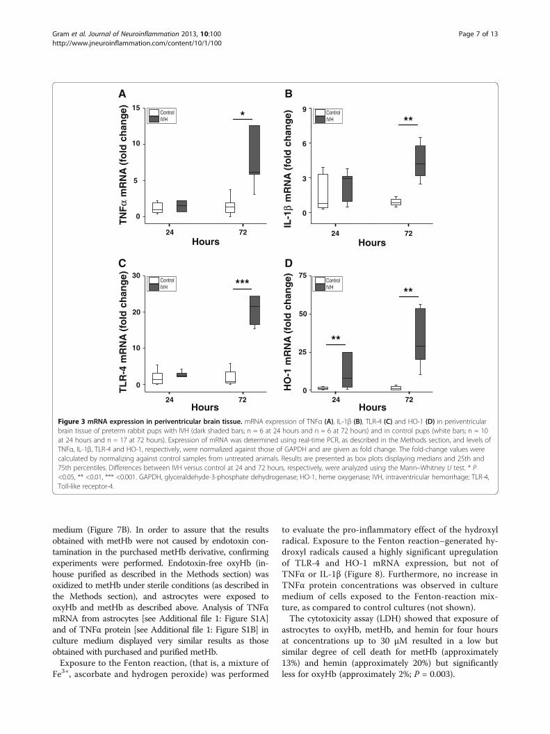
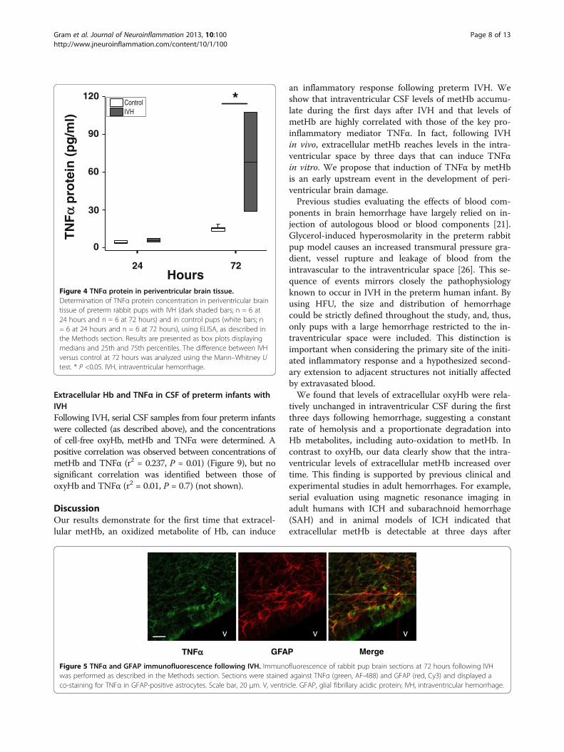
Inflammation in periventricular brain tissue following IVHThe median levels of mRNA expression for TNFα, IL-1β,TLR-4 and HO-1 were increased in periventricular braintissue in rabbit pups with IVH as compared to controlpups (Figure 3). The increases in TNFα, IL-1β and TLR-4mRNA were significant (P <0.05, TNFα; P <0.01, IL-1β; P<0.001, TLR-4) at 72 hours but not at 24 hours. Corres-pondingly, the median level of TNFα protein was in-creased at 72 hours in IVH pups (67.9 pg/mg totalprotein) as compared to control pups (15.4 pg/mg totalprotein; P = 0.02) but not at 24 hours (Figure 4). Inaddition, immunohistochemistry showed positive TNFαstaining in periventricular brain tissue in rabbit pups withIVH at 72 hours (Figure 5). Counter staining for astrocyteactivation (GFAP-positive staining) revealed the presenceof TNFα in GFAP-positive astrocytes but also in non–GFAP-positive cells.
Gram et al. Journal of Neuroinflammation 2013, 10:100 Page 5 of 13http://www.jneuroinflammation.com/content/10/1/100
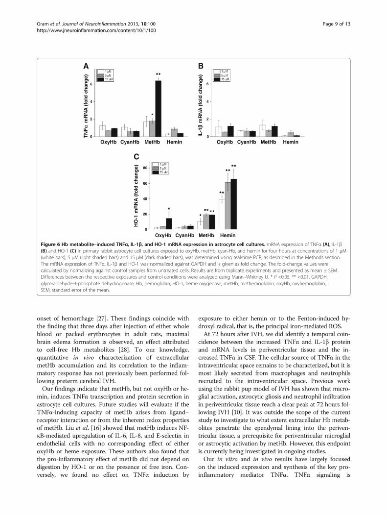
Exposure of astrocyte cultures to Hb metabolites and ROSThe strong correlation observed between extracellularmetHb and TNFα in intraventricular CSF suggested thatmetHb or metabolites of Hb may be causal initiators of in-flammation following IVH. These relationships were evalu-ated in primary rabbit astrocyte cell cultures. Exposing theastrocytes to oxyHb, cyan-Hb, metHb or hemin at concen-trations of 1 to 15 μM led to dose-dependent HO-1 mRNAexpression with each reagent as compared to controls(Figure 6C). TNFα mRNA expression increased dose-dependently only after exposure to metHb but to notoxyHb, cyan-Hb or hemin (Figure 6A) whereas no reagent
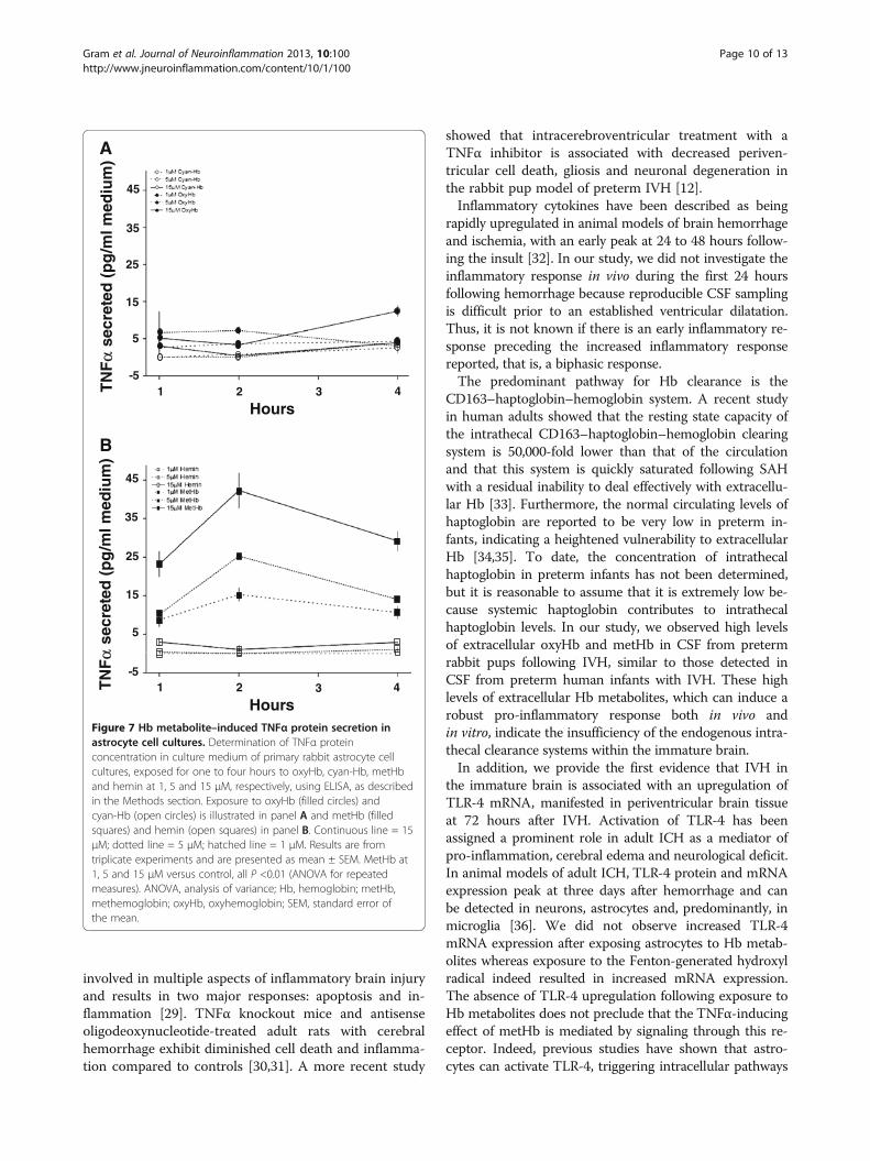
affected IL-1β mRNA expression as compared to controlcultures (Figure 6B). Furthermore, exposure to metHbcaused a significant, dose-dependent increase in TNFα pro-tein concentrations in culture medium at one, two, andfour hours of exposure, with the highest levels noted aftertwo hours (Figure 7B). Exposure to oxyHb at correspondingconcentrations resulted in a small but significant increasein levels of TNFα protein (Figure 7A). This increase wasnot observed following cyan-Hb exposure, strongly indicat-ing that conversion of oxyHb to metHb is necessary forTNFα induction. Additionally, exposure to free hemin didnot cause a measurable increase in TNFα protein in culture
A
Oxy
Hb
(μμM
)
(μμM
)
(μM)
-5
5
15
45
65
Hours24 48 72
25
55
35
B
-5
0
5
15
25
10
20
Met
Hb
Hours24 48 72
0
0.2
0.4
0.6
0.8
24 48 72
Met
Hb
/Oxy
Hb
Hours
0
10
20
40
60
30
50
24 48 72
Hours
DC
E
TN
Fα
(pg
/ml)
TN
Fα α
(pg
/ml)
MetHb
r2 = 0.896
0
10
20
30
40
50
60
0 5 10 20 25 30
Figure 2 Hb metabolites and TNFα in CSF from rabbit pups following IVH. OxyHb (A), metHb (B), and TNFα (D) were quantified inintraventricular CSF at 24 (n = 6), 48 (n = 6), and 72 (n = 10) hours, as described in the Methods section, and the ratio of oxyHb/metHb was calculated(C). Horizontal lines depict median values. Median values of metHb and TNFα were increased at 72 hours as compared to corresponding values at 24and 48 hours (P <0.01, Mann–Whitney U). The correlation between TNFα and metHb (E) at 72 hours was determined by linear regression analysis(r2 = 0.896, P <0.001). CSF, cerebrospinal fluid; Hb, hemoglobin; IVH, intraventricular hemorrhage; metHb, methemoglobin; oxyHb, oxyhemoglobin.
Gram et al. Journal of Neuroinflammation 2013, 10:100 Page 6 of 13http://www.jneuroinflammation.com/content/10/1/100
medium (Figure 7B). In order to assure that the resultsobtained with metHb were not caused by endotoxin con-tamination in the purchased metHb derivative, confirmingexperiments were performed. Endotoxin-free oxyHb (in-house purified as described in the Methods section) wasoxidized to metHb under sterile conditions (as described inthe Methods section), and astrocytes were exposed tooxyHb and metHb as described above. Analysis of TNFαmRNA from astrocytes [see Additional file 1: Figure S1A]and of TNFα protein [see Additional file 1: Figure S1B] inculture medium displayed very similar results as thoseobtained with purchased and purified metHb.
Exposure to the Fenton reaction, (that is, a mixture ofFe3+, ascorbate and hydrogen peroxide) was performed
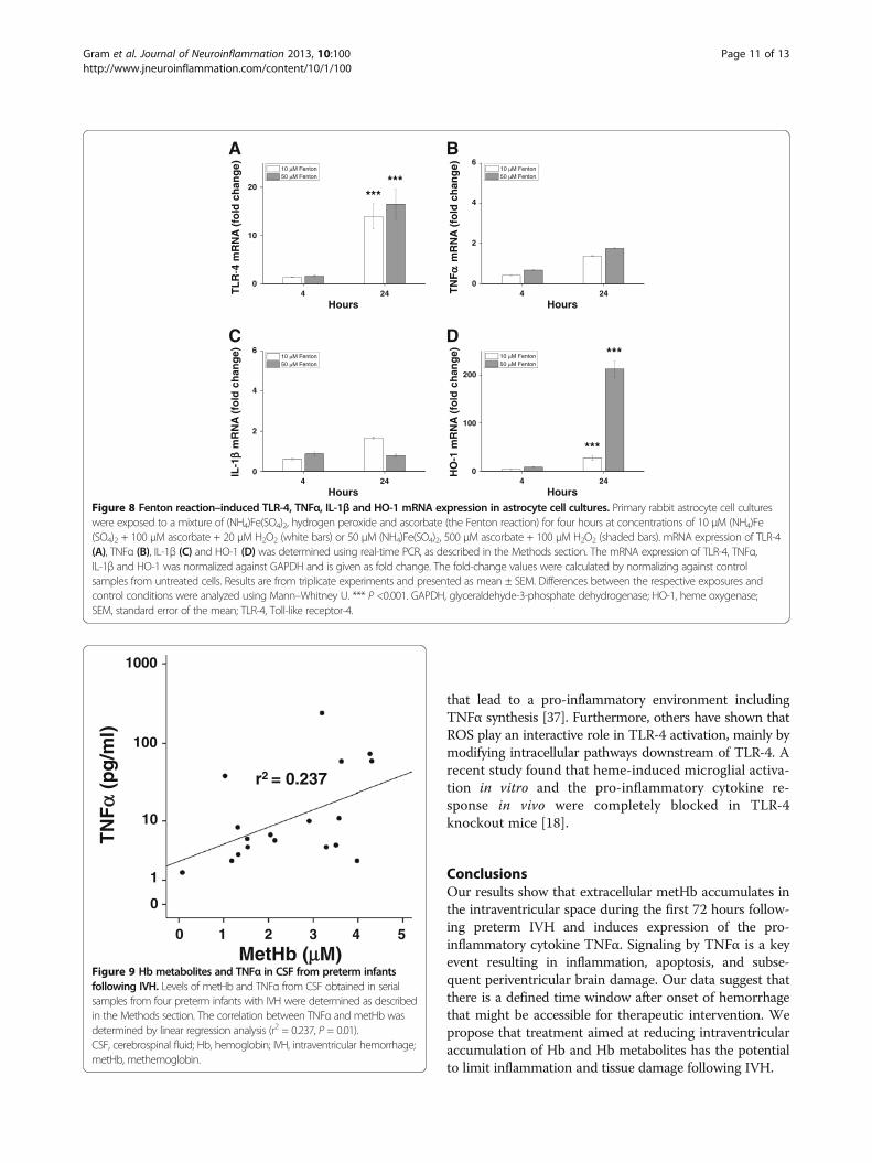
to evaluate the pro-inflammatory effect of the hydroxylradical. Exposure to the Fenton reaction–generated hy-droxyl radicals caused a highly significant upregulationof TLR-4 and HO-1 mRNA expression, but not ofTNFα or IL-1β (Figure 8). Furthermore, no increase inTNFα protein concentrations was observed in culturemedium of cells exposed to the Fenton-reaction mix-ture, as compared to control cultures (not shown).
The cytotoxicity assay (LDH) showed that exposure ofastrocytes to oxyHb, metHb, and hemin for four hoursat concentrations up to 30 μM resulted in a low butsimilar degree of cell death for metHb (approximately13%) and hemin (approximately 20%) but significantlyless for oxyHb (approximately 2%; P = 0.003).
Control IVH
mR
NA
(fo
ld c
han
ge) *
24 72
0
5
10
15
Hours
A
Control IVH **
0
3
6
9
24 72
IL-1
βm
RN
A (
fold
ch
ang
e)
B
Hours
Control IVH ***
0
10
20
30
24 72
TL
R-4
mR
NA
(fo
ld c
han
ge)
Hours
C Control IVH
**
**
75
24 720
25
50
Hours
HO
-1 m
RN
A (
fold
ch
ang
e)
D
TN
Fα
Figure 3 mRNA expression in periventricular brain tissue. mRNA expression of TNFα (A), IL-1β (B), TLR-4 (C) and HO-1 (D) in periventricularbrain tissue of preterm rabbit pups with IVH (dark shaded bars; n = 6 at 24 hours and n = 6 at 72 hours) and in control pups (white bars; n = 10at 24 hours and n = 17 at 72 hours). Expression of mRNA was determined using real-time PCR, as described in the Methods section, and levels ofTNFα, IL-1β, TLR-4 and HO-1, respectively, were normalized against those of GAPDH and are given as fold change. The fold-change values werecalculated by normalizing against control samples from untreated animals. Results are presented as box plots displaying medians and 25th and75th percentiles. Differences between IVH versus control at 24 and 72 hours, respectively, were analyzed using the Mann–Whitney U test. * P<0.05, ** <0.01, *** <0.001. GAPDH, glyceraldehyde-3-phosphate dehydrogenase; HO-1, heme oxygenase; IVH, intraventricular hemorrhage; TLR-4,Toll-like receptor-4.
Gram et al. Journal of Neuroinflammation 2013, 10:100 Page 7 of 13http://www.jneuroinflammation.com/content/10/1/100
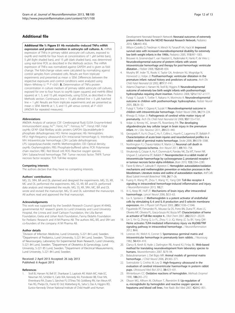
Extracellular Hb and TNFα in CSF of preterm infants withIVHFollowing IVH, serial CSF samples from four preterm infantswere collected (as described above), and the concentrationsof cell-free oxyHb, metHb and TNFα were determined. Apositive correlation was observed between concentrations ofmetHb and TNFα (r2 = 0.237, P = 0.01) (Figure 9), but nosignificant correlation was identified between those ofoxyHb and TNFα (r2 = 0.01, P = 0.7) (not shown).
DiscussionOur results demonstrate for the first time that extracel-lular metHb, an oxidized metabolite of Hb, can induce
an inflammatory response following preterm IVH. Weshow that intraventricular CSF levels of metHb accumu-late during the first days after IVH and that levels ofmetHb are highly correlated with those of the key pro-inflammatory mediator TNFα. In fact, following IVHin vivo, extracellular metHb reaches levels in the intra-ventricular space by three days that can induce TNFαin vitro. We propose that induction of TNFα by metHbis an early upstream event in the development of peri-ventricular brain damage.
Previous studies evaluating the effects of blood com-ponents in brain hemorrhage have largely relied on in-jection of autologous blood or blood components [21].Glycerol-induced hyperosmolarity in the preterm rabbitpup model causes an increased transmural pressure gra-dient, vessel rupture and leakage of blood from theintravascular to the intraventricular space [26]. This se-quence of events mirrors closely the pathophysiologyknown to occur in IVH in the preterm human infant. Byusing HFU, the size and distribution of hemorrhagecould be strictly defined throughout the study, and, thus,only pups with a large hemorrhage restricted to the in-traventricular space were included. This distinction isimportant when considering the primary site of the initi-ated inflammatory response and a hypothesized second-ary extension to adjacent structures not initially affectedby extravasated blood.
We found that levels of extracellular oxyHb were rela-tively unchanged in intraventricular CSF during the firstthree days following hemorrhage, suggesting a constantrate of hemolysis and a proportionate degradation intoHb metabolites, including auto-oxidation to metHb. Incontrast to oxyHb, our data clearly show that the intra-ventricular levels of extracellular metHb increased overtime. This finding is supported by previous clinical andexperimental studies in adult hemorrhages. For example,serial evaluation using magnetic resonance imaging inadult humans with ICH and subarachnoid hemorrhage(SAH) and in animal models of ICH indicated thatextracellular metHb is detectable at three days after
Control IVH
*
24 72
0
60
90
120
Hours
30
TN
Fα α
pro
tein
(p
g/m
l)
Figure 4 TNFα protein in periventricular brain tissue.Determination of TNFα protein concentration in periventricular braintissue of preterm rabbit pups with IVH (dark shaded bars; n = 6 at24 hours and n = 6 at 72 hours) and in control pups (white bars; n= 6 at 24 hours and n = 6 at 72 hours), using ELISA, as described inthe Methods section. Results are presented as box plots displayingmedians and 25th and 75th percentiles. The difference between IVHversus control at 72 hours was analyzed using the Mann–Whitney Utest. * P <0.05. IVH, intraventricular hemorrhage.
GFAP Merge
vvv
TNFαα
Figure 5 TNFα and GFAP immunofluorescence following IVH. Immunofluorescence of rabbit pup brain sections at 72 hours following IVHwas performed as described in the Methods section. Sections were stained against TNFα (green, AF-488) and GFAP (red, Cy3) and displayed aco-staining for TNFα in GFAP-positive astrocytes. Scale bar, 20 μm. V, ventricle. GFAP, glial fibrillary acidic protein; IVH, intraventricular hemorrhage.
Gram et al. Journal of Neuroinflammation 2013, 10:100 Page 8 of 13http://www.jneuroinflammation.com/content/10/1/100
onset of hemorrhage [27]. These findings coincide withthe finding that three days after injection of either wholeblood or packed erythrocytes in adult rats, maximalbrain edema formation is observed, an effect attributedto cell-free Hb metabolites [28]. To our knowledge,quantitative in vivo characterization of extracellularmetHb accumulation and its correlation to the inflam-matory response has not previously been performed fol-lowing preterm cerebral IVH.
Our findings indicate that metHb, but not oxyHb or he-min, induces TNFα transcription and protein secretion inastrocyte cell cultures. Future studies will evaluate if theTNFα-inducing capacity of metHb arises from ligand–receptor interaction or from the inherent redox propertiesof metHb. Liu et al. [16] showed that metHb induces NF-κB-mediated upregulation of IL-6, IL-8, and E-selectin inendothelial cells with no corresponding effect of eitheroxyHb or heme exposure. These authors also found thatthe pro-inflammatory effect of metHb did not depend ondigestion by HO-1 or on the presence of free iron. Con-versely, we found no effect on TNFα induction by
exposure to either hemin or to the Fenton-induced hy-droxyl radical, that is, the principal iron-mediated ROS.
At 72 hours after IVH, we did identify a temporal coin-cidence between the increased TNFα and IL-1β proteinand mRNA levels in periventricular tissue and the in-creased TNFα in CSF. The cellular source of TNFα in theintraventricular space remains to be characterized, but it ismost likely secreted from macrophages and neutrophilsrecruited to the intraventricular space. Previous workusing the rabbit pup model of IVH has shown that micro-glial activation, astrocytic gliosis and neutrophil infiltrationin periventricular tissue reach a clear peak at 72 hours fol-lowing IVH [10]. It was outside the scope of the currentstudy to investigate to what extent extracellular Hb metab-olites penetrate the ependymal lining into the periven-tricular tissue, a prerequisite for periventricular microglialor astrocytic activation by metHb. However, this endpointis currently being investigated in ongoing studies.
Our in vitro and in vivo results have largely focusedon the induced expression and synthesis of the key pro-inflammatory mediator TNFα. TNFα signaling is
Hemin
A
*
**
0
2
4
6
OxyHb CyanHb MetHb
mR
NA
(fo
ld c
han
ge)
0
2
4
6
OxyHb CyanHb MetHb Hemin
IL-1
ββm
RN
A (
fold
ch
ang
e)
B
****
**
*****
*
OxyHb CyanHb MetHb Hemin0
20
40
60
80
HO
-1 m
RN
A (
fold
ch
ang
e)
C
TN
Fα
Figure 6 Hb metabolite–induced TNFα, IL-1β, and HO-1 mRNA expression in astrocyte cell cultures. mRNA expression of TNFα (A), IL-1β(B) and HO-1 (C) in primary rabbit astrocyte cell cultures exposed to oxyHb, metHb, cyan-Hb, and hemin for four hours at concentrations of 1 μM(white bars), 5 μM (light shaded bars) and 15 μM (dark shaded bars), was determined using real-time PCR, as described in the Methods section.The mRNA expression of TNFα, IL-1β and HO-1 was normalized against GAPDH and is given as fold change. The fold-change values werecalculated by normalizing against control samples from untreated cells. Results are from triplicate experiments and presented as mean ± SEM.Differences between the respective exposures and control conditions were analyzed using Mann–Whitney U. * P <0.05, ** <0.01. GAPDH,glyceraldehyde-3-phosphate dehydrogenase; Hb, hemoglobin; HO-1, heme oxygenase; metHb, methemoglobin; oxyHb, oxyhemoglobin;SEM, standard error of the mean.
Gram et al. Journal of Neuroinflammation 2013, 10:100 Page 9 of 13http://www.jneuroinflammation.com/content/10/1/100
involved in multiple aspects of inflammatory brain injuryand results in two major responses: apoptosis and in-flammation [29]. TNFα knockout mice and antisenseoligodeoxynucleotide-treated adult rats with cerebralhemorrhage exhibit diminished cell death and inflamma-tion compared to controls [30,31]. A more recent study
showed that intracerebroventricular treatment with aTNFα inhibitor is associated with decreased periven-tricular cell death, gliosis and neuronal degeneration inthe rabbit pup model of preterm IVH [12].
Inflammatory cytokines have been described as beingrapidly upregulated in animal models of brain hemorrhageand ischemia, with an early peak at 24 to 48 hours follow-ing the insult [32]. In our study, we did not investigate theinflammatory response in vivo during the first 24 hoursfollowing hemorrhage because reproducible CSF samplingis difficult prior to an established ventricular dilatation.Thus, it is not known if there is an early inflammatory re-sponse preceding the increased inflammatory responsereported, that is, a biphasic response.
The predominant pathway for Hb clearance is theCD163–haptoglobin–hemoglobin system. A recent studyin human adults showed that the resting state capacity ofthe intrathecal CD163–haptoglobin–hemoglobin clearingsystem is 50,000-fold lower than that of the circulationand that this system is quickly saturated following SAHwith a residual inability to deal effectively with extracellu-lar Hb [33]. Furthermore, the normal circulating levels ofhaptoglobin are reported to be very low in preterm in-fants, indicating a heightened vulnerability to extracellularHb [34,35]. To date, the concentration of intrathecalhaptoglobin in preterm infants has not been determined,but it is reasonable to assume that it is extremely low be-cause systemic haptoglobin contributes to intrathecalhaptoglobin levels. In our study, we observed high levelsof extracellular oxyHb and metHb in CSF from pretermrabbit pups following IVH, similar to those detected inCSF from preterm human infants with IVH. These highlevels of extracellular Hb metabolites, which can induce arobust pro-inflammatory response both in vivo andin vitro, indicate the insufficiency of the endogenous intra-thecal clearance systems within the immature brain.
In addition, we provide the first evidence that IVH inthe immature brain is associated with an upregulation ofTLR-4 mRNA, manifested in periventricular brain tissueat 72 hours after IVH. Activation of TLR-4 has beenassigned a prominent role in adult ICH as a mediator ofpro-inflammation, cerebral edema and neurological deficit.In animal models of adult ICH, TLR-4 protein and mRNAexpression peak at three days after hemorrhage and canbe detected in neurons, astrocytes and, predominantly, inmicroglia [36]. We did not observe increased TLR-4mRNA expression after exposing astrocytes to Hb metab-olites whereas exposure to the Fenton-generated hydroxylradical indeed resulted in increased mRNA expression.The absence of TLR-4 upregulation following exposure toHb metabolites does not preclude that the TNFα-inducingeffect of metHb is mediated by signaling through this re-ceptor. Indeed, previous studies have shown that astro-cytes can activate TLR-4, triggering intracellular pathways
-5
5
15
25
35
45
1 2 3 4
Hours
secr
eted
(p
g/m
l med
ium
)
1 2 3 4
Hours
-5
5
15
25
35
45
A
secr
eted
(p
g/m
l med
ium
)
B
TN
Fα
TN
Fα
Figure 7 Hb metabolite–induced TNFα protein secretion inastrocyte cell cultures. Determination of TNFα proteinconcentration in culture medium of primary rabbit astrocyte cellcultures, exposed for one to four hours to oxyHb, cyan-Hb, metHband hemin at 1, 5 and 15 μM, respectively, using ELISA, as describedin the Methods section. Exposure to oxyHb (filled circles) andcyan-Hb (open circles) is illustrated in panel A and metHb (filledsquares) and hemin (open squares) in panel B. Continuous line = 15μM; dotted line = 5 μM; hatched line = 1 μM. Results are fromtriplicate experiments and are presented as mean ± SEM. MetHb at1, 5 and 15 μM versus control, all P <0.01 (ANOVA for repeatedmeasures). ANOVA, analysis of variance; Hb, hemoglobin; metHb,methemoglobin; oxyHb, oxyhemoglobin; SEM, standard error ofthe mean.
Gram et al. Journal of Neuroinflammation 2013, 10:100 Page 10 of 13http://www.jneuroinflammation.com/content/10/1/100
that lead to a pro-inflammatory environment includingTNFα synthesis [37]. Furthermore, others have shown thatROS play an interactive role in TLR-4 activation, mainly bymodifying intracellular pathways downstream of TLR-4. Arecent study found that heme-induced microglial activa-tion in vitro and the pro-inflammatory cytokine re-sponse in vivo were completely blocked in TLR-4knockout mice [18].
ConclusionsOur results show that extracellular metHb accumulates inthe intraventricular space during the first 72 hours follow-ing preterm IVH and induces expression of the pro-inflammatory cytokine TNFα. Signaling by TNFα is a keyevent resulting in inflammation, apoptosis, and subse-quent periventricular brain damage. Our data suggest thatthere is a defined time window after onset of hemorrhagethat might be accessible for therapeutic intervention. Wepropose that treatment aimed at reducing intraventricularaccumulation of Hb and Hb metabolites has the potentialto limit inflammation and tissue damage following IVH.
10 M Fenton 50 M Fenton
mR
NA
(fo
ld c
han
ge) 10 M Fenton
50 M Fenton
TL
R-4
mR
NA
(fo
ld c
han
ge)
0
10
20
4 24
Hours
A B
0
2
4
6
4 24
Hours
10 M Fenton 50 M Fenton
C D
4 24
Hours
IL-1
ββm
RN
A (
fold
ch
ang
e)
0
2
4
6 10 M Fenton 50 M Fenton
HO
-1 m
RN
A (
fold
ch
ang
e)
4 24
Hours
0
100
200
******
***
***
TN
Fαα
Figure 8 Fenton reaction–induced TLR-4, TNFα, IL-1β and HO-1 mRNA expression in astrocyte cell cultures. Primary rabbit astrocyte cell cultureswere exposed to a mixture of (NH4)Fe(SO4)2, hydrogen peroxide and ascorbate (the Fenton reaction) for four hours at concentrations of 10 μM (NH4)Fe(SO4)2 + 100 μM ascorbate + 20 μM H2O2 (white bars) or 50 μM (NH4)Fe(SO4)2, 500 μM ascorbate + 100 μM H2O2 (shaded bars). mRNA expression of TLR-4(A), TNFα (B), IL-1β (C) and HO-1 (D) was determined using real-time PCR, as described in the Methods section. The mRNA expression of TLR-4, TNFα,IL-1β and HO-1 was normalized against GAPDH and is given as fold change. The fold-change values were calculated by normalizing against controlsamples from untreated cells. Results are from triplicate experiments and presented as mean ± SEM. Differences between the respective exposures andcontrol conditions were analyzed using Mann–Whitney U. *** P <0.001. GAPDH, glyceraldehyde-3-phosphate dehydrogenase; HO-1, heme oxygenase;SEM, standard error of the mean; TLR-4, Toll-like receptor-4.
0 1 2 3 4 5MetHb (μμM)
100
1000
10
1
0
(pg
/ml)
r2 = 0.237
TN
Fαα
Figure 9 Hb metabolites and TNFα in CSF from preterm infantsfollowing IVH. Levels of metHb and TNFα from CSF obtained in serialsamples from four preterm infants with IVH were determined as describedin the Methods section. The correlation between TNFα and metHb wasdetermined by linear regression analysis (r2 = 0.237, P = 0.01).CSF, cerebrospinal fluid; Hb, hemoglobin; IVH, intraventricular hemorrhage;metHb, methemoglobin.
Gram et al. Journal of Neuroinflammation 2013, 10:100 Page 11 of 13http://www.jneuroinflammation.com/content/10/1/100
Additional file
Additional file 1: Figure S1 Hb metabolite–induced TNFα mRNAexpression and protein secretion in astrocyte cell cultures. A. mRNAexpression of TNFα in primary rabbit astrocyte cell cultures, exposed tooxyHb and metHb for four hours at concentrations of 1 μM (white bars),5 μM (light shaded bars), and 15 μM (dark shaded bars), was determinedusing real-time PCR, as described in the Methods section. The mRNAexpression of TNFα was normalized against GAPDH and is given as foldchange. The fold-change values were calculated by normalizing againstcontrol samples from untreated cells. Results are from triplicateexperiments and presented as mean ± SEM. Differences between therespective exposures and control conditions were analyzed usingMann–Whitney U. ** P <0.01. B. Determination of TNFα proteinconcentration in culture medium of primary rabbit astrocyte cell cultures,exposed for one to four hours to oxyHb (open squares) and metHb (filledsquares) at 1, 5, and 15 μM, respectively, using ELISA, as described in theMethods section. Continuous line = 15 μM; dotted line = 5 μM; hatchedline = 1 μM. Results are from triplicate experiments and are presented asmean ± SEM. MetHb at 1, 5, and 15 μM versus control, all P <0.01(ANOVA for repeated measures).
AbbreviationsANOVA: Analysis of variance; CSF: Cerebrospinal fluid; ELISA: Enzyme-linkedimmunosorbent assay; Fe3+: Ferric; Fe2+: Ferrous; Fe4+: Ferryl; HbF: FetaloxyHb; GFAP: Glial fibrillary acidic protein; GAPDH: Glyceraldehyde-3-phosphate dehydrogenase; HO: Heme oxygenase; Hb: Hemoglobin;HFU: High-frequency ultrasound; ICH: Intracranial hemorrhage; IL: Interleukin;IVH: Intraventricular hemorrhage; LDH: Lactate dehydrogenase;LPS: Lipopolysaccharide; metHb: Methemoglobin; OD: Optical density;oxyHb: Oxyhemoglobin; PBS: Phosphate-buffered saline; PCR: Polymerasechain reaction; RBC: Red blood cell; ROS: Reactive oxygen species;SAH: Subarachnoid hemorrhage; TNF: Tumor necrosis factor; TNFR: Tumornecrosis factor receptor; TLR: Toll-like receptor.
Competing interestsThe authors declare that they have no competing interests.
Authors’ contributionsMG, SS, SRH, BÅ and DL planned and designed the experiments. MG, SS, KR,MC and DL performed the experiments. MG, SS, KR, BÅ and DL performeddata analysis and interpreted the results. MG, SS, KR, SRH, MC, BÅ and DLwrote and revised the manuscript. MG, SS and DL submitted the manuscript.All authors read and approved the final manuscript.
AcknowledgementsThis work was supported by the Swedish Research Council (grant #14940),governmental ALF research grants to Lund University and Lund UniversityHospital, the Linnea and Josef Carlsson Foundation, the Lilla BarnetFoundation, Greta and Johan Kock Foundation, Fanny Ekdahls Foundationfor Pediatric Research and A1M-Pharma AB. The authors MG, BÅ, and SRH areco-founders of the company A1M Pharma AB.
Author details1Division of Infection Medicine, Lund University, S-221 84 Lund, Sweden.2Department of Pediatrics, Lund University, S-221 84 Lund, Sweden. 3Divisionof Neurosurgery, Laboratory for Experimental Brain Research, Lund University,S-221 84 Lund, Sweden. 4Department of Obstetrics & Gynecology, LundUniversity, S-221 85 Lund, Sweden. 5Department of Electrical Measurements,Lund University, S-221 84 Lund, Sweden.
Received: 2 April 2013 Accepted: 26 July 2013Published: 6 August 2013
References1. Stoll BJ, Hansen NI, Bell EF, Shankaran S, Laptook AR, Walsh MC, Hale EC,
Newman NS, Schibler K, Carlo WA, Kennedy KA, Poindexter BB, Finer NN,Ehrenkranz RA, Duara S, Sánchez PJ, O'Shea TM, Goldberg RN, Van Meurs KP,Faix RG, Phelps DL, Frantz ID 3rd, Watterberg KL, Saha S, Das A, Higgins RD,Eunice Kennedy Shriver National Institute of Child Health and Human
Development Neonatal Research Network: Neonatal outcomes of extremelypreterm infants from the NICHD Neonatal Research Network. Pediatrics2010, 126:443–456.
2. Wilson-Costello D, Friedman H, Minich N, Fanaroff AA, Hack M: Improvedsurvival rates with increased neurodevelopmental disability for extremelylow birth weight infants in the 1990s. Pediatrics 2005, 115:997–1003.
3. Brouwer A, Groenendaal F, van Haastert IL, Rademaker K, Hanlo P, de Vries L:Neurodevelopmental outcome of preterm infants with severeintraventricular hemorrhage and therapy for post-hemorrhagic ventriculardilatation. J Pediatr 2008, 152:648–654.
4. Murphy BP, Inder TE, Rooks V, Taylor GA, Anderson NJ, Mogridge N,Horwood LJ, Volpe JJ: Posthaemorrhagic ventricular dilatation in thepremature infant: natural history and predictors of outcome. Arch DisChild Fetal Neonatal Ed 2002, 87:F37–F41.
5. Adams-Chapman I, Hansen NI, Stoll BJ, Higgins R: Neurodevelopmentaloutcome of extremely low birth weight infants with posthemorrhagichydrocephalus requiring shunt insertion. Pediatrics 2008, 121:e1167–e1177.
6. Futagi Y, Suzuki Y, Toribe Y, Nakano H, Morimoto K: Neurodevelopmentaloutcome in children with posthemorrhagic hydrocephalus. Pediatr Neurol2005, 33:26–32.
7. Futagi Y, Toribe Y, Ogawa K, Suzuki Y: Neurodevelopmental outcome inchildren with intraventricular hemorrhage. Pediatr Neurol 2006, 34:219–224.
8. Khwaja O, Volpe JJ: Pathogenesis of cerebral white matter injury ofprematurity. Arch Dis Child Fetal Neonatal Ed 2008, 93:F153–F161.
9. Volpe JJ, Kinney HC, Jensen FE, Rosenberg PA: The developingoligodendrocyte: key cellular target in brain injury in the prematureinfant. Int J Dev Neurosci 2011, 29:423–440.
10. Georgiadis P, Xu H, Chua C, Hu F, Collins L, Huynh C, Lagamma EF, Ballabh P:Characterization of acute brain injuries and neurobehavioral profiles in arabbit model of germinal matrix hemorrhage. Stroke 2008, 39:3378–3388.
11. Northington FJ, Chavez-Valdez R, Martin LJ: Neuronal cell death inneonatal hypoxia-ischemia. Ann Neurol 2011, 69:743–758.
12. Vinukonda G, Csiszar A, Hu F, Dummula K, Pandey NK, Zia MT, Ferreri NR,Ungvari Z, LaGamma EF, Ballabh P: Neuroprotection in a rabbit model ofintraventricular haemorrhage by cyclooxygenase-2, prostanoid receptor-1or tumour necrosis factor-alpha inhibition. Brain 2010, 133:2264–2280.
13. Faivre B, Menu P, Labrude P, Vigneron C: Hemoglobin autooxidation/oxidationmechanisms and methemoglobin prevention or reduction processes in thebloodstream. Literature review and outline of autooxidation reaction. Artif CellsBlood Substit Immobil Biotechnol 1998, 26:17–26.
14. Fuang H, Wang PF, Zhou Y, Wang YC, Yang QW: Toll-like receptor 4signaling in intracerebral hemorrhage-induced inflammation and injury.J Neuroinflammation 2013, 10:27.
15. Xi G, Keep RF, Hoff JT: Mechanisms of brain injury after intracerebralhaemorrhage. Lancet Neurol 2006, 5:53–63.
16. Liu X, Spolarics Z: Methemoglobin is a potent activator of endothelialcells by stimulating IL-6 and IL-8 production and E-selectin membraneexpression. Am J Physiol Cell Physiol 2003, 285:C1036–C1046.
17. Figueiredo RT, Fernandez PL, Mourao-Sa DS, Porto BN, Dutra FF, Alves LS,Oliveira MF, Oliveira PL, Graca-Souza AV, Bozza MT: Characterization of hemeas activator of Toll-like receptor 4. J Biol Chem 2007, 282:20221–20229.
18. Lin S, Yin Q, Zhong Q, Lv FL, Zhou Y, Li JQ, Wang JZ, Su BY, Yang QW:Heme activates TLR4-mediated inflammatory injury via MyD88/TRIFsignaling pathway in intracerebral hemorrhage. J Neuroinflammation2012, 9:46.
19. Lorenzo AV, Welch K, Conner S: Spontaneous germinal matrix andintraventricular hemorrhage in prematurely born rabbits. J Neurosurg1982, 56:404–410.
20. Clancy B, Kersh B, Hyde J, Darlington RB, Anand KJ, Finlay BL: Web-basedmethod for translating neurodevelopment from laboratory species tohumans. Neuroinformatics 2007, 5:79–94.
21. Balasubramaniam J, Del Bigio MR: Animal models of germinal matrixhemorrhage. J Child Neurol 2006, 21:365–371.
22. Sveinsdottir S, Cinthio M, Ley D: High-frequency ultrasound in theevaluation of cerebral intraventricular haemorrhage in preterm rabbitpups. Ultrasound Med Biol 2012, 38:423–431.
23. Winterbourn CC: Oxidative reactions of hemoglobin. Methods Enzymol1990, 186:265–272.
24. Olsson MG, Allhorn M, Olofsson T, Åkerström B: Up-regulation ofa1-microglobulin by hemoglobin and reactive oxygen species inhepatoma and blood cell lines. Free Radic Biol Med 2007, 42:842–851.
Gram et al. Journal of Neuroinflammation 2013, 10:100 Page 12 of 13http://www.jneuroinflammation.com/content/10/1/100
25. Ruscher K, Freyer D, Karsch M, Isaev N, Megow D, Sawitzki B, Priller J,Dirnagl U, Meisel A: Erythropoietin is a paracrine mediator of ischemictolerance in the brain: evidence from an in vitro model. J Neurosci 2002,22:10291–10301.
26. Conner ES, Lorenzo AV, Welch K, Dorval B: The role of intracranialhypotension in neonatal intraventricular hemorrhage. J Neurosurg 1983,58:204–209.
27. Bradley WG Jr: MR appearance of hemorrhage in the brain. Radiology1993, 189:15–26.
28. Xi G, Keep RF, Hoff JT: Erythrocytes and delayed brain edema formationfollowing intracerebral hemorrhage in rats. J Neurosurg 1998, 89:991–996.
29. Ginis I, Jaiswal R, Klimanis D, Liu J, Greenspon J, Hallenbeck JM:TNF-alpha-induced tolerance to ischemic injury involves differentialcontrol of NF-kappaB transactivation: the role of NF-kappaB associationwith p300 adaptor. J Cereb Blood Flow Metab 2002, 22:142–152.
30. Mayne M, Fotheringham J, Yan HJ, Power C, Del Bigio MR, Peeling J, Geiger JD:Adenosine A2A receptor activation reduces proinflammatory events anddecreases cell death following intracerebral hemorrhage. Ann Neurol 2001,49:727–735.
31. Hua R, Walz W: Minocycline treatment prevents cavitation in rats after acortical devascularizing lesion. Brain Res 2006, 1090:172–181.
32. Maddahi A, Kruse LS, Chen QW, Edvinsson L: The role of tumor necrosisfactor-alpha and TNF-alpha receptors in cerebral arteries followingcerebral ischemia in rat. J Neuroinflammation 2011, 8:107.
33. Galea J, Cruickshank G, Teeling JL, Boche D, Garland P, Perry VH, Galea I:The intrathecal CD163-haptoglobin-hemoglobin scavenging system insubarachnoid hemorrhage. J Neurochem 2012, 121:785–792.
34. Kanakoudi F, Drossou V, Tzimouli V, Diamanti E, Konstantinidis T, Germenis A,Kremenopoulos G: Serum concentrations of 10 acute-phase proteins inhealthy term and preterm infants from birth to age 6 months. Clin Chem1995, 41:605–608.
35. Chavez-Bueno S, Beasley JA, Goldbeck JM, Bright BC, Morton DJ, Whitby PW,Stull TL: Haptoglobin concentrations in preterm and term newborns.J Perinatol 2011, 31:500–503.
36. Yao L, Kan EM, Lu J, Hao A, Dheen ST, Kaur C, Ling EA: Toll-like receptor 4mediates microglial activation and production of inflammatorymediators in neonatal rat brain following hypoxia: role of TLR4 inhypoxic microglia. J Neuroinflammation 2013, 10:23.
37. Gorina R, Font-Nieves M, Marquez-Kisinousky L, Santalucia T, Planas AM:Astrocyte TLR4 activation induces a proinflammatory environmentthrough the interplay between MyD88-dependent NFkappaB signaling,MAPK, and Jak1/Stat1 pathways. GLIA 2011, 59:242–255.
doi:10.1186/1742-2094-10-100Cite this article as: Gram et al.: Hemoglobin induces inflammation afterpreterm intraventricular hemorrhage by methemoglobin formation.Journal of Neuroinflammation 2013 10:100.
Submit your next manuscript to BioMed Centraland take full advantage of:
• Convenient online submission
• Thorough peer review
• No space constraints or color figure charges
• Immediate publication on acceptance
• Inclusion in PubMed, CAS, Scopus and Google Scholar
• Research which is freely available for redistribution
Submit your manuscript at www.biomedcentral.com/submit
Gram et al. Journal of Neuroinflammation 2013, 10:100 Page 13 of 13http://www.jneuroinflammation.com/content/10/1/100