Embed Size (px)
Citation preview

Invited Review
CALL FOR PAPERS Cardiovascular-Renal Mechanisms in Health and Disease
Dimethylarginine dimethylaminohydrolase (DDAH): expression, regulation,and function in the cardiovascular and renal systems
Fredrik Palm, Maristela L. Onozato, Zaiming Luo, and Christopher S. WilcoxDivision of Nephrology and Hypertension and Cardiovascular Kidney Hypertension Institute, Georgetown University,Washington, District of Columbia
Palm F, Onozato ML, Luo Z, Wilcox CS. Dimethylarginine dimethylamin-ohydrolase (DDAH): expression, regulation, and function in the cardiovascular andrenal systems. Am J Physiol Heart Circ Physiol 293: H3227–H3245, 2007. Firstpublished October 12, 2007; doi:10.1152/ajpheart.00998.2007.—Asymmetric(NG,NG)-dimethylarginine (ADMA) inhibits nitric oxide (NO) synthases (NOS).ADMA is a risk factor for endothelial dysfunction, cardiovascular mortality, andprogression of chronic kidney disease. Two isoforms of dimethylarginine dimeth-ylaminohydrolase (DDAH) metabolize ADMA. DDAH-1 is the predominant iso-form in the proximal tubules of the kidney and in the liver. These organs extractADMA from the circulation. DDAH-2 is the predominant isoform in the vascula-ture, where it is found in endothelial cells adjacent to the cell membrane and inintracellular vesicles and in vascular smooth muscle cells among the myofibrils andthe nuclear envelope. In vivo gene silencing of DDAH-1 in the rat and DDAH �/�mice both have increased circulating ADMA, whereas gene silencing of DDAH-2reduces vascular NO generation and endothelium-derived relaxation factor re-sponses. DDAH-2 also is expressed in the kidney in the macula densa and distalnephron. Angiotensin type 1 receptor activation in kidneys reduces the expressionof DDAH-1 but increases the expression of DDAH-2. This rapidly evolvingevidence of isoform-specific distribution and regulation of DDAH expression in thekidney and blood vessels provides potential mechanisms for nephron site-specificregulation of NO production. In this review, the recent advances in the regulationand function of DDAH enzymes, their roles in the regulation of NO generation, andtheir possible contribution to endothelial dysfunction in patients with cardiovascu-lar and kidney diseases are discussed.
nitric oxide synthase; hypertension; diabetes mellitus; chronic kidney disease;asymmetric dimethylarginine
ASYMMETRIC (NG,NG) dimethylarginine (ADMA) is an endoge-nous methylated amino acid that inhibits the constitutiveendothelial (e) or type III and neuronal (n) or type I isoformsof nitric oxide (NO) synthase (NOS) (49, 91, 103, 199). It is aless potent inhibitor of the inducible (i) or type II NOS isoform(41, 191, 213). Proteins are subject to methylation of arginineresidues by protein arginine methyltransferase (PRMT).S-adenosylmethionine, which is synthesized from methionineand ATP, serves as the methyl donor and, in the process, isconverted to S-adenosylhomocysteine, which itself can behydrolyzed to homocysteine. Remethylation of homocysteinein the “remethylation pathway” regenerates methionine (14,179). ADMA is released by protein hydrolysis and exportedfrom the cell and taken up by other cells via system y� carriersof the cationic amino acid (CAT) family (14, 196, 212).ADMA is eliminated both by renal excretion and metabolicdegradation. Its metabolism is facilitated by dimethylarginine
dimethylaminohydrolases (DDAHs), which are expressed astype 1 and 2 isoforms. Recent studies have shown differentialsites of expression of DDAH-1 and -2 in blood vessels and thekidneys and differential regulation of the renal expression ofthese isoforms by ANG II acting on type 1 receptors (AT1-Rs)(127). DDAH also is subject to extensive posttranscriptionalinhibition, for example by reactive oxygen species (ROS)generated by nicotinamide adenine dinucleotide phosphate(NADPH) oxidase or by homocysteine (65, 190). These con-cepts are described in greater details in the body of this reviewand are introduced in Fig. 11.
Since DDAH metabolizes ADMA (2) and regulates plasmalevels of ADMA, it can determine bioavailable NO (36, 204).NO activity in blood vessels mediates an important componentof the endothelium-dependent relaxation factor (EDRF) re-sponses in blood vessels (131), prevents vascular remodeling(52, 184) maintains a stable blood pressure (BP) and renal
Address for reprint requests and other correspondence: C. S. Wilcox,Georgetown Univ., Div. of Nephrology and Hypertension, 3800 Reservoir Rd.,N.W., PHC F6003, Washington, DC 20007 (e-mail: [email protected]).
1 This paper was presented at the 9th Cardiovascular-Kidney Interactions inHealth and Disease Meeting at Amelia Island Plantation, Florida, on May26–29, 2006.
Am J Physiol Heart Circ Physiol 293: H3227–H3245, 2007.First published October 12, 2007; doi:10.1152/ajpheart.00998.2007.
0363-6135/07 $8.00 Copyright © 2007 the American Physiological Societyhttp://www.ajpheart.org H3227
by 10.220.33.5 on October 23, 2017
http://ajpheart.physiology.org/D
ownloaded from

vascular resistance (RVR) (15, 38, 40), inhibits Na� entry intothe thick ascending limb (TAL) of the loop of Henle (LH)(128) and the collecting ducts (CDs) (163), and prevents saltsensitivity (143). Consequently, DDAH could have a pivotalrole in maintaining the homeostatic integrity of the cardiovas-cular and renal systems. This is the focus of this review.Readers are referred to recent reviews for further informationon the cardiovascular and renal actions of NO (50, 86, 87, 151,154, 195, 216, 217, 218) and ADMA (14, 18, 21, 34, 91, 99,189, 192, 193, 196, 197, 234, 235). The chemistry of DDAHhas been reviewed recently (78).
The type 1 isoform of DDAH in the rat is a 285-amino acidprotein with a molecular mass of �33 kDa (124). The Km formetabolism of ADMA is 0.18 mM. The plasma levels ofADMA in humans and rats in most studies are in the range of0.3 to 0.5 �mol/l (198). ADMA is accumulated in cells viaCATs that are widely expressed (77), accounting for higherintracellular concentrations of ADMA. The intracellularADMA concentrations in endothelial cells harvested from thecarotid artery of the rabbit are 10-fold higher than plasmalevels and increase further to �25-fold higher in vessels fromrabbits with streptozotocin-induced diabetes mellitus (105).Such high intracellular levels of ADMA should normally bewell above the Km for DDAH. Thus the intracellular concen-trations of ADMA should be very sensitive to changes in theactivity or expression of DDAH. This concept was testedrecently in the rat in our laboratory (204), which reported thata 30–40% reduction in DDAH-1 expression in the liver,kidneys, and blood vessels of rats 72 h after intravenousinjection of a small interference RNA (siRNA) targeted toDDAH-1 increases the serum ADMA concentrations by 20–30%. The authors report no changes in plasma ADMA levels inrats that had received a nontargeted control siRNA injection, oran injection of siRNA targeted to DDAH-2 (204). A limitationof this study is that tissue ADMA concentrations in the kidney,liver, and mesenteric artery were not evaluated. Therefore, thisstudy could not relate intracellular ADMA levels to DDAHexpression. Leiper et al. (90) reported that heterologous down-regulation of DDAH in the mouse increases the concentration
of ADMA in the plasma, brain, and lung by 20%. The modestincrease in tissue ADMA levels in this model may relate to themodest decreases in DDAH expression in the brain and the factthat DDAH-2 is the principal isoform expressed in the lung.Moreover, possible confounding effects of changes in arginineuptake or PRMT function were not evaluated.
This review will consider the development of ideas onDDAH; the regulation of the transcription, the chromosomallocation, the structure, and the regulation of the genes forDDAH; their sites of expression, posttranscriptional regulation,and protein-protein interactions; and their potential roles indisease states. The emphasis is on new findings in this rapidlydeveloping field.
Brief Overview of Development of Knowledge on DDAH
The isolation of ADMA and its stereoisomer NG,N�G-di-methylarginine (symmetric dimethylarginine; SDMA) fromhuman urine in 1970 by Kakimoto and Akazawa (70) estab-lished that they are normal products of metabolism and aresubject to renal elimination. Ogawa et al. (122–124) in 1987first described an enzyme that degrades ADMA, which wassubsequently termed NG,NG-dimethylarginine dimethylamino-hydrolase (EC 3.5.3.18). In 1999, Vallance, Leiper, and col-leagues (93, 188) reported that DDAH has two isoforms(DDAH-1 and DDAH-2) that are expressed in bacteria, sheep,mice, rats, and humans. The finding by Tojo et al. that DDAHand NOS isoforms have distinct sites of expression in bloodvessels and the kidney (182, 183) and that DDAH and NOS areco-expressed in cells (185) established that NO activity may beregulated by DDAH-induced changes in cellular ADMA con-centration in a cell-specific manner (93, 204, 214).
DDAH catalyzes the metabolism of one molecule of ADMAto one molecule of L-citrulline and one molecule of dimethyl-amine. There is no evidence to date of specific cofactorsrequired for the catalytic reaction. Purified DDAH has amaximum activity at pH 5.2 (124) to 6.5 (123). Its optimumtemperature is 55°C (123). When incubated at 37°C andbetween pH 5.0 and 8.5, the enzyme is stable for at least 1 h
Fig. 1. Cell diagram of the formation ofasymmetric methyl arginine (ADMA) in-volving protein arginine methyl transferase(PRMT) and the effect of ADMA to blocknitric oxide (NO) formation by nitric oxidesynthase (NOS), its metabolism by dimethy-larginine dimethylaminohydrolase (DDAH)type 1 or 2, and its export from the cell viathe cationic amino acid transporter (CAT),detailing some pathways of interaction withangiotensin II (ANG II) acting on type 1receptors (AT1-Rs) that activate protein ki-nase C (PKC) and the effect of superoxideanion (O2
•�) generated by nicotinamide ad-enine dinucleotide phosphate (NADPH) ox-idase.
Invited Review
H3228 DDAH IN CARDIOVASCULAR AND KIDNEY SYSTEMS
AJP-Heart Circ Physiol • VOL 293 • DECEMBER 2007 • www.ajpheart.org
by 10.220.33.5 on October 23, 2017
http://ajpheart.physiology.org/D
ownloaded from

(93, 122, 123, 188). Knipp et al. report that DDAH-1 is aZn(II)-containing protein (23, 80). Zn(II) is not involved in thecatalytic process, but it is required to stabilize the enzyme in afully active form (80). Most in vitro studies have been per-formed with DDAH-1. They report a wide variability ofresponse to temperature and pH that may relate to the amountof Zn(II)-free enzyme that occurs under the condition of thebuffer used to perform those experiments. In buffers that lackan appreciable metal-binding affinity, the effects of pH andtemperature on enzyme activity reflect the presence of a smallamount of Zn(II)-free DDAH-1. The Zn(II)-free enzyme hasmaximal activity at physiological pH (80).
Studies by Birdsey et al. (17) and Murray-Rust et al. (114)established that DDAH metabolizes ADMA intracellularly,whereas SDMA is not a substrate for DDAH. The specificaffinity of DDAH for ADMA has been explained by itsmolecular structure. Murray-Rust et al. (114) describe thatDDAH-1 has a substrate binding pocket that forms a small poreallowing the nonmethylated nitrogen side chain of ADMA toenter. On substrate binding, a loop closes the pore, and hydro-phobic interactions of Phe75 with its (CH2)3-chain anchors thesubstrate ADMA in the active site pocket. On the other hand,SDMA has methyl groups on both nitrogens that render itsterically and electrostatically unable to enter this acidic pocketof DDAH. This is further reviewed by Knipp et al. (78).Vallance et al. (198) demonstrated further that ADMA andSDMA accumulate in the plasma of patients with end-stagerenal disease (ESRD). This was postulated to be due to dimin-ished ADMA metabolism by DDAH and diminished SDMAexcretion by the functionally ineffective kidneys (189). Thusplasma ADMA will be dependent primarily on factors thataffect DDAH expression and activity, whereas plasma SDMAwill depend on the rate of renal excretion. Presently, there islittle information on the handling of ADMA or SDMA by thenephron.
The observation that DDAH activity and protein expressiondo not always correlate led to the discovery by Leiper et al.(93) of a second human DDAH isoform, subsequently namedDDAH-2. Their initial report was that DDAH-2 expressioncontributed only a minor fraction to total tissue DDAH activ-ity. However, later studies established a marked cellular dis-parity in the expression of the two isoforms, with DDAH-2making a predominant contribution in endothelial cells (188)where it determines NO bioactivity (204).
The crystal structure of DDAH-1 was published in 2007 byLeiper et al. (90). DDAH-2, whose human clone displays 62%homology at the amino acid level and 63% at the nucleotidelevel with human DDAH-1 (189), is considered to be theoriginal form of the DDAH gene (188). DDAH-1 in rats has itshighest expression in the kidney (122, 123). Human DDAH-1has 93% identity with the rat enzyme at the protein level anddisplays similar enzymatic properties (93). It is now recog-nized that there are two major pathways for the clearance ofADMA, whose relative importance has distinct species varia-tion (118).
ADMA is formed ubiquitously in all cells. It can be metab-olized intracellularly by DDAH to citrulline and dimethyl-amine or be exported from the cell to the plasma by CAT,which is involved in both cellular release and cellular uptake ofADMA (179). ADMA is metabolized extensively by the kid-neys and liver, which are principal sites of DDAH-1 expres-
sion. Some ADMA is excreted by the kidney (179). The sitesof gene expression for DDAH correspond generally to thosefor NOS. DDAH-1 is expressed at sites of nNOS expression,such as the brain, and DDAH-2 at sites of eNOS expressionsuch as endothelial cells, but there are exceptions to this rule(93). For example, the colocalization of DDAH and NOSisoforms in the kidney is more complex, as will be describedbelow.
Clearly, DDAH activity must be considered in the context offactors that regulate L-arginine availability and NOS activity.In 1992, Vallance et al. (198) reported that ADMA accumu-lates in the plasma of patients with ESRD in sufficient con-centration to inhibit NOS. This has led to the hypothesis thatADMA may contribute to the endothelial dysfunction, hyper-tension, atherosclerosis, and immune dysfunction of patientswith ESRD (198). Moncada, Vallance, and colleagues reportedfurther that ADMA causes dose-dependent vasoconstriction inrats (49) and humans (198). A growing body of epidemiolog-ical evidence links endothelial dysfunction with cardiovascularand renal disease risk factors or overt disease (56, 120, 121,178). In parallel, intense physiological and biochemical inves-tigation has thrown light on the mechanisms of ADMA accu-mulation and has highlighted the importance of DDAH activ-ity, which is the focus of this review.
The Genes and Proteins for DDAH
Gene and protein expression studies. The expression of theDDAH-2 gene is increased by all-trans-retinoic acid (3),pioglitazone (a peroxisome proliferator-activated receptor-gamma ligand with antioxidant action) (201), and estradiol,which counters the effects of oxidized low-density lipoprotein(LDL) to reduce DDAH-2 expression in human endothelialcells (112). The expression of the gene for DDAH-2 is de-creased by coupling factor 6 (CF6). CF6 is an essential com-ponent of the energy-transducing stalk of mitochondrial ATPsynthase that inhibits phospholipase A2, enhances ROS gener-ation, and induces vasoconstriction (175). The expression ofthe protein for DDAH-2 is upregulated by all-trans-retinoicacid (3), pioglitazone (201), and estradiol (112), and is down-regulated by CF6 (175), lipopolysaccharide (225), and highconcentrations of glucose that generate ROS (160). The proteinexpression for DDAH-1 is increased by IL-1� (191) but isdecreased by oxidized low-density lipoprotein (oxLDL) orTNF-� (64).
Chromosomal location of DDAH genes. Tran et al. (188)combined radiation hybrid mapping with fluorescence in situhybridization to locate the human DDAH-1 gene on chromo-some 1p22 and the DDAH-2 gene at the major histocompati-bility complex (MHC) III region of chromosome 6p21.3. Thesechromosomal regions contain clusters of homologous genes.This has led to the proposal that a duplication in the gene forDDAH occurred when these clusters appeared in the hominidgenome �450 million years ago (188).
Structure of the genes and proteins for DDAH. The DDAHshave little homology to other mammalian arginine-modifyingenzymes (93). However, Stone et al. (162a) report that bacteriaand primitive eukaryotes express protein arginine deiminase(PAD), which also metabolizes arginine to citrulline andbears several similarities to DDAH. PAD may represent an
Invited Review
H3229DDAH IN CARDIOVASCULAR AND KIDNEY SYSTEMS
AJP-Heart Circ Physiol • VOL 293 • DECEMBER 2007 • www.ajpheart.org
by 10.220.33.5 on October 23, 2017
http://ajpheart.physiology.org/D
ownloaded from

earlier evolutionary stage in the development of the DDAHgene (114, 144).
Human DDAH contains an 858-base pair (bp) open readingframe encoding a single 285-amino acid protein. Studies of thecoding region of both DDAH-1 and DDAH-2 by Leiper,Vallance, and colleagues (93, 188) have shown that it isdivided into six exons with highly conserved exon boundaries.In contrast, the length of the introns is distinctly differentbetween DDAH-1 and -2 (93, 188).
The crystal structure of DDAH isolated from Pseudomonasaureus (PaDDAH) by Murray-Rust et al. (114) depicts afive-blade �/�-propeller topology consisting of five ����-modules. The propeller-like arrangement of these modulesforms a narrow channel with a central negatively charged coreas the recognition site of the guanindinopropyl side chain of themethyl arginine substrate. It contains a conserved triad of His,Asp, and Cys amino acids, which catalyze the hydrolysis on themethyl arginine side chain.
A search of databases for expressed sequence tags from anumber of organisms suggested to Tran et al. (188) that allDDAH-1 transcripts initiate from a single start site in the5�-region of the open reading frame, but DDAH-2 transcriptshave three transcription start sites in the 5�-region. Theseauthors located a core promoter region of the DDAH-2 genethat contains a cytosine-phosphate diester-guanine (CpG) is-land in the 2 kilobases (kb) of DNA surrounding the transcrip-tion start sites. The presence of this island, and the lack of aTATA box in the promoter, may explain the widespreadexpression of the DDAH-2 in the fetus (188). The promoterregion of DDAH-2 contains candidate transcription factorbinding sites for early gene-2 factor, nerve growth factor-induced C/early growth response protein-2 gene, specificityprotein-1, and interferon regulatory factor-1 (68).
The crystal structure of bacterial DDAH (PaDDAH) indi-cates that the active site contains a Cys-His-Glu catalytic triad(114), which are features shared by a new superfamily ofarginine-modifying enzymes (48, 89, 114, 203). Despite thisinsight, studies have concluded that the active site variesamong different species. The PaDDAH has His162 and Glu114colocalized with Cys 249 on the coincident side of the site. Theproposed catalytic mechanism for ADMA by PaDDAH is thatADMA is held within the active site of DDAH by a network ofhydrogen bonds. The catalytic reaction occurs between thesulfur atom of Cys249 of DDAH and the carbon atom ofthe guanidine group of ADMA since replacement of the cystineresidue of PaDDAH by serine inactivates the enzyme despiteits retaining a normal solubility and folding. His162 andGlu114 maintain the reactive cystine in the active site inDDAH (114).
Further studies of the high-resolution crystal structure ofbovine DDAH-1 and the alignment of DDAH protein se-quences from mouse, sheep, and human sources, led to theconclusion that the catalytic site is composed of Cys273,Asp126, and His172 in DDAH-1; and Cys276, Asp125, andHis171 in DDAH-2. These amino acids are conserved in all themammalian species so far examined (47, 89, 114).
DDAH has a distinctive dimer interface, an apparentlyflexible loop that may close down the active site, and a uniquesubstrate binding mode. The recent report by Frey et al. (47) ofthe crystal structure of bovine DDAH-1 confirms that it alsocontains a propeller-like fold similar to other arginine-modi-
fying enzymes and a flexible loop that can adopt distinctconformations. This loop apparently may act as a lid to open orclose a channel providing access of substrates to the active site.
The mouse, bovine, and human protein sequences show ahomology of �92% for DDAH-1 and 95% for DDAH-2.Species-specific variations in the amino acid sequences arefound both in the substrate binding site and in the lid region.However, the amino acids that are proposed to be directlyinvolved in substrate binding are conserved within each iso-form among different species. The architecture of DDAH-2 issimilar to that of DDAH-1, except in the substrate bindingregion.
Epigenetic regulation of DDAH gene expression. The meth-ylation of mammalian genomic DNA at CpG islands and corehistones are posttranslational modifications that are essentialfor normal development and gene regulation (16, 147). Studiesin endothelial cells have not shown any effect of incubationwith ADMA on methylation of the heavily methylated RNAbinding protein Src-associated in mitosis, 68 kDa protein(Sam68) (137). Tomikawa et al. (187) analyzed the dynamicsof epigenetic regulation of the upstream region of the mouseDDAH-2 gene. They reported that the expression of this genein a stem cell population of trophoblast cell lineage is sup-pressed by a tissue-dependent, differentially methylated regionin the regulatory site. This DNA region is hypermethylated inundifferentiated stem cells, which have a reduced DDAH-2gene expression, but becomes hypomethylated when theDDAH-2 gene is expressed. Chromatin immunoprecipitationassays with antibodies to acetylated histone H3 and acetylatedhistone H4 reveal an increase in acetylation of these histones ascells differentiate. Inhibition of DNA methylation with 5�-Aza-2�-deoxycytidine (5-aza-dC) or inhibition of histone deacety-lation with trichostatin A enhances DDAH-2 gene expressionin undifferentiated cells. This important study identifies DNAmethylation and histone acetylation in the promoter region ofDDAH-2 as mechanisms that can control DDAH-2 gene tran-scription, at least in mice.
Single-nucleotide polymorphisms (SNIPs) of the DDAHgenes. Using polymerase chain reaction (PCR) and single-strand conformational analysis, Jones et al. (68) have identifiedsix common polymorphisms within the promoter region of thehuman gene for DDAH-2. A 6G/7G insertion/deletion poly-morphism variant at position �871 in the core promoter regionof DDAH-2 is associated with increased basal DDAH-2 pro-tein expression.
Valkonen et al. (192) studied 1,609 middle-aged Finnishmen who were participants in the Kuopio Ischemic HeartDisease Risk Factor Study. They identified another variant inthe DDAH-2 gene and six variants in the DDAH-1 gene. Aremarkable finding was an occurrence of coronary heart dis-ease that was 50-fold higher among the carriers for a DDAH-1mutation. About one-half of the relatives (spouses and siblings)of the 13 subjects identified as possessing the DDAH-1 muta-tion were carriers of this mutation. The family membersidentified in this study with this DDAH gene mutation had anincreased prevalence of hypertension. Recently, Ryan et al.(142) reported a study of 236 patients undergoing electivecardiac surgery in which 107 were found to be homozygouscarriers of a DDAH-2 �449G allele. The presence of this genemutation was associated with a doubled probability that vaso-pressor infusions would be required after cardiac surgery.
Invited Review
H3230 DDAH IN CARDIOVASCULAR AND KIDNEY SYSTEMS
AJP-Heart Circ Physiol • VOL 293 • DECEMBER 2007 • www.ajpheart.org
by 10.220.33.5 on October 23, 2017
http://ajpheart.physiology.org/D
ownloaded from

Presumably, this mutation affects vascular function under thestress of trauma.
Sites of DDAH Expression and Function
Organ distribution of DDAH. DDAH-1 and -2 are predomi-nantly cytoplasmic enzymes although some DDAH-1 is recoveredin the membrane fraction of endothelial cell lysates (17).
DDAH-1 is widely expressed, especially in liver and kidneyat sites of NOS expression (116, 117, 127, 185). The humankidney and liver are the major sites for metabolism of ADMA,leading to negative venoarterial gradients for ADMA acrossthese organs (116, 117). This suggests that DDAH-1 in theliver and kidneys may be the guardian of circulating ADMA.DDAH-1 also is expressed strongly in the pancreas, forebrain,aorta, and peritoneal neutrophils and macrophages (76, 188). Itis expressed at equivalent levels in fetal and adult tissues (188).A homozygous gene deletion for DDAH-1 is lethal in utero inthe mouse. Although the precise mechanism for this lethality isnot described in this work, this gene must fulfill some essentialroles during embryogenesis. DDAH-1 �/� mice have de-creased DDAH-1 protein expression in skeletal muscle, lung,brain, and heart with unaltered DDAH-2 expression. Theyshow abnormalities in pulmonary vasculature that lead topulmonary hypertension (90).
DDAH-2 is expressed at relatively high levels in all fetaltissues, while concentrations fall, and sites of expression be-come more selective, in adults (188). DDAH-2 predominates inthe vascular endothelium, which is the site of eNOS expression(3, 127, 188, 204). It is widely expressed in the heart andplacenta and is expressed heavily, but selectively, within thekidneys. DDAH-2 is also expressed in immune tissues thatexpress iNOS. These include the spleen, thymus, peripheralleukocytes, lymph nodes, and bone marrow (188). The expres-sion of DDAH-2 in cells of the immune system, coupled withthe observation that the gene for DDAH-2 is located onchromosome 6p21.3 in the region of MHC III which predis-poses to autoimmune diseases such as rheumatoid arthritis,raises the speculation that DDAH-2 may have a role in themodulation of host defense or immune tolerance in concertwith iNOS. Perhaps relevant to this is the report of an increasedrisk of cardiovascular events in patients with systemic lupuserythematosus who have elevated plasma levels of ADMA (28).
Expression and function of DDAH within blood vessels.DDAH-1 has been described in the endothelium (88). How-ever, the mRNA expression in the mesenteric resistance ves-sels of the rat for DDAH-2 is 5.1-fold greater than for DDAH-1(204). A human endothelioma cell line expresses �10-foldmore mRNA for DDAH-2 than DDAH-1 (204). Immunohis-tochemical staining of resistance vessels in the rat for DDAH-2is strongly positive but is negative for DDAH-1 (204). Immu-nohistochemical studies locate DDAH-2 in the endothelialcells, vascular smooth muscle cells, and adventitia of thesevessels. Overexpression of DDAH-2, but not DDAH-1, re-verses the impaired NO production of endothelial cells exposedto glycated protein (96). These observations indicate DDAH-2is the predominant isoform expressed in blood vessels andendothelium.
Recently, we have used an in vivo strategy of RNA inter-ference in the rat by rapid intravenous bolus injection of a largevolume of fluid containing small interference (si) RNAs
targeted to DDAH-1 or -2 or nontargeted control constructs(204). There is a 30–60% reduction in the mRNA and proteinexpression corresponding to the target gene without detectablechanges in the expression of the other isoform 3 days afterinjection of the specific targeted siRNA. Functional studies ofmesenteric blood vessels dissected from rats after silencing ofthe DDAH-2 gene show almost complete inhibition of theEDRF/NO response to acetylcholine and an equivalent reduc-tion in 4,5-diaminofluorescein acetoxymethyl ester-detectableNO activity, whereas these functions are minimally perturbedin vessels from rats after silencing of the DDAH-1 gene.Responses to the endothelium-independent vasodilator sodiumnitroprusside and contractile responses to phenylephrine arenot perturbed in vessels from mice after silencing of theDDAH-2 gene. DDAH-2 emerges from this study as the predom-inant isoform regulating bioactive NO in rat resistance vessels.However, primary cell cultures of pulmonary arterial endothe-lial cells from DDAH-1�/� mice produce significantly moreADMA, and less NO, than cells from DDAH-1 �/� mice (90).The vessels from these DDAH-1 �/� mice have increasedcontractions to phenylephrine, reduced relaxations to acetycho-line or the calcium ionophore A23187, and increased relax-ations to sodium nitroprusside (an NO donor). These functionalchanges suggest a reduced endogenous capacity to generateand respond to endothelial NO in vessels from DDAH �/�mice (90). Presently, it is not clear whether these differentconclusions concerning the primary role of DDAH-1 or -2 inendothelial cells represent species differences or differences incompensatory mechanisms to lifelong gene knockdown withthe gene deletion method compared with relatively briefknockdown with the RNA interference method.
DDAH-2 is located with eNOS in the cytosol of endothelialcells. Recent studies of the ultrastructural distribution ofDDAH-2 in the endothelial cells of the rat mesenteric resis-tance vessel in our laboratory (204) using preembedding im-munoelectron microscopy locate DDAH-2 in the apical mem-brane and confirms its expression in cytoplasmic vesicles. Theimmunogold method reveals DDAH-2 in the smooth musclefibrils and nuclei of vascular smooth muscle cells (204).Studies in vitro show that DDAH modulates iNOS expressionin vascular smooth muscle cells by regulating metabolism ofADMA. This involves changes in IL-1� activation. Thus IL-1�increases the protein expression of DDAH and the DDAHenzyme activity. Inhibition of DDAH with 4124W abolisheschanges in ADMA and attenuates NO synthesis after IL-1�stimulation. This suggests that DDAH may regulate cytokine-induced NO production and participate in the vascular injuryaccompanying atherosclerosis via effects on iNOS (191).
Studies by Achan et al. (3) relate beneficial effects of thevitamin A derivative all-trans-retinoic acid to its ability toenhance DDAH expression. It promotes angiogenic responsesof vascular smooth muscle cells and endothelial cells, inhibitscellular proliferation, promotes vascular differentiation, andretards the development of atherosclerosis (3). All-trans-reti-noic acid administration to endothelial cells increases thepromoter activity and gene and protein expression of DDAH-2,reduces ADMA, and increases NO synthesis, despite un-changed eNOS expression. The finding that inhibition ofDDAH blunts the increase in NO production induced byall-trans-retinoic acid implicates DDAH in these effects (3).
Invited Review
H3231DDAH IN CARDIOVASCULAR AND KIDNEY SYSTEMS
AJP-Heart Circ Physiol • VOL 293 • DECEMBER 2007 • www.ajpheart.org
by 10.220.33.5 on October 23, 2017
http://ajpheart.physiology.org/D
ownloaded from

DDAH is implicated further in endothelial repair and angio-genesis (4). Pulmonary artery endothelial cells from DDAH-1�/� mice have impaired motility that likely contributes to theassociated defect in angiogenesis (221). These effects aremediated via activation of RhoA and Rho kinase and can beprevented by overexpression of DDAH-1 or -2, or administra-tion of NO donors or 8-bromo-cGMP (191). Upregulation ofDDAH increases the mRNA expression for the potent angio-genesis promoting agent, vascular endothelial growth factor(VEGF), and enhances vessel tube formation (159).
A recent study by Patschan et al. (134) reports that ADMAuncouples NOS, leading simultaneously to reduced NO syn-thesis and increased superoxide generation. Collectively, thesereports establish the importance of DDAH in blood vessels forendothelial cell NO generation, EDRF/NO responses, andangiogenesis.
Expression and function of DDAH within the kidneys. Therat kidney expresses eNOS in microvascular endothelial cellsand cells of the thick ascending limb of the loop of Henle(TAL). nNOS is expressed in tubular epithelial cells of themacula densa segment, Bowman’s capsule of the glomerulus,and the collecting ducts (CDs). Although nNOS is not locatedimmunohistochemically in the proximal tubule (PT), otherstudies have shown that it regulates proximal tubule cellfunction (216). iNOS immunoreactivity is widely expressed inthe tubular epithelium, including the PT, TAL, distal convo-luted tubule (DCT), and intercalated cells of the CDs (182,185). DDAH is colocalized with eNOS, nNOS, and iNOS inthese cells (127, 182, 185).
Immunohistochemical studies in the rat have locatedDDAH-1 in the PT, especially the pars recta (127). This isconfirmed by a positive reaction in micro-Western analysis ofindividual PT segments dissected from the kidney, whereasglomeruli and more distal segments are negative. Immunore-active DDAH-2 is located in the TAL, macula densa segment,DCT, and cortical and medullary CDs. These findings also areconfirmed by a positive micro-Western analysis in the distalnephron segments (127). Electronmicroscopy has shown asubcellular localization of DDAH-2 in intracytoplasmic vesi-cles in macula densa cells (185). Thus DDAH expressionwithin the cells of the nephron is highly isoform specific. Thisis reminiscent of the highly localized, cell-specific sites ofexpression of the NOS isoforms. Such discreet cellular local-ization patterns, coupled with evolving evidence for differen-tial regulation of the individual isoforms of DDAH and NOS,for example by ANG II (127, 182, 183), provides for nephronsite-specific regulation of NO generation. This may endow thekidney with its capacity to respond homeostatically to factorssuch as angiotensin or salt that may require discreet changes insegmental nephron reabsorption or segmental vascular resis-tance. This concept is developed later in the discussion of thepathophysiology of diabetes mellitus.
Microperfusion of individual loops of Henle in the rat withADMA, NG-monomethyl-L-arginine (L-NMMA), or SDMAinhibit the uptake of coperfused L-[14C]arginine via system y�
transport (212). Similar microperfusions of ADMA orL-NMMA also enhance the maximal tubuloglomerular feed-back (TGF) response, whereas SDMA is ineffective. Thisdemonstrates that all three methyl arginines inhibit the uptakeof arginine from the lumen of the loop of Henle, whereas onlyADMA and L-NMMA inhibit NOS in the macula densa,
thereby enhancing TGF responses. Therefore, metabolism ofADMA by DDAH could enhance NOS substrate uptake intointracellular sites of NOS expression in the nephron as well asprotect tubular NOS from competitive inhibition by ADMA, atleast in the loop of Henle and macula densa segment (185).
DDAH Activity
DDAH activity measurements. DDAH hydrolyses ADMA orL-NMMA to L-citrulline and dimethylamine or monomethyl-amine, respectively. Determination of DDAH activity is basedon the degradation of the substrate or on the formation of thereaction products (81, 94, 101). Tissue can be incubated with[14C]ADMA or L-[14C]NMMA and the strongly cationicADMA separated from citrulline by an anionic exchange resin,yielding [14C]citrulline in the reaction products to quantitatethe degree of [14C]ADMA that has been metabolized (102,123). Some investigators have relied on unlabeled ADMA assubstrate, with measurement of ADMA and citrulline, forexample by high-pressure liquid chromatography (HPLC).Nonaka et al. (119) report a fast, sensitive column-switchingHPLC-fluorescence detection method for determination ofADMA in plasma and renal tissue. A simpler colorimetricmethod has been developed for L-citrulline production basedon its reaction with oximes, such as diacetyl monoxime, inwhich the color of the product is measured by spectrophoto-metric analysis (81). This assay requires attention to nonspe-cific color formation by urea and protein-bound L-citrulline(81, 173) and DDAH-independent production of L-citrulline byother enzymes, such as ornithine carbamoyltransferase andNOS (81, 101). Tain and Baylis (173) report the performanceof their modified time-saving and inexpensive colorimetricassay of L-citrulline accumulation, which correlates closelywith the direct measurement of renal ADMA consumption byHPLC.
DDAH activity assays based on the metabolism of[14C]ADMA or L-[14C] NMMA have been considered as the“gold standard” with high sensitivity and specificity (101), butthere are concerns whether nonradioactive ADMA in the sam-ple could compete with the tracer quantities of [14C]ADMA inthe assay. Recently, Maas et al. (101) have proposed usingdeuterium-labeled ADMA ([2H6]ADMA) as the substrate anddouble stable-isotope-labeled ADMA ([13C5-2H6]ADMA) asan internal standard in a stable-isotope-based assay suitable for96-well plates to determine simultaneously the formation andthe metabolism of ADMA. They applied this to renal and livertissues of mice by using liquid chromatography-tandem massspectrometry. This assay has the advantage of simultaneousdetermination of DDAH activity and endogenous formation ofADMA, SDMA, and L-arginine in tissue.
Nitrosylation. Activation of iNOS, for example during sep-sis, can generate sufficient NO to nitrosate and inactivateadjacent constitutive NOS isoforms (11). More recently, Leiperet al. (89) have reported that both purified recombinant bacte-rial DDAH and mammalian DDAH extracted from the cytosolof rat kidneys are inhibited reversely by NO donors. Inhibitionof DDAH by excessive NO occurs both in vitro and in vivo. Itis difficult to be certain of the concentrations of NO that arerequired to inactivate DDAH. However, after cytokine treat-ment of a murine endothelial cell line, there is induction ofiNOS, which produces sixfold greater amounts of NO, which
Invited Review
H3232 DDAH IN CARDIOVASCULAR AND KIDNEY SYSTEMS
AJP-Heart Circ Physiol • VOL 293 • DECEMBER 2007 • www.ajpheart.org
by 10.220.33.5 on October 23, 2017
http://ajpheart.physiology.org/D
ownloaded from

are sufficient to inhibit DDAH activity. Thus, after activationof iNOS, inactivation of DDAH by high ambient levels of NOmay put a brake on excessive NO production both by inhibitionof NOS by NO and by S-nitrosylation of DDAH expressed inadjacent cells. Inhibition of DDAH should limit ADMA me-tabolism and allow its accumulation to inhibit NOS. Theseconcepts require further study.
Leiper et al. (89) report that a specific antibody thatrecognizes S-nitrosocysteine residues does not react withnative DDAH but recognizes DDAH after treatment with theNO donor 2-(N,N-dimethylamino)-diazenolate-2-oxide �Na(DEANONOate). Thus NO can S-nitrosylate cysteines onDDAH. S-nitrosylation of PaDDAH involves a covalent at-tachment of a nitrogen monoxide group to the thiol side chainof the cysteine (Cys) 249 residue in the active site. Cys 221 and273 residues are the corresponding targets for NO in themammalian DDAH-1 (79, 89). Under basal conditions, DDAHis not detectably S-nitrosylated by the amount of NO synthe-sized by NOS. However, under some conditions where higherlevels of NO are generated, for example, in cells in whichiNOS is induced by cytokines, DDAH is S-nitrosylated andinactivated. Thus the amount of NO generated within cellsafter cytokine activation is sufficient to S-nitrosylate DDAH.Gow et al. (51) report that NO synthesized by each of the NOSisoforms S-nitrosylates a distinct subset of cellular proteins.This isoform-specific nitrosylation is proposed to result in partfrom the different localizations of the NOS isoforms withincell. Specifically, DDAH is S-nitrosylated after induction of theiNOS enzyme, which localizes to the cytosol. Although thenitrosylation reaction is covalent, the blockade is reversiblebecause incubation in the presence of the reducing agentdithiothreitol (DTT), which restores sulfhydryl groups on Cys,reverses the DEANONOate-induced inhibition of DDAH ac-tivity (89, 167). DTT alone has no effect on DDAH. Knipp etal. (79) investigated the effect of NO on the structure andactivity of DDAH-1. Using DEANONOate as an NO source,they found that whereas the native DDAH-1, which containsone tightly bound Zn2� (holo-form), is resistant to S-nitro-sylation, the zinc-depleted form of DDAH-1 (apo-form) issusceptible. Analysis by absorption spectroscopy, mass spec-trometry, and fluorometric S-NO quantification reveals two offive cysteine residues of the zinc-depleted DDAH-1 (Cys 221and 273) that react with NO, yielding cysteine-S-NO. Thesefindings suggest that an increase in NO generation leads toS-nitrosylation of zinc-depleted forms of DDAH, which atten-uates its activity, thereby permitting accumulation of ADMA,and inhibiting NOS activity.
Thiol oxidation. As described earlier, many factors havebeen identified that regulate the transcription or translation ofthe DDAH genes. In addition, DDAH activity is positivelyregulated by probucol (a potent antioxidant) (67), taurine (asemiessential amino acid with antioxidant properties) (174),insulin plus adiponectin (42), pravastatin (a cholesterol-lower-ing agent) (230), estradiol (59, 112), IL-1� (191), and fenofi-brate or pyrrolidine dithiocarbamate (an antagonist of nuclearfactor-B) (228). DDAH activity is negatively regulated byCF6 (175), lipopolysaccharide (225), glycosylated bovineserum albumin (229), the erythropoietin analogs, epoetin-� anddarbepoetin-� (146), high concentrations of glucose (160),oxLDL or TNF-� (64), cholesterol or homocysteine (167), andby cytomegalovirus infection (211). Almost all of these obser-
vations are performed in cultured endothelial cells or smoothmuscle cells. Whether the effects of these factors on DDAHactivity occurs in vivo needs to be elucidated.
Many of the inhibiting stimuli are accompanied by oxidativestress, whereas many of the activating stimuli are antioxidants.Redox modification of a sulfhydryl group in the catalyticregion of the active site of DDAH could confer reversiblesensitivity of the enzyme to oxidative stress (78, 167). Forexample, homocysteine oxidizes a sulfhydryl group in DDAHto form a mixed disulfide, which inactivates the enzyme (167).Tain and Baylis (173) report that a superoxide donor (2,3-dimethoxy-1,4 naphthoquinone) or NO donors (diethylamineNONOate or sodium nitrate) inhibit rat renal cortical DDAHactivity in vitro. However, the detailed molecular mechanismswhereby many of these factors regulate DDAH activity and/orexpression remain to be fully elucidated.
The flow of electrons through eNOS (223) or nNOS (138) isinterrupted after the first reduction step in the presence ofsuboptimal concentrations of the substrate, L-arginine, or thecofactor tetrahydrobiopterin (BH4). This directs NOS to gen-erate superoxide (O2
��) rather than NO. Under similar condi-tions, iNOS in murine macrophages (224) and in lipopoly-saccharide- and cytokine-stimulated mouse epithelial cells(213) also generates superoxide anion (O2
•�). Peroxynitrate(ONOO�) formed by the interaction of O2
•� and NO canoxidize BH4 to BH2 and perpetuate this uncoupling of NOSthat yields O2
•� (222). Coincident oxidation of key reactivethiols in DDAH could impair its activity and permit ADMAaccumulation. Inhibition of NOS by ADMA also can uncouplethe enzyme and dictate O2
•� generation by eNOS in endothe-lial cells (20) and by iNOS induced by lipopolysaccharides inmouse pulmonary epithelial cells (213), although high concen-trations of ADMA are required. These findings demonstrate theimportance of redox interruption of DDAH activity, withconsequent accumulation of cellular ADMA, which could be astarting point for further elevation of ADMA, inhibition ofNOS, and increase in O2
•� generation, thereby initiating afeed-forward reaction.
Effects of arginine and citrulline. L-Arginine stimulates NOproduction in models of kidney disease (14), hypercholester-olemia, oxidative stress, inflammation, ischemia, and dietarysalt restriction (31, 66, 77). However, a recent study in hepaticcells in vitro reports a dose-dependent inhibition of DDAHactivity by L-arginine, which competes with ADMA for bind-ing to DDAH (209), which should limit NO production. Nev-ertheless, the importance of this finding in vivo is not yet clearsince supplementation with L- but not D-arginine reverses manyof the vascular defects in DDAH-1 �/� mice (90) in whichNO generation is diminished because of the accumulation ofADMA. Citrulline also competes with ADMA for metabolismby DDAH, but at concentrations likely above those foundin vivo (102).
The finding that the bioactivity of NOS is limited by theavailability of L-arginine, despite the fact that the plasma levelsof L-arginine are many fold above the Km for NO generation byNOS, has been referred to as the arginine paradox (5, 39). Oneexplanation for this paradox is that NOS is subject to compet-itive inhibition by ADMA and therefore requires a highersubstrate concentration for maximal activity. Consequently, theL-arginine:ADMA ratio should give greater insight into NOSactivity than either component individually. A second expla-
Invited Review
H3233DDAH IN CARDIOVASCULAR AND KIDNEY SYSTEMS
AJP-Heart Circ Physiol • VOL 293 • DECEMBER 2007 • www.ajpheart.org
by 10.220.33.5 on October 23, 2017
http://ajpheart.physiology.org/D
ownloaded from

nation derives from the recent study of Joshi et al. (69) that theguanidinium group of the arginine molecule binds to �-2-adrenoreceptors on human umbilical vein endothelial cells(HUVECs). This is followed by an increase in intracellularcalcium via a pertussis toxin-sensitive G protein that activatesPKC, which stimulates NO formation by eNOS (69).
Pharmacological inhibition. Three classes of DDAH inhib-itors have been identified: pentafluorophenyl sulfonates (194);2-chloroacetamidine (162); and a class of compounds derivedthe reversible inhibitor S-2-amino-4 (3-methylguanidino)butanoic acid (4124W) (141).
4124W is a chain-shortened analog of L-NMMA that acts asa somewhat weak competitive inhibitor of DDAH (3, 102,191). MacAllister et al. (102) reported that 4124W inhibits themetabolism of L-[14C]NMMA to [14C]citrulline in rat liverhomogenates (IC50: 416 66 �M) and cultured human endo-thelial cells (IC50: 250 34 �M). The addition of 4124Wincreased the accumulation of endogenously generated ADMAin the supernatant of cultured human endothelial cells from3.1 0.3 to 5 0.7 �M. 4124W, in concentrations up to 1mM, had no direct effect on eNOS activity but caused endo-thelium-dependent contractions of rat aortic rings, and reversedbradykinin-induced, endothelium-dependent relaxations of hu-man saphenous veins. Moreover, 4124W dose dependentlyimpairs the NO generation by all-trans-retinoic acid, inhibitsIL-1�-induced upregulation of DDAH activity in rat VSMCs,and abolishes the reduction in ADMA by IL-1� in these cells(3, 191). Rossiter et al. (141) proposed that changing thecharge around the critical guanidine moiety could disruptADMA hydrolysis by DDAH. They tested this hypothesisusing a series of analogs of 4124W with varying guanidinesubstituents and amino acid moieties some of which inhibitedDDAH activity as selective, substrate-based inhibitors thatwere effective in vivo.
Hartzoulakis et al. (53) combined physicochemical filtering,virtual screening, and hit analysis to identify inhibitors ofPaDDAH. They isolated SR445, which had a much betteractivity than 4124W. They proposed that it inserts into theactive site of DDAH with the indole moiety of SR445 takingthe place of the arginine side chain of ADMA. SR445 is themost potent DDAH inhibitor identified to date with an IC50 of2 �M. It is structurally distinct from previous inhibitors of bothPaDDAH and mammalian DDAH.
A review of principal factors that regulate DDAH expressionand activity is presented in Fig. 2.
Regulation of Gene Expression or Activity by DDAHor ADMA
DDAH can modulate the expression of a wide range ofgenes secondary to changes in ADMA concentrations and NOgeneration (158). For example, ADMA enhances lipopolysac-charide (LPS)-induced tissue factor (TF) expression inHUVECs (225). Smith et al. (158) report results from a DNAmicroarray assay that 142 genes in human coronary arteryendothelial cells (HCAEC) are up- or downregulated bymore than 1.7 fold when incubated with 2 �M ADMA (56genes) or 100 �M ADMA (86 genes). Among these ADMA-stimulated genes are bone morphogenetic protein 2 induc-ible kinase (BMP2K), bone morphogenetic protein receptor1A (BMPR1A), small family decapentaplegic-5 (Smad5),
smooth muscle action (SMA)-related protein 5 (Smad5), andprotein arginine methyltransferase 3 (PRMT3). The authorsreport an increased expression of BMP2K and PRMT3 in thebrain, heart, and kidneys of DDAH-1 �/� mice that haveincreased plasma levels of ADMA. Bone morphogenetic pro-teins (BMPs) are multifunctional growth factors that belong tothe transforming growth factor-� (TGF-�) superfamily (61).The BMP signaling pathway is important in the skeleton,kidney, and vasculature. Activation of this pathway may un-derlie the increased vascular calcification and progressive fi-brosis of the kidney in chronic kidney disease (158). Interac-tion between the BMP and other major signaling pathwayssuch as TGF-�/activin and p38 mitogen-activated protein ki-nase is an important cell signaling pathway (57). BMP2K is aputative serine/threonine protein kinase that attenuates osteo-blast differentiation (71). Since ADMA reduces osteoblastdifferentiation and decreases osteocalcin expression, it is pos-sible that the effects of ADMA on osteocalcin may be mediatedby ADMA-induced changes in BMP2K (158). PRMT3 methy-lates protein arginine residues. Thus transcription of BMPR1Aby increased cellular ADMA could relate chronic inflammation
Fig. 2. Flow diagram of the action of ANG II acting on AT1-Rs and otherfactors that can increase or decrease the activity of DDAH, and majorconsequences for cardiovascular function. oxLDL, oxidized low-densitylipoprotein; CMV, cytomegalovirus; AGEs, advanced glycation end products;ROS, reactive oxygen species; PKA, protein kinase A; VEGF, vascularendothelial growth factor; eNOS, endothelial NOS; BP, blood pressure;NADPHox, NADPH oxidase. SNIPs, single nucleotide insertion polymor-phisms.
Invited Review
H3234 DDAH IN CARDIOVASCULAR AND KIDNEY SYSTEMS
AJP-Heart Circ Physiol • VOL 293 • DECEMBER 2007 • www.ajpheart.org
by 10.220.33.5 on October 23, 2017
http://ajpheart.physiology.org/D
ownloaded from

and fibrosis to oxidative stress or DDAH expression, whiletranscription of PRMT-3 by ADMA could be a positive-feedback amplification loop whereby ADMA enhances its ownproduction. These concepts await experimental analysis. Pres-ently, the signaling pathways and transcription factors thatmediate these effects of ADMA on gene regulation remain tobe established.
DDAH regulates the expression of genes required for angio-genesis. In a preliminary report, Wang et al. (207) demonstratesignificant downregulation of the mRNA and protein expres-sion for eNOS in mesenteric resistance vessels of rats in whichthe gene for DDAH-2 has been silenced using in vivo siRNA.This is specific for DDAH-2 since there are no effects ofsilencing of the gene for DDAH-1 or of injection of a nontar-geted siRNA. Smith et al. (159) reported that the mRNA forvascular endothelial growth factor (VEGF) is twice as high inECV304 and murine endothelial cells that overexpress DDAH.Recently, Wojciak-Stothard et al. (221) report that ADMAinduces stress fiber formation and inhibits the motility ofmouse cultured pulmonary microvascular endothelial cells(PMVECs). These defects are prevented by overexpressingDDAH. They report further that PMVECs cultured fromDDAH-1 �/� mice have reduced nitrite production and in-creased activity of the ras homolog gene family member A(RhoA), and defective angiogenic responses that are mimickedin DDAH-1 �/� mice by exogenous ADMA (158). Theypropose that ADMA activates RhoA and Rho kinase in endo-thelial cells, which impairs endothelial cell motility and angio-genesis.
There is growing evidence that an increase in NO mediatedby growth factors such as VEGF, TGF-�, and basic fibroblastgrowth factor (bFGF), plays a key role in angiogenesis (33,58). The capillary tube formation induced by these growthfactors is abolished by NOS antagonist (12, 133). Similarly,inhibition of NOS ablates the angiogenic effects of substance Por TGF-� in vitro (132, 233). VEGF-induced angiogenesis inthe rabbit cornea model is blocked by N�-nitro-L-argininemethyl ester (L-NAME) (232). Cooke (33) provides evidencethat ADMA acts as an endogenous inhibitor of angiogenesis byimpairing the NOS/NO pathway. Using a disc angiogenesissystem subcutaneously placed in normal or apo E-deficientmice, he reported that ADMA abrogates the effect of fibroblastgrowth factor on angiogenesis (33). This effect is reversed byL-arginine.
Hasegawa et al. (54) report that transfection of the gene forDDAH-2, but not DDAH-1, in primary cultures of bovineaortic endothelial cells transcriptionally activates VEGF. Theremarkable finding of these authors is that this is accomplishedin the absence of a change in NO generation by the directbinding of DDAH-2 to protein kinase A (PKA), leading to thephosphorylation of the transcription factor specificity protein 1(Sp1), which translocates to the nucleus where it binds to thepromoter region of the VEGF gene, leading to its transcription.This sequence of events is confirmed by the finding that ansiRNA targeted to Sp1 blocks the upregulation of VEGF byDDAH-2. Tokuo et al. (186) report an analogous pathwaywhereby DDAH-1 binds directly to the COOH-terminal do-main and to the cysteine/serine-rich domain of the smallGTPase rat sarcoma (Ras)-pathway regulator neurofibromin 1(NF-1), coinciding with the region containing specific PKAphosphorylation sites. Binding of DDAH-1 to NF-1 increases
NF-1 phosphorylation by PKA. These observations are ofspecial importance since they demonstrate that DDAH exertsits functions by two distinct mechanisms: hydrolysis of ADMAand protein-protein interaction. These protein-protein interac-tions that regulate gene transcription may also explain theunexpected finding in endothelial cell of rat mesenteric resis-tance vessels that DDAH-2 is located in the nucleus, which isnot a site of NOS expression (204). The significance of DDAHexpression in the nucleus is not yet clear. We recently reportedthat DDAH-2 is located in the nuclei of rat vascular smoothmuscle cells (207). The dual effects of DDAH-2 that togetherboth elicit VEGF gene transcription may contribute to itspotent effects on angiogenesis and endothelial cell motility (4,92). Angiogenesis begins with the proliferation and migrationof endothelial cells as new capillaries sprout from preexistingblood vessels. Angiogenic growth factors are being used toinduce “biological bypasses” to relieve ischemia and to pre-serve end-organ function (33).
Expression and Function of DDAH in Disease
Zoccali et al. have shown in a series of epidemiologicalstudies that ADMA is a strong independent risk factor forcardiovascular disease (238), progression of chronic kidneydisease (CKD) (237), and for the development of cardiovas-cular disease in patients with CKD (234, 236–238). The role ofDDAH in NO deficiency, which can cause hypertension, car-diovascular disease, or kidney disease, is reviewed below.
Hypertension and angiotensin. Achan et al. (2) reported thatinfusion of ADMA into normal human subject increases theirblood pressure only modestly because an increase in peripheraland pulmonary vascular resistances are offset by a decrease incardiac output and cardiac dysfunction. Leiper et al. (90)reported that DDAH-1 �/� mice accumulate ADMA, whichmay account for reduced vascular NO signaling, endothelialdysfunction, increased systemic vascular resistance, and ele-vated systemic and pulmonary artery pressures. Arrigoni et al.(8) showed that newborn piglets with pulmonary hypertensiondue to 3 days of hypobaric hypoxia have reduced DDAH-2protein expression and activity, which these authors propose asthe cause of the hypoxic pulmonary hypertension. Mice trans-genically overexpressing DDAH-1 have a reduced systolicblood pressure, systemic vascular resistance, and cardiac strokevolume (36). These findings suggest a particular role forDDAH in preventing pulmonary and systemic vasocon-striction.
On the other hand, there are no consistent changes in bloodpressure of conscious, unrestrained rats measured by telemetryover 3 days after intravenous injections of siRNAs targeted toDDAH-1 or -2, which downregulate the respective proteins by30–65% (204). The stable BP occurs despite a 30% increase inserum ADMA concentrations after silencing of the gene forDDAH-1 and almost complete abrogation of EDRF/NO sig-naling in mesenteric blood vessels after silencing of the genefor DDAH-2 (204). Thus moderate defects in DDAH over 3days are not sufficient to elevate BP in conscious rats.
Endothelial dysfunction and reduced constitutive NOS ac-tivity are apparent in resistance vessels dissected and studiedex vivo from patients with essential hypertension (205). Somestudies of human subjects with essential hypertension reportelevated plasma levels of ADMA (35, 62, 135, 170). However,
Invited Review
H3235DDAH IN CARDIOVASCULAR AND KIDNEY SYSTEMS
AJP-Heart Circ Physiol • VOL 293 • DECEMBER 2007 • www.ajpheart.org
by 10.220.33.5 on October 23, 2017
http://ajpheart.physiology.org/D
ownloaded from

others have failed to detect a relationship between bloodpressure and ADMA in human subjects (108, 129, 148, 152).
The effect of renin system intervention on DDAH activity inhuman subjects has not been studied, but the effects on circu-lating ADMA in hypertension are variable. Some studies reportthat angiotensin-converting enzyme inhibitors (ACEIs) orangiotensin receptor blockers (ARB) reduce plasma levels ofADMA in patients with hypertension, CKD, metabolic syn-drome, or diabetes mellitus (DM) (10, 30, 37, 62, 63, 115),whereas others report no changes in patients with hypertensionor coronary artery disease (37, 210). Some of these controver-sies may derive from problems in measuring ADMA.
Incubation of human umbilical vein endothelial cells withANG II over 24 h enhances their expression of PRMT, reducesDDAH activity, and doubles the release of ADMA into themedium (31). These effects are blocked by an AT1-R antago-nist. A subcutaneous infusion of ANG II into conscious micefor 4 wk at 60 mg �kg�1 �24 h�1 increases their circulatingADMA fourfold (55). This ADMA response is blunted intransgenic mice overexpressing DDAH-2. These data relate anincrease in ADMA with ANG II to a reduction in DDAHexpression or activity. On the other hand, Tojo et al. (182)reported that increasing ANG II generation in rats endog-enously by feeding a low-salt diet or exogenously by infusingANG II for 2 wk increases DDAH protein expression in therenal cortex and glomeruli. However, this study predated thediscovery of the two DDAH isoforms. A recent study byOnozato et al. (127) reports that DDAH-1 is expressed inproximal tubular cells and DDAH-2 in the afferent arteriole,the macula densa, and the collecting duct. The same study alsoreports that incubation of slices of rat kidney cortex with ANGII causes an AT1-R dependent downregulation of DDAH-1 buta corresponding AT1-R dependent upregulation of DDAH-2(127). Thus ANG II can exert isoform-specific effects onDDAH expression, at least in the rat kidney, both of whichentail AT1-R activation. Furthermore, serum levels of ADMAin the study by Onozato et al. (127) appeared to relate tochanges in DDAH-2 expression, which provides an explana-tion for the seemingly contradictory results from the previousstudy by Tojo et al. (182).
Recent reports suggest a possible role for DDAH in ANGII-induced inflammation. Tojo et al. (182) report that AT1-Ractivation enhances the expression of DDAH and eNOS in therat kidney. However, Chen et al. (31) report that incubation ofendothelial cells with ANG II increases the production of ROSand the release of ADMA and decreases the levels of nitrite/nitrate. This suggests that ANG II may generate sufficient ROSto uncouple eNOS (thereby reducing NO and increasing O2
•�
generation) and inactivate DDAH, thereby impairing NO gen-eration despite increased eNOS expression. Moreover, ADMAitself can contribute to uncoupling of NOS in vitro, whichprovides for amplification of ROS generation after inhibition ofDDAH activity (20, 213). The generation of ROS, especially ifcoincident with activation of NOS, is an important initial eventin inflammation. ADMA increases the generation of the down-stream proinflammatory mediators TNF-� and IL-8, and acti-vates the NF-� pathway and the binding of monocytes toendothelial cells (31). The finding that ANG II stimulatesTNF-�, which itself inhibits DDAH, identifies a potentialfeed-forward cycle of vascular inflammation and dysfunctionfollowing an initial reduction in DDAH activity (64).
The physiological regulation of DDAH activity in Pseudo-monas aeruginosa depends on the presence of an active-sitecysteine residue Cys249 (161). Stone et al. (161) showed thatDDAH activity varies according to the protonation state ofCys249 and demonstrated that metal ions inhibit binding ofADMA to Cys249. These results provide evidence for modu-lation of the reactivity of the active-site cysteine and thus formodulation of DDAH activity.
Pregnancy and preeclampsia. NO synthesis is increased inpregnancy and contributes to the reduced BP and peripheralvascular resistance (27). Plasma levels of ADMA normallydecline during pregnancy (46). However, pregnant womenwith a high-resistance placental circulation who are at risk forpreeclampsia and/or intrauterine growth restriction of the fetus(145) and those developing preeclampsia or the “hemolysiselevated liver enzymes and low platelet (HELLP)” syndromehave increased circulatory levels of ADMA (43, 46, 60, 136,145, 156). The effects of any elevation of ADMA may beespecially important in pregnancy since plasma levels ofL-arginine are reduced by �50% in women with normalpregnancy or preeclampsia (156).
One report concludes that the placenta is a source of ADMAsince the authors found that placental venous blood contains athreefold higher level of ADMA than maternal systemicplasma (104). In contrast, a recent report detected no venoar-terial gradient for ADMA, SDMA, or arginine across theplacenta of women with normal pregnancy or preeclampsia,and no significant effect of preeclampsia on placental DDAHactivity, which was 20-fold lower than in human liver tissue(156). Moreover, three studies have failed to detect an increasein plasma ADMA in women with preeclampsia (95, 100, 156),except for those with the HELLP syndrome, who have a 50%increase (156). Thus a defect in ADMA metabolism by DDAHin the placenta does not appear to be an adequate explanationfor preeclampsia, but further studies are required. In fact, theplasma levels of ADMA during pregnancy have been found tocorrelate closely with blood pressure and with the functions ofthe liver and kidney. Accordingly, elevated ADMA levels mayrather be a consequence of the preeclamptic and HELPPsyndromes rather than a cause (156).
Cardiovascular disease and atherosclerosis. Schnabel et al.(148) report that plasma ADMA is an independent predictor offuture cardiovascular events in patients with coronary arterydisease. However, a subsequent study reported elevated plasmaADMA concentrations in patients with incident coronary arterydisease only in smokers, those with elevated levels of plasmahomocysteine, or those with impaired renal function (208).Circulating levels of ADMA are increased in patients withestablished atherosclerosis (111), peripheral vascular disease(19), coronary artery disease (208), or left ventricular hyper-trophy (238). They are also elevated in subjects with many ofthe categories of risk for cardiovascular disease, includinghypercholesterolemia (18, 22), raised circulating level of lowdensity lipoproteins (64) or triglycerides (98), hyperhomocys-teinemia (165, 166, 167), raised C reactive protein (236),smokers (208), older age (97), postmenopausal women (153),and patients with carotid artery remodeling (236). In a study of363 patients with acute cerebrovascular disease, comparedwith 48 controls, ADMA was characterized as a weak, butindependent, marker for acute stroke, but as a stronger markerfor transient ischemic events (202).
Invited Review
H3236 DDAH IN CARDIOVASCULAR AND KIDNEY SYSTEMS
AJP-Heart Circ Physiol • VOL 293 • DECEMBER 2007 • www.ajpheart.org
by 10.220.33.5 on October 23, 2017
http://ajpheart.physiology.org/D
ownloaded from

The elevated plasma levels of ADMA in some conditions ofincreased cardiovascular risk have been linked experimentallyto changes in DDAH expression or activity. Thus hindlimbischemia in mice decreases the protein expression for DDAHand NOS and increases tissue ADMA concentrations (4). Highlevels of homocysteine, which are correlated with coronary andperipheral vascular disease, reduce DDAH enzyme activity inboth cultured endothelial and nonvascular cells, leading toaccumulation of ADMA and inhibition of NOS (167). Thiscould underlie an impaired endothelium-dependent NO-medi-ated vasodilatation induced by homocysteine in the pig aorta(167). However, more recent data has shown that ADMAaccumulates in patients with hyperhomocysteinemia only withthe development of renal dysfunction (215).
Extracts from cigarette smoke reduce NO production byeNOS in cultured pulmonary artery endothelial cells (168).Smokers have elevated plasma concentrations of ADMA andreduced eNOS expression (13, 66, 208), which correlate withelevated concentrations of homocysteine (208). Nicotinedownregulates the mRNA and the protein expression ofDDAH-2 and reduces DDAH activity in endothelial cells (66).A recent study reports that endothelial cell dysfunction inducedby nicotine is attenuated by the addition of L-arginine or byoverexpression of DDAH-2 (66). Thus cigarette smoke, and/orthe nicotine that it contains, could contribute to the endothelialdysfunction and cardiovascular disease of smokers by reducingDDAH-2 expression or function, thereby permitting the accu-mulation of ADMA that inhibits eNOS function and angiogen-esis (66).
CKD. Patients with CKD have elevated plasma levels ofADMA (198), impaired EDRF/NO responses in isolated resis-tance vessels (205), and a marked increase in the frequency ofcardiovascular events that are predicated by plasma levels ofADMA (237). An inverse relationship between plasma ADMAand glomerular filtration rate (GFR) is apparent in patients withcoronary artery disease, which has been implicated in theirincreased cardiovascular risk (208). Remarkably, these pro-cesses can start very early in the natural history of CKD andcan even predate any measurable reduction in GFR. Thus,among patients with autosomal dominant polycystic kidneydisease (ADPKD), endothelial dysfunction and reduced con-stitutive NOS activity of subcutaneous dermal resistance ves-sels (205) and elevated plasma levels of ADMA (74, 206) andlipid peroxidation products (206) are reported even in thosewith normal values for GFR. At this very early stage ofCKD-1, patients with ADPKD have cysts in their kidneys anda modest increase in albumin excretion, but normal clearanceof 51Cr-ethylenediamine tetraacetic acid (GFR marker) andnormal ambulatory 24 h blood pressure values (198). Thesedefects in ADMA metabolism in patients with early CKD areindependent of other factors that elevate plasma ADMA (198).They could contribute to the subsequent decline in renalfunction and the development of cardiovascular disease sinceZoccali et al. (139, 237) have shown that plasma levels ofADMA are independent predictors of these events in epidemi-ological studies of high-risk populations. Moreover, coronaryendothelial function is impaired in the early stage of CKD in adog model and has been related to downregulation the mRNAsfor eNOS and DDAH-2 (177). However, it has been questionedwhether elevated circulating levels of ADMA are merely amarker of kidney disease, akin to serum creatinine, or part of
the pathophysiological process (197). Indeed, plasma levels ofSDMA correlate more closely with parameters of renal func-tion than do plasma levels of ADMA (75). Thus it is importantto understand the regulation of DDAH in CKD.
Plasma concentrations of ADMA are elevated in subtotallynephrectomized rats in proportion to the degree of nephrec-tomy, despite increases in the renal clearance of ADMA, andcorrelate with increased blood pressure (106). Whereas hydral-azine treatment prevents the hypertension in this model, it doesnot prevent progressive reduction in GFR (106). Transfectionof the gene for DDAH-1 also prevents hypertension in thismodel (106). These findings demonstrate a pathogenic role fordefective DDAH in the elevation of blood pressure in a modelof CKD.
Elevated circulating levels of ADMA in CKD have beenrelated to a combination of a reduced renal ADMA excretionand a reduced catabolism of ADMA by DDAH (14). DDAH-2expression is downregulated in the kidneys of reduced renalmass models in the dog (125) or rat (106). A decreased activityof DDAH in CKD could also entail functional inhibition byoxidative stress (14).
There are other effects in addition to a rise in blood pressurethat may be a consequence of decreased DDAH activity in theremnant kidney model. Thus overexpression of DDAH-1 inrats with surgical reduction of renal mass prevents the progres-sive reduction in GFR and loss of peritubular capillaries,suppresses tubulointerstitial fibrosis and proteinuria, and re-duces the renal gene and protein expression of TGF-� (107).Overexpression of TGF-� by ADMA may contribute to theprogression of CKD since TGF is upregulated in many modelsof CKD and infusion of TGF causes hypertension, reducesGFR and renal medullary blood flow, and leads to fibrosis ofthe kidneys (72). Thus strategies to maintain DDAH activitymight have beneficial effects in preventing irreversible kidneyfailure and hypertension.
Liver disease. The healthy liver extracts and metabolizessubstantial quantities of L-arginine and ADMA from the cir-culation (214). Patients with liver failure have increased cir-culating levels of both L-arginine and ADMA (116, 157) thatare reversed by successful liver transplantation (157). Thisidentifies the liver as an important organ for ADMA elimina-tion in humans (157). An increase in plasma ADMA in patientswith alcoholic hepatitis is accompanied by decreased hepaticDDAH protein expression and increased hepatic protein ex-pression of PRMT-1 (113).
DM and insulin resistance. Several studies have reportedelevated concentrations of circulating ADMA in patients withDM Type I (6, 176) and II (1, 9), prior gestational diabetes(110), and in some rat models of insulinopenia (127, 226, 227)and insulin resistance (29, 94, 164, 172). The concentration ofADMA in endothelial cells stripped from the carotid artery ofthe rabbit are 10-fold higher than in plasma and increase afurther two- to threefold after induction of streptozotocin-induced DM (105). Plasma ADMA predicts progression toESRD in diabetic patients with CKD (139) and predicts mac-rovascular complications in a rat model of Type 1 DM (226).Treatment of diabetic patients with metformin lowers theirblood glucose concentration and their circulating levels ofADMA (9). The combination of streptozotocin administrationand high-fat diet elevates plasma ADMA levels consistently(94). There are some conflicting findings. Thus one study in
Invited Review
H3237DDAH IN CARDIOVASCULAR AND KIDNEY SYSTEMS
AJP-Heart Circ Physiol • VOL 293 • DECEMBER 2007 • www.ajpheart.org
by 10.220.33.5 on October 23, 2017
http://ajpheart.physiology.org/D
ownloaded from
Wistar rats in a model of streptozotocin-induced diabetesreports unaltered plasma ADMA after 4 wk of diabetes (130),whereas another apparently similar study in Sprague-Dawleyrats with a similar duration of streptozotocin-induced DMreports increased plasma levels of ADMA and SDMA (127).
The mechanism of elevated ADMA in models of DM hasbeen the focus of several studies (94, 127, 130, 171, 226, 227).Hyperglycemia can activate proinflammatory cytokines such asTNF-� that could impair endothelial function in diabetes (45,167). Eid et al. (42) report that TNF-� decreases DDAHactivity in human endothelial cells in culture and promotes theaccumulation of ADMA. The increase in ADMA in humancultured endothelial cells induced by TNF-� can be reversedby insulin or adiponectin, which increase DDAH activity.Endothelial cells exposed to glycosylated serum albumin havedecreased DDAH-2 gene expression, reduced DDAH activity,and consequently increased ADMA accumulation and de-creased NOS activity (96). These effects are prevented byoverexpression of the DDAH-2 gene (96).
We reported recently that early insulinopenic diabetic ratshave a 50% increase in renal cortical concentrations of ANG II,an increase in plasma levels of ADMA, a decrease in renalcortical expression of DDAH-1, but an increase in expressionof DDAH-2 (127). All of these changes are prevented by 2 wkof administration of the ARB telmisartan, which also reducesrenal cortical PRMT-1 expression. Consequently, a reductionin circulating levels of ADMA by an ARB in this model of DMType 1 may relate both to a decrease in ADMA synthesis byPRMT-1 and to an increase in renal ADMA metabolism byDDAH-1. The direct effects of AT1-R stimulation in thekidney were studied further in kidney slices from normal rats(127). Incubation with ANG II led to an AT1-R-dependentdownregulation of DDAH-1 expression and upregulation ofDDAH-2 expression, consistent with the effects of telmisartanon the expression of these isoforms of DDAH in the diabeticrat kidney.
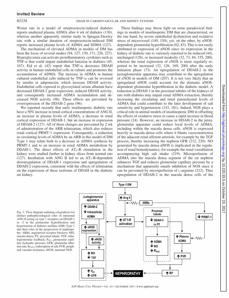
These findings may throw light on some paradoxical find-ings in models of insulinopenic DM that are characterized, onthe one hand, by severe endothelial dysfunction and oxidativestress of microvessel (149, 150), yet, on the other, by nNOS-dependent glomerular hyperfiltration (82, 83). This is not easilyattributed to expression of nNOS since its expression in thekidney of diabetic rats is variously reported to be reduced (44),unchanged (126), or increased modestly (32, 73, 84, 155, 200),whereas the renal expression of eNOS is more regularly re-ported to be increased (32, 126, 169, 200) after the earlyinitiation phase (73). An upregulation of DDAH-2 in thejuxtaglomerular apparatus may contribute to the upregulationof eNOS in models of DM (207). It is not very likely that anupregulated nNOS could account for the dramatic nNOS-dependent glomerular hyperfiltration in the diabetic model. Areduction in DDAH-1 in the proximal tubules of the kidneys ofrats with diabetes may impair renal ADMA extraction, therebyincreasing the circulating and renal parenchymal levels ofADMA that could contribute to the later development of saltsensitivity and hypertension (143, 181). Indeed, NOS plays acritical role in animal models of insulinopenic DM in offsettingthe effects of oxidative stress to cause a rapid increase in bloodpressure (24). However, an increase in DDAH-2 in the juxta-glomerular apparatus could reduce local levels of ADMA,including within the macula densa cells. nNOS is expressedheavily in macula densa cells where it blunts vasoconstrictionof the adjacent renal afferent arteriole, for example by the TGFprocess, thereby increasing the nephron GFR (212, 220). NOgenerated by macula densa nNOS is implicated in the regula-tion of renal hemodynamics, for example the renal vasodilationaccompanying high salt intake (219). Microperfusion ofADMA into the macula densa segment of the rat nephronenhances TGF and reduces glomerular capillary pressure by amechanism that apparently entails inhibition of NOS since itcan be prevented by microperfusion of L-arginine (212). Thusupregulation of DDAH-2 in the macula densa cells of the
Fig. 3. Flow diagram outlining a hypothesis fordistinct pathophysiological roles of intrarenalANG II acting on type 1 receptors on DDAH-1or -2 in the glomerular hyperfiltration andhypertension of diabetes mellitus (DM) Type 1and their roles in the progression of nephropa-thy. ARBs, angiotensin receptor blockers; MD,macula densa; PT, proximal tubule; TGF, tubu-loglomerular feedback; PGC, glomerular capil-lary hydraulic pressure; GFR, glomerular filtra-tion rate; RNaCl, reabsorption of salt; PVR, periph-eral vascular resistance; nNOS, neuronal NOS.
Invited Review
H3238 DDAH IN CARDIOVASCULAR AND KIDNEY SYSTEMS
AJP-Heart Circ Physiol • VOL 293 • DECEMBER 2007 • www.ajpheart.org
by 10.220.33.5 on October 23, 2017
http://ajpheart.physiology.org/D
ownloaded from

diabetic rat kidney may limit local ADMA accumulation,thereby enhancing nNOS-dependent afferent arteriolar vasodi-lation and glomerular hyperfiltration. Indeed, NO derived frommacula densa nNOS in rat models of early DM increases thesingle-nephron GFR even in the absence of activation of TGF(85, 180). These findings might help to explain the apparentlyparadoxical role of nNOS-dependent glomerular hyperfiltra-tion despite increased circulating levels of ADMA. Theseconcepts are outlined in Fig. 3 but require further experimentalinvestigation.
A second finding of potential clinical relevance to diabeticnephropathy is that the increase in ADMA and the decrease inrenal protein expression for DDAH-1 in the kidneys of dia-betic, insulinopenic rats are prevented by an ARB that alsoreduces the renal expression of PRMT-1 (127). Clinical trialsof patients with DM and nephropathy have reported thatpatients randomized to receive an ARB (26) or an ACEI (140),compared with equally antihypertensive treatments with drugsthat do not perturb the renin-angiotensin system, have a re-duced rate of loss of renal function. Studies by Anderson andBrenner (7), Brenner (25), and Zatz et al. (231) in a rat modelof insulinopenic DM have shown that ACEIs reduce theelevated levels of single-nephron glomerular filtration, glomer-ular capillary pressure, and BP. These actions of ACEIs orARBs on glomerular hemodynamics are not seen in normal ratsand depend on nNOS (82, 83). One potential explanation is thata reduction in ANG II generation or action on AT1-Rs reducesthe expression of DDAH-2 in the juxtaglomerular apparatus,leading to an increased intracellular ADMA that could inhibitnNOS activation specifically in those cells expressingDDAH-2. A reduced DDAH-2 expression within the juxtaglo-merular apparatus may allow sufficient accumulation ofADMA to inhibit nNOS-dependent glomerular hyperfiltration(Fig. 3). This hypothesis requires further study.
Several other mechanisms directly or indirectly related tohyperglycemia have been described that could contribute toimpaired DDAH activity in DM. ANG II stimulates oxidativestress (24) that could lead to posttranscriptional inhibition ofDDAH activity. ANG II induces the expression of proinflam-matory cytokines, growth factors, and adhesion molecules andthe transcription factor NF-B that can further promote ROSgeneration (45, 167). High glucose concentration itself caninhibit the expression of DDAH in cultured endothelial cells(160). Rats exposed to glycosylated proteins have endothelialdysfunction and impaired DDAH activity that can be restoredby insulin (42, 229). Thus dysfunction of DDAH could under-lie endothelial dysfunction in diabetes.
Conclusions and Clinical Perspectives
As research progresses, DDAH emerges as an importantregulator of NO bioavailability and the integrity of renal andvascular function. The finding that the gene for DDAH isevolutionarily well conserved suggests that the enzyme confersan important survival advantage. A decrease in DDAH expres-sion or activity is emerging as a potential mediator of cardio-vascular and kidney diseases that accompany ANG II action,oxidative stress, CKD, hypertension, inflammation, and insulinresistance. Several recent findings have clarified the regulationand action of DDAH. Immunohistochemical studies in the ratshow nephron site-specific expression of DDAH-1 and -2.
DDAH-2 is expressed not only at sites of NOS expression inendothelial cells but also in vascular smooth muscle cells andin the cell nucleus. DDAH-2 can undergo protein-proteininteraction, which leads to enhanced generation of VEGF thatcontributes to its role in promoting angiogenesis (54). Studiesof gene silencing or deletion in rodents lead to the conclusionthat plasma levels of ADMA are regulated by DDAH-1,whereas DDAH-2 can be of predominant importance for pre-serving endothelial function in resistance vessels. The recentfinding that gene transfer of DDAH prevents hypertension andprogressive loss of kidney function in a model of reduced renalmass implies that it plays a key role in the pathophysiologyof CKD.
NO contributes substantially to the regulation of BP, endo-thelial function, and host defense. It is therefore not surprisingthat circulating ADMA is a strong predictor of cardiovascularmorbidity and mortality in those with vascular disease and ofthe development of ESRD in those with CKD. Thus loweringof ADMA plasma concentrations in patients at risk could be amajor therapeutic goal. However, the findings that silencing ofthe DDAH-2 gene in the rat blocks EDRF/NO responseswithout a change in circulating ADMA demonstrates that thisgoal is inadequate. Apparently, ADMA can accumulate intra-cellular in endothelial cells sufficiently to block their produc-tion of NO without a spillover into the plasma that elevatescirculating ADMA levels.
Fig. 4. Flow diagram outlining the interactive roles of ROS and ADMA inrelating cardiovascular risk factors to pathophysiological changes in tissuesthat may underlie cardiovascular and kidney disease. For abbreviations, seelegends to Figs. 1, 2 and 3. SOD, superoxide dismutase; MMP, matrixmetalloproteinase; VSMC, vascular smooth muscle cell.
Invited Review
H3239DDAH IN CARDIOVASCULAR AND KIDNEY SYSTEMS
AJP-Heart Circ Physiol • VOL 293 • DECEMBER 2007 • www.ajpheart.org
by 10.220.33.5 on October 23, 2017
http://ajpheart.physiology.org/D
ownloaded from

Reversible inhibitors of DDAH may have a role in themanagement of diseases with excess NO production, such asseptic shock. However, interventions that enhance DDAHactivity, decrease ADMA production, and facilitate NO signal-ing are of far greater potential importance. Enhancing theactivity of DDAH or decreasing the activity of class 1 PRMTmay become novel therapeutic strategies for preventing pro-gressive cardiovascular and kidney diseases but currently lacka clear experimental basis.
Finally, the close interaction between the NOS/DDAH path-ways and those that generate or metabolize ROS may underliethe finding that both an elevation of ADMA and an elevationof ROS are implicated in the ensuing vascular and organdysfunction. These heuristic concepts are illustrated in Fig. 4.
ACKNOWLEDGMENTS
We are grateful to Emily Wing Kam Chan for preparing and editing thismanuscript.
GRANTS
This work was supported by National Institute of Diabetes and Digestiveand Kidney Diseases Grants DK-36079 and DK-49870 and National Heart,Lung, and Blood Institute Grant HL-68686. F. Palm is supported by K-99Grant (DK-077858). Z. Luo is supported by a National Institutes of HealthNephrology Fellowship Training Grant (DK-59274). Additional funds wereprovided by the George E. Schreiner Chair of Nephrology.
REFERENCES
1. Abbasi F, Asagmi T, Cooke JP, Lamendola C, McLaughlin T,Reaven GM, Stuehlinger M, Tsao P. Plasma concentrations of asym-metric dimethylarginine are increased in patients with type 2 diabetesmellitus. Am J Cardiol 88: 1201–1203, 2001.
2. Achan V, Broadhead M, Malaki M, Whitley G, Leiper J, MacAllisterR, Vallance P. Asymmetric dimethylarginine causes hypertension andcardiac dysfunction in humans and is actively metabolized by dimethy-larginine dimethylaminohydrolase. Arterioscler Thromb Vasc Biol 23:1455–1459, 2003.
3. Achan V, Tran CT, Arrigoni F, Whitley GS, Leiper JM, Vallance P.All trans retinoic acid increases nitric oxide synthesis by endothelialcells. Circ Res 90: 764–769, 2002.
4. Achan V, Ho HK, Heeschen C, Stuehlinger M, Jang JJ, Kimoto M,Vallance P, Cooke JP. ADMA regulates angiogenesis: genetic andmetabolic evidence. Vasc Med 10: 7–14, 2005.
5. Aisaka K, Gross SS, Griffiths OW, Levi R. L-Arginine availabilitydetermines the duration of acetylcholine-induced systemic vasodilationin vivo. Biochem Biophys Res Commun 163: 710–717, 1989.
6. Altinova AE, Arslan M, Sepici-Dincel A, Akturk M, Altan N,Toruner FB. Uncomplicated type 1 diabetes is associated with increasedasymmetric dimethylarginine concentrations. J Clin Endocrinol Metab92: 1881–1885, 2007.
7. Anderson S, Brenner BM. The role of intraglomerular pressure in theinitiation and progression of renal disease. J Hypertens Suppl 4: S236–S238, 1986.
8. Arrigoni FI, Vallance P, Haworth SG, Leiper JM. Metabolism ofasymmetric dimethylarginines is regulated in the lung developmentallyand with pulmonary hypertension induced by hypobaric hypoxia. Circu-lation 107: 1195–1201, 2003.
9. Asagami T, Abbasi F, Stuelinger M, Lamendola C, McLaughlin T,Cooke JP, Reaven GM, Tsao PS. Metformin treatment lowers asym-metric dimethylarginine concentrations in patients with type 2 diabetes.Metabolism 51: 843–846, 2003.
10. Aslam S, Santha T, Leone A, Wilcox C. Effects of amlodipine andvalsartan on oxidative stress and plasma methylarginines in end-stagerenal disease patients on hemodialysis. Kidney Int 70: 2109–2115, 2006.
11. Assreuy J, Cunha FQ, Liew FY, Moncada S. Feedback inhibition ofnitric oxide synthase activity by nitric oxide. Br J Pharmacol 108:833–837, 1993.
12. Babaei S, Teichert-Kuliszewska K, Monge JC, Mohamed F, BendeckMP, Stewart DJ. Role of nitric oxide in the angiogenic response in vitroto basic fibroblast growth factor. Circ Res 82: 1007–1015, 1998.
13. Barbera JA, Peinado VI, Santos S, Ramirez J, Roca J, Rodriguez-Roisin R. Reduced expression of endothelial nitric oxide synthase inpulmonary arteries of smokers. Am J Respir Crit Care Med 164: 709–713, 2001.
14. Baylis C. Arginine, arginine analogs and nitric oxide production inchronic kidney disease. Nat Clin Pract Nephrol 2: 209–220, 2006.
15. Baylis C, Mitruka B, Deng A. Chronic blockade of nitric oxidesynthesis in the rat produces systemic hypertension and glomerulardamage. J Clin Invest 90: 278–281, 1992.
16. Berger SL. The complex language of chromatin regulation duringtranscription. Nature 447: 407–412, 2007.
17. Birdsey GM, Leiper JM, Vallance P. Intracellular localization ofdimethylarginine dimethylaminohydrolase overexpressed in an endothe-lial cell line. Acta Physiol Scand 168: 73–79, 2000.
18. Boger RH, Bode-Boger SM, Szuba A, Tsao PS, Chan J, Tangphao O,Blaschke TF, John JS. Asymmetric dimethylarginine (ADMA): a novelrisk factor for endothelial dysfunction. Circulation 98: 1842–1847, 1998.
19. Boger RH, Bode-Boger SM, Thiele W, Creutzig A, Alexander K,Frolich JC. Restoring vascular nitric oxide formation by L-arginineimproves the symptoms of intermittent claudication in patients withperipheral arterial occlusive disease. J Am Coll Cardiol 32: 1336–1344,1998.
20. Boger RH, Bode-Boger SM, Tsao PS, Lin PS, Chan JR, Cooke JP. Anendogenous inhibitor of nitric oxide synthase regulates endothelialadhesiveness for monocytes. J Am Coll Cardiol 36: 2287–2295, 2000.
21. Boger RH, Cooke JP, Vallance P. ADMA: an emerging cardiovascularrisk factor. Vasc Med10, Suppl 1: S1–S2, 2005.
22. Boger RH, Sydow K, Borlak T, Thum T, Lenzen H, Schubert B,Tsikas D, Bode-Boger SM. LDL cholesterol upregulates synthesis ofasymmetrical dimethylarginine in human endothelial cells. Circ Res 87:99–105, 2000.
23. Bogumil R, Knipp M, Fundel SM, Vasak M. Characterization ofdimethylargininase from bovine brain: evidence for a zinc binding site.Biochemistry 37: 4791–4798, 1998.
24. Brands MW, Bell TD, Gibson B. Nitric oxide may prevent hypertensionearly in diabetes by counteracting renal actions of superoxide. Hyper-tension 43: 57–63, 2004.
25. Brenner BM. Hemodynamically mediated glomerular injury and theprogressive nature of kidney disease. Kidney Int 23: 647–655, 1983.
26. Brenner BM, Cooper ME, de Zeeuw D, Keane WF, Mitch WE,Parving HH, Remuzzi G, Snapinn SM, Zhang Z, Shahinfar S. Effectsof losartan on renal and cardiovascular outcomes in patients with type 2diabetes and nephropathy. N Engl J Med 345: 861–869, 2001.
27. Buhimschi IA, Saade GR, Chwalisz K, Garfield RE. The nitric oxidepathway in pre-eclampsia: pathophysiological implications. Hum ReprodUpdate 4: 25–42, 1998.
28. Bultink IE, Teerlink T, Heijst JA, Dijkmans BA, Voskuyl AE. Raisedplasma levels of asymmetric dimethylarginine are associated with car-diovascular events, disease activity, and organ damage in patients withsystemic lupus erythematosus. Ann Rheum Dis 64: 1362–1365, 2005.
29. Chan NN, Chan JC. Asymmetric dimethylarginine (ADMA): a poten-tial link between endothelial dysfunction and cardiovascular diseases ininsulin resistance syndrome? Diabetologia 45: 1609–1616, 2002.
30. Chen JW, Hsu NW, Wu TC, Lin SJ, Chang MS. Long-term angio-tensin-converting enzyme inhibition reduces plasma asymmetric dim-ethylarginine and improves endothelial nitric oxide bioavailability andcoronary microvascular function in patients with syndrome X. Am JCardiol 90: 974–982, 2002.
31. Chen MF, Xie XM, Yang TL, Wang YJ, Zhang XH, Luo BL, Li YJ.Role of asymmetric dimethylarginine in inflammatory reactions byangiotensin II. J Vasc Res 44: 391–402, 2007.
32. Choi KC, Kim NH, An MR, Kang DG, Kim SW, Lee J. Alterations ofintrarenal renin-angiotensin and nitric oxide systems in streptozotocin-induced diabetic rats. Kidney Int Suppl 60: S23–S27, 1997.
33. Cooke JP. NO and angiogenesis. Atheroscler Suppl 4: 53–60, 2003.34. Cooke JP. ADMA: its role in vascular disease. Vasc Med 10, Suppl 1:
S11–S17, 2005.35. Curgunlu A, Uzun H, Bavunoglu I, Karter Y, Genc H, Vehid S.
Increased circulating concentrations of asymmetric dimethylarginine(ADMA) in white coat hypertension. J Hum Hypertens 19: 629–633,2005.
36. Dayoub H, Achan V, Adimoolam S, Jacobi J, Stuehlinger M, WangB, Tsao PS, Kimoto M, Vallance P, Patterson A, Cooke JP. Dimeth-
Invited Review
H3240 DDAH IN CARDIOVASCULAR AND KIDNEY SYSTEMS
AJP-Heart Circ Physiol • VOL 293 • DECEMBER 2007 • www.ajpheart.org
by 10.220.33.5 on October 23, 2017
http://ajpheart.physiology.org/D
ownloaded from

ylarginine dimethylaminohydrolase regulates nitric oxide synthesis. Cir-culation 108: 3042–3047, 2003.
37. Delles C, Schneider MP, John S, Gekle M, Schmieder RE. Angioten-sin converting enzyme inhibition and angiotensin II AT1 receptor block-ade reduce the levels of asymmetrical NG,NG-dimethylarginine in humanessential hypertension. Am J Hypertens 15: 590–593, 2002.
38. Deng X, Welch WJ, Wilcox CS. Renal vasoconstriction during inhibi-tion of NO synthase: effects of dietary salt. Kidney Int 46: 639–646,1994.
39. Deng X, Welch WJ, Wilcox CS. Renal vasodilation with L-arginine:effects of dietary salt. Hypertension 26: 256–262, 1995.
40. Deng X, Welch WJ, Wilcox CS. Role of nitric oxide in short-term andprolonged effects of angiotensin II on renal hemodynamics. Hyperten-sion 27: 1173–1179, 1996.
41. Dowling RB, Newton R, Robichaud A, Cole PJ, Barnes PJ, Wilson R.Effect of inhibition of nitric oxide synthase on Pseudomonas aeruginosainfection of respiratory mucosa in vitro. Am J Respir Cell Mol Biol 19:950–958, 1998.
42. Eid HM, Lyberg T, Arnesen H, Seljeflot I. Insulin and adiponectininhibit the TNF-�-induced ADMA accumulation in human endothelialcells: the role of DDAH. Atherosclerosis 194: e1–e8, 2007.
43. Ellis J, Wennerholm UB, Bengtsson A, Lilja H, Pettersson A, SultanB, Wennergren M, Hagberg H. Levels of dimethylarginines and cyto-kines in mild and severe preeclampsia. Acta Obstet Gynecol Scand 80:602–608, 2001.
44. Erdely A, Freshour G, Maddox DA, Olson JL, Samsell L, Baylis C.Renal disease in rats with type 2 diabetes is associated with decreasedrenal nitric oxide production. Diabetologia 47: 1672–1676, 2004.
45. Evans JL, Goldfine ID, Maddux BA, Grodsky GM. Oxidative stressand stress-activated signaling pathways: a unifying hypothesis of type 2diabetes. Endocr Rev 23: 599–622, 2002.
46. Fickling SA, Williams D, Vallance P, Nussey SS, Whitley GS. Plasmaconcentrations of endogenous inhibitor of nitric oxide synthesis innormal pregnancy and pre-eclampsia. Lancet 342: 242–243, 1993.
47. Frey D, Braun O, Briand C, Vasak M, Grutter MG. Structure of themammalian NOS regulator dimethylarginine dimethylaminohydrolase: abasis for the design of specific inhibitors. Structure 14: 901–911, 2006.
48. Galkin A, Kulakova L, Sarikaya E, Lim K, Howard A, Herzberg O.Structural insight into arginine degradation by arginine deiminase, anantibacterial and parasite drug target. J Biol Chem 279: 14001–14008,2004.
49. Gardiner SM, Kemp PA, Bennett T, Palmer RMJ, Moncada S.Regional and cardiac haemodynamic effects of NG,NG,dimethyl-L-argi-nine and their reversibility by vasodilators in conscious rats. Br JPharmacol 110: 1457–1464, 1993.
50. Goligorsky MS. Endothelial cell dysfunction: can’t live with it, how tolive without it. Am J Physiol Renal Physiol 288: F871–F880, 2005.
51. Gow AJ, Chen Q, Hess DT, Day BJ, Ischiropoulos H, Stamler JS.Basal and stimulated protein S-nitrosylation in multiple cell types andtissues. J Biol Chem 277: 9637–9640, 2002.
52. Griendling KK, FitzGerald Ga. Oxidative stress and cardiovascularinjury. II. Animal and human studies. Circulation 108: 2034–2040, 2003.
53. Hartzoulakis B, Rossiter S, Gill H, O’Hara B, Steinke E, Gane PJ,Hurtado-Guerrero R, Leiper JM, Vallance P, Rust JM, Selwood DL.Discovery of inhibitors of the pentein superfamily protein dimethylargi-nine dimethylaminohydrolase (DDAH), by virtual screening and hitanalysis. Bioorg Med Chem Lett 17: 3953–3956, 2007.
54. Hasegawa K, Wakino S, Tanaka T, Kimoto M, Tatematsu S, KandaT, Yoshioka K, Homma K, Sugano N, Kurabayashi M, Saruta T,Hayashi K. Dimethylarginine dimethylaminohydrolase 2 increases vas-cular endothelial growth factor expression through Sp1 transcriptionfactor in endothelial cells. Arterioscler Thromb Vasc Biol 26: 1488–1494, 2006.
55. Hasegawa K, Wakino S, Tatematsu S, Yoshioka K, Homma K,Sugano N, Kimoto M, Hayashi K, Itoh H. Role of asymmetric dimeth-ylarginine in vascular injury in transgenic mice overexpressing dimeth-ylarginine dimethylaminohydrolase 2. Circ Res 101: e2–e10, 2007.
56. Heitzer T, Yla-Herttuala S, Luoma J, Kurz S, Munzel T, Just H,Olschewski M, Drexler H. Cigarette smoking potentiates endothelialdysfunction of forearm resistance vessels in patients with hypercholes-terolemia. Role of oxidized LDL. Circulation 93: 1346–1353, 1996.
57. Herpin A, Cunningham C. Cross-talk between the bone morphogeneticprotein pathway and other major signaling pathways results in tightlyregulated cell-specific outcomes. FEBS J 274: 2977–2985, 2007.
58. Ho QT, Kuo CJ. Vascular endothelial growth factor: biology andtherapeutic applications. Int J Biochem Cell Biol 39: 1349–1357, 2007.
59. Holden DP, Cartwright JE, Nussey SS, Whitley G. Estrogen stimu-lates dimethylarginine dimethylaminohydrolase activity and the metab-olism of asymmetric dimethylarginine. Circulation 108: 1575–1580,2003.
60. Holden DP, Fickling SA, Whitley GS, Nussey SS. Plasma concentra-tions of asymmetric dimethylarginine, a natural inhibitor of nitric oxidesynthase, in normal pregnancy and preeclampsia. Am J Obstet Gynecol178: 551–556, 1998.
61. Hruska KA, Saab G, Chaudhary LR, Quinn CO, Lund RJ, Suren-dran K. Kidney-bone, bone-kidney, and cell-cell communications inrenal osteodystrophy. Semin Nephrol 24: 25–38, 2004.
62. Ito A, Egashira K, Narishige T, Muramatsu K, Takeshita A. Renin-angiotensin system is involved in the mechanism of increased serumasymmetric dimethylarginine in essential hypertension. Jpn Circ J 65:775–778, 2001.
63. Ito A, Egashira K, Narishige T, Muramatsu K, Takeshita A. Angio-tensin-converting enzyme activity is involved in the mechanism ofincreased endogenous nitric oxide synthase inhibitor in patients with type2 diabetes mellitus. Circ J 66: 811–815, 2002.
64. Ito A, Tsao PS, Adimoolam S, Kimoto M, Ogawa T, Cooke JP. Novelmechanism for endothelial dysfunction:dysregulation of dimethylargi-nine dimethylaminohydrolase. Circulation 99: 3092–3095, 1999.
65. Jia SJ, Jiang DJ, Hu CP, Zhang XH, Deng HW, Li YJ. Lysophos-phatidylcholine-induced elevation of asymmetric dimethylarginine levelby the NADPH oxidase pathway in endothelial cells. Vascul Pharmacol44: 143–148, 2006.
66. Jiang DJ, Jia SJ, Yan J, Zhou Z, Yuan Q, Li YJ. Involvement ofDDAH/ADMA/NOS pathway in nicotine-induced endothelial dysfunc-tion. Biochem Biophys Res Commun 349: 683–693, 2006.
67. Jiang JL, Zhang XH, Li NS, Rang WQ, Feng Y, Hu CP, Li YJ, DengHW. Probucol decreases asymmetrical dimethylarginine level by alter-nation of protein arginine methyltransferase I and dimethylargininedimethylaminohydrolase activity. Cardiovasc Drugs Ther 20: 281–294,2006.
68. Jones LC, Tran CTL, Leiper JM, Hingorani AD, Vallance P. Com-mon genetic variation in a basal promoter element alters DDAH2expression in endothelial cells. Biochem Biophys Res Commun 310:836–843, 2003.
69. Joshi MS, Ferguson TB Jr, Johnson FK, Johnson RA, ParthasarathyS, Lancaster JR Jr. Receptor-mediated activation of nitric oxide syn-thesis by arginine in endothelial cells. Proc Natl Acad Sci USA 104:9982–9987, 2007.
70. Kakimoto Y, Akazawa S. Isolation and identification of NG,NG- andNG,NG�-dimethylarginine, N-mono-, di-, and trimethyllysine, and glu-cosylgalactosyl- and galactosyl-�-hydroxylysine from human urine.J Biol Chem 245: 5751–5758, 1970.
71. Kearns AE, Donohue MM, Sanyal B, Demay MB. Cloning andcharacterization of a novel protein kinase that impairs osteoblast differ-entiation in vitro. J Biol Chem 276: 42213–42218, 2001.
72. Kelly FJ, Anderson S, Thompson MM, Oyama TT, Kennefick TM,Corless CL, Roman RJ, Kurtzberg L, Pratt BM, Ledbetter SR. Acuteand chronic renal effects of recombinant human TGF-beta2 in the rat.J Am Soc Nephrol 10: 1264–1273, 1999.
73. Keynan S, Hirshberg B, Levin-Iaina N, Wexler ID, Dahan R, Rein-hartz E, Ovadia H, Wollman Y, Chernihovskey T, Iaina A, Raz I.Renal nitric oxide production during the early phase of experimentaldiabetes mellitus. Kidney Int 58: 740–747, 2000.
74. Kielstein JT, Boger RH, Bode-Boger SM, Frolich JC, Haller H, RitzE, Fliser D. Marked increase of asymmetric dimethylarginine in patientswith incipient primary chronic renal disease. J Am Soc Nephrol 13:170–176, 2002.
75. Kielstein JT, Salpeter SR, Bode-Boeger SM, Cooke JP, Fliser D.Symmetric dimethylarginine (SDMA) as endogenous marker of renalfunction - a meta-analysis. Nephrol Dial Transplant 21: 2446–2451,2006.
76. Kimoto M, Tsuji H, Ogawa T, Sasaoka K. Detection of NG,NG-dimethylarginine dimethylaminohydrolase in the nitric oxide-generatingsystems of rats using monoclonal antibody. Arch Biochem Biophys 300:657–662, 1993.
77. Kitiyakara C, Chabrashvili T, Jose P, Welch WJ, Wilcox CS. Effectsof dietary salt intake on plasma arginine. Am J Physiol Regul IntegrComp Physiol 280: R1069–R1075, 2001.
Invited Review
H3241DDAH IN CARDIOVASCULAR AND KIDNEY SYSTEMS
AJP-Heart Circ Physiol • VOL 293 • DECEMBER 2007 • www.ajpheart.org
by 10.220.33.5 on October 23, 2017
http://ajpheart.physiology.org/D
ownloaded from

78. Knipp M. How to control NO production in cells: N�,N�-dimethyl-L-arginine dimethylaminohydrolase as a novel drug target. Chembiochem7: 879–889, 2006.
79. Knipp M, Braun O, Gehrig PM, Sack R, Vasak M. Zn(II)-freedimethylargininase-1 (DDAH-1) is inhibited upon specific Cys-S-nitrosylation. J Biol Chem 278: 3410–3416, 2003.
80. Knipp M, Charnock JM, Garner CD, Vasak M. Structural andfunctional characterization of the Zn(II) site in dimethylargininase-1(DDAH-1) from bovine brain. Zn(II) release activates DDAH-1. J BiolChem 276: 40449–40456, 2001.
81. Knipp M, Vasak M. A colorimetric 96-well microtiter plate assay forthe determination of enzymatically formed citrulline. Anal Biochem 286:257–264, 2000.
82. Komers R, Anderson S. Paradoxes of nitric oxide in the diabetic kidney.Am J Physiol Renal Physiol 284: F1121–F1137, 2003.
83. Komers R, Lindsley JN, Oyama TT, Allison KM, Anderson S. Roleof neuronal nitric oxide synthase (NOS1) in the pathogenesis of renalhemodynamic changes in diabetes. Am J Physiol Renal Physiol 279:F573–F583, 2000.
84. Komers R, Lindsley JN, Oyama TT, Schutzer WE, Reed JF, MaderSL, Anderson S. Immunohistochemical and functional correlations ofrenal cyclooxygenase-2 in experimental diabetes. J Clin Invest 107:889–898, 2001.
85. Komine N, Thomson SC, Blantz RC, Gabbai FB. Role of maculadensa derived nitric oxide in the hyperfiltration of streptozotocin diabeticrats (Abstract). J Am Soc Nephrol 10: 684A, 1999.
86. Kone BC. Nitric oxide synthesis in the kidney: isoforms, biosynthesis,and functions in health. Semin Nephrol 24: 299–315, 2004.
87. Kone BC. NO break-ins at water gate. Hypertension 48: 29–30, 2006.88. Konishi H, Sydow K, Cooke JP. Dimethylarginine dimethylaminohy-
drolase promotes endothelial repair after vascular injury. J Am CollCardiol 49: 1099–1105, 2007.
89. Leiper J, Murray-Rust J, McDonald N, Vallance P. S-nitrosylation ofdimethylarginine dimethylaminohydrolase regulates enzyme activity:Further interactions between nitric oxide synthase and dimethylargininedimethylaminohydrolase. Proc Natl Acad Sci USA 99: 13527–13532,2002.
90. Leiper J, Nandi M, Torondel B, Murray-Rust J, Malaki M, O’HaraB, Rossiter S, Anthony S, Madhani M, Selwood D, Smith C, Wojciak-Stothard B, Rudiger A, Stidwill R, McDonald NQ, Vallance P.Disruption of methylarginine metabolism impairs vascular homeostasis.Nat Med 13: 198–203, 2007.
91. Leiper J, Vallance P. Biological significance of endogenous methyl-arginines that inhibit nitric oxide synthases. Cardiovasc Res 43: 542–548,2003.
92. Leiper J, Vallance P. New tricks from an old dog: nitric oxide-independent effects of dimethylarginine dimethylaminohydrolase.Arterioscler Thromb Vasc Biol 26: 1419–1420, 2006.
93. Leiper JM, Santa Maria J, Chubb A, MacAllister RJ, Charles IG,Whitley GS, Vallance P. Identification of two human dimethylargininedimethylaminohydrolases with distinct tissue distributions and homologywith microbial arginine deiminases. Biochem J 343: 209–214, 1999.
94. Lin KY, Ito A, Asagami T, Tsao PS, Adimoolam S, Kimoto M, TsujiH, Reaven GM, Cooke JP. Impaired nitric oxide synthase pathway indiabetes mellitus: role of asymmetric dimethylarginine and dimethylargi-nine dimethylaminohydrolase. Circulation 106: 987–992, 2002.
95. Lopez-Jaramillo P, Narvaez M, Calle A, Rivera J, Jacome P, RuanoC, Nava E. Cyclic guanosine 3�,5� monophosphate concentrations inpre-eclampsia: effects of hydralazine. Br J Obstet Gynaecol 103: 33–38,1996.
96. Lu CW, Xiong Y, He P. Dimethylarginine dimethylaminohydrolase-2overexpression improves impaired nitric oxide synthesis of endothelialcells induced by glycated protein. Nitric Oxide 16: 94–103, 2007.
97. Lu R, Hu CP, Wu XP, Liao EY, Lo YJ. Effect of age on bone mineraldensity and the serum concentration of endogenous nitric oxide synthaseinhibitors in rats. Comput Med 52: 224–228, 2003.
98. Lundman P, Eriksson MJ, Stuhlinger MC, Cooke JP, Hamsten A,Tornvall P. Mild to moderate hypertriglyceridemia in young men isassociated with endothelial dysfunction and increased plasma concentra-tions of asymmetric dimethylarginine. J Am Coll Cardiol 38: 111–116,2001.
99. Maas R. Pharmacotherapies and their influence on asymmetric dimethy-larginine (ADMA). Vasc Med 10 Suppl 1: S49–S57, 2005.
100. Maas R, Boger RH, Schwedhelm E, Casas JP, Lopez-Jaramillo P,Serrano N, Diaz LA. Plasma concentrations of asymmetric dimethyl-arginine (ADMA) in Colombian women with pre-eclampsia. JAMA 291:823–824, 2004.
101. Maas R, Tan-Andreesen J, Schwedhelm E, Schulze F, Boger RH. Astable-isotope based technique for the determination of dimethylargininedimethylaminohydrolase (DDAH) activity in mouse tissue. JChromatogr B Analyt Technol Biomed Life Sci 851: 220–228, 2007.
102. MacAllister RJ, Parry H, Kimoto M, Ogawa T, Russel RJ, HodsonH, Whitley GS, Vallance P. Regulation of nitric oxide synthesis bydimethylarginine dimethylaminohydrolase. Br J Pharmacol 119: 1533–1540, 1996.
103. MacAllister RJ, Whitley GS, Vallance P. Effects of guanidino anduremic compounds on nitric oxide pathways. Kidney Int 45: 737–742,1994.
104. Maeda T, Yoshimura T, Okamura H. Asymmetric dimethylarginine,an endogenous inhibitor of nitric oxide synthase, in maternal and fetalcirculation. J Soc Gynecol Investig 10: 2–4, 2003.
105. Masuda H, Goto M, Tamaoki S, Azuma H. Accelerated intimalhyperplasia and increased endogenous inhibitors of NO synthesis inrabbits with alloxan-induced hyperglycaemia. Br J Pharmacol 126:211–218, 1999.
106. Matsuguma K, Ueda S, Yamagishi S, Matsumoto Y, Kaneyuki U,Shibata R, Fujimura T, Matsuoka H, Kimoto M, Kato S, ImaizumiT, Okuda S. Molecular mechanism for elevation of asymmetricdimethylarginine and its role for hypertension in chronic kidney disease.J Am Soc Nephrol 17: 2176–2183, 2006.
107. Matsumoto Y, Ueda S, Yamagishi S, Matsuguma K, Shibata R,Fukami K, Matsuoka H, Imaizumi T, Okuda S. Dimethylargininedimethylaminohydrolase prevents progression of renal dysfunction byinhibiting loss of peritubular capillaries and tubulointerstitial fibrosis in arat model of chronic kidney disease. J Am Soc Nephrol 18: 1525–1533,2007.
108. Meinitzer A, Seelhorst U, Wellnitz B, Halwachs-Baumann G, BoehmBO, Winkelmann BR, Marz W. Asymmetrical dimethylarginine inde-pendently predicts total and cardiovascular mortality in individuals withangiographic coronary artery disease (the Ludwigshafen Risk and Car-diovascular Health study). Clin Chem 53: 273–283, 2007.
109. Mitchell JA, de Nucci G, Warner TD, Vane JR. Alkaline buffersrelease EDRF from bovine cultured aortic endothelial cells. Br J Phar-macol 103: 1295–1302, 1991.
110. Mittermayer F, Mayer BX, Meyer A, Winzer C, Pacini G, WagnerOF, Wolzt M, Kautzky-Willer A. Circulating concentrations of asym-metrical dimethyl-L-arginine are increased in women with previousgestational diabetes. Diabetologia 45: 1372–1378, 2002.
111. Miyazaki H, Matsuoka H, Cooke JP, Usui M, Ueda S, Okuda S,Imaizumi T. Endogenous nitric oxide synthase inhibitor: a novel markerof atherosclerosis. Circulation 99: 1141–1146, 1999.
112. Monsalve E, Oviedo PJ, Garcia-Perez MA, Tarin JJ, Cano A,Hermenegildo C. Estradiol counteracts oxidized LDL-induced asym-metric dimethylarginine production by cultured human endothelial cells.Cardiovasc Res 73: 66–72, 2007.
113. Mookerjee RP, Malaki M, Davies NA, Hodges SJ, Dalton RN,Turner C, Sen S, Williams R, Leiper J, Vallance P, Jalan R.Increasing dimethylarginine levels are associated with adverse clinicaloutcome in severe alcoholic hepatitis. Hepatology 45: 62–71, 2007.
114. Murray-Rust J, Leiper J, McAlister M, Phelan J, Tilley S, SantaMaria J, Vallance P, McDonald N. Structural insights into the hydro-lysis of cellular nitric oxide synthase inhibitors by dimethylargininedimethylaminohydrolase. Nat Struct Biol 8: 679–683, 2001.
115. Napoli C, Sica V, de NF, Pignalosa O, Condorelli M, Ignarro LJ,Liguori A. Sulfhydryl angiotensin-converting enzyme inhibition inducessustained reduction of systemic oxidative stress and improves the nitricoxide pathway in patients with essential hypertension. Am Heart J 148:e5, 2004.
116. Nijveldt RJ, Teerlink T, Siroen MPC, van Lambalgen AA, Rauw-erda JA, van Leeuwen PAM. The liver is an important organ in themetabolism of asymmetrical dimethylarginine (ADMA). Clin Nutr 22:17–22, 2003.
117. Nijveldt RJ, Teerlink T, van Guldener C, Prins HA, van LambalgenAA, Stehouwer CDA, Rauwerda JA, van Leeuwen PAM. Handling ofasymmetrical dimethylarginine and symmetrical dimethylarginine by therat kidney under basal conditions and during endotoxaemia. Nephrol DialTranplant 18: 2542–2550, 2003.
Invited Review
H3242 DDAH IN CARDIOVASCULAR AND KIDNEY SYSTEMS
AJP-Heart Circ Physiol • VOL 293 • DECEMBER 2007 • www.ajpheart.org
by 10.220.33.5 on October 23, 2017
http://ajpheart.physiology.org/D
ownloaded from

118. Nijveldt RJ, Siroen MP, Teerlink T, Van Leeuwen PA. Elimination ofasymmetric dimethylarginine by the kidney and the liver: a link to thedevelopment of multiple organ failure? J Nutr 134: 2848S-2852S, 2004.
119. Nonaka S, Tsunoda M, Aoyama C, Funatsu T. Determination ofNG,NG-dimethyl-L-arginine in rat plasma and dimethylarginine dimeth-ylaminohydrolase activity in rat kidney using a monolithic silica column.J Chromatogr B Analyt Technol Biomed Life Sci 843: 170–174, 2006.
120. O’Driscoll G, Green D, Maiorana A, Stanton K, Colreavy F, TaylorR. Improvement in endothelial function by angiotensin-converting en-zyme inhibition in non-insulin-dependent diabetes mellitus. J Am CollCardiol 33: 1506–1511, 1999.
121. O’Driscoll G, Green D, Rankin J, Stanton K, Taylor R. Improvementin endothelial function by angiotensin converting enzyme inhibition ininsulin-dependent diabetes mellitus. J Clin Invest 100: 678–684, 1997.
122. Ogawa T, Kimoto M, Sasaoka K. Occurrence of a new enzymecatalyzing the direct conversion of NG,NG-dimethyl-L-arginine toL-citrulline in rats. Biochem Biophys Res Commun 148: 671–677, 1987.
123. Ogawa T, Kimoto M, Sasaoka K. Purification and properties of a newenzyme, NG,NG-dimethylarginine dimethylaminohydrolase, from rat kid-ney. J Biol Chem 264: 10205–10209, 1989.
124. Ogawa T, Kimoto M, Watanabe H, Sasaoka K. Metabolism of NG,NG-and NG,N�G-dimethylarginine in rats. Arch Biochem Biophys 252: 526–537, 1987.
125. Okubo K, Hayashi K, Wakino S, Matsuda H, Kubota E, Honda M,Tokuyama H, Yamamoto T, Kajiya F, Saruta T. Role of asymmetricaldimethylarginine in renal microvascular endothelial dysfunction inchronic renal failure with hypertension. Hypertens Res 28: 181–189,2005.
126. Onozato ML, Tojo A, Goto A, Fujita T, Wilcox CS. Oxidative stressand nitric oxide synthase in rat diabetic nephropathy: effects of ACEI orARB. Kidney Int 61: 186–194, 2002.
127. Onozato ML, Tojo A, Leiper J, Fujita T, Palm F, Wilcox CS.Expression of DDAH and PRMT isoforms in the diabetic rat kidney;effects of angiotensin II receptor blocker. Diabetes. In press.
128. Ortiz PA, Hong NJ, Garvin JL. NO decreases thick ascending limbchloride absorption by reducing Na�-K�-2Cl� cotransporter activity.Am J Physiol Renal Physiol 281: F819–F825, 2001.
129. Paiva H, Laakso J, Kahonen M, Turjanmaa V, Koobi T, MajahalmeS, Lehtimaki T, Ruokonen I, Laaksonen R. Asymmetric dimethylargi-nine and hemodynamic regulation in middle-aged men. Metabolism 55:771–777, 2006.
130. Palm F, Buerk DG, Carlsson PO, Hansell P, Liss P. Reduced nitricoxide concentration in the renal cortex of streptozotocin-induced diabeticrats: effects on renal oxygenation and microcirculation. Diabetes 54:3282–3287, 2005.
131. Palmer RMJ, Ferrige AG, Moncada S. Nitric oxide release accountsfor the biological activity of endothelium-derived relaxing factor. Nature327: 524–526, 1987.
132. Papapetropoulos A, Desai KM, Rudic RD, Mayer B, Zhang R,Ruiz-Torres MP, Garcia-Cardena G, Madri JA, Sessa WC. Nitricoxide synthase inhibitors attenuate transforming-growth-factor-beta1-stimulated capillary organization in vitro. Am J Pathol 150: 1835–1844, 1997.
133. Papapetropoulos A, Garcia-Cardena G, Madri JA, Sessa WC. Nitricoxide production contributes to the angiogenic properties of vascularendothelial growth factor in human endothelial cells. J Clin Invest 100:3131–3139, 1997.
134. Patschan S, Li H, Brodsky S, Sullivan D, De Angelis DA, Patschan D,Goligorsky MS. Probing lipid rafts with proximity imaging: actions ofproatherogenic stimuli. Am J Physiol Heart Circ Physiol 290: H2210–H2219, 2006.
135. Perticone F, Sciacqua A, Maio R, Perticone M, Maas R, Boger RH,Tripepi G, Sesti G, Zoccali C. Asymmetric dimethylarginine, L-argi-nine, and endothelial dysfunction in essential hypertension. J Am CollCardiol 46: 518–523, 2005.
136. Pettersson A, Hedner T, Milsom I. Increased circulating concentrationsof asymmetric dimethylarginine (ADMA), an endogenous inhibitor ofnitric oxide synthesis in preeclampsia. Acta Obstet Gynecol Scand 77:808–813, 1998.
137. Polotskaia A, Wang M, Patschan S, Addabbo F, Chen J, GoligorskyMS. Regulation of arginine methylation in endothelial cells: role inpremature senescence and apoptosis. Cell Cycle 6: 2524–2530, 2007.
138. Pou S, Pou WS, Bredt DS, Snyder SH, Rosen GM. Generation ofsuperoxide by purified brain nitric oxide synthase. J Biol Chem 267:24173–24176, 1992.
139. Ravani P, Tripepi G, Malberti F, Testa S, Mallamaci F, Zoccali C.Asymmetrical dimethylarginine predicts progression to dialysis anddeath in patients with chronic kidney disease: a competing risks modelingapproach. J Am Soc Nephrol 16: 2449–2455, 2005.
140. Remuzzi G, Ruggenenti P, Perico N. Chronic renal disease: renopro-tective benefits of renin-angiotensin system inhibition. Ann Intern Med136: 604–615, 2002.
141. Rossiter S, Smith CL, Malaki M, Nandi M, Gill H, Leiper JM,Vallance P, Selwood DL. Selective substrate-based inhibitors of mam-malian dimethylarginine dimethylaminohydrolase. J Med Chem 48:4670–4678, 2005.
142. Ryan R, Thornton J, Duggan E, McGovern E, O’Dwyer MJ, RyanAW, Kelleher D, McManus R, Ryan T. Gene polymorphism andrequirement for vasopressor infusion after cardiac surgery. Ann ThoracSurg 82: 895–901, 2006.
143. Salazar FJ, Alberola A, Pinilla JM, Romero JC, Quesada T. Salt-induced increase in arterial pressure during nitric oxide synthesis inhi-bition. Hypertension 22: 49–55, 1993.
144. Santa MJ, Vallance P, Charles IG, Leiper JM. Identification ofmicrobial dimethylarginine dimethylaminohydrolase enzymes. MolMicrobiol 33: 1278–1279, 1999.
145. Savvidou MD, Hingorani AD, Tsikas D, Frolich JC, Vallance P,Nicolaides KH. Endothelial dysfunction and raised plasma concentra-tions of asymmetric dimethylarginine in pregnant women who subse-quently develop pre-eclampsia. Lancet 361: 1511–1517, 2003.
146. Scalera F, Kielstein JT, Martens-Lobenhoffer J, Postel SC, Tager M,Bode-Boger SM. Erythropoietin increases asymmetric dimethylargininein endothelial cells: role of dimethylarginine dimethylaminohydrolase.J Am Soc Nephrol 16: 892–898, 2005.
147. Schaefer CB, Ooi SK, Bestor TH, Bourc’his D. Epigenetic decisions inmammalian germ cells. Science 316: 398–399, 2007.
148. Schnabel R, Blankenberg S, Lubos E, Lackner KJ, Rupprecht HJ,Espinola-Klein C, Jachmann N, Post F, Peetz D, Bickel C, CambienF, Tiret L, Munzel T. Asymmetric dimethylarginine and the risk ofcardiovascular events and death in patients with coronary artery disease:results from the AtheroGene Study. Circ Res 97: e53–e59, 2005.
149. Schnackenberg CG, Wilcox CS. The SOD mimetic tempol restoresvasodilation in afferent arterioles of experimental diabetes. Kidney Int59: 1859–1864, 2001.
150. Schoonmaker GC, Fallet RW, Carmines PK. Superoxide anion curbsnitric oxide modulation of afferent arteriolar Ang II responsiveness indiabetes mellitus. Am J Physiol Renal Physiol 278: F302–F309, 2000.
151. Schulman IH, Zhou MS, Raij L. Interaction between nitric oxide andangiotensin II in the endothelium: role in atherosclerosis and hyperten-sion. J Hypertens Suppl 24: S45–S50, 2006.
152. Schulze F, Maas R, Freese R, Schwedhelm E, Silberhorn E, BogerRH. Determination of a reference value for NG,NG-dimethyl-L-argininein 500 subjects. Eur J Clin Invest 35: 622–626, 2005.
153. Scuteri A, Stuehlinger MC, Cooke JP, Wright JG, Lakatta EG,Anderson DE, Fleg JL. Nitric oxide inhibition as a mechanism for bloodpressure increase during salt loading in normotensive postmenopausalwomen. J Hypertens 21: 1339–1346, 2003.
154. Segal MS, Baylis C, Johnson RJ. Endothelial health and diversity in thekidney. J Am Soc Nephrol 17: 323–324, 2006.
155. Shin SJ, Lai FJ, Wen JD, Hsiao PJ, Hsieh MC, Tzeng TF, Chen HC,Guh JY, Tsai JH. Neuronal and endothelial nitric oxide synthaseexpression in outer medulla of streptozotocin-induced diabetic rat kid-ney. Diabetologia 43: 649–659, 2000.
156. Siroen MP, Teerlink T, Bolte AC, van Elburg RM, Richir MC,Nijveldt RJ, van der HB, Van Leeuwen PA. No compensatory upregu-lation of placental dimethylarginine dimethylaminohydrolase activity inpreeclampsia. Gynecol Obstet Invest 62: 7–13, 2006.
157. Siroen MP, Warle MC, Teerlink T, Nijveldt RJ, Kuipers EJ, Met-selaar HJ, Tilanus HW, Kuik DJ, van dS Jr, Meijer S, van der HB,Van Leeuwen PA. The transplanted liver graft is capable of clearingasymmetric dimethylarginine. Liver Transpl 10: 1524–1530, 2004.
158. Smith CL, Anthony S, Hubank M, Leiper JM, Vallance P. Effects ofADMA upon gene expression: an insight into the pathophysiologicalsignificance of raised plasma ADMA. PLoS Med 2: 1031–1043, 2005.
159. Smith CL, Birdsey GM, Anthony S, Arrigoni FI, Leiper JM, Val-lance P. Dimethylarginine dimethylaminohydrolase activity modulated
Invited Review
H3243DDAH IN CARDIOVASCULAR AND KIDNEY SYSTEMS
AJP-Heart Circ Physiol • VOL 293 • DECEMBER 2007 • www.ajpheart.org
by 10.220.33.5 on October 23, 2017
http://ajpheart.physiology.org/D
ownloaded from

ADMA levels VEGF expression and cell phenotype. Biochem BiophysRes Commun 308: 984–989, 2003.
160. Sorrenti V, Mazza F, Campisi A, Vanella L, Li VG, Di GC. Highglucose-mediated imbalance of nitric oxide synthase and dimethylargi-nine dimethylaminohydrolase expression in endothelial cells. Curr Neu-rovasc Res 3: 49–54, 2006.
161. Stone EM, Costello AL, Tierney DL, Fast W. Substrate-assistedcysteine deprotonation in the mechanism of dimethylargininase (DDAH)from Pseudomonas aeruginosa. Biochemistry 45: 5618–5630, 2006.
162. Stone EM, Person MD, Costello NJ, Fast W. Characterization of atransient covalent adduct formed during dimethylarginine dimethylami-nohydrolase catalysis. Biochemistry 44: 7069–7078, 2005.
162a.Stone EM, Schaller TH, Bianchi H, Person MD, Fast W. Inactivationof two diverse enzymes in the amidinotransferase superfamily by 2-chlo-roacetamidine: dimethylargininase and peptidylarginine deiminase. Bio-chemistry 44: 13744–13752, 2005.
163. Stoos BA, Garcia NH, Garvin JL. Nitric oxide inhibits sodium reab-sorption in the isolated perfused cortical collecting duct. J Am SocNephrol 6: 89–94, 1995.
164. Stuhlinger M, Abbasi F, Chu J, Lamendola C, McLaughlin T, CookeJP, Reaven GM, Tsao PS. Relationship between insulin resistance andan endogenous nitric oxide synthase inhibitor. JAMA 287: 1420–1426,2002.
165. Stuhlinger MC, Oka RK, Graf EE, Schmolzer I, Upson BM, KapoorO, Szuba A, Malinow MR, Wascher TC, Pachinger O, Cooke JP.Endothelial dysfunction induced by hyperhomocyst(e)inemia: role ofasymmetric dimethylarginine. Circulation 108: 933–938, 2003.
166. Stuhlinger MC, Stanger O. Asymmetric dimethyl-L-arginine (ADMA):a possible link between homocyst(e)ine and endothelial dysfunction.Curr Drug Metab 6: 3–14, 2005.
167. Stuhlinger MC, Tsao PS, Her JH, Kimoto M, Balint RF, Cooke JP.Homocysteine impairs the nitric oxide synthase pathway: Role of asym-metric dimethylarginine. Circulation 104: 2569–2575, 2001.
168. Su Y, Han W, Giraldo C, De LY, Block ER. Effect of cigarette smokeextract on nitric oxide synthase in pulmonary artery endothelial cells.Am J Respir Cell Mol Biol 19: 819–825, 1998.
169. Sugimoto H, Shikata K, Matsuda M, Kushiro M, Hayashi Y, Hira-gushi K, Wada J, Makino H. Increased expression of endothelial cellnitric oxide synthase (ecNOS) in afferent and glomerular endothelialcells is involved in glomerular hyperfiltration of diabetic nephropathy.Diabetologia 41: 1426–1434, 1998.
170. Surdacki A, Nowicki M, Sandmann J, Tsikas D, Boeger RH, Bode-Boger SM, Kruszelnicka-Kwiatkowska O, Kokot F, Dubiel JS,Froelich JC. Reduced urinary excretion of nitric oxide metabolites andincreased plasma levels of asymmetric dimethylarginine in men withessential hypertension. J Cardiovasc Pharmacol 33: 652–658, 1999.
171. Sydow K, Schwedhelm E, Arakawa N, Bode-Boger SM, Tsikas D,Hornig B, Frolich JC, Boger RH. ADMA and oxidative stress areresponsible for endothelial dysfunction in hyperhomocyst(e)inemia: ef-fects of L-arginine and B vitamins. Cardiovasc Res 57: 244–252, 2003.
172. Sydow K, Mondon CE, Cooke JP. Insulin resistance: potential role ofthe endogenous nitric oxide synthase inhibitor ADMA. Vasc Med 10,Suppl 1: S35–S43, 2005.
173. Tain YL, Baylis C. Determination of dimethylarginine dimethylamino-hydrolase activity in the kidney. Kidney Int 72: 886–886, 2007.
174. Tan B, Jiang DJ, Huang H, Jia SJ, Jiang JL, Hu CP, Li YJ. Taurineprotects against low-density lipoprotein-induced endothelial dysfunctionby the DDAH/ADMA pathway. Vascul Pharmacol 46: 338–345, 2007.
175. Tanaka M, Osanai T, Murakami R, Sasaki S, Tomita H, Maeda N,Satoh K, Magota K, Okumura K. Effect of vasoconstrictor couplingfactor 6 on gene expression profile in human vascular endothelial cells:enhanced release of asymmetric dimethylarginine. J Hypertens 24: 489–497, 2006.
176. Tarnow L, Hovind P, Teerlink T, Stehouwer CD, Parving HH.Elevated plasma asymmetric dimethylarginine as a marker of cardiovas-cular morbidity in early diabetic nephropathy in type 1 diabetes. DiabetesCare 27: 765–769, 2004.
177. Tatematsu S, Wakino S, Kanda T, Homma K, Yoshioka K, Hase-gawa K, Sugano N, Kimoto M, Saruta T, Hayashi K. Role of nitricoxide-producing and -degrading pathways in coronary endothelial dys-function in chronic kidney disease. J Am Soc Nephrol 18: 741–749, 2007.
178. Tawakol A, Omland T. Hyperhomocysteinemia is associated withimpaired endothelium-dependent vasodilatation in humans. Circulation95: 1119–1121, 1997.
179. Teerlink T. ADMA metabolism and clearance. Vasc Med 10, Suppl 1:S73–S81, 2005.
180. Thomson SC, Deng A, Komine N, Hammes JS, Blantz RC, GabbaiFB. Early diabetes as a model for testing the regulation of juxtaglomer-ular NOS I. Am J Physiol Renal Physiol 287: F732–F738, 2004.
181. Tian N, Thrasher KD, Gundy PD, Hughson MD, Manning RD Jr.Antioxidant treatment prevents renal damage and dysfunction and re-duces arterial pressure in salt-sensitive hypertension. Hypertension 45:934–939, 2005.
182. Tojo A, Kimoto M, Wilcox CS. Renal expression of constitutive NOSand DDAH: separate effects of salt intake and angiotensin. Kidney Int 58:2075–2083, 2000.
183. Tojo A, Madsen K, Wilcox CS. Expression of immunoreactive nitricoxide synthase isoforms in rat kidney: effects of dietary salt and losartan.Jpn Heart J 36: 389–398, 1995.
184. Tojo A, Onozato ML, Fujita T. Role of macula densa neuronal nitricoxide synthase in renal diseases. Med Mol Morphol 39: 2–7, 2006.
185. Tojo A, Welch WJ, Bremer V, Kimoto M, Kimura K, Omata M,Ogawa T, Vallance P, Wilcox CS. Colocalization of demethylatingenzymes and NOS and functional effects of methylarginines in ratkidney. Kidney Int 52: 1593–1601, 1997.
186. Tokuo H, Yunoue S, Feng L, Kimoto M, Tsuji H, Ono T, Saya H,Araki N. Phosphorylation of neurofibromin by cAMP-dependent proteinkinase is regulated via a cellular association of NG,NG-dimethylargininedimethylaminohydrolase. FEBS Lett 494: 48–53, 2001.
187. Tomikawa J, Fukatsu K, Tanaka S, Shiota K. DNA methylation-dependent epigenetic regulation of dimethylarginine dimethylaminohy-drolase 2 gene in trophoblast cell lineage. J Biol Chem 281: 12163–12169, 2006.
188. Tran CT, Fox MF, Vallance P, Leiper JM. Chromosomal localization,gene structure, and expression pattern of DDAH1: comparison withDDAH2 and implications for evolutionary origins. Genomics 68: 101–105, 2000.
189. Tran CTL, Leiper JM, Vallance P. The DDAH/ADMA/NOS pathway.Atherosclerosis Suppl 4: 33–40, 2003.
190. Tyagi N, Sedoris KC, Steed M, Ovechkin AV, Moshal KS, Tyagi SC.Mechanisms of homocysteine-induced oxidative stress. Am J PhysiolHeart Circ Physiol 289: H2649–H2656, 2005.
191. Ueda S, Kato S, Matsuoka H, Kimoto M, Okuda S, Morimatsu M,Imaizumi T. Regulation of cytokine-induced nitric oxide synthesis byasymmetric dimethylarginine. Circ Res 92: 226–233, 2003.
192. Valkonen VP, Tuomainen TP, Laaksonen R. DDAH gene and cardio-vascular risk. Vasc Med 10, Suppl 1: S45–S48, 2005.
193. Vallance P. Importance of asymmetrical dimethylarginine in cardiovas-cular risk. Lancet 358: 2096–2097, 2001.
194. Vallance P, Bush HD, Mok BJ, Hurtado-Guerrero R, Gill H, Ros-siter S, Wilden JD, Caddick S. Inhibition of dimethylarginine dimeth-ylaminohydrolase (DDAH) and arginine deiminase (ADI) by pentaflu-orophenyl (PFP) sulfonates. Chem Commun (Camb ) 44: 5563–5565,2005.
195. Vallance P, Leiper J. Blocking NO synthesis: how, where and why? NatRev Drug Discov 1: 939–950, 2002.
196. Vallance P, Leiper J. Cardiovascular biology of the asymmetric dimethy-larginine:dimethylarginine dimethylaminohydrolase pathway. ArteriosclerThromb Vasc Biol 24: 1023–1030, 2004.
197. Vallance P, Leiper J. Asymmetric dimethylarginine and kidney dis-ease—marker or mediator? J Am Soc Nephrol 16: 2254–2256, 2005.
198. Vallance P, Leone A, Calver A, Collier J, Moncada S. Accumulationof an endogenous inhibitor of nitric oxide synthesis in chronic renalfailure. Lancet 339: 572–575, 1992.
199. Vallance P, Leone A, Calver A, Collier J, Moncada S. Endogenousdimethylarginine as an inhibitor of nitric oxide synthesis. J CardiovascPharmacol 20: S60–S62, 1992.
200. Veelken R, Hilgers KF, Hartner A, Haas A, Bohmer KP, Sterzel RB.Nitric oxide synthase isoforms and glomerular hyperfiltration in earlydiabetic nephropathy. J Am Soc Nephrol 11: 71–79, 2000.
201. Wakino S, Hayashi K, Tatematsu S, Hasegawa K, Takamatsu I,Kanda T, Homma K, Yoshioka K, Sugano N, Saruta T. Pioglitazonelowers systemic asymmetric dimethylarginine by inducing dimethylargi-nine dimethylaminohydrolase in rats. Hypertens Res 28: 255–262, 2005.
202. Wanby P, Teerlink T, Brudin L, Brattstrom L, Nilsson I, PalmqvistP, Carlsson M. Asymmetric dimethylarginine (ADMA) as a risk markerfor stroke and TIA in a Swedish population. Atherosclerosis 185: 271–277, 2006.
Invited Review
H3244 DDAH IN CARDIOVASCULAR AND KIDNEY SYSTEMS
AJP-Heart Circ Physiol • VOL 293 • DECEMBER 2007 • www.ajpheart.org
by 10.220.33.5 on October 23, 2017
http://ajpheart.physiology.org/D
ownloaded from

203. Wang C, Xu D, Zhang L, Xie D, Guo H. Molecular dynamics anddensity functional studies of substrate binding and catalysis of argininedeiminase. J Phys Chem B 111: 3267–3273, 2007.
204. Wang D, Gill P, Chabrashvili T, Onozato ML, Raggio J, MendoncaM, Dennehy K, Li M, Modlinger P, Leiper J, Vallance P, Adler O,Leone A, Tojo A, Welch WJ, Wilcox CS. Isoform-specific regulationby NG,NG-dimethylarginine dimethylaminohydrolase of rat serum asym-metric dimethylarginine and vascular endothelium-derived relaxing fac-tor/NO. Circ Res 101: 627–635, 2007.
205. Wang D, Iversen J, Wilcox CS, Strandgaard S. Endothelial dysfunc-tion and reduced nitric oxide in resistance arteries in autosomal polycys-tic kidney disease. Kidney Int 64: 1381–1388, 2003.
206. Wang D, Strandgaard S, Raggio J, Leone A, Wilcox CS. Plasmaasymmetric dimethylarginine in patients with ADPKD relationship tocNOS activity and endothelial function (Abstract). J Am Soc Nephrol 16:360A, 2005.
207. Wang D, Wang W, Gill P, Welch WJ, Wilcox CS. DDAH-2 regulatesendothelial function, eNOS expression and nitric oxide activity in ratmesenteric resistance vessels (Abstract). Proc AHA 61st Annu HighBlood Pressure Res Conf 152: LB32, 2007.
208. Wang J, Sim AS, Wang XL, Salonikas C, Naidoo D, Wilcken DE.Relations between plasma asymmetric dimethylarginine (ADMA) andrisk factors for coronary disease. Atherosclerosis 184: 383–388, 2006.
209. Wang J, Sim AS, Wang XL, Wilcken DE. L-arginine regulates asym-metric dimethylarginine metabolism by inhibiting dimethylarginine di-methylaminohydrolase activity in hepatic (HepG2) cells. Cell Mol LifeSci 63: 2838–2846, 2006.
210. Warnholtz A, Ostad MA, Heitzer T, Thuneke F, Frohlich M,Tschentscher P, Schwedhelm E, Boger R, Meinertz T, Munzel T.AT1-receptor blockade with irbesartan improves peripheral but notcoronary endothelial dysfunction in patients with stable coronary arterydisease. Atherosclerosis 194: 439–445, 2007.
211. Weis M, Kledal TN, Lin KY, Panchal SN, Gao SZ, Valantine HA,Mocarski ES, Cooke JP. Cytomegalovirus infection impairs the nitricoxide synthase pathway: role of asymmetric dimethylarginine in trans-plant arteriosclerosis. Circulation 109: 500–505, 2004.
212. Welch WJ, Wilcox CS. Macula densa arginine delivery and uptake inthe rat regulates glomerular capillary pressure: effects of salt intake.J Clin Invest 100: 2235–2242, 1997.
213. Wells SM, Holian A. Asymmetric dimethylarginine induces oxidativeand nitrosative stress in murine lung epithelial cells. Am J Respir CellMol Biol 36: 520–528, 2007.
214. Wilcken DE, Sim AS, Wang J, Wang XL. Asymmetric dimethylargi-nine (ADMA) in vascular, renal and hepatic disease and the regulatoryrole of L-arginine on its metabolism. Mol Genet Metab 91: 309–317,2007.
215. Wilcken DE, Wang J, Sim AS, Green K, Wilcken B. Asymmetricdimethylarginine in homocystinuria due to cystathionine beta-synthasedeficiency: relevance of renal function. J Inherit Metab Dis 29: 30–37,2006.
216. Wilcox CS. L-Arginine-NO pathway. In: The Kidney: Physiology andPathophysiology, edited by Seldin DW and Giebisch G. New York:Raven, 2000, p. 849–871.
217. Wilcox CS. Oxidative stress and nitric oxide deficiency in the kidney: acritical link to hypertension? Am J Physiol Regul Integr Comp Physiol289: R913–R935, 2005.
218. Wilcox CS. Nitric oxide synthase and cyclooxygenase in the kidneys. In:Principles of Molecular Medicine, edited by Runge MS and Patterson C.Totowa, NJ: Humana, 2006, p. 606–612.
219. Wilcox CS, Deng X, Welch WJ. NO generation and action duringchanges in salt intake: roles of nNOS and macula densa. Am J PhysiolRegul Integr Comp Physiol 274: R1588–R1593, 1998.
220. Wilcox CS, Welch WJ, Murad F, Gross SS, Taylor G, Levi R,Schmidt HH. Nitric oxide synthase in macula densa regulates glomer-ular capillary pressure. Proc Natl Acad Sci USA 89: 11993–11997, 1992.
221. Wojciak-Stothard B, Torondel B, Tsang LY, Fleming I, FisslthalerB, Leiper JM, Vallance P. The ADMA/DDAH pathway is a criticalregulator of endothelial cell motility. J Cell Sci 120: 929–942, 2007.
222. Xia Y, Dawson VL, Dawson TM, Snyder SH, Zweier JL. Nitric oxidesynthase generates superoxide and nitric oxide in arginine-depleted cellsleading to peroxynitrite-mediated cellular injury. Proc Natl Acad SciUSA 93: 6770–6774, 1996.
223. Xia Y, Tsai A, Berka V, Zweier JL. Superoxide generation fromendothelial nitric-oxide synthase. J Biol Chem 273: 25804–25808, 1998.
224. Xia Y, Zweier JL. Superoxide and peroxynitrite generation from induc-ible nitric oxide synthase in macrophages. Proc Natl Acad Sci USA 94:6954–6958, 1997.
225. Xin HY, Jiang DJ, Jia SJ, Song K, Wang GP, Li YJ, Chen FP.Regulation by DDAH/ADMA pathway of lipopolysaccharide-inducedtissue factor expression in endothelial cells. Thromb Haemost 97: 830–838, 2007.
226. Xiong Y, Fu Y, Fu S, Zhou H. Elevated levels of the serum endogenousinhibitor of nitric oxide synthase and metabolic control in rats withstreptozotocin-induced diabetes. J Cardiovasc Pharmacol 42: 191–196,2003.
227. Xiong Y, Lei M, Fu S, Fu Y. Effect of diabetic duration on serumconcentrations of endogenous inhibitor of nitric oxide synthase inpatients and rats with diabetes. Life Sci 77: 149–159, 2005.
228. Yang TL, Chen MF, Luo BL, Xie QY, Jiang JL, Li YJ. Fenofibratedecreases asymmetric dimethylarginine level in cultured endothelial cellsby inhibiting NF-kappaB activity. Naunyn Schmiedebergs Arch Pharma-col 371: 401–407, 2005.
229. Yin QF, Fu SH, He P, Xiong Y. Dimethylarginine dimethylaminohy-drolase inhibition and asymmetric dimethylarginine accumulation con-tribute to endothelial dysfunction in rats exposed to glycosylated protein:effects of aminoguanidine. Atherosclerosis 190: 53–61, 2007.
230. Yin QF, Xiong Y. Pravastatin restores DDAH activity and endothelium-dependent relaxation of rat aorta after exposure to glycated protein.J Cardiovasc Pharmacol 45: 525–532, 2005.
231. Zatz R, Dunn BR, Meyer TW, Anderson S, Rennke HG, BrennerBM. Prevention of diabetic glomerulopathy by pharmacological amelio-ration of glomerular capillary hypertension. J Clin Invest 77: 1925–1930,1986.
232. Ziche M, Morbidelli L, Choudhuri R, Zhang HT, Donnini S,Granger HJ, Bicknell R. Nitric oxide synthase lies downstream fromvascular endothelial growth factor-induced but not basic fibroblastgrowth factor-induced angiogenesis. J Clin Invest 99: 2625–2634, 1997.
233. Ziche M, Morbidelli L, Masini E, Amerini S, Granger HJ, MaggiCA, Geppetti P, Ledda F. Nitric oxide mediates angiogenesis in vivoand endothelial cell growth and migration in vitro promoted by substanceP. J Clin Invest 94: 2036–2044, 1994.
234. Zoccali C. ADMA: a critical cardio-renal link in heart failure? Eur J ClinInvest 33: 361–362, 2003.
235. Zoccali C. Traditional and emerging cardiovascular and renal riskfactors: an epidemiologic perspective. Kidney Int 70: 26–33, 2006.
236. Zoccali C, Benedetto F, Maas R, Mallamaci F, Tripepi G, MalatinoLS, Boger RH. Asymmetric dimethylarginine, C-reactive protein, andcarotid intima-media thickness in end-stage renal disease. J Am SocNephrol 13: 490–496, 2002.
237. Zoccali C, Bode-Boger SM, Mallamaci F, Benedetto F, Tripepi G,Malatino LS, Cataliotti A, Bellanuova I, Fermo I, Frolich JC, BogerRH. Plasma concentration of asymmetrical dimethylarginine and mor-tality in patients with end-stage renal disease: a prospective study. Lancet358: 2113–2117, 2001.
238. Zoccali C, Mallamaci F, Maas R, Benedetto FA, Tripepi G, MalatinoLS, Cataliotti A, Bellanuova I, Boger R on behalf of the CREEDInvestigators. Left ventricular hypertrophy, cardiac remodeling andasymmetric dimethylarginine (ADMA) in hemodialysis patients. KidneyInt 62: 339–345, 2002.
Invited Review
H3245DDAH IN CARDIOVASCULAR AND KIDNEY SYSTEMS
AJP-Heart Circ Physiol • VOL 293 • DECEMBER 2007 • www.ajpheart.org
by 10.220.33.5 on October 23, 2017
http://ajpheart.physiology.org/D
ownloaded from


![PS 54 SAMUEL C BARNES DISTRICT #13 · VgZ cdi ^cXajYZY ^c V hX]dda¾h ZcgdaabZci# HijYZcih XaVhh^ÅZY Wn hX]ddah Vh »egZ"Åghi¼ VgZ ^cXajYZY ^c Åghi \gVYZ Xdjcih# & HX]dda Egd[^aZ](https://img.pdfslide.net/doc/110x75/5f3a52fcef58422aae435bc7/ps-54-samuel-c-barnes-district-13-vgz-cdi-cxajyzy-c-v-hxddah-zcgdaabzci-hijyzcih.jpg)


![IS 61 LEONARDO DA VINCI DISTRICT #24 · VgZ cdi ^cXajYZY ^c V hX]dda¾h ZcgdaabZci# HijYZcih XaVhh^ÅZY Wn hX]ddah Vh »egZ"Åghi¼ VgZ ^cXajYZY ^c Åghi \gVYZ Xdjcih# & HX]dda Egd[^aZ](https://img.pdfslide.net/doc/110x75/5eda0f02b3745412b570afed/is-61-leonardo-da-vinci-district-24-vgz-cdi-cxajyzy-c-v-hxddah-zcgdaabzci.jpg)