Embed Size (px)
Citation preview
Diminished Brain Glucose Metabolism Is a Significant Determinant for FallingRates of Systemic Glucose Utilization during Sleep in Normal HumansPatrick J. Boyle, James C. Scott, Andrew J. Krentz, Roger J. Nagy, Eileen Comstock, and Charles HoffmanDivision of Endocrinology and Metabolism, Department of Medicine, and the Department of Anesthesiology, University of NewMexico,Albuquerque, NewMexico 87131
Abstract
Systemic glucose utilization declines during sleep in man. Wetested the hypothesis that this decline in utilization is largelyaccounted for by reduced brain glucose metabolism. 10 normalsubjects underwent internal jugular and radial artery cannula-tion to determine cerebral blood flow by N20 equilibrium tech-nique and to quantitate cross-brain glucose and oxygen differ-ences before and every 3 h during sleep. Sleep stage was gradedby continuous electroencephalogram, and systemic glucoseturnover was estimated by isotope dilution. Brain glucose me-tabolism fell from 33.6±2.2 ,gmol/100 g per min (mean±SE)before sleep (2300 h) to a mean nadir of 24.3±1.1 gmol/100 gper min at 0300 h during sleep (P = 0.001). Correspondingrates of systemic glucose utilization fell from 13.2±0.8 to11.0±0.5 gmol/ kg per min (P = 0.003). Diminished brain glu-cose metabolism was the product of a reduced arteriovenousglucose difference, 0.643±0.024 to 0.546±0.020 mmol/liter (P= 0.002), and cerebral blood flow, 50.3±2.8 to 44.6±1.4 cc/100 g per min (P = 0.021). Brain oxygen metabolism fell com-mensurately from 153.4±11.8 to 128.0±8.4 Mmol/100 g permin (P = 0.045). The observed reduction in brain metabolismoccurred independent of stage of central nervous system electri-cal activity (electroencephalographic data), and was moreclosely linked to duration of sleep. Weconclude that a declinein brain glucose metabolism is a significant determinant of fall-ing rates of systemic glucose utilization during sleep. (J. Clin.Invest. 1994. 93:529-535.) Key words: brain * glucose metabo-lism * oxygen metabolism , sleep * electroencephalogram
Introduction
Major changes in metabolism occur during sleep. These alter-ations include such diverse events as decrements in central ner-vous system electrical activity (1), reduced basal metabolicrate (2), decreased cardiac output (3), as well as concomitantreductions in rates of systemic glucose turnover in normal (4)and diabetic humans (5). In direct response to sleep, adreno-corticotrophin (6), and growth hormone (7) are released fromthe anterior pituitary and exert major influences on systemicglucose metabolism during the ensuing hours (8- 1 0). Two re-cent reviews consolidate the fragments of our present knowl-
Address correspondence to Dr. Patrick J. Boyle, University of NewMexico School of Medicine, Department of Medicine/Endo - 5ACC,Albuquerque, NM87131.
Received for publication 21 December 1992 and in revisedform 21September 1993.
edge of the metabolism of sleep (11, 12), yet substantial detailsregarding interrelationships between brain and whole body fuelkinetics are incomplete.
Despite the above mentioned well-known physiologic re-sponses during sleep, direct relationships between brain glu-cose metabolism and falling systemic glucose turnover havenever been investigated. Mangold demonstrated stable cerebralblood flow and oxygen consumption in sleep-deprived humansbefore sleep and immediately after the onset of it (13). Unfortu-nately, these early studies were not continued though the re-mainder of the sleep period, and quantification of brain glu-cose metabolism was not included. Subsequently, a number ofinvestigations have quantitated cerebral blood flow (14-16),oxygen metabolism ( 1 7), or glucose metabolism (18-2 1 ) im-mediately before the onset of sleep and generally once moreduring the night. None of these investigations included serial,simultaneous measurements of whole brain blood flow withoxygen and glucose metabolism to evaluate any direct effectthat duration of sleep might have on these parameters. Like-wise, no previous investigation has included measurement ofsystemic glucose turnover to assess potential associations withcentral nervous system metabolism.
The brain's major fuel is glucose, and during the postab-sorptive period, brain glucose metabolism accounts for > 50%of systemic glucose utilization (22). Because of the major con-tribution the brain plays in total glucose consumption, it isreasonable to suggest that minor decrements in brain glucosemetabolism could account for a substantial part of the fall insystemic glucose metabolism during the night. In this investiga-tion, we demonstrate that whole brain glucose metabolism fallsin parallel with systemic glucose metabolism and quantita-tively accounts for a significant fraction of the reduction ob-served during sleep in normal humans.
Methods
Subjects. 12 normal volunteers (7 men and 5 women) gave their in-formed consent to participate in investigations approved by the Hu-man Research Review Committee at the University of New Mexico.Their ages were between 19 and 39 (mean±SE, 25±2 yr). All werewithin 10% of their ideal body weight. None was taking any medica-tion, and all had normal screening laboratory chemistry studies beforebeing included for participation.
Protocol. On the night before the night of formal investigation,subjects were admitted to the General Clinical Research Center andallowed to sleep from 2300 to 0300 h, and were then kept awake forthe remainder of the night and the next day. At 1500 h, eight electroen-cephalogram (EEG)' leads were placed (C3, C4, 01, 02, LOC, ROCreferenced to M1 and M2 [ international 1020 electrode placement sys-tem], as well as a chin muscle channel for electromyography). Theevening meal was completed by 1730 h, and the subjects were allowed
1. Abbreviation used in this paper: EEG, electroencephalogram.
Sleep and Brain Glucose Metabolism 529
J. Clin. Invest.© The American Society for Clinical Investigation, Inc.0021-9738/94/02/0529/07 $2.00Volume 93, February 1994, 529-535

to rest until 1800 h. Under sterile conditions an 18-gauge flexible, 8-inch cannula was introduced into the right internal jugular vein andadvanced to the level of the internal jugular bulb. Subjects characteris-tically noted a discomfort in their ear when the cannula reached thejugular bulb, and it was then withdrawn 2 mm. Roentgenogram of thelateral skull confirmed the location of the cannula tip in all partici-pants. Left radial arterial and antecubital intravenous cannulas wereintroduced for blood sampling and infusions, respectively. 2 h beforebeginning blood sampling for glucose kinetics, a primed continuousinfusion of 6,6-D2-glucose was started to allow estimation of rates ofsystemic glucose production and utilization.
Measurements of cerebral blood flow were conducted after the tech-nique of Kety and Schmidt (23) and are described in detail below. Ingeneral, two measurements were made before sleep and then repeatedduring sleep at - 0100, 0300, and 0500 h. All but one individual wasable to sleep the entire night. This subject remained awake throughoutthe night, and his data are not included in further group analyses. Cere-bral blood flow was determined by having subjects rest with their eyesclosed in a small plastic hood with air flow maintained at 30 liter/min.Air flow was continued throughout the night so as not to wake subjectsby interrupting and restarting the flow. 9% nitrous oxide was intro-duced at the start of each blood flow measurement while blood waswithdrawn continuously from the jugular venous and arterial vessels at7 cc/min by a syringe pump (model 22; Harvard Apparatus, SouthNatick, MA). Samples of blood (0.3 cc) for N20 content were collectedsimultaneously from the arterial and jugular venous cannulas at 30-sintervals for 6 min and then every 2 min until equilibrium was estab-lished at 20 min. Arterial and jugular venous samples for oxygen satura-tion and glucose concentration were collected at the end of each bloodflow determination. Glucose concentration was quantitated by glucoseoxidase technique on a glucose analyzer (Beckman Instruments, Ful-lerton, CA) with each sample determined 10 times to yield coefficientsof variation of 0.5% in the range of glucose concentrations encoun-tered. Oxygen saturation of hemoglobin was determined on an opticaldiffraction grating instrument (OSM3; Radiometer, Copenhagen,Denmark) with a coefficient of variation of 0.85%.
An electroencephalograph instrument (model 6SS; Grass Instru-ment Co, Quincy, MA) was used to monitor the eight channel continu-ous EEGrecording. Sleep was staged according to Rechtschaffen andKales (24). Arterial blood for insulin, growth hormone, glucagon, andsystemic glucose kinetics was collected hourly. The venous nitrous ox-ide concentration reached equilibrium with the arterial compartmentin all but one subject, and for that person was taken as evidence ofcontamination of venous return from extracerebral structures (25).The data for this individual were excluded from further analysis, leav-ing 10 subjects for data comparison.
Because rates of systemic glucose production and utilization werehigher than expected basal values seen in other studies performedunder normal postabsorptive conditions (26, 27), we postulated that aportion of the total systemic glucose production during the initial hoursof sleep was caused by ongoing gut glucose absorption. Seven of theabove subjects returned to be fed their final meal before sleep at 1200 h.The meal was identical to the one they had previously consumed by1730 h. At 2000 h, a primed continuous infusion of 6,6-D2-glucose wasbegun to allow estimation of rates of systemic glucose turnover. Sam-ples for glucose and glucose kinetics were collected every 60 min duringthe night while the subjects slept.
Analytical methods. Plasma insulin (28), C-peptide (28), glucagon(29), and growth hormone (30) were measured by radioimmunoassay.Samples for glucose turnover were prepared as previously described(31 ), and rates of glucose production and utilization were calculatedfrom modified forms of Steel's nonsteady state formulas (31 ). Sampleswere analyzed for changes in trace amounts of N20 content with a N20trace infrared spectrophotometer (Traverse Medical Monitors, Saline,MI) after the method of Robertson et al. (32).
Calculations and statistical methods. Cerebral blood flow (CBF)was calculated from the following form of the Fick equation (23):
CBF S
L(A - V)dt
where Veq is the relative concentration of N20 at equilibrium, and S isthe partition coefficient of nitrous oxide between blood and brain,known to be 1.0 (23). The integrated area between the arterial andvenous nitrous oxide curves, the denominator of the above equation,was calculated by simultaneous curve fitting of the arterial, and venousdata sets to the class of sigmoidal curves were predicted by the equa-tion:
A-D1 + (X/b)C
where A is the minimum point of the curve, b is the slope at the mid-point of the curve, C is the middle point of the curve, and D is themaximum point of the curve. Multiple iterations with forced conver-gence at equilibrium were performed with the SAS Procedure NLINusing a derivative-free option (33) to provide the coefficients that bestdescribed each set of curves. The derived coefficients were then used togenerate data points every 0.1 min, and sequential trapezoidal summa-tion was used to calculate the final area between the curves. Brainglucose metabolism is the product of the arterial jugular venous glucosedifference and the calculated cerebral blood flow. Brain oxygen metabo-lism was calculated similarly. Comparisons of interest were made withtwo-tailed paired Student's t tests. Changes in glucose kinetics, glucoseconcentration, and hormone levels were made by repeated measuresanalysis of variance (33).Results
Glucose concentration and rates ofsystemic glucose productionand utilization (Fig. 1). Systemic glucose concentration ap-peared to decline slightly during the night, but this changefailed to achieve significance. Rates of systemic glucose produc-tion and utilization declined in parallel until the final hours ofthe night and then plateaued at normal fasting rates (ANOVA,P < 0.002).
Growth hormone, insulin, C-peptide, and glucagon levels(Fig. 2). Mean growth hormone values rose significantly dur-ing the night (P < 0.01). Individual growth hormone spikeswere observed at differing times in each individual duringsleep. The mean, peak growth hormone concentration was59 1.6±97.1 pmol/liter (13.4±2.2 ng/ml) during the night
Q6.0
_ 5
v E 5.0
40
.c 3
ED 30
825
20
,, 16
o .'. E 14
02 12E:
C:10
2200 2400 0200 0400Hour
110
100 5
go E Figure 1. Mean±SE ar-80 terial glucose concen-7 tration (top panel) re-
mained steady, while6 E mean rate of brain glu-
8 cose utilization (middle5 panel) was stable at the
4 two presleep measure-ments and then fell pro-gressively during the
2.5 night (*P < 0.01 and2 **P < 0.001). Systemica glucose production and
2.0 E utilization (bottompanel) fell significantlyduring the night (AN-OVA, P < 0.002).
530 P. J. Boyle, J. C. Scott, A. J. Krentz, R. J. Nagy, E. Comstock, and C. Hoffman
: :A1~
NS ** *'
Glucose Utilization
Glucose Production
2200 2400 0200 0400
Hour
2.5
2.0 -
1.5
1.0
120
110 E
100 =
90on
Figure 2. Mean±SEgrowth hormone, insu-lin, C-peptide, and glu-cagon levels before andduring sleep. Insulinconcentrations ap-peared to decreaseslightly; however, thistrend was not signifi-cant. Only growth hor-mone changed signifi-cantly during sleep(ANOVA, P < 0.00 1).
(data not shown). Insulin levels tended to decline slightly over
the night. However, this trend was not statistically significant.C-peptide levels appeared to rise slightly at 0300 h, but theoverall concentration remained constant overnight. Glucagonappeared to fall slightly during sleep but this difference was,
likewise, not significant.Individual rates of brain glucose metabolism and stage of
sleep (Fig. 3). All subjects had substantial decrements in ratesof brain glucose metabolism during sleep. Values observed at0100 h were intermediate between wakeful measurements andnadir values that were approached by 0300 h. Only slight fur-ther decrements were observed during the remainder of thesleep period between 0300 and 0500 h in most subjects. Nadir
4.w
_RR
00 7200 7
E35~~~~~~~~~~~0~~~
W 6oW C
Figure 3. Individual rates of brain glucose metabolism before and
during sleep correlated with level of sleep. Numerals and letters rep-
resent the stage of sleep. W, awake; I, dropout of waves and ap-
pearance of 6l waves; II, III, and IV, increasing percent of time spent
in delta wave (slow wave) sleep; R, rapid eye movement sleep.
values were observed in stages II, III, and IV slow wave sleep.Thus, no relationship between stage of sleep and rate of brainglucose metabolism was apparent. Two out of the three sub-jects in whom REMsleep was observed demonstrated a pro-nounced rise in brain glucose metabolism to levels encoun-tered before sleep.
Comparison of 2300 h (presleep) and 0300 h rates of brainmetabolism (Table I). The mean rate brain glucose metabo-lism immediately before sleep (2300 h) and the mean rate at0300 h are summarized in Table I. Overall, there was a 27% fallin brain glucose metabolism during sleep (P = 0.001). Thisdecrement was the result of an overall 15% decrease in thearterial-venous glucose difference, P = 0.002, and a 7.3% de-crease in cerebral blood flow, P = 0.021. The correspondingrates of brain oxygen metabolism fell by 12.0%, P = 0.045. Theratio of brain oxygen/glucose metabolism increased from4.85±0.13 to 5.33±0.22, P = 0.05. Related rates of systemicglucose production and utilization fell 19. 1%, P = 0.003. Quan-titatively, the change in the amount of glucose each subject'swhole brain used, calculated by multiplying individual rates ofbrain glucose metabolism by the subject's brain weight (de-rived from nomograms of normal brain weight based on ageand sex [(34]), fell from 390.1±27.2 to 320.2± 13.9 prmol /min(P = 0.005), while estimated whole body glucose utilization(individual systemic kinetic data multiplied by the subject'sbody weight) fell from 976±37 to 874±20 Mrmol/min (P= 0.003). Therefore, the fall in brain glucose metabolism is asignificant contributor to the fall in whole body systemic glu-cose utilization.
Comparison of nocturnal rates of systemic glucose utiliza-tion occurring after varYing time interval from final meal beforesleep (Fig. 4). Rates of systemic glucose turnover fell signifi-cantly on both study nights (2200 vs 0400 h, P < 0.002). At
- 10 h into the fast during each of the investigations (e.g., at0400 h, when the meal was served at 1730 h [closed bars], andat 2200 h, when the meal was served at 1200 h [open bars]),there was no significant difference between rates of glucoseproduction. Rate of glucose production at 2200 h (presleep)was significantly lower when the interval after the previousmeal was prolonged (P < 0.001 ), thus supporting the conceptthat a portion, but not all, of systemic glucose production aftera normally timed evening meal is most likely caused by ongo-ing food absorption (i.e., a combination of the endogenousglucose production and gut glucose absorption).
Brain glucose metabolism and systemic glucose utilizationin one subject unable to sleep throughout the night (Fig. 5). Theone participant who was unable to sleep maintained the samerate of brain glucose metabolism (within 10%) at all timepoints during the night. On the other hand, systemic glucoseproduction and utilization decreased by 20% between 2300and 0300 h, just as it did in the other subjects who slept. Theevening meal for this subject was served at 1700 h. The shadedregion represents 95% confidence intervals of systemic glucoseutilization in sleeping subjects.
Discussion
Wehave demonstrated that cerebral blood flow, brain glucose,and oxygen metabolism all decrease during sleep in normalman. These reductions began early during sleep with most indi-viduals, having initial decrements discernible by 0100 h and
Sleep and Brain Glucose Metabolism 531
0E
-E0=
0.8
0.6
.0 v
a ECL
0.41
35.2200 2400 0200 0400
co I-m:s 30
2
251
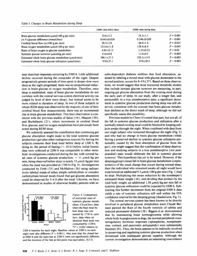
Table I. Changes in Brain Metabolism during Sleep
2300h value before sleep 0300h value during sleep
Brain glucose metabolism (Aimol/l00 g per min) 33.6±2.2 24.3±1.1 P = 0.001(A-V) glucose difference (mmol/liter) 0.643±0.024 0.546±0.020 P = 0.001Cerebral blood flow (cc/100 g per min) 50.3±2.8 44.6±1.4 P = 0.021Brain oxygen metabolism (,gmol/100 g per min) 153.4±11.8 128.4±8.4 P = 0.045Ratio of brain oxygen to glucose metabolism 4.85±0.13 5.33±0.22 P = 0.05Systemic glucose turnover (,gmol/kg per min) 13.6±0.8 11.0±0.5 P = 0.003Estimated whole brain glucose metabolism (Amol/min) 390.1±27.2 320.3±13.9 P = 0.005Estimated whole body glucose utilization (imol/min) 976±37.1 874±20.5 P = 0.003
near maximal responses occurring by 0300 h. Little additionaldecline occurred during the remainder of the night. Despiteprogressively greater periods of time spent in deeper slow wavesleep as the night progressed, there was no proportional reduc-tion in brain glucose or oxygen metabolism. Therefore, oncesleep is established, rates of brain glucose metabolism do notcorrelate with the central nervous system electrical activity (asjudged by level of slow wave sleep), but instead seems to bemore related to duration of sleep. In two of three subjects inwhomREMsleep was observed for the majority of one of theircerebral blood flow measurements, there was an incrementalrise in brain glucose metabolism. This later observation is con-sistent with the previous studies of Sakai ( 16), Maquet (20),and Buchsbaum (21), where increments in cerebral bloodflow, glucose, and/or oxygen metabolism have previously beennoted during REMsleep.
Weindirectly assessed the contribution that continuing gutglucose absorption might make to the total systemic glucoseproduction (gut plus other endogenous sources) by having thesubjects consume their final meal before sleep at 1200 h. Bydoing so, the period of fasting (- 10 h) before initial kineticdata were collected at 2200 h was comparable to other meta-bolic investigations conducted during the day with normal ba-sal rates of systemic glucose production, -11 mol/kg permin, being observed before sleep vs nearly 15 ,qmol/kg per minwhen the meal was provided at 1700 h (Fig. 4). Investigationsconducted by Firth (35) and McMahon (36) using radioac-tively labeled meals of either simple carbohydrate or complexcarbohydrate/mixed meals found that gut glucose absorptioncould be observed for 5-6 h after the meal. Likewise, we havedemonstrated in studies of otherwise healthy patients with in-
Final Meal Final MealI at 1730h at 1230h
2200h 0400h
Hour
3.0 Figure 4. Comparisonof nocturnal rates of
2.5 systemic glucose metab-E olism. Closed bars, data= when the final meal be-
2.0 E fore sleep was con-
sumed by 1730 h; openbars, data when an
5 identical final meal wasconsumed by 1230 h.*P < 0.002 relative to
2200 h baseline for each night. Baseline values at 2200 h on eachnight were also different (P < 0.001). Also note that the closed barat 0400 h and the open bar at 2200 h were not significantly different,and the duration of the fast at this point was equivalent, 10.5 h.
sulin-dependent diabetes mellitus that food absorption, as-sessed by labeling a mixed meal with glucose deuterated in thesecond position, occurs for 8-9 h (37). Based on these observa-tions, we would suggest that most nocturnal metabolic studiesthat include isotopic glucose turnover are measuring, in part,ongoing gut glucose absorption from the evening meal duringthe early part of sleep. In our study, after a longer fast, andpresumably in a true postabsorptive state, a significant decre-ment in systemic glucose production during sleep was still ob-served, consistent with the concept that brain glucose metabo-lism declines as the direct result of sleep, although we did notspecifically assess this possibility.
Previous studies by Clore (4) noted that part, but not all, ofthe fall in systemic glucose production and utilization after anormally timed evening meal could be blocked by keeping sub-jects awake through the night. Whencombined with data fromour single subject who remained throughout the night (Fig. 5)and who had no change in brain glucose metabolism (whilehaving a preserved decline in systemic glucose utilization pre-sumably caused by the final absorption of glucose from thegut), one might suggest that the combination of sleep depriva-tion and studying subjects in a true postabsorptive (not post-prandial) state would obliterate the fall in systemic glucoseturnover. This hypothesis has yet to be tested. However, if thesleeping group's mean fall in brain glucose metabolism is repre-sentative of the usual change that occurs during normal sleep,then the individual who remained awake all night would haveexperienced an additional 9.3 ,gmol/ 100 g per min (Fig. 1 ) hadhe slept. Multiplying this mean reduction by the nonsleeper'sestimated brain weight (34), and dividing that product by histotal body weight, an additional 1.58 ,gmol/kg per min fall insystemic glucose utilization could be expected by 0300 h. Sub-tracting this further decrement from his original 0300 h datayields a rate of systemic utilization still well within the 95%confidence interval for the sleeping group (shaded area, Fig. 5).
The central nervous system has been known to be directlyinvolved in peripheral glucose metabolism since Claude Ber-nard pierced the floor of the fourth ventricle of rabbits andinduced permanent diabetes (38). Biggers et al. demonstratedthat by maintaining brain normoglycemia while allowingwhole body hypoglycemia in dogs, the normal peripheral coun-terregulatory hormone responses (epinephrine, norepineph-rine, cortisol, and pancreatic polypeptide) were substantiallyblunted (39). Thus, the brain appears to be indirectly involvedin preserving and regulating systemic glucose production whenthreatened with inadequate glucose supplies. Although thecurrent investigation demonstrates an interesting concordance
532 P. J. Boyle, J. C. Scott, A. J. Krentz, R. J. Nagy, E. Comstock, and C. HoJlinan
a, 16
0~
10
(I 1
wEn
' 20 30
EH~~~~~~~~~~~25O
15 82
1010
2200 2400 0200 0400
HourFigure 5. Data from an individual subject unable to sleep throughoutthe night (evening meal served at 1730 h). The rate of systemic glu-cose production and utilization fell in parallel from 2200 and 0300 hand then plateaued. Rate of brain glucose metabolism remained con-stant throughout the night. For reference, the shaded area represents95% confidence intervals for systemic glucose turnover data in sub-jects able to sleep.
between falling rates of brain glucose metabolism and systemicglucose utilization, it cannot delineate cause and effect. Levelsof the key regulators of hepatic glucose production (insulin andglucagon) did not change appreciably during the night, suggest-ing that decrements in production are unlikely to be the pri-mary event explaining our findings. More likely, the fall insystemic utilization occurs as result of a normal decline inbrain glucose metabolism, and hepatic production then fol-lows, so that euglycemia is preserved. Alternatively, one mightconsider that a primary fall in hepatic production occurs, fol-lowed secondarily by a reduction in brain glucose utilization.Sleep is associated with reductions in glycerol and free fattyacid concentrations, which are known to reduce fructose 2,6-bisphosphate levels (40, 41). Thus, decrements in these meta-bolic intermediates may indirectly lead to the fall in hepaticglucose production. Howsuch a peripheral mechanism for de-creased hepatic glucose output might lead to reduction in brainmetabolism is not clear, since the systemic glucose concentra-tion is not rate-limiting for brain metabolism until it falls belownormal range (42). Subjects' glucose concentrations remainedwithin the normal range during our studies; therefore, brainmetabolism should not have been limited by supply and itseems unlikely that a decrement in hepatic glucose output isthe primary metabolic event in this sequence of events. As afinal, and more unlikely option, hepatic glucose output andbrain metabolism may be regulated independently and the factthat euglycemia is maintained throughout the night is serendip-itous.
Rates of glucose production have previously been observedto increase in the final hours of a night of sleep in normalsubjects (4). Because of a concomitant increase in glucose uti-lization that occurred in those studies, there was no change inglucose concentration. In general, such increments occur dur-ing the hours of 0500 to 0800 h and are thought to be related tosecretion of growth hormone during the first hours of sleep(43). In patients with insulin-dependent diabetes, this in-creased rate of hepatic glucose production generally results in arise in plasma glucose concentration when coupled with stableinsulin levels and is referred to as the "dawn phenomena"(44). During these investigations, we failed to observe any in-crease in glucose production and utilization during the final
hours of observed sleep, most likely because of the fact that ourfinal data was collected at 0500 h, before the rise in glucoseproduction normally begins. If one extrapolates this proposalto patients with insulin-dependent diabetes, where modest hy-perinsulinemia occurs routinely during the night, then thestage should be set for nocturnal hypoglycemia. In the DiabetesControl and Complications Trial, a multicenter, randomizedtrial comparing near normalization of blood glucose levels tostandard insulin therapy in > 1,400 patients with insulin-de-pendent diabetes, the insulin replacement schemes used rou-tinely produce stable or moderate hyperinsulinemia duringsleep (45). Consistent with the above extrapolation, > 50% ofthe severe hypoglycemic events experienced by the patients inthe Diabetes Control and Complications Trial have occurredtemporally with our reported nocturnal nadirs in brain glucoseutilization and systemic glucose production (46).
In the current study, the overall pattern of brain oxygenmetabolism fell gradually over the night. The magnitude of thechange was not nearly as great as that observed for glucoseconsumption but is in keeping with the overall reduction insystemic oxygen utilization reported by Brebbia (47). OnlyMangold ( 13) and Madsen ( 17) have previously investigatednocturnal brain oxygen metabolism, and they came to dispa-rate conclusions. Mangold's original observation of unchangedoxygen utilization has been questioned, since when one of thesix participants he studied is excluded from analysis, there is astatistically significant decrease in oxygen metabolism. Mad-sen found a 25% reduction in brain oxygen metabolism duringdeep sleep and an increase during REMsleep in subjects stud-ied on several different nights. In the present investigation, thequantity of oxygen required (on a molar basis) to metabolizeglucose (at the nocturnal glucose metabolism nadir) was signifi-cantly greater than the oxygen/glucose ratio seen before sleep.Several possible metabolic events could explain this observa-tion: (a) oxidative glucose disposal could continue unchangedwhile there is a decrease in nonoxidative glucose disposal inbrain tissue (i.e., less glycogen formation); (b) an alternate fuelsource such as ketone bodies may be oxidized to meet meta-bolic demands as glucose metabolism falls off; or (c) there is arelative decrease in anaerobic metabolism. With respect to thefirst possibility, the brain is unable to synthesize or store morethan a few minutes' worth of glucose for later use (48). Thus, alarge enough decrease in glycogen synthesis to account for the25% decrease in systemic glucose utilization we report seemsunlikely, although no data exist on glycogen content before andafter sleep. The second explanation also seems unlikely, sincewe failed to observe an increase in the arteriovenous ketonedifference across the brain (data not shown). Other substrates(such as nonesterified fatty acids and amino acids) are notmajor energy substrates for brain metabolism (42) and werenot assessed in this investigation. The final explanation is per-haps most reasonable. Fox et al. have demonstrated that duringvisual stimulation, brain glucose metabolism increases in thevisual cortex, while oxygen utilization remains constant (49),implying that a relative increase in anaerobic metabolism oc-curs with increased neuronal activity. During sleep, corticalneuronal activity decreases (the opposite outcome of the Foxexperiment) with median firing rates falling by 25%comparingwakefulness to slow wave sleep ( 1). In further support of anoverall reduction in electrical activity, we and others ( 14-16 )have observed a decline in cerebral blood flow, a parameter
Sleep and Brain Glucose Metabolism 533

generally believed to be coupled to neuronal activity (50).Therefore, if sleep is viewed physiologically as a relative oppo-site of the wakeful state (e.g., less visual stimulation and re-duced overall neuronal activity), and by the above reasoningresults in a decrease in anaerobic glycolysis, then the oxygen/glucose ratio would be expected to rise.
In summary, whether or not the primary metabolic eventresponsible for the fall in whole body glucose production andutilization is a fall in brain glucose metabolism during sleepremains unclear. However, several independent conclusionscan be made: (a) rates of brain glucose and oxygen metabolismdecrease during the night (the former more than the later); (b)quantitatively, the fall in brain glucose utilization accounts fora significant part of the decrement in measured peripheral glu-cose utilization; (c) the fall in brain glucose metabolism occurssecondary to reduced cerebral blood flow and diminished brainglucose arteriovenous glucose difference; and (d) these changesin brain energy and flow dynamics during sleep occur indepen-dent of stage of slow wave electrical activity (except duringREMsleep where brain metabolism increases in most subjects)and instead appear to be more related to duration of sleep.
Acknowledgments
The expertise of Dr. Clifford Qualls in developing the curve fitting andintegration programs is gratefully acknowledged. The care of the nurs-ing staff on the General Clinical Research Center, especially Ms. LizCandelaria, and the technical assistance of Ms. Rose Mills and Ms.Ellen Russo with the placement of the EEGleads and interpretation ofthe electroencephalograms is also gratefully acknowledged. The edito-rial assistance of Ms. Natalie Pacheco and Ms. Carolyn King are alsoacknowledged.
Dr. Boyle was a recipient of a Research and Development Awardfrom the American Diabetes Association. This project was supportedin part by dedicated Health Research Funds of the University of NewMexico and by a grant from General Clinical Research Program, Na-tional Center for Research Resources, NIH 5 MO1 RR00997-15.
References
1. Hobsen, J. A., and R. W. McCarley. 1971. Cortical unit activity in sleep andwaking. Electroencephalogr. Clin. Neuroph.vsio/. 30:97-112.
2. Ravussin, E., S. Lillioja, T. E. Anderson, L. Christin, and C. Bogardus.1986. Determinants of 24-hour energy expenditure in man. J. Clin. Invest.78:1568-1578.
3. Miller, J. C., and S. M. Horvath. 1976. Cardiac output during human sleep.A vial. Space Environ. Med. 47:1046-105 1.
4. Clore, J. N., J. E. Nestler, and W. G. Blackard. 1989. Hepatic glucoseoutput during sleep. Diabets. 38:285-290.
5. Campbell, P. J., G. B. Bolli, P. E. Cryer, and J. E. Gerich. 1985. Pathogene-sis ofthe dawn phenomenon in patients with insulin-dependent diabetes mellitus.N. Engi. J. Med. 312:1473-1479.
6. Tanaka, K., W. E. Nicholson, and D. N. Orth. 1978. Diurnal /3-MSH(LPH) and ACTH in man. J. Clin. Endocrinol. & Melab. 46:883-890.
7. Takahashi, Y., D. M. Kipnis, and W. H. Daughaday. 1968. Growth hor-mone secretion during sleep. J. Clin. Invest. 47:2079-2090.
8. Boyle, P. J., A. Avogaro, L. Smith, S. D. Shah, P. E. Cryer, and J. V.Santiago. 1991. Absence of the dawn phenomenon and abnormal lipolysis in typeI (insulin-dependent) diabetic patients with chronic growth hormone deficiency.Diabetologia. 35:372-379.
9. Boyle, P. J., A. Avogaro, L. Smith, D. M. Bier, A. S. Pappu, D. R. Illing-worth, and P. E. Cryer. 1992. Role of GHin regulating nocturnal rates of lipolysisand plasma mevalonate levels in normal and diabetic humans. Am. J. Physiol.263 (Endocrino/. Melab. 26) :E I 68-E 172.
10. Ashmore, J., and D. Morgan. 1967. Metabolic effects of adrenal glucocor-ticoid hormones. In The Adrenal Cortex. A. B. Eisenstein, editor. Little, Brownand Company, Boston. pp. 249-267.
I 1. Franzini, C. 1992. Brain metabolism and blood flow during sleep. J. SleepRes. 1:3-16.
12. Sawaya, R., and D. H. Ingvar. 1989. Cerebral blood flow and metabolismin sleep. Adca. Neitro/. Scand. 80:481-491.
13. Mangold, R., L. Sokoloff, E. Conner, J. Kleinerman, P. G. Therman, andS. S. Kety. 1955. The effects of sleep and lack of sleep on the cerebral circulationand metabolism of normal young men. J. Clin. Invest. 34:1092-1 100.
14. Madsen, P. L., S. Holm, S. Vorstrup, L. Friberg, N. A. Lassen, and G.Wilschiodz. 1991. Human regional cerebral blood flow during rapid-eye-move-ment sleep. J. Cereb. Blood Flow Metab. 1 1:502-507.
15. Townsend, R. E., P. N. Prinz, and W. D. Obrist. 1973. Human cerebralblood flow during sleep and waking. J. App/. Phvsiol. 35:620-625.
16. Sakai, F., J. S. Meyer, 1. Karacan, S. Derman, and M. Yamamoto. 1980.Normal human sleep: regional cerebral hemodynamics. Ann. Neurol. 7:471-478.
17. Madsen, P. L., J. F. Schmidt, G. Wildschiodtz, L. Friberg, S. Holm, S.Vorstrup, and N. A. Lassen. 1991. Cerebral 02 metabolism and cerebral bloodflow in humans during deep and rapid-eye-movement sleep. Am. Physiol. Soc.16 1:2597-260 1.
18. Kennedy, C., J. C. Gillin, W. Mendelson, S. Suda, M. Miyaoka, M. Ito, R.K. Nakamura, F. 1. Storch, K. Pettigrew, M. Mishkin, and L. Sokoloff. 1982.Local cerebral glucose utilization in non-rapid eye movement sleep. Nature(Lond.) 297:325-327.
19. Heiss, W. D., G. Pawlik, K. Herholz, R. Wagner, and K. Wienhard. 1985.Regional cerebral glucose metabolism in man during wakefulness, sleep, anddreaming. Brain Rets. 327:362-366.
20. Maquet, P., D. Dive, E. Salmon, B. Sadzot, G. Franco, R. Poirrier, R. VonFrenckell, and G. Franck. 1990. Cerebral glucose utilization during sleep-wakecycle in man determined by positron emission tomography and [ '8F]-2-fluoro-2-deoxy-D-glucose method. Brain Res. 513:136-143.
21. Buchsbaum, M. S., J. C. Gillin, J. Wu, E. Hazlett, N. Sicotte, R. M.Dupont, and W. E. Bunney, Jr. 1989. Regional cerebral glucose metabolic rate inhuman sleep assessed by positron emission tomography. Life Sci. 45:1349-1356.
22. Sherwin, R. S. 1980. Role of the liver in glucose homeostasis. DiabetesCare. 3:261-265.
23. Kety, S. S.. and C. F. Schmidt. 1948. The nitrous oxide method for thequantitative determination of cerebral blood flow in man: theory, procedure andnormal values. J. Clin. Invest. 27:476.
24. Rechtschaffen, A., and A. Kales. 1968. A Manual of Standardized Termi-nology, Techniques, and a Scoring System for Sleep Stages of Human Subjects.Brain Information Service/ Brain Research Institute, UCLA, Los Angeles.
25. Lassen, N. A., and M. H. Lane. 1961. Validity of internaljugular blood forstudy of cerebral blood flow and metabolism. J. App/. Phvsio/. 16:313-320.
26. Boyle, P. J., S. B. Liggett, S. D. Shah, and P. E. Cryer. 1988. Directmuscarinic cholinergic inhibition of hepatic glucose production in humans. J.C/in. Inivest. 82:445-449.
27. Boyle, P. J., S. D. Shah, and P. E. Cryer. 1989. The prevention of hypogly-cemia during fasting in humans: roles of insulin, glucagon and catecholamines.Am. J. P/wsioL. 256 (Endocrinol. Metab. 19):E65 I-E661.
28. Kuzuya, H., P. M. Blix, D. L. Horwitz, D. N. Steiner, and A. H. Ruben-stein. 1977. Determination of free and total insulin C-peptide in insulin-treateddiabetics. Diabeltes. 26:22-29.
29. Ensinck, J. W. 1983. Immunoassays for glucagon. In Glucagon Handbookof Experimental Pharmacology. P. Lefebvre, editor. Springer-Verlag, NewYork.pp. 203-221.
30. Schalch, D. S., and M. L. Parker. 1964. A sensitive double antibodyimmunoassay for human growth hormone in plasma. Naittre(Lond.). 203:1141-2.
31. Argoud, G., D. S. Schade, and R. P. Eaton. 1987. Underestimation ofhepatic glucose production by radioactive and stable tracers. Am. J. Physiol.193:E606-E6 15.
32. Robertson, C. S., R. K. Narayan, Z. L. Gokaslan, R. Pahwa, R. G. Gross-man, P. Caram, Jr., and E. Allen. 1989. Cerebral arteriovenous oxygen differenceas an estimate of cerebral blood flow in comatose patients. J. Neurosurg. 70:222-230.
33. SAS Institute Inc. 1988. SAS/STATT Users Guide, Release 6.03 Edi-tion. SAS Institute Inc. Cary, NC.
34. Sunderman, F. W., and F. Boerner. 1949. Normal Values in ClinicalMedicine. W. B. Saunders Company, Philadelphia, pp. 664.
35. Firth, R. G., P. M. Bell, H. M. Marsh, 1. Hansen, and R. A. Rizza. 1986.Postprandial hyperglycemia in patients with noninsulin dependent diabetes mel-litus: role of hepatic extrahepatic tissues. J. Clin. Invest. 77:1525-1531.
36. McMahon, M., H. Marsh, and R. Rizza. 1989. Comparison of the patterof postprandial carbohydrate metabolism after ingestion of a glucose drink or amixed meal. J. Clin. Endocrinol. & Melab. 68:647-653.
37. Boyle, P. J., E. Comstock, A. O'Connor, R. J. Nagy, and D. S. Schade.1992. Declining gut glucose absorption is the mechanism for onset of nocturnalhypoglycemia. Diabetes. 41:30A.
38. Bernard, C. 1957. An Introduction to the Study of Experimental Medi-cine. (1865.) (English translation by H. C. Green. 1927. Macmillan JournalsLtd., London.) Republished by Dover Publications, New York. p. 173.
39. Biggers, D. W., S. R. Myers, D. Neal, R. Stinson, N. B. Cooper, J. B.
534 P. J. Boyle, J. C. Scott, A. J. Krentz, R. J. Nagy, E. Comstock, and C. Hoffman

Jaspan, P. E. Williams, A. D. Cherrington, and R. T. Frizzell. 1989. Role of brainin counterregulation of insulin-induced hypoglycemia in dogs. Diahetes. 38:7-16.
40. Uyeda, K., E. Furuya, C. S. Richards, and M. Yokahama. 1984. Fructose-2,6-P2, chemistry and biological function. Mol. Cell. Biochern. 48:97-120.
41. Hue. L., and M. H. Rider. 1984. Role of fructose 2,6-bisphosphate in thecontrol of glycolysis in mammalian tissues. Bioc/ten. J. 245:313-24.
42. Pardridge. W. H. 1983. Brain metabolism from the blood-brain barrier.PArsiol. Rev. 63:1481-1535.
43. Campbell, P. J., G. B. Bolli, P. E. Cryer, and J. E. Gerich. 1985. Pathogene-sis ofthe dawn phenomenon in patients with insulin-dependent diabetes mellitus:accelerated glucose production and impaired glucose utilization due to nocturnalsurges in growth hormone secretion. N. Engi. J. Med. 312:1473.
44. Boyle, P. J., A. Avogaro, L. Smith. S. D., P. E. Cryer, and J. V. Santiago.1992. Absence of the dawn phenomenon and abnormal lipolysis in type I (insu-
lin-dependent) diabetic patients with chronic growth hormone deficiency. Diabe-tologia. 35:373-379.
45. Schade. D. S., J. B. Santiago, J. S. Skyler, and R. A. Rizza. 1983. IntensiveInsulin Therapy. Excerpta Medica, Inc., Princeton, NJ. pp. 129-148.
46. The DCCTResearch Group. 1991. Epidemiology of severe hypoglycemiain the DCCT. Ain. J. Med. 90:450-459.
47. Brebbia, D. R., and K. Z. Altshuler. 1965. Oxygen consumption rate andelectroencephalographic stage of sleeo. Science (Wash. DC). 150:1621-1623.
48. Siesjo, B. K. 1978. Utilization of substrates of brain tissues. In BrainEnergy Metabolism. Wiley, NY. pp. 1-20.
49. Fox, P. T., M. E. Raichle, M. A. Mintun, and C. Dence. 1988. Nonoxida-tive glucose consumption during focal physiologic neural activity. Science(W'ash. DC). 2:462-464.
50. Lou, H. C.. L. Edvinsson, and E. T. MacKenzie. 1987. The concept ofcoupling blood flow to brain function: revision required? Ann. Neuro/. 22:289-297.
Sleep and Brain Glucose Metabolism 535


![Glucose Metabolism Is Required for Platelet ... · Glucose Metabolism To determine glucose uptake, washed platelets in 1 mmol/L glucose DMEM were incubated with 10 mmol/L [3H]2-deoxy-D-glucose](https://img.pdfslide.net/doc/110x75/5f7630d406ba0e330e387389/glucose-metabolism-is-required-for-platelet-glucose-metabolism-to-determine.jpg)