Embed Size (px)
Citation preview
Direct Ischaemia imaging and other Competing technologies
Dr. NATHAN BETTER Associate Professor of Medicine University of Melbourne Departments of Cardiology & Nuclear Medicine Royal Melbourne Hospital December, 2012
CLINICAL CASE
34 year old male Atypical chest pain. No cardiac risk factors. Examination – normal. BP 120/70 ECG – Normal. CXR/Bloods- Normal
Which test next?
CHOICES
Nil Exercise ECG Stress echo Stress nuclear Cardiac CT Coronary angiography

A standard treadmill stress test was performed
Robert Bruce (The AGE 1/3/04)
20/11/16 – 12/2/04 First published 1949 Multistage test 1963 Emeritus Professor of Medicine, U of Washington. “You would never buy a used car without taking it for
a drive and seeing how the engine performed while it was running, and the same is true for evaluating the function of the heart.”

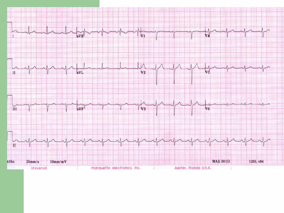
Exercise test
9 minutes Bruce Ceased due to SOB No chest pain HR Rest - 72 Peak - 162 BP Rest - 130/75 Peak - 200/80
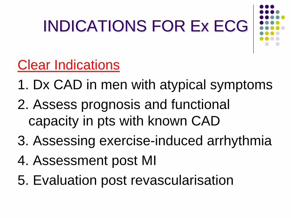
INDICATIONS FOR Ex ECG
Clear Indications 1. Dx CAD in men with atypical symptoms 2. Assess prognosis and functional
capacity in pts with known CAD 3. Assessing exercise-induced arrhythmia 4. Assessment post MI 5. Evaluation post revascularisation

INDICATION Ex ECG Possible indications 1. Dx CAD in women with typical and atypical pain 2. RBBB, Digoxin 3. Known CAD - functional capacity, response to
treatment 4. Functional capacity in some pts with valvular heart
disease – Moderate to severe AS (ESC 2005) 5. Asymptomatic males > 40 or high risk job with 2+
risk factors or a sedentary pt going to begin a vigorous exercise program
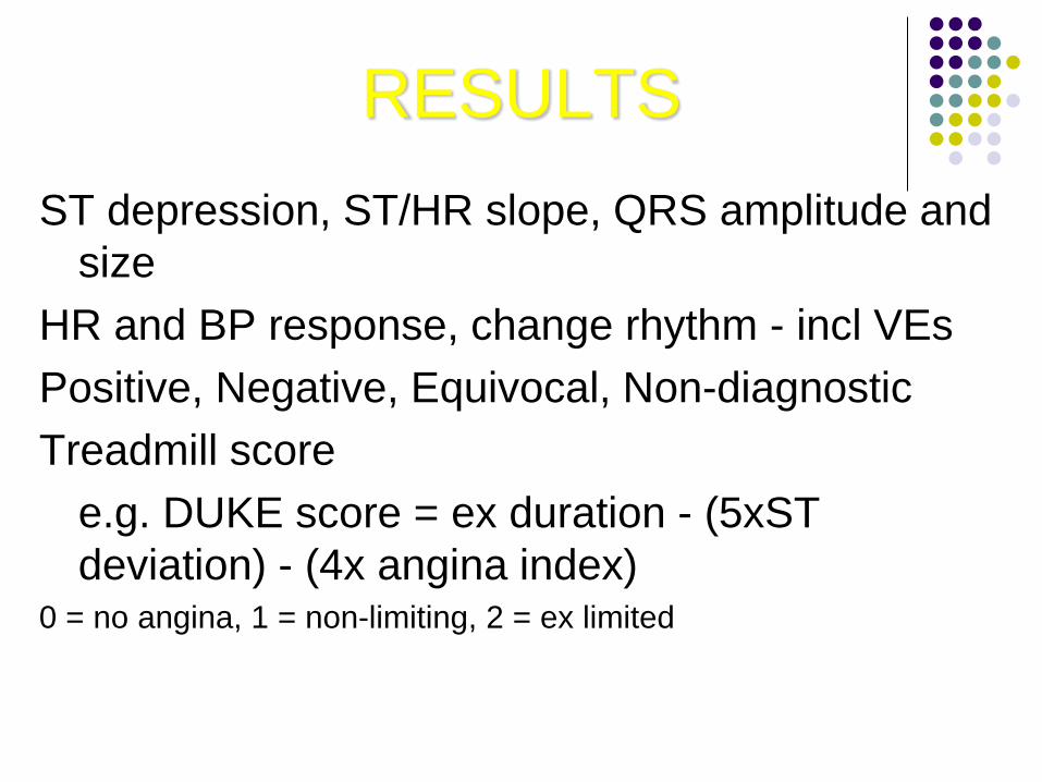
RESULTS ST depression, ST/HR slope, QRS amplitude and
size HR and BP response, change rhythm - incl VEs Positive, Negative, Equivocal, Non-diagnostic Treadmill score e.g. DUKE score = ex duration - (5xST
deviation) - (4x angina index) 0 = no angina, 1 = non-limiting, 2 = ex limited
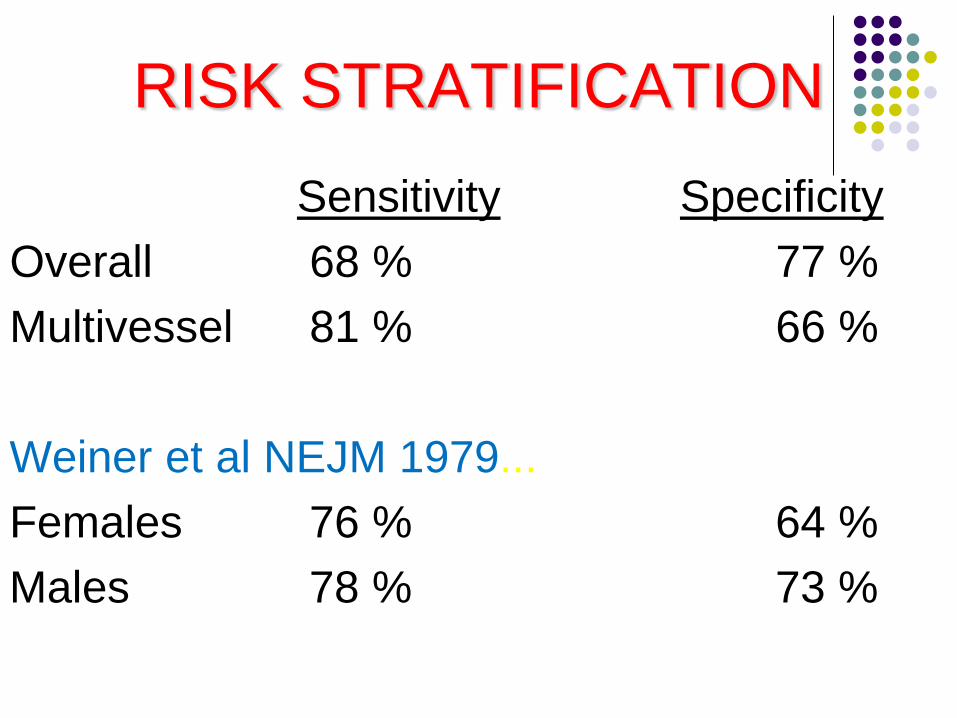
RISK STRATIFICATION Sensitivity Specificity Overall 68 % 77 % Multivessel 81 % 66 % Weiner et al NEJM 1979... Females 76 % 64 % Males 78 % 73 %

Where to now?
Normal – stop
Abnormal – further tests, depending on results
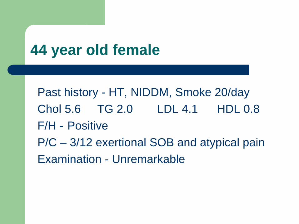
44 year old female
Past history - HT, NIDDM, Smoke 20/day Chol 5.6 TG 2.0 LDL 4.1 HDL 0.8 F/H - Positive P/C – 3/12 exertional SOB and atypical pain Examination - Unremarkable

CHOICES
Nil Exercise ECG Stress echo Stress nuclear Cardiac CT Coronary angiography
STRESS ECHOCARDIOGRAPHY

Ischaemic Cascade
Perfusion
Diastolic Function
Systolic Function
ECG changes
Angina
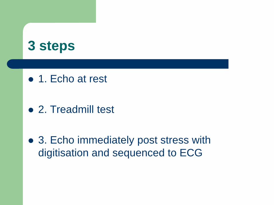
3 steps
1. Echo at rest
2. Treadmill test
3. Echo immediately post stress with digitisation and sequenced to ECG

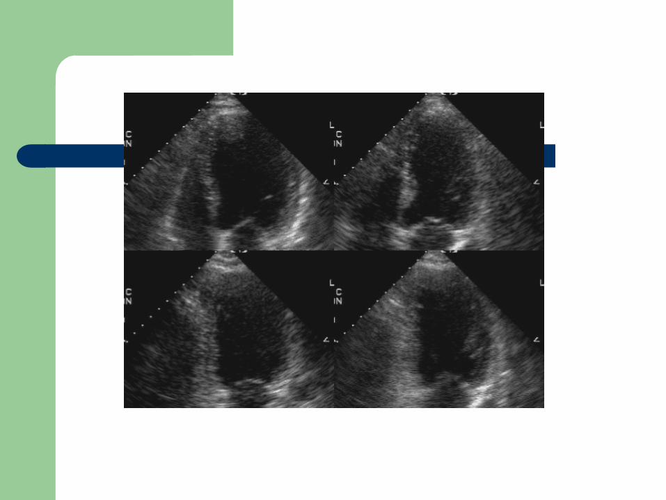
Interpretation
Rest Stress Result Normal Normal Normal N/Hypo akinesis/dys Ischaemia Akinesis Normal/hypo Viable Akinesis/dys Akinesis/dys Infarct/Necrosis
Indications
Ischaemia assessment Valvular heart disease – asymptomatic AS (incl.
LV function normal or poor), AR and MR. Viability Advantages – no ionising radiation. Slightly
greater specificity
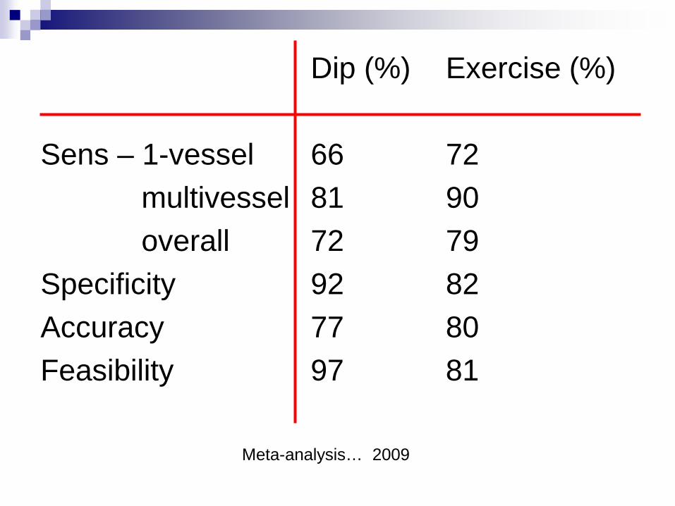
Dip (%) Exercise (%) Sens – 1-vessel 66 72 multivessel 81 90 overall 72 79 Specificity 92 82 Accuracy 77 80 Feasibility 97 81
Meta-analysis… 2009
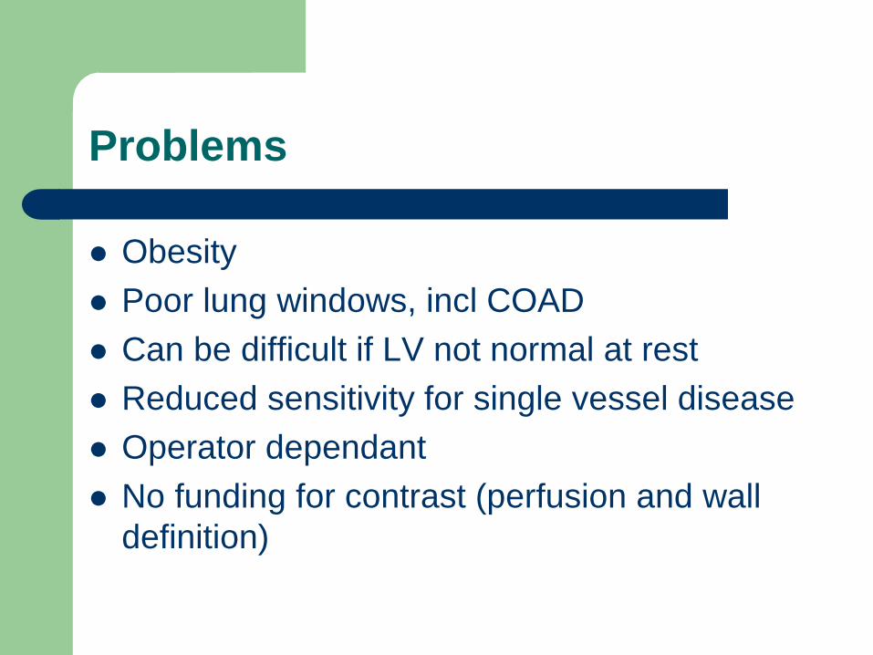
Problems
Obesity Poor lung windows, incl COAD Can be difficult if LV not normal at rest Reduced sensitivity for single vessel disease Operator dependant No funding for contrast (perfusion and wall
definition)

Clinical case
65 year old male Diabetic, hypertensive, LDL chol 4.8 Chest pain FI – Exertional, but pleuritic
(atypical) Examination – Well 150/90 ECG – SR. LBBB.

CHOICES
Nil Exercise ECG Stress echo Stress nuclear Cardiac CT Coronary angiography

Persantin-exercise Tc-99m sestamibi study
Off caffeine 24 hours 0.568 mg/kg IV Persantin 4 mins SLR Peak HR 98 Peak BP 120/80 No pain Equivocal ECG Rest and stress images Gating
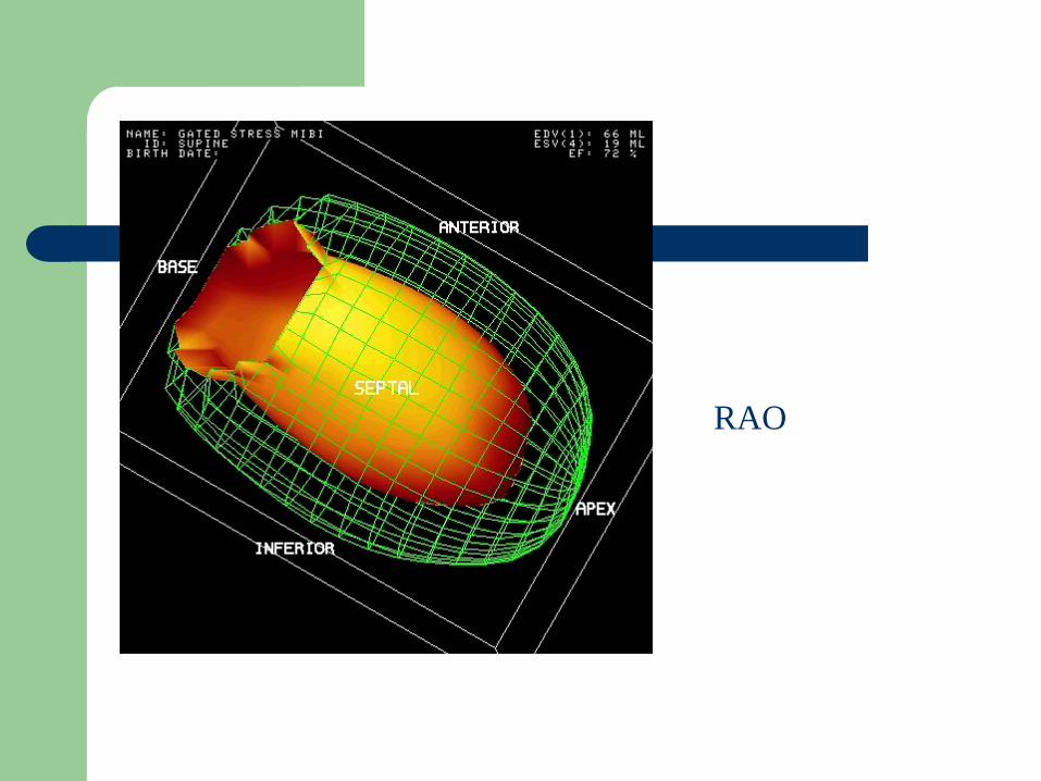
RAO

Management
Risk factor control Reassurance +++++++++++++
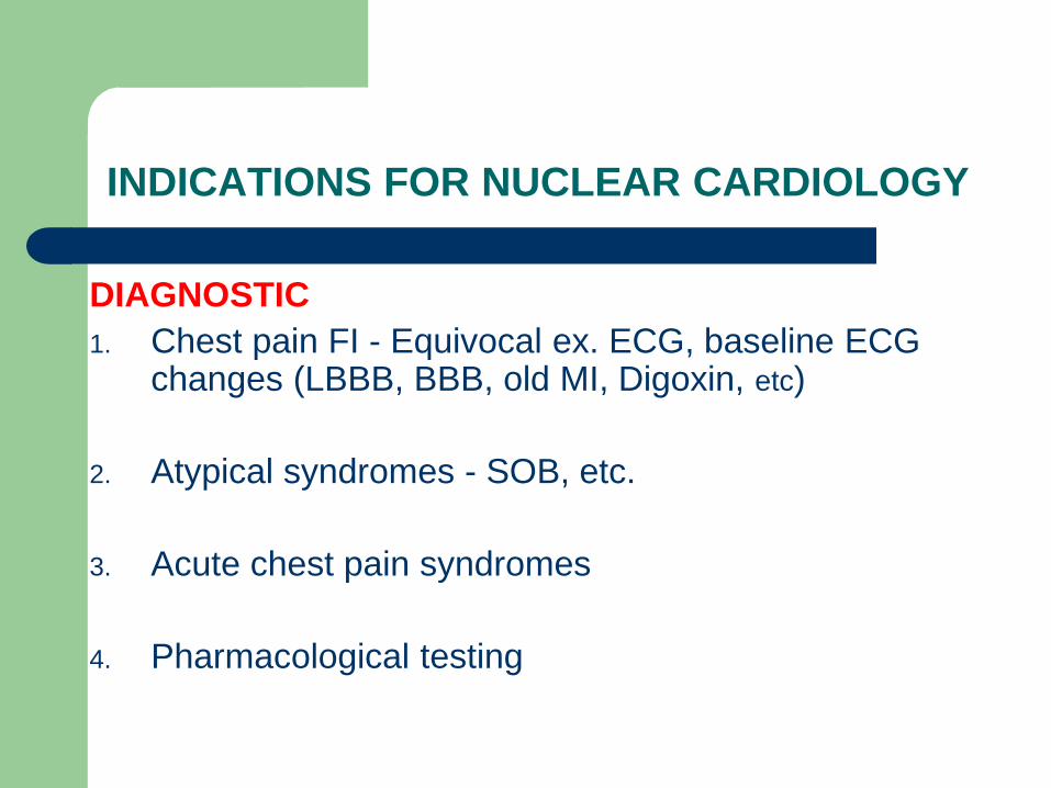
INDICATIONS FOR NUCLEAR CARDIOLOGY
DIAGNOSTIC 1. Chest pain FI - Equivocal ex. ECG, baseline ECG
changes (LBBB, BBB, old MI, Digoxin, etc)
2. Atypical syndromes - SOB, etc.
3. Acute chest pain syndromes
4. Pharmacological testing
INDICATIONS FOR NUCLEAR CARDIOLOGY
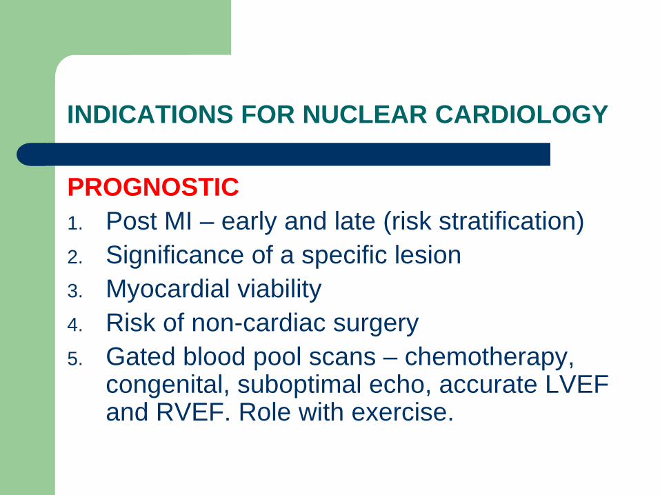
PROGNOSTIC 1. Post MI – early and late (risk stratification) 2. Significance of a specific lesion 3. Myocardial viability 4. Risk of non-cardiac surgery 5. Gated blood pool scans – chemotherapy,
congenital, suboptimal echo, accurate LVEF and RVEF. Role with exercise.

Clinical Case
50 year old male Positive FH Slightly atypical pain Low-intermediate probability of IHD Examination / ECG - Normal

CHOICES
Nil Exercise ECG Stress echo Stress nuclear Cardiac CT Coronary angiography

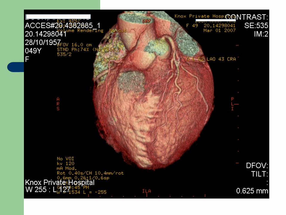
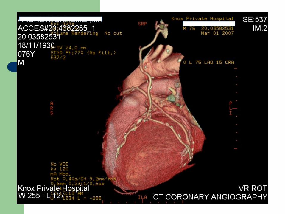
Cardiac CT

POSSIBLE Cardiac CT Indications
Screening high risk asymptomatic individuals Triple rule out in acute chest pain (with PE and
dissection) Graft and stent patency Equivocal diagnostic functional studies (if no
known prior CAD) Coronary calcium score Diagnosis in low-intermediate risk Anomolous coronary artery origins Evaluating coronary arteries prior to cardiac non-
coronary surgery
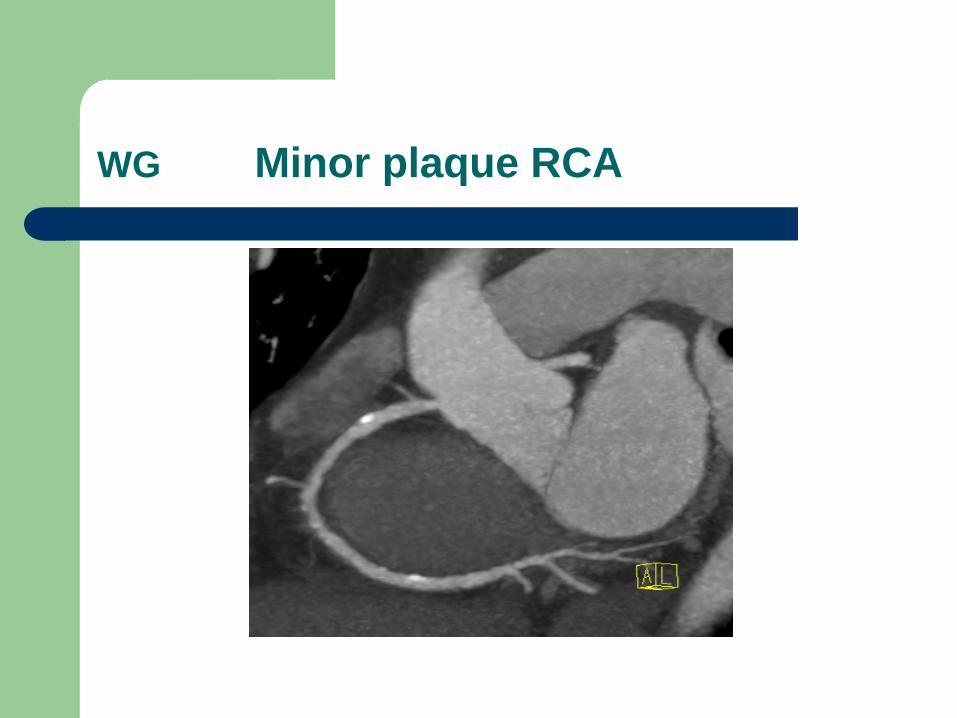
WG Minor plaque RCA

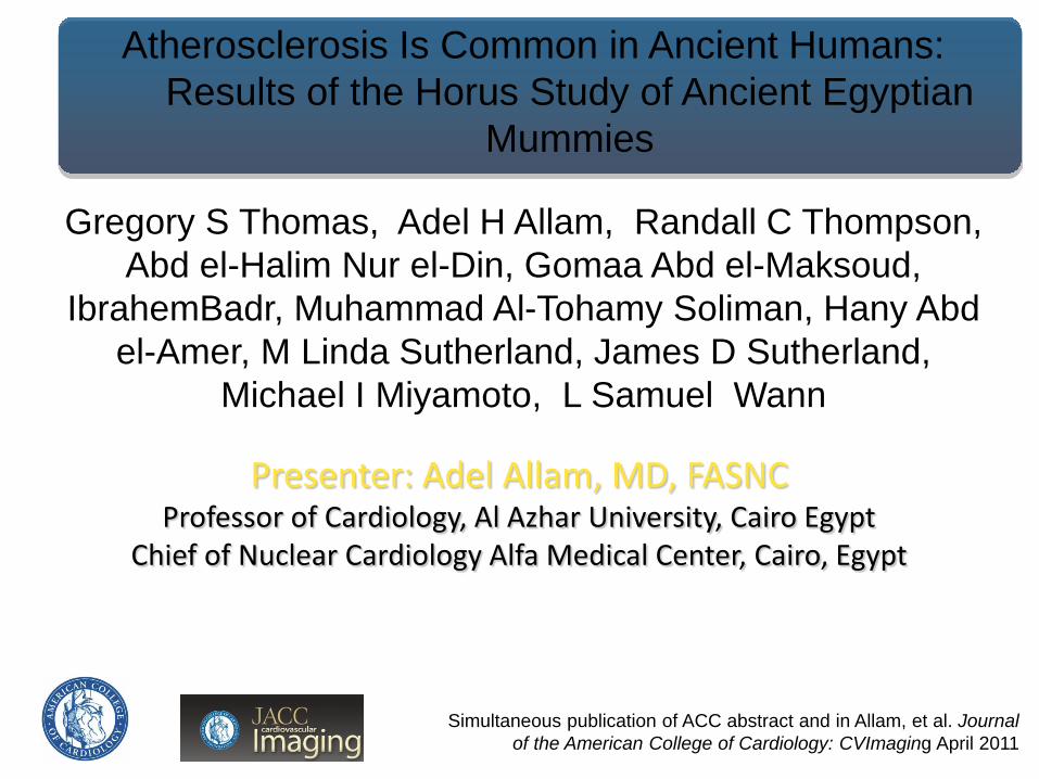
Atherosclerosis Is Common in Ancient Humans: Results of the Horus Study of Ancient Egyptian
Mummies
Presenter: Adel Allam, MD, FASNC Professor of Cardiology, Al Azhar University, Cairo Egypt
Chief of Nuclear Cardiology Alfa Medical Center, Cairo, Egypt
Gregory S Thomas, Adel H Allam, Randall C Thompson, Abd el-Halim Nur el-Din, Gomaa Abd el-Maksoud,
IbrahemBadr, Muhammad Al-Tohamy Soliman, Hany Abd el-Amer, M Linda Sutherland, James D Sutherland,
Michael I Miyamoto, L Samuel Wann
Simultaneous publication of ACC abstract and in Allam, et al. Journal of the American College of Cardiology: CVImaging April 2011
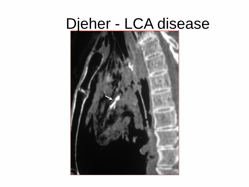
Djeher - LCA disease
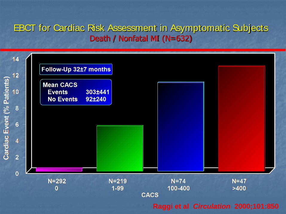
EBCT for Cardiac Risk Assessment in Asymptomatic Subjects Death / Nonfatal MI (N=632)
Raggi et al Circulation 2000;101:850

Events by CAC stratification
Multi-Ethnic Study of Atherosclerosis 2010
CAC group Patients, n (%)
Events, n (%) Event rate (per 1000 person-years)
Hazard ratio (95% CI)
Coronary heart disease
•CAC=0 444 (47) 2 (0.5) 0.8 1.0 (reference)
•CAC 1–100 267 (28) 7 (3) 4.8 4.6 (0.9–23.4)
•CAC >100 239 (25) 25 (11) 20.2 24.8 (5.4–11.5)
Cardiovascular disease events •CAC=0 444 (47) 9 (2) 3.7 1.0 (reference)
•CAC 1–100 267 (28) 12 (5) 8.4 1.8 (0.7–4.6)
•CAC >100 239 (25) 32 (13) 26.4 6.0 (2.5–14.6)
CIRC 4/11 - CAC score useful in low-int risk Framingham pts, NOT very low probability group

Cumulative rate of stress cardiac imaging (%)
CAC score Year 1 Year 2 Year 4 0–10 (n=773) 2.3 7.6 16.8 11–100 (n=287) 18.0 26.7 33.6 101–399 (n=187) 16.7 19.8 28.9 400–999 (n=83) 36.9 41.2 42.3
>1000 (n=31) 44.5 58.7 86.8
Shaw LJ et al. J Am Coll Cardiol 2009; 54:1258-1267.
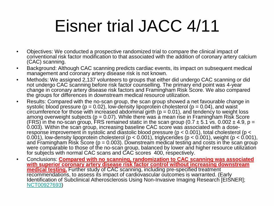
Eisner trial JACC 4/11 • Objectives: We conducted a prospective randomized trial to compare the clinical impact of
conventional risk factor modification to that associated with the addition of coronary artery calcium (CAC) scanning.
• Background: Although CAC scanning predicts cardiac events, its impact on subsequent medical management and coronary artery disease risk is not known.
• Methods: We assigned 2,137 volunteers to groups that either did undergo CAC scanning or did not undergo CAC scanning before risk factor counselling. The primary end point was 4-year change in coronary artery disease risk factors and Framingham Risk Score. We also compared the groups for differences in downstream medical resource utilization.
• Results: Compared with the no-scan group, the scan group showed a net favourable change in systolic blood pressure (p = 0.02), low-density lipoprotein cholesterol (p = 0.04), and waist circumference for those with increased abdominal girth (p = 0.01), and tendency to weight loss among overweight subjects (p = 0.07). While there was a mean rise in Framingham Risk Score (FRS) in the no-scan group, FRS remained static in the scan group (0.7 ± 5.1 vs. 0.002 ± 4.9, p = 0.003). Within the scan group, increasing baseline CAC score was associated with a dose-response improvement in systolic and diastolic blood pressure (p < 0.001), total cholesterol (p < 0.001), low-density lipoprotein cholesterol (p < 0.001), triglycerides (p < 0.001), weight (p < 0.001), and Framingham Risk Score (p = 0.003). Downstream medical testing and costs in the scan group were comparable to those of the no-scan group, balanced by lower and higher resource utilization for subjects with normal CAC scans and CAC scores 400, respectively.
• Conclusions: Compared with no scanning, randomization to CAC scanning was associated with superior coronary artery disease risk factor control without increasing downstream medical testing. Further study of CAC scanning, including pre-specified treatment recommendations, to assess its impact of cardiovascular outcomes is warranted. (Early Identification of Subclinical Atherosclerosis Using Non-Invasive Imaging Research [EISNER]; NCT00927693)

MSCT
Multi-slice CT coronary angiography 64 slice 128 slice Dual source 256 slice 320 slice
Use prospective gating – keep dose down!!!

Summary of PPV/NPV of MSCT status… Hachamovitch and Di Carli JNC 10/07
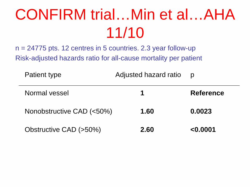
CONFIRM trial…Min et al…AHA 11/10
n = 24775 pts. 12 centres in 5 countries. 2.3 year follow-up Risk-adjusted hazards ratio for all-cause mortality per patient
Patient type Adjusted hazard ratio p
Normal vessel 1 Reference
Nonobstructive CAD (<50%) 1.60 0.0023
Obstructive CAD (>50%) 2.60 <0.0001
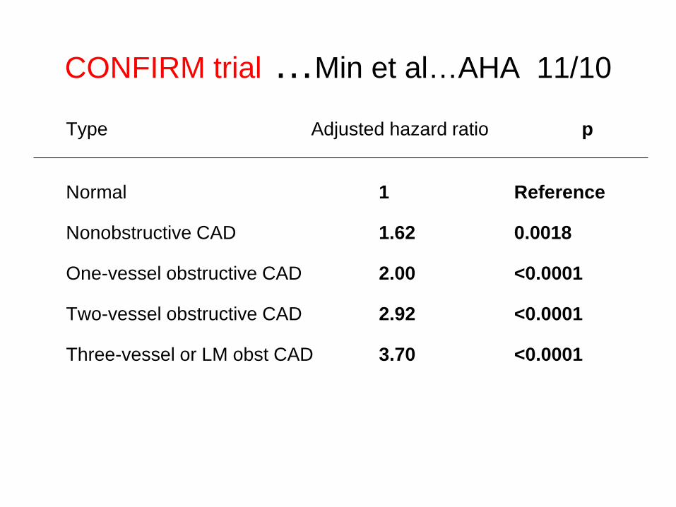
CONFIRM trial …Min et al…AHA 11/10
Type Adjusted hazard ratio p
Normal 1 Reference
Nonobstructive CAD 1.62 0.0018
One-vessel obstructive CAD 2.00 <0.0001
Two-vessel obstructive CAD 2.92 <0.0001
Three-vessel or LM obst CAD 3.70 <0.0001

Review of CTCA v. MPI in U.S. – 300,000 medicare pts Shreibati et al…. JAMA 11/11
Significant findings included the following: 1. Compared to patients who underwent MPS, those who underwent CCTA were
nearly twice as likely to undergo subsequent cardiac catheterization (22.9% versus 12.1% for MPS).
2. CCTA patients were more than twice as likely to undergo coronary artery bypass graft surgery (3.71%) compared to MPS patients (1.29%).
3. CCTA patients remained a little healthier over time, with a slightly lower likelihood of hospitalization for acute heart attack (0.19%) in the first 180 days after their first test than patients undergoing MPS (0.43%). But patients undergoing CCTA had a similar likelihood of all-cause mortality (1.05%) compared to patients whose testing began with MPS (1.28%).
4. As for costs, both average total spending ($29,719) and spending related to coronary artery disease ($14,943) over the 180-day follow-up period were significantly higher among patients undergoing CCTA, who had nearly 50% higher CAD-related average expenditures than patients undergoing MPS.
5. However, CCTA patients had lower associated spending with echocardiography (-$4,981) and exercise ECG (-$7,449) versus MPS patients.
6. Overall spending related to coronary artery disease was $11,437 for CCTA versus $7,430 for MPS. Average total spending was also significantly lower for patients undergoing MPS ($29,719 for CCTA versus $14,943 for MPS).
Conclusion – costs escalate if you do inappropriate pts


Fused images…. Rispler et al JACC 3/07
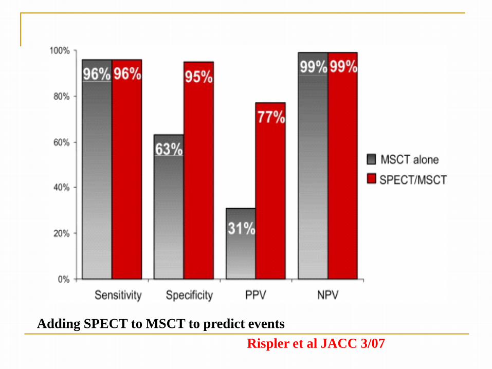
Rispler et al JACC 3/07 Adding SPECT to MSCT to predict events
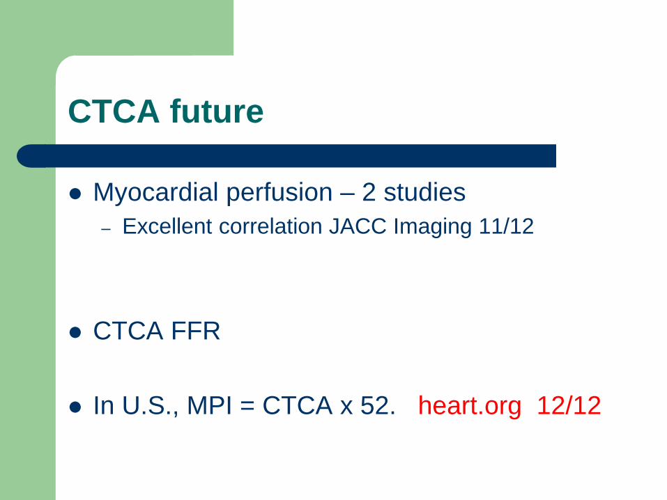
CTCA future
Myocardial perfusion – 2 studies – Excellent correlation JACC Imaging 11/12
CTCA FFR
In U.S., MPI = CTCA x 52. heart.org 12/12
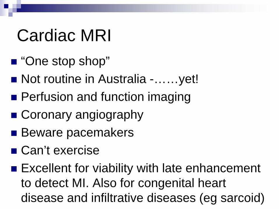
Cardiac MRI “One stop shop” Not routine in Australia -……yet! Perfusion and function imaging Coronary angiography Beware pacemakers Can’t exercise Excellent for viability with late enhancement
to detect MI. Also for congenital heart disease and infiltrative diseases (eg sarcoid)

Tissue Characterisation
Late Gadolinium Enhancement (LGE)
• 10 minutes after gadolinium • White - myocardial necrosis or scar • Ischaemic and non-ischaemic conditions
– Differentiated by pattern of LGE
Myocardial Infarction
Subendocardial involvement
Myocarditis
Subendocardial sparing

LGE Viability Imaging
LV function recovered Developed LV aneurysm
Subendocardial scar only - viable
Transmural scar Non-viable

Stress Perfusion MRI
• Pharmacological stress • Adenosine/dipyridamole or
dobutamine • In magnet during stress • Limited patient monitoring
• Robust technique • Technical expertise
essential
• Qualitative or quantitative assessment

Stress Perfusion MRI vs SPECT • CE-MARC Study
– 752 patients, cath as gold standard
– Comprehensive MRI including LGE
– MRI more accurate than SPECT • SPECT sensitivity 66.5 % • MRI sensitivity 86.5 %
• MR IMPACT I and II 2-3
– Similar overall accuracy – SPECT sensitivity poor - 59% 3
1. CE-MARC. Lancet 2011 2. MR IMPACT I EHJ 2008 3. MR IMPACT II EHJ 2012

After STICH is Viability Imaging Dead?
• Viability did not predict mortality benefit from CAGS
• Nuclear, stress echo (no MRI)
• Not randomized based on viability • Viability may have affected enrollment • Await LV function data
• However, is there enough other evidence
to deny surgery based on lack of viability?
Bonow. NEJM 2011
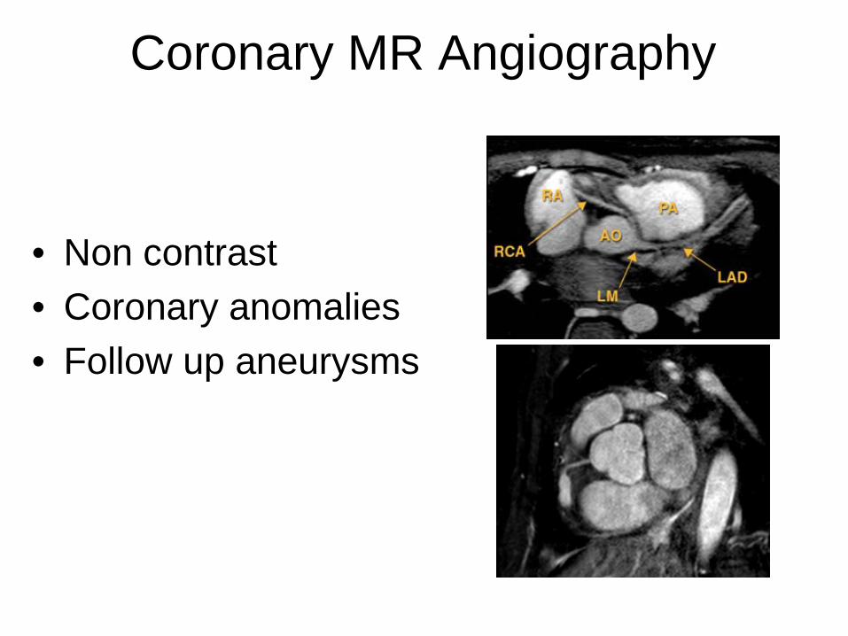
Coronary MR Angiography
• Non contrast • Coronary anomalies • Follow up aneurysms
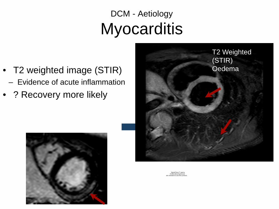
DCM - Aetiology
Myocarditis
• T2 weighted image (STIR) – Evidence of acute inflammation
• ? Recovery more likely
QuickTime™ and aH.264 decompressor
are needed to see this picture.
LGE
T2 Weighted (STIR) Oedema
Hypertrophic Cardiomyopathy (HCM)
• Prevalence 1:500
• Most common cause of sudden death in young adults
• Autosomal dominant – Sarcomeric proteins
• Diagnosis based largely on imaging
• Hypertrophy in absence of other underlying case

CMR Diagnosis of HCM Hypertrophy Missed on Echocardiography
• Regions frequently missed on echo: – Apical – Anterolateral – Inferoseptal
• Comprises 12% of all HCM patients Maron JACC 2009

Arrhythmogenic Right Ventricular Dysplasia (ARVD)
• RV cardiomyopathy • Arrhythmias, sudden death • Autosomal dominant
• New criteria place greater emphasis on volumes and
RV ejection fraction
• Fat in RV free wall no longer part of criteria – Also good for SARCOID!!!
Marcus. Circulation 2010


Putting it all together….

CT for equivocal MPI …Abidov et al JNC 12/10
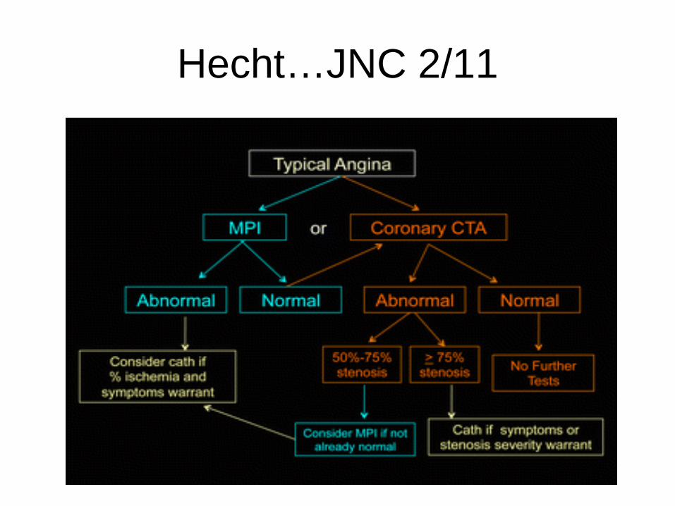
Hecht…JNC 2/11
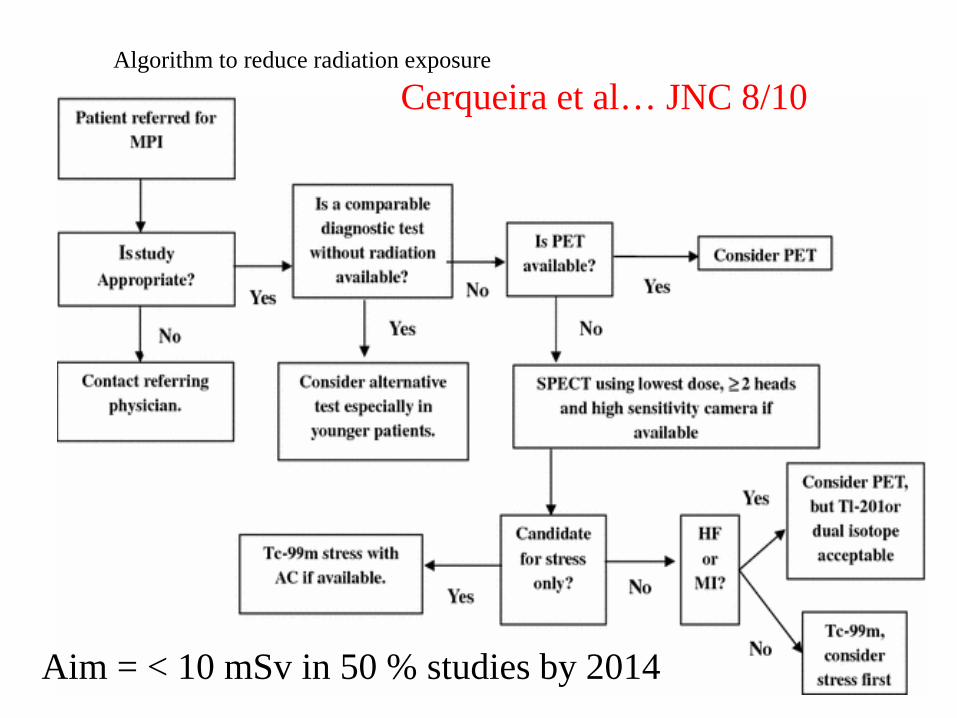
Algorithm to reduce radiation exposure
Cerqueira et al… JNC 8/10
Aim = < 10 mSv in 50 % studies by 2014
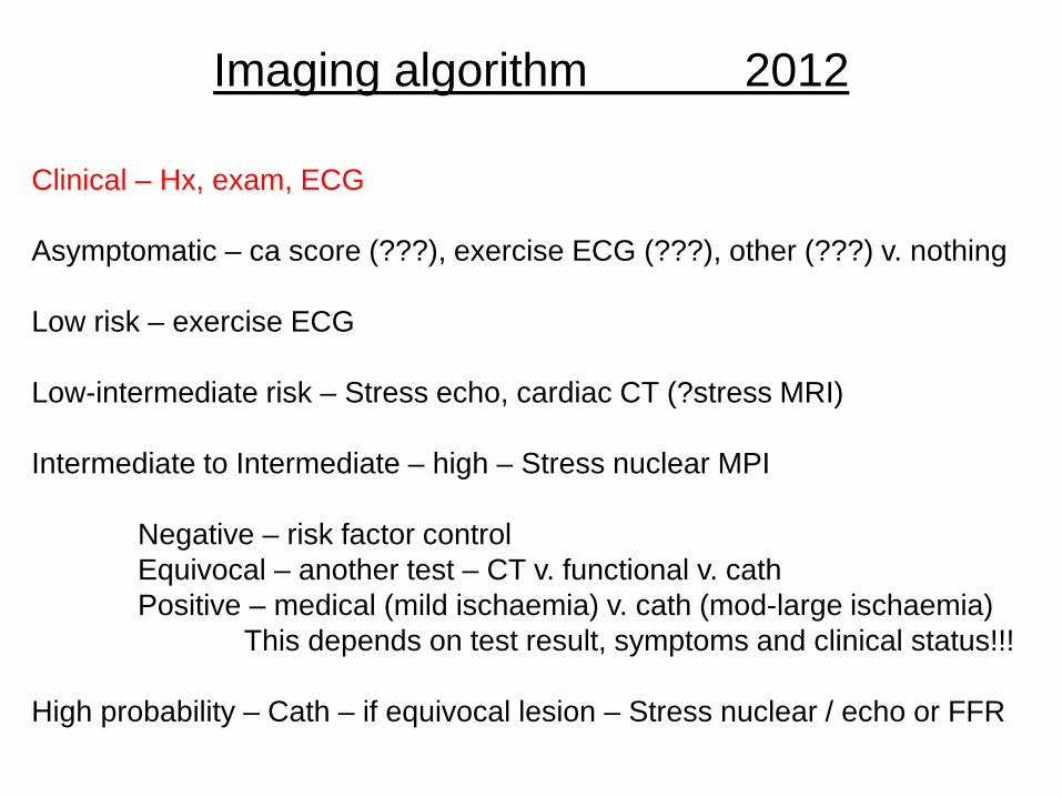
Imaging algorithm 2012
Clinical – Hx, exam, ECG Asymptomatic – ca score (???), exercise ECG (???), other (???) v. nothing Low risk – exercise ECG Low-intermediate risk – Stress echo, cardiac CT (?stress MRI) Intermediate to Intermediate – high – Stress nuclear MPI Negative – risk factor control Equivocal – another test – CT v. functional v. cath Positive – medical (mild ischaemia) v. cath (mod-large ischaemia) This depends on test result, symptoms and clinical status!!! High probability – Cath – if equivocal lesion – Stress nuclear / echo or FFR

CONCLUSION
In 2012, we have choices with variety in availability, cost, radiation, ability to exercise, sensitivity v. specificity and prognostic power.
9/05 – Time Magazine – “Use the test that is
done best in your institution” P. Douglas ACC president.


![Myocardial Ischaemia - national audit project [MINAP] 2011 - UCL](https://img.pdfslide.net/doc/110x75/620349a224f6b61e9c664083/myocardial-ischaemia-national-audit-project-minap-2011-ucl.jpg)