Embed Size (px)
Citation preview
Direct Stimulation of Adult Neural Stem/Progenitor CellsIn Vitro and Neurogenesis In Vivo by Salvianolic Acid BPengwei Zhuang1, Yanjun Zhang1*, Guangzhi Cui1, Yuhong Bian2, Mixia Zhang1, Jinbao Zhang1,
Yang Liu1, Xinpeng Yang1, Adejobi Oluwaniyi Isaiah1, Yingxue Lin1, Yongbo Jiang1
1 Tianjin State Key Laboratory of Modern Chinese Medicine, Key Laboratory of Traditional Chinese Medicine Pharmacology, Chinese Materia Medica College, Tianjin
University of Traditional Chinese Medicine, Tianjin, China, 2 Chinese Medical College, Tianjin University of Traditional Chinese Medicine, Tianjin, China
Abstract
Background: Small molecules have been shown to modulate the neurogenesis processes. In search for new therapeuticdrugs, the herbs used in traditional medicines for neurogenesis are promising candidates.
Methodology and Principal Findings: We selected a total of 45 natural compounds from Traditional Chinese herbalmedicines which are extensively used in China to treat stroke clinically, and tested their proliferation-inducing activities onneural stem/progenitor cells (NSPCs). The screening results showed that salvianolic acid B (Sal B) displayed marked effectson the induction of proliferation of NSPCs. We further demonstrated that Sal B promoted NSPCs proliferation in dose- andtime-dependent manners. To explore the molecular mechanism, PI3K/Akt, MEK/ERK and Notch signaling pathways wereinvestigated. Cell proliferation assay demonstrated that Ly294002 (PI3K/Akt inhibitor), but neither U0126 (ERK inhibitor) norDAPT (Notch inhibitor) inhibited the Sal B-induced proliferation of cells. Western Blotting results showed that stimulation ofNSPCs with Sal B enhanced the phosphorylation of Akt, and Ly294002 abolished this effect, confirming the role of Akt in SalB mediated proliferation of NSPCs. Rats exposed to transient cerebral ischemia were treated for 4 weeks with Sal B from the7th day after stroke. BrdU incorporation assay results showed that exposure Sal B could maintain the proliferation of NSPCsafter cerebral ischemia. Morris water maze test showed that delayed post-ischemic treatment with Sal B improved cognitiveimpairment after stroke in rats.
Significance: Sal B could maintain the NSPCs self-renew and promote proliferation, which was mediated by PI3K/Akt signalpathway. And delayed post-ischemic treatment with Sal B improved cognitive impairment after stroke in rats. Thesefindings suggested that Sal B may act as a potential drug in treatment of brain injury or neurodegenerative diseases.
Citation: Zhuang P, Zhang Y, Cui G, Bian Y, Zhang M, et al. (2012) Direct Stimulation of Adult Neural Stem/Progenitor Cells In Vitro and Neurogenesis In Vivo bySalvianolic Acid B. PLoS ONE 7(4): e35636. doi:10.1371/journal.pone.0035636
Editor: Josef Priller, Charite-Universitatsmedizin Berlin, Germany
Received December 2, 2011; Accepted March 19, 2012; Published April 24, 2012
Copyright: � 2012 Zhuang et al. This is an open-access article distributed under the terms of the Creative Commons Attribution License, which permitsunrestricted use, distribution, and reproduction in any medium, provided the original author and source are credited.
Funding: This work was supported by the National Natural Science Foundation of China (nos. 30873395 and 30472177), National Basic Research Program ofChina (no. 2011CB505302), National Science & Technology Major Project ‘‘Key New Drug Creation and Manufacturing Program’’ (2012ZX09101202) and theProgram for Changjiang Scholars and Innovative Research Team in University (no. IRT0973). The funders had no role in study design, data collection and analysis,decision to publish, or preparation of the manuscript.
Competing Interests: The authors have declared that no competing interest exist.
* E-mail: [email protected]
Introduction
Ischemic brain damage is one of the most dangerous ailments
that lead to learning and memory disability, physical dysfunction
and even death. Up to now, no effective treatment has been
reported [1]. Neurons as terminally differentiated cells cannot
regenerate after injury in traditional view. However, appropriate
exercise training can facilitate some neurological function recovery
after stroke in clinical practice [2,3], with the evidence that
neurogenesis occurs in the adult brain. Neural stem/precursor
cells (NSPCs) had been found and confirmed in adult brain in past
decades that it can differentiate into neurons or glial cells as a
result of neurogenesis [4–7], NSPCs can be stimulated in several
pathological conditions, such as neurological diseases, cerebral
ischemic in adult brain, and many reports showed that they are an
excellent candidate for developing therapeutic strategies to repair
the injured CNS [8,9]. Although the NSPCs would be stimulated
to proliferation and differentiation during the brain injury, often
this response is not sufficient to overcome the damage. It is
essential to study the signalling mechanisms that are activated by
small molecular materials in the NSPCs to enhance their response
pharmacologically. NSPCs proliferation and neurogensis involves
a series of intracellular signaling pathways [10,11]. Among these
pathways, the activation of Notch, mitogen-activated protein
kinases (MAPKs) and phosphatidylinositol-3-kinase (PI3K)/Akt
pathways are known to play major roles in cell growth and survival
responses [12–14]. Numerous studies have shown that small
molecular materials such as growth factors [15], retinoic acid [16]
and Traditional Chinese Medicine (TCM) active constituent
[17,18] can regulate the biological characteristics of neural stem
cell and promote neurogenesis. Therefore, regulation of neuro-
genesis by NSPCs is anticipated as a noble therapeutic strategy for
brain damage.
Herbs have been used for treating diseases for centuries, and a
lot of natural compounds that with neural beneficial from
medicinal plants had been discovered [19]. Treatment of stroke
PLoS ONE | www.plosone.org 1 April 2012 | Volume 7 | Issue 4 | e35636
by TCM has a wealth of clinical experience and theoretical basis,
and a large number of effective clinical prescriptions have been
accumulated. In recent years a large number of studies have
shown that TCM prescription and its active ingredient can
improve cerebral ischemic injury in experimental animal [20,21].
Ginsenoside Rb1 and Rg1, for example, improved spatial learning
and increase hippocampal synaptophysin level in mice [22].
Curcumin had been demonstrated to stimulate developmental and
adult hippocampal neurogenesis, and a biological activity that may
enhance neural plasticity and repair [23]. A recent report has
shown that NeuroAid (MLC601 and MLC901), a Traditional
Chinese Medicine is used in China for patients after stroke,
reduced the increase in escape latency and in swim distance
induced by ischemia [24]. With an extensive clinical experience,
there are ample opportunities to discover natural compounds that
effectively promote the proliferation of NSPCs and neurogenesis
from TCM.
Sal B was discovered in an in vitro screening assay for searching
the NSPCs proliferation-inducing natural molecules. It is one of
the major ingredients in the water-soluble extracts of S. miltiorrhiza
Bunge which has been reported to reduce cerebrovascular disease
[25–27]. As a well-known Chinese herbal medicine, Danshen
(Salvia miltiorrhiza) has been widely used in traditional Chinese
medicinal preparation for the treatment of ischemic disease, and
the medicinal properties of this plant have been extensively studied
[28,29]. Several studies have demonstrated the effect of salvianolic
acids on preventing brain injury [27,30]. Importantly, Sal B was
reported to be capable of improving the recovery of motor
function after cerebral ischemia in rats [31,32]. However, research
on the mechanism of Sal B in treatment of stroke, and the
molecular mechanisms responsible for the reported beneficial
cerebrovascular effects of Sal B are fairly rare. Meanwhile, the
effect of delayed treatment with Sal B after ischemic stroke is still
unclear. Therefore, the effect of chronic Sal B treatment beginning
seven days after ischemic stroke on neurological deficits and
pathophysiology after the transient cerebral ischemic model in rats
were studied in our research.
Considering the significance of neurogenesis-related brain
recovery, the present study was undertaken to examine the
promotive effects of Sal B on NSPCs proliferation and
neurogenesis. We provide evidence that Sal B increasing the
proliferation of NSPCs in vitro and in vivo. These actions are at
least in part mediated by altering the PI3K/Akt signaling
pathway. Additionally, delayed post-ischemic administration of
Sal B had beneficial effects on the recovery of cognitive function.
The stimulative effects of Sal B on NSPCs self-renew and
neurogenesis might be associated with a favorable outcome for
stroke and other neurological disease.
Results
Salvianolic acid B induced the proliferation of culturedNSPCs in vitro
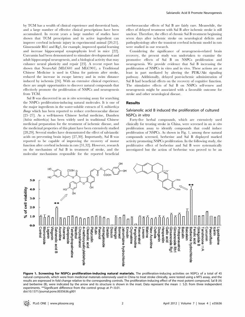
Forty-five herbal compounds, which are extensively used
clinically for treating stroke in China, were screened in an in vitro
proliferation assay to identify compounds that could induce
proliferation of NSPCs. As shown in Fig. 1, among these natural
compounds screened, berberine and Sal B displayed marked
activity promoting NSPCs proliferation. In the following study, the
proliferative effect of berberine and Sal B were systematically
investigated but the action of berberine was proved to be an
Figure 1. Screening for NSPCs proliferation-inducing natural materials. The proliferation-inducing activities on NSPCs of a total of 45natural compounds, which were from medicinal materials extensively used in China to treat stroke clinically, were tested using a MTS assay, and theresults are expressed in fold change relative to the corresponding controls. The proliferation-inducing effect of the most potent compound, Sal B (A)and berberine (B), were indicated by the arrow and its structure is shown in the inset. Data represent the mean 6 S.D. from three independentexperiments. **Significant difference from the control group at P,0.01.doi:10.1371/journal.pone.0035636.g001
Salvianolic Acid B Promote Neurogenesis
PLoS ONE | www.plosone.org 2 April 2012 | Volume 7 | Issue 4 | e35636
illusion by BrdU incorporation assay (See Figure S1 in the
Supporting Information).
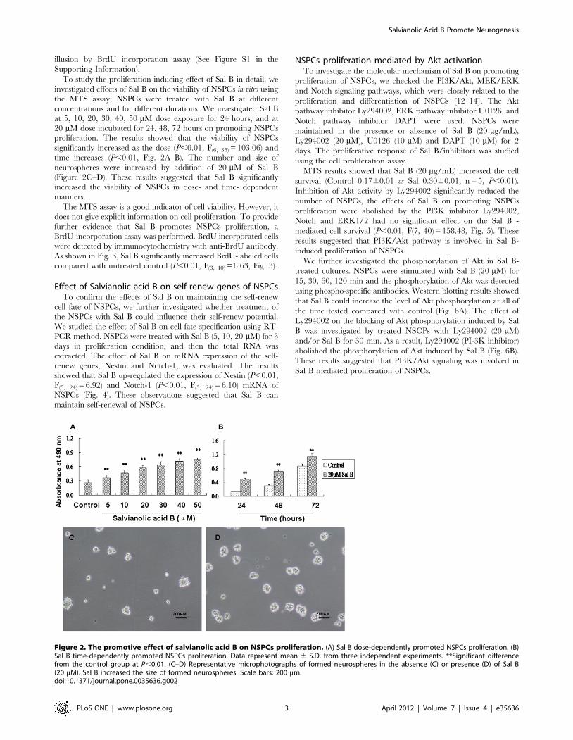
To study the proliferation-inducing effect of Sal B in detail, we
investigated effects of Sal B on the viability of NSPCs in vitro using
the MTS assay, NSPCs were treated with Sal B at different
concentrations and for different durations. We investigated Sal B
at 5, 10, 20, 30, 40, 50 mM dose exposure for 24 hours, and at
20 mM dose incubated for 24, 48, 72 hours on promoting NSPCs
proliferation. The results showed that the viability of NSPCs
significantly increased as the dose (P,0.01, F(6, 35) = 103.06) and
time increases (P,0.01, Fig. 2A–B). The number and size of
neurospheres were increased by addition of 20 mM of Sal B
(Figure 2C–D). These results suggested that Sal B significantly
increased the viability of NSPCs in dose- and time- dependent
manners.
The MTS assay is a good indicator of cell viability. However, it
does not give explicit information on cell proliferation. To provide
further evidence that Sal B promotes NSPCs proliferation, a
BrdU-incorporation assay was performed. BrdU incorporated cells
were detected by immunocytochemistry with anti-BrdU antibody.
As shown in Fig. 3, Sal B significantly increased BrdU-labeled cells
compared with untreated control (P,0.01, F(3, 40) = 6.63, Fig. 3).
Effect of Salvianolic acid B on self-renew genes of NSPCsTo confirm the effects of Sal B on maintaining the self-renew
cell fate of NSPCs, we further investigated whether treatment of
the NSPCs with Sal B could influence their self-renew potential.
We studied the effect of Sal B on cell fate specification using RT-
PCR method. NSPCs were treated with Sal B (5, 10, 20 mM) for 3
days in proliferation condition, and then the total RNA was
extracted. The effect of Sal B on mRNA expression of the self-
renew genes, Nestin and Notch-1, was evaluated. The results
showed that Sal B up-regulated the expression of Nestin (P,0.01,
F(5, 24) = 6.92) and Notch-1 (P,0.01, F(5, 24) = 6.10) mRNA of
NSPCs (Fig. 4). These observations suggested that Sal B can
maintain self-renewal of NSPCs.
NSPCs proliferation mediated by Akt activationTo investigate the molecular mechanism of Sal B on promoting
proliferation of NSPCs, we checked the PI3K/Akt, MEK/ERK
and Notch signaling pathways, which were closely related to the
proliferation and differentiation of NSPCs [12–14]. The Akt
pathway inhibitor Ly294002, ERK pathway inhibitor U0126, and
Notch pathway inhibitor DAPT were used. NSPCs were
maintained in the presence or absence of Sal B (20 mg/mL),
Ly294002 (20 mM), U0126 (10 mM) and DAPT (10 mM) for 2
days. The proliferative response of Sal B/inhibitors was studied
using the cell proliferation assay.
MTS results showed that Sal B (20 mg/mL) increased the cell
survival (Control 0.1760.01 vs Sal 0.3060.01, n = 5, P,0.01).
Inhibition of Akt activity by Ly294002 significantly reduced the
number of NSPCs, the effects of Sal B on promoting NSPCs
proliferation were abolished by the PI3K inhibitor Ly294002,
Notch and ERK1/2 had no significant effect on the Sal B -
mediated cell survival (P,0.01, F(7, 40) = 158.48, Fig. 5). These
results suggested that PI3K/Akt pathway is involved in Sal B-
induced proliferation of NSPCs.
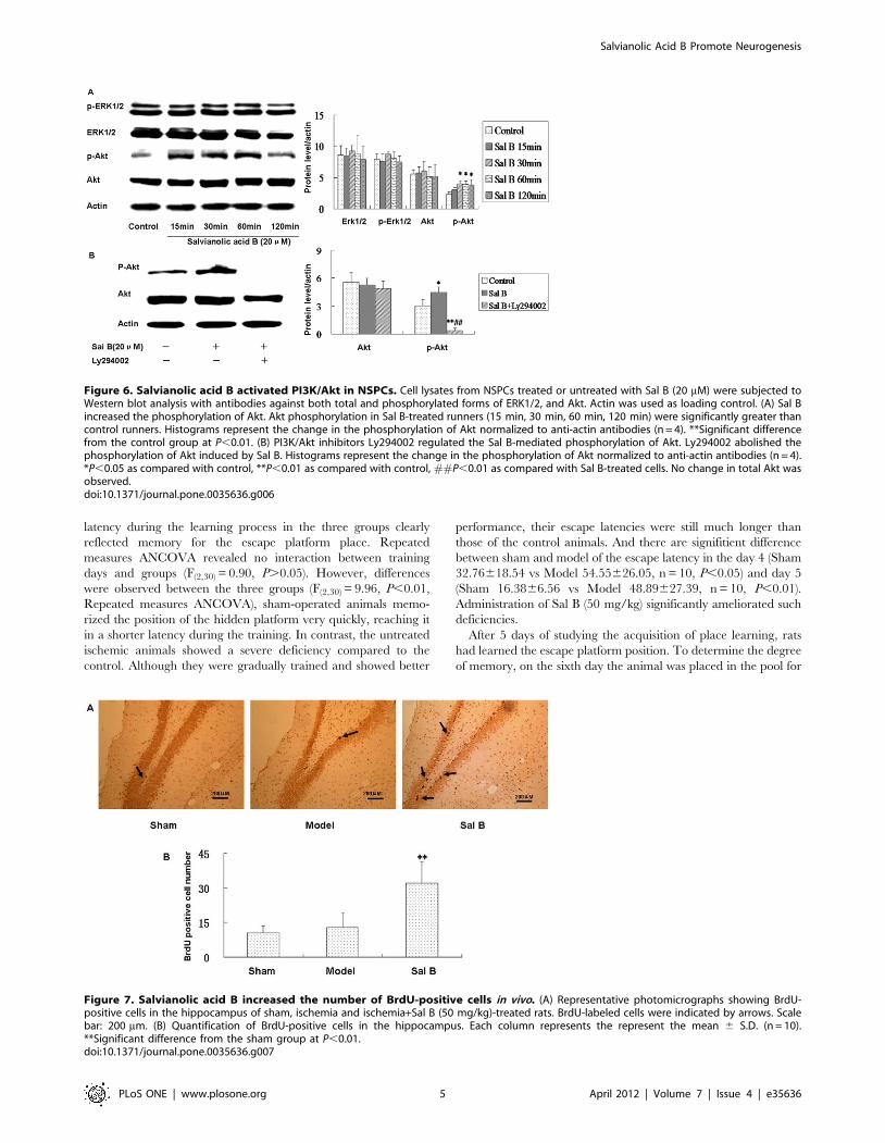
We further investigated the phosphorylation of Akt in Sal B-
treated cultures. NSPCs were stimulated with Sal B (20 mM) for
15, 30, 60, 120 min and the phosphorylation of Akt was detected
using phospho-specific antibodies. Western blotting results showed
that Sal B could increase the level of Akt phosphorylation at all of
the time tested compared with control (Fig. 6A). The effect of
Ly294002 on the blocking of Akt phosphorylation induced by Sal
B was investigated by treated NSCPs with Ly294002 (20 mM)
and/or Sal B for 30 min. As a result, Ly294002 (PI-3K inhibitor)
abolished the phosphorylation of Akt induced by Sal B (Fig. 6B).
These results suggested that PI3K/Akt signaling was involved in
Sal B mediated proliferation of NSPCs.
Figure 2. The promotive effect of salvianolic acid B on NSPCs proliferation. (A) Sal B dose-dependently promoted NSPCs proliferation. (B)Sal B time-dependently promoted NSPCs proliferation. Data represent mean 6 S.D. from three independent experiments. **Significant differencefrom the control group at P,0.01. (C–D) Representative microphotographs of formed neurospheres in the absence (C) or presence (D) of Sal B(20 mM). Sal B increased the size of formed neurospheres. Scale bars: 200 mm.doi:10.1371/journal.pone.0035636.g002
Salvianolic Acid B Promote Neurogenesis
PLoS ONE | www.plosone.org 3 April 2012 | Volume 7 | Issue 4 | e35636
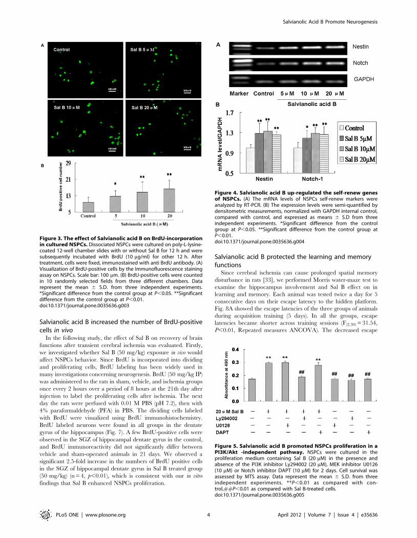
Salvianolic acid B increased the number of BrdU-positivecells in vivo
In the following study, the effect of Sal B on recovery of brain
functions after transient cerebral ischemia was evaluated. Firstly,
we investigated whether Sal B (50 mg/kg) exposure in vivo would
affect NSPCs behavior. Since BrdU is incorporated into dividing
and proliferating cells, BrdU labeling has been widely used in
many investigations concerning neurogenesis. BrdU (50 mg/kg IP)
was administered to the rats in sham, vehicle, and ischemia groups
once every 2 hours over a period of 8 hours at the 21th day after
injection to label the proliferating cells after ischemia. The next
day the rats were perfused with 0.01 M PBS (pH 7.2), then with
4% paraformaldehyde (PFA) in PBS. The dividing cells labeled
with BrdU were visualized using BrdU immunohistochemistry.
BrdU labeled neurons were found in all groups in the dentate
gyrus of the hippocampus (Fig. 7). A few BrdU-positive cells were
observed in the SGZ of hippocampal dentate gyrus in the control,
and BrdU immunoreactivity did not significantly differ between
vehicle and sham-operated animals in 21 days. We observed a
significant 2.5-fold increase in the numbers of BrdU positive cells
in the SGZ of hippocampal dentate gyrus in Sal B treated group
(50 mg/kg) (n = 4, p,0.01), which is consistent with our in vitro
findings that Sal B enhanced NSPCs proliferation.
Salvianolic acid B protected the learning and memoryfunctions
Since cerebral ischemia can cause prolonged spatial memory
disturbance in rats [33], we performed Morris water-maze test to
examine the hippocampus involvement and Sal B effect on in
learning and memory. Each animal was tested twice a day for 5
consecutive days on their escape latency to the hidden platform.
Fig. 8A showed the escape latencies of the three groups of animals
during acquisition training (5 days). In all the groups, escape
latencies became shorter across training sessions (F(2,30) = 31.54,
P,0.01, Repeated measures ANCOVA). The decreased escape
Figure 3. The effect of Salvianolic acid B on BrdU-incorporationin cultured NSPCs. Dissociated NSPCs were cultured on poly-L-lysine-coated 12-well chamber slides with or without Sal B for 12 h and weresubsequently incubated with BrdU (10 mg/ml) for other 12 h. Aftertreatment, cells were fixed, immunostained with anti BrdU antibody. (A)Visualization of BrdU-positive cells by the Immunofluorescence stainingassay on NSPCs. Scale bar: 100 mm. (B) BrdU-positive cells were countedin 10 randomly selected fields from three different chambers. Datarepresent the mean 6 S.D. from three independent experiments.*Significant difference from the control group at P,0.05. **Significantdifference from the control group at P,0.01.doi:10.1371/journal.pone.0035636.g003
Figure 4. Salvianolic acid B up-regulated the self-renew genesof NSPCs. (A) The mRNA levels of NSPCs self-renew markers wereanalyzed by RT-PCR. (B) The expression levels were semi-quantified bydensitometric measurements, normalized with GAPDH internal control,compared with control, and expressed as means 6 S.D from threeindependent experiments. *Significant difference from the controlgroup at P,0.05. **Significant difference from the control group atP,0.01.doi:10.1371/journal.pone.0035636.g004
Figure 5. Salvianolic acid B promoted NSPCs proliferation in aPI3K/Akt -independent pathway. NSPCs were cultured in theproliferation medium containing Sal B (20 mM) in the presence andabsence of the PI3K inhibitor Ly294002 (20 mM), MEK inhibitor U0126(10 mM) or Notch inhibitor DAPT (10 mM) for 2 days. Cell survival wasassessed by MTS assay. Data represent the mean 6 S.D. from threeindependent experiments. **P,0.01 as compared with con-trol,##P,0.01 as compared with Sal B-treated cells.doi:10.1371/journal.pone.0035636.g005
Salvianolic Acid B Promote Neurogenesis
PLoS ONE | www.plosone.org 4 April 2012 | Volume 7 | Issue 4 | e35636
latency during the learning process in the three groups clearly
reflected memory for the escape platform place. Repeated
measures ANCOVA revealed no interaction between training
days and groups (F(2,30) = 0.90, P.0.05). However, differences
were observed between the three groups (F(2,30) = 9.96, P,0.01,
Repeated measures ANCOVA), sham-operated animals memo-
rized the position of the hidden platform very quickly, reaching it
in a shorter latency during the training. In contrast, the untreated
ischemic animals showed a severe deficiency compared to the
control. Although they were gradually trained and showed better
performance, their escape latencies were still much longer than
those of the control animals. And there are signifitient difference
between sham and model of the escape latency in the day 4 (Sham
32.76618.54 vs Model 54.55626.05, n = 10, P,0.05) and day 5
(Sham 16.3866.56 vs Model 48.89627.39, n = 10, P,0.01).
Administration of Sal B (50 mg/kg) significantly ameliorated such
deficiencies.
After 5 days of studying the acquisition of place learning, rats
had learned the escape platform position. To determine the degree
of memory, on the sixth day the animal was placed in the pool for
Figure 6. Salvianolic acid B activated PI3K/Akt in NSPCs. Cell lysates from NSPCs treated or untreated with Sal B (20 mM) were subjected toWestern blot analysis with antibodies against both total and phosphorylated forms of ERK1/2, and Akt. Actin was used as loading control. (A) Sal Bincreased the phosphorylation of Akt. Akt phosphorylation in Sal B-treated runners (15 min, 30 min, 60 min, 120 min) were significantly greater thancontrol runners. Histograms represent the change in the phosphorylation of Akt normalized to anti-actin antibodies (n = 4). **Significant differencefrom the control group at P,0.01. (B) PI3K/Akt inhibitors Ly294002 regulated the Sal B-mediated phosphorylation of Akt. Ly294002 abolished thephosphorylation of Akt induced by Sal B. Histograms represent the change in the phosphorylation of Akt normalized to anti-actin antibodies (n = 4).*P,0.05 as compared with control, **P,0.01 as compared with control, ##P,0.01 as compared with Sal B-treated cells. No change in total Akt wasobserved.doi:10.1371/journal.pone.0035636.g006
Figure 7. Salvianolic acid B increased the number of BrdU-positive cells in vivo. (A) Representative photomicrographs showing BrdU-positive cells in the hippocampus of sham, ischemia and ischemia+Sal B (50 mg/kg)-treated rats. BrdU-labeled cells were indicated by arrows. Scalebar: 200 mm. (B) Quantification of BrdU-positive cells in the hippocampus. Each column represents the represent the mean 6 S.D. (n = 10).**Significant difference from the sham group at P,0.01.doi:10.1371/journal.pone.0035636.g007
Salvianolic Acid B Promote Neurogenesis
PLoS ONE | www.plosone.org 5 April 2012 | Volume 7 | Issue 4 | e35636
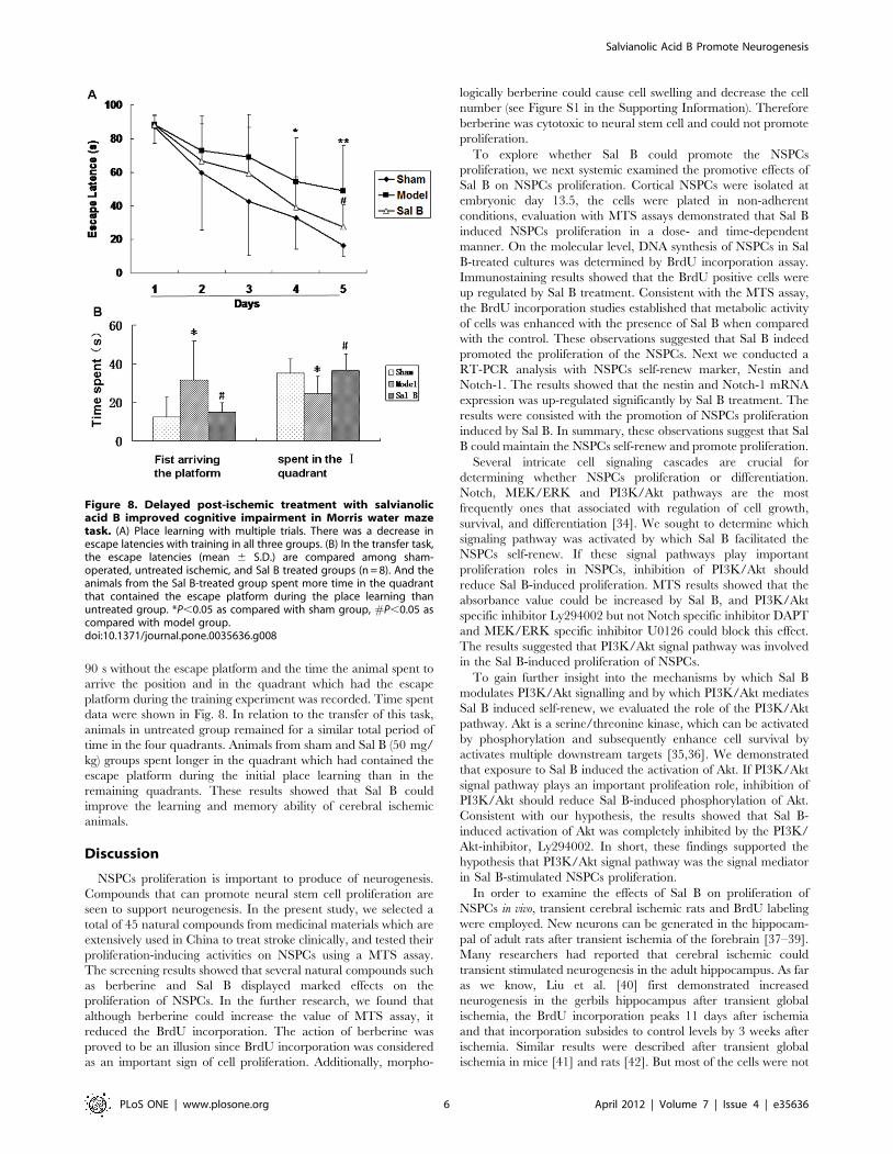
90 s without the escape platform and the time the animal spent to
arrive the position and in the quadrant which had the escape
platform during the training experiment was recorded. Time spent
data were shown in Fig. 8. In relation to the transfer of this task,
animals in untreated group remained for a similar total period of
time in the four quadrants. Animals from sham and Sal B (50 mg/
kg) groups spent longer in the quadrant which had contained the
escape platform during the initial place learning than in the
remaining quadrants. These results showed that Sal B could
improve the learning and memory ability of cerebral ischemic
animals.
Discussion
NSPCs proliferation is important to produce of neurogenesis.
Compounds that can promote neural stem cell proliferation are
seen to support neurogenesis. In the present study, we selected a
total of 45 natural compounds from medicinal materials which are
extensively used in China to treat stroke clinically, and tested their
proliferation-inducing activities on NSPCs using a MTS assay.
The screening results showed that several natural compounds such
as berberine and Sal B displayed marked effects on the
proliferation of NSPCs. In the further research, we found that
although berberine could increase the value of MTS assay, it
reduced the BrdU incorporation. The action of berberine was
proved to be an illusion since BrdU incorporation was considered
as an important sign of cell proliferation. Additionally, morpho-
logically berberine could cause cell swelling and decrease the cell
number (see Figure S1 in the Supporting Information). Therefore
berberine was cytotoxic to neural stem cell and could not promote
proliferation.
To explore whether Sal B could promote the NSPCs
proliferation, we next systemic examined the promotive effects of
Sal B on NSPCs proliferation. Cortical NSPCs were isolated at
embryonic day 13.5, the cells were plated in non-adherent
conditions, evaluation with MTS assays demonstrated that Sal B
induced NSPCs proliferation in a dose- and time-dependent
manner. On the molecular level, DNA synthesis of NSPCs in Sal
B-treated cultures was determined by BrdU incorporation assay.
Immunostaining results showed that the BrdU positive cells were
up regulated by Sal B treatment. Consistent with the MTS assay,
the BrdU incorporation studies established that metabolic activity
of cells was enhanced with the presence of Sal B when compared
with the control. These observations suggested that Sal B indeed
promoted the proliferation of the NSPCs. Next we conducted a
RT-PCR analysis with NSPCs self-renew marker, Nestin and
Notch-1. The results showed that the nestin and Notch-1 mRNA
expression was up-regulated significantly by Sal B treatment. The
results were consisted with the promotion of NSPCs proliferation
induced by Sal B. In summary, these observations suggest that Sal
B could maintain the NSPCs self-renew and promote proliferation.
Several intricate cell signaling cascades are crucial for
determining whether NSPCs proliferation or differentiation.
Notch, MEK/ERK and PI3K/Akt pathways are the most
frequently ones that associated with regulation of cell growth,
survival, and differentiation [34]. We sought to determine which
signaling pathway was activated by which Sal B facilitated the
NSPCs self-renew. If these signal pathways play important
proliferation roles in NSPCs, inhibition of PI3K/Akt should
reduce Sal B-induced proliferation. MTS results showed that the
absorbance value could be increased by Sal B, and PI3K/Akt
specific inhibitor Ly294002 but not Notch specific inhibitor DAPT
and MEK/ERK specific inhibitor U0126 could block this effect.
The results suggested that PI3K/Akt signal pathway was involved
in the Sal B-induced proliferation of NSPCs.
To gain further insight into the mechanisms by which Sal B
modulates PI3K/Akt signalling and by which PI3K/Akt mediates
Sal B induced self-renew, we evaluated the role of the PI3K/Akt
pathway. Akt is a serine/threonine kinase, which can be activated
by phosphorylation and subsequently enhance cell survival by
activates multiple downstream targets [35,36]. We demonstrated
that exposure to Sal B induced the activation of Akt. If PI3K/Akt
signal pathway plays an important prolifeation role, inhibition of
PI3K/Akt should reduce Sal B-induced phosphorylation of Akt.
Consistent with our hypothesis, the results showed that Sal B-
induced activation of Akt was completely inhibited by the PI3K/
Akt-inhibitor, Ly294002. In short, these findings supported the
hypothesis that PI3K/Akt signal pathway was the signal mediator
in Sal B-stimulated NSPCs proliferation.
In order to examine the effects of Sal B on proliferation of
NSPCs in vivo, transient cerebral ischemic rats and BrdU labeling
were employed. New neurons can be generated in the hippocam-
pal of adult rats after transient ischemia of the forebrain [37–39].
Many researchers had reported that cerebral ischemic could
transient stimulated neurogenesis in the adult hippocampus. As far
as we know, Liu et al. [40] first demonstrated increased
neurogenesis in the gerbils hippocampus after transient global
ischemia, the BrdU incorporation peaks 11 days after ischemia
and that incorporation subsides to control levels by 3 weeks after
ischemia. Similar results were described after transient global
ischemia in mice [41] and rats [42]. But most of the cells were not
Figure 8. Delayed post-ischemic treatment with salvianolicacid B improved cognitive impairment in Morris water mazetask. (A) Place learning with multiple trials. There was a decrease inescape latencies with training in all three groups. (B) In the transfer task,the escape latencies (mean 6 S.D.) are compared among sham-operated, untreated ischemic, and Sal B treated groups (n = 8). And theanimals from the Sal B-treated group spent more time in the quadrantthat contained the escape platform during the place learning thanuntreated group. *P,0.05 as compared with sham group, #P,0.05 ascompared with model group.doi:10.1371/journal.pone.0035636.g008
Salvianolic Acid B Promote Neurogenesis
PLoS ONE | www.plosone.org 6 April 2012 | Volume 7 | Issue 4 | e35636
fully mature during the 2- to 5-week period after ischemia and
could not form a functional link [42]. Therefore, in our view, the
behavior performance could not be better at that time. In
accordance with previous reports, the NSPCs responded to the
injury by proliferation in this study. Cerebral ischemic made the
NSPCs proliferation after 7 days (See Figure S2 in the Supporting
Information), but the proliferation ability of the rats in the vehicle
group declined quickly 28 days after cerebral ischemic. However,
Sal B (25 mg/kg) treatment significantly increased the number of
BrdU-positive cells in the dentate gyrus 21 days after injection,
suggested that Sal B exposure could maintain the ability of the
NSPCs proliferation in vivo.
Since Sal B promote NSPCs proliferation in vitro and in vivo, we
next sought to correlate the regeneration of new neurons and
recovery of brain functions. The present study demonstrated that
delayed post-ischemic treatment (7 days after ischemic stroke) with
Sal B (25 mg/kg) improved cognitive impairment after stroke in
rats. Since NSPCs proliferation in hippocampus was important site
for spatial learning and memory [43,44], we speculated that the
enhancement of functional recovery by Sal B might be dependent
on its action on NSPCs proliferation. But more extensive
experiments were still necessary to demonstrate that Sal B could
cross over the blood–brain barrier and act on NSPCs. Further
study will be carried out to clarify this point. Although our present
study showed that Sal B promoted the adult hippocampus
neurogenesis and improved the cognitive functions in cerebral
ischemia rats, there is no direct evidence to show that Sal B could
pass through the blood-brain barrier [45], and the exact
mechanism(s) by which Sal B acts on adult neurogenesis remain
unclear. More advanced research is needed in the future to further
clarify the pathways and mechanisms of Sal B in promoting adult
neurogenesis including using appropriate tool drugs to block
certain pathways to confirm NSPCs proliferation by Sal B on
contributing to the cognitive improvement.
In conclusion, the results of this work clearly demonstrated that
Sal B was capable of promoting proliferation of NSPCs and
improving the learning and memory ability of cerebral ischemic
rats. Additionally, we confirmed that Sal B promoteed NSPCs self-
renew and neurogensis were at least in part mediated by the
PI3K/Akt signaling pathway. These findings suggested that Sal B
may act as a potential drug in treatment brain injury or
neurodegenerative disease.
Materials and Methods
ReagentsSalvianolic acid B (Sal B, purity .99%) was purchased from the
Chinese National Institute for the Control of Pharmaceutical and
Biological Products (Beijing, China), When used, it was freshly
prepared in phosphate buffer solution (PBS); B27 (without retinoic
acid) was purchased from Invitrogen (Carlsbad, CA, USA);
Recombinant human fibroblast growth factor 2 (FGF-2) was from
Millipore (Temecula, CA); Antibodies for ERK1/2, phospho-
ERK1/2, Akt, phosphor-Akt (Ser-473) were obtained from Cell
Signaling Technology (Beverly, MA). Antibody for b-actin was
purchased from senta; 1,4-diamino-2,3-dicyano-1,4-bis(2-amino-
phenylthio)-butadiene (U0126) and 2-(4-morpholinyl)-8-phenyl-
4H-1-benzopyran-4-one (Ly294002) was from CalBiochem (San
Diego, CA). U0126 and Ly294002 were solubilized in dimethyl-
sulphoxide.
Cell culturePrimary neurospheres were isolated from the cerebral cortex of
13.5-day-embryonic Wistar rats, according to the method
described by Davis et al [46]. Briefly, the cerebral cortex was
carefully isolated from adjacent tissues, and collected in cold
serum-free medium consisting of DMEM/F-12 (1:1; Invitrogen,
Carlsbad, CA, USA). The tissue was digested at 37uC for 10 min
by acctuase (Millipore, Temecula, CA), and mechanically
disrupted into single cells by filtering through a nylon mesh of
70 mm. The dissociated cells were then plated at a concentration of
26105 cells/ml in T75 culture flasks for 20 ml, and cultured in
neurosphere proliferation media consisting of Dulbecco’s Modified
Eagle’s Medium (DMEM): F12 supplemented with 2% (v/v) B27
supplement (Invitrogen, Carlsbad, CA, USA), 20 ng/ml epidermal
growth factor (EGF; PEPROTECH, Rocky Hill, NJ, USA), and
20 ng/ml fibroblast growth factor 2 (FGF2). After 6 days in
culture, the proliferating cells formed the neurospheres, which
were suspended in the medium. Subsequently, the neurospheres
were passaged by treatment with accutase about 5 min at 37uCuntil they were gently dissociated, and then subcultured as single
cells in a new T75 culture flask at a density of 20,000 cells/ml in
the fresh culture medium. The procedure of subculture was
repeated again to achieve the purified cortical NSPCs and
proliferating neurospheres. The 3–5 passages of NSPCs were
used for the following experiments.
MTS assayFor in vitro cell proliferation assay, A Cell Titer 96 AQueous One
Solution Cell Proliferation Assay (Promega, Charbonnieres-les-
Bains, France) was used. NSPCs were plated at 30, 000 cells/well
in a 96-well plate in the presence and absence of Sal B (0.5, 1, 5,
10, 20 mM), U0126 (10 mg/ml), DAPT (10 mg/ml) and Ly294002
(20 mg/ml) in a 96-well plate. Cell proliferation was assessed at day
2 of cell culture. According to the manufacturer’s recommenda-
tions, 40 ml MTS solution was then added into each of the wells.
Then cells were incubated for 2 h at 37uC in the humidified 5%
CO2 atmosphere incubator, and results were obtained at a
wavelength of 490 nm using a microplate reader (FlexStation 3,
Molecular Devices, USA). The same volume of medium without
cells was used as blank. Results were expressed in Optical Density
(OD).
In vitro BrdU-incorporation assayFor in vitro BrdU-incorporation assay, cultured NSPCs were
incubated with or without Sal B for 24 h, the cells were labeled
with BrdU (10 mg/ml) during the last 12 h of incubation, then the
cells were plated onto poly-L-lysine coated slides for 2 h, fixed in
4% paraformaldehyde, washed with phosphate-buffered saline
(PBS) and incubated in 2 N HCl at 37uC for 10 min. After
washing with PBS, the cells were incubated with mouse anti-BrdU
antibody (1:100, senta) at 4uC for 24 h in PBS. After washing in
PBS, they were then incubated at room temperature for 1 h in
PBS containing FITC conjugated anti-mouse IgG secondary
antibody (1:200, senta). BrdU-positive cells were evaluated using a
fluorescent microscope (Leica, German). BrdU positive cells were
counted in 10 randomly selected fields from three different
chambers.
RT-PCR analysisCells were harvested and total RNA was isolated from treated or
untreated NSPCs with TRIzol (Roche), the first strand cDNA was
synthesized from 0.4 mg of total RNA using Reverse Transcriptase
(Takala) and Random primer as described in the manufacture’s
instructions. After synthesis, 5 ml of cDNA was used in PCR
reaction with gene-specific primers, The sequences of the PCR
primer pairs (59 to 39) that were used for each gene are as follows:
rat nestin, 59- TTCCCTTCCCCCTTGCCTAATACC-39 (for-
Salvianolic Acid B Promote Neurogenesis
PLoS ONE | www.plosone.org 7 April 2012 | Volume 7 | Issue 4 | e35636
ward) and 59- TGGGCTGAGCTGTTTTCTACTTTT-39 (re-
verse); Notch-1, 59- ATGGCCTCCAACGATACTCCT-39 (for-
ward) and 59- ACATGTACCCCCATAGTGGCA-39 (reverse).
Products were analyzed on 1.5% agarose gel and visualized by
ethidium bromide staining. The relative amount of each transcript
was normalized to the level of b-actin.
Western blot analysisProtein extracts were prepared and subjected to Western blot
analysis. Cells were harvested and lysed with RIPA lysis buffer.
The protein concentrations of the lysates were determined with a
Bradford protein assay kit (Bio-Rad) according to the manufac-
turer’s instructions. An equal amount of protein was fractionated
by SDS-polyacrylamide gel electrophoresis (PAGE) and trans-
ferred onto polyvinylidine difluoride membranes. After blocking
with 5% skim milk in TBS-T, the membrane was probed with
primary antibodies (goat anti-actin 1:1000, Santa Cruz; rabbit
anti-ERK1/2 1:500, rabbit anti-phospho-ERK1/2 1:300, rabbit
anti-Akt 1:500 or rabbit anti-phospho-Akt (Ser-473) 1:300, Cell
Signaling Technology) in a blocking solution of non-fat milk (5%).
Secondary horseradish peroxidase-conjugated rabbit anti-goat
1:2000, goat anti-rabbit 1:2000 (Santa Cruz) antibody in non-fat
milk blocking solution (5%) was then applied. The immuno-
reactivity was visualized with ECL Western blotting detection
reagents (Millipore).
Induction of the transient global ischemiaAdult (8–10 week-old) male Wistar rats weighing 200–250 g
were subjected to transient forebrain ischemia by a method
combining those described previously [47,48]. The rats were
housed singly in temperature-controlled conditions with a 12 hr
light/dark cycle (lights on: 8:00 AM) following surgery. They had
access to food and water ad libitum. Experimental procedures
were conducted in accordance with recommendations of the
Animal Ethics Committee of Tianjin University of Traditional
Chinese Medicine (TCM-2009-034-E01). In brief, twelve hours
before the induction of ischemia, rats were anesthetized with
sodium pentobarbital, the bilateral carotid arteries were exposed
to facilitate the occluding on the following day, and then the
vertebral arteries were irreversibly occluded by electrocoagulation.
The next day, ischemia was induced by bilaterally occluding
carotid arteries with aneurysm clips, and carotid arteries were
clamped for 6 min exactly. Rats lost their righting reflex during
ischemic, the clips were removed to restore cerebral blood flow.
The rats that remain their righting reflex in 1 min after occluding
of the both carotid arteries were considered to be failure of
ischemia and eliminated. Sham-operated rats were anesthetized,
the carotid arteries were isolated, but they were not clamped.
Drug adminstrationThe study was carried out on rats divided into three groups
(n = 13). (i) Sham group+vehicle (Sham), (ii) Ischemic group+-vehicle (Model) and (iii) Ischemic group+Sal B (Sal B). Seven days
after cerebral ischemia, Sal B (50 mg/kg) was dissolved in distilled
water, and injected i.p. once daily for 4 weeks, and three mice in
each group were used for histological analysis at day 21 after
innjection. All controls received an amount of vehicle equivalent to
drug treatment conditions.
In vivo proliferation assayFor in vivo proliferation analysis of Sal B, that had not been
behaviorally tested were evaluated for BrdU labeling. BrdU
(50 mg/kg; Sigma) was injected i.p. once every 2 hours over a
period of 8 hours at the 21th day after injection to label the
proliferating cells. Twenty-four hours after the last BrdU injection,
rars were anesthetized with ether and perfused with PBS, followed
by a cold 4% paraformaldehyde solution. Brains were collected
and post-fixed overnight in a 4% paraformaldehyde solution at
4uC. Coronal sections (20 mm thickness) were obtained throughout
the hippocampus. For analysis concerning BrdU immunohisto-
chemistry, sections were incubated in 50% formamide/26saline
sodium citrate (SSC) for 2 h at 65uC, followed by a rinse with PBS.
Sections were then incubated in 2 N HCl for 30 min at 37uC to
denature double-stranded DNA, and rinsed in 0.1 M borate buffer
(pH 8.5). After blocking for 2 h with 1% BSA in PBS, sections
were incubated overnight at 4uC with mouse anti-BrdU
monoclonal antibody (1:100; senta). Followed by rinsing in PBS,
sections were incubated for 2 h at RT with biotinylated goat anti-
mouse IgG (1:200; senta), and incubated for 2 h at RT with the
ABC kit. BrdU positive cells were visualized by incubating sections
with Vector DAB.
Morris water maze taskThe learning and memory ability was examined using the
Morris water-maze [49]. A cylindrical tank 1.5 m in diameter was
filled with water (2261uC), and a transparent platform 10 cm in
diameter was placed at a constant position in the center of one of
the four quadrants within the tank. The platform was set 2 cm
below the water level where the rats could not see it directly. Rats
were allowed to swim freely for 1 min to become habituated to the
apparatus. From the next day, in the hidden platform trials,
acquisition trials were carried out 2 times per day for 5 days. In
each trial, rats were placed into the water at a fixed starting
position, and the time taken to escape onto the hidden platform
and the swimming path length were measured. Rats were given
90 s to find the hidden platform during each acquisition trial. If it
failed to locate the platform within 90 s, it was guided there. The
rat was allowed to stay on the platform for 20 s. Performance was
tested 24 h after the final training day in a probe trial during
which the platform was removed, the rat was placed in the start
and its behavior was monitored for 90 s.
Statistical analysisSPSS11.5 for Windows (SPSS Inc.) was used to analyze the
data. Data are expressed as the mean 6 S.D. and analyzed by
ANOVA followed by the post-hoc Least Significant Difference
(LSD) test. Differences were considered statistically significant if P
values were less than 0.05. To analyze water-maze place-
navigation performance, the average escape latency of 5 trials
per day per animal was calculated and evaluated by repeated-
measures ANCOVA.
Supporting Information
Figure S1 Berberine failed to promote the NSPCsproliferation. (A) Berberine increased the value of MTT assay.
(B) Berberine reduced the BrdU incorporation. **Significant
difference from the control group at P,0.01. (C) Morphologically
berberine caused cell swelling and decreased the cell number.
Scale bar: 200 mm.
(TIF)
Figure S2 Regeneration of Hippocampal NSPCs Follow-ing Ischemia. BrdU positive cells in the granule cell layer in the
intact (A) and ischemic (B, DAI7) animals. The inset shows an
enlarged display of a typical BrdU positive cell. Scale bar: 200 mm.
(TIF)
Salvianolic Acid B Promote Neurogenesis
PLoS ONE | www.plosone.org 8 April 2012 | Volume 7 | Issue 4 | e35636
Acknowledgments
We would like to thank Dr. Hui Wang and Prof. Yan Zhu for their helpful
discussion and critical reading of our manuscript. We would also like to
thank Mr. Huaien Bu for technical assistant of the Statistical analysis.
Author Contributions
Conceived and designed the experiments: YJZ PWZ. Performed the
experiments: PWZ GZC MXZ XPY YL JBZ AOI. Analyzed the data: YJZ
PWZ YHB. Contributed reagents/materials/analysis tools: JBZ YXL YBJ.
Wrote the paper: YJZ PWZ AOI.
References
1. Doyle KP, Simon RP, Stenzel-Poore MP (2008) Mechanisms of ischemic brain
damage. Neuropharmacology 55: 310–318.
2. Duncan P, Studenski S, Richards L, Gollub S, Lai SM, et al. (2003) Randomized
clinical trial of therapeutic exercise in subacute stroke. Stroke 34: 2173–2180.
3. Kwakkel G, van Peppen R, Wagenaar RC, Wood Dauphinee S, Richards C, et
al. (2004) Effects of augmented exercise therapy time after stroke: a meta-
analysis. Stroke 35: 2529–2539.
4. Reynolds BA, Weiss S (1992) Generation of neurons and astrocytes from isolated
cells of the adult mammalian central nervous system. Science 255: 1707–1710.
5. Weiss S, Reynolds BA, Vescovi AL, Morshead C, Craig CG, et al. (1996) Is
there a neural stem cell in the mammalian forebrain? Trends Neurosci 19:
387–393.
6. Reynolds BA, Weiss S (1996) Clonal and population analyses demonstrate that
an EGF-responsive mammalian embryonic CNS precursor is a stem cell. Dev
Biol 175: 1–13.
7. McKay R (1997) Stem cells in the central nervous system. Science 276: 66–71.
8. Ming GL, Song H (2011) Adult neurogenesis in the mammalian brain:
significant answers and significant questions. Neuron 70: 687–702.
9. Lie DC, Song H, Colamarino SA, Ming GL, Gage FH (2004) Neurogenesis in
the adult brain: new strategies for central nervous system diseases. Annu Rev
Pharmacol Toxicol 44: 399–421.
10. Johnson MA, Ables JL, Eisch AJ (2009) Cell-intrinsic signals that regulate adult
neurogenesis in vivo: insights from inducible approaches. BMB Rep 42:
245–259.
11. Khodosevich K, Monyer H (2010) Signaling involved in neurite outgrowth of
postnatally born subventricular zone neurons in vitro. BMC Neurosci 11: 18.
12. Imayoshi I, Sakamoto M, Yamaguchi M, Mori K, Kageyama R (2010) Essential
roles of Notch signaling in maintenance of neural stem cells in developing and
adult brains. J Neurosci 30: 3489–3498.
13. Choi YS, Cho HY, Hoyt KR, Naegele JR, Obrietan K (2008) IGF-1 receptor-
mediated ERK/MAPK signaling couples status epilepticus to progenitor cell
proliferation in the subgranular layer of the dentate gyrus. Glia 56: 791–800.
14. Bruel-Jungerman E, Veyrac A, Dufour F, Horwood J, Laroche S, et al. (2009)
Inhibition of PI3K-Akt signaling blocks exercise-mediated enhancement of adult
neurogenesis and synaptic plasticity in the dentate gyrus. PLoS One 4: e7901.
15. Leker RR, Lasri V, Chernoguz D (2009) Growth factors improve neurogenesis
and outcome after focal cerebral ischemia. J Neural Transm 116: 1397–1402.
16. Jacobs S, Lie DC, DeCicco KL, Shi Y, DeLuca LM, et al. (2006) Retinoic acid is
required early during adult neurogenesis in the dentate gyrus. Proc Natl Acad
Sci U S A 103: 3902–3907.
17. Yabe T, Hirahara H, Harada N, Ito N, Nagai T, et al. (2010) Ferulic acid
induces neural progenitor cell proliferation in vitro and in vivo. Neuroscience
165: 515–524.
18. Heurteaux C, Gandin C, Borsotto M, Widmann C, Brau F, et al. (2010)
Neuroprotective and neuroproliferative activities of NeuroAid (MLC601,
MLC901), a Chinese medicine, in vitro and in vivo. Neuropharmacology 58:
987–1001.
19. Kim H (2005) Neuroprotective herbs for stroke therapy in traditional eastern
medicine. Neurol Res 27: 287–301.
20. Feigin VL (2007) Herbal medicine in stroke: does it have a future? Stroke 38:
1734–1736.
21. Wu B, Liu M, Liu H, Li W, Tan S, et al. (2007) Meta-analysis of traditional
Chinese patent medicine for ischemic stroke. Stroke 38: 1973–1979.
22. Mook-Jung I, Hong HS, Boo JH, Lee KH, Yun SH, et al. (2001) Ginsenoside
Rb1 and Rg1 improve spatial learning and increase hippocampal synaptophysin
level in mice. J Neurosci Res 63: 509–515.
23. Kim SJ, Son TG, Park HR, Park M, Kim MS, et al. (2008) Curcumin stimulates
proliferation of embryonic neural progenitor cells and neurogenesis in the adult
hippocampus. J Biol Chem 283: 14497–14505.
24. Quintard H, Borsotto M, Veyssiere J, Gandin C, Labbal F, et al. (2011)
MLC901, a traditional Chinese medicine protects the brain against global
ischemia. Neuropharmacology 61: 622–631.
25. Kim DH, Park SJ, Kim JM, Jeon SJ, Kim DH, et al. (2011) Cognitive
dysfunctions induced by a cholinergic blockade and Ab(25–35) peptide are
attenuated by salvianolic acid B. Neuropharmacology 61: 1432–1440.
26. Tang MK, Ren DC, Zhang JT, Du GH (2002) Effect of salvianolic acids from
Radix Salviae miltiorrhizae on regional cerebral blood flow and plateletaggregation in rats. Phytomedicine 9: 405–409.
27. Chen T, Liu W, Chao X, Zhang L, Qu Y, et al. (2011) Salvianolic acid B
attenuates brain damage and inflammation after traumatic brain injury in mice.Brain Res Bull 84: 163–168.
28. Zhou L, Zuo Z, Chow MS (2005) Danshen: an overview of its chemistry,pharmacology, pharmacokinetics, and clinical use. J Clin Pharmacol 45:
1345–1359.29. Ji XY, Tan BK, Zhu YZ (2000) Salvia miltiorrhiza and ischemic diseases. Acta
Pharmacol Sin 21: 1089–1094.
30. Jiang M, Wang XY, Zhou WY, Li J, Wang J, et al. (2011) Cerebral protection ofsalvianolic acid A by the inhibition of granulocyte adherence. Am J Chin Med
39: 111–120.31. Tang M, Feng W, Zhang Y, Zhong J, Zhang J (2006) Salvianolic acid B
improves motor function after cerebral ischemia in rats. Behav Pharmacol 17:
493–498.32. Du GH, Qiu Y, Zhang JT (2000) Salvianolic acid B protects the memory
functions against transient cerebral ischemia in mice. J Asian Nat Prod Res 2:145–152.
33. Yonemori F, Yamada H, Yamaguchi T, Uemura A, Tamura A (1996) Spatial
memory disturbance after focal cerebral ischemia in rats. J Cereb Blood FlowMetab 16: 973–980.
34. Shioda N, Han F, Fukunaga K (2009) Role of Akt and ERK signaling in theneurogenesis following brain ischemia. Int Rev Neurobiol 85: 375–387.
35. Chan CB, Liu X, Pradoldej S, Hao C, An J, et al. (2011) Phosphoinositide 3-kinase enhancer regulates neuronal dendritogenesis and survival in neocortex.
J Neurosci 31: 8083–8092.
36. Le Belle JE, Orozco NM, Paucar AA, Saxe JP, Mottahedeh J, et al. (2011)Proliferative neural stem cells have high endogenous ROS levels that regulate
self-renewal and neurogenesis in a PI3K/Akt-dependant manner. Cell Stem Cell8: 59–71.
37. Nakatomi H, Kuriu T, Okabe S, Yamamoto S, Hatano O, et al. (2002)
Regeneration of hippocampal pyramidal neurons after ischemic brain injury byrecruitment of endogenous neural progenitors. Cell 110: 429–441.
38. Wang C, Zhang M, Sun C, Cai Y, You Y, et al. (2011) Sustained increase inadult neurogenesis in the rat hippocampal dentate gyrus after transient brain
ischemia. Neurosci Lett 488: 70–75.39. Yagita Y, Kitagawa K, Ohtsuki T, Takasawa Ki, Miyata T, et al. (2001)
Neurogenesis by progenitor cells in the ischemic adult rat hippocampus. Stroke
32: 1890–1896.40. Liu J, Solway K, Messing RO, Sharp FR (1998) Increased neurogenesis in the
dentate gyrus after transient global ischemia in gerbils. J Neurosci 18: 7768 –7778.
41. Takagi T, Nozaki K, Takahashi J, Yodoi J, Ishikawa M, et al. (1999)
Proliferation of neuronal precursor cells in the dentate gyrus is accelerated aftertransient forebrain ischemia in mice. Brain Res 831: 283–287.
42. Kee NJ, Preston E, Wojtowicz JM (2001) Enhanced neurogenesis after transientglobal ischemia in the dentate gyrus of the rat. Exp Brain Res 136(3): 313–20.
43. Canales JJ (2010) Comparative neuroscience of stimulant-induced memorydysfunction: role for neurogenesis in the adult hippocampus. Behav Pharmacol
21: 379–393.
44. Banta Lavenex P, Lavenex P (2009) Spatial memory and the monkeyhippocampus: not all space is created equal. Hippocampus 19: 8–19.
45. Xu M, Fu G, Qiao X, Wu WY, Guo H, et al. (2007) HPLC method forcomparative study on tissue distribution in rat after oral administration of
salvianolic acid B and phenolic acids from Salvia miltiorrhiza. Biomed
Chromatogr 21(10): 1052–63.46. Davis AA, Temple S (1994) A self-renewing multipotential stem cell in
embryonic rat cerebral cortex. Nature 372: 263–266.47. Nitatori T, Sato N, Waguri S, Karasawa Y, Araki H, et al. (1995) Delayed
neuronal death in the CA1 pyramidal cell layer of the gerbil hippocampus
following transient ischemia is apoptosis. J Neurosci 15: 1001–1011.48. Pulsinelli WA, Brierley JB, Plum F (1982) Temporal profile of neuronal damage
in a model of transient forebrain ischemia. Ann Neurol 11: 491–498.49. Morris R (1984) Developments of a water-maze procedure for studying spatial
learning in the rat. J Neurosci Methods 11: 47–60.
Salvianolic Acid B Promote Neurogenesis
PLoS ONE | www.plosone.org 9 April 2012 | Volume 7 | Issue 4 | e35636