Embed Size (px)
Citation preview
Disease Modeling& Genome Editing
using Human iPS Cells
Jean J. Kim, Ph.D. Director, Human Stem Cell Core (HSCC)
Assistant Professor, Molecular & Cellular BiologyStem Cells & Regenerative Medicine (STaR) Center
Center for Cell and Gene Therapy
Baylor College of Medicine
ATC Seminar SeriesFebruary 12, 2019
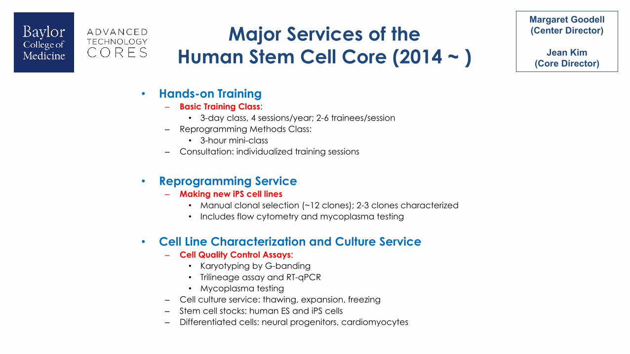
Major Services of the Human Stem Cell Core (2014 ~ )
• Hands-on Training– Basic Training Class:
• 3-day class, 4 sessions/year; 2-6 trainees/session– Reprogramming Methods Class:
• 3-hour mini-class– Consultation: individualized training sessions
• Reprogramming Service– Making new iPS cell lines
• Manual clonal selection (~12 clones); 2-3 clones characterized• Includes flow cytometry and mycoplasma testing
• Cell Line Characterization and Culture Service– Cell Quality Control Assays:
• Karyotyping by G-banding• Trilineage assay and RT-qPCR• Mycoplasma testing
– Cell culture service: thawing, expansion, freezing– Stem cell stocks: human ES and iPS cells– Differentiated cells: neural progenitors, cardiomyocytes
Margaret Goodell(Center Director)
Jean Kim (Core Director)
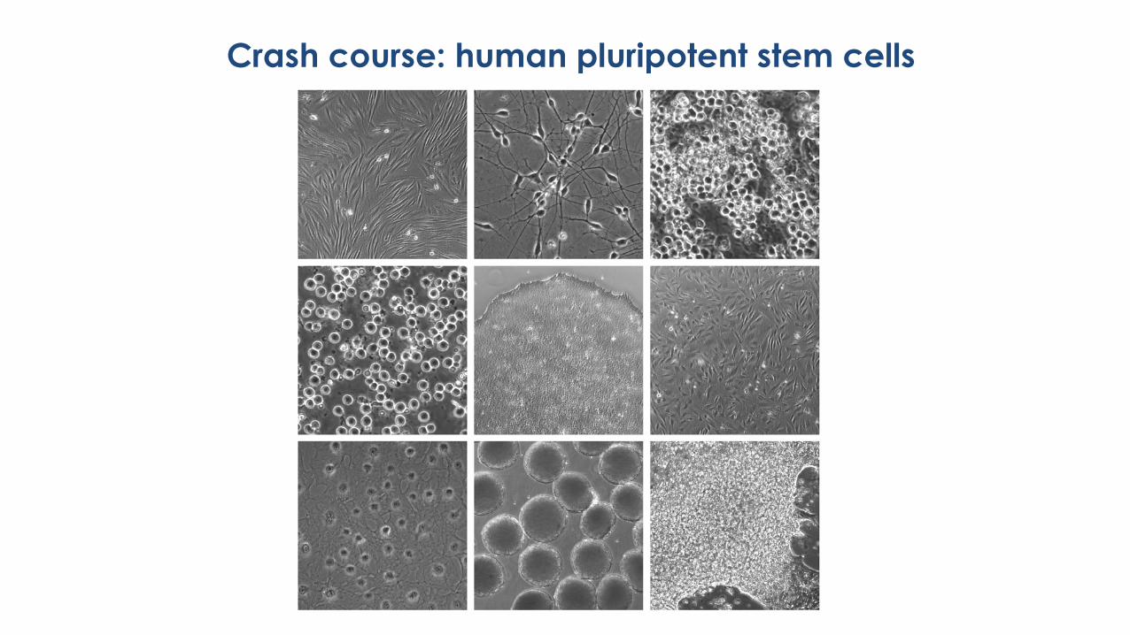
Crash course: human pluripotent stem cells
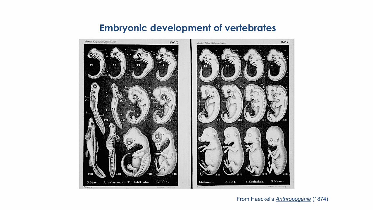
Embryonic development of vertebrates
From Haeckel's Anthropogenie (1874)
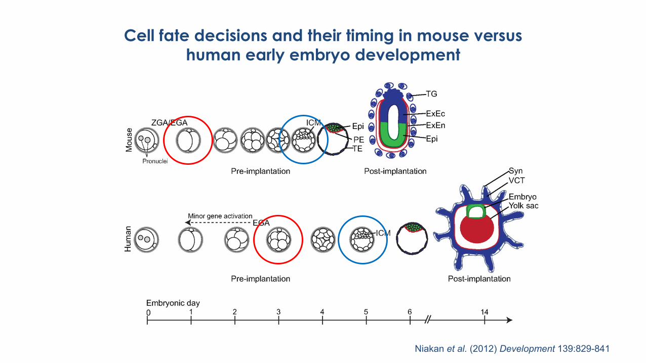
Cell fate decisions and their timing in mouse versus human early embryo development
Niakan et al. (2012) Development 139:829-841
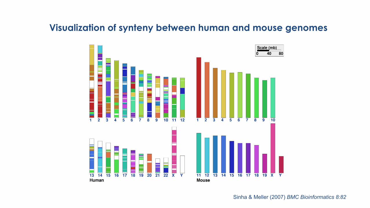
Visualization of synteny between human and mouse genomes
Sinha & Meller (2007) BMC Bioinformatics 8:82
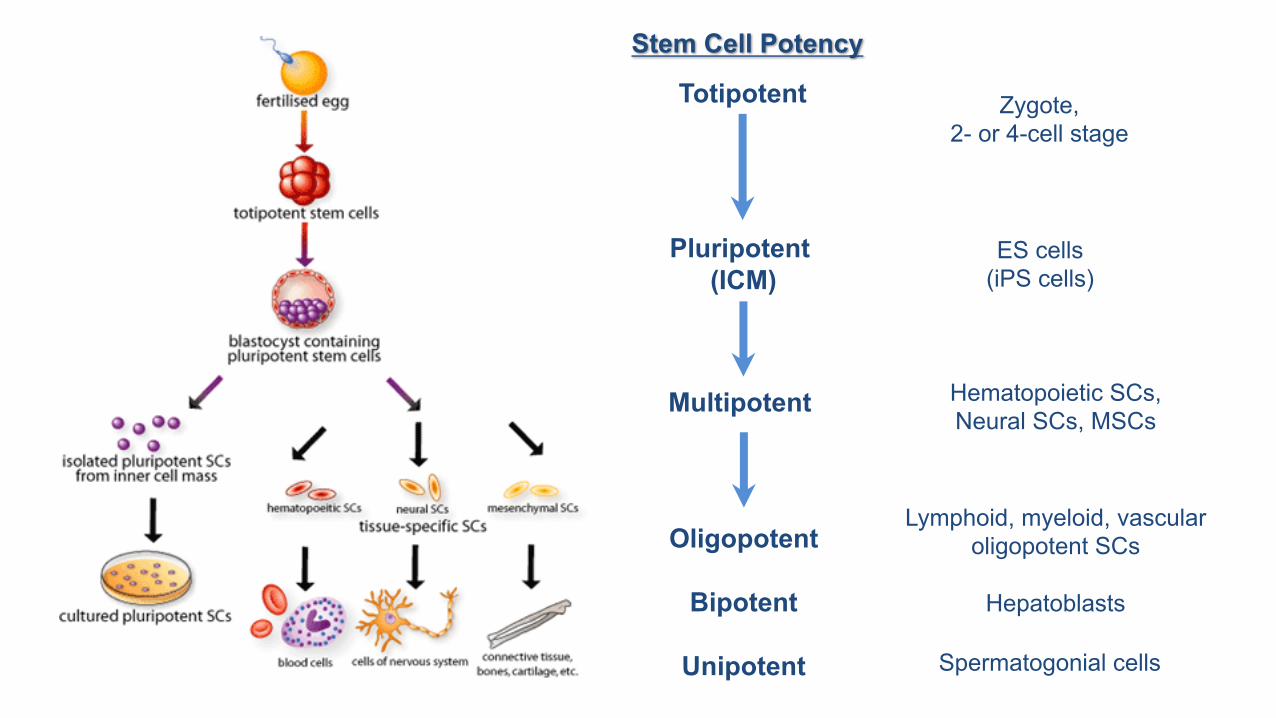
Totipotent
Pluripotent (ICM)
Multipotent
Oligopotent
Bipotent
Unipotent
Stem Cell Potency
Zygote,2- or 4-cell stage
ES cells(iPS cells)
Hematopoietic SCs, Neural SCs, MSCs
Lymphoid, myeloid, vascular oligopotent SCs
Hepatoblasts
Spermatogonial cells
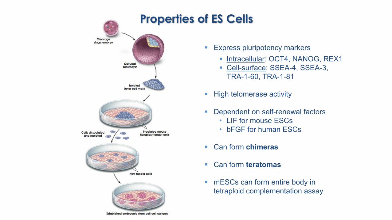
Properties of ES Cells
§ Express pluripotency markers § Intracellular: OCT4, NANOG, REX1§ Cell-surface: SSEA-4, SSEA-3,
TRA-1-60, TRA-1-81
§ High telomerase activity
§ Dependent on self-renewal factors• LIF for mouse ESCs • bFGF for human ESCs
§ Can form chimeras
§ Can form teratomas
§ mESCs can form entire body in tetraploid complementation assay
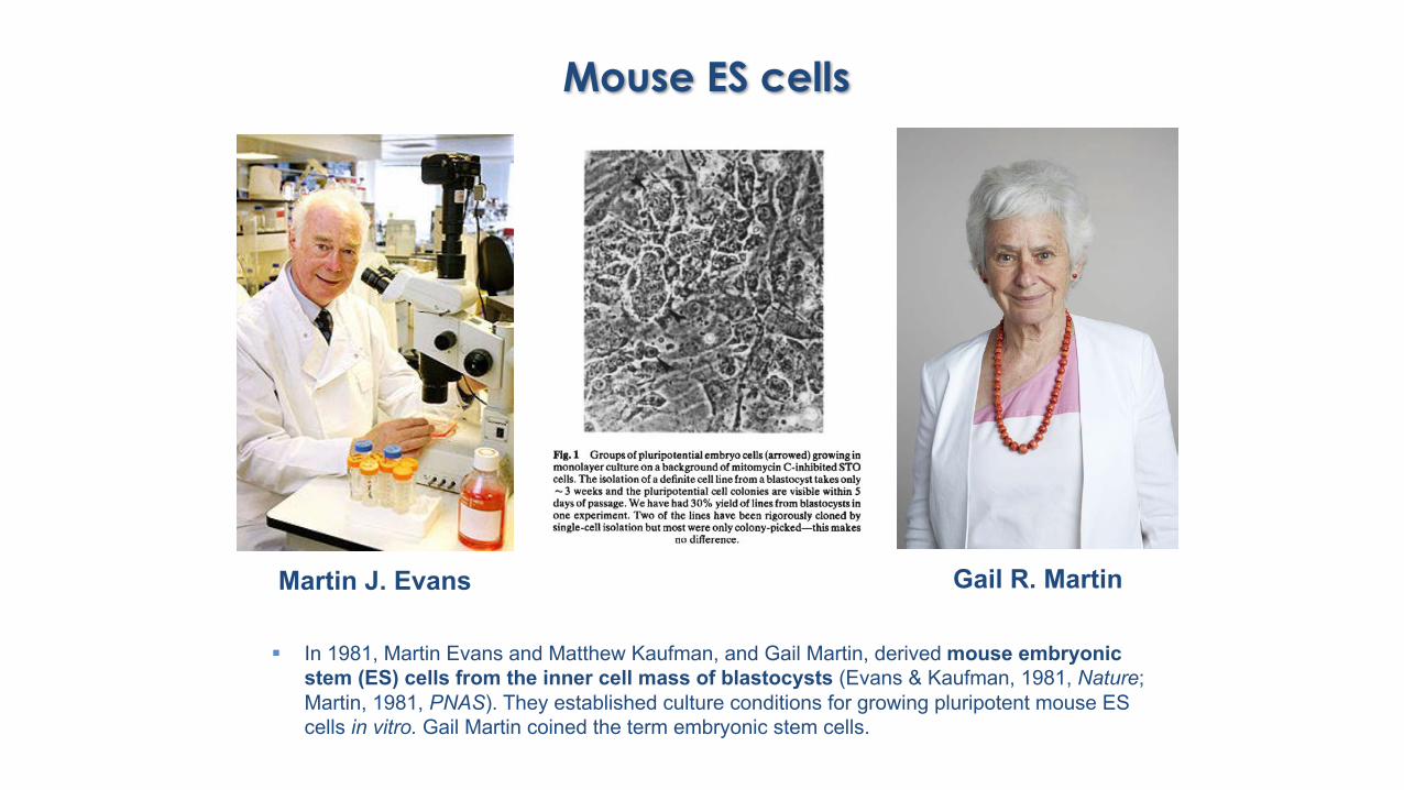
§ In 1981, Martin Evans and Matthew Kaufman, and Gail Martin, derived mouse embryonic stem (ES) cells from the inner cell mass of blastocysts (Evans & Kaufman, 1981, Nature; Martin, 1981, PNAS). They established culture conditions for growing pluripotent mouse ES cells in vitro. Gail Martin coined the term embryonic stem cells.
Mouse ES cells
Martin J. Evans Gail R. Martin
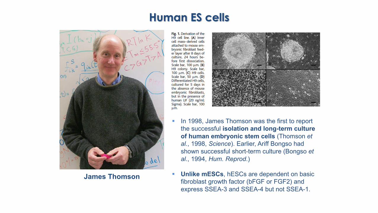
§ In 1998, James Thomson was the first to report the successful isolation and long-term culture of human embryonic stem cells (Thomson et al., 1998, Science). Earlier, Ariff Bongso had shown successful short-term culture (Bongso et al., 1994, Hum. Reprod.)
§ Unlike mESCs, hESCs are dependent on basic fibroblast growth factor (bFGF or FGF2) and express SSEA-3 and SSEA-4 but not SSEA-1.
Human ES cells
James Thomson
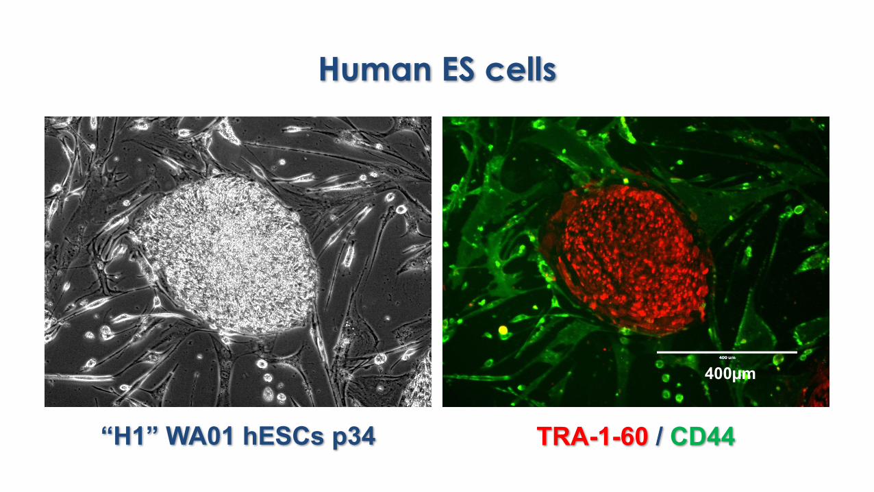
Human ES cells
“H1” WA01 hESCs p34 TRA-1-60 / CD44
400µm
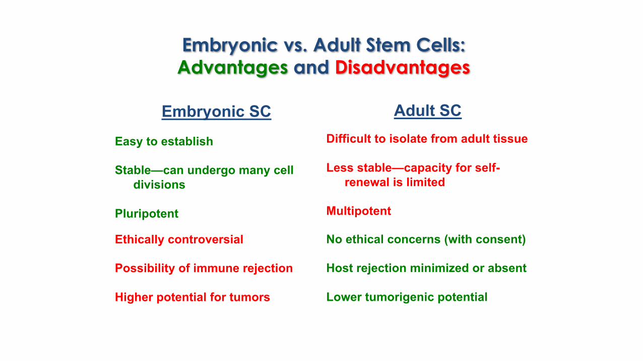
Embryonic vs. Adult Stem Cells: Advantages and Disadvantages
Embryonic SCEasy to establish
Stable—can undergo many cell divisions
Pluripotent
Adult SCDifficult to isolate from adult tissue
Less stable—capacity for self-renewal is limited
Multipotent
Ethically controversial
Possibility of immune rejection
Higher potential for tumors
No ethical concerns (with consent)
Host rejection minimized or absent
Lower tumorigenic potential

“Reprogrammed” Stem Cells— the best of both worlds?
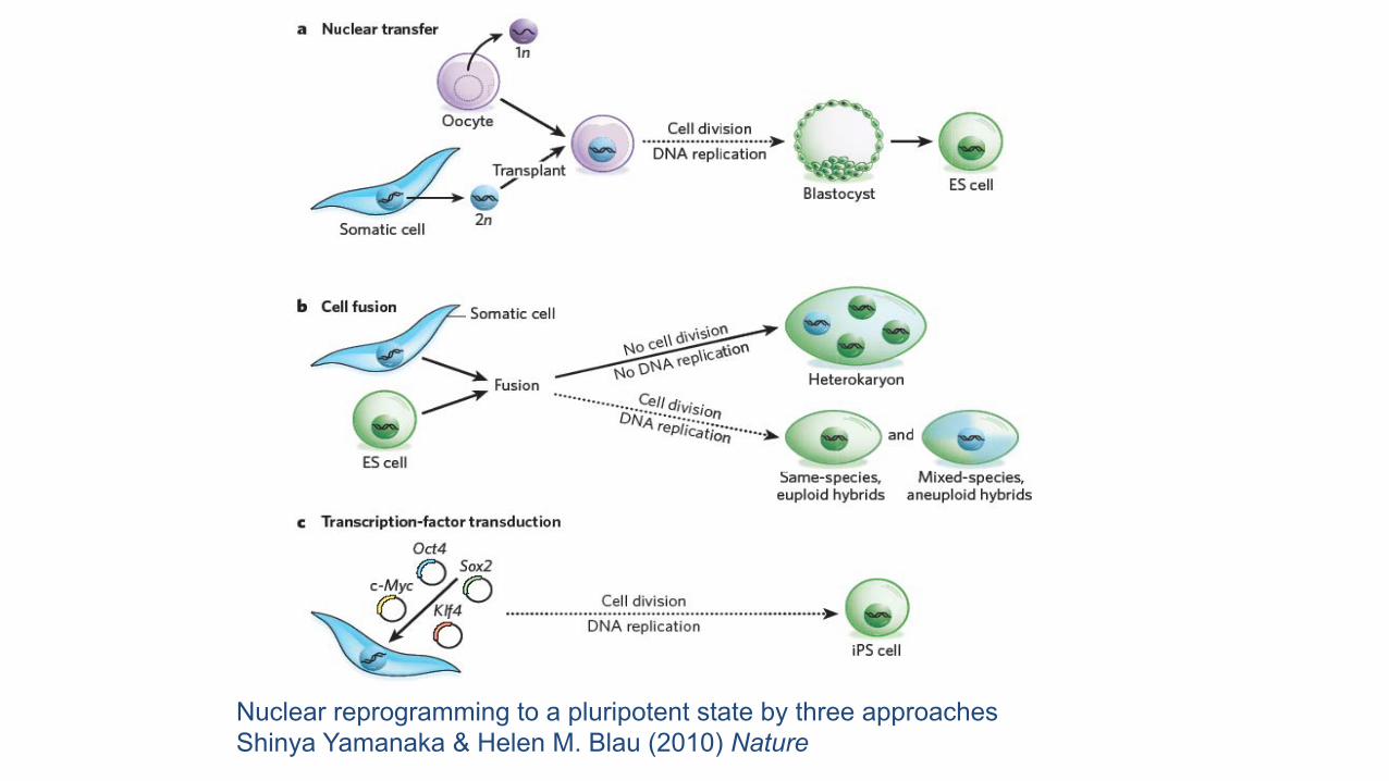
Nuclear reprogramming to a pluripotent state by three approachesShinya Yamanaka & Helen M. Blau (2010) Nature
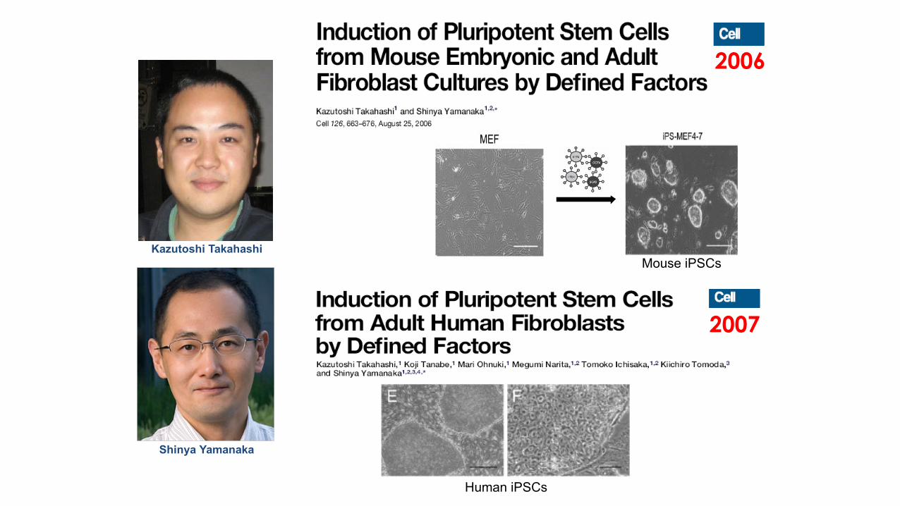
Shinya Yamanaka
Kazutoshi Takahashi
Human iPSCs
2006
2007
Mouse iPSCs
24 factors
Induction of pluripotent stem cells from mouse embryonic and adult fibroblast cultures by defined factors Takahashi and Yamanaka (2006) Cell 126, 663-676.
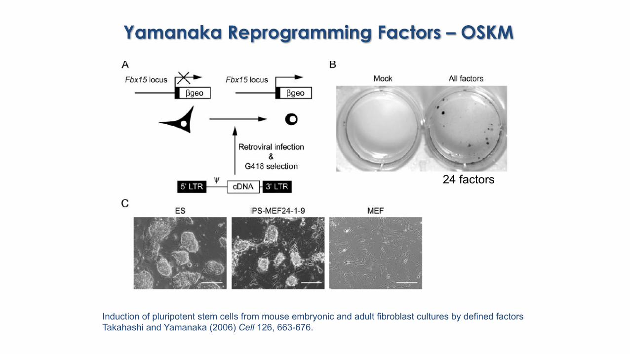
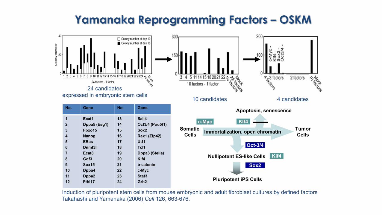
Yamanaka Reprogramming Factors – OSKM
Yamanaka Reprogramming Factors – OSKM
10 candidates 4 candidates
24 candidates expressed in embryonic stem cells
No. Gene No. Gene
123456789101112
Ecat1Dppa5 (Esg1)Fbxo15NanogERasDnmt3lEcat8Gdf3Sox15Dppa4Dppa2Fthl17
131415161718192021222324
Sall4Oct3/4 (Pou5f1)Sox2Rex1 (Zfp42)Utf1Tcl1Dppa3 (Stella)Klf4b-cateninc-MycStat3Grb2
Induction of pluripotent stem cells from mouse embryonic and adult fibroblast cultures by defined factors Takahashi and Yamanaka (2006) Cell 126, 663-676.
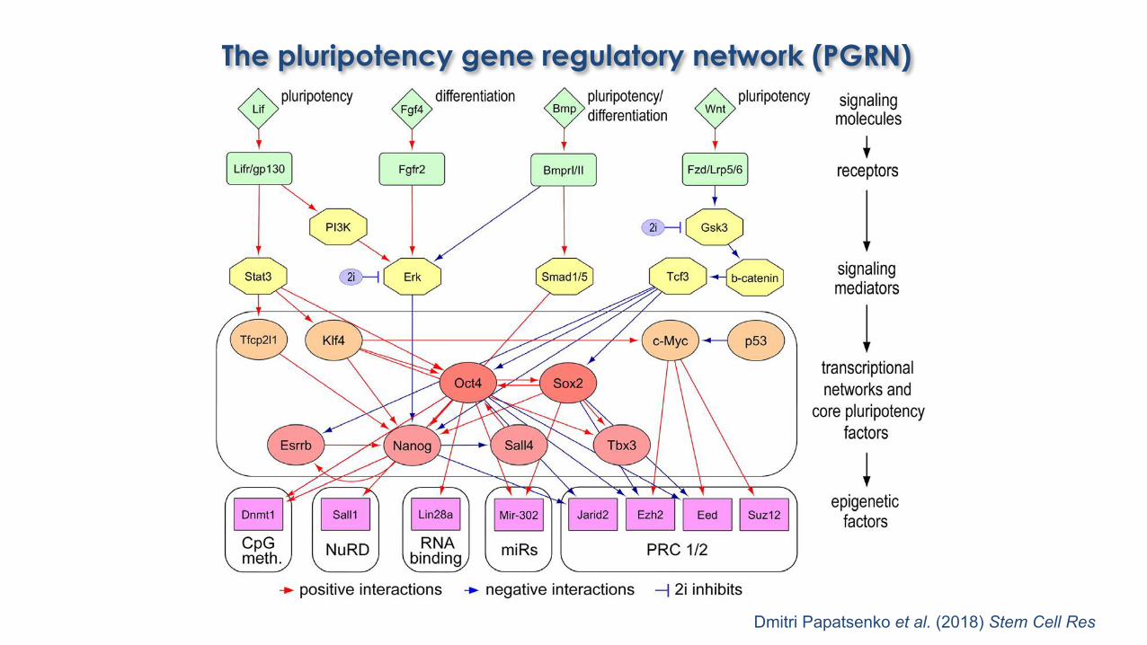
The pluripotency gene regulatory network (PGRN)
Dmitri Papatsenko et al. (2018) Stem Cell Res

Published iPS cell derivation strategies
§ Integration-free (“footprint-free”)• Adenoviruses• Sendai virus (RNA-based reproductive cycle)• Nucleofection of Episomal oriP/EBNA-1 vectors• Plasmid transfections (regular, minicircles)• Repeated mRNA transfection• Self-replicating synthetic RNA (Simplicon)• Protein transduction, small molecules, miRNAs etc.
§ Integration-based• Standard Moloney viruses (retroviral silencing)• Dox-inducible lentiviruses• Transposase-mediated integration (piggyBac)• Excisable lentivirus (4F cassette)
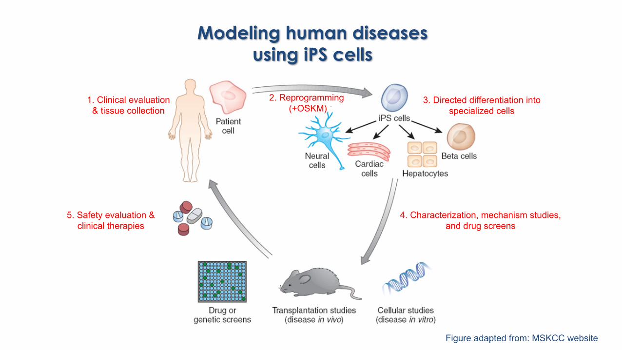
Modeling human diseases using iPS cells
Figure adapted from: MSKCC website
2. Reprogramming (+OSKM)
4. Characterization, mechanism studies, and drug screens
3. Directed differentiation into specialized cells
5. Safety evaluation & clinical therapies
1. Clinical evaluation& tissue collection
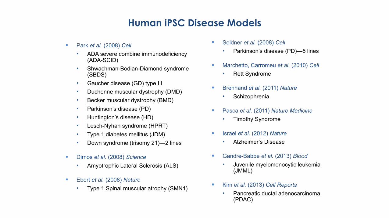
§ Park et al. (2008) Cell• ADA severe combine immunodeficiency
(ADA-SCID)• Shwachman-Bodian-Diamond syndrome
(SBDS)• Gaucher disease (GD) type III• Duchenne muscular dystrophy (DMD)• Becker muscular dystrophy (BMD)• Parkinson’s disease (PD)• Huntington’s disease (HD)• Lesch-Nyhan syndrome (HPRT)• Type 1 diabetes mellitus (JDM)• Down syndrome (trisomy 21)—2 lines
§ Dimos et al. (2008) Science• Amyotrophic Lateral Sclerosis (ALS)
§ Ebert et al. (2008) Nature• Type 1 Spinal muscular atrophy (SMN1)
Human iPSC Disease Models
§ Soldner et al. (2008) Cell• Parkinson’s disease (PD)—5 lines
§ Marchetto, Carromeu et al. (2010) Cell• Rett Syndrome
§ Brennand et al. (2011) Nature• Schizophrenia
§ Pasca et al. (2011) Nature Medicine• Timothy Syndrome
§ Israel et al. (2012) Nature• Alzheimer’s Disease
§ Gandre-Babbe et al. (2013) Blood• Juvenile myelomonocytic leukemia
(JMML)
§ Kim et al. (2013) Cell Reports• Pancreatic ductal adenocarcinoma
(PDAC)
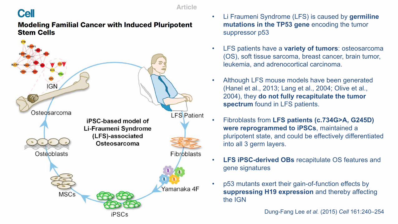
Dung-Fang Lee et al. (2015) Cell 161:240–254
• Li Fraumeni Syndrome (LFS) is caused by germilinemutations in the TP53 gene encoding the tumor suppressor p53
• LFS patients have a variety of tumors: osteosarcoma (OS), soft tissue sarcoma, breast cancer, brain tumor, leukemia, and adrenocortical carcinoma.
• Although LFS mouse models have been generated (Hanel et al., 2013; Lang et al., 2004; Olive et al., 2004), they do not fully recapitulate the tumor spectrum found in LFS patients.
• Fibroblasts from LFS patients (c.734G>A, G245D)were reprogrammed to iPSCs, maintained a pluripotent state, and could be effectively differentiated into all 3 germ layers.
• LFS iPSC-derived OBs recapitulate OS features and gene signatures
• p53 mutants exert their gain-of-function effects by suppressing H19 expression and thereby affecting the IGN
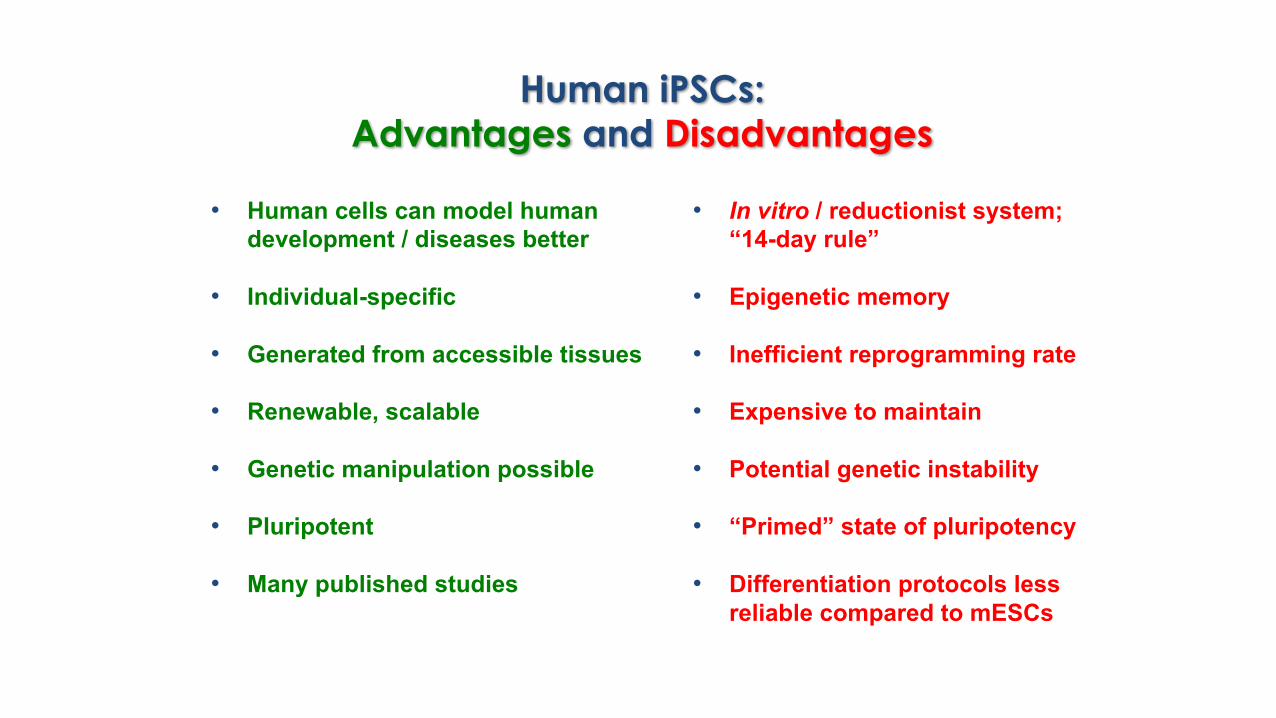
Human iPSCs: Advantages and Disadvantages
• Human cells can model human development / diseases better
• Individual-specific
• Generated from accessible tissues
• Renewable, scalable
• Genetic manipulation possible
• Pluripotent
• Many published studies
• In vitro / reductionist system; “14-day rule”
• Epigenetic memory
• Inefficient reprogramming rate
• Expensive to maintain
• Potential genetic instability
• “Primed” state of pluripotency
• Differentiation protocols less reliable compared to mESCs
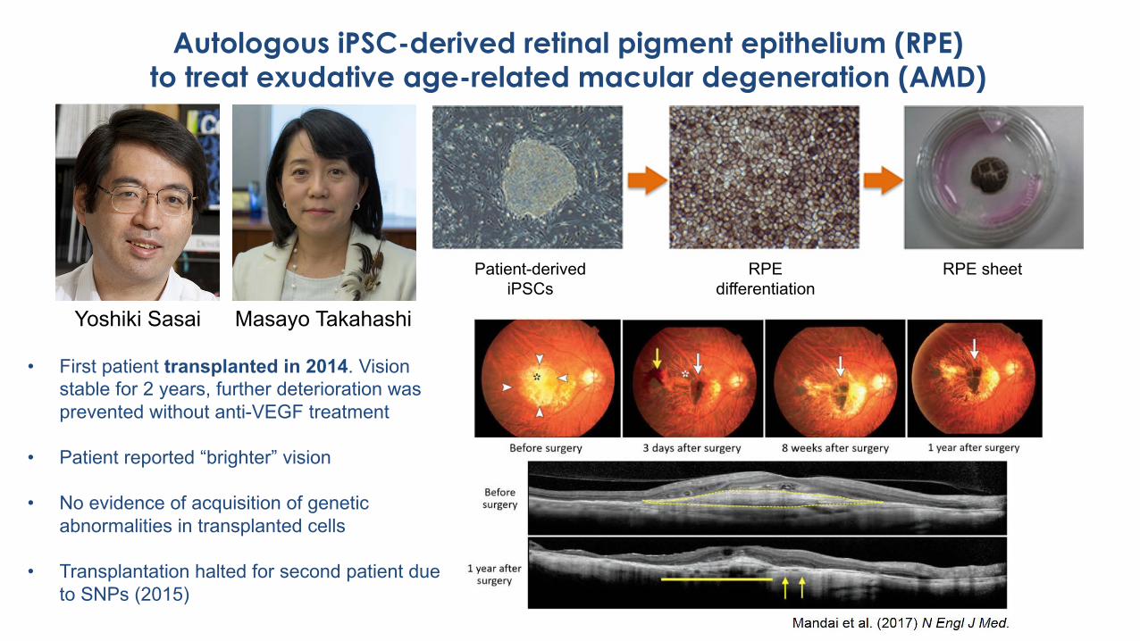
Autologous iPSC-derived retinal pigment epithelium (RPE) to treat exudative age-related macular degeneration (AMD)
Masayo TakahashiYoshiki Sasai
Patient-derived iPSCs
RPE differentiation
RPE sheet
• First patient transplanted in 2014. Vision stable for 2 years, further deterioration was prevented without anti-VEGF treatment
• Patient reported “brighter” vision
• No evidence of acquisition of genetic abnormalities in transplanted cells
• Transplantation halted for second patient due to SNPs (2015)
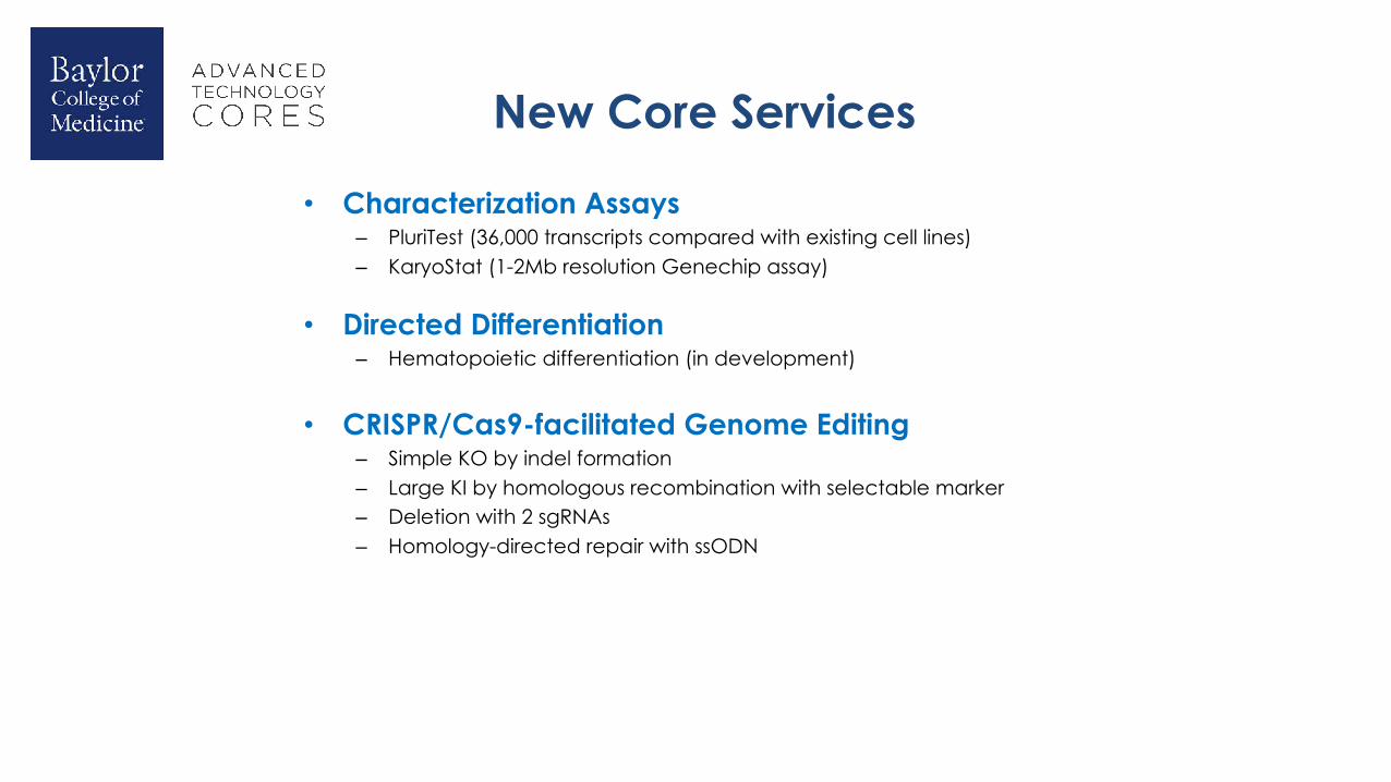
New Core Services
• Characterization Assays– PluriTest (36,000 transcripts compared with existing cell lines)– KaryoStat (1-2Mb resolution Genechip assay)
• Directed Differentiation– Hematopoietic differentiation (in development)
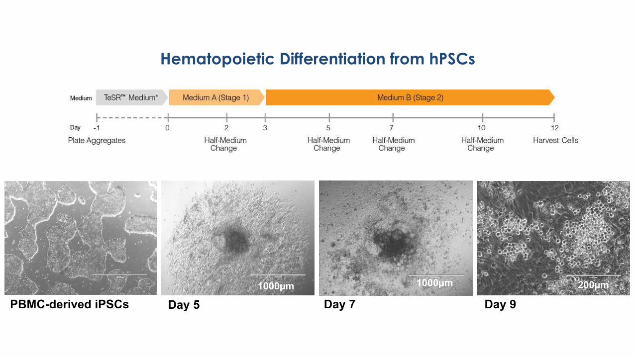
Hematopoietic Differentiation from hPSCs
PBMC-derived iPSCs Day 9200µm
Day 7
1000µm
Day 51000µm
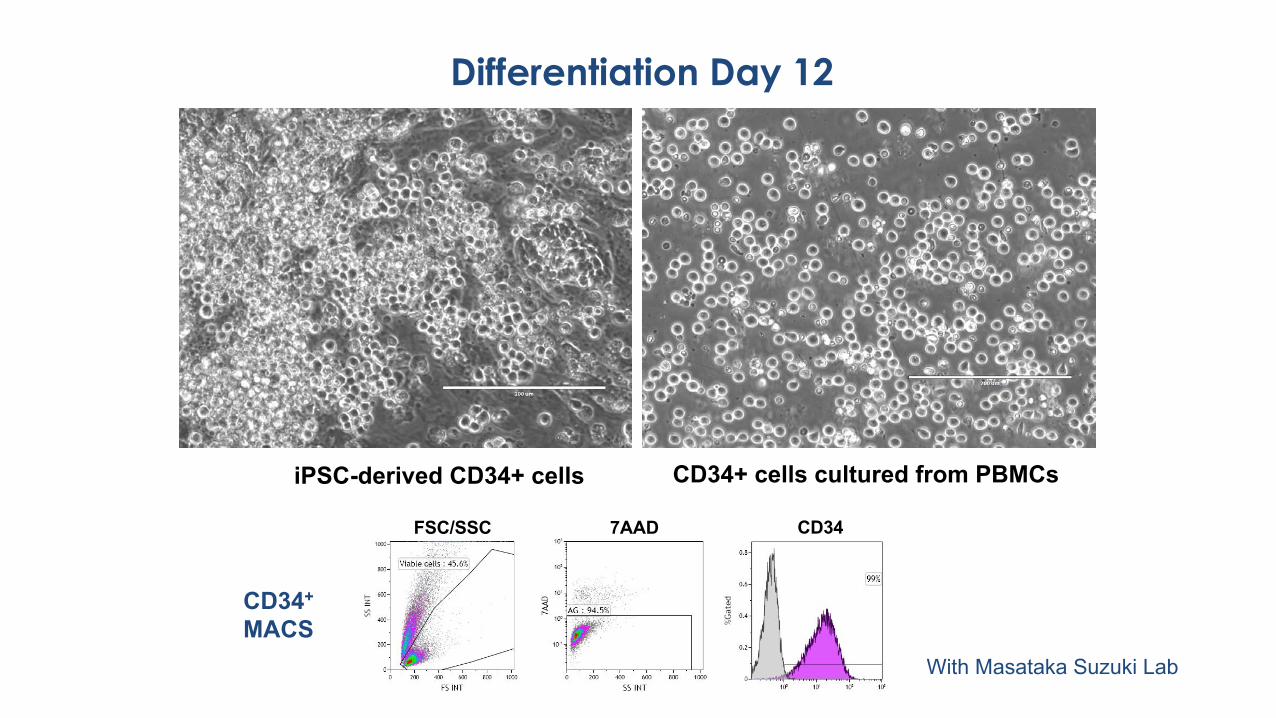
Differentiation Day 12
200µmiPSC-derived CD34+ cells CD34+ cells cultured from PBMCs
CD34FSC/SSC 7AAD
CD34+
MACSWith Masataka Suzuki Lab
New Core Services
• Characterization Assays– PluriTest (36,000 transcripts compared with existing cell lines)– KaryoStat (1-2Mb resolution Genechip assay)
• Directed Differentiation– Hematopoietic differentiation (in development)
• CRISPR/Cas9-facilitated Genome Editing– Simple KO by indel formation– Large KI by homologous recombination with selectable marker– Deletion with 2 sgRNAs– Homology-directed repair with ssODN
Part 1Cell Modeling with hPSCs
Part 2Genome Editing in hPSCs
Acknowledgments
Advanced Technology CoresDean Edwards
Jennifer McCulloughMyeshia Brown
Human Stem Cell CorePing Zhang
Anel LaGrone
Mauro Costa-Mattioli Lab (BCM)Ping Jun Zhu
Christian Schaaf (BCM; now U. Cologne)Madelyn Gillentine
Aryeh Warmflash (Rice U.)Kinshuk MitraWilliam Feist
Goodell Lab (BCM)Ayala Tovy
Raghav Ramabadran
Huda Zoghbi (BCM)Li Wang
Yingyao ShaoXiangling Meng
Wan Hee Yoon (OMRF)