Embed Size (px)
Citation preview

Diseases of Lates calcarifer by S Gibson-Kueh Page i
Diseases of Asian seabass (or barramundi), Lates
calcarifer Bloch
Susan GIBSON-KUEH, B.V.Sc, M.Sc (Aquatic Veterinary Studies)
This thesis is presented for the degree of Doctor of Philosophy of
Murdoch University, 2012.
Supervisors: Associate Prof. Philip K. Nicholls
Prof. (adjunct) Brian Jones

Diseases of Lates calcarifer by S Gibson-Kueh Page ii
Declaration
I declare that this thesis is an account of my research and contains work which
has not been previously submitted for a degree at any tertiary education
institution. Contributions by co-authors have been duly acknowledged.
Susan Gibson-Kueh

Diseases of Lates calcarifer by S Gibson-Kueh Page iii
Acknowledgements
Andrew Thompson, Andrea Valigurova & Miloslav Jirku for expert advice on
apicomplexans,
Una Ryan & RongChang Yang for molecular and phylogenetic analysis on the
Eimeria and Cryptosporidium from L. calcarifer,
Thuy, Alain, Diana Chee, Neil Wendover & John Jardine for some of the L.
calcarifer materials included in this study,
Aileen Elliot, Peter Fallon, Michael Slaven, Gerard Spoelstra, Wai-Yee Lee &
Micky Leong for technical laboratory support,
Jing Chen, Yahui Wang, Sophie Tay from the Animal & Plant Health Laboratories for
Red Sea bream iridovirus PCR and viral isolation on the scale drop cases,
Merck Aquatic Animal Health Laboratory in Singapore for the Red Sea bream
iridovirus PCR on L. calcarifer tissues from Farm A,
Mark Bennett & Masa Wayan for good advice on in situ hybridization,
Linda McInnes for providing the dioxygenin labeled Trypanosoma irwini DNA
probes
Jun Kurita for the Red Sea bream iridovirus monoclonal antibodies,
Jimmy Turnbull for introducing the mantra ‘the presence of a pathogen does not
equal the presence of a disease’
Mary Ng, who let me into the world of electron microscopy, what a great tool!
Hugh Ferguson, my teacher since Stirling, for providing expert opinion on the
pathology associated with scale drop syndrome…it was a great adventure
learning from you, and
Alan Lymbery for supporting all aquaculture related endeavours at Murdoch
University.
My supervisors, Phil & Brian for being patient with me.
To my husband Kelvin, and my daughters Leia, Sara, Rebecca & Clare, for all their
support and love.

Diseases of Lates calcarifer by S Gibson-Kueh Page iv
Preface
Chapter 1 serves as a brief introduction to husbandry practices and diseases
previously reported in cultured Lates calcarifer. It also includes a section on the
interactions between the host, environment and pathogens which need to be
considered in the investigation and managing of disease outbreaks. Chapters 2 to
4 are based on published papers while Chapter 5 is a manuscript intended for
publication in a scientific journal. There has been a need to adapt the chapters
based on published papers to integrate them into a thesis. Chapter 6 discusses
management strategies in relation to specific diseases in L. calcarifer at the
htachery, nursery and growout levels. Citations style is in keeping with that used
in Journal of Fish Diseases. Citations with more than 2 authors are quoted in full
when it appears in text within each chapter for the first time, and thereafter only
as first author & et al.

Diseases of Lates calcarifer by S Gibson-Kueh Page v
Abstract
Other than the study by Griffiths (2009) on gill diseases, there has been no
comprehensive study and report on the major diseases of Asian seabass (or
barramundi) Lates calcarifer Bloch. It is a food fish species of growing
importance in Asia and Australia. This study investigates some of the major
diseases encountered in the various stages of the culture of L. calcarifer, at the
histopathological, ultrastructural and molecular levels. Culture practices can
have significant impacts on fish health. Disease outbreaks are influenced by
factors involving the host, environment and pathogen. Current knowledge on
diseases of L. calcarifer, and these factors which may influence disease outbreaks
are discussed in Chapter 1.
This is the first report of an intestinal Eimeria infection in L. calcarifer.
The Eimeria infection was associated with severe pathology and significant
mortality in the absence of other pathogens. It was detected in diseased L.
calcarifer in all five nurseries in Ca Mau, Vietnam. Although these were small
scale nurseries which stocked an average of 3000 to 5000 fish at any one time, a
mortality rate of up to 30% was reported and is the cause of significant economic
losses for these nurseries. Moderate to heavy Eimeria infestation were observed
in greater than 80% of diseased fish examined. This high rate of Eimeria
infestation is suspected to be linked to the low daily water exchange rates
practised in these nurseries. However, the examination of only diseased fish does
not allow the determination of prevalence. A systemic iridovirus infection was
concurrently observed in some of the fishes but was not consistently present
when compared to the Eimeria infection. Molecular analysis showed that the
Eimeria of L. calcarifer from Vietnam formed clades with the Eimeria detected in

Diseases of Lates calcarifer by S Gibson-Kueh Page vi
L. calcarifer cultured in Australia, but clustered separately from other known
Eimeria species. Although Cryptosporidium was detected in these L. calcarifer
tissues, it could not be demonstrated histologically or ultrastructurally,
suggesting a low grade infestation or perhaps an environmental contaminant in
fish tissues tested. In situ hybridization using labeled PCR products showed that
labeled DNA probes generated from 18S PCR products could not be used to
distinguish between closely related genera such as Cryptosporidium and Eimeria.
Future investigation to determine the origin, transmission and risk factors
associated with this Eimeria infestation in L. calcarifer are needed.
‘Scale drop syndrome’ is a novel disease first reported in L. calcarifer
in Penang, Malaysia in 1992. Cases with similar gross and clinical presentations
were observed in Singapore in 2002, 2006 and 2009. Affected fish have loose
scales, which dropped off easily when handled. The disease was initially
observed in 100-300g fish, and later in larger fish up to 5kg bodyweight.
Cumulative mortalities of 40 to 50% were reported by farms, posing significant
economic losses of larger more valuable fish. This investigation forms the first
pathological description of ‘scale drop syndrome’ (SDS) in L. calcarifer. To aid
recognition of new cases for study, a case definition was developed for ‘scale
drop syndrome’ in L. calcarifer as a systemic vasculitis associated with tissue
necrosis in all major organs including the skin, with apparent targeting of cells of
epithelial origin. Attempts to isolate or detect the causative agent(s) by cell
culture, PCR and immunohistochemistry have proven unsuccessful. Further
studies to elucidate the definitive aetiology, isolate the causal agent(s) and
reproduce the disease will help better understanding and control of SDS.

Diseases of Lates calcarifer by S Gibson-Kueh Page vii
Although systemic iridoviral disease has been previously reported in
many freshwater and marine fish species, this study forms the first report of this
disease in L. calcarifer. Systemic iridoviral disease was observed in 5 to 20g L.
calcarifer usually 2 to 3 weeks post-transfer into sea cages at two farms.
Inclusion bodies suggestive of a systemic iridovirus infection were observed in
clinically healthy L. calcarifer from the land-based nursery of one of these two
farm; the presence of an iridovirus infection was supported by positive PCR
results using Red Sea bream iridovirus (RSIV) primer 1. The presence of
inclusions was not accompanied by any tissue necrosis in these clinically healthy
fish. This finding suggested that the systemic iridovirus infection occurred before
stocking at sea, and did not originate from wild fish or older fish in adjacent sea
cages as initially suspected by this farm. Immunohistochemistry on tissues of
clinical cases of systemic iridovirus gave positive results using the Red Sea
bream iridovirus monoclonal antibody (RSIV M10), although intensity varied
between tissues, possibly related to varying exposure of different tissues to
fixation chemicals. Inclusion bodies in clinically healthy fish from the same farm
did not show positive reaction with RSIV M10. This may be due to a lack of
antigenic expression by the viral infected cells at this early stage of infection.
Viral nervous necrosis (VNN) is a serious disease of hatchery reared L.
calcarifer fry in this study. Mortalities of 50 to 100% were reported in 3wo fry.
VNN can be difficult to diagnose in older fry, where it can be associated with few
vacuolations or an absence of viral inclusions
‘Pot belly disease’ (PBD) was previously reported in L. calcarifer fry less
than 1g, in association with an intracellular coccobacillus infection and
mortalities of 80 to 100%. In this study, PBD was observed in 120g L. calcarifer

Diseases of Lates calcarifer by S Gibson-Kueh Page viii
at two sea cage farms, in association with significant granulomatous enteritis.
The extent of the granulomatous enteritis is likely to have an effect on affected
fish. It was observed concurrently with systemic iridoviral disease at one farm
and nocardiosis at another farm. Diagnosis by histopathology and the lack of
other confirmatory tests for PBD may result in underdiagnosis of this disease.
The epidemiology of PBD needs further study to establish origin and modes of
transmission, to facilitate better disease control.
Diseases associated with infections by ubiquitous bacteria such as Vibrio,
Tenacibaculum were commonly observed in L. calcarifer post-handling.
Tenacibaculosis and vibriosis often occurred concurrently with other diseases
such as streptococcosis, systemic iridviral disease or PBD. Streptococcosis can
affect fish up to 3kg bodyweight, resulting in significant mortalities greater than
40 to 50%. Like SDS, because streptococcosis can affect up to market size fish,
they can cause considerable economic losses. Although vaccines against
Streptococcosis are available, conflicting views are held on the efficacy of
Streptococcus vaccines by various research groups. Overall, the South-east Asian
L. calcarifer farms which practiced vaccination against Streptococcus iniae
reported a reduction of mortality, especially in fish greater than 1 to 1.5kg
bodyweight.
Nocardiosis has been reported as an emerging disease in marine food fish
species caused by acid fast filamentous branching bacterium. Although
nocardiosis was observed histopathologically in L. calcarifer at two sea cage
farms, the numbers of samples examined were small and no other tests were
attempted due to lack of suitable samples. More intensive and extensive study is
needed to determine the significance of nocardiosis in L. calcarifer. Chronic

Diseases of Lates calcarifer by S Gibson-Kueh Page ix
granulomatous enteritis was not uncommon in the cases submitted to the Fish
Health Laboratory in Perth. Although the peritonitis was associated with heavy
bacteria infection, it is unclear if these are secondary invaders. Schipps, Bosmans
& Humphreys (2009) reported that Vibrio harveyi and Photobacterium damsela
damsela vaccinations appeared to be not efficacious, suggesting that these
bacteria were not the primary cause of the disease.
It is well recognized that disease outbreaks in farmed fish are influenced
by the interaction between host, the environment and pathogens. While serious
diseases are often reported in association with specific aquatic pathogens, not
much is known about the risk factors which trigger fish disease outbreaks.
Disease outbreaks often occur after stressful events such as net transfers, recent
handling or poor water quality. In fact, diseases are often caused by ubiquitous
pathogens that are commonly present in the culture environment. Although
further research is necessary to gather more information to improve diagnosis
and management of specific diseases, general health management strategies can
be applied at the various stages in the culture of L. calcarifer to minimize disease
outbreaks. This is discussed for L. calcarifer in Chapter 6.
Observations of types of disease agents may be influenced by site
conditions or the types of tests or materials examined. For example, some
parasites may be more prevalent in certain sites where intermediate hosts
abound, or loosely attached ectoparasites may be lost unless wet mount
microscopic examinations of fresh tissues were carried out. The study of
emerging diseases such as scale drop syndrome (SDS) or pot belly disease (PBD)
in L. calcarifer has been hampered by lack of confirmatory diagnostic tools and
inadequate knowledge on critical epidemiological factors such as mode of

Diseases of Lates calcarifer by S Gibson-Kueh Page x
transmission or potential reservoirs. While ideally identification and isolation of
the causal agent will help fulfil Koch’s postulates, it may be possible to improve
the understanding of disease via cohabitation or infectivity trials using tissue
homogenates from diseased fish when pure isolates are not available. There is a
need to conduct research to not only establish a definitive aetiology, but also to
identify risk factors to facilitate successful disease control. The successful
management of disease in aquaculture does not lie in any one strategy but an
integrated management of all risks encountered during the culture cycle against
disease occurrence or incursions.

Diseases of Lates calcarifer by S Gibson-Kueh Page xi
Table of Contents
Diseases of Asian seabass (or barramundi), Lates calcarifer Bloch ............................... i
Declaration ......................................................................................................................................... ii
Acknowledgements ........................................................................................................................ iii
Preface ................................................................................................................................................ iv
Table of Contents ............................................................................................................................ xi
Legends of Figures ........................................................................................................................ xv
Lists of Tables ................................................................................................................................. xxi
List of abbreviations ................................................................................................................... xxii
Chapter 1 Introduction .......................................................................................................... 1
1.1 Culture of Asian seabass (or barramundi), Lates calcarifer Bloch ............ 1
1.1.1 Importance as an aquaculture food fish species .......................................... 1
1.1.2 Production of juveniles of L. calcarifer ............................................................ 2
1.1.3 Grow-out of L. calcarifer ........................................................................................ 3
1.2 Disease outbreaks are influenced by interactions between host, aquatic
environment and pathogen ..................................................................................................... 4
1.2.1 Host factors .................................................................................................................... 5
1.2.2 Environment ................................................................................................................. 7
1.2.3 Pathogens ....................................................................................................................... 8
1.3 Infectious diseases with a major impact on the culture of L. calcarifer 11
1.3.1 Viral diseases .............................................................................................................. 11
1.3.2 Bacterial diseases ...................................................................................................... 12
1.3.3 Monogenean parasites ............................................................................................ 14
1.3.4 Protozoan parasites with a direct life cycle .................................................... 14
1.4 Other infectious diseases observed in cultured L. calcarifer ..................... 16

Diseases of Lates calcarifer by S Gibson-Kueh Page xii
1.5 Non-infectious diseases reported in L. calcarifer ........................................... 18
1.6 Aims of present study ............................................................................................... 19
Chapter 2 An intestinal Eimeria infection in juvenile Asian seabass, Lates
calcarifer Bloch cultured in Vietnam – a first report ....................................................... 20
2.1 Introduction ......................................................................................................................... 21
2.2 Materials and methods .................................................................................................... 22
2.2.1 Background information on samples examined ........................................... 22
2.2.2 Light microscopy (LM) ......................................................................................... 23
2.2.3 Transmission electron microscopy (TEM)................................................... 23
2.3. Results ................................................................................................................................... 24
2.3.1 Field observations made on L. calcarifer nurseries sampled in this
study .......................................................................................................................................... 24
2.3.2 Histopathology ........................................................................................................... 24
2.3.3 Ultrastructural observations by TEM ................................................................ 30
2.4 Discussion ............................................................................................................................. 33
Chapter 3 The molecular characterization of an Eimeria and Cryptosporidium
detected in Asian seabass (Lates calcarifer) cultured in Vietnam .............................. 37
3.1 Introduction ......................................................................................................................... 39
3.2 Materials and methods .................................................................................................... 40
3.2.1 General .......................................................................................................................... 40
3.2.2 18S Polymerase Chain Reaction (PCR) and sequencing ............................ 41
3.2.3 Phylogenetic analysis............................................................................................... 42
3.2.4 In situ hybridization using dioxygenin labeled 18S PCR products ........ 43
3.3 Results .................................................................................................................................... 44
3.3.1 Sequence and phylogenetic analysis of Eimeria ............................................ 44

Diseases of Lates calcarifer by S Gibson-Kueh Page xiii
3.3.2 Sequence and phylogenetic analysis of Cryptosporidium .......................... 45
3.3.3 In-situ hybridization (ISH) .................................................................................... 48
3.4 Discussion ............................................................................................................................. 50
Chapter 4 The pathology of ‘scale drop syndrome’ in Asian seabass, Lates
calcarifer Bloch ............................................................................................................................... 54
4.1 Introduction .................................................................................................................. 56
4.2 Materials and methods ............................................................................................. 57
4.2.1 Background .............................................................................................................. 57
4.2.2 Light microscopy (LM) ......................................................................................... 57
4.2.3 Transmission electron microscopy (TEM)................................................... 57
4.2.4 Immunohistochemistry ....................................................................................... 57
4.2.5 PCR and tissue culture ......................................................................................... 58
4.3 Results ............................................................................................................................. 58
4.3.1 History, clinical & gross observations............................................................ 58
4.3.2 Histopathology ........................................................................................................ 61
4.4 Discussion ...................................................................................................................... 68
Chapter 5 Other viral and bacterial diseases observed in cultured Asian
seabass (or barramundi), Lates calcarifer Bloch ............................................................... 72
5.1 Introduction .................................................................................................................. 73
5.2 Materials and methods ............................................................................................. 74
5.2.1 Background .............................................................................................................. 74
5.2.2 Light microscopy (LM) ......................................................................................... 76
5.2.3 Transmission electron microscopy (TEM)................................................... 76
5.2.4 Immunohistochemistry using RSIV M10 ...................................................... 78
5.2.5 Polymerase chain reaction & in situ hybridization ................................... 79

Diseases of Lates calcarifer by S Gibson-Kueh Page xiv
5.3 Diseases observed in L. calcarifer tissues examined .................................... 80
5.3.1 Systemic iridoviral disease ................................................................................ 81
5.3.2 Viral nervous necrosis (VNN) ........................................................................... 85
5.3.3 ‘Pot Belly Disease’ (PBD) ..................................................................................... 87
5.3.4 Streptococcosis ....................................................................................................... 87
5.3.5 Vibriosis and tenacibaculosis ............................................................................ 89
5.3.5 Nocardiosis ............................................................................................................... 90
5.3.6 Chronic peritonitis ................................................................................................. 91
5.4 Discussion ...................................................................................................................... 94
Chapter 6 Disease management and future research strategies ......................... 98
6.1 Hatchery ................................................................................................................................ 98
6.2 Nursery ............................................................................................................................... 100
6.3 Grow-out ............................................................................................................................ 103
6.4 Conclusion & future research .................................................................................... 105
References ..................................................................................................................................... 109
Appendix 1.1 – Response to questionaire from Farm A .............................................. 137
Appendix 1.2 - Response to questionaire from Farm B ............................................... 139

Diseases of Lates calcarifer by S Gibson-Kueh Page xv
Legends of Figures
Figure 1 Eimeria infection on the brush border of intestinal mucosa in juvenile
Lates calcarifer from Vietnam and increased mononuclear infiltrate (Inf) in
lamina propria. Meronts (Me) with merozoites arranged in rosettes,
macrogamonts (Ma) with foamy cytoplasm, microgamonts (Mi) with
peripherally arranged nuclei and more intensely basophilic trophozoites (T)
(H&E). ................................................................................................................................................. 28
Figure 2 (a) Meronts (Me) with merozoites arranged in parallel and merozoites
(z) apparently still within parasitophorus envelopes. (Giemsa). (b) An unusually
large meront (arrow) with at least 18 merozoites. (H&E) (c) Microgamonts (*)
with peripherally arranged nuclei and microgametes (arrows) (Giemsa). ............ 28
Figure 3 (a) Sporulating oocysts (*) had four sporocysts. Sporulated oocysts
(arrows) in faecal materials within intestinal lumen had four pairs of sporozoites
and a thin membranous wall. (H&E) (b) Unsporulated oocyst observed in
discharged tank waste water. (c) Sporulated oocyst from waste water with
oocyst residual body (R) and sporozoites in pairs, bounded by thin sporocyst
membrane. Sporocysts (S) had no stieda bodies or suture lines (Nomarski
interference microscopy). ............................................................................................................ 29
Figure 4 The intestinal epithelium was denuded (arrows) in some areas with
corresponding intense inflammatory (Inf) in the lamina propria, and a significant
amount of sloughed cellular debri (D) in intestinal lumen (Giemsa). ....................... 29
Figure 5 Macrogamonts (Ma) with amylopectin granules and microgamonts with
microgametes (MiG) at the microvillous brush border of intestines. ‘X’ was
presumably a microgamont from which microgametes had been released, thus

Diseases of Lates calcarifer by S Gibson-Kueh Page xvi
giving a crenate appearance. Parasitophorous envelopes (PE), residual body (Re)
in microgamont. ............................................................................................................................. 31
Figure 6 Rodlet cells (Ro) often associated with parasitism in fish were observed
within blood vessels (bv) in intestines. One of the rodlet cells appeared to be in
the process of exiting the blood vessel (*). Vascular endothelial cells (E),
fibroblasts (F) that produced the collagen (C) of blood vessel wall. ......................... 31
Figure 7(a) Meront (Me) with finger-like attachment organelles (fao) and
residual body (Re). Merozoites (z) had apical complexes (A) at various stages of
formation. Trophozoites (T) and developing meront (dMe) in epicytoplasmic
position. (b) Meront with at least 8 merozoites (z) and finger-like attachment
organelles (fao). .............................................................................................................................. 32
Figure 8 Macrogamont with abundant amylopectin (A) granules and finger-like
attachment organelles (fao) extended into host cell but limited to the
epicytoplasmic boundary. parasitophorus envelope (PE). ........................................... 32
Figure 9 Phylogenetic tree of Eimeria detected in L. calcarifer from Vietnam and
Australia, with GenBank database accession numbers JF261140 (VTASB1),
JF261139 (VTASB2), and JF261138 (AUBarra1) and JF261137 (AUBarra2),
respectively. ..................................................................................................................................... 46
Figure 10 Phylogenetic tree of Cryptosporidium detected in L. calcarifer from
Vietnam, with GenBank database accession numbers JF285332 (H08384) and
JF285333 (H08391). ..................................................................................................................... 47
Figure 11 Epicytoplasmic Asian seabass Eimeria in intestines showed positive
red fluorescence in ISH using DIG-labeled Eimeria 18S PCR products and
Permanent Red (Dako). Inset (to same scale) shows no fluorescence in ISH using
negative control DIG-labeled Trypanosoma 18S PCR products. .................................. 49

Diseases of Lates calcarifer by S Gibson-Kueh Page xvii
Figure 12 Epicytoplasmic Asian seabass Eimeria in intestines showed mild to
moderate red fluorescence in ISH using DIG-labeled Crytopsoridium 18S PCR
products............................................................................................................................................. 49
Figure 13 Darkened skin and loss of scales (arrows) over extensive caudal half
of body with loss of colour in Asian seabass, Lates calcarifer Bloch with ‘scale
drop syndrome’. ............................................................................................................................. 60
Figure 14 Foci of fatty liver (arrows) in L. calcarifer with ‘scale drop syndrome’.
............................................................................................................................................................... 60
Figure 15 (a) Scale loss was associated with intense dermal perivasculitis
(arrows) in scale beds. (b) Necrotic scale bed (N), seen grossly in fig. 12 as areas
of skin with loss of colour and scales. (c) Ventricle showing necrosis of coronary
blood vessels (arrows) in compact myocardium (H&E). ............................................... 62
Figure 16 (a) Multifocal areas of splenic necrosis (*) were associated with
reactive (RxE) and necrotic ellipsoids (NeE), shown at higher magnification in
Figs. b and c (arrows) respectively. (d) Thrombus formation (arrow) in an
ellipsoid (H&E). .............................................................................................................................. 62
Figure 17 (a) Severe endothelial inflammation (E Inf) in renal artery. Such a
lesion would have compromised blood supply to the kidney and produced severe
renal necrosis if not for its portal blood supply. (b) Brain with pronounced
perivascular inflammation (arrows) (H&E) ........................................................................ 63
Figure 18 (a) Cytoplasmic inclusion bodies (IB) were often present in renal
interstitium of L. calcarifer with SDS. The IB can be hard to distinguish due to the
presence of haematopoietic cells. Karyorrhexis (arrows) in renal tubules suggest
specific targeting of epithelial cells. (b) IB with a basophilic hyaline appearance
in degenerated glomeruli. These IB are suggestive of a systemic iridovirus

Diseases of Lates calcarifer by S Gibson-Kueh Page xviii
infection (Gibson-Kueh et al., 2003), which are now endemic in many L. calcarifer
farms in South East Asia, so that they may be an incidental finding of a common
pathogen concurrently with other diseases. (H&E) ......................................................... 63
Figure 19 (a) Hydropic change (D) in choroid rete of eye. (b) The heart was
often a good place to look for viral inclusion bodies without the ‘clutter’ of
haematopoietic tissues. Enlarged basophilic inclusion bodies (IB) can be seen
bulging from the endocardium. There was a subacute to chronic pericarditis (*).
(c) Hypertrophied myocardial endothelium (E) needs to be differentiated from
(d) basophilic inclusion bodies (IBs) located on endothelium. The presence of the
IB typical of systemic iridoviral disease must be taken into context of a disease
that can be endemic, and may occur concurrently with other diseases (H&E) ... 64
Figure 20 (a) Prominent hepatic lobulation in L. calcarifer with SDS. This may be
attributed to increased cell death. Fatty liver or hepatic lipidosis (Fa) typified by
vacuolation can be focal to extensive involving whole segments of livers, as seen
grossly in fig. 13. Liver with reduced glycogen reserves stained more intensely
basophilic (NG). (b) Diffuse karyorrhexis (arrows) and nuclear chromatin
margination (MNC) in liver (H&E). ......................................................................................... 65
Figure 21 Multifocal areas of gastric glands necrosis (*) associated with
perivasculitis (arrows) in L. calcarifer with SDS. Demarcated area is shown at
higher magnification in inset: paler areas of gastric gland necrosis and karyolysis
(arrows) in gastric mucosa (H&E). ......................................................................................... 65
Figure 22 L. calcarifer with SDS have remarkably normal renal haematopoietic
cells (Hc). Necrotic cells have marginated chromatin (MNC) or presence of very
electron dense lamellae (L) in nucleus. Red blood cells (rbc) in blood vessel.
Demarcated area is shown at higher magnification in inset: double enveloped

Diseases of Lates calcarifer by S Gibson-Kueh Page xix
hexagonal virions with ribosomes absent, presumably as virions have budded
out from cytoplasm of cell. ......................................................................................................... 67
Figure 23 (a)Enveloped hexagonal virions in spleen in remains of cytoplasmic
ribosomes (r), with a size range of 188 to 269 nm (n=8). (b) Smaller enveloped
hexagonal virions measuring 133nm (n=2) in cytoplasmic remnants in kidney.
These virions have electron-lucent nucleocapsids. .......................................................... 67
Figure 24 Systemic iridovirus infections in (a) clinically healthy 0.2g L.
calcarifer, and (b) clinically diseased 4g L. calcarifer. Tissue degeneration with
hydropic changes and cell deaths (D) were more evident in the clinically
diseased 4g fish in b. Inclusion bodies (IB) could be observed in both fish. .......... 83
Figure 25 Cytoplasmic inclusion bodies in cardiac tissue with positive red
fluorescence in immunohistochemistry using RSIV M10 and Permanent Red
(Dako). Inset shows no fluorescence in negative control. ............................................. 84
Figure 26 PCR on alcohol fixed L. calcarifer tissues from diseased fish with
histopathological signs of a systemic iridovirus infection showed an expected
570bp product (Kurita et al. 1998). Lane 1 & 11: 100bp ladder, Lane 2: negative
control ultrapure water, Lanes 3-5: Fish 1 (triplicates), Lanes 6-8: Fish 2
(triplicates), Lanes 9-10: Fish 3 (duplicates) ...................................................................... 84
Figure 27 (a, b & d) Viral encephalopathy and retinopathy or viral nervous
necrosis (VNN) in 12 do L. calcarifer fry from Farm A (a) Severe vacuolation
(arrows) in the brain and eye. (b) Vacuoles were often associated with basophilic
inclusion bodies (arrows). (c) VNN in 22 do fry submitted to the Fish Health
Laboratory in Perth, with mild vacuolation (arrows) and no inclusion bodies.
(H&E) (d) Nerve cells with dendritic processes (arrows) and electron dense
inclusions (IB) from VNNV infection (TEM). This explains the location of

Diseases of Lates calcarifer by S Gibson-Kueh Page xx
vacuolation in grey matter of brain where the cell bodies of nerve cells are
located. ............................................................................................................................................... 86
Figure 28 (a) Pot belly disease (PBD) in nursery-reared 1.8g L. calcarifer with
granulomatous enteritis (G) as well as unaffected parts of intestines (N). (b) PBD
in 80-120g fish in sea cages. Although associated pathology was less marked in
these older fish than in younger fry, the granulomatous enteritis has affected
significant portions of intestine. Insets: Higher magnification with clusters of
large coccobacilli (arrows. (H&E). .......................................................................................... 88
Figure 29 (a) Coccoid-shaped bacteria, presumably Streptococcus species were
observed within blood vessels (arrows) in various organs including the brain,
and (b) within phagocytic endothelium lining the heart (arrows). (c) Clusters of
filamentous rods (arrows), presumably Tenacibaculum maritimum associated
with gill necrosis. ........................................................................................................................... 89
Figure 30 Severe granulomatous response (arrows) was observed in the choroid
rete of the eye of this severly affected 50g L. calcarifer from Farm B. The
filamentous rods stained positively with Fite Faraco (arrows), a modified Ziehl
Neelsen stain (inset). Granulomatous lesions were also observed in the
intestines, liver, gills, heart, kidney and spleen of this fish although with few
filamentous bacteria (not shown) (H&E). ............................................................................ 90

Diseases of Lates calcarifer by S Gibson-Kueh Page xxi
Lists of Tables
Table 1 Size range of various stages of Eimeria in L. calcarifer .................................. 27
Table 2 Summary of general information on Farm A to D ............................................ 77
Table 3 Diseases observed in L. calcarifer cases from Farms A to D in South East
Asia, and cases submitted to AAHL ......................................................................................... 92
Table 4 Diseases observed in L. calcarifer cases received at the Fish Health
Laboratory, WA Department of Fisheries 2002-2008 .................................................... 93

Diseases of Lates calcarifer by S Gibson-Kueh Page xxii
List of abbreviations
AP alkaline phosphatase
DIG dioxygenin
DO dissolved oxygen
FFPE formalin fixed paraffin embedded
IHC immunohistochemistry
ISH in-situ hybridization
PBD pot belly disease (or big belly)
ppm Parts per million
ppt Parts per thousand
RSIV Red Sea bream iridovirus
SDS scale drop syndrome
SSC saline sodium citrate
TBS tris-buffered saline
VNN viral nervous necrosis

Diseases of L. calcarifer Introduction
Diseases of Lates calcarifer by S Gibson-Kueh Page 1
Chapter 1 Introduction
This is a description of the diseases of Asian seabass (or barramundi) Lates
calcarifer Bloch from various farms in Southeast Asia. It includes description of
diseases based on histological materials obtained from the Aquatic Animal
Health Laboratory, AgriFood & Veterinary Authority of Singapore and the Fish
Health Laboratory, Department of Fisheries, Western Australia. Chapter 1 puts in
perspective the importance of L. calcarifer as an aquaculture food fish species.
Culture practices can have significant impacts on fish health at the farm level.
Hence, the different production phases of L. calcarifer are outlined in brief to
explain the background against which diseases may occur. In addition, sections
on the host, environmental and pathogen factors that may influence disease
outbreaks and hence are important considerations in conducting disease
investigations are discussed. This chapter also provides an overview of the
diseases or disease agents which have been previously reported, to which this
study expands the repertoire of known diseases of L. calcarifer.
1.1 Culture of Asian seabass (or barramundi), Lates calcarifer Bloch
1.1.1 Importance as an aquaculture food fish species
Asian seabass (or barramundi) Lates calcarifer Bloch is a cultured food fish
species of rapidly growing importance in Asia and Australia. It is a warm water
fish species, and can be cultured in areas where the winter water temperature
remains above 25oC. Being a euryhaline fish species, it is tolerant of a wide range
of salinities from freshwater to full strength seawater. The culture of L. calcarifer
is divided into specialized operations in hatcheries, nurseries and grow-out
farms (Barlow 1998; Rimmer 2006).

Diseases of L. calcarifer Introduction
Diseases of Lates calcarifer by S Gibson-Kueh Page 2
Farms are generally small to medium scale though some larger grow out
farms have reported an annual production of 300-400 metric tones, with
projections of up to 1000 metric tonnes in 2011 (Queensland Department of
Primary Industries and Fisheries 2008; Marine Produce Australia 2010;
Forristall 2010). The latest global production of L. calcarifer was reported as
49,299 metric tones (FAO 2006). Thailand and Indonesia are currently the
largest producers of cultured L. calcarifer at 15,700 and 4,417 tonnes,
respectively (Kongkeo, Wayne, Murdjani, Bunliptanon & Chien 2010). The
culture of L. calcarifer is well established in Australia and Malaysia, and there is
growing interest in L. calcarifer farming in Singapore, Vietnam and India. The
aquaculture production of L. calcarifer at 3,300 metric tonnes has exceeded wild
fisheries at 1,500 metric tonnes in Australia (Queensland Department of Primary
Industries and Fisheries 2008).
1.1.2 Production of juveniles of L. calcarifer
The spawning of L. calcarifer in captivity was pioneered more than 40 years ago
in Thailand in 1971 (Tattanong & Maneewongsa 1988). The production of L.
calcarifer is well established with the hatchery and nursery phase carried out in
separate operations to grow-out facilities. Broodstock gonadal maturation and
larvae rearing need to be carried out in saline water (Tattanong & Maneewongsa
1988; Barlow 1998). Some hatcheries maintain their own broodstock while
others buy in fertilized eggs to hatch. The hatchery stage is very specialized and
labour intensive due to the need for live feed production. Eggs hatch in less than
a day. Fry need to be fed live feed such as rotifers when 2 do and artemia starting
at 14 do. Larvae are weaned onto an artificial feed starting at 20 do. Earlier
introduction of a special weaning microdiet diet at 8 do has almost eradicated

Diseases of L. calcarifer Introduction
Diseases of Lates calcarifer by S Gibson-Kueh Page 3
mortality associated with failure to wean, and advanced weaning to 16 to 20 do
versus the conventional 30 do. The early nursery stages (20-35mm bodylength)
need to be graded every three to four days while larger fish (50-100mm
bodylength) need to be graded weekly to control cannibalism (Schipp, Bosmans
& Humphrey 2007).
1.1.3 Grow-out of L. calcarifer
In Southeast Asia, L. calcarifer are traditionally grown out in small scale farms
with an average of 30 sea cages, in 5 to 8m or 10 to12m deep waters in sheltered
coastal seas (Anil, Santhosh, Jasmine, Saleela, George, Kingsly, Unnikrishnan, Rao
& Rao 2010; Joseph, Joseph, Ignatius, Rao, Sobhana, Prema & Varghese 2010).
Culture of L. calcarifer in brackish water ponds occurs in Thailand, Malaysia and
Vietnam. Sea-cages approximately 3 to 5m long by 3 to 5m wide and 2 to 3m
deep are each stocked with 1000 to 3000 fish fingerlings for grow-out. Average
stocking density is typically kept less than 15-16 kg fish per cubic metre water
throughout the culture period (Gibson-Kueh S., personal observations). Some of
the larger farms in Southeast Asia and Australia grow out L. calcarifer in circular
steel cages that measure 12, 18 or 28m diameter and 10m deep. These circular
steel cages are stocked with 50,000 to 100,000 fish fingerlings. The fish
fingerlings are stocked into the smaller 12 or 18m cages, and transferred into the
larger 28m diameter cages as the fish grow (Appendix 1. Questionaire - Farm A).
One marine cage farm exists in Western Australia while most of the L. calcarifer
culture in Australia occurs in fresh or saltwater ponds in Queensland and
Northern Territory. The fish are reared in cages suspended by a solid frame in
these ponds. Up to 4 cages measuring 3m x 2m, and up to 1.5m deep are
suspended in one pond (Australian Barramundi Farmers Association 2008;

Diseases of L. calcarifer Introduction
Diseases of Lates calcarifer by S Gibson-Kueh Page 4
Queensland Department of Primary Industries and Fisheries 2009; Marine
Produce Australia 2010).
Mariculture of L. calcarifer involves the stocking of 5 to 20g fish sourced
from nurseries into sea cages or ponds. Farmed L. calcarifer can grow up to 350g
in six months and 2 kg in 2 years (Rimmer 2006). In Queensland, Australia, L.
calcarifer are grown to 500g bodyweight in net cages suspended in freshwater
ponds. Farms in Asia generally produce 700-800g fish for the fresh fish markets.
There is a trend for farms in both Asia and Australia to produce fish for the
lucrative fillet market with the production of 3 to 5kg fish (Rimmer 2006;
Queensland Department of Primary Industries and Fisheries 2008; Forristall
2010; Yeow 2010).
Fish are fed mainly commercial fish pellets. In smaller farms in Asia,
feeding may be supplemented with by-catches of wild marine fish termed trash
(or bait) fish. Farms are moving away from the feeding of trash fish to pellets,
partly due to inadequate supply and better awareness of potential sources of
disease. The feeding of trash fish gives very poor food conversion ratios from 6:1
to 10:1 as compared to 1.2:1 to 2:1 for commercial pelleted diets (Schipp et al.
2007).
1.2 Disease outbreaks are influenced by interactions between host, aquatic
environment and pathogen
Disease outbreaks in aquaculture are influenced by interactions between the
host, the aquatic environment and pathogens. Althought not exhaustive, the
sections below introduce some of the important factors which need to be taken
into account when conducting fish disease investigations. These factors should
also be taken into consideration to ensure successful disease management in

Diseases of L. calcarifer Introduction
Diseases of Lates calcarifer by S Gibson-Kueh Page 5
farmed fish. The increasing replacement of fish meals by alternative plant
protein sources can cause diseases as a result of mineral, amino acid and fatty
acid deficiencies (Oliva-Teles 2012). Hence adequate nutrition is an important
consideration in disease investigations, and some cases reported in L. calcarifer
are mentioned in Section 1.5. The review by Glencross (2006) serves as a good
guide to nutritional needs of L. calcarifer.
1.2.1 Host factors
Fish are poikilothermic animals that thermoconform to enviromental
temperature, which influences their metabolism, growth and immunity. Stress in
fish is defined as a state when the adaptive responses to maintain homeostasis
are exhausted. This can lead to disease caused by normally benign organisms
present in the culture environment. Hence it is important that any
microbiological findings in diseased fish must be interpreted in the light of
recent background information or history on husbandry (Roberts & Rodger
2012).
Fish are the first animal phyla to possess both the innate and adaptive
immune response. The adaptive immune system of fish is known to mature much
later in marine than freshwater fish species (Magnadottir 2010). Hence both the
species and age of fish determine the ability of fish to respond to disease.
Although the adaptive immune system in fish is responsible for induction of
lasting immunity in vaccinations, the innate system is necessary to prime the
adaptive immune system during an antigenic response. Stressors such as
transport, handling and high stocking density are known to suppress the
immune system and hence disease resistance in fish. Constant challenge from
increased bacterial or high organic loads can deplete the immune system of fish,
Diseases of L. calcarifer Introduction
Diseases of Lates calcarifer by S Gibson-Kueh Page 6
leaving them more prone to infections by ubiquitous and normally benign organisms
present in the culture environment. For example, concurrent ectoparasitisism by
Trichodina species have been found to increase losses from Streptococcus
infections in channel catfish, Ictalurus punctatus Rafinesque (Evans, Klesius,
Pasnik & Shoemaker 2007).
Host responses in fish can be acute or chronic inflammatory responses,
and are often accompanied by tissue degeneration and necrosis. These host
responses themselves contribute towards the clinical signs observed, so that
disease management must not only take into consideration the elimination of the
causative agent(s) but also supportive therapy to enhance tissue healing. For
example, external bacterial or parasitic infections may result in damage to gills
and skin. The gill is an important organ in gas exchange and waste excretion. The
skin is vital in providing a barrier to support homeostasis and osmoregulation.
The severity of tissue damage, intrinsic ability of affected organs to regenerate
and acute versus chronic tissue responses must be taken into account in disease
management protocols (Roberts & Rodger 2012). Good aeration to ensure
optimal oxygenation and increased water exchanges to keep nitrogenous and
organic wastes low are beneficial in disease management. Dropping salinity in
seawater or increasing salinity in freshwater tolerant fish species closer to that
of the osmolarity of body fluids will help osmoregulation. Dehydration from
osmoregulatory dysfunction in marine fish may present with lethargy, anorexia
and darkened bodies (Greenwell, Sherrill & Clayton 2003). Avoiding unnecessary
handling procedures in inappetant sick fish will help conserve energy for vital
processes such as oxygen uptake, osmoregulation and tissue repair (Tseng &
Hwang 2008).

Diseases of L. calcarifer Introduction
Diseases of Lates calcarifer by S Gibson-Kueh Page 7
1.2.2 Environment
Aquaculture may be carried out in inland ponds, tanks or in sea cages, involving
static, through-flow or closed recirculation systems, and be semi-intensive or
intensive. Static or recirculation systems with low water exchange rates may
result in the build up of nitrogenous wastes and potential disease agents.
Intensive aquaculture with high biomass needs good aeration to maintain
optimal dissolved oxygen (DO) levels and high water exchange rates to keep
nitrogenous waste levels low (Shepherd 1993). Net management and removal of
mortalities take up a large proportion of labour in sea cages. Net management
ensure adequate water flow through sea cages while removal of mortalities
minimizes disease spread and serves as a record for reliable assessment of stock
numbers. Accurate records of stock number are important for feed management
(Grant 1993). Although fish biomass may impact disease spread in a particular
area, it has been shown via epidemiological modelling that disease spread is
favoured by multiple small farms more than a few large farms in one area
(Salama & Murray 2011).
Poor water quality can predispose fish to serious disease outbreaks.
Although many water quality parameters can affect fish health, the dissolved
oxygen (DO) level ranks among the most important. DO levels must be kept
above 4-5ppm, and even at optimal levels of 8ppm is several thousand times less
than in atmospheric air. High temperatures are associated with lower DO, which
may exacerbate severity of disease. Parasitic infestations resulting in severe
mortality have been associated with high temperature, presumably by a
corresponding drop in DO and increased pathogen proliferation rates (Khan
2012). Rapid diurnal fluctuations of parameters such as temperature, DO and pH

Diseases of L. calcarifer Introduction
Diseases of Lates calcarifer by S Gibson-Kueh Page 8
are stressful to fish (Roberts 2012). Fish stocking density, microbial loads and
water exchange rates in aquaculture systems can affect DO levels. Ammonia can
be toxic to fish even at low levels. Ammonia and other organic wastes may be
derived from fish wastes fed high protein diets or uneaten feed, resulting in
increased bacterial loads which can overwhelm the immune system of fish
(Olafsen 2001). Ammonia levels must be kept in check by good water exchanges
or use of biological filters, andn even at sublethal levels can compromise the
health of fish. High stocking densities are commonly practiced in aquaculture to
maximize returns (Southworth, Stone & Engle 2006). High stocking density
increases opportunity for pathogens to contact host and also host to host
transmissions, hence affecting the dynamics which may lead to disease outbreaks
(Krkosek 2010). Although management procedures maybe in place in intensive
aquaculture, it is equally important to routinely monitor both water quality and fish
health to allow early intervention before disease occurs.
1.2.3 Pathogens
Ubiquitous organisms can cause disease in the stressed fish host. Parasites may
have direct life cycles, being able transmit to another fish host or they may
require intermediate hosts for transmission. Protozoans may proliferate by
binary fission while metazoan parasites such as nematodes and trematodes
produce eggs or live young. Modes of proliferation affect the rate at which
parasites increase in numbers in a fish population and cause disease. Parasites
may be able to persist in the environment as resistant cysts, making them
difficult to eradicate (Wootten 2012). Protozoans can rapidly reach large
numbers in aquaculture systems with a high stocking density and hence
increased accessibility to fish hosts. Severe disease is attributed to some

Diseases of L. calcarifer Introduction
Diseases of Lates calcarifer by S Gibson-Kueh Page 9
protozoans such as Ichthyophthirius multifiliis and Ichthyohodo necator more so
than others such as Trichodina or Epistylis (Rintamaki-Kinnunen & Valtonen
1997). Recent genetic studies suggested that I. necator isolates are comprised of
several species. This knowledge can be used to conduct studies on the likely
pathogenicity of different Ichthyobodo species (Callahan, Litaker & Noga 2005).
For years, amoebic gill disease (AGD) was thought to be due to an opportunistic
amoeba infestation during the warmer summer months. Each infected fish and
even dead fish can carry thousands of amoebae, posing an important source of
AGD transmission (Munday, Zilberg & Findlay 2001). However, it was only with
the recent identification and isolation of Neoparamoeba perurans as the specific
aetiological agent of AGD that the disease could be reproduced experimentally.
These findings will allow improved understanding of the epidemiology and
pathogenesis of AGD (Crosbie, Bridle, Cadoret & Nowak 2012). Besides
protozoans, monogeneans have been the dominant parasites of cultured fish. The
high density of a single species of fish in many aquaculture facilities suits
transmission of parasites that do not need intermediate hosts (Mladineo 2005).
There are many bacterial and viral pathogens reported to cause serious
disease in cultured fin fish. Most fish bacteria reported to cause serious disease
in cultured fish are commonly present in the environment. Vibrio and Aeromonas
species are amongst the most commonly reported fish bacterial pathogens,
usually in association with poor water quality or after handlng. Strict
intracellular bacteria belonging to chlamydiaceae, rickettsiaceae and
francisellaceae have been reported as emerging fish pathogens. These
intracellular bacteria require specialised culture techniques which may not be
readily available in some diagnostic laboratories (Roberts 2012). Intracellular

Diseases of L. calcarifer Introduction
Diseases of Lates calcarifer by S Gibson-Kueh Page 10
bacteria possess mechanisms to escape the host immune system, and are not
affected by many common antibiotics. Ubiquitous bacteria such as
Flavobacterium spp., Aeromonas spp. and Vibrio spp. could be reisolated from
water up to 4 months after inoculation, while Aeromonas salmonicida, which is
considered an obligate fish bacterium, could not be recovered from water 14d
after inoculation. Infected or dead fish would therefore be more important
sources of transmission for obligate bacterial pathogens (Femandez , Rodriguez
& Nieto 1992).
Water current is less effective than aerosols for long range transmission
of viral pathogens and wild reservoir fish species generally occur at lower
densities. Hence, it would logical to assume that diseased or carrier fish within
aquaculture facilities are more important potential sources of viral disease
agents. However, some viral diseases such as infectious haematopoietic necrosis,
viral haemorrhagic septicaemia, infectious salmon anemia and epizootic
haematopoietic necrosis were shown to have originally spread from wild to
farmed fish. Systemic iridoviruses such as the Red Sea bream iridovirus and
megalocytiviruses such as infectious spleen and kidney necrosis virus have been
both reported in greater than 30 species of cultured marine fish in Asia. Although
recent molecular work has resulted in the placement of these systemic viruses
into specific groups, more research is needed to ascertain epidemiological
factors such as host range, potential reservoirs and modes of transmission
necessary for successful disease management (Walker & Winton 2010).
Infectious pancreatic necrosis (IPN) caused by aquabirnaviruses was previously
reported as a serious disease of salmonid fry but has been recently recognised
also as serious disease of post smolts in sea cages. The emergence of IPN in older

Diseases of L. calcarifer Introduction
Diseases of Lates calcarifer by S Gibson-Kueh Page 11
fish may be due to changes in characteristics of the virus, the fish host or the
environment. The most important concern is the persistence of the IPNV in
subclinical carrier fish that survive disease outbreaks. Aquabirnaviruses are
increasingly detected in non-salmonid species although their exact contribution
to disease is unclear (Crane & Hyatt 2011). Viral nervous necrosis caused by
nodaviruses has been reported in more than 40 species of marine and
freshwater fish world-wide. Recent research on nodaviruses has shown that
several sub-genotpes appeared to be restricted to certain locations associated
with climatic conditions (Crane & Hyatt 2011). Viruses have been increasingly
detected due to the availability and application of cell culture and molecular
techniques. This will facilitate the better understanding of the epidemiology of
different virus strains within families detected in various fish species, and aid
future disease management.
1.3 Infectious diseases with a major impact on the culture of L. calcarifer
1.3.1 Viral diseases
Significant diseases reported as limiting the culture of L. calcarifer include viral
nervous necrosis (VNN). VNN is usually observed in 15-18 do L. calcarifer fry,
often resulting in mortality of 50-100%. Fry affected with VNN exhibit lethargy,
anorexia, pale discolouration and abnormal swimming in a corkscrew or darting
fashion. A dietary deficiency of unsaturated fatty acids and high levels of
undissociated ammonia were initially suspected as possible causes for the
vacuolative encephalopathy and retinopathy, until a picorna-like virus was
identified in brain and eye tissues of affected fry (Glazebrook, Heasman & de
Beer 1990; Munday, Langdon, Hyatt & Humphrey 1992). The viral causative
agent of VNN was eventually assigned to the family Nodaviridae based on
Diseases of L. calcarifer Introduction
Diseases of Lates calcarifer by S Gibson-Kueh Page 12
morphological and biochemical properties (Mori, Nakai, Muroga, Arimoto,
Mushiake & Furusawa 1992). Nearly two decades after VNN was first reported, it
remains a significant cause of mortality in hatcheries in the Philippines, India
and Malaysia where the culture of L. calcarifer is gaining popularity (Maeno, De
La Pena & Cruz-Lacierda 2004; Parameswaran, Rajesh Kumar, Ishaq Ahmed &
Sahul Hameed 2008; Ransangan & Mannin 2010).
1.3.2 Bacterial diseases
Streptococcosis due to Streptococcus iniae is a serious disease in cultured warm
water fish species including L. calcarifer. In younger fish, streptococcosis may
present with minimal clinical signs and severe mortality up to 70%. In subacute
cases, affected fish display bilateral exopthalmos, darkened bodies and
reddening of skin at the base of the fins and on the ventral abdomen. Fish up to
1.5 and 3 kg bodyweight can be affected (Creeper & Buller 2006;
http://www.thefishsite.com/articles/1086/diseases-of-farmed-barramundi-in-
asia, assessed 18 Dec 2012). In subacute cases, cumulative mortality can be up to
50% over several weeks. Large numbers of gram positive cocci in pairs or chains
are often observed free within blood vessels or within histiocytes of major
organs such as the spleen, kidney, liver and brain (Bromage, Thomas & Owens
1999; Creeper & Buller 2006; Humphrey, Benedict & Small 2010). Griffiths
(2009) reported the importance of S. iniae as a serious pathogen in two farms in
South Australia.
Flavobacterium (previously Cytophaga or Flexibacter) and Tenacibaculum
species are ubiquitous gram negative filamentous bacteria with gliding motility,
in the freshwater and marine environment, respectively. Flavobacteriosis and
tenacibaculosis are serious disease of cultured finfish worldwide in the

Diseases of L. calcarifer Introduction
Diseases of Lates calcarifer by S Gibson-Kueh Page 13
freshwater and marine environment, respectively. Ulcerative cutaneous lesions
are observed on body surfaces of the head, mouth, fins, flanks and gills. Mats of
filamentous bacteria may or may not be present on these cutaneous ulcerations.
Outbreaks of disease have been reported following stressful events such as
handling, high stocking density or poor water quality (Carson, Schmidtke &
Munday 1993; Avendano-Herrera, Toranzo & Magarinos 2006; Labrie, Ng,
Komar & Sheehan 2007). Tenacibaculosis can occur concurrently with vibriosis,
and both produce similar external cutaneous ulcerations (Handlinger, Soltani &
Percival 1997). Tenacibaculosis was reported as most significant in 1-100g L.
calcarifer in Southeast Asia. Cumulative losses may reach 50-60%
(http://www.thefishsite.com/articles/1086/diseases-of-farmed-barramundi-in-
asia, assessed 18 Dec 2012). Bacterial septicaemias attributed to opportunistic
pathogens such as Aeromonas species in freshwater, and Vibrio species and
Photobacterium damselae subsp. damselae in sea water have been reprted to
cause serious mortalities in cultured L. calcarifer (Ruangpan 1988; Sreevetana
1992; Azad, Thirunavukkarasu, Kailasam & Rajan 2004: Kanchanopas-Barnette,
Labella, Alonso, Manchado, Castro & Borrego 2009).
Pot-belly disease was reported in L. calcarifer fry from 3 weeks old, with
mortality of up to 80%. Affected fish were darkened and emaciated, with
abdominal distensions. Multifocal to coalescing granulomatous enteritis with
presence of clusters of large gram negative coccobacilli were observed
histopathologically. The disease often extended systemically with granulomatous
lesions in peritoneum, spleen and kidney in association with these large
coccobacilli. Although attempts at culture were unsuccessful, these coccobacilli
Diseases of L. calcarifer Introduction
Diseases of Lates calcarifer by S Gibson-Kueh Page 14
reacted positively with Immunohistochemistry using a polyclonal antibody
against Edwardsiella ictaluri (Gibson-Kueh, Crumlish & Ferguson 2004a).
1.3.3 Monogenean parasites
Neobenedenia melleni infestation was associated with large losses of 200,000 fish
at a sea cage farm in Queensland, Australia. These are large monogenean
parasites that measure 2230-4190 by 997-1560 µm, and are armed with hooks
which can cause significant damage to the host during their feeding activity
(Deveney, Chisholm & Whittington 2001). Neobenedenia was considered the
most serious ectoparasite in L. calcarifer in sea cages in Southeast Asia. Juvenile
L. calcarifer were infested with Neobenedenia in both cases: 124-174mm fish in
Queensland and 15-100g fish in Southeast Asia. Affected fish quickly become
inappetant, lethargic, and develop exophthalmos, skin and body rot resulting in
cumulative mortality of 30-40%
(http://www.thefishsite.com/articles/1086/diseases-of-farmed-barramundi-in-
asia, assessed 18 Dec 2012).
1.3.4 Protozoan parasites with a direct life cycle
Protozoan parasites can cause significant disease and mortality in closed systems
such as tanks or ponds which, with high stocking density, provide ideal
conditions for their rapid proliferation. Heavy infestations of protozoan parasites
result in irritation and tissue responses such as hyperplasia, oedema, hydropic
degeneration and necrosis in skin and gills. Cryptocaryon irritans referred to as
‘marine Ich’ or ‘marine white spot’, and its freshwater counterpart
Ichthyophthirius multifiliis both burrow under the epithelium of fish, resulting in
damage to skin when the parasite matures, and exit the host to form very

Diseases of L. calcarifer Introduction
Diseases of Lates calcarifer by S Gibson-Kueh Page 15
resistant cysts in the pond or tank bottom. Each cyst produces up to 200
infective stages, which results in a greater exponential increase in parasite
numbers compared to other protozoan parasites. The infective stages of both C.
irritans and I. multifiliis must find hosts within 12-24 h or perish, and hence
fallowing of tanks and ponds in between batches of fish can be useful in
managing these parasites (Colorni & Burgess 1997; Matthews 2005). Extensive
damage by heavy protozoan parasites will affect the ability of skin to function
effectively as an osmotic barrier, resulting in osmoregulatory dysfunction such as
water influx in freshwater or dehydration in sea water, both of which can be fatal
to fish host. Ichthyophthirius multifiliis was associated with 50% mortality in a
batch of L. calcarifer undering a purging process but not in the other fish in the
main tank at one farm in South Australia, suggesting a stress related disease
outbreak. Although the increase in mucus and epithelial hyperplasia on gills may
be caused directly by invading I. mulitfiliis, predisposition by poor water quality
or overcrowding in the purging tanks cannot be ruled out (Griffiths 2009).
Ichthyophthirius multifiliis infectations are often associated with presence of
grossly visible white spots on skin and gills. The absence of grossly visible white
spots in this case reported by Griffiths (2009) suggests an acute infection, and
presence of smaller invading or developing parasites embedded in skin and gill
epithelium. Other external protozoan parasites reported in L. calcarifer included
Ichthyobodo and Trichodina (Ruangpan 1988; Leong 1997). Low grade
Icthyobodo infestations were reported in all fish examined from disease
outbreaks at three land-based farms in South Australia, together with concurrent
epitheliocyctis and streptococcosis or filamentous bacteria on gills (Griffiths
2009). The precise contribution of Icthyobodo infestations to disease in these

Diseases of L. calcarifer Introduction
Diseases of Lates calcarifer by S Gibson-Kueh Page 16
cases were uncertain, disease outbreak was post-handling in one farm, dissolved
oxygen level was reported to frequently drop below 4ppm at another farm, and
high stocking density exceeding 80kg fish/m3 water was practiced at the third
farm. A blood borne Trypanosoma sp. has been associated with severe mortalities
in the Northern Territory (Schipp et al. 2007). Trypanosoma infestations were
also observed in diseased L. calcarifer in sea cages in Singapore, in association
with concurrent parasitic or bacterial infections, although their exact
contribution towards disease needs further investigation (Chee D., personal
communications).
1.4 Other infectious diseases observed in cultured L. calcarifer
Although the diseases described in this section have been observed in L.
calcarifer, their impacts are either unknown or they are not associated with
significant losses under culture conditions.
Lymphocystis was one of the first viral diseases described in cultured L.
calcarifer. It is caused by an iridovirus that can result in extensive white
verrucous lesions on the skin and fins that can contribute to carcass rejection.
Although mentioned in several previous reports, this disease is not currently a
major problem in cultured L. calcarifer (Ruangpan 1988; Schipp et al. 2007;
Gibson-Kueh S., personal observations). It is likely that better husbandry
practices have resulted in fewer external skin injuries, which can predispose fish
to lymphocystis. Epitheliocystis has been reported in 8- and 12-week old L.
calcarifer with no clinical disease in sea-cages in Queensland (Anderson & Prior
1992). Similarly, the extent of the pathological effect by epitheliocystis on gills of
L. calcarifer in sea cages surveyed by Griffiths (2009) is mostly mild. Epizootic
ulcerative syndrome (EUS) has been reported as serious epizootics in wild fish

Diseases of L. calcarifer Introduction
Diseases of Lates calcarifer by S Gibson-Kueh Page 17
including L. calcarifer in fresh and brackish water systems in Australia. However,
EUS has not been seen in L. calcarifer in marine environments or farms (Schipp
et al. 2007).
Although myxosporean parasites with with Henneguya type spores were
found in gills of all fish examined from a L. calcarifer sea cage farm located in a
tidal river sea cage farm, these fish were clinically healthy with minimal gill
pathology (Griffiths 2009). An extensive study carried out in a L. calcarifer sea-
cage farm in Indonesia showed high prevalence of single host parasites, notably
the diplectanid monogeneans. Prevalence of these diplectanid parasites was high
irrespective of seasons, but no mention was made of any pathology or other ill
effects (Ruckert, Palm & Klimpel 2008). Diplectanid monogeneans infesting gills
have also been reported in L. calcarifer cultured in China (Wu, Li, Zhu & Xie 2005;
Yang, Kritsky, Sun, Zhang, Shi & Agrawal 2006). These reports were based mainly
on morphological and molecular descriptions, and few included history,
pathology or assessment of impacts on fish. Diplectanid parasites armed with
multiple hooks are relatively large parasites measuring close to 500m, and
could potentially cause significant tissue damage in infected fish. Low grade
dactylogyrid infestation in cage cultured L. calcarifer in Lake Argyle was
associated with mild to moderate gill hyperplasia, although its precise
contribution to disease outbreaks was unknown (Griffiths 2009).
Blood flukes or sanguinicolid infestations were reported to cause minimal
pathological changes in L. calcarifer stocked in sea-cages off Malaysia (Herbert,
Shaharom & Anderson 1995). Sanguinicolid eggs lodged in gills or other organs
can cause severe reactions, and may result in clinical disease in heavy
infestations with release of large numbers of eggs into the circulation.

Diseases of L. calcarifer Introduction
Diseases of Lates calcarifer by S Gibson-Kueh Page 18
Lernanthropus infestation of gills can cause anemia, but other than the report by
Ruangpan (1988), a recent report was limited to morphological descriptions (Ho
& Kim 2004).
1.5 Non-infectious diseases reported in L. calcarifer
One of the major hurdles in the early culture of L. calcarifer was cannibalism
during larviculture and the initial grow-out stages. Cannibalism has been quite
successfully managed by regular grading of fish (see 1.1.2; Questionaire Farm A
Appendix 2). Although the level of larval deformities has been reduced with
essential fatty acid enrichment of live feed, it still occurs to a significant level in
many hatcheries (Schipp et al. 2007). Several studies have shown that dietary
Vitamin C prevents spinal and jaw deformities. Current commercial diets are
generally adequately supplemented with Vitamin C levels although this can be
altered by feed storage conditions. However, that larval deformity continues to
be observed in larval stage L. calcarifer suggests that this is likely to be
multifactorial (Fraser, Anderson & de Nys 2004; Fraser & De Nys 2005; Fraser &
De Nys 2011). Additionally, apoptosis has been observed in gills and kidneys of
fish fed diets deficient in Vitamin C (Phromkunthong, Boonyaratpalin & Starch
1997).
Weaning is a stressful period during larviculture, and failure of a
significant percentage of fry to wean onto an artificial diet can result in mortality
of these fry from starvation once live feed is withdrawn. One of the major
breakthroughs in L. calcarifer larviculture is the use of microdiets. Microdiet can
be introduced as early as 8 days post hatch, allowing full weaning to formulated
diet at day 16-20 versus day 35. This not only dramatically reduces the amount
of artemia that needs to be fed, but reduces mortality as a result of failure of fry

Diseases of L. calcarifer Introduction
Diseases of Lates calcarifer by S Gibson-Kueh Page 19
to wean onto an artificial diet (Schipp et al. 2007). Artemia is an expensive feed
commodity in the culture of fin fish fry.
Recently, myoskeletal abnormalities have been reported in juvenile L.
calcarifer with the use of saline ground water at old mining sites, in association
with potassium deficiency (Partridge & Creeper 2004). Such cases of skeletal
myopathy were observed to respond to potassium supplementation in water but
not in the diet (Partridge G.J., personal communication). Vitamin E deficiency
was implicated in L. calcarifer with myopathy (Bowater & Burren 2007). Stephen
& Ingram (2006) reported gill necrosis and mortality due to aluminium toxicity
in L. calcarifer kept in ponds with low pH.
1.6 Aims of present study
Other than the study by Griffiths (2009) on gill diseases, there has been no
comprehensive study and report on the major diseases of Asian seabass (or
barramundi) Lates calcarifer Bloch, a food fish species of growing importance in
Asia and Australia. This study aims to investigate the diseases encountered in the
various phases of the culture of L. calcarifer. It hopes to improve the recognition
of novel or emerging diseases and to increase knowledge on diseases previously
reported in the culture of L. calcarifer, and set the platform for future research.

Diseases of L. calcarifer Intestinal Eimeria infection
Diseases of Lates calcarifer by S Gibson-Kueh Page 20
Chapter 2 An intestinal Eimeria infection in juvenile Asian seabass, Lates
calcarifer Bloch cultured in Vietnam – a first report
Based on an adaptation of the published article: Gibson-Kueh S., Thuy N.T.N.,
Elliot A., Jones J.B., Nicholls P.K. & Thompson R.C.A. (2011) An intestinal Eimeria
infection in juvenile Asian seabass (Lates calcarifer) cultured in Vietnam - a first
report. Veterinary Parasitology 181, 106-112.
ABSTRACT
This is the first report of an intestinal Eimeria infection in Asian seabass, Lates
calcarifer Bloch at the histopathological and ultrastructural levels. The Eimeria
infection was often associated with severe pathology and significant mortality in
the absence of other pathogens. This showed that it is an important disease of
juvenile L. calcarifer in small scale nurseries in Vietnam. Heavy infection and
high prevalence of the Eimeria infection are suspected to be linked to the low
daily water exchange rates practised in these nurseries. A systemic iridovirus
infection was concurrently observed in some of the fishes but was not
consistently present when compared to the Eimeria infection.

Diseases of L. calcarifer Intestinal Eimeria infection
Diseases of Lates calcarifer by S Gibson-Kueh Page 21
2.1 Introduction
Piscine apicomplexan parasites exhibiting epicytoplasmic development on host
cells may belong to the genera Cryptosporidium, Eimeria, Epieimeria or Goussia
Cryptosporidium is typically epicytoplasmic while Eimeria and Goussia may be
either epicytoplasmic or intracytoplasmic parasites (Davies & Ball 1993; Paperna
1995; Paperna & Vilenkin 1996; Alvarez-Pellitero & Sitja-Bobadilla 2002;
Alvarez-Pellitero, Quiroga, Sitja-Bobadilla, Redondo, Palenzuela, Padros, Vazquez
& Nieto 2004; Molnar 2006). There have been two reports of Cryptospordium,
but none to date of Eimeria in L. calcarifer (Glazebrook & Campbell 1987; Gabor,
Srivastava, Titmarsh, Dennis, Gabor & Landos 2011). A review of numerous
reports on histopathology and ultrastructure of piscine Eimeria or Goussia
showed that it was difficult to distinguish between these two groups of parasites
in host tissues based on morphology (Molnar & Baska 1986; Landsberg &
Paperna 1987; Molnar 1989; Lukes & Dykova 1990; Szekely & Molnar 1992;
Landsberg 1993; Benajiba, Marques, Lom & Bouix 1994; Costa & MacKenzie
1994: Alvarez-Pellitero, Palenzuela & Sitja-Bobadilla 1997; Baska 1997). Recent
work based on molecular analysis appeared useful in the specific identification
as well as phylogenetic placement of Cryptosporidium in fish (Ryan, O'Hara &
Xiao 2004; Murphy, Bradway, Walsh, Sanders & Snekvik 2009; Palenzuela,
Alvarez-Pellitero & Sitjà-Bobadilla 2010; Reid, Lymbery, Ng, Tweedle & Ryan
2010; Zanguee, Lymbery, Lau, Suzuki, Yang, Ng & Ryan 2010).
The classification of apicomplexan parasites belonging to the genus
Eimeria or Goussia depended largely on the morphology of sporulated oocysts by
light and electron microscopy. Oocysts of Eimeria and Epieimeria have four dizoic
sporocysts each with a stieda body or polar plug. In Goussia, oocysts are

Diseases of L. calcarifer Intestinal Eimeria infection
Diseases of Lates calcarifer by S Gibson-Kueh Page 22
characterized by four dizoic sporocysts each with a suture line. Cryptosporidium
oocysts have four naked sporozoites (Davies & Ball 1993; Molnar 2006). Key
distinguishing features of oocysts were often not easily observed. Parasites with
indistinct suture lines and the absence of stieda bodies in their sporocysts were
often ascribed to the genus Goussia (Upton, Reduker, Current & Duszynski 1984;
Molnar & Ogawa 2000; Molnar, Avenant-Oldewage & Szekely 2004).
Meronts, gamonts and oocysts of piscine Cryptosporidium with a size
range of 3 to 5m are much smaller than corresponding stages of Eimeria and
Goussia, in the size range of 5 to 20m. The presence of invaginating feeder
organelles at the attachment juncture of Cryptosporidium distinguishes it from
the other genera (Valigurova, Jirku, Koudela, Gelnar, Modry & Slapeta 2008). The
attachment organelles of epicytoplasmic Eimeria and Goussia vary
ultrastructurally from monopodial to multiple finger-like attachment organelles
(Paperna 1991; Benajiba et al. 1994; Alvarez-Pellitero et al. 1997; Lukes 1992;
Lukes & Stary 1992).
This is the first report of an intestinal Eimeria infection in juveniles of L.
calcarifer, at the histopathological and ultrastructural levels. The Eimeria
infection was often associated with severe pathology even in the absence of
other significant pathogens, and is therefore a significant disease of L. calcarifer
in small scale nurseries in Ca Mau, Vietnam.
2.2 Materials and methods
2.2.1 Background information on samples examined
The emerging L. calcarifer industry in Vietnam depends on the grow-out of
juvenile fish from small scale nurseries, from which the samples in this study
were taken. Diseased juvenile L. calcarifer 2.5 to 7cm in body length were

Diseases of L. calcarifer Intestinal Eimeria infection
Diseases of Lates calcarifer by S Gibson-Kueh Page 23
sampled from a total of five nurseries in Ca Mau, Vietnam. It is estimated that
these nurseries stock 3000 to 5000 fish at any one time. These fish samples were
collected by field officers from the Minh Hai Sub-Institute for Fisheries Research,
Ca Mau, Vietnam in Jan to Mar 2008, Mar and Dec 2009, and Nov to Dec 2010, as
part of their active disease surveillance programs. Fixed tissue samples were
sent to Murdoch University, Australia for this study. A total of 181 fish were
processed for examination by histopathology and 10 fish for transmission
electron microscopy. Alcohol fixed oocysts obtained from discharged waste
water from culture tanks were also examined.
2.2.2 Light microscopy (LM)
Tissues were fixed in 10% phosphate buffered formalin for at least 24 h,
dehydrated in an ethanol-xylene series and embedded in paraffin blocks.
Formalin fixed bony tissues were decalcified in 5% nitric acid overnight prior to
dehydration and embedded in paraffin blocks. 5µm tissue sections were
dewaxed in xylene, rehydrated in an ethanol series and stained by haematoxylin
& eosin (H&E) or Giemsa stain.
2.2.3 Transmission electron microscopy (TEM)
Tissues were fixed in 5% glutaraldehyde in phosphate-buffered saline (PBS) at
4oC overnight, washed in several changes of PBS and post-fixed in Dalton’s
chrome osmic acid for1 h at 4oC. Fixed tissues were dehydrated through a graded
ethanol series to propylene oxide followed by immersion in a 60:40 solution of
propylene oxide/epoxy resin for 1 h, pure epoxy resin on a rotator overnight and
baked in an oven at 60oC for 24 h. Ultra-thin sections were stained with uranyl
acetate and lead citrate for viewing on a Philips CM100 Bio TEM.
Diseases of L. calcarifer Intestinal Eimeria infection
Diseases of Lates calcarifer by S Gibson-Kueh Page 24
2.3. Results
2.3.1 Field observations made on L. calcarifer nurseries sampled in this
study
The L. calcarifer nurseries in Ca Mau, Vietnam were mainly small scale with less
than five ½- to 1-tonne tanks. These nurseries obtained their fry from hatcheries
in Vung Tau or Khanh Hoa Province in Vietnam, or as imported fry from
Thailand. Fiber glass or cement tanks were mainly used as holding facilities with
static or closed water recirculation systems. Partial daily water exchange rates
less than 20-30% were practised. In earthen ponds which were less commonly
used, the fry were kept in nets suspended in the water column. Salinity of rearing
water ranged from 15 to 25 parts per thousand (ppt). Stocking density varied
from 280 to 350 fish/m3 water. Fish were fed commercial feed pellets
supplemented with coarsely chopped trash fish. The trash fish fed consisted of
wild fish caught from the sea.
Nurseries stocked 1 to 3 cm L. calcarifer fry obtained from hatcheries, and
grown to 7 to 9 cm body length fish to sell to grow-out farmers. Nursery reared
2.5 to 7.0 cm body length L. calcarifer juveniles were reported to suffer low grade
clinical disease soon after stocking, with a cumulative mortality of up to 30% of
stocked fish. Clinical signs and lesions included fish hanging at water surface,
inappetance, lethargy, darkened bodies, tail rot and scale loss.
2.3.2 Histopathology
An Eimeria infection was observed in greater than 60% of diseased L. calcarifer
sampled from each of the five nurseries, often as early as the first week post
stocking. The fish sampled in this study were kept in cement or fiber glass tanks,
or ponds, and in salinities of 15 or 25 ppt. Fish were found to be infected with

Diseases of L. calcarifer Intestinal Eimeria infection
Diseases of Lates calcarifer by S Gibson-Kueh Page 25
Eimeria in both tanks and ponds, and irrespective of salinities of rearing water.
Cytoplasmic inclusion bodies suggestive of systemic iridoviral infection were
observed histopathologically in approximately 20% of diseased L. calcarifer with
a concurrent Eimeria infestation. Low grade to heavy gill trichodinid infestation
was sometimes observed but not associated with any significant pathological
changes.
The primary infection site of the L. calcarifer Eimeria was the small
intestine. Both merogony and gamogony were epicytoplasmic and occurred
simultaneously (Fig. 1). Infection levels varied from light to heavy, often with
obliteration of the microvillous brush border. Meronts were much smaller than
gamonts, and had merozoites arranged in rosettes or in parallel (Figs 1 & 2a).
Intracytoplasmic meronts or unusually large meronts with at least 18 merozoites
were occasionally observed (Fig. 2b). Macrogamonts with foamy cytoplasm due
to the presence of amylopectin granules often outnumbered microgamonts.
Microgamonts were smaller than macrogamonts and had peripherally arranged
nuclei (Figs 1 & 2c). Mature microgamonts had numerous flagellated
microgametes (Fig. 2c). Meronts measured 4.8 x 3.5 m (n=5), macrogamonts
13.1 x 7.6 m (n=10) and microgamonts 8.1 x 6.0 m (n=5). Table 1 shows a
summary of the size range of meronts, gamonts and oocysts.
Sporulated oocysts were very rarely observed in histological tissue
sections, in fact in only 1 out of 181 fish examined, and measured 18.5 x 12.3 µm
(n = 5). These oocysts in faecal materials within the intestinal lumen had four
pairs of sporozoites held loosely within a thin membranous oocyst wall (Fig. 3a).
Both unsporulated and sporulated oocysts were readily observed in faeces
collected from tank bottom and in waste water from rearing tanks by wet mount

Diseases of L. calcarifer Intestinal Eimeria infection
Diseases of Lates calcarifer by S Gibson-Kueh Page 26
microscopic examinations (Figs 3b & c). Nomarski interference microscopy on
alcohol fixed sporulated oocysts showed the absence of Stieda bodies and suture
lines. Alcohol fixed sporulated oocysts measured 36.6 x 22.8 µm (n=5). A residual
body was present in oocysts, and each pair of sporozoites was held together by a
thin sporocyst membrane (Fig. 3c).
Squamous to cuboidal intestinal epithelium and low grade to severe
mononuclear inflammatory infiltrates in the lamina propria were frequently
observed in association with the Eimeria infection. The inflammation was
sometimes extended into the mucosal epithelium. There were often focal to
extensive areas of intestinal mucosal degeneration, necrosis and sloughed
necrotic cells in intestinal lumen (Fig. 4). Extra-intestinal parasite stages were
not commonly observed, except for two macrogamonts in renal tubules from 1
fish. Other abnormalities observed included dermatitis in caudal peduncle, renal
glomerular degeneration, moderately reactive spleens with white pulps depleted
of leucocytes, and reduced levels of hepatic glycogen stores.

Diseases of L. calcarifer Intestinal Eimeria infection
Diseases of Lates calcarifer by S Gibson-Kueh Page 27
Table 1 Size range of various stages of Eimeria in L. calcarifer
Stage Size range Mean size
Histology tissue sections
meronts 4.6-5.6 x 3.1-5.2 m 4.8 x 3.5 m
(n=5)
macrogamonts 8.0-14.4 x 4.8-9.6 m 13.1 x 7.6 m
(n=10)
microgamonts 7.2-10.0 x 5.6-8.3 m 8.1 x 6.0 m
(n=5)
Sporulated
oocysts
17.9-20.0 x 12.1-14.8 µm 18.5 x 12.3 µm
(n = 5)
Alcohol fixed samples from tank water discharge
Sporulated
occysts
34.2-38.2 x 15.8-23.4 µm 36.6 x 22.8 µm
(n=5)

Diseases of L. calcarifer Intestinal Eimeria infection
Diseases of Lates calcarifer by S Gibson-Kueh Page 28
Figure 1 Eimeria infection on the brush border of intestinal mucosa in juvenile
Lates calcarifer from Vietnam and increased mononuclear infiltrate (Inf) in
lamina propria. Meronts (Me) with merozoites arranged in rosettes,
macrogamonts (Ma) with foamy cytoplasm, microgamonts (Mi) with peripherally
arranged nuclei and more intensely basophilic trophozoites (T) (H&E).
Mi
Me
Ma
T Inf
a
Me
z
Figure 2 (a) Meronts (Me) with merozoites arranged in parallel and merozoites
(z) apparently still within parasitophorus envelopes (Giemsa). (b) An unusually
large meront (arrow) with at least 18 merozoites (H&E). (c) Microgamonts (*)
with peripherally arranged nuclei and microgametes (arrows) (Giemsa)
b
10 m
c
10 m
*

Diseases of L. calcarifer Intestinal Eimeria infection
Diseases of Lates calcarifer by S Gibson-Kueh Page 29
Figure 3 (a) Sporulating oocysts (*) had four sporocysts. Sporulated oocysts (arrows) in faecal materials within intestinal lumen had four pairs of sporozoites and a thin membranous wall (H&E). (b) Unsporulated oocyst observed in discharged tank waste water. (c) Sporulated oocyst from waste water with oocyst residual body (R) and sporozoites in pairs, bounded by thin sporocyst membrane. Sporocysts (S) had no stieda bodies or suture lines (Nomarski interference microscopy).
*
* a
b
c
R
S
Figure 4 The intestinal epithelium was denuded (arrows) in some areas with
corresponding intense inflammation (Inf) in the lamina propria, and a
significant amount of sloughed cellular debris (D) in the intestinal lumen
(Giemsa).
D
Inf Inf

Diseases of L. calcarifer Intestinal Eimeria infection
Diseases of Lates calcarifer by S Gibson-Kueh Page 30
2.3.3 Ultrastructural observations by TEM
Parasitic stages were observed within complete parasitophorous envelopes at
extracytoplasmic positions on the microvillous brush border of intestinal
epithelium. Shortening or loss of microvilli and necrosis of affected intestinal
epithelium were often observed. Both meronts and gamonts had finger-like
attachment organelles that extended into host cells but were limited to the
extracytoplasmic cellular boundaries (Figs 5, 7a, 7b & 8). A large number of
rodlet cells were often seen in association with these parasitic infections, and
sometimes within blood vessels in the intestine (Fig. 6).
Meronts had up to eight merozoites, each with apical complexes in
various stages of formation (Figs 7a & b). Macrogamonts had abundant
amylopectin granules (Fig. 8) while microgamonts had flagellated microgametes
with a large residual body (Fig. 5).

Diseases of L. calcarifer Intestinal Eimeria infection
Diseases of Lates calcarifer by S Gibson-Kueh Page 31
Figure 5 Macrogamonts (Ma) with amylopectin granules and microgamonts with microgametes (MiG) at the microvillous brush border of intestines. ‘X’ was presumably a microgamont from which microgametes had been released, thus giving a crenate appearance. Parasitophorous envelopes (PE), residual body (Re) in microgamont.
Figure 6 Rodlet cells (Ro) often associated with parasitism in fish were observed within blood vessels (bv) in intestines. One of the rodlet cells appeared to be in the process of exiting the blood vessel (*). Vascular endothelial cells (E), fibroblasts (F) that produced the collagen (C) of blood vessel wall.
5 Ma
X
MiG
PE
10m
Re
6
*
Ro
E
C C
E
F
bv
bv
C
5 µm

Diseases of L. calcarifer Intestinal Eimeria infection
Diseases of Lates calcarifer by S Gibson-Kueh Page 32
Figure 7(a) Meront (Me) with finger-like attachment organelles (fao) and residual body (Re). Merozoites (z) had apical complexes (A) at various stages of formation. Trophozoites (T) and developing meront (dMe) in epicytoplasmic position. (b) Meront with at least 8 merozoites (z) and finger-like attachment organelles (fao).
A
A
Re
T
fao Me
z
z
z
z
T dMe
2m
A a
2 µm
z
z
fao
b
Figure 8 Macrogamont with abundant amylopectin (A) granules and finger-like attachment organelles (fao) extended into host cell but limited to the epicytoplasmic boundary. parasitophorus envelope (PE).
fao
A
A
2 µm
PE

Diseases of L. calcarifer Intestinal Eimeria infection
Diseases of Lates calcarifer by S Gibson-Kueh Page 33
2.4 Discussion
This is the first report of a natural Eimeria infection in L. calcarifer. The mortality
of 30% was based on what was reported by these nurseries. A total of 181 fish
were processed for examination by histopathology. The Eimeria infection was
observed in nearly all fish sampled over the period samples were taken in 2008
to 2010. However, an estimation of prevalence was not possible as only diseased
fish were examined. The Eimeria infection was associated with significant
intestinal necrosis and inflammation. Cumulative losses of 30% of an average
stock of 3000 to 5000 fish would constitute a significant loss for these small scale
nurseries.
While it does not supply all the answers, consistent findings of the Eimeria
infection in association with severe pathology showed that it is a significant
disease under nursery culture conditions in Ca Mau, one which will need to be
managed. Significant pathology was frequently reported in fish with
apicomplexan infections (Benajiba et al. 1994; Morrison, Leger & Morrison 1993;
Jendrysek, Steinhagen, Drommer & Korting 1994; Hemmer, Steinhagen,
Drommer & Korting 1998; Molnar 2006; Gjurcevic, Kozaric, Bambir, Petrinec,
Kuzir, Gudan & Bazdaric 2008). A systemic iridoviral disease was concurrently
observed but was not consistently present when compared to the Eimeria
infection in diseased fish. Nonetheless, iridovirus is a serious pathogen which can
co-contribute to losses during the culture cycle (Gibson-Kueh, Netto, Ngoh-Lim,
Chang, Ho, Qin, Chua, Ng & Ferguson 2003).
The L. calcarifer Eimeria did not possess the feeder organelles typical of
Cryptosporidium but had finger-like attachment organelles very similar to
epicytoplasmic species of Eimeria and Goussia. Although the sporocysts in
Diseases of L. calcarifer Intestinal Eimeria infection
Diseases of Lates calcarifer by S Gibson-Kueh Page 34
oocysts examined in this study lack Steida bodies, this was also the case in some
previously described piscine Eimeria (Upton et al. 1984; Landsberg & Paperna
1987; Paperna 1995; Molnar 2006). Therefore, we will refer to the L. calcarifer
apicomplexan parasite in this study as Eimeria. Molecular studies conducted on
the L. calcarifer apicomplexan parasite, presented in Chapter 3, further support
its identity as Eimeria based on the morphology reported here. Since sporulated
oocysts were rarely observed in tissue sections of L. calcarifer with an Eimeria
infection, it is presumed that sporulation was mainly exogenous. Both
unsporulated and sporulated oocysts were readily observed in faecal materials
collected from tank bottoms. The L. calcarifer Eimeria oocysts in histological
tissue sections were almost half the size of alcohol fixed oocysts obtained from
waste water, likely due to the dehydration process used in histology. Shrinkage
due to fixation was suggested as a possible cause for differences in oocyst size
observed in fresh and fixed materials in a study of eimeriids in littleneck clams,
Protothaca staminea (Desser & Bower 1997). There is also the possibility that
more than one species of Eimeria were involved.
A study on Goussia carpelli in common carp, Cyprinus carpio suggested the
correlation of infection rates to stress and immunosupression (Steinhagen,
Hespe, Ellmer & Korting 1998). The diseased fish examined in this study were
sampled during the initial post-stocking period when the fish were still
recovering from transport and acclimatization stress. Depletion of splenic white
pulp of leucocytes in diseased L. calcarifer examined in this study can be
expected to have an impact on immunity, and may explain the heavy Eimeria
infection often observed.

Diseases of L. calcarifer Intestinal Eimeria infection
Diseases of Lates calcarifer by S Gibson-Kueh Page 35
The origin of the Eimeria infection in L. calcarifer from nurseries in Ca
Mau is unknown, and warrants further study. Intestinal coccidiosis was observed
to occur in cyprinids and cichlids, with losses occurring in 2 to 3 weeks shortly
after hatch (Paperna 1996). The feeding of trash (or bait) fish is a possible source
of infection, and future studies can examine trash fish for Eimeria or look at L.
calcarifer from nurseries that do not feed trash fish. Direct transmission of
Eimeria via ingestion of sporulated oocysts of Eimeria vanasi was reported by
Kim and Paperna (1992) although others such as Fournie and Overstreet (1983)
reported obligate intermediate hosts for another piscine coccidia, Calyptospora
funduli (Paperna 1996).
The stocking of fish in static or closed recirculation aquaculture systems
with relatively low daily water exchange rates (20-30%) may cause infective
stages to build-up and explain Eimeria infection in greater than 80% of all sick
fish examined. Large scale L. calcarifer hatcheries and nurseries in Indonesia,
Singapore and Australia practised very high water exchange rates of 100 to
300% an hour (Schipp et al. 2007; Appendix 3.2). Whether the Eimeria infection
will persist in older fish as a chronic infection or were present in fish before
being stocked in nurseries in Vietnam remains to be elucidated and is vital
information for its effective management. Experimental challenge or co-
habitation trials will complement what has been learnt from examination of the
naturally infected fish in this study.
There are currently no treatment options. The sequestering of these
epicytoplasmic parasites in parasitophorus envelopes away from the intestinal
lumen and host cell cytoplasm makes it resistant to currently available
therapeutic drugs (Sterling 2000). Recent research revealed that addition of

Diseases of L. calcarifer Intestinal Eimeria infection
Diseases of Lates calcarifer by S Gibson-Kueh Page 36
proteins produced by Cryptosporidium competitively inhibited their attachment
to intestinal epithelium (Tzipori and Ward 2002). A similar approach could be
applied for this epicytoplasmic Eimeria.

Diseases of L. calcarifer Eimeria & Cryptosporidium - molecular characterization
Diseases of Lates calcarifer by S Gibson-Kueh Page 37
Chapter 3 The molecular characterization of an Eimeria and
Cryptosporidium detected in Asian seabass (Lates calcarifer) cultured in
Vietnam
Based on an adaptation of the published article: Gibson-Kueh S., Yang R., Thuy
N.T.N., Jones J.B., Nicholls P.K. & Ryan U. (2011). The molecular characterization
of an Eimeria and Cryptosporidium detected in Asian seabass (Lates calcarifer)
cultured in Vietnam. Veterinary Parasitology 181, 91-96
ABSTRACT
An intestinal Eimeria was previously reported as a significant pathogen of Asian
seabass (Lates calcarifer) in nurseries in Vietnam. In the present study, both
Eimeria and Cryptosporidium were detected by sequence analyses of fragments
of the 18S rRNA gene amplified from these Vietnamese L. calcarifer tissues.
Based on these analyses, the Eimeria from the Vietnamese L. calcarifer formed
clades with the Eimeria detected in L. calcarifer tissues from Australia, but
clustered separately from other known Eimeria and Goussia species. The
Cryptosporidium detected in L. calcarifer from Vietnam clustered closest with C.
parvum and C. hominis. In situ hybridization using DIG-labeled DNA probes
generated from 18S PCR products on the Vietnamese L. calcarifer wax block
tissues showed that this method could not be used to distinguish between
Eimeria and Cryptosporidium, due to the conserved nature of the 18S locus. A
previously published study on the morphology of parasite developmental stages
and oocysts in the Vietnamese L. calcarifer tissues showed only an intestinal
Eimeria infection. The Cryptosporidium could be present at very low levels
undetectable by microscopy in intestines or, being ubiquitous, was a possible

Diseases of L. calcarifer Eimeria & Cryptosporidium - molecular characterization
Diseases of Lates calcarifer by S Gibson-Kueh Page 38
contaminant from feed or water. While molecular analysis is a very useful tool in
the study of disease and identification of aetiological agents, this study reiterates
the importance of demonstrating organisms in situ in tissues.

Diseases of L. calcarifer Eimeria & Cryptosporidium - molecular characterization
Diseases of Lates calcarifer by S Gibson-Kueh Page 39
3.1 Introduction
The exact identity of an apicomplexan parasite of Lates calcarifer was initially
unclear based on morphology, and hence molecular analysis for both Eimeria
and Cryptosporidium was undertaken. A great variety of coccidian parasites
belonging to the genera Eimeria or Epieimeria have been reported in fish but
these are based solely on morphological decriptions (Upton, Reduker, Current &
Duszynski 1984; Paperna 1991; Landsberg 1993; Sitja-Bobadilla, Palenzuela &
Alvarez-Pellitero 1996). Genetic sequences for piscine-derived Eimeria are not
available. An intestinal Eimeria was previously reported as a significant pathogen
of L. calcarifer in nurseries in Vietnam (Gibson-Kueh, Thuy, Elliot, Jones, Nicholls
& Thompson 2011a).
Cryptosporidium is an apicomplexan parasite that is now recognized as
being more closely related to gregarines than to coccidia (Barta & Thompson
2006). There are two well recognised species of Cryptosporidium in fish:
Cryptosporidium molnari in gilthead sea bream (Sparus aurata), European sea
bass (Dicentrarchus labrax), and Cryptosporidium scophthalmi in turbot (Psetta
maxima, syn. Scophthalmus maximus) (Alvarez-Pellitero & Sitja-Bobadilla 2002;
Alvarez-Pellitero, Quiroga, Sitja-Bobadilla, Redondo, Palenzuela, Padros, Vazquez
& Nieto 2004). Other novel Cryptosporidium species have been reported in
aquarium fish such as angelfish and guppies (Ryan, O’Hara & Xiao 2004; Murphy,
Bradway, Walsh, Sanders & Snekvik 2009; Zanguee, Lymbery, Lau, Suzuki, Yang,
Ng & Ryan 2010), and in wild fish including mullet and whiting (Reid, Lymbery,
Ng, Tweedle & Ryan 2010). There are two reports of Cryptosporidium infections
in Asian seabass (or barramundi), Lates calcarifer (Glazebrook & Campbell 1987;

Diseases of L. calcarifer Eimeria & Cryptosporidium - molecular characterization
Diseases of Lates calcarifer by S Gibson-Kueh Page 40
Gabor, Srivastava, Titmarsh, Dennis, Gabor & Landos 2011). Genetic sequences
are however only available for C. molnari (GenBank accession number
HM243547) (Palenzuela, Alvarez-Pellitero & Sitja-Bobadilla 2010). A total of 9
additional species/genotypes have been identified in fish using molecular tools:
piscine genotype 1 from a guppy (Poecilia reticulata) (Ryan et al. 2004), piscine
genotype 2 from a freshwater angelfish (Pterophyllum scalare) (Murphy et al.
2009), piscine genotype 3 from sea mullets (Mugil cephalus) (Reid et al. 2010),
piscine genotypes 4-6 from ornamental fish (Zanguee et al. 2010) and C. parvum,
C. xiaoi and pig genotype II in whiting (Sillago vittata) (Reid et al. 2010).
In the present study, analysis of a partial 18S rRNA gene sequences,
detected in tissues of these nursery reared L. calcarifer from Vietnam, identified
novel species of Eimeria and Cryptosporidium. These are the first piscine-derived
Eimeria partial 18S gene sequences to be published.
3.2 Materials and methods
3.2.1 General
Fixed tissues from 211 fish were originally sampled from five nurseries in Ca
Mau, Vietnam between 2008 and 2010. Of these samples, 181 fish were
previously examined by histopathology while 10 fish were examined by electron
microcopy, and found to be infected by an intestinal Eimeria. Intestinal tissues
from 10 formalin fixed fish sampled in 2008, and 10 alcohol fixed fish sampled in
2010, both from Vietnam were processed for DNA extraction. Polymerase chain
reaction (PCR) amplification of 18S rRNA gene was performed on DNA extracted
from both formalin and alcohol fixed Vietnamese L. calcarifer tissues. Included in

Diseases of L. calcarifer Eimeria & Cryptosporidium - molecular characterization
Diseases of Lates calcarifer by S Gibson-Kueh Page 41
this study were wax block tissues from one case of cultured barramundi (also
Lates calcarifer) from Western Australia (WA).
3.2.2 18S Polymerase Chain Reaction (PCR) and sequencing
DNA was extracted from formalin and alcohol fixed tissues of L. calcarifer from
Vietnam and wax block tissues of L. calcarifer from Australia, using a Qiagen
DNeasy tissue kit (Qiagen, Germany). Nested PCR of a fragment of the 18S locus
of Cryptosporidium was conducted as previously described (Ryan, Xiao, Read,
Zhou, Lal & Pavlasek 2003). For Eimeria, a hemi-nested PCR was used. The
primary amplification was conducted using the primers EIF1 5’- GCT TGT CTC
AAA GAT TAA GCC (Power, Richter, Emery, Hufschmid & Gillings 2009) and EIR3
5’ – ATG CAT ACT CAA AAG ATT ACC (this study). Diluted amplicons (1:10) of
the primary PCR were used as template for a secondary amplification using the
primers EIF3 5’- CTA TGG CTA ATA CAT GCG CAA TC (this study) and EIR3. The
PCR was performed in a 25 µl reaction mixture that contained approximately 15
ng of DNA, 1 x PCR buffer (FisherBiotech Perth, Western Australia), 0.2 mM
deoxynucleoside triphosphates, 2.5 mM MgCl2, 5% (wt/vol) dimethyl sulfoxide,
0.2 µmol of each primer and 1 U of Tth+ DNA polymerase (FisherBiotech Perth,
Western Australia). Reactions were initially denatured at 94°C for 3 min. and
then subjected to 45 cycles of 94°C for 30 sec, 60°C for 30 sec, and 72°C for 90
sec. The final extension was 72°C for 7 min. The reaction mixture and cycling
program were identical for both the primary and secondary PCRs but the cycle
number was lowered to 35 for the primary PCR. An Eimeria isolate from Western
Grey Kangaroo designated WGK2533 (GenBank Accession number JF419349)

Diseases of L. calcarifer Eimeria & Cryptosporidium - molecular characterization
Diseases of Lates calcarifer by S Gibson-Kueh Page 42
and a Cryptosporidium isolate from mullet (Reid et al. 2010) was used as positive
controls. Sterile distilled water served as negative controls in both PCR.
PCR products were purified using QIAquick PCR purification spin columns
(Qiagen, Germany), and sequenced using an ABI Prism Dye Terminator Cycle
Sequencing kit (Applied Biosystems, Foster City, California). All commercial kits
were used according to the manufacturer’s instructions unless otherwise
specified. PCR products were sequenced in both directions and analyzed using
SeqEd version 1.0.3 (Applied Biosystems). For all DNA extracts from fish tissue
samples that were positive at the 18S locus for Cryptosporidium by PCR, attempts
were also made to amplify the actin locus as previously described (Ng, Pavlasek
& Ryan 2006).
3.2.3 Phylogenetic analysis
Nucleotide sequences were analyzed using Chromas lite version 2.0
(http://www.technelysium.com.au) and aligned with reference genotypes from
GenBank using Clustal W (http://www.clustalw.genome.jp). Phylogenetic trees
were constructed using additional isolates from Genbank. Distance estimation
was conducted using TREECON (Van de Peer & De Wachter 1994), based on
evolutionary distances calculated with the Tamura-Nei model and grouped using
Neighbour-Joining. Parsimony analyses were conducted using MEGA version 3.1
(MEGA3.1: Molecular Evolutionary Genetics Analyses software, Arizona State
University, Tempe, Arizona, USA). Bootstrap analyses were conducted using
1,000 replicates to assess the reliability of inferred tree topologies. Maximum
Likelihood (ML) analyses were conducted using the program PhyML (Dereeper,
Guignon, Blanc, Audic, Buffet, Chevenet, Dufayard, Guindon, Lefort, Lescot,

Diseases of L. calcarifer Eimeria & Cryptosporidium - molecular characterization
Diseases of Lates calcarifer by S Gibson-Kueh Page 43
Claverie & Gascuel 2008) and the reliability of the inferred trees was assessed by
the approximate likelihood ratio test (aLRT) (Anisimova & Gascuel 2006).
3.2.4 In situ hybridization using dioxygenin labeled 18S PCR products
The in-situ hybridization (ISH) steps were adapted from published literature
(Bearham, Spiers, Raidal, Jones & Nicholls 2007a; Bennett, Woolford, O’Hara,
Warren & Nicholls 2008). QIAquick PCR purification spin column (Qiagen,
Germany) purified products from both Eimeria and Cryptosporidium 18S PCR
described above were used to generate dioxygenin (DIG) labeled DNA probes
using DIG-nick translation mix (Roche, Germany) according to the
manufacturer’s instructions. The DIG-nick translation kit is expected to produce
a cocktail of DIG-labeled DNA probes ranging between 200 to 500bp from the
PCR products. DIG labeled DNA probes made from 1 g of PCR products were
used to make up 1 ml of DNA probe mixture that contained 50% formamide,
10% dextran sulphate and 2x saline sodium citrate (SSC) buffer. The DNA probe
mixture was stored at 4oC until required. A negative control DIG-labeled DNA
probe generated from 18S PCR products to detect Trypanosoma irwini was
kindly provided by Linda McInnes, School of Veterinary and Biomedical Sciences,
Murdoch University. All incubations were at room temperature unless otherwise
stated.
Formalin fixed paraffin embedded (FFPE) 5µm tissue sections on
silanised slides were deparaffinized in two changes of xylene and rehydrated
through an ethanol series to tap water. Tissue sections in Tris EDTA buffer (pH
9) bath were subjected to ‘reheat’ for 4 minutes and ‘low heat’ function for 4
minutes in a domestic microwave (Kambrook Model KER-686LE), followed by

Diseases of L. calcarifer Eimeria & Cryptosporidium - molecular characterization
Diseases of Lates calcarifer by S Gibson-Kueh Page 44
cooling in running tap water. Tissue sections were incubated in tris-buffered
saline (TBS) 0.05% Tween 20 for 5 minutes before tapping off. DIG labeled DNA
probe mixture (30-50µl) were added to each tissue section which were then
cover-slipped, and incubated at 95oC for 15 minutes. The hybridization step was
carried out at 42oC overnight (approximately 16-20 hours) in a moist chamber.
Cover-slips were removed and tissue sections washed for several seconds with
2x SSC buffer containing 0.05% Tween 20. A blocking solution (0.1% bovine
serum albumin in TBS 0.05% Tween 20) was added to tissue sections for 10
minutes before tapping off. Alkaline phosphatase (AP) conjugated anti-
dioxygenin (anti-DIG) (Roche, Germany) antibody diluted 1:600 in blocking
solution was added to each tissue section for a 60-minute incubation. The AP
conjugated anti-DIG was rinsed off followed by a 5-minute bath in TBS 0.05%
Tween 20. Liquid Permanent Red (Dako, USA) with 300 µg/ml levamisole to
block endogenous alkaline phosphatase was added to each slide for 20 minutes,
and rinsed off with tap water. Tissue sections were counterstained with
haematoxylin for 30 seconds and rinsed in tap water followed by a few short
dips in Scott’s solution. Slides were flicked dry prior to cover-slipping with
Faramount Mounting Media (Dako, USA) for visualization on an Olympus BX51
fluorescent microscope using the U-MWIBA2 filter.
3.3 Results
3.3.1 Sequence and phylogenetic analysis of Eimeria
Partial 18S rRNA gene sequences were obtained from two alcohol fixed
Vietnamese L. calcarifer tissue samples (VTASB1 and VTASB2) and two from
Australian L. calcarifer wax block tissue samples (AUBara1 and AUBara2). The

Diseases of L. calcarifer Eimeria & Cryptosporidium - molecular characterization
Diseases of Lates calcarifer by S Gibson-Kueh Page 45
partial 18S gene sequences VTASB2, AUBara1 and AUBara2 measured 1475bp,
while VTBSB1 measured 1468bp. Neighbour-joining, parsimony and ML analysis
of the 18S rRNA partial sequences from these four samples and a range of
Eimeria, Goussia and other species obtained from GenBank produced similar
results and showed that these isolates grouped separately from known Eimeria
and Goussia species (Fig. 9 - NJ tree shown). VTASB1 & 2 and AUBarra1 & 2
shared 99.93% - 98.1% similarity to each other and formed a distinct clade, but
shared only 92.8- 88.7% similarity with all other species.
The unique partial 18S rRNA gene sequences of the Vietnamese L.
calcarifer and Australian L. calcarifer Eimeria genotypes have been deposited in
the GenBank database under accession numbers JF261140 (VTASB1), JF261139
(VTASB2), JF261138 (AUBarra1) and JF261137 (AUBarra2), respectively.
3.3.2 Sequence and phylogenetic analysis of Cryptosporidium
Partial 18S rRNA gene sequences were obtained from two formalin fixed
Vietnamese L. calcarifer tissue samples (H08384 and H08391). These sequences
obtained were 518 and 519bp, respectively. Neighbour-joining, parsimony and
ML analysis of the 18S rRNA sequences from these two samples and a range of
Cryptosporidium species and genotypes obtained from GenBank produced
similar results and showed that these DNA sequences grouped most closely with
C. parvum and C. hominis (Fig. 10 - NJ tree shown). H08384 and H08391 had 3
single nucleotide polymorphisms (SNPs) from each other, and between 7 and 12
SNPs from C. parvum KSU isolate type B (GenBank accession no. AF308600).
H08384 and H08391 had between 11-17 SNPs from C. parvum (GQ121019) and

Diseases of L. calcarifer Eimeria & Cryptosporidium - molecular characterization
Diseases of Lates calcarifer by S Gibson-Kueh Page 46
between 14-17 SNPs from C. hominis. H08384 and H08391 shared 98.5% genetic
similarity with each other, 97-98% similarity with the C. parvum KSU type B
Figure 9 Phylogenetic tree of Eimeria detected in L. calcarifer from Vietnam and Australia, with GenBank database accession numbers JF261140 (VTASB1), JF261139 (VTASB2), and JF261138 (AUBarra1) and JF261137 (AUBarra2), respectively.

Diseases of L. calcarifer Eimeria & Cryptosporidium - molecular characterization
Diseases of Lates calcarifer by S Gibson-Kueh Page 47
Figure 10 Phylogenetic tree of Cryptosporidium detected in L. calcarifer from Vietnam, with GenBank database accession numbers JF285332 (H08384) and JF285333 (H08391).
This study
0.1
Eimeria faurei
C. baileyi
C. saurophilum
C. fayeri
C. suis
C.meleagridis
C. wrairi
C. fragile
Piscine genotype 6 HM991857
Piscine genotype 5 HM989834
C. ryanae
C. serpentis
C. hominis
Pig II
Piscine genotype 3 GQ925452
Whiting 7
C. galli
C. parvum type B (AF308600)
H08384
H08391
C.parvum
Whiting1
C. ubiquitum
C. macropodum
C. canis
C. felis
Whiting 5
Whiting 4
C. bovis
C. xiaoi
C. andersoni
C. muris
Piscine genotype 1 AY524773
C. molnari_HM243547
Piscine genotype 2 FJ769050
Piscine genotype 4 HM989833
100
70
100
82
96
54
66
59
61
82
77
50
75
100
91
75
100
84
79
61
100
62
63
91
100
93
82
59

Diseases of L. calcarifer Eimeria & Cryptosporidium - molecular characterization
Diseases of Lates calcarifer by S Gibson-Kueh Page 48
isolate, and 96-97.5% similarity with C. parvum and C. hominis. Unfortunately all
attempts to amplify at the actin locus were unsuccessful.
The unique partial 18S rRNA sequences of the Cryptosporidium genotypes
detected in Vietnamese L. calcarifer have been deposited in the GenBank
database under accession numbers JF285332 (H08384) and JF285333
(H08391).
3.3.3 In-situ hybridization (ISH)
The epicytoplasmic intestinal Eimeria in tissue sections of L. calcarifer from
Vietnam showed positive red fluorescence in ISH carried out using DIG-labeled
Eimeria 18S PCR products (Fig. 11). Negative controls using DIG-labeled 18S PCR
products generated to detect Trypanosoma irwini showed negative fluorescence
(inset of Fig. 11). Low to moderate level red fluorescence was observed on the
same organisms in tissue sections using DIG-labeled Cryptosporidium 18S PCR
products (Fig. 12).

Diseases of L. calcarifer Eimeria & Cryptosporidium - molecular characterization
Diseases of Lates calcarifer by S Gibson-Kueh Page 49
Figure 11 Epicytoplasmic Asian seabass Eimeria in intestines showed positive red fluorescence in ISH using DIG-labeled Eimeria 18S PCR products and Permanent Red (Dako). Inset (to same scale) shows no fluorescence in ISH using negative control DIG-labeled Trypanosoma 18S PCR products.
Figure 12 Epicytoplasmic Asian seabass Eimeria in intestines showed mild to moderate red fluorescence in ISH using DIG-labeled Crytopsoridium 18S PCR products.

Diseases of L. calcarifer Eimeria & Cryptosporidium - molecular characterization
Diseases of Lates calcarifer by S Gibson-Kueh Page 50
3.4 Discussion
Vietnamese and Australian L. calcarifer are the same fish species, but are from
distinct geographically separated populations. Analysis of Eimeria from both
Vietnamese and Australian L. calcarifer revealed that they are unique and form a
clade basal to the rest of Eimeriidae, which is clearly distinct from previously
described clades including anuran Eimeria and Goussia (Jirku, Jirku, Obornik,
Lukes & Modry 2009a; Jirku, Jirku, Obornik, Lukes & Modry 2009b). They
represent the first published sequences of piscine Eimeria and are most probably
conspecific due to their high sequence similarity.
Phylogenetic analysis of partial 18S sequences revealed that the Eimeria
sequences were genetically very distinct and likely to be a separate species. In order
to confirm the species status of the novel Eimeria, sequence and phylogenetic analysis
at another locus such as the mitochrondial cytochrome oxidase gene (COI) is
required. Previous studies have reported heterozygous alleles in Eimeria species at the
18S locus (Hill, Richter & Power 2012). Studies comparing the analysis of the 18S
and COI genes indicate the latter has higher resolving power for Eimeria species,
especially with respect to recent speciation studies (Ogedengbe, Hanner & Barta
2011). COI has become the target gene for the Barcode of Life project that aims to
use the marker for rapid identification of animals, including parasites (Ratnasingham
& Hebert 2007). One drawback of using this gene in the context of piscine studies is
the paucity of Eimeria species sequences while available for hosts such as poultry,
rodents and more recently marsupials (Hill et al. 2012). Analyzing the isolates at
multiple loci will provide a more in-depth analysis of the species status and evolution
of piscine-derived Eimeria.

Diseases of L. calcarifer Eimeria & Cryptosporidium - molecular characterization
Diseases of Lates calcarifer by S Gibson-Kueh Page 51
Cryptosporidium is known to have four type A and one type B ribososmal
units which differ genetically (Le Blancq, Khramtsov, Zamani, Upton & Wu 1997).
Analysis of the two Cryptosporidium partial 18S rRNA sequences detected in
Vietnamese L. calcarifer in the present study revealed that they clustered closest
with C. parvum type B. Analysis at a second locus is therefore essential to
determine the true identity and phylogenetic placement of this Cryptosporidium.
Unfortunately all attempts to amplify at the actin locus as described by Ng et al.
(2006) were unsuccessful.
Maximum likelihood (ML) is considered the most accurate method in
molecular phylogenetics, based on analysis of the specific DNA sequence using
alignment and an evolutionary model. Although neighbour-joining (NJ) or
maximum parsimony (MP) may produce the correct topology, they may not give
good estimates of branch length (Sleator 2011). However, when the extent of
sequence divergence is approximately 5% and greater than 1,000 nucleotides
are used, all three (NJ, MP and ML) methods show essentially the same efficiency
in obtaining the correct topology and in estimating branch lengths (Tateno ,
Naoko & Neij 1994). The Eimeria 18S gene sequences measured 1475bp and
1468bp, and when compared with other GenBank genotypes, sequence
differences were greater than 5%. The Crytopospridium 18S gene sequences of L.
calcarifer measured 418 and 419bp, and when compared with other GenBank
genotypes, sequence differences was less than 5% with some isolates such as C.
parvum. However, neighbour-joining, parsimony and maximum-likelihood
analysis of the 18S rRNA sequences compared with species and genotypes

Diseases of L. calcarifer Eimeria & Cryptosporidium - molecular characterization
Diseases of Lates calcarifer by S Gibson-Kueh Page 52
obtained from GenBank produced similar results for both the Eimeria and
Cryptosporidium detected in L. calcarifer.
Previous studies in fish for which morphological and genetic data are
available have identified piscine-derived Cryptosporidium species as genetically
very distinct. For example, C. molnari, and piscine genotype 1 and 2, which were
all identified in the stomach, clustered separately in a clade basal to other gastric
Cryptosporidium species (Ryan et al. 2004; Murphy et al. 2009; Palenzuela et al.
2010). While C. scophthalmi has been identified in turbot intestines (Alvarez-
Pellitero et al., 2004), no molecular data are available for comparison. In
addition, morphological but not molecular descriptions have been reported in
the intestinal villi of L. calcarifer (Glazebrook & Campbell 1987; Gabor et al.
2011) and carp, Cyprinus carpio (Pavlasek 1983). Although both Eimeria and
Cryptosporidium were detected at the molecular level in the L. calcarifer tissues
from Vietnam, only Eimeria could be identified at the light and electron
microscopic levels (Gibson-Kueh et al. 2011a). This could possibly be due to a
low rate of infection by Cryptosporidium. Being ubiquitous organisms,
Cryptosporidium might be a contaminant in the L. calcarifer intestines from feed
or culture water, and was not a true colonizing infection. This was further
supported by the clustering of this Cryptosporidium closest to mammalian
genotypes (Fig. 10).
Positive fluorescence was obtained for Vietnamese L. calcarifer Eimeria in
tissues by ISH using DIG-labeled DNA probes generated from Eimeria and
Cryptosporidium but not the negative control (Trypanosoma irwini) 18S PCR
products. The fluorescence was more intense with ISH using the DIG-labeled

Diseases of L. calcarifer Eimeria & Cryptosporidium - molecular characterization
Diseases of Lates calcarifer by S Gibson-Kueh Page 53
Eimeria as compared with Cryptosporidium PCR products (Figs 11 & 12). The
DIG-nick translation mix (Roche, Germany) results in the production of a cocktail
of DNA probes of lengths varying between 200-500bp. Polynucleotide probes are
useful for detecting a wide range of species within a phylum, but will not be as
species specific as fixed length oligonucleotide probes (Bearham, Spiers, Raidal,
Jones & Nicholls 2007b). However, this study showed that ISH using DIG labeled
18S PCR generated products could not be used to distinguish between Eimeria
and Cryptosporidium due to the conserved nature of the 18S locus.
Future studies on piscine Eimeria and Cryptosporidium should include
both molecular and ultrastructural characterizations. Descriptions of natural
infections at the histopathological and ultrastructural levels are inadequate for
the identification of these parasites, which undergo complex life cycles. While
molecular analysis is a very useful tool in the identification of aetiological agents
in disease, this study reiterates the importance of demonstrating organisms in
situ in tissues, particularly for ubiquitous organisms which can easily be
environmental contaminants in fish tissues. Demonstratration of presence of
associated tissue pathology is very important to determine the significance of
such parasite infestations, as are studies to assess the significance of risk factors
for expression of clinical disease.

Diseases of L. calcarifer Scale drop syndrome
Diseases of Lates calcarifer by S Gibson-Kueh Page 54
Chapter 4 The pathology of ‘scale drop syndrome’ in Asian seabass, Lates
calcarifer Bloch
Based on an adaptation of the published article: Gibson-Kueh S., Chee D., Chen J.,
Wang Y.H., Tay S., Leong L.N., Ng M.L., Jones J.B., Nicholls P.K. & Ferguson H.W.
(2012) The pathology of ‘scale drop syndrome’ in Asian seabass, Lates calcarifer
Bloch, a first description. Journal of Fish Diseases 35, 19-27
Abstract
This is the first pathological description of ‘scale-drop syndrome’ (SDS) in Asian
seabass, Lates calcarifer Bloch. Cumulative mortality was estimated at 40-50%.
The vasculitis in all major organs including the skin and associated tissue
necrosis was distinctive. The dermis overlying scale beds was often necrotic and
associated with scale loss. Necrosis of splenic ellipsoids, renal glomeruli and
choroid rete glands of eye were further hallmarks of a disease with systemic
vascular involvement. The brain was not spared vascular damage, and the
resulting multifocal encephalomalacia probably accounts for the spiral
swimming behaviour in some affected fish. Other lesions included accentuated
hepatic lobulation and gastric gland necrosis. Nuclear chromatin margination
and karyolysis in hepatocytes, renal tubular epithelium, and gastric and
intestinal epithelium suggest specific targeting of cells. Basophilic cytoplasmic
inclusions were present in spleen, kidney, liver, heart and choroid rete but they
were not prominent. Using transmission electron microscopy, two morphological
forms of virions were observed: single- and double-enveloped hexagonal virions.
Based on size and morphology, these virions resemble iridovirus or herpesvirus.

Diseases of L. calcarifer Scale drop syndrome
Diseases of Lates calcarifer by S Gibson-Kueh Page 55
The cause of SDS is unknown, but the pathological changes, especially the
vasculitis, suggest an infectious aetiology, possibly viral.

Diseases of L. calcarifer Scale drop syndrome
Diseases of Lates calcarifer by S Gibson-Kueh Page 56
4.1 Introduction
‘Scale-drop syndrome’ (SDS) was first reported in Asian seabass (or barramundi)
Lates calcarifer Bloch by farmers in Penang, Malaysia in 1992. It manifested as a
disease associated with loose scales, which dropped off easily when fish are handled.
‘Scale drop sundrome’ was initially observed in 200 to 300g L. calcarifer in Penang.
Affected fish were lethargic, swam sluggishly at the water surface and typically died 3
to 4 days after onset of clinical signs. In 1998 to 2000, SDS in Penang was reported to
affect larger as well as smaller fish than earlier cases. Larger 700 to 800g fish with
SDS appeared to survive longer after initial onset of clinical signs. The impact of SDS
was great enough that farmers in Penang farm L. calcarifer only in earthen ponds
since 2002. SDS appeared to be less of a problem in fish kept in ponds (Leong TS,
personal communication). The author only managed to obtain this information from
Dr T.S. Leong after the publication of the paper on which this chapter is based. There
is an MSc and corresponding PhD thesis by the same student at the University Sains
Malaysia (USM) on SDS in L. calcarifer. It is unfortunate that attempts to obtain a
copy of both theses were unsuccessful.
Cases with similar gross lesions and clinical presentations were observed
in 100-300g L. calcarifer in sea cages in Singapore in 2002, 2006 and 2009. The
disease was initially believed to be caused by Tenacibaculum maritimum, a
serious bacterial pathogen of cultured marine food fish and usually associated
with dermal ulcerations (Avendano-Herrera, Toranzo & Magarinos 2006).
This is the first published description of the clinical presentation and
pathology of SDS in L. calcarifer. While the precise aetiology of SDS remains to be
elucidated, this report provides the case definition for this serious emerging
Diseases of L. calcarifer Scale drop syndrome
Diseases of Lates calcarifer by S Gibson-Kueh Page 57
disease in an economically important marine food fish, to facilitate better
recognition, future epidemiological studies and disease control.
4.2 Materials and methods
4.2.1 Background
Fish with typical ‘scale drop’ lesions were submitted for diagnostic purposes to
the Aquatic Animal Health Laboratory, AgriFood & Veterinary Authority of
Singapore in 2002, 2006 and 2009. These materials were subsequently
examined at Murdoch University mainly by light and electron microscopy. 50 fish
were examined by light microscopy and 10 fish by transmission electron
microscopy.
4.2.2 Light microscopy (LM)
Tissues were processed for light microscopy as outlined in 2.2.2. Tissue sections
were stained by haematoxylin & eosin (H&E) stain.
4.2.3 Transmission electron microscopy (TEM)
Tissues were processed for electron microscopy as outlined in 2.2.3. Ultra-thin
sections were stained with uranyl acetate and lead citrate for viewing on a
Philips CM100 Bio TEM.
4.2.4 Immunohistochemistry
Immunohistochemistry was carried out on paraffin block tissues using the Red
Sea bream iridovirus (RSIV) monoclonal antibody M10 as outlined in 5.2.4. The
RSIV M10 was kindly supplied by Dr Jun Kurita, National Research Institute of
Aquaculture, Fisheries Research Agency, Mie, Japan (Nakajima & Sorimachi
1995; Nakajima, Maeno, Fukudome, Fukuda, Tanaka, Matsuoka & Sorimachi

Diseases of L. calcarifer Scale drop syndrome
Diseases of Lates calcarifer by S Gibson-Kueh Page 58
1995). L. calcarifer tissues from farm A in Indonesia (Chapter 5) diagnosed
histopathologically with a systemic iridoviral disease was found to produce
positive fluorescence in immunohistochemistry using the monoclonal antibody
RSIV M10, and served as positive control in testing L. calcarifer tissues with SDS.
4.2.5 PCR and tissue culture
Polymerase chain reaction (PCR) using RSIV primer set 1 which targets a large
range of iridoviruses (Kurita, Nakajima, Hirono & Aoki 1998), and viral isolation
on ATCC gruntfin, Haemulon sciurus Shaw (GF) and Asian seabass, Lates
calcarifer Bloch (SB) cells (Chong, Ngoh & Ng 1987) were carried out by the
Aquatic Animal Health Laboratory, AgriFood & Veterinary Authority of
Singapore.
4.3 Results
4.3.1 History, clinical & gross observations
‘Scale drop syndrome’ (SDS) was observed in 100-300g fish more than 3 to 4
months post-stocking in cages off the coast of Singapore. Fish were almost
always described as eating well with no signs of disease until onset of clinical
‘scale drop syndrome’. The disease appeared to progress within a few index
cages and then ‘spread’ onto surrounding cages stocked with L. calcarifer. The
disease did not affect other fish species stocked in the same farm. Based on
reports by farms, daily mortality of up to 1-2% was observed in affected cages
over a period of more than 3-4 weeks, with average cumulative losses of 40-50%
of stocked fish. Severely affected fish stopped schooling, and occasionally
showed abnormal neurological behaviour characterized by spiral swimming.
Gross observations included darkened bodies, scale loss over extensive areas

Diseases of L. calcarifer Scale drop syndrome
Diseases of Lates calcarifer by S Gibson-Kueh Page 59
with loss of skin colour (Fig. 13), tail/fin erosion, pallor of gills, focal to extensive
areas of hepatic lipidosis (Fig. 14), petechial to ecchymotic haemorrhage in the
liver, kidney and spleen, splenomegaly or atrophied shrunken spleen, and
renomegaly. Although low to moderate levels of parasites such as trichodinids,
monogeneans and myxosporeans were observed by wet mount microscopic
examinations in gills of some fish, they were not consistently present in affected
fish and therefore were considered opportunistic or incidental infestations.

Diseases of L. calcarifer Scale drop syndrome
Diseases of Lates calcarifer by S Gibson-Kueh Page 60
Figure 13 Darkened skin and loss of scales (arrows) over extensive caudal half of body with loss of colour in Asian seabass, Lates calcarifer Bloch with ‘scale drop syndrome’.
Figure 14 Foci of fatty liver (arrows) in L. calcarifer with ‘scale drop syndrome’.

Diseases of L. calcarifer Scale drop syndrome
Diseases of Lates calcarifer by S Gibson-Kueh Page 61
4.3.2 Histopathology
The most distinctive histopathological feature of SDS was the vasculitis in all
major organs and associated tissue degeneration, haemorrhage and necrosis of
varying severity (Figs 15a-c & 16a-d). In some cases, thrombosis was evident
(Fig. 16d). Marked inflammatory endothelial reactions in major blood vessels of
organs such as the gills, liver, kidney and spleen, resulted in reduced luminal
diameter (Fig. 17a).
The dermis overlying scale beds was often necrotic (Fig. 15b),
corresponding to areas with loss of scales and skin colour (Fig. 13). Extensive gill
epithelial necrosis also occurred. Ellipsoidal necrosis in spleen with multifocal to
more extensive coalescing areas of infarction and haemorrhage, renal
glomerular and tubular necrosis, necrosis of coronary blood vessels and choroid
rete glands of eye were further hallmarks of a disease with widespread systemic
vascular damage (Figs 15c, 16a-d, 18a-b & 19a). The brain was not spared the
perivascular inflammation (Fig. 17b) with associated multifocal
encephalomalacia, neuronophagia and gliosis. Other observations included
accentuated hepatic lobulation, likely stemming from increased cell loss or
karyolysis, and distinct multifocal necrosis of gastric glands (Figs 20a-b & 21).
Nuclei with marginated chromatin were often present in spleen, kidney, liver,
stomach and intestines (Fig. 20b).
Inclusion bodies in spleen, kidney, liver, heart and choroid rete were not
as prominent in SDS as those seen in typical systemic iridoviral disease with
megalocytosis, but they were present nonetheless. Inclusions were basophilic
and had a hyaline appearance. They were observed within shrunken necrotic
glomeruli, in renal interstitia, spleen and in cardiac endothelium (Figs 18a-b, 19b

Diseases of L. calcarifer Scale drop syndrome
Diseases of Lates calcarifer by S Gibson-Kueh Page 62
& d). Inclusion bodies were often observed in a perivascular location, and in the
heart were readily distinguished from hypertrophied endocardium (Figs 19b-d).
100m
a
Figure 15 (a) Scale loss was associated with intense dermal perivasculitis (arrows) in scale beds. (b) Necrotic scale bed (N), seen grossly in fig. 12 as areas of skin with loss of colour and scales. (c) Ventricle showing necrosis of coronary blood vessels (arrows) in compact myocardium. (H&E)
N
50m
b
20 m
c
Figure 16 (a) Multifocal areas of splenic necrosis (*) were associated with reactive (RxE) and necrotic ellipsoids (NeE), shown at higher magnification in Figs. b and c (arrows) respectively. (d) Thrombus formation (arrow) in an ellipsoid. (H&E)
200 m
a
Rx
*
Ne
*
d
20 m
b
20 m
c
20 m
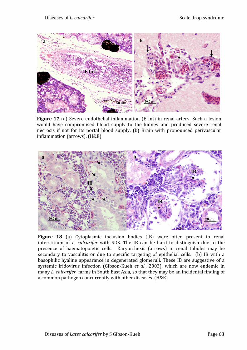
Diseases of L. calcarifer Scale drop syndrome
Diseases of Lates calcarifer by S Gibson-Kueh Page 63
Figure 18 (a) Cytoplasmic inclusion bodies (IB) were often present in renal interstitium of L. calcarifer with SDS. The IB can be hard to distinguish due to the presence of haematopoietic cells. Karyorrhexis (arrows) in renal tubules may be secondary to vasculitis or due to specific targeting of epithelial cells. (b) IB with a basophilic hyaline appearance in degenerated glomeruli. These IB are suggestive of a systemic iridovirus infection (Gibson-Kueh et al., 2003), which are now endemic in many L. calcarifer farms in South East Asia, so that they may be an incidental finding of a common pathogen concurrently with other diseases. (H&E)
b
20 m
IB
IB
a
IB
Figure 17 (a) Severe endothelial inflammation (E Inf) in renal artery. Such a lesion would have compromised blood supply to the kidney and produced severe renal necrosis if not for its portal blood supply. (b) Brain with pronounced perivascular inflammation (arrows). (H&E)
50 m
E Inf
a
b

Diseases of L. calcarifer Scale drop syndrome
Diseases of Lates calcarifer by S Gibson-Kueh Page 64
Figure 19 (a) Hydropic change (D) in choroid rete of eye. (b) The heart was often a good place to look for viral inclusion bodies without the ‘clutter’ of haematopoietic tissues. Enlarged basophilic inclusion bodies (IB) can be seen bulging from the endocardium. There was a subacute to chronic pericarditis (*). (c) Hypertrophied myocardial endothelium (E) needs to be differentiated from (d) basophilic inclusion bodies (IBs) located on endothelium. The presence of the IB typical of systemic iridoviral disease must be taken into context of a disease that can be endemic, and may occur concurrently with other diseases. (H&E)
* IB
b
IB
s
d
50 m D
a
c
E

Diseases of L. calcarifer Scale drop syndrome
Diseases of Lates calcarifer by S Gibson-Kueh Page 65
Figure 20 (a) Prominent hepatic lobulation in L. calcarifer with SDS. This may be attributed to increased cell death. Fatty liver or hepatic lipidosis (Fa) typified by vacuolation can be focal to extensive involving whole segments of livers, as seen grossly in fig. 13. Liver with reduced glycogen reserves stained more intensely basophilic (NG). (b) Diffuse karyorrhexis (arrows) and nuclear chromatin margination (MNC) in liver. (H&E)
200m
NG
Fa
a
b
20 m
MNC
20m
*
*
200m
*
Figure 21 Multifocal areas of gastric glands necrosis (*) associated with perivasculitis (arrows) in L. calcarifer with SDS. Demarcated area is shown at higher magnification in inset: paler areas of gastric gland necrosis and karyolysis (arrows) in gastric mucosa. (H&E)

Diseases of L. calcarifer Scale drop syndrome
Diseases of Lates calcarifer by S Gibson-Kueh Page 66
4.3.3 Transmission electron microscopy
Transmission electron microscopic changes were dominated by tissue
degeneration and necrosis. Although the identity of the necrotic cells was
unclear, renal haematopoietic cells were remarkably unaffected. Necrotic cells
often had marginated nuclear chromatin or the presence of electron dense
lamellae (Fig. 22). Enveloped hexagonal virions measuring 188 to 269 nm (n=8)
were observed in spleen and kidney (Fig. 23a). Where virions were observed
together with ribosomes, they were presumed to be located within remains of
cytoplasm (Figs 23a & b). The double enveloped virions in the kidney appeared
to have budded from the cytoplasm, as evidenced by the absence of ribosomes
(Fig. 22 inset). Much smaller hexagonal virions with an average diameter of
133nm (n=2) and electron-lucent cores were also occasionally observed (Fig.
23b).
4.3.4 Immunohistochemistry, PCR and viral isolation
Fluorescent immunohistochemistry on paraffin block tissues using the Red Sea
bream iridovirus (RSIV) monoclonal antibody M10 (Nakajima & Sorimachi 1995;
Nakajima et al. 1995) gave negative results. Lates calcarifer tissues with a
systemic iridoviral disease served as positive control (Fig. 25).
Polymerase chain reaction (PCR) using RSIV primer set 1 which targets a
large range of iridoviruses (Kurita et al. 1998) also gave negative results.
Attempts at viral isolation on ATCC gruntfin, Haemulon sciurus Shaw (GF) and
Asian seabass, Lates calcarifer Bloch (SB) cells (Chong et al. 1987) were not
successful (Wang Y.H., unpublished data).

Diseases of L. calcarifer Scale drop syndrome
Diseases of Lates calcarifer by S Gibson-Kueh Page 67
Figure 22 L. calcarifer with SDS have remarkably normal renal haematopoietic cells (Hc). Necrotic cells have marginated chromatin (MNC) or presence of very electron dense lamellae (L) in nucleus. Red blood cells (rbc) in blood vessel. Demarcated area is shown at higher magnification in inset: double enveloped hexagonal virions with ribosomes absent, presumably as virions have budded out from cytoplasm of cell.
5 m
rbc
MNC
MNC
Hc
Hc
L
200nm
Figure 23 (a)Enveloped hexagonal virions in spleen in remains of cytoplasmic ribosomes (r), with a size range of 188 to 269 nm (n=8). (b) Smaller enveloped hexagonal virions measuring 133nm (n=2) in cytoplasmic remnants in kidney. These virions have electron-lucent nucleocapsids.
100 nm
b
200 nm
r
r
a

Diseases of L. calcarifer Scale drop syndrome
Diseases of Lates calcarifer by S Gibson-Kueh Page 68
4.4 Discussion
This first pathological description of scale-drop syndrome (SDS) provides a case
definition for future work, including epidemiological studies. The cause of SDS is
not known, but it appears to be infectious. Moreover, the histopathological
changes and the presence of enveloped hexagonal virions in the absence of other
consistent causal agents support the possibility of a viral aetiology.
The lesions seen in SDS differed in several ways from other systemic
iridoviral diseases (Reddacliff & Whittington 1996; Gibson-Kueh, Netto, Ngoh-
Lim, Chang, Ho, Qin, Chua, Ng & Ferguson 2003). In systemic iridoviral disease,
inclusion bodies were shown to be viral infected fibroblasts located under
vascular endothelium, which eventually rupture to release virions into the
circulation, and resulting in vascular damage and necrosis in spleen and kidney.
This may be accompanied by limited to extensive necrosis of parenchyma in
organs such as the spleen or just the glomerulus in the kidney (Gibson-Kueh et al.
2003). In other iridoviral disease such as epizootic haematopoietic necrosis, the
main lesion observed was necrosis of haematopoietic tissues in spleen and
kidney (Reddacliff & Whittington 1996). In contrast, the pattern of tissue
necrosis in SDS suggested that infarction was linked directly to the vasculitis that
was such a prominent feature overall. Vascular compromise in the skin would be
expected to affect the health of scale beds and lead to scale loss, one of the main
clinical features of SDS. The extensive necrosis of the dermis (Fig. 13 & 15b)
where an estimated 80% of lymph, almost four times the volume of circulating
blood is located, would in itself be fatal (Ferguson 2006). The karyorrhexis in
liver, kidney, spleen, stomach and intestines suggest a disease associated with
specific target cells, possibly of epithelial origin.

Diseases of L. calcarifer Scale drop syndrome
Diseases of Lates calcarifer by S Gibson-Kueh Page 69
There was no histopathological evidence for an overt bacterial
infection including Tenacibaculum. The vasculitis of SDS may, however, be the
result of an immune hypersensitivity reaction to bacterial antigens such as that
suggested for strawberry disease in salmonids (Ferguson, Girons, Rizgalla,
LaPatra, Branson, McKenzie, Davies, Collins, Diab & Crumlish 2006).
Based on size and morphology, some of the observed virions
resembled iridovirus (Fig. 23a) but fluorescent immunohistochemistry on
paraffin block tissues using the Red Sea bream iridovirus (RSIV) monoclonal
antibody M10 (Nakajima & Sorimachi 1995; Nakajima et al. 1995) gave negative
results. Polymerase chain reaction (PCR) using RSIV primer set 1 which targets a
large range of iridoviruses (Kurita et al. 1998) also gave negative results.
Attempts at viral isolation on ATCC gruntfin, Haemulon sciurus Shaw (GF) and
Asian seabass, Lates calcarifer Bloch (SB) cells (Chong et al. 1987) were not
successful (Wang Y.H., unpublished data). There are many explanations why
virus isolation may be unsuccessful. These include the choice of inappropriate
cell lines, or possibly the low numbers of virions present in tissues, even those
with significant pathological changes. The enveloped nature of virions may also
explain the difficulty in culturing a potentially fragile virus. While it is very
tempting to implicate the virus-like particles in SDS, it is important to remember
that they were relatively hard to find. Moreover, as presented in Chapter 5,
systemic iridoviral disease is now endemic in some L. calcarifer farms and their
presence in fish with SDS could be an incidental finding of a common pathogen.
The vasculitis and associated necrosis that were the hallmarks of SDS
are not typical of iridoviral disease, but they were seen in herpesvirus infections,
as were cells with marginated nuclear chromatin in target organs such as liver,

Diseases of L. calcarifer Scale drop syndrome
Diseases of Lates calcarifer by S Gibson-Kueh Page 70
spleen, kidney and gastrointestinal tracts (Wolf, Herman & Carlson 1972; Plumb
& Gaines 1975; Plumb, Gaines, Mora & Bradley 1975; Hedrick, Gilad, Yun,
Spangenburg, Marty, Nordhausen, Kebus, Bercovier & Eldar 2000; Garver, Al-
Hussinee, Hawley, Schroeder, Edes, LePage, Contador, Russell, Lord, Stevenson,
Souter, Wright & Lumsden 2010). Although Cowdry type A inclusions were not
observed in SDS, this was also the case in herpesviral disease of pilchards (Jones,
Hyatt, Hine, Whittington, Griffin & Bax 1997). The electron-dense lamellae
observed in the nucleus in Fig. 22 resemble those described in cell culture of
channel catfish herpevirus (CCV). The extensive release of CCV into supernatant
as early as 4 hours post-inoculation into cell culture may explain the intense
vasculitis often observed in herpesviral disease (Wolf & Darlington 1971).
Assuming that SDS is indeed infectious, what was its source in these L.
calcarifer? It is possible that the fish were infected after stocking in sea cages. As
there are often multi-age groups of L. calcarifer in the vicinity of affected cages or
farms, it is likely that the infection originated from older carrier fish. Carrier fish
have been implicated in a recent review on iridoviruses causing epizootic
haematopoietic necrosis and it has long been suspected in channel catfish virus
disease (CCVD) (Plumb 1986; Whittington, Becker & Dennis, 2010). If infection
was present in fish from early on, it is peculiar that the disease onset occurred
well after the initial stressful period post-stocking when most viral diseases
usually occur, e.g. ‘sleepy grouper disease’ (SGD) or other systemic iridoviral
disease (Chua, Ng, Ng, Loo & Wee 1994; Gibson-Kueh, Ngoh-Lim, Netto, Kurita,
Nakajima & Ng 2004b). There is a possibility that the susceptibility of fish to SDS
was inversely related to age, with implications of maternal antibody protection
as observed in CCVD (Hanson, Rudis & Petrie-Hanson 2004).
Diseases of L. calcarifer Scale drop syndrome
Diseases of Lates calcarifer by S Gibson-Kueh Page 71
With the increasing culture of L. calcarifer, SDS is expected to occur
more frequently. Since the publication of SDS by Gibson-Kueh et al. (2012), the
size range of L. calcarifer affected by SDS has been extended to 3 to 5kg fish. SDS
affects larger more valuable fish and will result in greater economic losses. The
case definition for SDS is proposed as a systemic vasculitis in Asian seabass, L.
calcarifer Bloch, associated with tissue necrosis in all major organs including the
skin, with apparent targeting of cells of epithelial origin. The inclusion bodies
observed were thought to be due to an underlying systemic iridoviral infection,
although this needs further investigation. Further study of this disease to
elucidate its definitive aetiology via the use of degenerate PCRs designed to
detect large DNA viruses (Hanson, Rudis, Vasquez-Lee & Montgomery 2006) and
attempts to isolate the agent via use of more cell lines are underway.
Cohabitation of naïve fish with L. calcarifer with overt clinical signs of SDS, or
subjecting clinically healthy L. calcarifer from farms with history of SDS to
external stressors under laboratory conditions to see if SDS can be reproduced,
may indeed help us understand this disease better.

Diseases of L. calcarifer Other viral & bacterial diseases
Diseases of Lates calcarifer by S Gibson-Kueh Page 72
Chapter 5 Other viral and bacterial diseases observed in cultured Asian
seabass (or barramundi), Lates calcarifer Bloch
Abstract
This is a first report of systemic iridoviral disease in Asian seabass (or
barramundi) Lates calcarifer. The iridoviral disease was observed in 5 to 20g L.
calcarifer two to three weeks post-transfer into sea-cages at two farms.
Clinically healthy L. calcarifer fry had histopathological lesions indicative of
systemic iridovirus infection and were tested positive by PCR using Red Sea
bream iridovirus (RSIV) primer 1. This suggested that the iridovirus infection
occurred before stocking in sea cages, and did not originate from wild fish or
older fish in adjacent cages. Viral nervous necrosis (VNN) remained a significant
disease of L. calcarifer fry. Lower mortality and less severe vacuolative
encelopathy and retinopathy were observed in older L. calcarifer fry with VNN.
Pot belly disease (PBD) was previously reported in 3-week-old L. calcarifer fry.
In this study, extensive granulomatous enteritis associated with intracellular
coccobacilli similar to PBD in fry was observed in 120g L. calcarifer in sea-cages.
Nocardiosis is an emerging disease of cultured marine food fish. This is a first
description of nocardiosis in L. calcarifer in sea cages. Streptococcosis,
tenacibaculosis and vibriosis were often observed concurrently with systemic
iridoviral disease and PBD.

Diseases of L. calcarifer Other viral & bacterial diseases
Diseases of Lates calcarifer by S Gibson-Kueh Page 73
5.1 Introduction
Systemic iridoviral disease has been previously reported in many marine fish
species (Chua, Ng, Ng, Loo & Wee 1994; Matsuoka, Inouye & Nakajima 1996;
Chou, Hsu & Peng 1998; Nakajima, Inouye & Sorimachi 1998; Gibson-Kueh,
Ngoh-Lim, Netto, Kurita, Nakajima & Ng 2004b). Histologically, iridovirus
infections are recognized by the presence of large numbers of basophilic
hypertrophied cells in all major organs (Gibson-Kueh, Netto, Ngoh-Lim, Chang,
Ho, Qin, Chua, Ng & Ferguson 2003). Hence systemic iridovirus has been
recently termed megalocytivirus (Mahardika, Haryanti, Muzaki & Miyazaki
2008).
Viral nervous necrosis (VNN) is a serious disease of cultured marine fish
including Asian seabass (or barramundi) Lates calcarifer fry. VNN produced very
distinctive histopathology described as vacuolative encephalopathy and
retinopathy (Glazebrook, Heasman & de Beer 1990; Munday, Langdon, Hyatt &
Humphrey 1992; Maeno, De La Pena & Cruz-Lacierda 2004; Parameswaran,
Rajesh Kumar, Ishaq Ahmed & Sahul Hameed 2008). Both systemic iridoviral
disease and VNN can be confirmed via PCR (Chang, Ngoh & Kueh 1997; Kurita,
Nakajima, Hirono & Aoki 1998; Gibson-Kueh et al. 2004b).
‘Pot belly disease’ (PBD) was previously reported in L. calcarifer fry with
mortalities of 80 to 100%. ‘Pot belly disease’ is characterized by perforating
granulomatous enteritis and peritonitis in association with large intracellular
gram negative coccobacilli (Gibson-Kueh, Crumlish & Ferguson 2004a). All
attempts to isolate the bacterium associated with PBD have been unsuccessful.
Molecular methods to test for PBD are not available, and diagnosis is solely by
histopathology.
Diseases of L. calcarifer Other viral & bacterial diseases
Diseases of Lates calcarifer by S Gibson-Kueh Page 74
Streptococcosis is a serious bacterial disease of farmed marine finfish
including L. calcarifer, and can cause severe mortality in fish up to 3kg
bodyweight (Creeper & Buller 2006; Labrie, Komar & Sheehan 2007; Wendover
2009). Outbreaks of streptococcosis have been associated with stormy weather
conditions on several occasions and in some cases are linked to increased river
discharges (Siung-Chang & Lum-Kong 2001; Creeper & Buller 2006; Humphrey,
Benedict & Small 2010).
Although vibriosis and tenacibaculosis can cause serious mortality in
cultured L. calcarifer, they are generally considered as opportunistic diseases in
stressed fish (Ruangpan 1988; Azad, Thirunavukkarasu, Kailasam & Rajan 2004;
Avendano-Herrera et al. 2006; Labrie et al. 2007; Humphrey et al. 2010).
Nocardiosis is an emerging disease in cultured marine finfish. Nocardia
can be recognized histologically as gram positive, acid fast with Fite Faraco stain,
filamentous and often branching bacteria in association with systemic
granulomatous necrotic lesions. Culture techniques and PCR have been
established for the detection of Nocardia (Kudo, Hatai & Seino 1988; Labrie, Ng,
Tan, Komar, Ho & Grisez 2008).
This is a first report of systemic iridoviral disease and nocardiosis in L.
calcarifer. Pot belly disease was found to affect older L. calcarifer than previously
reported. Viral and bacterial diseases reported in this study can cause significant
mortality in cultured L. calcarifer.
5.2 Materials and methods
5.2.1 Background
Table 2 is a summary of the general information on Farms A to D including
location, proximity to other farms, general husbandry practices and number of

Diseases of L. calcarifer Other viral & bacterial diseases
Diseases of Lates calcarifer by S Gibson-Kueh Page 75
years each farm has been in operation. Appendix 1.1 is a questionaire completed
by Farm A and appendix 1.2 by Farm B, on husbandry practices.
Materials examined included fixed L. calcarifer tissues sent to Murdoch
University, Perth by Farm A in Indonesia between 2007 and 2010. The range of
diseases encountered in Farm A is well documented. Farm A has a history of
systemic iridoviral disease in 10-20g L. calcarifer fingerlings two to three weeks
post transfer into sea cages. As the farm was comtemplating using an iridovirus
vaccine, the purpose of this study was to determine if the systemic iridovirus
infection occurred before transfer to sea cages. Lates calcarifer tissues were
examined by histopathology at Murdoch University and corresponding samples
sent to Merck Aquatic Animal Health Laboratory in Singapore for testing by PCR.
All samples examined at Murdoch University were either formalin fixed or
alcohol fixed L. calcarifer tissues, and presumptive diagnoses are based mainly
on histopathological evidence. PCR, immunohistochemistry and in-situ
hybridization was carried out only on selected L. calcarifer tissues from Farm A
with histopathological evidence of a systemic iridoviral disease.
Histology slides from L. calcarifer cases submitted to Aquatic Animal
Health Laboratory (AAHL), Agri-Food & Veterinary Authority of Singapore in
Singapore from 1993 to 2006, and Fish Health Laboratory, WA Department of
Fisheries from 2003 to 2008 were also examined. Reference to specific bacteria
species are based on results available in case records from routine culture or
PCR.

Diseases of L. calcarifer Other viral & bacterial diseases
Diseases of Lates calcarifer by S Gibson-Kueh Page 76
5.2.2 Light microscopy (LM)
Tissues were processed for light microscopy as outlined in 2.2.2. Tissue sections
were stained by haematoxylin & eosin (H&E) and Fite Faraco, a modified Ziehl
Neelsen stain.
5.2.3 Transmission electron microscopy (TEM)
Tissues were processed for electron microscopy as outlined in 2.2.3. Ultra-thin
sections were stained with uranyl acetate and lead citrate for viewing on a
Philips CM100 Bio TEM.

Diseases of L. calcarifer Other viral & bacterial diseases
Diseases of Lates calcarifer by S Gibson-Kueh Page 77
Table 2 Summary of general information on Farm A to D
Location Years in operation
Fish species cultured
Farm A island farm in Indonesia with the nearest fish farm 10km away
10 Mainly L. calcarifer
produces fry from its own broodstock in a land based hatchery. Nursery fish are routinely vaccinated with the Intervet Streptococcus vaccine at 10g bodyweight. 20-40g fish are stocked into 12m or 18m diameter circular cages that are 10m deep, or square cages.
50,000 to 100,000 fish are stocked into circular cages 12 or 18 m in diameter, and 10m deep. Estimated >500,000 fish on farm at any one time.
Farm B island farm in Indonesia with the nearest fish farm 200-300m away
5 Mainly L. calcarifer
buys in 30 do L. calcarifer fry to grow to 2-8g fish before stocking into sea cages. The fish are stocked mainly into 640 wooden cages 3.25 x 3.25 x 2.5m. The farm vaccinates fish against streptococcosis.
1000-3000 fish are stocked per cage. Estimated >500,000 fish on farm at any one time.
Farm C sea cage farm located in close proximity to at least 20 other farms in a narrow strait off Singapore.
>10 Stocks multiple fish species besides L. calcarifer
small scale farm with less than 50 wooden cages each measuring approximately 3 x 3 x 2.5m, and buys in L. calcarifer fingerlings for stocking directly into sea cages
1000-3000 fish are stocked per cage. Estimated >20,000 fish on farm at any one time.
Farm D sea cage farm located a 45-minute boat ride from the next nearest fish farms in Malaysia.
3 Mainly L. calcarifer
small scale farm with less than 50 wooden cages each measuring approximately 3 x 3 x 2.5m, and buys in L. calcarifer fingerlings for stocking directly into sea cages
1000-3000 fish are stocked per cage. Estimated >50,000 fish on farm at any one time.

Diseases of L. calcarifer Other viral & bacterial diseases
Diseases of Lates calcarifer by S Gibson-Kueh Page 78
5.2.4 Immunohistochemistry using RSIV M10
Formalin fixed paraffin embedded (FFPE) L. calcarifer tissue sections of selected
cases from Farm A with histopathological signs of a systemic iridoviral disease
(Gibson-Kueh et al. 2003) were tested by immunohistochemistry (IHC) using a
monoclonal antibody against the Red Sea bream iridovirus (RSIV M10)
(Nakajima & Sorimachi, 1995). The RSIV M10 was kindly supplied by Dr Jun
Kurita, National Research Institute of Aquaculture, Fisheries Research Agency,
Mie, Japan. The immunohistochemistry method after Adams and de Mateo
(1994) with modifications as described below was used. Negative control is
carried out with substitution of the RSIV M10 with blocking solution.
FFPE 5µm tissue sections on silanised slides were deparaffinized in two
changes of xylene and rehydrated through an ethanol series to tap water.
Antigen retrieval was performed by placing rehydrated tissue sections in Tris
EDTA buffer (pH 9) bath in a domestic microwave (Kambrook Model KER-
686LE) and subjected to ‘reheat’ for 4 minutes and ‘low heat’ function for 4
minutes, followed by cooling in running tap water.
A blocking step was carried out by adding 0.1% bovine serum albumin in
Tris buffered saline (TBS) (pH 7) with 0.05% Tween 20 directly to tissue
sections for a 10-minute incubation. After the blocking solution was tapped off,
the primary antibody RSIV M10 diluted 1/10 in blocking solution was added to
each tissue section for a 30-minute incubation. FFPE tissues incubated with
blocking solution instead of RSIV M10 served as the negative control. The
primary antibody was rinsed off, followed by addition of TBS (pH7) with 0.05%
Tween 20 to tissue sections for a 5-minute incubation.

Diseases of L. calcarifer Other viral & bacterial diseases
Diseases of Lates calcarifer by S Gibson-Kueh Page 79
Polyclonal rabbit anti-mouse immunoglobulin conjugated with alkaline
phosphatase (Dako, US) diluted 1:50 in blocking solution was added to tissue
sections for a 30-minute incubation, followed by rinsing in TBS (pH7) with
0.05% Tween 20 added to each slide for 5-minute incubation. Liquid permanent
red (Dako, US) with levamisole (300μg/ml) to block endogenous alkaline
phosphatase activity was added to tissue sections for a 20- minute incubation,
and rinsed off with tap water. Tissue sections were counterstained with
haematoxylin for 1 minute, rinsed in tap water and flicked dry prior to cover-
slipping with Farmount mounting media (Dako, US).
5.2.5 Polymerase chain reaction & in situ hybridization
Red Sea bream iridovirus (RSIV) PCR as published by Kurita et al. (1998) on L.
calcarifer tissues from Farm A was carried out by the Merck Aquatic Animal
Health Laboratory in Singapore. RSIV PCR was carried out at Murdoch University
to generate dioxigenin labeled DNA probes from PCR products for in situ
hybridization (ISH) on formalin fixed paraffin embedded (FFPE) tissues. DNA was
extracted from alcohol fixed L. calcarifer tissues from Farm A with histopathological
evidence of systemic iridoviral disease, using a QIAamp DNA FFPE Tissue kit
(Qiagen, Germany), according to the manufacturer’s instructions.
Briefly, PCR reaction mixes made up using TopTaq Master Mix (Qiagen, US)
according to the manufacturer’s instructions, containing 0.2 M of RSIV primer set 1
and 1 l of extracted DNA were incubated in an Eppendorf Mastercycler under PCR
conditions as described by Kurita et al., 1998. After amplification, 10 l of PCR
products from each reaction tube was analyzed in a 1 % agarose gel in 0.5x TBE
buffer with SybrSafe, and visualized under UV illumination.

Diseases of L. calcarifer Other viral & bacterial diseases
Diseases of Lates calcarifer by S Gibson-Kueh Page 80
RSIV PCR products obtained from fish tissues from Farm A were purified
using QIAquick PCR purification spin columns (Qiagen, Germany) and labeled with
dioxigenin (DIG) using DIG-nick translation mix (Roche, Germany), according to
the manufacturer’s instructions. The DIG-nick translation kit results in the
production of a mixture of DIG labeled DNA probes between 200 to 500bp in size
from purified PCR products. In situ hybridization was carried out using these DIG-
labeled DNA probes on FFPE L. calcarifer tissues with overt clinical and
histopathological evidence of a systemic iridoviral disease from the same Farm A.
PCR products were also purified using QIAquick PCR purification spin columns
(Qiagen, Germany), and send to a commercial company for sequencing.
DIG labeled DNA probes made from 1 g of PCR products were used to
make up 1 ml of DNA probe mixture that contained 50% formamide, 10%
dextran sulphate and 2x Saline Sodium Citrate (SSC) buffer. The DNA probe
mixture was stored at 4oC until required. A negative control DIG labeled DNA
probe generated from PCR products to detect a papilloma virus of bandicoot was
kindly provided by Mark Bennett, School of Veterinary and Biomedical Sciences,
Murdoch University (Bennett, Woolford, O’Hara, Warren & Nicholls 2008).
The ISH steps are carried out as described in Section 3.2.4 for the
detection of Eimeria in L. calcarifer tissues except for the use of DIG labeled RSIV
PCR product as the DNA probe.
5.3 Diseases observed in L. calcarifer tissues examined
Table 3 is a summary of the diseases observed in L. calcarifer from Farms A to D,
and cases submitted to the Aquatic Animal Health Laboratory from 1993 to
2003. Table 4 is a summary of the diseases observed in L. calcarifer cases
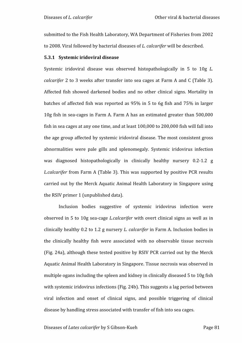
Diseases of L. calcarifer Other viral & bacterial diseases
Diseases of Lates calcarifer by S Gibson-Kueh Page 81
submitted to the Fish Health Laboratory, WA Department of Fisheries from 2002
to 2008. Viral followed by bacterial diseases of L. calcarifer will be described.
5.3.1 Systemic iridoviral disease
Systemic iridoviral disease was observed histopathologically in 5 to 10g L.
calcarifer 2 to 3 weeks after transfer into sea cages at Farm A and C (Table 3).
Affected fish showed darkened bodies and no other clinical signs. Mortality in
batches of affected fish was reported as 95% in 5 to 6g fish and 75% in larger
10g fish in sea-cages in Farm A. Farm A has an estimated greater than 500,000
fish in sea cages at any one time, and at least 100,000 to 200,000 fish will fall into
the age group affected by systemic iridoviral disease. The most consistent gross
abnormalities were pale gills and splenomegaly. Systemic iridovirus infection
was diagnosed histopathologically in clinically healthy nursery 0.2-1.2 g
L.calcarifer from Farm A (Table 3). This was supported by positive PCR results
carried out by the Merck Aquatic Animal Health Laboratory in Singapore using
the RSIV primer 1 (unpublished data).
Inclusion bodies suggestive of systemic iridovirus infection were
observed in 5 to 10g sea-cage L.calcarifer with overt clinical signs as well as in
clinically healthy 0.2 to 1.2 g nursery L. calcarifer in Farm A. Inclusion bodies in
the clinically healthy fish were associated with no observable tissue necrosis
(Fig. 24a), although these tested positive by RSIV PCR carried out by the Merck
Aquatic Animal Health Laboratory in Singapore. Tissue necrosis was observed in
multiple ogans including the spleen and kidney in clinically diseased 5 to 10g fish
with systemic iridovirus infections (Fig. 24b). This suggests a lag period between
viral infection and onset of clinical signs, and possible triggering of clinical
disease by handling stress associated with transfer of fish into sea cages.

Diseases of L. calcarifer Other viral & bacterial diseases
Diseases of Lates calcarifer by S Gibson-Kueh Page 82
Tissues of clinically diseased L. calcarifer from Farm A with
histopathological evidence of a systemic iridovirus infection reacted positively
with Red Sea Bream iridovirus (RSIV) monoclonal antibody M10 by fluorescent
IHC (Fig. 25). Negative control tissue with substitution of the RSIV M10 by
blocking solution showed no fluorescence (Fig. 25 inset). Sections of intestines,
spleen, liver and kidney with inclusion bodies gave varying degrees of positive
reaction with the RSIV M10 IHC, with the most intense results observed in heart
tissue (Fig. 25). Immunohistochemistry using RSIV M10 on tissues with inclusion
bodies but from clinically healthy L. calcarifer gave negative results. It is possible
that the antigen against which the monoclonal RSIV M10 is not yet expressed in
these early viral inclusions in subclinical fish.
RSIV PCR on DNA extracted from alcohol fixed L. calcarifer tissues from
Farm A with histopathological evidence of systemic iridoviral disease produced the
expected 570bp product (Fig. 26). In situ hybridization using DIG labeled PCR
products generated using RSIV primer 1 on tissues with histopathological signs
of a systemic iridovirus infective did not yield any reactions, despite use of
optimization steps such as microwave treatment in Tris EDTA buffer (pH 9). To
show that the PCR products were generated from iridoviral DNA, the DNA
sequence of the purified PCR products carried out using RSIV primer 1 was
analysed using the blast function on GenBank, and found to be 99% homologous
to aligned sequences of three iridovirus isolates: an infectious spleen and kidney
necrosis virus (AF371960), iridovirus of barramundi (FJ222554) and grouper
iridovirus (AY059399) (unpublished data).
Diseases of L. calcarifer Other viral & bacterial diseases
Diseases of Lates calcarifer by S Gibson-Kueh Page 83
PCR using RSIV primer set 1 (Kurita et al. 1998) on alcohol fixed spleen
tissues of L. calcarifer from Farm A with overt clinical and histopathological signs
of systemic iridoviral disease produced the expected 570bp product (Fig. 26).
Attempts to perform in situ hybridizations (ISH) using DIG-labeled RSIV PCR
products on FFPE L. calcarifer tissues from fish with clinical and
histopathological signs of systemic iridoviral disease from Farm A gave negative
results.
Figure 24 Systemic iridovirus infections in (a) clinically healthy 0.2g L. calcarifer, and (b) clinically diseased 4g L. calcarifer. Tissue degeneration with hydropic changes and cell deaths (D) were more evident in the clinically diseased 4g fish in b. Inclusion bodies (IB) could be observed in both fish.
IB IB
20m
a
IB
IB D
D
20m
b

Diseases of L. calcarifer Other viral & bacterial diseases
Diseases of Lates calcarifer by S Gibson-Kueh Page 84
Figure 26 PCR on alcohol fixed L. calcarifer tissues from diseased fish with histopathological signs of a systemic iridovirus infection showed an expected 570bp product (Kurita et al. 1998). Lane 1 & 11: 100bp ladder, Lane 2: negative control ultrapure water, Lanes 3-5: Fish 1 (triplicates), Lanes 6-8: Fish 2 (triplicates), Lanes 9-10: Fish 3 (duplicates)
1 2 3 4 5 6 7 8 9 10 11
500bp
Figure 25 Cytoplasmic inclusion bodies in cardiac tissue with positive red fluorescence in immunohistochemistry using RSIV M10 and Permanent Red (Dako). Inset shows no fluorescence in negative control.

Diseases of L. calcarifer Other viral & bacterial diseases
Diseases of Lates calcarifer by S Gibson-Kueh Page 85
5.3.2 Viral nervous necrosis (VNN)
Viral nervous necrosis (VNN) was diagnosed histopathologically in 2 to 3 week-
old L. calcarifer with mortalities of up to 100% at Farm A. Mortality was reported
as less than 50% in older fry with VNN at Farm A. Clinical signs observed
included inappetance and pale fish, with or without abnormal neurological signs
(Table 3).
Marked vacuolation and abundant basophilic inclusion bodies were
observed histopathologically in the grey matter of the brain and in the retina of
less than 3-week old fry affected by VNN (Figs 27a & b). In two cases of VNN
submitted to the Fish Health Laboratory in Perth, in fry older than 3 weeks of
age (Table 4), the vacuolation in brain and eye was less marked and inclusion
bodies present in smaller numbers or not at all (Fig. 27c). Vacuolation was
confined to the grey matter of the brain and spinal cord. TEM showed specific
targeting of nerve cells by VNNV (Fig. 27d).

Diseases of L. calcarifer Other viral & bacterial diseases
Diseases of Lates calcarifer by S Gibson-Kueh Page 86
a
200m
Figure 27 (a, b & d) Viral encephalopathy and retinopathy or viral nervous necrosis (VNN) in 12 do L. calcarifer fry from Farm A (a) Severe vacuolation (arrows) in the brain and eye. (b) Vacuoles were often associated with basophilic inclusion bodies (arrows). (c) VNN in 22 do fry submitted to the Fish Health Laboratory in Perth, with mild vacuolation (arrows) and no inclusion bodies. (H&E) (d) Nerve cells with dendritic processes (arrows) and electron dense inclusions (IB) from VNNV infection (TEM). This explains the location of vacuolation in grey matter of the brain where the cell bodies of neurons are located.
b
50m
2m
IB
IB
IB
IB
d
100m
c

Diseases of L. calcarifer Other viral & bacterial diseases
Diseases of Lates calcarifer by S Gibson-Kueh Page 87
5.3.3 ‘Pot Belly Disease’ (PBD)
Mortality of 90 to 95% was reported in L. calcarifer fry at Farm A, in association
with ‘pot belly disease’ (PBD) (Table 3). PBD was also observed in older L.
calcarifer examined in this study: 1 to 5g L. calcarifer in the land-based nursery
of Farm A, and up to 120g L. calcarifer at two sea cage Farms B and D.
While the granulomatous enteritis tended to be less severe in older fish, it
affected significant portions of the intestine in affected fish (Figs 28a & b).
Clusters of large coccobacilli were observed in both groups of older fish (Figs 28a
& b insets). Remnants of peritonitis with and without the presence of coccobacilli
were observed in some fish. PBD often occurred concurrently with other
diseases such as systemic iridoviral disease in L. calcarifer in sea cages (Table 3).
The contribution of PBD to percentage mortality in grow-out cages in not known,
as it is often complicated by other concurrent diseases.
5.3.4 Streptococcosis
Streptococcosis was observed in 4.5 to 15g, 100 to 500g and up to 3kg L.
calcarifer with cumulative mortality greater than 40 to 50% (Tables 3 & 4). In
fish smaller than 15g, non-specific clinical signs such as darkened bodies,
lethargy and anorexia were observed. In 100 to 200g fish, additional clinical
signs such as exophthalmos, cloudy eyes and abnormal neurological signs were
observed (Table 3). Onset of mortality tended to be more acute, occurring over
several days in smaller fish while larger fish exhibited low grade mortality over
several weeks. Large numbers of gram positive, coccoid-shaped bacteria in pairs
or chains were present within tissues and blood vessels in all organs including
the brain (Fig. 29a) and intracellularly within phagocytic cells in spleen, kidney

Diseases of L. calcarifer Other viral & bacterial diseases
Diseases of Lates calcarifer by S Gibson-Kueh Page 88
and cardiac endothelium (Fig. 29b). Severe multi-organ multifocal to coalescing
necrosis may be observed.
Figure 28 (a) Pot belly disease (PBD) in nursery-reared 1.8g L. calcarifer with granulomatous enteritis (G) as well as unaffected parts of intestines (N). (b) PBD in 80-120g fish in sea cages. Although associated pathology was less marked in these older fish than in younger fry, the granulomatous enteritis has affected significant parts of intestine. Insets: Higher magnification with clusters of large coccobacilli (arrows). (H&E)
G
G
N
a
500m
20µm
N
G
G
b
1mm
20m

Diseases of L. calcarifer Other viral & bacterial diseases
Diseases of Lates calcarifer by S Gibson-Kueh Page 89
5.3.5 Vibriosis and tenacibaculosis
Vibriosis and tenacibaculosis often occurred post handling in L. calcarifer less
than 25g (Tables 3 & 4). Mats of filamentous bacteria were observed
histopathologically on skin, mouth, eye and gill in association with severe tissue
necrosis in tenacibaculosis (Fig. 29c). Tenacibaculosis was often observed
concurrently with other diseases such as systemic iridoviral disease, PBD,
streptococcosis or vibriosis (Table 3). Filamentous and/or rod shaped bacteria
were frequently observed concurrently on necrotic skin and gill lesions. Based
on case records of materials examined, the most common rod shaped bacteria
isolated were Vibrio species. Vibriosis may progress systemically resulting in
multifocal to extensive necrosis of spleen, kidney and liver, whereas
tenacibaculosis typically presents as external infection of skin, gill, eye and
mouth.
20m
a
10m
b
Figure 29 (a) Coccoid-shaped bacteria, presumably Streptococcus species were observed within blood vessels (arrows) in various organs including the brain, and (b) within phagocytic endothelium lining the heart (arrows). (c) Clusters of filamentous rods (arrows), presumably Tenacibaculum maritimum associated with gill necrosis.
100m
c

Diseases of L. calcarifer Other viral & bacterial diseases
Diseases of Lates calcarifer by S Gibson-Kueh Page 90
5.3.5 Nocardiosis
Nocardiosis was observed in 50 to 120g L. calcarifer at two sea-cage farms: Farm
B in Indonesia and Farm D in Malaysia (Table 3). Moderate to severe
granulomatous lesions were observed in multiple organs including the spleen,
kidney, liver, intestines, heart, eye and gills. Filamentous rods stained positively
with Fite Faraco, a modified Ziehl Neelsen stain, were observed in some of these
granulomatous lesions (Fig. 30 inset). Filamentous bacteria were present in
larger numbers in the choroid rete of the eye rather than other organs, in one
severely affected fish from Farm B (Fig. 30). In both these two cases of
nocardiosis in L. calcarifer in sea-cage farms, it occurred concurrently with PBD.
Both Farms B and D also culture pompano, Trachinotus blochii, in which
nocardiosis was frequently observed.
Figure 30 Severe granulomatous response (arrows) was observed in the choroid rete of the eye of this severely affected 50g L. calcarifer from Farm B. The branching filamentous rods stained positively with Fite Faraco (arrows), a modified Ziehl Neelsen stain (inset). Granulomatous lesions were also observed in the intestines, liver, gills, heart, kidney and spleen of this fish although with few to no filamentous bacteria (not shown). (H&E)
10m

Diseases of L. calcarifer Other viral & bacterial diseases
Diseases of Lates calcarifer by S Gibson-Kueh Page 91
5.3.6 Chronic peritonitis
Moderate to severe chronic peritonitis was observed in both clinically healthy as
well as diseased fish samples submitted to the Fish Health Laboratory in Perth
(Table 4). Low grade mortalities less than 1% occured 2-4 weeks post stocking.
Affected fish have abdominal swelling and blood stained ascitic fluid. According
to the case history, mortality was associated with increased water temperatures
and feeding rates.
Adequate glycogen stores in the liver and the presence of food in
gastrointestinal tracts were observed in clinically healthy fish with chronic
peritonitis. Severe granulomatous peritonitis was associated with the presence
of mainly gram negative rods, and the occasional gram positive cocci and
coccobacilli. Pericarditis may be observed. The presence of ingested materials
associated with early granuloma formation in the peritoneal cavity suggested
gastrointestinal perforation. Bacterial culture was generally insignificant. The
aetiology of chronic peritonitis is uncertain.

Diseases of L. calcarifer Other viral & bacterial diseases
Diseases of Lates calcarifer by S Gibson-Kueh Page 92
Table 3 Diseases observed in L. calcarifer cases from Farms A to D in South East Asia, and cases submitted to AAHL
Size /no. examined Clinical History Diagnosis based on histopathological observations
Hatchery Farm A
2-4 wo fry x60
Inappetance and pale fish, with or without abnormal nervous signs. 50-100% mortality reported.
Viral nervous necrosis (Munday et al. 2002)
>3wo fry Skinny, darkened bodies, off feed, not schooling. 90-95% mortality reported.
Pot belly disease (Gibson-Kueh et al., 2004a)
Land-based nursery, Farm A
0.2 to 1.2g ( x67 fish) Clinically healthy Systemic iridovirus infection (Gibson-Kueh et al., 2003). The presence of inclusion bodies was associated with minimal tissue necrosis or inflammation. Observations correlated with positive PCR results using RSIV primer 1 (Kurita et al., 1998).
1 to 5g (x20 fish) Skinny, darkened bodies, off feed, not schooling. Enlarged abdomens.
Pot belly disease. Concurrent systemic iridoviral infection.
4.5 to 15g (x15 fish) Darkened bodies, lethargy, anorexia Streptococcosis with abundant gram positive cocci in all tissues including blood.
Sea cages
Farm A, C 5 to 10g x14 fish
Recently stocked. Darkened bodies, not schooling, swimming near surface, pale gills & enlarged spleens. Cumulative mortality 75 to 95% over 3 weeks.
Clinical systemic iridoviral disease associated with tissue necrosis. Secondary bacterial dermatitis and branchitis associated with rod shaped or filamentous bacteria, presumably Vibrio species or Tenacibaculum maritimum. Concurrent pot belly disease at Farm A.
Farm B, D 50 to120g x4 fish
Moribund fish with pale gills, systemic granuloma Nocardiosis with acid fast filamentous bacteria and concurrent pot belly disease
7 to 30g x10 fish
Recently stocked, bodyrot Dermatitis, branchitis and conjunctivitis associated with rod shaped bacteria presumably Vibrio sp.
100-200g x30 fish
Darkened bodies, lethargy, anorexia May have exophthalmos, cloudy eyes, exhibit abnormal neurological signs – spinning 0.5-2% daily mortality, cumulative exceeding 40-50% over 2-3 weeks.
Streptococcosis with abundant gram positive cocci in all tissues including blood. Bacteria isolates were tested as Streptococcus iniae by PCR (Zlotkin, Hershko & Eldar 1998) by AAHL. Multi-organ inflammation and necrosis.

Diseases of L. calcarifer Other viral & bacterial diseases
Diseases of Lates calcarifer by S Gibson-Kueh Page 93
Table 4 Diseases observed in L. calcarifer cases received at the Fish Health Laboratory, WA Department of Fisheries 2002-2008
Size /no. examined Clinical History Diagnosis based on histopathological observations
Land-based nursery >3 wo fry x60 2 cases
Abnormal swimming behavior. High mortality Viral nervous necrosis
Land-based nursery (marine) 0.8g x7 fish 1 case
Stocked as 0.15g fry from a local hatchery, with no significant post transfer losses. 60% mortality overnight followed by low grade mortality.
Tenacibaculosis. Severe oral epidermal ulcerations on mandible and necrotic gills were observed in association with filamentous rods. Mucoid cell metaplasia observed in gills. Other observations include presence of ingesta in stomach and adequate hepatic glycogen stores, supporting an acute disease onset.
Land-based recirculation tanks (marine) 5g x 40 fish 6 cases
Sea-cages >10g x6 fish 1 case
Low grade Mortalities (<1%) 2-4 weeks post stocking. Abdominal swelling and blood stained ascitic fluid. Affected fish often still have food in gastrointestinal tracts. One case history noted that mortality was associated with increased water temperatures and feeding rates.
Chronic peritonitis, uncertain aetiology. May present as chronic serositis of abdominal organs to severe granulomatous peritonitis with presence of mainly gram negative rods, and few gram positive cocci and coccobacilli in both clinically healthy as well as diseased fish. Pericarditis observed. Adequate glycogen stores in liver. The presence of ingested materials associated with early granuloma formation in peritoneal cavity suggested gastrointestinal perforation. Bacterial culture generally insignificant.
Sea-cages 200g x2 fish 1 case
>50% mortality over 3 days, peaking on day 2. Affected fish had pale gills, finrot and white skin patches.
Streptococcosis. Severe bacteriaemia with abundant coccoid shaped bacteria in spleen and kidney in association with severe necrosis. Acute branchitis. Bacteria isolated were identified as Streptococcus iniae.
Freshwater cages
350 to 500g x6 fish 1 case
Index case in 2.5 to 3kg fish, with cumulative mortality of 50%. Disease was subsequently observed in 350-500g fish in vicinity of the larger ones. Affected fish were lethargic and had exophthalmia, empty gastrointestinal tracts and enlarged spleens.
Streptococcosis. The publication by Creeper & Buller 2006 contains the full case details. Bacteria isolated were identified as S. iniae by biochemical methods. Affected fish had severe bacteraemia with abundant coccoid shaped bacteria in spleen and kidney, in association with severe necrosis. One fish had severe necrotizing branchitis with mats of filamentous bacteria, presumably Flavobacterium columnare.
Sea cages 5 to 10g x 11 fish 1 case
Recently graded and develop redness in eyes and base of operculum the next day.
Tenacibaculosis and Vibriosis. Necrosis of tissue on head/mandible and gills associated with filamentous bacteria in three fish, presumably Tenacibaculum maritimum. Rest of fish had dermatitis associated with rod shape bacteria, presumably Vibrio species. Case records showed that Vibrio species was isolated from gills while bacteria were not isolated from internal organs.

Diseases of L. calcarifer Other viral & bacterial diseases
Diseases of Lates calcarifer by S Gibson-Kueh Page 94
5.4 Discussion
Although systemic iridoviral disease has been previously reported in many
marine fish species, this study forms the first report of this disease in L.
calcarifer. Our findings of systemic iridoviral infection in clinically healthy fish in
the land-based nursery of Farm A level suggested that the infection occurred
before stocking into sea cages, and did not originate from wild fish or older fish
in adjacent sea cages. The possibility of a systemic iridovirus infection in
clinically healthy fish reiterates the importance of routinely screening fish for
subclinical infections, and will impact any vaccination programs.
Attempts to perform in situ hybridizations (ISH) using DIG-labeled RSIV
PCR products on FFPE L. calcarifer tissues with histopathological signs of
systemic iridoviral disease from Farm A gave negative results. PCR products
were generated from and tested on tissues of fish with clinical systemic iridoviral
disease from farm A. It is suspected that ISH may have been affected by the nitric
acid decalcification process used on L. calcarifer tissues, which may result in
denatured DNA (Kucukodaci, Haholu, Sucullu & Baloglu 2012). Although
immunohistochemistry gave positive results using the RSIV M10, the intensity of
the antibody reaction varied between tissues such as intestines, liver, spleen and
kidney with abundant inclusion bodies, and was most intense in heart tissues.
This may be related to the prolonged exposure of different tissues in specimens
fixed as whole fish to histoprocessing chemicals and masking of antigen (Webster,
Miller, DuSold & Ramos-Vara 2010).
Viral nervous necrosis can be difficult to diagnose histopathologically in
cases where it is associated with few vacuolations and an absence of inclusion
bodies. In such cases, diagnosis should be verified by PCR (Chang et al. 1997).

Diseases of L. calcarifer Other viral & bacterial diseases
Diseases of Lates calcarifer by S Gibson-Kueh Page 95
In a previous publication of PBD, the causative agent was identified as a
large gram negative coccobacillus, tested positive by immunohistochemistry
using polyclonal antibodies to Edwardsiella ictaluri (Gibson-Kueh et al. 2004a).
Culture and PCR detection methods for the bacteria causative agent for pot belly
disease have been established by Global Aquatic Animal Health laboratory,
Merck Animal Health Singapore, although this has not been published and hence
is not available for use at diagnostic laboratories
(http://www.thefishsite.com/articles/1086/diseases-of-farmed-barramundi-in-
asia, accessed 18 Dec 2012). Pot belly disease’ (PBD) which was originally
described only in L. calcarifer fry were observed in 120g fish in sea cages in this
study. These observations showed that PBD can occur in much older fish than
previously reported. Granulomatous enteritis affecting significant parts of
intestines are likely to have an effect on affected fish. The epidemiology of PBD in
all phases of the culture cycle of L. calcarifer needs further study, to facilitate
better disease control.
External lesions caused by Vibrio and Tenacibaculum cannot be
distinguished grossly, and usually present as ulcerations on the skin of body,
head or mouth areas or as necrosis of the gills . While the presence of rod-shaped
bacteria can signify a Vibrio infection and filamentous rods indicate a
Tenacibaculum infection, the results are best verified by bacteria culture from
freshly dead fish when possible. When bacterial cultures cannot be performed
within 6 hours on freshly dead fish by the laboratory, primary culture plates
performed at the farm level will produce optimal results (Buller 2004).
Streptococcosis is best recognized by the presence of gram positive cocci
in pairs or chains within blood vessels, and often associated with mortality in the

Diseases of L. calcarifer Other viral & bacterial diseases
Diseases of Lates calcarifer by S Gibson-Kueh Page 96
absence of other clinical signs particularly in juvenile L. calcarifer less than 15g
bodyweight. In such cases, the quickest method of diagnosis is the examination
of gram stained blood or tissue smears.
The diagnosis of nocardiosis was presumptive, based on histopathology
and positive reaction with Fite Faraco, a modified acid fast stain. Although there
is a possibility that acid fast organisms may be Mycobacterium, the acid fast
bacterium detected in this case was very long and filamentous rather than the
rod shape typical for Mycobacterium. Confirmation by isolation and further
identification of the bacterium by PCR are always recommended to avoid
misdiagnosis (Chinabut 1999). However, in this study, only histological materials
were available for examinations. Although in-situ hybridization could be carried
out with labeled DNA probes generated from PCR products, this was not carried
due to prior experience with poor success of ISH on decalcified tissues, the only
materials available from these cases. Further studies are necessary to establish if
nocardiosis in farmed L. calcarifer occurred due to a spill over of pathogens from
the presence of a highly susceptible fish host such as pompano, Trachinotus
blochii (Power & Mitchell 2004; Labrie et al. 2008).
Chronic granulomatous peritonitis was not uncommon in the cases
submitted to the Fish Health Laboratory in Perth. Anecdotal evidence suggested
that chronic granulomatous peritonitis can be managed by feeding rates.
Although the peritonitis was associated with abundant bacteria identified as
Vibrio harveyi and Photobacterium damsela subspecies damselae, the exact
aetiology is unclear and warrants future study. This may explain why attempts at
vaccinations with Vibrio harveyi and Photobacterium damsela damsela have not
been efficacious at controlling the disease (Schipp, Bosmans & Humphrey 2007).

Diseases of L. calcarifer Other viral & bacterial diseases
Diseases of Lates calcarifer by S Gibson-Kueh Page 97
Live fish challenge trials using filtered and non-filtered tissue homogenates to try
reproduce the disease may be useful in at least ruling out an infectious cause.
Examination of clinically healthy fish from farms with a history of chronic
peritonitis may be useful in greater understanding of this disease.

Diseases of L. calcarifer Disease management & research strategies
Diseases of Lates calcarifer by S Gibson-Kueh Page 98
Chapter 6 Disease management and future research strategies
It is well recognized that disease outbreaks in farmed fish are influenced by the
interaction between host, the environment and pathogens. While serious
diseases are often reported in association with specific aquatic pathogens, not
much is known about the risk factors which trigger fish disease outbreaks.
Implementation of management strategies based on assessment of risk factors in
shrimp farms has resulted in increased production and reduced incidence of
disease outbreaks (Corsin, Turnbull, Hao, Mohan, Phi, Phuoc, Tinh & Morgan
2001; Subasinghe 2005). Disease outbreaks often occur after stressful events
such as net transfers, recent handling or poor water quality (Magnadottir 2010).
In fact, diseases are often caused by ubiquitous pathogens that are commonly
present in the culture environment, such as the bacteria Vibrio species or the
parasite Trichodina species (Leong 1997: Tendencia 2002; Ruckert, Palm &
Klimpel 2008). The health and hence immune status of the fish host are
influenced by nutrition, culture conditions and husbandry practices (Oliva-Teles
2012). Although further research is necessary to gather more information to
improve diagnosis and management of specific diseases, general health
management strategies can be applied at the various stages in the culture of L.
calcarifer to minimize disease outbreaks.
6.1 Hatchery
Vertical transfer and horizontal transmission have both been implicated as
important modes of disease spread in outbreaks of viral nervous necrosis (VNN)
(Azad, Jithendran, Shekkar, Thirunavukkarasa & de la Pena 2006). Reducing
stocking densities (< 10 fry/L) has been found to lower the incidence of

Diseases of L. calcarifer Disease management & research strategies
Diseases of Lates calcarifer by S Gibson-Kueh Page 99
outbreaks of viral nervous necrosis (VNN) (Munday, Kwang & Moody 2002; Azad
et al. 2006). Hatcheries reported fewer incidences of pot belly disease (PBD)
with a lower stocking density and stringent disinfection of incoming water
supply (Gibson-Kueh S., personal observations). Although the importance of
vertical or horizontal transmissions is unknown in PBD, fewer incidences of
disease outbreaks with lower stocking densities may be related to reduced
horizontal transmission. Although not proven for PBD, water has been
implicated as a potential source of VNNV (Hick, Schipp, Bosmans, Humphrey &
Whittington 2011). Regular daily removal of dead fish from rearing tanks will
help reduce the load of potentially infectious materials and hence minimize
disease transmission (Manin & Ransangan 2011). An all-in-all-out stocking
management in hatcheries will allow adequate disinfection in between batches of
fry (Hick et al. 2011).
Weaning fry from live feed to an artificial pellet is a stressful period
during the hatchery phase. Any stressful events can lower the immunity of fry,
and predispose them to disease (Magnadottir 2010). Changes in feed offered to
fry should be carried out gradually and carefully monitored to ensure that they
are feeding well. Microdiets have been reported to improve the weaning process
from live to an inert feed and reduce weaning induced mortality (Schipp, Bosman
& Humphrey 2007).
Some studies have supported the use of immunostimulants to boost the
innate immunity and hence disease resistance of young fry (Bricknell & Dalmo
2005). Probiotics have been reported to promote a healthy gut environment and
out-compete potentially pathogenic bacteria, and are now incorporated into
some commercial fish diets (Irianto & Austin 2002; Vine, Leukes & Kaiser 2006;

Diseases of L. calcarifer Disease management & research strategies
Diseases of Lates calcarifer by S Gibson-Kueh Page 100
Gomez, Geovanny, Balcázar, & Shen 2007; Zhou, Buentello & Gatlin 2010). This
has become quite popular in hatcheries, with some farms even putting probiotics
into the culture water (Gibson-Kueh S, personal observations). Tenacibaculum
maritimum were found to remain viable longer in sterile than natural seawater,
suggesting that natural microbial population in natural seawater may play a part
in keeping this bacterium in check (Avendaño-Herrera, Irgang, Magariños,
Romalde & Toranzo 2006).
Some farms have reported good results treating PBD with antibiotics with
intracellular effects such as florfenicol. Diseased fry often show few clinical signs
or abnormalities, so that it is difficult to determine the exact cause of disease.
Hence, treatment needs to be preceded by proper laboratory disease diagnosis
for optimal disease control and to prevent emergence of antibiotic resistance.
Experimental trials have proved vaccination to be efficacious for
controlling VNN (Husgar, Grotmol, Hjeltnes, Rødseth & Biering 2001; Thiery,
Cozien, Cabon, Lamour, Baud & Schneemann 2006; Pakingking Jr., Seron, de la
Pena, Yamashita & Nakai 2009; Kai, Su, Tai & Chi 2010). However, occurrence of
VNN in fry less than 3 wo makes a vaccination program not viable in L. calcarifer.
The identification of VNNV infection in broodstock based on available tests is
currently unreliable (Huang, Tan, Chang, Munday, Mathew, Ngoh & Kwang 2001;
Munday et al. 2002).
6.2 Nursery
Most of the good husbandry practices discussed under the hatchery phase will also be
beneficial at the nursery phase. In addition, cannibalism is a serious problem in the
culture of L. calcarifer at nursery and early growout phases, due to rapid growth and

Diseases of L. calcarifer Disease management & research strategies
Diseases of Lates calcarifer by S Gibson-Kueh Page 101
resulting large disparity in fish size. This can result in severe losses directly, or
indirectly, and can cause skin damage on the head and tail that may predispose to
bacterial infections. The early nursery stages (20-35mm bodylength) need to be
graded every three to four days while larger fish (50-100mm bodylength) need to be
graded weekly to reduce disparity in size (Schipp et al. 2007). The grading procedure
itself can result in external injuries that can be predisposed to secondary bacterial
infection such as tenacibaculosis and vibriosis. As such, the grading procedure should
be carried out with well designed systems.
In heavily stocked tanks with rapidly growing fish, the oxygen level can
easily drop below 4ppm and organic load may build up. Trichodiniasis in eels,
Anguilla anguilla in recirculation systems was found to be positively correlated
to high organic loads (Madsen, Buchmann & Mellergaard 2000). Low daily water
exchange rates were suspected to be one of the reasons for severe intestinal
Eimeria infestations in L. calcarifer in small scale nurseries in Vietnam, likely
from build up of infective parasite stages (Gibson-Kueh, Thuy, Elliot, Jones,
Nicholls & Thompson 2011). Higher water exchanges ensure optimal DO levels
and prevent build-up of organic matter and bacteria, which can predispose to
disease outbreaks (Wedekind, Gessner, Vazquez, Maerki & Steiner 2010).
Streptococcosis and tenacibaculosis were associated with higher
mortality in warmer conditions with less dissolved oxygen (Wakabayashi 1991;
Soltani, Munday & Burke 1995; Handlinger, Soltani & Percival 1997; Ferguson, St
John, Roach, Willoughby, Parker & Ryan 2000; Creeper & Buller 2006; Speare &
Ferguson 2006; Bromage & Owens 2009). Management of diseases with
pronounced effects on blood oxygen should include avoiding any stressful
handling procedures, ensuring good aeration and withholding of feed until

Diseases of L. calcarifer Disease management & research strategies
Diseases of Lates calcarifer by S Gibson-Kueh Page 102
mortality has abated. High mortality is often experienced even with antibiotic
therapy in Streptococcus outbreaks at both the nursery and growut stages. A
monovalent inactivated vaccine (Norvax® Strep Si) marketed by Intervet can be
used as an immersion or an injectable (http://aqua.merck-animal-
health.com/products/norvax-strep-si/productdetails_127_112317.aspx
accessed 13 Dec 2012). Bath vaccinations may be an option for streptococcosis
in fish from 3g bodyweight. This is usually followed by intraperitoneal
vaccinations in fish greater than 20g, to extend protection throughout the grow-
out period. However, vaccinations may not cross protect against different
biotypes of Streptococcus and hence proper diagnosis is critical in developing a
successful vaccination program (Wendover, Aguirre, Zanolo & Cericarto 2011).
Commercial vaccines against tenacibaculosis are currently not available.
It is important to screen nursery fish for any underlying infections which
may become clinical with transport, transfer to sea or any handling procedures
including grading. Systemic iridoviral infections can be present in clinically
healthy 0.2 to 1.2g L. calcarifer well before clinical disease is observed. Although
vaccination against systemic iridovirus has been shown to be efficacious, this
early preclinical infection may impact the success of vaccination programs
(Nakajima, Maeno., Honda, Yokoyama, Tooriyama & Manabe 1999; Mahardika,
Haryanti, Muzaki & Miyazaki 2008) A new commercial iridovirus vaccine
(AQUAVAC® IridoV) has been released by Intervet in Singapore
(http://www.fishupdate.com/news/fullstory.php/aid/18164/AQUAVAC_AE_IridoV_r
eceives_first_market_authorisation_in_Singapore.html accessed 13 Dec 2012). It is
vital to know the health status of fish before deciding on any treatment or
control programs.

Diseases of L. calcarifer Disease management & research strategies
Diseases of Lates calcarifer by S Gibson-Kueh Page 103
6.3 Grow-out
Fish should ideally be vaccinated against significant diseases that can occur on
each farm before stocking into sea cages. Treatment of fish in sea cages can be
difficult. Bath treatments are laborious and stressful to fish. Fish are often not
eating by the time disease is noticed so that oral medications are impossible.
Removal of mortality is very important in disease outbreaks, critical to reduce
the exposure of other fish to infectious materials. Failure of removal of dead fish
also attracts large predators and results in net damage.
Although many diseases such as vibriosis and tenacibaculosis are
precipitated by transfers or grading, these husbandry procedures cannot be
avoided. It is important that farm personnel understand basic fish health, so that
these procedures can be carried out with minimal stress to fish. Handling time
can be reduced with proper planning and the right equipment, and the
conditions kept optimal with appropriate aeration, good water exchanges and
anaesthetics. Cannibalism can be quite a problem in L. calcarifer in early grow-
out phase, so that grading should be continued until fish are at least 300g
bodyweight. Large size disparity will result in cannibalism and direct mortality
or predisposition to opportunistic bacterial or parasitic parthogens.
Outbreaks of streptococcosis in L. calcarifer in Lake Argyle in Western
Australia and Northern Territory in Australia were associated with extreme
weather conditions and increased suspended solids (Creeper & Buller 2006;
Humphrey et al. 2010). Suspended solids have been documented to act as
physical carriers of infectious agents (Siung-Chang & Lum-Kong 2001; Simon,
Grossart, Schweitser & Ploug 2002; Lyons, Smolowitz, Uhlinger, Gast & Ward
Diseases of L. calcarifer Disease management & research strategies
Diseases of Lates calcarifer by S Gibson-Kueh Page 104
2005). Locating future sea cage sites well away from discharges from major
rivers may reduce outbreaks of streptococcosis.
Parasitism or immunosuppression have been shown to reduce the
efficacy of Streptococcus iniae vaccination in fish (Martins, Shoemaker, Xu &
Klesius 2011; Eldar, Horovitcz & Bercovier 1997). The efficacy of vaccination
should be supported by concurrent parasite control programs and husbandry
practices that minimize stress to fish. Although vaccines are available, conflicting
views are held on the efficacy of Streptococcus vaccines by various research
groups (Eldar, Shapiro, Bejerano & Bercovier 1995; Bachrach, Zlotkin, Hurvitz,
Evans & Eldar 2001; Agnew & Barnes 2007). Overall, the farms in Southeast Asia
which practiced vaccination against S. iniae experienced benefit in the reduction
of mortality in L. calcarifer (http://www.thefishsite.com/articles/299/duration-
of-immunity-of-norvax-strep-si-under-normal-farming-conditions accessed 18
Dec 2012; Wendover N., personal communications).
Anecdotal evidence from established farms in Southeast Asia and
Australia has shown that the stocking of 30 to 40g L. calcarifer in sea-cages is
associated with a higher percentage survival. The adaptive immune system is
known to develop much later in marine fish species than their freshwater
counterparts (Magnadottir 2010). Age related susceptibility to disease has been
suggested by studies of disease outbreaks in farmed L. calcarifer in Asia further
supporting the stocking of older more immune competent fish into sea cages
(Tan, Komar & Enright 2006).
As there are often multi-age groups of L. calcarifer each farm site, older
carrier fish can be a source of infectious agents. Carrier fish have been implicated
in a recent review on iridoviruses causing epizootic haematopoietic necrosis and

Diseases of L. calcarifer Disease management & research strategies
Diseases of Lates calcarifer by S Gibson-Kueh Page 105
it has long been suspected in channel catfish virus disease (CCVD) (Plumb 1986;
Whittington, Becker & Dennis, 2010). There should be some thought given to
managing sites as single year classes to facilitate disease control in the future.
6.4 Conclusion & future research
Although low to moderate levels of parasites such as trichodinids, monogeneans
and myxosporeans (presumably Henneguya based on spore morphology) were
observed by wet mount microscopic examinations in gills of some fish with scale
drop sundrome, they were not consistently present in affected fish and therefore
were considered opportunistic or incidental infestations. Disease outbreaks due
to myxosporean parasites are known to be affected by the abundance of
oligochaete intermediate hosts in rivers (Kaeser & Sharpe 2008). Occurrence of
parasites can be quite site specific, as Griffiths (2009) found that although
epitheliocystis was found at a seacage and in wild fish in a river, it was not
observed in L. calcarifer in pond culture. It was noted that the sea cages were
near river systems and the ponds had water drawn from a river. A survey of
parasite infestations in grey mullet, Liza aurata in the Mediterraneans suggested
that helminth loads was determined both by local environmental conditions and
conduciveness to parasite transmissions (Miguez-Lozano, Pardo-Carranza,
Blasco-Costa & Balbuena 2012). Only land based nurseries in Vietnam were
included in this study, and maybe the reason why only low grade to heavy gill
trichodinid infestation was sometimes observed (2.3.2 para 1) but not other
parasites. Direct microscopic examinations of wet mounts of gills and skin
scrapings may be the best method to detect parasites which are not firmly
attached to tissues such as Trichodina species, which may drop off from fixed

Diseases of L. calcarifer Disease management & research strategies
Diseases of Lates calcarifer by S Gibson-Kueh Page 106
tissues processed for histology. Most of the results in this study are based on
histological materials rather then wet mount examinations as in the study by
Griffiths (2009), and may explain the difference in incidences of some types of
loosely attached parasites observed.
The study of emerging diseases such as scale drop syndrome (SDS) or pot
belly disease (PBD) in L. calcarifer has been hampered by lack of confirmatory
diagnostic tools and inadequate knowledge on critical epidemiological factors
such as mode of transmission or potential reservoirs. The aetiological agent has
not been identified or isolated in SDS, and it can currently only be recognized by
the pathological lesions observed in diseased fish. Koch postulates outline the
importance of demonstrating the causal agent in diseased tissues, isolating the
organism in pure form for use in replicating the disease and re-isolating the
organism from experimental animals to proove causality in a specific disease
(Walker, LeVine & Jucker 2006). Along the same lines, demonstrating and trying
to isolate a causal agent would be the next steps in future research on SDS. While
amoebic gill disease (AGD) was described 21 years ago, Koch’s postulates were
fulifilled only recently when the aetiological agent was identified as
Neoparameoba perurans and successfully cultured in-vitro (Crosbie, Bridle,
Cadoret & Nowak 2012). While ideally identification and isolation of the causal
agent will help fulfil Koch’s postulates, it may be possible to improve the
understanding of disease via cohabitation or infectivity trials using tissue
homogenates from diseased fish when pure isolates are not available. The case
definition of scale drop syndrome (SDS) established in this study will help
identify cases in L. calcarifer in which identification and isolation of the
aetiological agent can be attempted. SDS has been reported in fish several

Diseases of L. calcarifer Disease management & research strategies
Diseases of Lates calcarifer by S Gibson-Kueh Page 107
months after stocking in sea cages, and whether the disease is spread from older
carrier fish on site or is a subclinical disease needs further study. The reservoir
of the PBD bacterium and how the organism is transmitted to fry, and its
persistence in the environment needs to be established. This study showed that
PBD can occur in much older L. calcarifer than initially reported in fry. It is
possible that the PBD bacterium can persist in adult fish, some of which may be
used as broodstocks. The epidemiology of PBD in all phases of the culture cycle
of L. calcarifer needs further study, to facilitate better disease control.
Examination of apparently healthy as well as diseased fish upon stocking and
serial follow-up examinations during the nursery phase will complement what
has been learnt from examination of the L. calcarifer naturally infected with
Eimeria in nurseries in Vietnam.
There is a need to extend knowledge of disease beyond mere detection of
the causal agent, as disease onset is also influenced by both the health status of
host and the environmental conditions. There is a need to conduct research to
not only establish a definitive aetiology, but also to identify risk factors to
facilitate successful disease control. The successful management of disease in
aquaculture does not lie in any one strategy but an integrated management of all
risks encountered during the culture cycle against disease occurrence or
incursions. Ferguson, 2006 in the Preface of his book Systemic Pathology of Fish
(2nd Ed.) laments the over-dependence on the detection of disease agents by the
myriad of molecular diagnostic tools available today, without understanding the
disease process and the impacts on their hosts.
It is not easy to establish disease risk factors and understand the
epidemiology of disease at the farm level. Diagnosis has often to depend on

Diseases of L. calcarifer Disease management & research strategies
Diseases of Lates calcarifer by S Gibson-Kueh Page 108
examination of fixed tissue samples as farms are often located in remote areas.
Farm staff turnover is often high, and loss of staff trained in fish health makes
even appropriate sample submission a big challenge. Government laboratories
offer mainly diagnostic services, and research on novel diseases is often carried
out at universities located far away from farms. Culture and PCR detection
methods for the bacterial causative agent for pot belly disease have been
established by Global Aquatic Animal Health laboratory, Merck Animal Health
Singapore, although this has not been published and hence is not available for
use at diagnostic laboratories
(http://www.thefishsite.com/articles/1086/diseases-of-farmed-barramundi-in-
asia, assessed 18 Dec 2012). These drawbacks make progress on establishing
epidemiology of emerging fish diseases slow at best.

Diseases of L. calcarifer
Diseases of Lates calcarifer by S Gibson-Kueh Page 109
References
Adams A. & de Mateo M. (1994) Immunohistochemical detection of fish
pathogens. In: Techniques in Fish Immunology (ed. by J.S. Stolen, A.F.
Fletcher, D.P. Rowley, S.L. Anderson, J.T. Kaattari & S.A. Smith), pp. 133–
144. SOS Publications, Fair Haven, NJ.
Agnew W. & Barnes C.A. (2007) Streptococcus iniae: An aquatic pathogen of
global veterinary significance and a challenging candidate for reliable
vaccination. Veterinary Microbiology 122, 1–15.
Alvarez-Pellitero P. & Sitja-Bobadilla A. (2002) Cryptosporidium molnari n. sp.
(Apicomplexa: Cryptosporidiidae) infecting two marine fish species,
Sparus aurata L. and Dicentrarchus labrax L. International Journal for
Parasitology 32, 1007–1021.
Alvarez-Pellitero P., Palenzuela O. & Sitja-Bobadilla A. (1997) Ultrastructure and
cytochemistry study of Eimeria sparis (Protozoa: Apicomplexa) stages from
the intestine of gilthead sea bream Sparus aurata L. (Pisces: Teleostei).
Parasitology Research 83, 126–136.
Alvarez-Pellitero P., Quiroga M.I., Sitja-Bobadilla A., Redondo M.J., Palenzuela O.,
Padros F., Vazquez S. & Nieto J.M. (2004) Cryptosporidium scophthalmi n. sp.
(Apicomplexa: Cryptosporidiidae) from cultured turbot Scophthalmus
maximus. Light and electron microscope description and histopathological
study. Diseases of Aquatic Organisms 62, 133–145.
Anderson I.G. & Prior H.C. (1992) Subclinical epitheliocystis in barramundi, Lates
calcarifer, reared in sea cages. Australian Veterinary Journal 69, 226–227.

Diseases of L. calcarifer
Diseases of Lates calcarifer by S Gibson-Kueh Page 110
Anil M.K., Santhosh B., Jasmine S., Saleela K.N., George R.M., Kingsly H.J.,
Unnikrishnan C., Rao G.H. & Rao G.S. (2010) Growth performance of
seabass Lates calcarifer (Bloch) in sea cage at Vizhinjam Bay along the
south-west coast of India. Indian Journal of Fisheries 57, 65–69.
Anisimova M. & Gascuel O. (2006) Approximate likelihood-ratio test for
branches: A fast, accurate, and powerful alternative. Systematic Biology 55,
539–552.
Australian Barramundi Farmers Association (2008) Barramundi.
http://www.abfa.org.au/barramundi.html (Accessed 20 July 2011).
Avendaño-Herrera R., Irgang R., Magariños B., Romalde J.L. & Toranzo A.E.
(2006) Use of microcosms to determine the survival of the fish pathogen
Tenacibaculum maritimum in seawater. Environmental Microbiology 8,
921–928.
Avendano-Herrera R., Toranzo A.E. & Magarinos B. (2006) Tenacibaculosis
infection in marine fish caused by Tenacibaculum maritimum: a review.
Diseases of Aquatic Organisms 71, 255-266.
Azad I.S., Jithendran K.P., Shekhar M.S., Thirunavukkarasu A.R. & de la Pena L.D.
(2006) Immunolocalisation of nervous necrosis virus indicates vertical
transmission in hatchery produced Asian sea bass (Lates calcarifer Bloch) –
A case study. Aquaculture 255, 39–47.
Azad I.S., Thirunavukkarasu A.R., Kailasam M., & Rajan J.J.S. (2004) Virulence and
histopathology of Vibrio anguillarum-like (VAL) bacterium isolated from
hatchery produced juveniles of Lates calcarifer (Bloch). Asian Fisheries
Science 17, 101–110.

Diseases of L. calcarifer
Diseases of Lates calcarifer by S Gibson-Kueh Page 111
Bachrach G., Zlotkin A., Hurvitz A., Evans D.L. & Eldar A. (2001) Recovery of
Streptococcus iniae from diseased fish previously vaccinated with a
Streptococcus vaccine. Applied and Environmental Microbiology 67, 3756–
3758.
Barlow C.G (1998) Barramundi. In: The New Rural Industries, a Handbook for
Farmers and Investors (ed. by K.W. Hyde), pp.93–100. Rural Industries
Research & Development Corporation, Canberra.
Barta J.R. & Thompson R.C (2006) What is Cryptosporidium? Reappraising its
biology and phylogenetic affinities. Trends in Parasitolology 22, 463–468.
Baska F. (1997) Epicellular and nodular coccidiosis in the intestine of barbel
Barbus barbus. Diseases of Aquatic Organisms 29, 49–56.
Bearham D., Spiers Z., Raidal S., Jones B.J. & Nicholls P.K. (2007)a Molecular
characterization of a haplosporidian parasite infecting rock oysters
Saccostrea cuccullata in north Western Australia. Journal of Invertebrate
Patholology 95, 33–40.
Bearham D., Spiers Z., Raidal S., Jones B.J. & Nicholls P.K. (2007)b Detection of
Minchinia sp., in rock oysters Saccostrea cuccullata (Born, 1778) using DNA
probes. Journal of Invertebrate Pathology 97, 50–60.
Benajiba M.H., Marques A., Lom J. & Bouix G. (1994) Ultrastructure and
sporogony of Eimeria (Syn Epieimeria) Anguillae (Apicomplexa) in the eel
(Anguilla anguilla). Journal of Eukaryotic Microbiology 41, 215–222.
Bennett M.D., Woolford L., O’Hara A.J., Warren K.S. & Nicholls P.K. (2008) In situ
hybridization to detect bandicoot papillomatosis carcinomatosis virus type
1 in biopsies from endangered western barred bandicoots (Perameles
bougainville). Journal of General Virology 89, 419–423.

Diseases of L. calcarifer
Diseases of Lates calcarifer by S Gibson-Kueh Page 112
Bowater R.O. & Burren B. (2007) Lateral muscular myopathy associated with
Vitamin E deficiency in farmed barramundi, Lates calcarifer (Bloch).
Journal of Fish Diseases 30, 117–121.
Bricknell I. & Dalmo R.A. (2005) The use of immunostimulants in fish larval
aquaculture. Fish & Shellfish Immunology 19, 457-472.
Bromage E. & Owens L. (2009) Environmental factors affecting the susceptibility
of barramundi to Streptococcus iniae. Aquaculture 290, 224–228.
Bromage E.S., Thomas A. & Owens L. (1999) Streptococcus iniae, a bacterial
infection in barramundi Lates calcarifer. Diseases of Aquatic Organisms 36,
177–181.
Buller N. (2004) Bacteria from Fish and Other Aquatic Animals - A Practical
Identification Manual, p. 84. CABI Publishing, UK.
Callahan H.A., Litaker R.W. & Noga E.J. (2005) Genetic relationships among
members of the Ichthyobodo necator complex: implications for the
management of aquaculture stocks. Journal of Fish Diseases 28, 111–118.
Carson J., Schmidtke L.M. & Munday, B.L. (1993) Cytophaga johnsonae: A putative
skin pathogen of juvenile farmed barramundi, Lates calcarifer Bloch. Journal
of Fish Diseases 16, 209–218.
Chang S.F., Ngoh G.H. & Kueh S. (1997) Detection of viral nervous necrosis
nodavirus by reverse transcription polymerase chain reaction in locally farmed
marine food fish. Singapore Veterinary Journal 21, 39–44.
Chinabut S. (1999) Mycobacteriosis and nocardiosis. In: (ed. by P.T.K. Woo & D.W.
Bruno) Fish Diseases and Disorders Vol. 3, pp. 319-340. CAB International,
UK.

Diseases of L. calcarifer
Diseases of Lates calcarifer by S Gibson-Kueh Page 113
Chong S.Y., Ngoh G.H. & Ng M.K. (1987) Growth of lymphocystis virus in a
seabass (Lates calcarifer Bloch) cell line. Singapore Veterinary Journal 11,
78–89.
Chou H.Y., Hsu C.C. and Peng T.Y. (1998) Isolation and characterization of a
pathogenic iridovirus from cultured grouper (Epinephalus sp.) in Taiwan.
Fish Pathology 33, 201–206.
Chua F.H.C., Ng M.L., Ng K.L., Loo J.J. & Wee J.Y. (1994) Investigation of outbreaks
of a novel disease, ‘Sleepy Grouper Disease’, affecting the brown-spotted
grouper (Epinephelus tauvina Forskal). Journal of Fish Disease 17, 417–
427.
Colorni A. & Burgess P. (1997) Cryptocaryon irritans Brown 1951, the cause of
‘white spot disease’ in marine fish: an update. Aquarium Sciences and
Conservation 1, 217–238.
Corsin F., Turnbull J.F., Hao N.V., Mohan C.V., Phi T.T., Phuoc L.H., Tinh N.T.N. &
Morgan K.L. (2001) Risk factors associated with white spot syndrome
virus infection in a Vietnamese rice-shrimp farming system. Diseases of
Aquatic Organisms 47, 1–12.
Costa G. & MacKenzie K. (1994) Histopathology of Goussia clupearum (Protozoa:
Apicomplexa: Coccidia) in some marine fish from Scottish waters. Diseases
of Aquatic Organisms 18, 195–202.
Crane M. & Hyatt A. (2011) Viruses of Fish: An Overview of Significant Pathogens.
Viruses 3, 2025-2046.
Creeper J.H. & Buller N.B. (2006) An outbreak of Streptococcus iniae in
barramundi (Lates calcarifera) in freshwater cage culture. Australian
Veterinary Journal 84, 408–411.

Diseases of L. calcarifer
Diseases of Lates calcarifer by S Gibson-Kueh Page 114
Crosbie P.B.B., Bridle A.R., Cadoret K. & Nowak B.F. (2012) In vitro cultured
Neoparamoeba perurans causes amoebic gill disease in Atlantic salmon and
fulfils Koch’s postulates. International Journal for Parasitology 42, 511–515.
Davies A.J. & Ball S.J. (1993) The biology of fish coccidia. Advances in Parasitology
32, 293–366.
Dereeper A., Guignon V., Blanc G., Audic S., Buffet S., Chevenet F., Dufayard J.F.,
Guindon S., Lefort V., Lescot M., Claverie J.M. & Gascuel O. (2008)
Phylogeny.fr: robust phylogenetic analysis for the non-specialist. Nucleic
Acids Research 36, W465–469.
Desser S.S. & Bower S.M. (1997) Margolisiella kabatai gen. et sp. n. (Apicomplexa:
Eimeriidae), a parasite of native littleneck clams, Protothaca staminea,
from British Columbia, Canada, with a taxonomic revision of the coccidian
parasites of bivalves (Mollusca: Bivalvia). Folia Parasitologica 44, 241-
247.
Deveney M.R., Chisholm L.A. & Whittington I.D. (2001) First published record of
the pathogenic monogenean parasite Neobenedenia melleni (Capsalidae)
from Australia. Diseases of Aquatic Organisms 46, 79–82.
Eldar A., Horovitcz A. & Bercovier H. (1997) Development and efficacy of a
vaccine against Streptococcus iniae infection in farmed rainbow trout.
Veterinary Immunology and Immunopathology 56, 175–183.
Eldar A., Shapiro O., Bejerano Y. & Bercovier H. (1995) Vaccination with whole-
cell vaccine and bacterial protein extract protects tilapia against
Streptococcus difficile meningoencephalitis. Vaccine 13, 867–870.
Evans J.J., Klesius P.H., Pasnik D.J. & Craig A Shoemaker C.A. (2007) Influence of
natural Trichodina sp. parasitism on experimental Streptococcus iniae or

Diseases of L. calcarifer
Diseases of Lates calcarifer by S Gibson-Kueh Page 115
Streptococcus agalactiae infection and survival of young channel catfish
Ictalurus punctatus (Rafinesque). Aquaculture Research 38, 664-667.
FAO (2006) Cultured Aquatic Species Information Programme. FAO Fisheries
and Aquaculture Department, Rome.
http://www.fao.org/fishery/culturedspecies/Lates_calcarifer/en#tcNA0
0EA (assessed 20 July 2011).
Ferguson H.W. (2006) Skin In: Systemic Pathology of Fish (ed. by H.W. Ferguson)
pp. 65–89. Scotian Press, London.
Ferguson H.W., Girons A., Rizgalla G., LaPatra S., Branson E., McKenzie K., Davies
M., Collins R.O., Diab A. & Crumlish M. (2006) Strawberry disease in
rainbow trout in Scotland: pathology and association with Flavobacterium
psychrophilum. Veterinary Record 158:630–632.
Ferguson H.W., St John V.S., Roach C.J., Willoughby S., Parker C. & Ryan R. (2000)
Caribbean reef fish mortality associated with Streptococcus iniae.
Veterinary Record 147, 662–664.
Femandez A.I.G. , Rodriguez L.A. & Nieto T.P. (1992) Survival of bacterial fish
pathogens in sea water. Aquaculture 107, 271-276.
Forristall A. (2010) Barramundi producer gears up for expansion.
http://www.seafoodsource.com/newsarticledetail.aspx?id=4294990500
(Accessed 20 July 2011).
Fraser M.R. & de Nys R. (2005) The morphology and occurrence of jaw and
operculum deformities in cultured barramundi (Lates calcarifer) larvae.
Aquaculture 250, 496–503.

Diseases of L. calcarifer
Diseases of Lates calcarifer by S Gibson-Kueh Page 116
Fraser M.R. & de Nys R. (2011) A quantitative determination of deformities in
barramundi (Lates calcarifer Bloch) fed a vitamin deficient diet.
Aquaculture Nutrition 17, 235–243.
Fraser M.R., Anderson T.A. & de Nys R. (2004) Ontogenic development of the
spine and spinal deformities in larval barramundi (Lates calcarifer)
culture. Aquaculture 242, 697–711.
Gabor L.J., Srivastava M., Titmarsh J., Dennis M., Gabor M. & Landos M. (2011)
Cryptosporidiosis in intensively reared barramundi (Lates calcarifer).
Journal of Veterinary Diagnostic Investigation 23, 383–386.
Garver K.A., Al-Hussinee L., Hawley L.M., Schroeder T., Edes S., LePage V.,
Contador E., Russell S., Lord S., Stevenson R.M.W., Souter B., Wright E. &
Lumsden J.S. (2010) Mass mortality associated with koi herpesvirus in
wild common carp in Canada. Journal of Wildlife Diseases 46, 1242–1251.
Gibson-Kueh S., Chee D., Chen J., Wang Y.H., Tay S., Leong L.N., Ng M.L., Jones J.B.,
Nicholls P.K. & Ferguson H.W. (2012) The pathology of ‘scale drop
syndrome’ in Asian seabass, Lates calcarifer Bloch, a first description.
Journal of Fish Diseases 35, 19–27.
Gibson-Kueh S., Crumlish M. & Ferguson H.W. (2004)a A novel ‘skinny pot-belly’
disease in Asian seabass fry, Lates calcarifer (Bloch). Journal of Fish
Diseases 27, 731–735.
Gibson-Kueh S., Netto P., Ngoh-Lim G.H., Chang S.F., Ho L.L., Qin Q.W., Chua F.H.C.,
Ng M.L. & Ferguson H.W. (2003) The pathology of systemic iridoviral
disease in fish. Journal of Comparative Pathology 129, 111–119.
Gibson-Kueh S., Ngoh-Lim G.H., Netto P., Kurita J., Nakajima K. & Ng M.L. (2004)b
A systemic iridovirus in mullet, Mugil cephalus L., and tiger grouper,

Diseases of L. calcarifer
Diseases of Lates calcarifer by S Gibson-Kueh Page 117
Epinephelus fuscoguttatus Forsskal: a first report and study. Journal of Fish
Diseases 27, 693–699.
Gibson-Kueh S., Thuy N.T.N., Elliot A., Jones J.B., Nicholls P.K. & Thompson R.C.A.
(2011)a An intestinal Eimeria infection in juvenile Asian seabass (Lates
calcarifer) cultured in Vietnam – A first report. Veterinary Parasitology
181, 106–112.
Gibson-Kueh S., Yang R., Thuy N.T.N., Jones J.B., Nicholls P.K. & Ryan U. (2011)b
The molecular characterization of an Eimeria and Cryptosporidium
detected in Asian seabass (Lates calcarifer) cultured in Vietnam.
Veterinary Parasitology 181:91–96.
Gjurcevic E., Kozaric Z., Bambir S., Petrinec Z., Kuzir S., Gudan A. & Bazdaric B.
(2008) Histological investigations of Eimeria infection in large-scaled
gurnards, Lepidotrigla cavillone (Lacepède, 1801) from the Novigrad Sea,
Croatia. Acta Parasitologica 53, 81–84.
Glazebrook J. S., Heasman M. P. & de Beer S. W. (1990) Picorna-like viral particles
associated with mass mortalities in larval barramundi, Lates calcarifer
Bloch. Journal of Fish Diseases 13, 245–249.
Glazebrook J.S. & Campbell R.S.F. (1987) Diseases of barramundi, (Lates
calcarifer) in Australia. In: Management of wild and cultured sea
bass/barramundi (Lates calcarifer) (ed. by J.W. Copeland & D.L. Gray), pp.
204–209. Australian Centre for International Agricultural Research,
Canberra, Australia.
Glencross B. (2006) The nutritional management of barramundi, Lates calcarifer
– a review. Aquaculture Nutrition 12, 291–309.
Diseases of L. calcarifer
Diseases of Lates calcarifer by S Gibson-Kueh Page 118
Gomez R., Geovanny D., Balcazar J.L. & Shen M.A. (2007) Probiotics as Control
Agents in Aquaculture. Journal of Ocean University of China (Oceanic and
Coastal Sea Research) 6, 76–79.
Grant A. (1993) Basic husbandry on fish farms. In: Aquaculture for Veterinarians
– Fish Husbandry and Medicine (ed. by L. Brown), pp 31-42. Pergamon
Press, UK.
Greenwell M.G, Sherrill J. & Clayton L.A. (2003) Osmoregulation in fish:
mechanisms and clinical implications. The Veterinary Clinics Exotic Animal
Practice 6, 169–189.
Griffiths N.R. (2009) Gill disease in barramundi (Lates calcarifer). MSc thesis,
Murdoch University, 212 pp.
Handlinger J., Soltani M. & Percival S. (1997) The pathology of Flexibacter
maritimus in aquaculture species in Tasmania, Australia. Journal of Fish
Diseases 20, 159–168.
Hanson L.A., Rudis M.R. & Petrie-Hanson L. (2004) Susceptibility of channel
catfish fry to channel catfish virus (CCV) challenge increases with age.
Diseases of Aquatic Organisms 62, 27–34.
Hanson L.A., Rudis M.R., Vasquez-Lee M. & Montgomery R.D. (2006) A broadly
applicable method to characterize large DNA viruses and adenoviruses
based on the DNA polymerase gene. Virology Journal 3:28.
http://www.virologyj.com/content/3/1/28 (assessed 29 Mar 2011)
Hedrick R.P., Gilad O., Yun S., Spangenburg J.V., Marty G.D., Nordhausen R.W.,
Kebus M.J., Bercovier H. & Eldar A. (2000) A herpesvirus associated with
mass mortalities of juvenile and adult koi, a strain of common carp.
Journal of Aquatic Animal Health 12, 44–57.

Diseases of L. calcarifer
Diseases of Lates calcarifer by S Gibson-Kueh Page 119
Hemmer N., Steinhagen D., Drommer W. & Korting W. (1998) Changes of
intestinal epithelial structure and cell turnover in carp Cyprinus carpio
infected with Goussia carpelli (Protozoa: Apicomplexa). Diseases of Aquatic
Organisms 34, 39–44.
Herbert B.W., Shaharom F.M. & Anderson I.G. (1995) Histopathology of cultured
sea bass (Lates calcarifer) (Centropomidae) infected with Cruoricola lates
(Trematoda: Sanguinicolidae) from Pulau Ketam, Malaysia. International
Journal for Parasitology 25, 3–13.
Hick P., Schipp G., Bosmans J., Humphrey J. & Whittington R. (2011) Recurrent
outbreaks of viral nervous necrosis in intensively cultured barramundi
(Lates calcarifer) due to horizontal transmission of betanodavirus and
recommendations for disease control. Aquaculture 319, 41–52.
Hill N.J., Richter C. & Power M.L. (2012) Pinning down a polymorphic parasite:
New genetic and morphological descriptions of Eimeria macropodis from
the Tammar wallaby (Macropus eugenii). Parasitology International 61,
461–465.
Ho J.S. & Kim I.H. (2004) Lernanthropid copepods (Siphonostomatoida) parasitic
on fishes of the Gulf of Thailand. Systematic Parasitology 58, 17–21.
Huang B., Tan C., Chang S.F., Munday B., Mathew J.A., Ngoh G.H. & Kwang J. (2001)
Detection of nodavirus in barramundi, Lates calcarifer (Bloch), using
recombinant coat protein-based ELISA and RT-PCR. Journal of Fish Diseases
24, 135–141.
Humphrey J.D. Benedict S. & Small L. (2010) Streptococcosis, trypanosomiasis,
vibriosis and bacterial gill disease in sea-caged barramundi at Port Hurd,

Diseases of L. calcarifer
Diseases of Lates calcarifer by S Gibson-Kueh Page 120
Bathurst Island, July - August 2005. Fishery Report No. 98, 27pp. Northern
Territory Government, Australia.
Husgar S., Grotmol S., Hjeltnes B.K., Rodseth O.M. & Biering E. (2001) Immune
response to a recombinant capsid protein of striped jack nervous necrosis
virus (SJNNV) in turbot Scophthalmus maximus and Atlantic halibut
Hippoglossus hippoglossus, and evaluation of a vaccine against SJNNV.
Diseases of Aquatic Organisms 45, 33–44.
Inouye K., Yamano K., Maeno Y., Nakajima K., Matsuoka M., Wada Y. & Sorimachi
M. (1992) Iridovirus infection of cultured Red Sea bream, Pagrus major.
Gyobyo Kenkyu 27, 19–27.
Irianto A. & Austin B. (2002) Probiotics in aquaculture. Journal of Fish Diseases
25, 633–642.
Jendrysek S., Steinhagen D., Drommer W. & Korting W. (1994) Carp coccidiosis:
intestinal histo- and cytopathology under Goussia carpelli infection.
Diseases of Aquatic Organisms 20, 171–182.
Jirku M., Jirku M., Obornik M., Lukes J. & Modry D. (2009)a Goussia Labbe, 1896
(Apicomplexa, Eimeriorina) in amphibian: diversity, biology, molecular
phylogeny and comments on the status of the genus. Protist 160, 123–
136.
Jirku M., Jirku M., Obornik M., Lukes J. & Modry D. (2009)b Model for Taxonomic
Work on Homoxenous Coccidia: Redescription, Host Specificity, and
Molecular Phylogeny of Eimeria ranae Dobell, 1909, with a Review of
Anuran-Host Eimeria (Apicomplexa: Eimeriorina). Journal of Eukaryotic
Microbiology 56, 39–51.

Diseases of L. calcarifer
Diseases of Lates calcarifer by S Gibson-Kueh Page 121
Jones J.B., Hyatt A.D., Hine P.M., Whittington R.J., Griffin D.A. & Bax N.J. (1997)
Special topic review: Australasian pilchard mortalities. World Journal of
Microbiology & Biotechnology 13, 383–392.
Joseph I., Joseph S., Ignatius B., Rao G.S., Sobhana K.S., Prema D. & Varghese M.
(2010) A pilot study on culture of Asian seabass, Lates calcarifer (Bloch)
in open sea cage at Munambam, Cochin coast, India. Indian Journal of
Fisheries 57, 29–33.
Kaeser A.J. & Sharpe W.E. (2008) The Ecology of Tubifex tubifex in two Myxobolus
cerebralis Enzootic Streams in Pennsylvania. Journal of Freshwater Ecology 23,
575-588.
Kai Y.H., Su H.M., Tai K.T. & Chi S.C. (2010) Vaccination of grouper broodfish
(Epinephelus tukula) reduces the risk of vertical transmission by nervous
necrosis virus. Vaccine 28, 996–10
Kanchanopas-Barnette P., Labella A., Alonso C.M., Manchado M., Castro D. &
Borrego J.J. (2009) The first isolation of Photobacterium damselae subsp.
damselae from Asian seabass Lates calcarifer. Fish Pathology 44, 47–50.
Khan R. A. (2012) Host-parasite interactions in some fish species. Journal of
Parasitology Research, Article ID 237280, 7 pp.
Kongkeo H., Wayne C., Murdjani M., Bunliptanon P. & Chien T. (2010) Current
practices of marine finfish cage culture in China, Indonesia, Thailand and
Vietnam. http://library.enaca.org/AquacultureAsia/Articles/april-june-
2010/8-cage-culture-asia.pdf (Accessed 20 July 2011).
Krkosek M. (2010) Host density thresholds and disease control for fisheries and
aquaculture. Aquaculture Environmental Interactions 1, 21-32.
Diseases of L. calcarifer
Diseases of Lates calcarifer by S Gibson-Kueh Page 122
Kucukodaci Z.., Haholu A., Sucullu I. & Baloglu H. (2012) The effects of
suboptimal formalin fixation on DNA content of formalin fixed paraffin
embedded tissues. Turkish Journal of Medical Sciences 32, 74–79.
Kudo T., Hatai K. & Seino A. (1988) Nocardia seriolae sp. nov. causing nocardiosis
of cultured fish. International Journal of Systemic Bacteriology 38, 173–178.
Kurita J., Nakajima K., Hirono I. & Aoki T. (1998) Polymerase chain reaction
(PCR) amplification of DNA of Red Sea bream iridovirus (RSIV). Fish Path-
ology 33, 17–23.
Labrie L., Ng J., Komar C. & Sheehan B. (2007) Bacterial diseases of finfish in the
South East Asian region. http://aqua.merck-animal-
health.com/news/2007-10-10.aspx (Accessed 20 July 2011).
Labrie L., Ng J., Tan Z., Komar C., Ho E. & Grisez L. (2008) Nocardial infections in
fish: an emerging problem in both freshwater and marine aquaculture
systems in Asia In: Diseases in Asian Aquaculture VI (ed. by M.G. Bondad-
Reantaso, C.V. Mohan, M. Crumlish & R.P. Subasinghe), pp. 297–312. Fish
Health Section, Asian Fisheries Society, Manila, Philippines.
Landsberg J.H. & Paperna I. (1987) Intestinal infections by Eimeria (s. l.) vanasi n.
sp. (Eimeriidae, Apicomplexa, Protozoa) in cichlid fish. Annales de
Parasitologie Humaine et Comparee 62, 283–293.
Landsberg J.H. (1993) Two new species of coccidian parasites (Apicomplexa,
Eimeriorina) from red drum Sciaenops ocellatus. Diseases of Aquatic
Organisms 16, 83–90.
Le Blancq S.M., Khramtsov N.V., Zamani F., Upton S.J., & Wu T.W. (1997)
Ribosomal RNA gene organization in Cryptosporidium parvum. Molecular
and Biochemical Parasitology 90, 463–478.
Diseases of L. calcarifer
Diseases of Lates calcarifer by S Gibson-Kueh Page 123
Leong T.S. (1997) Control of parasites in cultured marine finfishes in Southeast
Asia – an overview. International Journal for Parasitology 27, 1177–1184.
Lukes J. (1992) Life cycle of Goussia pannonica (Molnar, 1989) (Apicomplexa,
Eimeriorina), an extracytoplasmic coccidium from the white bream Blicca
bjoerkna. Journal of Eukaryotic Microbiology 39, 484–494.
Lukes J. & Dykova I. (1990) Goussia janae n. sp. (Apicomplexa, Eimeriorina) in
dace Leuciscus leuciscus and chub L. Cephalus. Diseases of Aquatic Organisms
8, 85–90.
Lukes J. & Stary V. (1992) Ultrastructure of the life cycles stages of Goussia janae
(Apicomplexa, Eimeriidae) with X-ray microanalysis of accompanying
precipitates. Canadian Journal of Zoology 70, 2382–2397.
Lyons M.M., Smolowitz R., Uhlinger K.R., Gast R.J. & Ward J.E. (2005) Lethal
marine snow: Pathogen of bivalve mollusc concealed in marine aggregates.
Limnology and Oceanography 50, 1983–1988.
Madsen H.C.K., Buchmann K. & Mellergaard S. (2000) Association between
trichodiniasis in eel (Anguilla anguilla) and water quality in recirculation
systems. Aquaculture 187, 275–281.
Maeno Y., De La Pena, L. D. & Cruz-Lacierda E. R. (2004) Mass mortality
associated viral nervous necrosis in hatchery-reared sea bass Lates
calcarifer in the Philippines. Japan Agricultural Research Quarterly 38, 69–
73.
Magnadottir B. (2010) Immunological control of fish diseases. Marine
Biotechnology 12, 361–379.
Mahardika K., Haryanti, Muzaki A. & Miyazaki T. (2008) Histopathological and
ultrastructural features of enlarged cells of humpback grouper Cromileptes

Diseases of L. calcarifer
Diseases of Lates calcarifer by S Gibson-Kueh Page 124
altivelis challenged with Megalocytivirus (family Iridoviridae) after
vaccination. Diseases of Aquatic Organisms 79, 163–168.
Manin B.O & Ransangan J. (2011) Experimental evidence of horizontal
transmission of betanodavirus in hatchery-produced Asian seabass, Lates
calcarifer and brown-marbled grouper, Epinephelus fuscoguttatus
fingerling. Aquaculture 321, 157–165.
Marine Produce Australia (2010) Annual Report.
http://www.marineproduce.com/annual_reports.html (Accessed 20 July
2011)
Martins M.L., Shoemaker C.A., Xu D. & Klesius P.H. (2011) Effect of parasitism on
vaccine efficacy against Streptococcus iniae in Nile tilapia. Aquaculture
314, 18–23.
Matthews R.A. (2005) Ichthyophthirius multifiliis Fouquet and ichthyophthiriosis
in freshwater teleosts. Advances in Parasitology 59, 160-205.
Matsuoka S., Inouye K. & Nakajima K. (1996) Cultured fish species affected by red
sea bream iridoviral disease from 1991 to 1995. Fish Pathology 31, 233–
234.
Miguez-Lozano R., Pardo-Carranza T.V., Blasco-Costa I. & Balbuena J.A. (2012)
Spatial structure of helminth communities in the golden grey mullet, Liza
aurata (Actinopterygii: Mugilidae), from the Western Mediterranean.
Journal of Parasitology 98, 904–912.
Mladineo I. (2005) Parasite communities of Adriatic cage-reared fish. Diseases of
Aquatic Organisms 64, 77–83.
Molnar K. (1989) Nodular and epicellular coccidiosis in the intestine of cyprinid
fishes. Diseases of Aquatic Organisms 7, 1–12.

Diseases of L. calcarifer
Diseases of Lates calcarifer by S Gibson-Kueh Page 125
Molnar K. (2006) Phylum Apicomplexa. In: Fish Diseases and Disorders, Volume 1:
Protozoan and Metazoan Infections, 2nd Edition (ed. by P.T.K. Woo), pp.
183–204. CAB International, UK.
Molnar K. & Baska F. (1986) Light and electron microscopic studies on
Epieimeria anguillae (Leger and Hollande, 1922), a coccidium parasitizing
the European eel, Anguilla anguilla. Journal of Fish Diseases 9, 99–110.
Molnar K. & Ogawa K. (2000) A survey on coccidian infection of Lake Biwa fishes
in Japan, with the description of four new species of Goussia Labbe, 1986
(Apicomplexa). Systematic Parasitology 47, 215–222.
Molnar K., Avenant-Oldewage A. & Szekely C. (2004) A survey of coccidian
infection of freshwater fishes in South Africa, with the description of
Goussia anopli n. sp. (Apicomplexa: Eimeriidae). Systematic Parasitology
59, 75–80.
Mori K., Nakai T., Muroga K., Arimoto M., Mushiake K. & Furusawa I. (1992)
Properties of a new virus belonging to nodaviridae found in larval striped
jack (Pseudocaranx dentex) with nervous necrosis. Virology 187, 368-71.
Morrison C.M., Leger J.P. & Morrison C.A. (1993) Light and electron microscopic
study of the pathology and merogony of Goussia gadi (Apicomplexa:
Coccidia) in the swimbladder wall of haddock Melanogrammus aeglefinus.
Diseases of Aquatic Organisms 17, 113–125.
Munday B. L., Kwang J. & Moody N. (2002) Betanodavirus infections of teleost
fish: a review. Journal of Fish Diseases 25, 127–142.
Munday B.L., Langdon J.S., Hyatt A.D. & Humphrey J.D. (1992) Mass mortality
associated with a viral-induced vacuolating encephalopathy and

Diseases of L. calcarifer
Diseases of Lates calcarifer by S Gibson-Kueh Page 126
retinopathy of larval and juvenile barramundi, Lates calcarifer.
Aquaculture 103, 197–211.
Munday B.L., Zilberg D. & Findlay V. (2001) Gill disease of marine fish caused by
infection with Neoparamoeba pemaquidensis. Journal of Fish Diseases 24,
497-507.
Murphy B.G., Bradway D., Walsh T., Sanders G.E. & Snekvik K. (2009) Gastric
cryptosporidiosis in freshwater angelfish (Pterophyllum scalare). Journal of
Veterinary Diagnostic Investigations 21, 722–727.
Nakajima K., Inouye K. & Sorimachi M. (1998) Viral diseases in cultured marine
fish in Japan. Fish Pathology 33, 181–188.
Nakajima K., Maeno Y., Honda A., Yokoyama K., Tooriyama T. & Manabe S. (1999)
Effectiveness of a vaccine against red sea bream iridoviral disease in a field
trial test. Diseases of Aquatic Organisms 36: 73-75.
Nakajima K., Maeno Y., Fukudome M., Fukuda Y., Tanaka S., Matsuoka S. &
Sorimachi M. (1995) Immunofluorescence test for the rapid diagnosis of
Red Sea bream iridovirus infection using monoclonal antibody. Fish
Pathology 30, 115–119.
Nakajima K. & Sorimachi M. (1995) Production of monoclonal antibodies against
Red Sea bream iridovirus. Fish Pathology, 30, 47–52.
Ng J., Pavlasek I. & Ryan U. (2006) Identification of novel Cryptosporidium
genotypes from avian hosts. Applied Environmental Microbiology 72, 7548–
7553.
Ogedengbe J.D., Hanner R.H. & Barta J.R. (2011) DNA barcoding identifies Eimeria
species and contributes to the phylogenetics of coccidian parasites

Diseases of L. calcarifer
Diseases of Lates calcarifer by S Gibson-Kueh Page 127
(Eimeriorina, Apicomplexa, Alveolata). International Journal of Parasitology
41, 843–850.
Olafsen J.A. (2001) Interactions between fish larvae and bacteria in marine
aquaculture. Aquaculture 200, 223–247.
Oliva-Teles A. (2012) Nutrition and health of aquaculture fish. Journal of Fish
Diseases 35, 83–108.
Pakingking Jr. R., Seron R., dela Pena L., Mori K., Yamashita H. & Nakai T. (2009)
Immune responses of Asian sea bass, Lates calcarifer Bloch, against an
inactivated betanodavirus vaccine. Journal of Fish Diseases 32, 457–463.
Palenzuela O., Alvarez-Pellitero P. & Sitjà-Bobadilla A. (2010) Molecular
characterization of Cryptosporidium molnari reveals a distinct piscine clade.
Applied Environmental Microbiology 76, 7646–7649.
Paperna I. (1991) Fine structure of Eimeria (s. l.) vanasi merogony stages in the
intestinal mucosa of cichlid fishes. Diseases of Aquatic Organisms 10, 195–
201.
Paperna I. (1995) Ultrastructural and developmental affinities of piscine
coccidian. Diseases of Aquatic Organisms 22, 67–76.
Paperna I. (1996) Parasites, infections and diseases of fishes in Africa – an
update. CIFA Technical Paper No. 31, pp. 81–83. FAO, Rome.
Paperna I. & Vilenkin, M. (1996) Cryptosporidiosis in the gourami Trichogaster
leeri: description of a new species and a proposal for a genus,
Piscicryptosporidium, for species infecting fish. Diseases of Aquatic
Organisms 27, 95–101.

Diseases of L. calcarifer
Diseases of Lates calcarifer by S Gibson-Kueh Page 128
Parameswaran V., Rajesh Kumar S., Ishaq Ahmed V. P. & Sahul Hameed A.S.
(2008) A fish nodavirus associated with mass mortalities in hatchery-
reared Asian sea bass, Lates calcarifer. Aquaculture 275, 366–369.
Partridge G.J. & Creeper J. (2004) Skeletal myopathy in juvenile barramundi,
Lates calcarifer (Bloch), cultured in potassium-deficient saline
groundwater. Journal of Fish Diseases 27, 523–530.
Pavlasek I. (1983) Cryptosporidium sp. in Cyprinus carpio Linne, 1758 in
Czechoslovkia. Folia Parasitology 30, 248.
Phromkunthong W., Boonyaratpalin M. & Starch V. (1997) Different
concentrations of ascorbyl-2-monophosphate-magnesium as dietary
sources of vitamin C for seabass, Lates calcarifer. Aquaculture 151, 225–
243.
Plumb J.A. (1986) Channel catfish virus disease. US Fish & Wildlife Publications
fish disease leaflet 73, 7pp.
Plumb J.A. & Gaines J.L. (1975) Channel catfish virus disease. In: The pathology of
fishes (ed. by W.E. Ribelin & G. Migaki), pp. 287–301. University of
Wisconsin Press, Madison.
Plumb J.A., Gaines J.L., Mora E.C. & Bradley G.G. (1975) Histopathology and
electron microscopy of channel catfish virus in infected channel catfish,
Ictalurus punctatus (Rafinesque). Journal of Fish Biology 6, 661–664.
Power A.G. & Mitchell C.E. (2004) Pathogen spillover in disease epidemics. The
American Naturalist 164, pp. S79-S89.
http://www.jstor.org/stable/10.1086/424610 (assessed 05 Dec 2012).
Diseases of L. calcarifer
Diseases of Lates calcarifer by S Gibson-Kueh Page 129
Power M.L., Richter C., Emery S., Hufschmid J. & Gillings M.R. (2009) Eimeria
trichosuri: Phylogenetic position of a marsupial coccidium, based on 18S
rDNA sequences. Experimental Parasitolology 122, 165–168.
Queensland Department of Primary Industries and Fisheries (2008) Queensland
barramundi farming status report 2008.
http://www.dpi.qld.gov.au/documents/Fisheries_Aquaculture/Barramun
di-status-report-2008.pdf (Accessed 20 July 2011).
Queensland Department of Primary Industries and Fisheries (2009) Barramundi.
http://www.dpi.qld.gov.au/28_13323.htm (Accessed 20 July 2011).
Ransangan J. & Manin B.O. (2010) Mass mortality of hatchery-produced larvae of
Asian seabass, Lates calcarifer (Bloch), associated with viral nervous
necrosis in Sabah,Malaysia. Veterinary Microbiology 145 (2010) 153–157
Ratnasingham S. & Hebert P.D. (2007) BOLD: the barcode of life data system.
Molecular Ecology Notes 7, 355–364. http://www.barcodinglife.org
(Accessed 20 Nov 2012).
Reddacliff L.A. & Whittington R.J. (1996) Pathology of epizootic haematopoietic
necrosis virus (EHNV) infection in rainbow trout (Oncorhychus mykiss
Walbaum) and redfin perch (Perca fluviatilis L.). Journal of Comparative
Pathology 115, 103–115.
Reid A., Lymbery A., Ng J., Tweedle S. & Ryan U. (2010) Identification of novel and
zoonotic Cryptosporidium species in marine fish. Veterinary Parasitology
168, 190–195.
Rimmer M.A. (2006) Lates calcarifer. In: FAO Fisheries and Aquaculture
Department Cultured Aquatic Species Information Programme (online),

Diseases of L. calcarifer
Diseases of Lates calcarifer by S Gibson-Kueh Page 130
Rome. http://www.fao.org/fishery/culturedspecies/Lates_calcarifer/en
(Accessed 23 October 2011).
Rintamaki-Kinnunen P. & Valtonen E.T. (1997) Epizootiology of protozoans in
farmed salmonids at northern latitudes. International Journal of
Parasitology 27, 89-99.
Roberts R.J. (2012) The aquatic environment. In: Fish Pathology 4th Ed. (ed. By R.J
Roberts), pp. 1-16. Blackwell Publishing Ltd., UK.
Roberts R.J. & Rodger H.D. (2012) The pathophysiology and systematic pathology
of teleosts. In: Fish Pathology 4th Ed. (ed. By R.J Roberts), pp. 62-143.
Blackwell Publishing Ltd., UK.
Ruangpan L. (1988) Diseases of cultured seabass, Lates calcarifer.
http://www.fao.org/docrep/field/003/ab707e/AB707E10.htm#ch10
(Accessed 20 July 2011).
Ruckert S., Palm H.W. & Klimpel S. (2008) Parasite fauna of seabass (Lates
calcarifer) under mariculture conditions in Lampung Bay, Indonesia.
Journal of Applied Ichthyology 24, 321–327.
Ryan U., O'Hara A. & Xiao L. (2004) Molecular and biological characterization of
Cryptosporidium molnari-like isolate from a guppy (Poecilia reticulata).
Applied Environmental Microbiology 70, 3761–3765.
Ryan U., Xiao L., Read C., Zhou L., Lal A.A. & Pavlasek I. (2003) Identification of
novel Cryptosporidium genotypes from the Czech Republic. Applied
Environmental Microbiology 69, 4302–4307.
Salama N.K.G. & Murray A.G. (2011) Farm size as a factor in hydrodynamic
transmission of pathogens in aquaculture fish production. Aquaculture
Environment Interactions 2, 61–74.

Diseases of L. calcarifer
Diseases of Lates calcarifer by S Gibson-Kueh Page 131
Schipp G., Bosmans J. & Humphrey J. (2007) Barramundi Farming Handbook, 3rd
ed., pp. 23–24. Department of Primary Industry, Fisheries and Mines,
Northern Territory Government, Australia.
Shepherd J. (1993) Aquaculture systems. In: Aquaculture for Veterinarians – Fish
Husbandry and Medicine (ed. by L. Brown), pp 43-56. Pergamon Press, UK.
Simon M., Grossart H., Schweitser B. & Ploug H. (2002) Microbial ecology of
organic aggregates in aquatic ecosystems. Aquatic Microbial Ecology 26,
175–211.
Sitja-Bobadilla A., Palenzuela O. & Alvarez-Pellitero P. (1996) Light microscopic
description of Eimeria sparis sp. nov. and Goussia sparis sp. nov. (Protozoa:
Apicomplexa) from Sparus aurata L. (Pisces: Teleostei). Parasitology
Research 82, 323–332.
Siung-Chang A.M. & Lum-Kong A. (2001) Possible link between reef-fish
mortalities in the South East Carribean and South American river discharge
(July – October 1999). Bulletin of Marine Science 68, 343–340.
Sleator R.D. (2011) Phylogenetics. Archives of Microbiology 193, 235–239.
Soltani M., Munday B.L. & Burke C.M. (1995) The relative susceptibility of fish to
infections by Flexibacter columnaris and Flexibacter maritimus.
Aquaculture 140, 259–264.
Southworth B.E., Stone N. & Engle C.R. (2006) Production characteristics, water
quality, and costs of producing channel catfish Ictalurus punctatus at
different stocking densities in single-batch production. Journal of the
World Aquaculture Society 37, 21-31.

Diseases of L. calcarifer
Diseases of Lates calcarifer by S Gibson-Kueh Page 132
Speare D.J. & Ferguson H.W. (2006) Gills and Pseudobranchs. In: Systemic
Pathology of Fish (ed. by H.W. Ferguson), pp. 25-61. Scotian Press,
London.
Sreevatana W. (1992) Fish diseases in Thailand: status and problems. In:
Proceedings of the Aquaculture Workshop for SEAFDEC/AQD Training
Alumni, pp 62–68. SEAFDEC Aquaculture Department, Iloilo, Philippines.
Steinhagen D., Hespe K., Ellmer B. & Korting W. (1998) Goussia carpelli
(Portozoa: Coccidia) infection in stressed and immunosuppressed common
carp Cyprinus carpio. Diseases of Aquatic Organisms 34, 199–204.
Stephens F.J. & Ingram M. (2006) Two cases of fish mortality in low pH,
aluminium rich water. Journal of Fish Disease 29, 765–770.
Sterling C.S. (2000) Cryptosporidiosis: the treatment dilemma. Journal of Medical
Microbiology 49, 207–208.
Subasinghe R.P. (2005) Epidemiological approach to aquatic animal health
management: opportunities and challenges for developing countries to
increase aquatic production through aquaculture. Preventive Veterinary
Medicine 67, 117–124.
Szekely C. & Molnar K. (1992) Goussia trichogaster n. sp. (Apicomplexa:
Eimeriidae) infecting the aquarium-cultured golden gourami Trichogaster
trichopterus trichopterus. Diseases of Aquatic Organisms 13, 79–81.
Tan Z., Komar C. & Enright W.J. (2006) Health management practices for cage
aquaculture in Asia - a key component for sustainability. Oral presentation
at 2nd International Symposium on Cage Aquaculture in Asia. Asian
Fisheries Society.

Diseases of L. calcarifer
Diseases of Lates calcarifer by S Gibson-Kueh Page 133
Tateno Y., Naoko T. & Neij M. (1994) Relative efficiencies of the maximum-
likelihood, neighbor-joining, and maximum-Parsimony methods when
substitution rate varies with site. Molecular Biology and Evolution 11, 26 l–
277.
Tendencia E.A. (2002) Vibrio harveyi isolated from cage-cultured seabass Lates
calcarifer Bloch in the Philippines. Aquaculture Research 33, 455-458.
Tattanong T. & Maneewongsa S. (1988) Broodstock rearing of seabass.
http://www.fao.org/docrep/field/003/ab707e/AB707E04.htm#ch4
(Accessed 20 July 2011).
Thiery R., Cozien J., Cabon J., Lamour F., Baud M.,1 & Schneemann A. (2006)
Induction of a protective immune response against viral nervous necrosis
in the European sea bass Dicentrarchus labrax by using betanodavirus
virus-Like particles. Journal of Virology 80, 10201–10207.
Tseng Y.C. & Hwang P.P. (2008) Some insights into energy metabolism for
osmoregulation in fish. Comparative Biochemistry and Physiology 148, 419–
429.
Tzipori S. & Ward H. (2002) Cryptosporidiosis: biology, pathogenesis and
disease. Microbes and Infection 4, 1047–1058.
Upton S.J., Reduker D.W., Current W.L. & Duszynski D.W. (1984) Taxonomy of
North American fish Eimeriidae. In: National Oceanic and Atmospheric
Administration (NOAA) Technical Report NMFS 11, 18 pp. Department of
Commerce, US.
Valigurova A., Jirku M., Koudela B., Gelnar M., Modry D. & Slapeta J. (2008)
Cryptosporidia: epicellular parasites embraced by the host cell membranes.
International Journal of Parasitology 38, 913–922.

Diseases of L. calcarifer
Diseases of Lates calcarifer by S Gibson-Kueh Page 134
Van de Peer Y. & De Wachter R. (1994) TREECON for Windows: a software
package for the construction and drawing of evolutionary trees for the
Microsoft Windows environment. Computer Applications in the Biosciences
10, 569–570.
Vine N.G., Leukes W.D. & Kaiser H. (2006) Probiotics in marine larviculture.
Federation of European Microbiological Societies Microbiological Review 30,
404–427.
Wakabayashi H. (1991) Effect of environmental conditions on the infectivity of
Flexibacter columnaris to fish. Journal of Fish Disease 14, 279–290.
Walker P.J. & Winton J.R. (2010) Emerging viral diseases of fish and shrimp.
Veterinary Research 41, 51-68.
Webster J.D., Miller M.A., DuSold D. & Ramos-Vara J. (2010) Effects of prolonged
formalin fixation on the immunohistochemical detection of infectious agents
in formalin-fixed, paraffin-embedded tissues Veterinary Pathology 47, 529–
535.
Wedekind C., Gessner M. O., Vazquez F., Maerki M. & Steiner D. (2010) Elevated
resource availability sufficient to turn opportunistic into virulent fish
pathogens. Ecology 91, 1251–1256
Wendover N. (2009) Managing tilapia health in complex commercial systems In:
Symposium Proceedings on Managing Streptococcus in Warmwater Fish,
pp. 4-8. World Aquaculture Society Conference, Veracruz, Mexico.
Wendover N., Aguirre M., Zanolo R. & Cericarto L. (2011) Streptococcus in tilapia:
vaccine development, field experiences in Asia. Global Aquaculture
Advocate September/October 2011 issue, 58-60.

Diseases of L. calcarifer
Diseases of Lates calcarifer by S Gibson-Kueh Page 135
http://pdf.gaalliance.org/pdf/GAA-Wendover-Sept11.pdf (assessed 12
Dec 2012)
Whittington R.J., Becker J.A. & Dennis M.M. (2010) Iridovirus infections in fish –
critical review with emphasis on ranaviruses. Journal of Fish Diseases 33,
95–122.
Wolf K. & Darlington R.W. (1971) Channel catfish virus: a new Herpesvirus of
Ictalurid fish. Journal of Virology 8, 525–533.
Wolf K., Herman R.L. & Carlson C. (1972) Fish viruses: histopathologic changes
associated with experimental catfish virus disease. Journal of the Fisheries
Research Board of Canada 29, 145–150.
Wootten (2012) The parasitology of teleosts. In: Fish Pathology 4th Ed. (ed. By R.J
Roberts), pp. 292-338. Blackwell Publishing Ltd., UK.
Wu X.Y., Li A.X., Zhu X.Q. & Xie M.Q. (2005) Description of Pseudorhabdosynochus
seabassi sp. n. (Monogenea: Diplectanidae) from Lates calcarifer and
revision of the phylogenetic position of Diplectanum grouperi
(Monogenea: Diplectanidae) based on rDNA sequence data. Folia
Parasitologica 52, 231–240.
Yang T., Kritsky D.C., Sun Y., Zhang J., Shi & Agrawal N. (2006) Diplectanids
infesting the gills of the barramundi Lates calcarifer (Bloch) (Perciformes:
Centropomidae), with the proposal of Laticola n. g. (Monogenoidea:
Diplectanidae). Systemic Parasitology 63, 127–141.
Yeow L.B. (2010) Nothing fishy about this barramundi.
http://www.channelnewsasia.com/stories/specialreport/view/3612/1/
eyeonbusiness/others/nothing-fishy-about-this-barramundi/ (Accessed
20 July 2011).

Diseases of L. calcarifer
Diseases of Lates calcarifer by S Gibson-Kueh Page 136
Zanguee N., Lymbery J.A., Lau J., Suzuki A., Yang R., Ng J. & Ryan U. (2010)
Identification of novel Cryptosporidium species in aquarium fish.
Veterinary Parasitology 174, 43–48.
Zhou Q.-C., Buentello J.A. & Gatlin D.M. (2010) Effects of dietary prebiotics on
growth performance, immune response and intestinal morphology of red
drum (Sciaenops ocellatus). Aquaculture 309, 253–257.

Diseases of L. calcarifer
Diseases of Lates calcarifer by S Gibson-Kueh Page 137
Appendix 1.1 – Response to questionaire from Farm A Farm name : Farm A (sea-cage farm with own broodstock, hatchery and nursery) Location : Island A off Indonesia Tel : -withheld- Email : -withheld- Objective of questionnaire: Collect information on the main diseases observed in Asian seabass/barramundi production Date questionnaire filled up: 10 Dec 2008 Number of years farm in operation: 10 years on pilot and commercial scale. Nearest fish farm(s) in km (if any): floating cages for Pomfret 10km away Fish species farmed other than barramundi: Epinephelus fuscoguttatus, Lutjanus argentimaculatus, Rachycentrum canadum on pilot
scale Water source: Sea water Salinity of water used: Full salinity 34ppt Number of tanks/ponds/cages on farm: Information not supplied Number of Asian seabass on farm currently of each age group: All from larvae to market size (3kg) and captive broodstocks Size of ponds or cages in metres: Cages 12 and 18 meters diameter, and 10 meter depth. Stocking density of fry in hatchery: Mesocosm technique 10-30/liter Age & size of fry in hatchery: All sizes from day 0 to day 30 Feed type & rates in hatchery: Rotifer artemia weaning feed and pellet Stocking density of fry in nursery: 2-5 kg/m3 Age & size of fry in nursery: From day 30 to 10-20g Feed types & rates in nursery: Pellet of different sizes; Feeding rates from 10% to 3% Age & size of fry or fingerlings at stocking in growout facilities: 10-30g all vaccinated by injection against Streptococcus Initial stocking density at grow-out: 50.000-100-000 in 12 meter circular cages 10.000 to 20.000 in square cages Time from stocking in growout to first grading: Graded prior to stocking. Fish from one cage may be split into different cages as fish
grows

Diseases of L. calcarifer
Diseases of Lates calcarifer by S Gibson-Kueh Page 138
Frequency of grading: No grading from 100g to 3kg Frequency of net changes: 5-8 months Water exchange rate in tank/pond: 100% per hour at nursery stage Temperature range of culture water: 28-29oC all over the year If water is recirculated, how is water managed or treated? Through flow water system Type of vaccination used if any: Routine Streptococcus vaccination When do diseases occur in the culture cycle? Larvae, juveniles and adults
Describe the main diseases observed? VNN, Iridovirus, Big belly (synonymous with pot belly), Tenacibaculum maritimum, Benedenia, Streptococcus
What are the main disease agents known to occur on farm, as diagnosed by laboratory? As above What percentage losses are associated with the main diseases observed? 100% (VNN) 75% (irido) variable on others. Are the following disease conditions experienced on farm? Yes/No Please indicate percentage affected or associated losses experienced? Swimbladder hyperinflation: Associated with VNN and Big Belly Scale loss: Not frequent and occurring after rough current or rough handling Enlarged Abdomen: Big belly disease Unexplained low grade mortality: Not observed Sudden overnight mortality with unknown cause : Not observed If not applicable, please write NA. If unknown, please indicate.
Contact for Human Research Ethics Committee: Dr Erich von Dietze Tel: 9360 6677, Email: [email protected]
Contact for Chief Investigator/co-investigators: Dr Philip Nicholls: 618 9360 2599, Email: [email protected] Dr Brian Jones: 618 9368 3649, Email: [email protected] Dr Susan Kueh: 0430 100 931, Email: [email protected]

Diseases of L. calcarifer
Diseases of Lates calcarifer by S Gibson-Kueh Page 139
Appendix 1.2 - Response to questionaire from Farm B
Farm name : Farm B (seacage grow-out farm with own nursery, buys in fry) Location : Island B off Indonesia Tel : -withheld- Email : -withheld- Objective of questionnaire: Collect information on the main diseases observed in Asian seabass/barramundi production Date questionnaire filled up: 02 May 2008 Number of years farm in operation: 4-5 years Nearest fish farm(s) in km (if any): Nearest fish farms 200 to 300m away Fish species farmed other than barramundi: Crimson snapper, pompano, tiger grouper Water source: Sea water Salinity of water used: Full salinity 30-34ppt Number of tanks/ponds/cages on farm: 640 wooden framed and 4 steel cages Number of Asian seabass on farm currently of each age group: 1.2 million fish as on 2008 Size of ponds or cages in metres: Wooden cages 3.25 x 3.25 x 2.5m Stocking density of fry in hatchery: NA Age & size of fry in hatchery: NA Feed type & rates in hatchery: NA Stocking density of fry in nursery: 2-3kg biomass/metric tonne Age & size of fry in nursery: From day 30 to 2 to 8g Feed types & rates in nursery: Pellet of different sizes as fish grows Age & size of fry or fingerlings at stocking in growout facilities: 2 to 8g fish Initial stocking density at grow-out: Information not supplied Time from stocking in growout to first grading: Information not supplied

Diseases of L. calcarifer
Diseases of Lates calcarifer by S Gibson-Kueh Page 140
Frequency of grading: Information not supplied Frequency of net changes: Every week for small fish to every 2 weeks for larger fish in nets with larger mesh size Water exchange rate in tank/pond: 100% per hour at nursery stage Temperature range of culture water: 28-30oC all over the year If water is recirculated, how is water managed or treated? NA Type of vaccination used if any: Routine Streptococcus vaccination via intraperitonal route When do diseases occur in the culture cycle? Within first month of stocking at sea Describe the main disease signs observed? Scale loss, gulping at water surface and darkened fish What are the main disease agents known to occur on farm, as diagnosed by laboratory? Tenacibaculum maritimum & Streptococcus sp. What percentage losses are associated with the main diseases observed? 10-20% mortality Are the following disease conditions experienced on farm? Yes/No Please indicate percentage affected or associated losses experienced? Swimbladder hyperinflation: Not observed Scale loss: 10% mortality Enlarged Abdomen: Less than 1% Unexplained low grade mortality: Not observed Sudden overnight mortality with unknown cause : Not observed If not applicable, please write NA. If unknown, please indicate.
Contact for Human Research Ethics Committee: Dr Erich von Dietze Tel: 9360 6677, Email: [email protected]
Contact for Chief Investigator/co-investigators: Dr Philip Nicholls: 618 9360 2599, Email: [email protected] Dr Brian Jones: 618 9368 3649, Email: [email protected] Dr Susan Kueh: 0430 100 931, Email: [email protected]