Embed Size (px)
Citation preview


Aus dem Institut für Tierernährung des Fachbereichs Veterinärmedizin
der Freien Universität Berlin
Studies on the influence of zinc on trace element status of neonatal piglets and copper concentration in kidney of weaned piglets
including zinc-related genes
Inaugural Dissertation zur Erlangung des Grades eines Doktors der Veterinärmedizin
an der Freien Universität Berlin
vorgelegt von Alina Zetzsche
Tierärztin aus Rüdersdorf
Berlin 2019
Journal Nr.: 4133
Gedruckt mit Genehmigung des Fachbereichs Veterinärmedizin
der Freien Universität Berlin Dekan: Univ.-Prof. Dr. Jürgen Zentek Erster Gutachter: Univ.-Prof. Dr. Jürgen Zentek
Zweiter Gutachter: PD Dr. Soroush Sharbati
Dritter Gutachter: Prof. Dr. Dr. habil. Wilhelm Windisch
Deskriptoren (nach CAB-Thesaurus): piglets; zinc; trace elements; jejunum; pancreas; liver; mineral absorption; cell structure; polymerase chain reaction Tag der Promotion: 08.11.2019
III
Contents
LIST OF TABLES ................................................................................................................... V
LIST OF FIGURES ............................................................................................................... VI
LIST OF ABBREVIATIONS ............................................................................................. VII
CHAPTER 1: GENERAL INTRODUCTION .......................................................................1
CHAPTER 2: LITERATURE REVIEW ...............................................................................2 2.1 Zinc Metabolism ................................................................................................................... 2
2.1.1 Zinc absorption and bioavailability ....................................................................... 3 2.1.2 Zinc excretion ........................................................................................................ 4 2.1.3 Factor affecting zinc absorption ............................................................................ 5
2.2 Cellular zinc homeostasis and zinc transporter ..................................................................... 8 2.3 Zinc deficiency and genetic disorder in zinc metabolism ................................................... 11 2.4 Zinc poisoning .................................................................................................................... 13 2.5 Requirement and recommendation of zinc ......................................................................... 13 2.6 Zinc in sow milk ................................................................................................................. 14 2.7 Copper ................................................................................................................................. 15 2.8 Copper absorption and distribution ..................................................................................... 15
2.8.1 Copper transporter and chaperons ....................................................................... 16 2.8.2 Copper deficiency and genetic disorder in copper metabolism .......................... 17
2.9 Copper in swine nutrition .................................................................................................... 17
CHAPTER 3: OBJECTIVES OF THIS THESIS ................................................................19
CHAPTER 4: INFLUENCE OF FORMULA VERSUS SOW MILK FEEDING ON TRACE ELEMENT STATUS AND EXPRESSION OF ZINC-RELATED GENES IN THE JEJUNUM, LIVER AND PANCREAS OF NEONATAL PIGLETS ......................20
4.1 Introduction ......................................................................................................................... 21 4.2 Material and Methods ......................................................................................................... 22
4.2.1 Animals, diets and housing ................................................................................. 22 4.2.2 Sampling ............................................................................................................. 22 4.2.3 Gene expression analysis .................................................................................... 22 4.2.4 Chemical analyses ............................................................................................... 23 4.2.5 Statistical analysis ............................................................................................... 23
4.3 Results ................................................................................................................................. 23 4.3.1 Performance ........................................................................................................ 23 4.3.2 Organ trace mineral concentration ...................................................................... 23 4.3.3 Gene expression .................................................................................................. 23 4.3.4 Correlation analysis ............................................................................................. 24
4.4 Discussion ........................................................................................................................... 24
IV
CHAPTER 5: ACCUMULATION OF COPPER IN THE KIDNEY OF PIGS FED HIGH DIETARY ZINC IS DUE TO METALLOTHIONEIN EXPRESSION WITH MINOR EFFECTS ON GENES INVOLVED IN COPPER METABOLISM .................35
5.1 Introduction ......................................................................................................................... 37 5.2 Material and Methods ......................................................................................................... 37
5.2.1 Animals, diets and sampling ............................................................................... 37 5.2.2 Chemical analyses ............................................................................................... 38 5.2.3 Gene expression .................................................................................................. 38 5.2.4 Statistical analyses .............................................................................................. 38
5.3 Results ................................................................................................................................. 39 5.3.1 Performance ........................................................................................................ 39 5.3.2 Organ trace mineral concentration ...................................................................... 39 5.3.3 Gene expression .................................................................................................. 39
5.4 Discussion ........................................................................................................................... 40
CHAPTER 6: GENERAL DISCUSSION AND CONCLUSION .......................................51
CHAPTER 7: SUMMARY / ZUSAMMENFASSUNG .......................................................58
REFERENCES ........................................................................................................................63
PUBLICATION LIST ............................................................................................................80
DANKSAGUNG ......................................................................................................................81
EIDESSTATTLICHE ERKLÄRUNG .................................................................................83
V
List of Tables
Table 2.1 Overview of commonly occurring components, influencing Zn absorption efficiency in pigs ................................................................................................................................................. 7
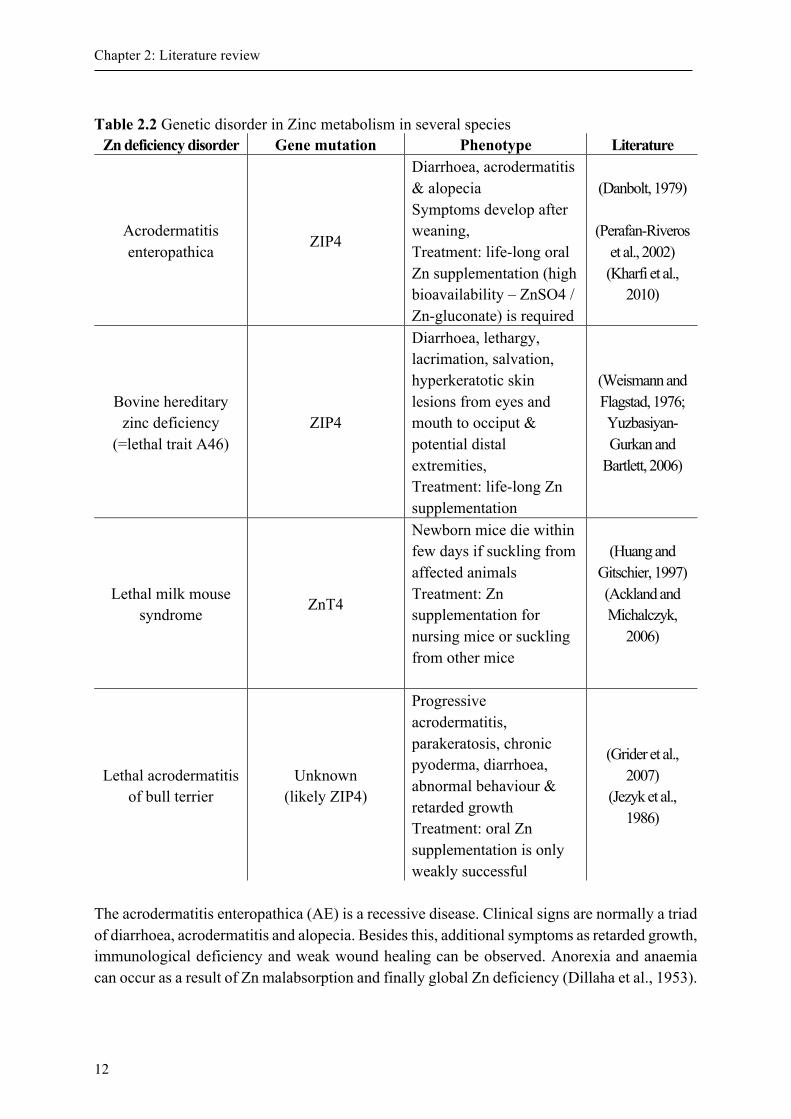
Table 2.2 Genetic disorder in Zinc metabolism in several species ........................................................ 12 Table 4.1 Chemical composition of sow milk and formula in the study. .............................................. 30 Table 4.2 Primers used in this study. ..................................................................................................... 31 Table 4.3 Concentration of Zn, Cu, Mn and Fe in tissues in piglets fed sow milk or formula * ........... 32 Table 4.4 Relative Gene expression in liver, jejunum and pancreas tissue in formula and sow milk fed
piglets* ................................................................................................................................... 33 Table 4.5 Correlation coefficients for liver tissueA ................................................................................ 34 Figure 5. 1Concentration of Zn (A) and Cu (B) in the kidney of piglets fed diets containing normal
(100 mg Zn/kg; NZn; n = 16), very high (2000 mg Zn/kg; HZn; n = 16) dietary zinc oxide for two and four weeks, and piglets fed HZn for two weeks followed by NZn for another two weeks (CZn; n = 8). Superscripts indicate significant (P < 0.05) differences. ............... 42
Figure 5. 2 Pearson correlation between Zn and Cu concentration in the kidney of piglets fed diets containing normal (100 mg Zn/kg; NZn; n = 16), very high (2000 mg Zn/kg; HZn; n = 16) dietary zinc oxide for two and four weeks, and piglets fed HZn for two weeks followed by NZn for another two weeks (CZn; n = 8). ............................................................................. 43
Table 5. 1 Ingredients and chemical composition of the diets used in the study ................................... 44 Supplemental Table S 5. 1 Primes used in this study ............................................................................ 46 Supplemental Table S 5. 2 Trace element concentration (mg/kg DM) in different organs after two and
four feeding weeks of normal (NZn), high (HZn) and changed (CZn) dietary Zn concentration in weaning piglets. .......................................................................................... 47
VI
List of Figures Figure 5. 1Concentration of Zn (A) and Cu (B) in the kidney of piglets fed diets containing normal
(100 mg Zn/kg; NZn; n = 16), very high (2000 mg Zn/kg; HZn; n = 16) dietary zinc oxide for two and four weeks, and piglets fed HZn for two weeks followed by NZn for another two weeks (CZn; n = 8). Superscripts indicate significant (P < 0.05) differences. ............... 42
Figure 5. 2 Pearson correlation between Zn and Cu concentration in the kidney of piglets fed diets containing normal (100 mg Zn/kg; NZn; n = 16), very high (2000 mg Zn/kg; HZn; n = 16) dietary zinc oxide for two and four weeks, and piglets fed HZn for two weeks followed by NZn for another two weeks (CZn; n = 8). ............................................................................. 43
VII
List of abbreviations ADG Average daily weight gain AE Acrodermatits enteropathica Atox-1 Antioxidant 1 copper chaperon ATP7A Copper-transporting P-type ATPase α ATP7B Copper-transporting P-type ATPase ß BHK Baby hamster kidney cell BHZD Bovine hereditary zinc deficiency BW Body weight CCS Cu chaperon protein Cd Cadmium COX 17 Cytochrome c oxidase copper chaperon CP Ceruloplasmin Ctr1 Copper transporting protein 1 Cu Copper CZn Changed dietary zinc group from high to normal dietary zinc supplementation DM Dry matter DMT1 Divalent metal ion transporter 1 DNA Desoxyribonucleic acid Fe Iron FO Formula FSH Follicle stimulating hormone GH Growth hormone GIT Gastrointestinal tract GSH Glutathione HZn High dietary zinc group IGF Insulin like growth factor IL Interleukin LH Luteinizing hormone lm lethal milk allel MD Menke disease miRNA micro RNA mRNA Messenger ribonucleic acid MT Metallothionein MTF-1 Metal transcription factor 1 NRC National Research Council NZn Normal dietary zinc group PCR Polymerase chain reaction RNA Ribonucleic acid SLC Solute carrier SOD1 Cu/Zn superoxide dismutase 1 TGN trans-Golgi network WD Wilson disease ZIP Zrt-and Irt-like protein Zn Zinc ZnO Zinc Oxide ZnSO4 Zinc Sulphate ZnT Zinc transporter protein

Chapter 1: General introduction
Chapter 1: General introduction Zinc (Zn) has been first described as an essential trace element for Aspergillus niger already in 1869 (Edwards and Baker, 2000; Raulin, 1869). From there, it took another 60 years before Zn was first shown to being indispensable for men and animals as well (Prasad et al., 1961; Todd et al., 1934). As an essential trace mineral, Zn is involved in manifold biological functions but it is also potentially toxic when exceeding the required amounts in the cell or body. There are currently more than 6000 Zn proteins and more than 300 Zn-dependent metalloenzymes described, giving an idea of its outstanding importance for the host organism (Andreini and Bertini, 2012; Bettger and O'Dell, 1981; Romeo et al., 2014; Suzuki et al., 2011). Well-known metalloenzymes are carbonic anhydrase, pancreatic carboxipeptidase and several dehydrogenases like, for instance, lactate-, maltate- and alcohol dehydrogenase and arginase (Lloyd, 1978a). The Zn status can influence appetite, likely through an alteration in hypothalamic neurotransmitter metabolism by the influence in leptin system (Lobo et al., 2012). Further hormones, as insulin, glucagon, follicle stimulating hormone (FSH) and luteinizing hormone, insulin like growth factor 1 (IGF-1) and growth hormone (GH) are under direct influence of Zn and, in this way, protects the cell from free radicals (Frassinetti et al., 2006; Hansen et al., 1997; Lloyd et al., 1978a; Malhotra et al., 1961; Maret, 2005; Suttle, 2010b). Thus, Zn has also anti-oxidant properties (Prasad et al., 2004). Finally, Zn is an essential element for the immune system, and low Zn supply can lead to immunodeficiency (Frassinetti et al., 2006). The zinc finger proteins are an example for the structural role of Zn. These proteins change their structure in presence of Zn and subsequently can bind to the DNA helix to initiate gene expression (Hambidge, 2001). Hence, Zn is involved in DNA replication, DNA stabilization and RNA transcription (Frassinetti et al., 2006; Suttle, 2010b). A temporal Zn (over-)supplementation has been used for the treatment of diarrhoea in humans (mostly in developing countries) und animals. For decades, dietary ZnO was applied in high doses up to 3000 mg/kg feed in swine production to combat post-weaning diarrhoea in piglets (Starke et al., 2014). Around weaning, piglets have to adjust to a new environment, the change from liquid milk to solid feed and arrange new hierarchy in the new social group. Among others, this is an extreme stressful situation leading to low feed intake, an imbalanced microbiota and diarrhoea (Starke et al., 2014). In the European Union, the maximum allowance for Zn in animal feed (except fish, pet animals and in milk replacers) is 150 mg/kg feed (EC 1334/2003). In contrast, in Asia and the Americas, weaned piglets receive pharmacological high Zn amounts up to 3000 mg/kg feed. In Europe, national exceptions from the maximum allowance still exist in several countries (e.g. Belgium, Denmark and Spain), but this will not longer be possible in the future. Also, ZnO is still used as carrier substance for in-feed medications. Although, only short-term use is often propagated, high doses of ZnO are sometimes fed to piglets for several weeks. The toxic potential of the heavy metal Zn raises the question about possible adverse effects of Zn-rich diets in young piglets. In addition, although nutritional recommendations are available for pigs after the weaning, the increasing practice to feed milk replacers during the suckling period raises the question about the metabolic reactions to the dietary Zn level, since no recommendations are yet available.
1
Chapter 2: Literature review
Chapter 2: Literature review 2.1 Zinc Metabolism The Zn homeostasis is an essential process for the survival of an organism (Roohani et al., 2013). Although the dietary Zn intake can vary, the body Zn homeostasis is maintained within narrow margins by intestinal absorption and endogenous excretion (King et al., 2000; Weigand and Kirchgessner, 1980). Thereby, the body Zn content remain constant at approximately 30 mg/kg fresh tissue or averaging 2,1 g Zn in a pig of 100 kg body weight (BW) and 1.4 to 2.3g Zn in total adult body of humans. Zn contents in respective tissue concentration between mammalian species show only small differences (Stefanidou et al., 2016; Underwood, 1977; Dourmad, 2015). However, Rincker et al. analysed nursery pig at the age of 19 ± 3days and 54 ± 3days and measured a total body Zn content of 59.7 and 74.4 mg/kg, respectively. This is in good concordance to reports of rising Zn concentrations during the suckling period (Rincker et al., 2004). With approximately 57% the main percentage of body Zn is located in the skeletal muscles. It is followed by bone (29%), skin (6%), liver (5%) and brain (1.5%). In kidney tissue 0.7 % of total body Zn is found. Hair, blood plasma and heart muscle contain approximately 0.1 to 0.4 % of total body Zn content (Suttle, 2010b; Yoshida et al., 2008). Indeed, the highest concentration per gram tissue is situated in retina (200 – 500 mg/kg dry matter (DM)), hair and wool (100– 200 mg/kg DM), liver (196 mg/kg DM), testes (app. 100 mg/kg DM) and bones (70-90 mg/kg DM) (Galin et al., 1962; Suttle, 2010b).
The Zn homeostasis is controlled at organism and cellular level (Wang and Zhou, 2010). While each organ and each cell necessarily needs Zn, some organs are more relevant for the total body Zn metabolism and storage. This metabolic highly relevant organs are intestine, liver, pancreas, kidney, bone and skin (Cotzias et al., 1962; King and Turnlund, 1989; King et al., 2000; Sullivan et al., 1981).
After ingestion, Zn is absorbed along the entire small intestine (see Chapter 2.1.1 Zinc absorption and bioavailability), whereby the jejunum has the highest absorption rate in comparison to the remaining sections of small intestine (Lee et al., 1989). From the enterocytes Zn is transferred into the portal blood stream via Zn transporters and loosely bound to albumin (70%) or α-Macroglobulin (20%). The remaining Zn in blood plasma is bound to amino acids, transferrin, histidine-rich glycoprotein and metallothionein (MT) (King, 1999; Reyes, 1996). Zn, which is bound to albumin, is available for hepatocytes (Cousins and Swerdel, 1985). In hepatocytes, the Zn metabolism is regulated through proteins (see Chapter 2.2 Cellular zinc homeostasis and zinc transporter). Therefore, the liver is an important organ in Zn metabolism by involvement in Zn excretion, distribution via systemic circulation and incorporation into proteins.
Approximately 90% of the body Zn content is present in slow-exchangeable Zn pool organs, namely skeletal muscles (60%) and bones (30%) (Chesters, 1982). These organs are important for the total body Zn homeostasis and the compartments, which store Zn inside the cells, are necessary for the homeostasis of mentioned organs as well. The remaining 10% of body Zn is rapidly exchangeable and thereby available for maintaining Zn homeostasis within
2
Chapter 2: Literature review
a short time. Organs with rapidly turn-over rates of Zn are e.g. liver, pancreas and gut (Chesters, 1982; Miller et al., 1994).
The pancreas has exocrine and endocrine functions. Thereby over 85% of the pancreas tissue contains of exocrine cells (Gorelick and Jamieson, 2006). And while 95% of the exocrine component are acinus cells, the remaining 5% are part of the duct system (Miller et al., 1991). The acinar cells can produce and store several enzymes and zymogens (Miller et al., 1991). Pancreatic Zn is localized mainly in the exocrine part of pancreas, more exactly in granules of acinar cells (Guo et al., 2010). If the organ is stimulated, the acinar cells release their products into the acinus and the duct system transport secretions to the duodenum. Thus, the pancreas plays an important role in Zn homeostasis by facilitating Zn secretion into the intestinal lumen. In turn, excessive dietary Zn results in altered pancreas cell structure, increase of proteins involved in oxidative stress including necrosis and even death of the animals (Gabrielson et al., 1996; Pieper et al., 2015). 2.1.1 Zinc absorption and bioavailability For the body Zn homeostasis, the gastrointestinal system plays the most important role (Wang et al., 2010 ). For the uptake mechanism into enterocytes mainly via transporters (see chapter 2.2 Cellular zinc homeostasis and zinc transporter), Zn ions have to be soluble. While the Zn solubility rises with decreasing pH, Zn is predominantly absorbed in small intestine in non-ruminant animals and humans (Annenkov, 1979). In the gut two possible mechanisms for Zn uptake exist
1) active and 2) passive transport (Martin et al., 2013; Solomons and Cousins, 1984; Suttle, 2010b).
The active, carrier-mediated transport is facilitated through specific Zn transporter (see chapter 2.2 Cellular zinc homeostasis and zinc transporter). Under normal physiological conditions this transport process is not saturated, but saturable in general (Cousins, 1985). The second way of Zn absorption is the para-cellular way or diffusion. This is an energy- independent, non-saturable process (Lichten and Cousins, 2009; Martin et al., 2013).
The absorption rate depends on diet composition and the form of the diet. For example, absorption efficiency of solid diets is less effective in comparison to aqueous solutions in fasted men (Roohani et al., 2013). Further, for the bioavailability of Zn, three factors seem to be important
1) individual body Zn status 2) Zn content of the diet and 3) availability of Zn from the diet components and interaction with other diet ingredients (Lönnerdal, 2000). The availability depends on the solubility of Zn and the enhancers or inhibitors, which
are possibly within the diet (see Chapter 2.1.3 Factor affecting zinc absorption). Moreover, absorption efficiency can depend on Zn concentration in supplied diets (Sandström and Cederblad, 1980). Thus, during periods of insufficient Zn concentration in diets, the fractional Zn absorption rises, whereas it decreases during excess Zn intake. However, the fractional Zn absorption differs between individuals of the same group (King et al., 2000). On
3
Chapter 2: Literature review
the contrary, another study showed, that Zn uptake efficiency seems to be unaffected by Zn status, whereas rather the excretion of endogenous Zn depends on Zn status (Krebs, 2013). Taken into account the endogenous Zn excretion, the real Zn absorption rate can only be measured by stable isotope technique (Sandström et al., 1993). The trace minerals contents in blood plasma or serum were bad biomarkers for status of trace minerals (except selenium) since plasma concentration is carefully regulated to ensure mineral maintenance of the organs (Hambidge, 2003).
Nevertheless, average Zn absorption for adult humans is approximately 33% (Cousins, 1985; Turnlund et al., 1984). On the other hand, for pigs receiving maize-soy diet apparent Zn absorption depend on age and is between 16% (age of 37 – 44 days) and 24% (age of 51 to 58 days) (Pallauf and Rimbach, 1992). This is in good concordance to Chu et al. who reported a true absorption of approximately 20% in growing pigs (Chu et al., 2008). If womens´ dietary Zn intake is restricted during lactation, the Zn absorption rate rises up to 70%, however other studies did not present similar results, conceivably because of different food limitation (King, 2002)
Hence, Zn absorption rate vary widely from 15 to 60 % and depend on numerous factors, including stage of life, since the Zn uptake is greater in growing compared to adult organism (McDowell, 2003; Weigand and Kirchgessner, 1979). 2.1.2 Zinc excretion Zn is mainly excreted via the faeces. In the intestine, Zn is excreted into the gut lumen through pancreatic juice, bile, gastrointestinal secretions, transepithelial flux or bound in desquamate enterocytes (Chesters, 1997; Krebs, 2000). For homeostatic adaptation, the pancreas play a more important role in comparison to liver, since during dietary Zn deficiency pancreatic Zn excretion decreased while Zn excretion through bile did not change (Sullivan et al., 1981). The adjustments of endogenous excretion respond immediately but in relatively small amounts. Even so, endogenous Zn is from major importance for Zn homeostasis (Weigand and Kirchgessner, 1978). On the contrary to that, adaptation of absorption process to dietary Zn is slowly but can cope with larger fluctuations (Krebs, 2000). Nevertheless, both adaptation processes are important to maintain homeostasis. Finally, a small amount of Zn is excreted via urine and integumental losses (e.g. sweat, hair and wool growth and seminal emission). Under normal conditions, Zn losses through this route make up to approximately 10 - 20% (King et al., 2000; McDowell, 2003). In addition, neither urinary nor integumental losses adapt during high dietary Zn supply (King and Keen, 1999). Nevertheless, urinary losses can rise after trauma and increased muscle catabolism and decrease during extremely low Zn intake (Hambidge et al., 1986; Johnson et al., 1993). Since the renal excretion is quantitatively insignificant, adjustments after low Zn contents in diets occur only if Zn intakes are extremely low (King et al., 2000). However, the mechanism of Zn excretion via urine is still unclear. Adaptation processes in Zn absorption and excretion can maintain body Zn content throughout a 10-fold change in Zn intake in animals and human (King et al., 2000).
4
2.1.3 Factor affecting zinc absorption There are numerous of dietary factors, which can negatively affect Zn uptake and thereby reduce Zn bioavailability of primordially Zn-rich diets. The most noted factors are e.g., other divalent ions such as calcium, magnesium and iron (Fe) and chelating agents like EDTA or phytate (Lloyd et al., 1978b). In Table 2.1 an overview of some important factors is given.
Dietary phytate can supress reabsorption of Zn and stimulation of mucosal cell sloughing and intestinal fluid secretion (Brink et al., 1992; Krebs, 2000). Phytate is composed of several phosphorylated forms of inositol phosphate (Sandberg and Ahderinne, 1986).
The most common form is hexaphosphate, but tri-, tetra- and pentaphosphate are also present. In rat pups model only penta- and hexaphosphate inhibited Zn absorption, while other inositol phosphates had no influence (Lönnerdal et al., 1989). These findings were supported by a trial with human subject groups (Sandström and Sandberg, 1992). Likewise in a pig trial phytate impaired Zn absorption by the formation of poorly soluble complexes (Turnbull et al., 1990). Moreover, feed processing types can influence the composition of phosphorus (Beauchemin and Holthausen, 2001; Schlemmer et al., 2009). Regarding negligible endogenous phytase activity in the gut of mammals, phytase can be added to diets, to improve Zn absorption. But till date, it is not clear if phytase improves bioavailability without affecting apparent absorption of Zn, or if apparent absorption rate of Zn is increased by phytase (Adeola et al., 1995; Nasi et al., 1995; Yokoyama et al., 1993). Nevertheless, apparent digestibility of Zn in piglets increases curvilinear with rising phytase levels in pigs (Windisch, 1996a). A supplementation of 700 units of microbial phytase can reduce Zn supply by 35 mg/kg feed for weaned piglets (Revy et al., 2006).
In addition, the Zn source influence the efficiency of Zn absorption. According to the European Union register of feed additives pursuant to Regulation No. 1831/2003 Zn can be added in various forms including Zn acetate, Zn oxide, Zn hydroxide, Zn sulphate, Zn chloride, Zn chelate of amino acids, Zn chelate of glycine, Zn chelate of the hydroxy analogue of methionine and Zn chelate of methionine.
At the moment several Zn sources, organic and inorganic, are commercially available. Zn oxide (ZnO) is the most common inorganic Zn source, but it is relatively insoluble at neutral pH and therefore considered to have a lower bioavailability. On the contrary, Zn sulphate (ZnSO4) has an increased absorption rate in comparison to ZnO. In piglets, ZnO has 68% bioavailable Zn, relative to ZnSO4 (Wedekind et al., 1994). Furthermore, apparent Zn digestibility is higher from Zn-Methionine (Zn-Met) compared to ZnO (Nitrayova et al., 2012). Nevertheless, the ratio of Zn and Methionine effect the bioavailability. In this way Zn-Met complex with a 1:1 ratio did not have this positive effect which was observed in Zn-Met (1:2) (Nitrayova at al., 2012; van Heugten et al., 2003). Moreover, compared to ZnO the relative bioavailability of tetrabasic Zn chloride (TBZC) is higher (Zhang and Guo, 2007). While ZnSO4 seems to be higher bioavailable than Zn-Lysininate (Zn-Lys), it is followed by Zn-Met and ZnO (Schell and Kornegay, 1996). On the contrary, another study showed following ranking: ZnSO4 > ZnMet > ZnO > ZnLys (Wedekind et al., 1994).
5
Chapter 2: Literature review
Chapter 2: Literature review
Finally, the bioavailability of Zn from organic sources, relative to inorganic Zn sources may depend on the amount of antagonistic dietary factors as Ca, phytate or fibre (Wedekind et al., 1994). Currently, the use of ZnO and ZnSO4 as inorganic sources of Zn is more common although the use of organically bound Zn as Zn-yeast and Zn-glycine seems to be an option of replace high Zn concentration of inorganic Zn in diets through enhanced digestibility and retention and thereby decrease Zn excretion and environmental pollution (Nitrayova et al., 2012). The interactions of different trace elements with Zn are manifold. A well-known interdependency is the antagonism between Zn and Copper (Cu), although the reasons are not yet clear. It is postulated that Cu can inhibit the Zn absorption (Van Campen, 1969). On the other hand, Zn can inhibit Cu contribution into blood stream and thereby result in general Cu deficiency after long periods of high Zn supply (Baker and Ammermann, 1995; Ritchie et al., 1963). Both, Fe and cobalt can inhibit Zn uptake as well as the transfer across the basolateral side of enterocytes (Flanagan et al., 1980; Vallberg et al., 1984). But the interaction between Fe and Zn is controversial discussed and the results are inconsistent. Thus, it seems that Fe:Zn ratio has to be greater than 2, suggesting a common pathway between both trace elements (Solomon, 1986, Flanagan et al., 1980). In addition, an interaction between both elements appears less, if Zn and Fe intake is closer to “physiological” level (Lönnerdal, 2000).
Phosphorus can reduce Zn absorption only if it is Phytate-Phosphate like it appears from 50 to 80 % in legumes and crops (Rimbach et al., 1996).
Ca have no influence on Zn absorption in the absence of Phytate (Forbes et al., 1984). However, Ca can raise the adverse effect of Phytate by building insoluble Ca-Zn-Phytate-complexes (Fordyce et al., 1987; Windisch, 1996b).
Zn itself can have an influence on Zn absorption. If diets have extremely high Zn contents, relative Zn absorption efficiency is reduced, but finally leads to an enhanced absolute Zn uptake (Liu et al., 2014).
Further intrinsic factors can also have an influence on Zn absorption. In this way, some luminal factors like pH and therefore Zn solubility and the intensity of digestion can have an impact (Krämer and Rimbach, 1994). To summarize, stress, infections, functionality of liver and kidney, hormone status and also anabolic requirement (as lactation, gravidity or growth) are the most common intrinsic factors which can influence Zn absorption (Krämer and Rimbach, 1994; Rimbach et al., 1996). Consequently, intrinsic factors can enhance (e.g., citric acid), competitively inhibit (e.g., Cu, Fe) or not competitively inhibit Zn absorption (e.g., Phytate).
6
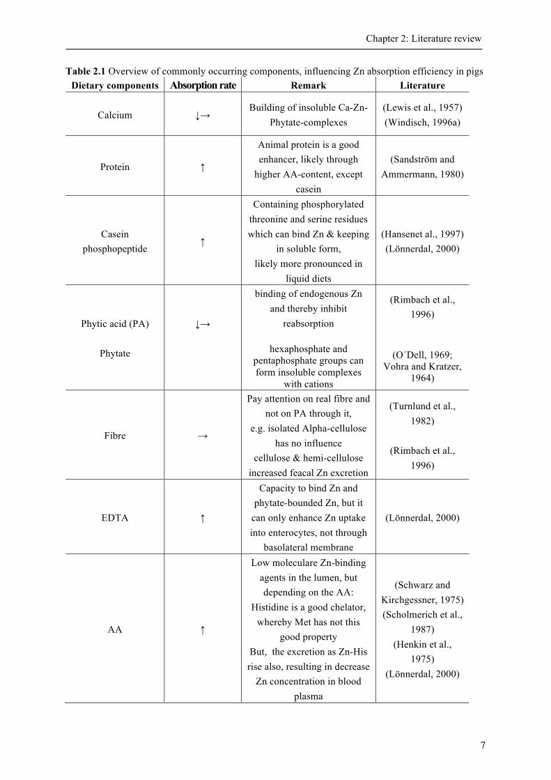
Table 2.1 Overview of commonly occurring components, influencing Zn absorption efficiency in pigs Dietary components Absorption rate Remark Literature
Calcium ↓→ Building of insoluble Ca-Zn-
Phytate-complexes (Lewis et al., 1957) (Windisch, 1996a)
Protein ↑
Animal protein is a good enhancer, likely through
higher AA-content, except casein
(Sandström and Ammermann, 1980)
Casein phosphopeptide
↑
Containing phosphorylated threonine and serine residues which can bind Zn & keeping
in soluble form, likely more pronounced in
liquid diets
(Hansenet al., 1997) (Lönnerdal, 2000)
Phytic acid (PA)
Phytate
↓→
binding of endogenous Zn and thereby inhibit
reabsorption
hexaphosphate and pentaphosphate groups can form insoluble complexes
with cations
(Rimbach et al., 1996)
(O´Dell, 1969; Vohra and Kratzer,
1964)
Fibre →
Pay attention on real fibre and not on PA through it,
e.g. isolated Alpha-cellulose has no influence
cellulose & hemi-cellulose increased feacal Zn excretion
(Turnlund et al., 1982)
(Rimbach et al.,
1996)
EDTA ↑
Capacity to bind Zn and phytate-bounded Zn, but it
can only enhance Zn uptake into enterocytes, not through
basolateral membrane
(Lönnerdal, 2000)
AA ↑
Low moleculare Zn-binding agents in the lumen, but depending on the AA:
Histidine is a good chelator, whereby Met has not this
good property But, the excretion as Zn-His rise also, resulting in decrease
Zn concentration in blood plasma
(Schwarz and Kirchgessner, 1975) (Scholmerich et al.,
1987) (Henkin et al.,
1975) (Lönnerdal, 2000)
7
Chapter 2: Literature review
Chapter 2: Literature review
Organic acid ↑ Citric acid
(Höhler and Pallauf, 1994)
(Pabón and Lönnerdal, 1993)
Pectin ↓→
Depending on rate of methylation: low methylated
apple pectine decrease bioavailablity, high
methylated pectine has no influence
(Bagheri and Gueguen, 1985; Sandberg et al.,
1983)
PA, phytic acid; EDTA, Ethylenediaminetetraacetic acid; AA, amino acid 2.2 Cellular zinc homeostasis and zinc transporter Around 95 % of body Zn is intracellular, whereby 40 % of the cellular Zn can be found in the nucleus. The amount of free cytosolic Zn is very low, likely due to the cytotoxic potential of double positively charged Zn ions (Outten and O`Halloran, 2001). A closely controlled Zn concentration in cytoplasm is essential for the cellular function. For this, the cells have the ability to regulate the in- and efflux of Zn and can store surplus Zn e.g. in cellular vesicles (Sekler et al., 2007).
To date, numerous Zn transporters are known. They are subdivided into two families which both related to solute-carrier (SLC) families, namely ZIP (Zinc-regulated, Iron-regulated transporter-like protein; SLC39) and ZnT (Zinc transporter, SLC30) family (Lichten and Cousins, 2009). While ZIP family members are responsible for enhancing, ZnT family members decreasing Zn concentration in cellular cytoplasm (Eide, 2004; Liuzzi and Cousins, 2004). Furthermore, both families differ from each other by their structure. ZnT proteins have six transmembrane domains with intracellular N- and C-terminus and an intracellular loop with histidine residues. On the contrary ZIP proteins have eight transmembrane domains with extracellular N- and C-terminus and an intracellular histidine-rich loop. Nevertheless, there are two exceptions. ZnT5 have 12 transmembrane domains and ZIP14 have an extracellular loop (Romeo et al., 2014).
The distribution of the ten known ZnTs and the 14 known ZIPs is tissue-specific different (Cousins, 2010). However, most of the ZnT proteins were located at intracellular compartments including Golgi apparatus and endoplasmatic reticulum. Only ZnT1 is located at the plasma membrane (Romeo et al., 2014). This is in contrast to ZIP proteins, which were generally located in the plasma membrane and only ZIP7, 9, 11 and 13 have localisations in Golgi apparatus or nucleus. Following, a brief overview of important transporting proteins in several tissues is given.
8
Jejunum ZIP4 is a transporter for cellular uptake into cells of the gastro intestinal tract and kidney
across the apical membrane (Romeo et al., 2014; Wang and Zhou, 2010; Huang and Gitschier, 1997). It is the crucial transporter for Zn absorption in the small intestine and thus, numerous diseases are known due to mutations in ZIP4 gene (see Chapter 2.3 Zinc deficiency and genetic disorder in zinc metabolism) (Liuzzi et al., 2009). During periods of dietary Zn restriction ZIP4 is up-regulated in enterocytes, to increase Zn absorption rate in the small intestine (Weaver et al., 2007). Vice versa, there are decreased ZIP4 mRNA levels in enterocytes after periods of excess dietary Zn intake in pigs (Martin et al., 2013). On the contrary, ZIP5 translation is also Zn-responsive, but during Zn deficiency it is internalized and degraded in enterocytes, while excess Zn results in rapid re-synthesis and translocation at basolateral enterocyte side, suggesting serosal-to-mucosal transport of Zn by oppose ZIP4 and ZIP5 transports (Liuzzi et al., 2009).
The ubiquitously expressed ZnT1 can facilitate both, Zn efflux into intracellular vesicles and across basolateral membranes (Wang et al., 2010 ). The most important function of ZnT1 is facilitating the export of Zn from one compartment to another, e.g., from enterocytes into the blood stream and thereby have a crucial role in Zn providing for the hole organism, or from exocrine cells to the pancreatic ducts and though for Zn excretion (Palmiter and Huang, 2004). Liver
ZIP14 is the most important transporter for Zn uptake into hepatocytes. It is localized at the plasma membrane and up-regulated during acute-phase response to inflammation through interleukin (IL) 6 (Liuzzi et al., 2005). ZIP5 is located at the basolateral side of hepatocytes, which is contrary to ZIP4 localization and its translation is responsive to Zn concentration (Liuzzi et al., 2009).
ZnT2 in liver is responsible to sequester Zn in intracellular organelles, particularly late endosomes (Palmiter and Huang, 2004). Moreover, ZnT2 mRNA contents are strongly increasing after oral doses of Zn in mice (Liuzzi et al., 2001). ZnT7 gene was also detected in high amounts in liver tissue (Liuzzi et al., 2009).
Since ZnT1 is ubiquitously expressed in the body, it is also important in liver tissue (Palmiter and Huang, 2004).
Pancreas
ZIP 14 gene expression is high in pancreatic tissue and the localization is plasma membrane, similar to liver tissue (Taylor et al., 2005). In human pancreas, high ZIP5 mRNA were measured and the translation of basolateral localized ZIP5 is sensitive to distinct Zn levels (Wang et al., 2004).
ZnT1 is localized at the plasma membrane, whereas ZnT2 is additionally associated with the zymogen granules (acidic intragranule pH) in acinar cells, suggesting several functions in Zn trafficking (Guo et al., 2010). ZnT2 is up-regulated in pancreas during high dietary Zn intake (Guo et al., 2010). In ß-cells of pancreatic tissue ZnT8 transports Zn which accumulates in this insulin-secreting cells, were two Zn2+ ions are bound to one insulin-hexamer (Liuzzi and Cousins, 2004).
9
Chapter 2: Literature review

Chapter 2: Literature review
Kidney Abundant ZIP4 expression was found in kidney tissue, as it is potentially involved in
reabsorption of Zn (Liuzzi and Cousins, 2004). Furthermore, ZIP5 was detected in kidney, whereby its expression has no response to Zn concentration, but the translation is responded to Zn (Liuzzi et al., 2009). Moreover, ZIP14 is found in kidney cells. To date it is speculative, if this protein only plays a critical role during growth and development. However, the role of ZIP14 in response to inflammation and its responsiveness to IL-6 signalling is certainly interesting (Liuzzi and Cousins, 2004; Liuzzi et al., 2009).
ZnT1 was found on the basolateral side in kidneys´ thick ascending and distal convoluted tubules, indication a function in recovery of Zn from glomerular filtrate (Liuzzi et al., 2009). ZnT2 is sensible to Zn supply and mRNA levels strongly rises after high dietary intake (Palmiter and Huang, 2004). Indeed, it is not clear, if the ZnT2 protein is localized to late endosomes under physiological conditions because studied baby hamster kidney (BHK) cells appropriate intracellular targeting may depend on chaperones that are not present in mentioned cells (Palmiter and Huang, 2004). ZnT4 protein was found in rat normal kidney (NRK) cells, although neither the response to oral Zn intake nor location in kidney cells is clear to date (Palmiter and Huang, 2004). Moreover, mRNA of ZnT6 & 7 was found in kidney tissue. However, ZnT7 proteins of mentioned transporters were not found by Western blot (Huang et al., 2002).
In addition to ZnTs and ZIPs, which represent specific Zn transporter, a further non-specific transporter is involved in cellular Zn homeostasis. Divalent metal ion transporter 1 (DMT1 or divalent cation transporter 1, DCT1) can transport Fe2+ into enterocytes and out of endosomes (Garrick et al., 2003). Furthermore, this transporter plays a role in non-transferrin bound iron uptake (Garrick et al., 2003). The DMT1 can transport several ions. To date, affinity to metal ions seems to have the following ranking: Mn2+>?Cd2+>?Fe2+ >Pb2+~Co2+~Ni2+ >Zn2+ (Garrick et al., 2006). In this way, DMT1 can play a role in Zn homeostasis. Finally, a storage protein, metallothionein (MT), has an influence on Zn homeostasis inside the cells. MT is a small (7 kDa) cysteine-rich metal binding protein with the property of heavy metal detoxification, anti-oxidation, protection against DNA damage and homeostasis of Zn and Cu (Kagi and Valee, 1960; Martinez et al., 2004; Thirumoorthy et al., 2011). For MTs´ anti-oxidant capability, the Zn concentration is important. During pathological processes of oxidative stress and concomitant Zn mobilization, anti-oxidant properties of MT were enhanced (Maret, 2009). MT consist of four iso-enzymes and can be divided into four classes, with partially tissue-specific distribution. While MT-1 and MT-2 are presented in all cells, MT-3 was only detected in brain and male reproductive organs, kidney and in small amounts in both, pancreas and intestine. MT-4 is restricted to stratified squamous epithelia, including gastrointestinal tract and skin (Cherian et al., 2003; Thirumoorthy et al., 2011). Depending on dietary amounts of Zn and Cu, MT-1 and -2 are mainly synthesized in liver and kidney in humans, where MT expression rises with high dietary Zn contents (Romeo et al., 2014; Thirumoorthy et al., 2011). In addition, they can sequester Zn in mucosal cells of the gut
10

and thereby have an influence in Zn absorption (Romeo et al., 2014). Thus, high intracellular Zn levels result in increased MT synthesis and vice versa (Andrews, 2001). The MT synthesis is influenced by Zn through direct, reversible binding to the Zn-finger domain of metal transcription factor-1 (MTF-1). After adopting a DNA-binding conformation and translocation into nucleus, the MTF-1 binds to metal-response element in gene promoters and thereby increase transcription of genes which are involved in Zn homeostasis, including MT (Andrews, 2001). Since there is no specific functional test for measurements of Zn status in blood plasma until today, it is suggested to use MT for Zn status diagnostics (King and Turnlund, 1989). In blood, MT is detectable in plasma and erythrocytes. On the contrary to erythrocyte MT, plasma MT seems to be receptive to changes in hepatic MT levels. However, hepatic MT concentration is not only sensitive to Zn, but also to stress and inflammation (Cousins and Leinart, 1988; Martinez et al., 2004; Ruttkay-Nedecky et al., 2013). In addition cytokines such as IL-1, IL-6 and glucocorticoids can regulate synthesis of MT (Andrews, 2001). Nevertheless, the quantity is also depended on the supply with essential sulphur-containing amino acids cysteine and histidine (Thirumoorthy et al., 2011). Although the binding affinity to Zn is relatively high, it is low in comparison to cadmium. The latter can replace bounded Zn and thereby MT play a crucial role in detoxification of heavy metals (Romeo et al., 2014). 2.3 Zinc deficiency and genetic disorder in zinc metabolism Regarding the number of physiological functions Zn, deficiency can appear with non-specific symptoms. While marginal lack in Zn supply can result in loss of appetite, delayed wound healing, delayed bone maturation, growth retardation and lethargy, severe deficiency lead to alopecia, hyperkeratosis and parakeratosis in pigs, diarrhoea, default spermatogenesis and increased susceptibility to infection (Hambidge et al., 1986; Prasad, 2012). Female rats are unable to conceive whereas already pregnant rats bear malformed puppies reasoned by Zn deficiency periods (Shrader and Hurley, 1972). In both, animals and men pathological dermal appearance including increased dermal thickness and thus developing chaps´ exudates of serum and blood appear as brown crusts were described and known as parakeratosis of swines (Chesters, 1983; Romanucci et al., 2011). Concerning also oesophageal epithelia, ruminants and swine lose weight when those affected patients’ left untreated (Fell et al., 1973; Miller and Miller, 1962). Since Zn is essential for hair and claws, affected goats and sheep have curled wool and deformed claws. Curled feathers and thickened hocks were already observed in Zn deficient chickens (O´Dell ,1958). During phases of Zn deficiency, rats stop growing and consequently reach a normal whole-body Zn concentration (Williams and Mills, 1970). Nevertheless, there are some tissues, which lose Zn in favour to other tissues. Thus, e.g. liver and bone Zn concentration decline, while muscle Zn concentration is conserved (King, 1990). If the gene mutation results in Zn deficiency the clinical signs are similar to severe dietary Zn deficiency. An overview of important diseases is given in Table 2.2.
11
Chapter 2: Literature review
Chapter 2: Literature review
Table 2.2 Genetic disorder in Zinc metabolism in several species
Zn deficiency disorder Gene mutation Phenotype Literature
Acrodermatitis enteropathica
ZIP4
Diarrhoea, acrodermatitis & alopecia Symptoms develop after weaning, Treatment: life-long oral Zn supplementation (high bioavailability – ZnSO4 / Zn-gluconate) is required
(Danbolt, 1979)
(Perafan-Riveros et al., 2002) (Kharfi et al.,
2010)
Bovine hereditary zinc deficiency
(=lethal trait A46) ZIP4
Diarrhoea, lethargy, lacrimation, salvation, hyperkeratotic skin lesions from eyes and mouth to occiput & potential distal extremities, Treatment: life-long Zn supplementation
(Weismann and Flagstad, 1976; Yuzbasiyan-Gurkan and
Bartlett, 2006)
Lethal milk mouse syndrome
ZnT4
Newborn mice die within few days if suckling from affected animals Treatment: Zn supplementation for nursing mice or suckling from other mice
(Huang and Gitschier, 1997) (Ackland and Michalczyk,
2006)
Lethal acrodermatitis of bull terrier
Unknown (likely ZIP4)
Progressive acrodermatitis, parakeratosis, chronic pyoderma, diarrhoea, abnormal behaviour & retarded growth Treatment: oral Zn supplementation is only weakly successful
(Grider et al., 2007)
(Jezyk et al., 1986)
The acrodermatitis enteropathica (AE) is a recessive disease. Clinical signs are normally a triad of diarrhoea, acrodermatitis and alopecia. Besides this, additional symptoms as retarded growth, immunological deficiency and weak wound healing can be observed. Anorexia and anaemia can occur as a result of Zn malabsorption and finally global Zn deficiency (Dillaha et al., 1953).
12
Bovine hereditary Zn deficiency (BHZD, or bovine hereditary Zn parakeratosis) is an autosomal recessive gene defect which result in reduces milk uptake in calves. This leads to decreased immune response and diarrhoea. In cases of extreme diarrhoea an acidotic situation can lead to dehydration and death (Machen et al., 1996).
In case of a homozygote lm/lm mice, the pups will die within a few days of suckling, since the Zn concentration in milk is reduced by 58% (Ackland and Michalczyk, 2006; Lee et al., 1992). Hence, the ZnT4 seems to have a major role in Zn release to the milk.
Bull terriers, which suffer from lethal acrodermatits show similar symptoms like AE or BHZD patients. But it is not as easy to treat as mentioned diseases. While 880 mg Zn/day twice a day alleviated the skin lesions only in part (Grider et al., 2007), AE patients were successfully treated with 1-3 mg Zn/kg body weight per day (Kharfi et al., 2010). 2.4 Zinc poisoning In general, compared to other species, pigs are relative tolerable to high levels of Zn (Maret and Sandstead, 2006). Nevertheless, acute and chronical intoxications are described whereby most of them are due to oral exposure (Plumlee, 2003). In animal husbandry, zinc-based paints, galvanized tray or pipes, galvanized nuts and wires of cages and errors in calculation of diets can lead to toxicosis (Plumlee, 2003). The critical dose depends on the bioavailability of the source. For example, 1000 ppm dietary Zn from Zn-lactate results in lame and unthrifty pigs within two months, while pigs with diets containing 1000 ppm ZnSO4 did not show signs of intoxication (Miller et al., 1991). Further clinical signs of toxicosis in swine can include anorexia, lethargy, abnormal articular cartilarges and pancreatic failure (Plumlee, 2003)
In 1996 a case report was published, where they described a trial with two piglets, which were fed with total parenteral nutrition (Gabrielson et al., 1996). The 90 ppm Zn containing diet led to pancreatic epithelial cell necrosis, diffuse acinar atrophy and marked interstitial fibrosis in addition to Zn accumulation in liver tissue and finally in piglets´ death. This report points towards the essential role of the intestinal tract for absorption and excretion of Zn. In addition, Zn concentration in liver tissue was nearly tenfold higher in Zn poisoned piglets in comparison to control pigs.
Moreover, chronical Zn surplus could result in anaemia, Cu deficiency, decline in chaperones ceruloplasmin and cytochrome oxidase Cu chaperone and changes in immunological parameters (Maret and Sandstead, 2006). However, it is not advisable to enhance both, Zn and Cu to prevent Cu deficiency due to possible Cu intoxication (de Romana et al., 2011) (see Chapter 2.9 Copper in swine nutrition).
2.5 Requirement and recommendation of zinc The Zn requirements in pigs were determined by several scientific authorities (e.g., Gesellschaft für Ernährungsphysiologie (GfE) and Nutritional Research Council (NRC)) to formulate
13
Chapter 2: Literature review
recommendations for optimal dietary Zn supply. The requirement of Zn depends on species, sex, stage of life and BW. Thus, the Zn requirement is higher for gilts than for barrows and it is highest for boars (NRC, 2012).
While 33 ppm dietary Zn are adequate for sows through 5 parities, the number of weaned pigs per litter increased when sows received 200 ppm Zn (Hedges et al., 1976; Payne et al., 2006). Moreover, during lactation, Zn supply has to exceed 33 ppm to acquire essential Zn concentration in milk (NRC, 2001).
The NRC recommended 100 mg Zn/kg feed for piglets with BW between 5 and 11 kg. This amount (100 mg/kg DM (88%) applies also for piglets up to a BW of 30 kg (GfE). Subsequently, the amounts of Zn per kg diet (90 % DM) decrease from 80 and 60 to finally 50 mg/kg for 11-25, 25-50 and 50-135 kg pigs, respectively (GfE & NRC, 2012).
These dietary recommendations already take into account interactions with other dietary compounds (see chapter 2.1.3 Factor affecting zinc absorption) which could negatively affect the Zn absorption. On the contrary, extremely high Zn contents can result in Zn poisoning, including clinical signs of depressed feed intake and performance, haemorrhage in axillary space, lymph nodes, spleen and intestine, anaemia, swollen joints, gastric ulceration and death (Brink et al., 1959). Furthermore, it has to be considered that the European Union restricted the dietary Zn concentration to a maximum of 150 mg/kg for pigs to prevent environmental pollution through pigs´ manure (EU 1334/2003). This upper limit ensures a coverage of the dietary recommendations. 2.6 Zinc in sow milk Milk is secreted through the mammary gland of the sow and thereby serves as Zn source for the suckling piglet. Thereby the sow delivers an easily digestible source of energy, lipids amino acids, vitamins and biologically active components (Hurley, 2015). The secret of the mammary gland within the first 24 hours after parturition is called colostrum. Zn is an essential trace element for normal growth and development and therefore the bioavailability of Zn is very important since milk is the only source of suckling animals (Pabón and Lönnerdal, 2000). Colostrum has a higher concentration of micro minerals including Zn and Cu in comparison to milk. The Zn amount in sow milk has a range of 5.1 to 8.3 µg/ml (Farmer, 2015). This relatively low Zn concentration, nevertheless, seems to be sufficient to meet the requirements for the suckling piglet, likely due to absent substrates like phytate (Krebs et al., 1996). Furthermore, the fractional Zn absorption from breast-fed human infants approached the Zn absorption of 60%, which is the fractional absorption from Zn administered water (Krebs et al., 1996). However, wide differences between several milk sources are shown (Pabón and Lönnerdal, 2000). The bioavailability of Zn from human milk was higher compared to cows´ milk and soy-based formula (Sandstrom et al., 1983). However, the bioavailability was analysed in suckling rats. Trace element status of sow milk suckling piglets, cow´s milk and soy based formula fed piglets were compared and both, serum Zn concentration and MT content in liver tissue was lower in soy formula compared to sow milk and cows´ milk formula (Ronis et al., 2014; Sandström and Keen, 1983). The diversity could be explained by varying
14
Chapter 2: Literature review
composition and binding rate of Zn to ligands like citrate, casein and albumin (Pabón and Lönnerdal, 2000). Surprisingly, Zn bioavailability was only 28% from human milk, 24% from whey-adjusted cows´ milk formula and 10% from soy formula in suckling rats (Sandstrom et al., 1983). For suckling piglets differences in trace mineral composition and bioavailability between breast milk and infant soy or cow milk formulas may affect metal homeostasis, since significant differences in serum Zn concentration, expression of Zn transporters, binding proteins, and Zn-regulated genes were measured (Ronis et al., 2015). But, it is yet unclear whether piglet Zn metabolism is influenced by higher levels of inorganic Zn sources in the milk replacer. 2.7 Copper Similar to Zn, Cu is also a micro-mineral. Taken into account partly similar metabolic pathways and interaction with Zn, it is likely that the Zn status in the body has direct or indirect effects on Cu metabolism. 2.8 Copper absorption and distribution Cu homeostasis is regulated through absorption and excretion, similar to Zn. Thereby Cu is absorbed from enterocytes and mainly excreted via bile. The Cu absorption occurs along the small intestine, primarily in the duodenum, but presumably also in stomach and distal jejunum (Cater and Mercer, 2005; Mason, 1979). Approximately 12 to 60 % of the dietary Cu will be absorbed (King and Turnlund, 1989). The efficiency depends on the amount of Cu in the diet, inhibitory or enhancing ingredients in the diet and the whole body Cu status (de Romana, 2011). Cu absorption enhancing components in humans are e.g., inulin and short chain fructo-oligosaccharids. Up to now, Zn, ascorbic acid and phenol seems to have no influence on Cu absorption (de Romana et al., 2011).
The apical Cu uptake into the enterocyte is not completely understood. To date, an involvement of Cu transport protein 1 (Ctr1) and divalent metal transporter protein 1 (DMT1) is discussed (Cater and Mercer, 2005). Both are carrier-mediated processes (Danks, 1995; Gross et al., 1989). Following absorption, Cu is immediately chelated by MT or bound to Cu chaperons (de Romana et al., 2011). Subsequently, Cu is bound to albumin, transcuprein or linked in Cu-histidine-complexes. In this way, Cu is transported to the liver via the portal blood stream (Bal et al., 1998; de Romana et al., 2011). Cu uptake into hepatocytes is facilitated mainly through Ctr1. In hepatocytes, Cu can be stored bound to MT or glutathione (GSH), or via chaperons transferred to trans-golgi-network (TGN) or proteins like SOD1 (de Romana et al., 2010). Recapitulating, similar to Zn, Cu is either stored in the liver or distributed to other organs through ceruloplasmin (CP) which is given into the blood stream or, if surplus Cu, is excreted via bile (Miller et al., 1991). Thus, the liver is the most important organ to enable Cu homeostasis.
In contrast to Zn, Cu, when released into the gut via bile, is not more available for absorption. Cu can also be excreted via urine and sweat, which play only a minor role under normal physiological conditions (Cater and Mercer., 2005).
15
Chapter 2: Literature review

Although albumin is by far the most abundant protein in blood plasma, the major part (70%) of Cu is bound to CP and only the minor ratio is bound to albumin and transcuprein (Cater and Mercer, 2005). For the uptake of Cu into the cells, Cu must dissociate from the transporting proteins again (see Chapter 2.8.1 Copper transporter and chaperons) (Miller et al., 1991).
Cu is a critical metal since it can exist in two redox states in the body and can thereby result in various problems by changing this states through donation or acceptance of electrons (Hill and Link, 2009). To prevent the production of free radicals the bounding to proteins is necessary. Hence, the body Cu regulation is tightly managed and the delivery to cells is metabolically regulated, which is very similarly to Zn. 2.8.1 Copper transporter and chaperons Cu uptake into cytoplasm is facilitated through several transporters.
One of them is DMT1. This transporter does not only transport Cu. Instead, this protein is known to facilitate the uptake of Cu2+, Fe2+, Zn2+ and Mn2+ into cells (Cater and Mercer, 2005; Garrick et al., 2006). A more specific high-affinity transporter, which is critical for Cu uptake, is Ctr1. The Ctr1 is found ubiquitously in all tissues with the highest abundance in liver tissue. The protein has three histidine- and methionine-rich transmembrane domains which probably form a channel to transport Cu2+ into cells (Hill and Link, 2009). Indeed, different authors reported a reduction of Cu2+ to Cu1+ at the apical membrane through the reductases Steap2 and Dyctb and a transport of Cu1+, a soft Lewis acid, through the membrane channel, where sulfur ligand of the methionine rich motif is a soft Lewis base (Cater and Mercer, 2005; de Romana et al., 2011; Guo et al., 2004). Inside the cell Cu is delivered to Cu chaperons (as Cu1+) to prevent the accumulation of free Cu ions in the cytoplasm (Bertinato and L'Abbe, 2004; Cater, 2005). If Cu is in excess, Ctr1 is internalized into the plasma membrane and degraded in endosomal compartments (Cater and Mercer, 2005; Hill and Link, 2009).
One of the Cu chaperons is named cytochrome c oxidase Cu chaperon (COX17), which transfers Cu to the mitochondria. Following, Cu is translocated into the intermembrane space and incorporated into cytochrome c oxidase by other proteins (Bertinato and L'Abbe, 2004). This is an important step since cytochrome c oxidase is the most efficient ATP-producing enzyme in cells (Hill and Link, 2009).
A further Cu chaperon protein (CCS) delivers Cu to the enzyme SOD. The 70 kDA protein CCS is suggested as a biomarker for Cu status, since CCS gene was found to be up-regulated in liver and red blood cells in Cu-deficient rats (Bertinato et al., 2003). CCS is necessary to convert apo-SOD to holo-SOD and thereby built an essential antioxidant enzyme which remove superoxide radicals and thus, have a considerable proportion in cellular protection. Primarily, SOD can be found in cytoplasm, but one to two percent also occurs in mitochondrial intermembrane space (Field et al., 2002). Contrary to CCS, SOD activity did not change in liver, brain and red blood cells, when rats received Cu-deficient diets about 6 weeks (Bertinato and L'Abbe, 2004). In addition, CCS gene expression in red blood cells were responsive even to a mild Cu-deficiency (Iskandar et al., 2005).
16
Chapter 2: Literature review
The Cu chaperon Atox-1 directly interact with two ATPases, ATP7A and ATP7B, and delivers Cu to mentioned Cu transporting ATPases. The proteins are normally located at the TGN. There, Cu is incorporated into enzymes including lysyl oxidase, CP and others (Cater and Mercer, 2005). In case of rising Cu concentration inside the cells, ATP7A translocates to plasma membrane and eliminates excess Cu. Different from this, ATP7B helps to store Cu in vesicular compartments during phases of increased cellular Cu concentration. In this way, Ctr1 and ATP7A and B play a critical role for cellular Cu homeostasis (Petris et al., 2003). 2.8.2 Copper deficiency and genetic disorder in copper metabolism Certain genetic disorders illustrate the importance of ATP7A and B for Cu metabolism. Patients suffering from Wilson disease (WD) have a defect in ATP7B protein (Pechanova et al.2010). This results in inability of Cu excretion via bile and consequently in Cu accumulation in liver tissue. Thus, clinical signs are liver dysfunction, neurological dysfunction, loss of red blood cell integrity and finally death (Hill et al., 1987). But it is possible to use the metabolic interaction of Cu and Zn to protect patients against too much Cu uptake by adding pharmacological Zn concentration to the diet. Thus, the high Zn amounts in gastro-intestinal-tract cause to MT up-regulation in enterocytes (Hill and Spears, 2000). The higher affinity of MT to Cu result in bonds of Cu in enterocytes as Cu-MT (Huang and Gitschier, 1997). Afterwards, sloughed cells with high amounts of Cu-MT were excrete via faeces.
A further genetic disorder concern Bedlington terrier. The disease has a very similar outcome, although the metabolism is different to WD. It is an autosomal recessive gene defect of the MURR1 (=COMMD1). Beside other functions, this protein normally interacts with ATP7B (Bertinato and L'Abbe, 2004). Absense of the interaction leads to Cu excretion failure and Cu accumulates in liver tissue.
Menke´s disease (MD) patients´ have a defect in ATP7A. This protein is, beside others, important for the delivery of Cu across the blood-brain barrier (Hill and Link, 2009). Thus, patients show neuronal degeneration and demyelination (Turmer and Moller, 2010). Apart from this, patients show a lack of pigmentation in skin and hair, growth retardation, neurological dysfunctions and connective tissue abnormalities and die early in life (Cater and Mercer, 2005; Tumer and Moller, 2010). Except brain and liver, which have low Cu contents, other tissues like intestine, kidney, spleen, pancreas, skeletal muscle and placenta show Cu accumulation, since Cu elimination through ATP7A is disturbed (Kaler, 2013). Nevertheless, Cu does not reach toxic amounts, probably due to defective Cu export from enterocytes (Tumer and Moller, 2010).
2.9 Copper in swine nutrition Cu is involved in various metabolic processes and is inevitable for many proteins. Cu is an essential co-factor for many enzymes including e.g., cytochrome c oxidase and SOD1. In addition, an adequate Cu supply is necessary for the immune system. Furthermore, lysyl oxidase, which is critical for normal elastin and collagen fibres and therefore, among others, form cartilages and blood vessels, depends on Cu (Miller et al., 1991). The involvement of the
17
Chapter 2: Literature review
different micro minerals in similar metabolic processes can e.g. results in disturbance of Fe metabolism with secondary anaemia in case of Cu deficiency (de Romana et al., 2011).
High levels of Cu can be toxic, especially as a chronic effect (de Romana et al., 2011). Clinical signs of acute Cu toxicity include vomiting, abdominal pain, paralysis, convulsions, anaemia and finally death. Similar to Zn, pigs are considered quite tolerant to high dietary Cu contents. Thus, 250 ppm Cu is already lethal to ruminants, while the toxic margin for pigs is 400 - 500 ppm Cu (NRC, 2005).
However, Cu can be used to enhance performance in rearing and fattening pigs if the supply is about 100 to 200 mg/kg feed (GfE, 2006). In case of such high Cu contents in feed, absorption of several essential minerals like Fe and Zn can be reduced, which results in deficiency of mentioned minerals if they are supplemented in marginal levels. To prevent this, Fe and Zn should be supplemented 150 mg/kg feed, each. Nevertheless, the positive effect on growth performance and reduced digestive diseases like diarrhoea of high Zn supply about 2000 to 3000 ppm are not additive if 250 ppm Cu is supplied in addition (Hess et al., 2000). This is similar to findings of Smith et al. (1997) who did not find additive responses on average daily feed intake and ADG for 14 and 28 days after weaning.
However, pharmacological high dietary Cu contents should be avoid regarding ecotoxicity. That is why, the upper limit of dietary Cu is restricted to 170 mg/kg feed for piglets up to 12 weeks and only 25 mg/kg for older piglets, sows and boars (EU regulation Nr. 1334/200). Thus, the current dietary recommendation is 8 to 20 mg Cu/kg DM (90%) for lactating sows and decrease from 6 mg Cu/kg DM (BW of 5 to 11 kg) to 3mg Cu/kg DM for BW of 75 to 135 kg (Gfe, 2006 & NRC, 2012).
Zn and Cu absorption can inhibit each other, probably through antagonistic ionic effects (Gfe, 2006) and chronic Zn toxicity can result in Cu deficiency in pigs and man (Fosmire, 1990; Pritchard et al., 1985; Suttle, 2010b). On the other hand, Zn and Cu are absorbed through different transporters in small intestine (Bertinato and L'Abbe, 2004; Cousins, 2010; Eide, 2004; Garrick et al., 2003; Lichten and Cousins, 2009). Furthermore, some researches showed accumulation of Cu in kidney tissue after periods of high dietary Zn intake (Carlson et al., 1999; Martinez et al., 2004). This is in good accordance to findings of our institute (unpublished data). This indicates that Cu absorption is probably not inhibited and same chaperons of Cu and Zn or binding likely to MT, like it is described for intestinal epithelial cells, could be a possibility of the antagonistic effect (Fosmire, 1990).
18
Chapter 2: Literature review
Chapter 3: Objectives of this thesis
Chapter 3: Objectives of this thesis As the literature overview show, certain questions in metabolism of Zn and Cu in pigs are still unresolved. The interaction of Zn and Cu in absorption process is known, but does not explain why several researches showed increased Cu contents in piglets´ kidney tissue after longer period of high dietary Zn intake (Carlson et al., 1999; Martinez et al., 2004). This rises the question weather there is an impairment of Cu utilization beyond absorption process. Furthermore, through rising numbers of born piglets per sow and year, alternative rearing systems come into our focus and bring up the question of a good trace mineral composition of formula milk. To date, recommendation of formula milk and comparisons of formula vs. sow milk suckling piglets are rare. With this thesis, two major questions should be answered: 1. How does a higher Zn level from an inorganic source (ZnO) affect the trace element status and indicators of homeostasis in suckling or formula-fed piglets? 2. Does a high dietary level influence the intermediary Zn and Cu metabolism and what is the role of the kidney in this process?
19
Chapter 4: Influence of formula versus sow milk feeding on trace element status and expression of zinc-related genes in the jejunum, liver and pancreas of neonatal piglets
This chapter is published in Archives of Animal Nutrition (2015)
The manuscript received: April 24, 2015 Accepted: July 7, 2015 Published August, 25, 2015
Authors: Alina Zetzsche, Robert Pieper, Jürgen Zentek Freie Universität Berlin, Königin-Luise-Strasse 49, D-14195 Berlin, Germany
Abstract: Increasing litter sizes in modern swine production have raised an urgent need for artificial rearing strategies and formula feeding. The current experiment was conducted to study the influence of formula trace element concentration according to recommendations for weaned piglets on mRNA concentration of zinc (Zn) -related genes in jejunum, liver and pancreas of neonatal piglets. Eight artificially reared piglets were fed a cow-milk based formula (FO) containing 100mg Zn/kg dry matter. Eight of their sow-reared (SM) littermates were used as control. After 14 days, all sixteen piglets were killed and jejunum, liver and pancreas evaluated for Zn, copper (Cu), manganese (Mn) and iron (Fe) concentration and mRNA concentration of metal and Zn specific transporters, metallothioneins (MTs) and interleukin 6 (IL-6). Feeding FO resulted in significantly higher Zn concentrations in liver tissue (p < 0.05). Furthermore, Fe and Mn concentrations in liver and jejunal tissue were higher (p < 0.05) in the FO group, whereas neither Zn transporters nor MTs in jejunal and pancreas tissue showed differences between both groups. MT mRNA concentration was higher (p < 0.05), whereas Zn transporter protein 1 (ZnT1) and divalent metal-ion transporter 1 (DMT1) mRNA concentration was lower (p < 0.05) in the liver of FO piglets. Besides Zn-induced expression of transporters and MTs, significantly increased IL-6 expression in the FO group suggests the involvement of cytokine-mediated Mn and Fe sequestration in the liver and jejunum. The results reveal that dietary trace element concentration used in the study likely exceeded the requirements of the neonatal pig as reflected by homeostatic counter regulation in different organs.
Key words: neonatal piglets, formula, artificial rearing, zinc, gene expression
20
Chapter 4: Influence of formula versus sow milk feeding on trace element status and expression of zinc-related genes in the jejunum, liver and pancreas of neonatal piglets
DOI: 10.1080/1745039X.2015.1073003
4.1 Introduction In pigs, the genetic selection for hyper-proliferation of sows during the past decades has
increased the number of life born pigs, but also the number of low birth-weight piglets per litter (Foxcroft, 2012). As the ability of sows to raise large litters with > 14 piglets is limited, there is an increased need for alternative rearing systems for piglets using formula (FO) feeding. To date there is limited information available about formula composition and how this will affect the development and health of the neonate. This includes the dietary level of trace elements. Zinc (Zn) is involved in a multitude of different physiological processes (Suttle, 2010a). The Zn requirement of young pigs fed milk based diets was estimated as being approximately 14-20 mg Zn/kg diet (Shanklin et al., 1968). Current recommendations for Zn in diets for young weaned piglets are 100 mg Zn/kg feed and take into account some safety margins due to possible interactions of Zn with phytate, calcium or other factors (GfE, 2006; NRC, 2012). In addition, differences may occur between organic and inorganic Zn source in terms of their bioavailability (Schlegel et al., 2013). Other publications indicated a Zn concentration between 50 and 60 mg/kg feed would meet the requirements in cereal-based diets for young pigs (Brugger et al., 2014; Paulicks et al., 2011; Revy et al., 2006). In corn, soybean, whey based diets 75 mg Zn/kg feed were considered to be sufficient (Hill et al., 2014). Literature data about Zn concentration in sow milk and colostrum ranges between 5.1 to 16.1 mg/L (Farmer, 2015; Hill et al., 1983a) and it can be assumed that Zn in milk is highly bioavailable compared to inorganic sources in pig diets. However, it is yet not clear how piglet formula supplemented with zinc according to the current requirements will affect the trace element metabolism in the very young animal compared to sow-reared counterparts.
Usually, Zn homeostasis in the body is controlled by intestinal absorption and secretion in narrow margins (Weigand and Kirchgessner, 1980). This is accomplished through specific transport proteins such as Zrt-, Irt-like (ZIP-like) and Zn transporter protein-like (ZnT-like) families. While ZIP family members facilitate Zn uptake into the cells, ZnTs facilitate the Zn efflux from cytosol (Lichten and Cousins, 2009). Some of these transporters are distributed ubiquitously in the jejunum, liver, pancreas and kidney, whereas others show strong tissue specificity. In the small intestine, for example, ZnT1, ZnT2 and ZIP4 are involved in Zn homeostasis (Lichten and Cousins, 2009). The divalent metal-ion transporter 1 (DMT1) appears to play a more minor role in Zn homeostasis (Kordas and Stoltzfus, 2004) but is involved in iron (Fe) and copper (Cu) uptake. Within the cell, Zn is mainly bound to metallothioneins (MTs), a family of low molecular weight proteins with a high cysteine content and binding affinity towards heavy metal ions such as Zn, Cu, cadmium (Lloyd) and others. Thus, interactions between trace elements may occur at absorption and intracellular metabolism level with possible consequences for the animal.
The current study was performed to evaluate the effect of trace element level in piglet formula using recommendations for weaning piglets on trace element status and expression of Zn-related genes in neonatal piglets.
21
Chapter 4: Influence of formula versus sow milk feeding on trace element status and expression of zinc-related genes in the jejunum, liver and pancreas of neonatal piglets
Chapter 4: Influence of formula versus sow milk feeding on trace element status and expression of zinc-related genes in the jejunum, liver and pancreas of neonatal piglets
4.2 Material and Methods The study followed the institutional and national guidelines for the care and use of animals, and the study was approved by the State Office of Health and Social Affairs ‘Landesamt für Gesundheit und Soziales Berlin’ (LaGeSo Reg. Nr.281/13). 4.2.1 Animals, diets and housing Sixteen new-born piglets from a total of 4 litters were used in this study. Eight randomly selected piglets (4 male, 4 female) with a mean body weight (BW) of 1.42 ± 0.2 kg were removed from their mothers 4 h after birth (FO group) and placed (2 piglets each) in artificial acryl glass rearing pens (60 x 60 x 100 cm). Another 8 piglets (mean BW 1.36 ± 0.2 kg) were selected and suckled by their mothers together with the remaining littermates (SM group). The artificial rearing units were equipped with a heating lamp (allowing an ambient temperature of 32 ± 1 °C), ventilation and ad libitum water supply. Within the following 12 h, all FO piglets were successfully trained to drink the formula from trays. From this time point on, FO piglets were offered the pre-mixed and pre-warmed formula (1:4 w/w; 37 °C) every 2 h starting from 6:00 am until 12:00 pm. The formula was composed of skimmed milk powder (63 %), whey powder (15 %), soy oil (19.9 %), limestone (1.0 %), mineral and vitamin premix (1.0 %) and methionine (0.1 %). The composition of the mineral and vitamin premix has been published previously (Martin et al., 2013b). The chemical composition of the formula and average chemical composition of sow milk from sow-reared litters is provided in Table 1. The BW, food intake and fecal score (based on a subjective scoring system from 1 = entirely liquid to 5 = hard pellets) were recorded daily. Place table 1 approximately here. 4.2.2 Sampling
At 14 ± 1 d of age, FO and SM piglets were killed for tissue and digesta sampling 4 h after the last meal. Pigs were sedated with 20 mg/kg BW of ketamine hydrochloride (Ursotamin®, Serumwerk Bernburg AG, Germany) and 2 mg/kg BW of azaperone (Stresnil®, Jansen-Cilag, Neuss, Germany) prior to euthanization by intracardial injection of 10 mg/kg BW of T61® (Intervet, Unterschleißheim, Germany). Jejunal, liver and pancreas tissue were collected, snap-frozen in liquid nitrogen and stored at -80 °C until further analyses. 4.2.3 Gene expression analysis
Analysis of mRNA concentration in jejunum, liver and pancreas tissue was accomplished as described previously (Villodre Tudela et al., 2015). Briefly, total RNA was extracted using the NucleoSpin® RNAII kit (Marchery-Nagel GmbH & Co. KG, Düren, Germany). The mRNA quality and quantity was determined on an Agilent 2100 Bioanalyzer (Agilent, Waldbronn, Germany) followed by reverse-transcription of 100 ng RNA into cDNA in a final volume of 20 µl using Super Script® III Reverse Transcriptase First-Strand cDNA Synthesis System (Invitrogen, Carlsbad, CA). Primers for ZIP4, ZnT1, ZnT2, DMT1, MT-1a, MT-2b, MT-3 and interleukin 6 (IL-6) were used (Table 2). Gene expression data were normalized using β2-microglobulin, succinate dehydrogenase subunit A (SDHA) and β-actin as
22
housekeeping genes and fold expression was calculated based on mean ct values of the housekeeping genes using the real-time PCR efficiency (Pfaffl, 2001).
Place Table 2 approximately here 4.2.4 Chemical analyses
Weende crude nutrients (dry matter, ash, crude protein, ether extract) were determined using standard procedures (Naumann and Bassler, 2004). Lactose was determined enzymatically (ENZYTECTM Lactose/D-galactose kit, R-Biopharm, Darmstadt, Germany). Trace mineral content in feedstuffs and organs was determined by atomic absorption spectrometry in an AAS vario 6 spectrometer (Analytik Jena, Jena, Germany) after hydrolysis of samples in concentrated hydrochloric acid as described in detail by Pieper et al. (2015). The Amino acid analyses were performed on a Biochrom 20 Plus amino acid analyser (Amersham Pharmacia Biotech, Piscataway, USA) after hydrolysis of lyophilized samples in 6 M aqueous HCl at 110 °C for 24 h. Methionine and cysteine were measured after oxidation (H2O2/formic acid). 4.2.5 Statistical analysis
For statistical analysis, the Mann-Whitney test was used to analyze group differences using SPSS (version 21.0, Chicago, USA). Additionally, Spearman correlation analysis was performed. Differences at p < 0.05 were considered significantly. Data were given as mean ± SE unless otherwise stated. 4.3 Results 4.3.1 Performance Average daily gain (223 ± 64 g vs 187 ± 21 g; p = 0.15) and final BW (5.1 ± 0.7 kg versus 5.0 ± 0.3 kg; p = 0.63) did not differ significantly between SM and FO fed piglets, respectively. More liquid faeces (p < 0.05) was determined in FO fed piglets during the entire period as reflected in lower faecal scores (2.7 ± 0.7 versus 3.0 ± 0.3 for FO and SM piglets, respectively). No other clinical signs of impaired health were determined during the entire period. 4.3.2 Organ trace mineral concentration Liver tissue Zn concentration was higher (p < 0.01) in FO than in SM fed piglets (Table 3). Similarly, Mn and Fe concentration in jejunum (p < 0.01) and liver (p < 0.05) tissue in FO fed group was higher compared to the SM group. There were no differences in Cu concentration of examined tissues between both groups. Place Table 3 approximately here 4.3.3 Gene expression As presented in Table 4 mRNA concentration of the three tested MT iso-enzymes MT1a (p < 0.01), MT2b (p < 0.05) and MT3 (p < 0.01) were increased in liver tissue in FO fed piglets in comparison to SM piglets. While ZnT2 tended to be higher, ZnT1 mRNA concentration was lower (p < 0.05) in liver tissue of FO fed piglets. Additionally, DMT1 mRNA concentration
23
Chapter 4: Influence of formula versus sow milk feeding on trace element status and expression of zinc-related genes in the jejunum, liver and pancreas of neonatal piglets
Chapter 4: Influence of formula versus sow milk feeding on trace element status and expression of zinc-related genes in the jejunum, liver and pancreas of neonatal piglets
was lower (p < 0.01) in liver tissue of FO compared to SM fed piglets. In FO group the IL-6 mRNA concentration was higher (p < 0.05) than in SM group in liver tissue. The mRNA concentration of MTs, DMT1, ZnT1 and 2 and ZIP4 in pancreas and jejunal tissue did not differ between both groups. Place Table 4 approximately here 4.3.4 Correlation analysis Spearman’s Correlation Coefficients are given in Table 5. While ZnT1 mRNA concentration was negatively correlated (R = -0.67; p < 0.01), ZnT2 mRNA concentration in liver tissue was positively (R = 0.53; p < 0.05) correlated to liver Zn concentration. Furthermore liver Zn concentration showed a strong positive correlation to MTs (p < 0.01) whereby correlation to MT1a (R = 0.89) was higher than to MT3 (R = 0.82) and MT2b (R = 0.70). While liver Mn concentration was positively correlated to MT1a (R = 0.58; p < 0.05), MT2b (R = 0.68; p < 0.01) and MT3 (R = 0.67; p < 0.01), liver Fe concentration was only positively correlated to MT1a (R = 0.67; p < 0.01) and MT3 (R = 0.79; p < 0.01). Moreover, Mn and Fe concentrations in liver tissue showed positive correlation to liver Zn concentration (R = 0.63; p < 0.01). Fe concentrations in jejunum and liver were strongly positive correlated (R = 0.67; p < 0.01) to each other (Data not shown). DMT1 mRNA concentration in liver tissue was negatively correlated to liver Zn concentration (R = -0.52; p < 0.05). IL-6 showed a strong positive correlation to MT1a (R = 0.82, p < 0.01), MT3 (R = 0.88, p < 0.01) and the trace elements Zn (R = 0.78, p < 0.01), Mn (R = 0.59, p < 0.05) and Fe (R = 0.73, p < 0.05). 4.4 Discussion
The formula used in the present study differed from sow milk regarding contents of protein, ether extract, lactose, potassium, Fe, Zn and Mn. Nevertheless, the formula composition is in good accordance with commercially available milk replacers and those used in previous studies (Comstock et al., 2014; Thymann et al., 2006; Wang et al., 2013a).
As expected, Zn concentrations in liver tissue were higher in FO fed piglets as compared to SM pigs. The trace element concentration in the formula was adjusted to meet the recommendations for very young (weaned) piglets (GfE, 2006; NRC, 2012) which usually takes into account some safety margins due to possible interactions with other dietary factors such as phytate. Since the formula was devoid of plant-based ingredients and phytate, one could assume that the availability of trace elements in the formula was higher compared to nursery or weaning diets. However, very limited information is to date available regarding the trace element requirements of suckling or FO fed piglets. Since the use of artificial rearing systems is currently increasing to address increasing litter sizes in swine, it is important to assess whether recommendations for weaned piglets could be used for the pre-weaning, suckling piglet as well.
Zn concentration in the formula was adjusted to nearly 100 mg Zn/kg, which exceeds the normal concentration in sow milk approximately three- to five-fold. Usually, Zn homeostasis is tightly controlled in narrow margins (Weigand and Kirchgessner, 1980). Nevertheless, Zn in very high dietary concentrations in weaned piglets results in Zn accumulation in several tissues including bone, liver, intestine and pancreas (Davin et al., 2013;
24
Martin et al., 2013b; Pieper et al., 2015). In the present study, no change in Zn concentration in jejunal tissue was determined, which indicate that Zn uptake was not counter-regulated at the intestinal level, which led to subsequent Zn accumulation in the liver. The homeostasis of cellular Zn is mainly regulated through 2 families of Zn transporters (Lichten and Cousins, 2009). Whereas ZnT1 occurs ubiquitously, ZnT2 mRNA is tissue specifically distributed and occurs solely in small intestine, liver, kidney, placenta, mammary gland and testis (Palmiter and Huang, 2004). No significant changes in ZIP4 and ZnT1 gene expression were determined in jejunum and pancreas of FO or SM fed piglets, respectively. However, ZnT2 mRNA concentration increased numerically in liver tissue of FO fed piglets. Dietary Zn intake can influence ZnT2 mRNA concentration in rats liver tissue (Liuzzi et al., 2001), likely to sequester excessive Zn into endosomal vesicles and thus protect cells from Zn toxicity (Palmiter and Huang, 2004). This is at least in part supported by our findings in FO fed group. Surprisingly ZnT1 mRNA concentration in liver tissue decreased in FO fed piglets and showed a negative correlation to liver Zn concentration. Reasons are yet not clear but one might speculate that post-translational modification of ZnT1 mRNA concentrations could also play a role (McMahon, 1998).
The major organ in the maintaining of Zn hodemeostasis is the pancreas (Oberleas, 1996). As presented in previous studies, high dietary Zn intake results in increase Zn concentration in various organs including pancreas and jejunum (Davin et al., 2013). In this study, neither the pancreas nor the jejunum showed significantly higher tissue Zn concentration in FO group despite the higher Zn concentrations in formula milk. This is partly in contrast to previous studies where Zn concentration in pancreatic tissue of 28 days old suckling piglets was higher when the sows received high dietary Zn concentrations (Hill et al., 1983b). Although speculative, this could indicate on the one hand that whole body Zn homeostasis was maintained in FO fed piglets during the experimental period and on the other hand, that Zn concentration in sow milk was sufficient to meet the requirements.
Trace element concentration was higher in formula milk compared to sow milk and significantly higher Fe and Mn concentrations were determined in jejunal and liver tissue of piglets receiving the formula diet. Fe homeostasis is mainly regulated by the liver and at small intestinal level. Fe is absorbed in small intestine by haem carrier protein or DMT1 into enterocytes (Garrick et al., 2003; West and Oates, 2008). No changes in jejunal DMT1 gene expression were determined in the current study. However, a reduced DMT1 abundance was determined in liver tissue, which is in good concordance to Hansen et al. (Hansen et al., 2009). This could be also explained by the fact that Zn is involved in DMT1 gene expression (Kordas and Stoltzfus, 2004; Yamaji et al., 2001). Thus, a higher dietary Zn concentrations could result in decreased DMT1 mRNA concentration (Yamaji et al., 2001) as also indicated by the negative correlation coefficient (R = -0.52) in the liver tissue of the presented study. Interestingly, FO fed piglets had significantly higher Fe concentration in jejunum and nearly threefold higher Fe concentration in liver tissue compared to SM fed piglets. The liver acts as an important storage organ to protect different tissues from Fe induced cellular damage during excess Fe conditions (Anderson and Shah, 2013). Considering a fivefold higher formula Fe concentration, one could assume that the Fe concentration supplied with the formula in the current study by far exceeded the actual requirements. On the other hand, inflammatory processes and pro-inflammatory
25
Chapter 4: Influence of formula versus sow milk feeding on trace element status and expression of zinc-related genes in the jejunum, liver and pancreas of neonatal piglets
Chapter 4: Influence of formula versus sow milk feeding on trace element status and expression of zinc-related genes in the jejunum, liver and pancreas of neonatal piglets
cytokines could result in increased Fe sequestration in the liver and jejunum through cytokine induced hepcidin synthesis (Nemeth et al., 2004). Hepcidin inhibits ferroportin thereby inhibiting Fe efflux into plasma, which in turn leads to enhanced Fe concentration in liver, jejunal and splenic tissue (Ganz and Nemeth, 2012). Pro-inflammatory cytokine expression (interleukin 8, interferon γ and tumor necrosis factor α) and the relative proportion of CD2+/CD5+ T cells and CD2+/Cd5- Natural Killer cells was higher in jejunal tissue for FO fed pigs in the present study compared to SM group (unpublished data). Therefore, we determined IL-6 mRNA concentration in the liver, since this cytokine is involved in Fe metabolism. Indeed, increased IL-6 mRNA concentration in liver tissue may explain, at least in part, a higher Fe sequestration in the organ. A similar effect could be assumed for Mn, although less is yet known about metabolic regulation of this trace element. Since Mn is important for growth and development the absorption rate is higher in suckling animals (20 %) in comparison to adult animals (between 1 % and 5 %) (Jeroch et al., 2008). Mn is absorbed in small intestine and is mainly excreted via bile (Pallauf et al., 2012). Similar to Fe, Mn is transported across the cellular membrane by DMT1, although the affinity to Fe is higher (Fleming and Andrews, 1998; Gunshin et al., 1997).
MTs are small (7 kDa) cysteine-rich proteins which are able to bind divalent metal ions as Zn, Cu, Cd and Fe with different binding affinities. The mRNA concentration of all three MTs was significantly higher in liver of FO fed piglets. The MT synthesis is among others induced by elevated metal abundance including Zn, Cu and Cd (Kägi, 1991). The transcriptional factor MTF-1 induces gene expression by binding to metal responsive elements of the MT gene (Heuchel et al., 1994). In addition, metals such as Mn, Fe and Silver can induce MT expression to a lesser extent (Fleet et al., 1990). Indeed, metals such as Mn and Fe can raise MT expression through cytokine-mediated signaling pathways (Kobayashi et al., 2007; Yang et al., 2001). Thus, Mn-induced MT expression depends on IL-6 production which in turn activates MTF-1. Taken into account that enhanced Zn, Mn or Fe concentration in liver tissue can result in elevated MT mRNA concentration in FO fed piglets through different modes of action than only liver Zn concentration.
In conclusion, the data indicate that dietary recommendation for trace elements in very young weaned piglets are likely too high for formula fed piglets due to accumulation of trace elements including Fe, Zn and Mn in organs including the jejunum and liver. However, further dose-response studies are needed to estimate the specific trace element requirements for the nutrition of artificially reared piglets. Acknowledgements The study was financially supported by the German Research Foundation (DFG) through research grant #SFB852/1. A. Zetzsche was financially supported through a stipend of the Integrated Research training group “Biology of Nutrition” of the SFB852/1. We are grateful to the staff of the Institute of Animal Nutrition at the Freie Universität Berlin for excellent support during the animal experiments and laboratory analyses.
26
References
Anderson ER, Shah YM. 2013. Iron homeostasis in the liver. Compr Physiol. 3:315–330.
Brugger D, Buffler M, Windisch W. 2014. Development of an experimental model to assess the bioavailability of zinc in practical piglet diets. Arch Anim Nutr. 68:73–92.
Comstock SS, Reznikov EA, Contractor N, Donovan SM. 2014. Dietary bovine lactoferrin alters mucosal and systemic immune cell responses in neonatal piglets. J Nutr. 144:525–532.
Davin R, Manzanilla EG, Klasing KC, Pérez JF. 2013. Effect of weaning and in-feed high doses of zinc oxide on zinc levels in different body compartments of piglets. J Anim Physiol Anim Nutr (Berl). 97:6–12.
Farmer C. 2015. The gestating and lactating sow. Wageningen: Wageningen Academic Publishers; p. 210.
Fleet JC, Golemboski KA, Dietert RR, Andrews GK, McCormick CC. 1990. Induction of hepatic metallothionein by intraperitoneal metal injection: an associated inflammatory response. Am J Physiol. 258:G926–933.
Fleming MD, Andrews NC. 1998. Mammalian iron transport: an unexpected link between metal homeostasis and host defense. J Lab Clin Med. 132:464–468.
Foxcroft GR. 2012. Reproduction in farm animals in an era of rapid genetic change: will genetic change outpace our knowledge of physiology? Reprod Domest Anim. 47:313–319.
Ganz T, Nemeth E. 2012. Hepcidin and iron homeostasis. Biochim Biophys Acta. 1823:1434–1443.
Garrick MD, Dolan KG, Horbinski C, Ghio AJ, Higgins D, Porubcin M, Moore EG, Hainsworth LN, Umbreit JN, Conrad ME, et al. 2003. DMT1: a mammalian transporter for multiple metals. Biometals. 16:41–54.
Gunshin H, Mackenzie B, Berger UV, Gunshin Y, Romero MF, Boron WF, Nussberger S, Gollan JL, Hediger MA. 1997. Cloning and characterization of a mammalian proton-coupled metal-ion transporter. Nature. 388:482–488.
Hansen SL, Trakooljul N, Liu H-C, Moeser AJ, Spears JW. 2009. Iron transporters are differentially regulated by dietary iron, and modifications are associated with changes in manganese meta- bolism in young pigs. J Nutr. 139:1474–1479.
Heuchel R, Radtke F, Georgiev O, Stark G, Aguet M, Schaffner W. 1994. The transcription factor MTF-1 is essential for basal and heavy metal-induced metallothionein gene expression. Embo J. 13:2870–2875.
Hill GM, Mahan DC, Jolliff JS. 2014. Comparison of organic and inorganic zinc sources to maximize growth and meet the zinc needs of the nursery pig. J Anim Sci. 92:1582–1594.
Hill GM, Miller ER, Ku PK. 1983a. Effect of dietary zinc levels on mineral concentration in milk. J Anim Sci. 57:123–129.
27
Chapter 4: Influence of formula versus sow milk feeding on trace element status and expression of zinc-related genes in the jejunum, liver and pancreas of neonatal piglets
Chapter 4: Influence of formula versus sow milk feeding on trace element status and expression of zinc-related genes in the jejunum, liver and pancreas of neonatal piglets
Hill GM, Miller ER, Whetter PA, Ullrey DE. 1983b. Concentration of minerals in tissues of pigs from dams fed different levels of dietary zinc. J Anim Sci. 57:130–138.
Jeroch H, Drochner W, Simon O. 2008. Ernährung landwirtschaftlicher Nutztiere. 2nd ed. Stuttgart: Eugen Ulmer KG; p. 89. Kägi JH. 1991. Overview of metallothionein. Methods Enzymol. 205:613–626.
Kobayashi K, Kuroda J, Shibata N, Hasegawa T, Seko Y, Satoh M, Tohyama C, Takano H, Imura N, Sakabe K, et al. 2007. Induction of metallothionein by manganese is completely dependent on interleukin-6 production. J Pharmacol Exp Ther. 320:721–727.
Kordas K, Stoltzfus RJ. 2004. New evidence of iron and zinc interplay at the enterocyte and neural tissues. J Nutr. 134:1295–1298.
Lichten LA, Cousins RJ. 2009. Mammalian zinc transporters: nutritional and physiologic regulation. Annu Rev Nutr. 29:153–176.
Liuzzi JP, Blanchard RK, Cousins RJ. 2001. Differential regulation of zinc transporter 1, 2, and 4 mRNA expression by dietary zinc in rats. J Nutr. 131:46–52.
Martin L, Pieper R, Schunter N, Vahjen W, Zentek J. 2013. Performance, organ zinc concentration, jejunal brush border membrane enzyme activities and mRNA expression in piglets fed with different levels of dietary zinc. Arch Anim Nutr. 67:248–261.
McMahon R, Cousins J. 1998. Regulation of the zinc transporter ZnT-1 by dietary zinc. Proc Natl Acad Sci USA. 95:4841–4846.
Naumann C, Bassler R. 2004. Die chemische Untersuchung von Futtermitteln. Methodenbuch. Darmstadt (Germany): VDLUFA-Verlag.
Nemeth E, Rivera S, Gabayan V, Keller C, Taudorf S, Pedersen BK, Ganz T. 2004. IL-6 mediates hypoferremia of inflammation by inducing the synthesis of the iron regulatory hormone hepcidin. J Clin Invest. 113:1271–1276.
[NRC] National Research Council. 2012. Nutrient requirements of swine. 11th revised ed. Washington (DC): National Research Council, National Academy Press.
Oberleas D. 1996. Mechanism of zinc homeostasis. J Inorg Biochem. 62:231–241.
Pallauf J, Kauer C, Most E, Habicht SD, Moch J. 2012. Impact of dietary manganese concentration on status criteria to determine manganese requirement in piglets. J Anim Physiol Anim Nutr (Berl). 96:993–1002.
Palmiter RD, Huang L. 2004. Efflux and compartmentalization of zinc by members of the SLC30 family of solute carriers. Pflügers Arch. 447:744–751.
Paulicks BR, Ingenkamp H, Eder K. 2011. Bioavailability of two organic forms of zinc in comparison to zinc sulphate for weaning pigs fed a diet composed mainly of wheat, barley and soybean meal. Arch Anim Nutr. 65:320–328.
28
Pfaffl MW. 2001. A new mathematical model for relative quantification in real-time RT-PCR. Nucleic Acids Res. 29:45e–45.
Pieper R, Martin L, Schunter N, Villodre Tudela C, Weise C, Klopfleisch R, Zentek J, Einspanier R, Bondzio A. 2015. Impact of high dietary zinc on zinc accumulation, enzyme activity and proteomic profiles in the pancreas of piglets. J Trace Elem Med Biol. 30:30–36.
Revy PS, Jondreville C, Dourmad JY, Nys Y. 2006. Assessment of dietary zinc requirement of weaned piglets fed diets with or without microbial phytase. J Anim Physiol Anim Nutr (Berl). 90:50–59.
Schlegel P, Sauvant D, Jondreville C. 2013. Bioavailability of zinc sources and their interaction with phytates in broilers and piglets. Animal. 7:47–59.
Shanklin SH, Miller ER, Ullrey DE, Hoefer JA, Luecke RW. 1968. Zinc requirement of baby pigs on casein diet. J Nutr. 96:101–108.
Suttle N. 2010. Mineral nutrition of livestock. 4th ed. Wallingford (UK): CABI Publishing; p. 426–458.
Thymann T, Burrin DG, Tappenden KA, Bjornvad CR, Jensen SK, Sangild PT. 2006. Formula- feeding reduces lactose digestive capacity in neonatal pigs. Br J Nutr. 95:1075–1081.
Villodre Tudela C, Boudry C, Stumpff F, Aschenbach JR, Vahjen W, Zentek J, Pieper R. 2015. Down-regulation of monocarboxylate transporter 1 (MCT1) gene expression in the colon of piglets is linked to bacterial protein fermentation and pro-inflammatory cytokine-mediated signalling. Br J Nutr. 113:610–617.
Wang M, Radlowski EC, Monaco MH, Fahey Jr. GC, Gaskins HR, Donovan SM. 2013. Mode of delivery and early nutrition modulate microbial colonization and fermentation products in neonatal piglets. J Nutr. 143:795–803.
Weigand E, Kirchgessner M. 1980. Total true efficiency of zinc utilization: determination and homeostatic dependence upon the zinc supply status in young rats. J Nutr. 110:469–480.
West AR, Oates PS. 2008. Subcellular location of heme oxygenase 1 and 2 and divalent metal transporter 1 in relation to endocytotic markers during heme iron absorption. J Gastroenterol Hepatol. 23:150–158.
Yamaji S, Tennant J, Tandy S, Williams M, Singh Srai SK, Sharp P. 2001. Zinc regulates the function and expression of the iron transporters DMT1 and IREG1 in human intestinal Caco-2 cells. FEBS Lett. 507:137–141.
29
Chapter 4: Influence of formula versus sow milk feeding on trace element status and expression of zinc-related genes in the jejunum, liver and pancreas of neonatal piglets
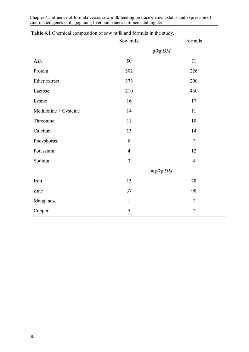
Table 4.1 Chemical composition of sow milk and formula in the study. Sow milk Formula
g/kg DM
Ash 50 71
Protein 302 226
Ether extract 373 200
Lactose 210 460
Lysine 16 17
Methionine + Cysteine 14 11
Threonine 11 10
Calcium 13 14
Phosphorus 8 7
Potassium 4 12
Sodium 3 4
mg/kg DM
Iron 13 70
Zinc 37 98
Manganese 1 7
Copper 5 7
30
Chapter 4: Influence of formula versus sow milk feeding on trace element status and expression of zinc-related genes in the jejunum, liver and pancreas of neonatal piglets
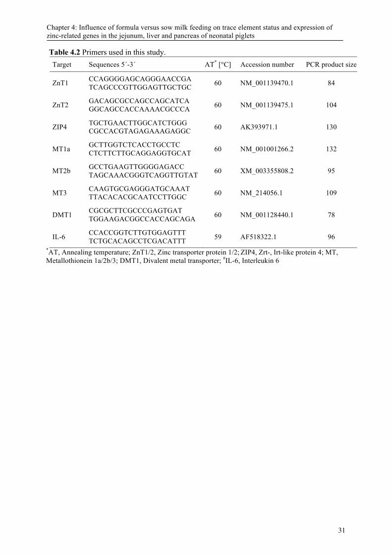
Table 4.2 Primers used in this study. Target Sequences 5´-3´ AT* [°C] Accession number PCR product size
ZnT1 CCAGGGGAGCAGGGAACCGA TCAGCCCGTTGGAGTTGCTGC 60 NM_001139470.1 84
ZnT2 GACAGCGCCAGCCAGCATCA GGCAGCCACCAAAACGCCCA 60 NM_001139475.1 104
ZIP4 TGCTGAACTTGGCATCTGGG CGCCACGTAGAGAAAGAGGC 60 AK393971.1 130
MT1a GCTTGGTCTCACCTGCCTC CTCTTCTTGCAGGAGGTGCAT 60 NM_001001266.2 132
MT2b GCCTGAAGTTGGGGAGACC TAGCAAACGGGTCAGGTTGTAT 60 XM_003355808.2 95
MT3 CAAGTGCGAGGGATGCAAAT TTACACACGCAATCCTTGGC 60 NM_214056.1 109
DMT1 CGCGCTTCGCCCGAGTGAT TGGAAGACGGCCACCAGCAGA 60 NM_001128440.1 78
IL-6 CCACCGGTCTTGTGGAGTTT TCTGCACAGCCTCGACATTT 59 AF518322.1 96
*AT, Annealing temperature; ZnT1/2, Zinc transporter protein 1/2; ZIP4, Zrt-, Irt-like protein 4; MT, Metallothionein 1a/2b/3; DMT1, Divalent metal transporter; #IL-6, Interleukin 6
31
Chapter 4: Influence of formula versus sow milk feeding on trace element status and expression of zinc-related genes in the jejunum, liver and pancreas of neonatal piglets
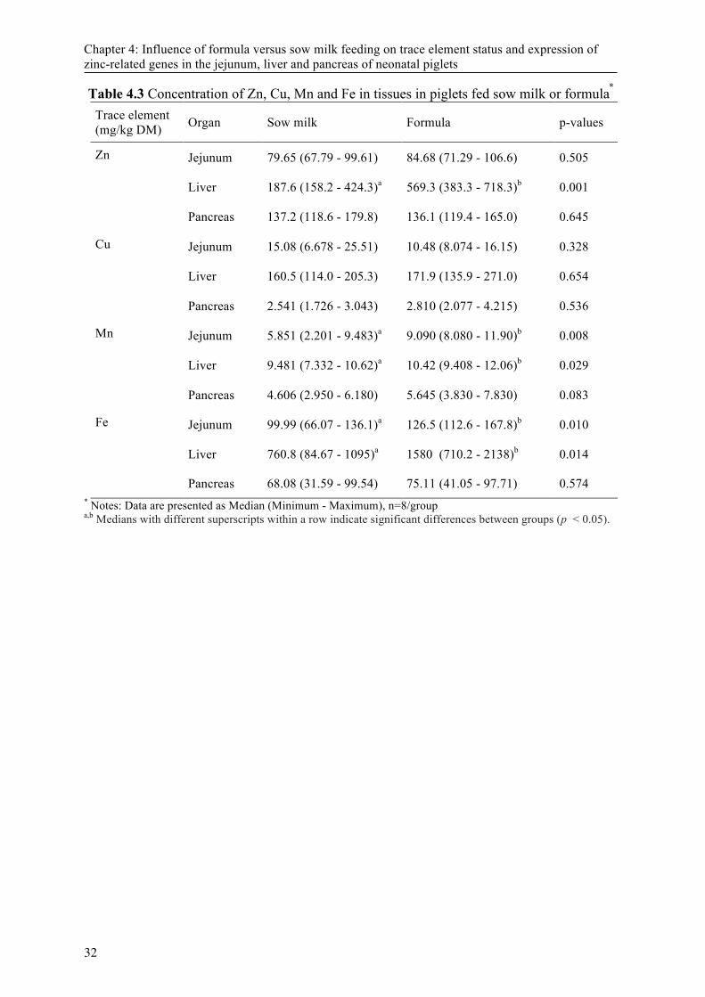
Chapter 4: Influence of formula versus sow milk feeding on trace element status and expression of zinc-related genes in the jejunum, liver and pancreas of neonatal piglets
Table 4.3 Concentration of Zn, Cu, Mn and Fe in tissues in piglets fed sow milk or formula *
Trace element (mg/kg DM) Organ Sow milk Formula p-values
Zn Jejunum 79.65 (67.79 - 99.61) 84.68 (71.29 - 106.6) 0.505
Liver 187.6 (158.2 - 424.3)a 569.3 (383.3 - 718.3)b 0.001
Pancreas 137.2 (118.6 - 179.8) 136.1 (119.4 - 165.0) 0.645
Cu Jejunum 15.08 (6.678 - 25.51) 10.48 (8.074 - 16.15) 0.328
Liver 160.5 (114.0 - 205.3) 171.9 (135.9 - 271.0) 0.654
Pancreas 2.541 (1.726 - 3.043) 2.810 (2.077 - 4.215) 0.536
Mn Jejunum 5.851 (2.201 - 9.483)a 9.090 (8.080 - 11.90)b 0.008
Liver 9.481 (7.332 - 10.62)a 10.42 (9.408 - 12.06)b 0.029
Pancreas 4.606 (2.950 - 6.180) 5.645 (3.830 - 7.830) 0.083
Fe Jejunum 99.99 (66.07 - 136.1)a 126.5 (112.6 - 167.8)b 0.010
Liver 760.8 (84.67 - 1095)a 1580 (710.2 - 2138)b 0.014
Pancreas 68.08 (31.59 - 99.54) 75.11 (41.05 - 97.71) 0.574 * Notes: Data are presented as Median (Minimum - Maximum), n=8/group a,b Medians with different superscripts within a row indicate significant differences between groups (p < 0.05).
32
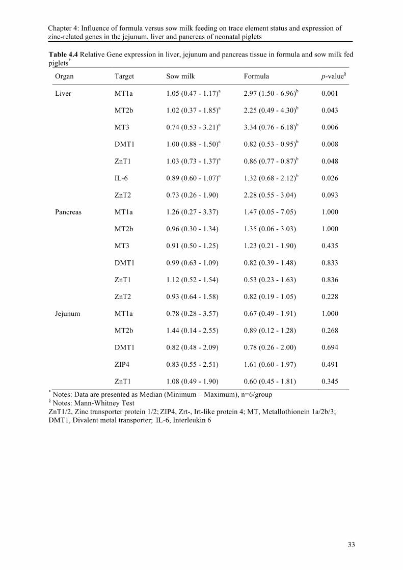
Table 4.4 Relative Gene expression in liver, jejunum and pancreas tissue in formula and sow milk fed piglets*
Organ Target Sow milk Formula p-value§
Liver MT1a 1.05 (0.47 - 1.17)a 2.97 (1.50 - 6.96)b 0.001
MT2b 1.02 (0.37 - 1.85)a 2.25 (0.49 - 4.30)b 0.043
MT3 0.74 (0.53 - 3.21)a 3.34 (0.76 - 6.18)b 0.006
DMT1 1.00 (0.88 - 1.50)a 0.82 (0.53 - 0.95)b 0.008
ZnT1 1.03 (0.73 - 1.37)a 0.86 (0.77 - 0.87)b 0.048
IL-6 0.89 (0.60 - 1.07)a 1.32 (0.68 - 2.12)b 0.026
ZnT2 0.73 (0.26 - 1.90) 2.28 (0.55 - 3.04) 0.093
Pancreas MT1a 1.26 (0.27 - 3.37) 1.47 (0.05 - 7.05) 1.000
MT2b 0.96 (0.30 - 1.34) 1.35 (0.06 - 3.03) 1.000
MT3 0.91 (0.50 - 1.25) 1.23 (0.21 - 1.90) 0.435
DMT1 0.99 (0.63 - 1.09) 0.82 (0.39 - 1.48) 0.833
ZnT1 1.12 (0.52 - 1.54) 0.53 (0.23 - 1.63) 0.836
ZnT2 0.93 (0.64 - 1.58) 0.82 (0.19 - 1.05) 0.228
Jejunum MT1a 0.78 (0.28 - 3.57) 0.67 (0.49 - 1.91) 1.000
MT2b 1.44 (0.14 - 2.55) 0.89 (0.12 - 1.28) 0.268
DMT1 0.82 (0.48 - 2.09) 0.78 (0.26 - 2.00) 0.694
ZIP4 0.83 (0.55 - 2.51) 1.61 (0.60 - 1.97) 0.491
ZnT1 1.08 (0.49 - 1.90) 0.60 (0.45 - 1.81) 0.345 * Notes: Data are presented as Median (Minimum – Maximum), n=6/group § Notes: Mann-Whitney Test ZnT1/2, Zinc transporter protein 1/2; ZIP4, Zrt-, Irt-like protein 4; MT, Metallothionein 1a/2b/3; DMT1, Divalent metal transporter; IL-6, Interleukin 6
33
Chapter 4: Influence of formula versus sow milk feeding on trace element status and expression of zinc-related genes in the jejunum, liver and pancreas of neonatal piglets
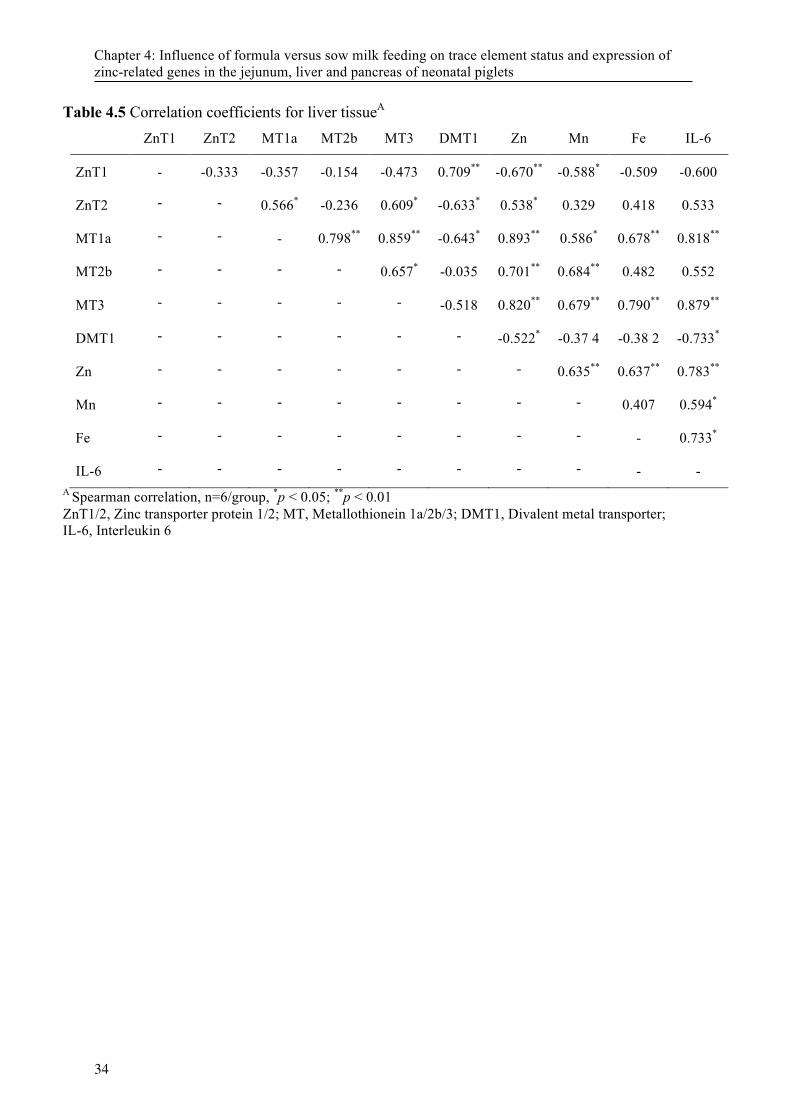
Chapter 4: Influence of formula versus sow milk feeding on trace element status and expression of zinc-related genes in the jejunum, liver and pancreas of neonatal piglets
Table 4.5 Correlation coefficients for liver tissueA ZnT1 ZnT2 MT1a MT2b MT3 DMT1 Zn Mn Fe IL-6
ZnT1 - -0.333 -0.357 -0.154 -0.473 0.709** -0.670** -0.588* -0.509 -0.600
ZnT2 - - 0.566* -0.236 0.609* -0.633* 0.538* 0.329 0.418 0.533
MT1a - - - 0.798** 0.859** -0.643* 0.893** 0.586* 0.678** 0.818**
MT2b - - - - 0.657* -0.035 0.701** 0.684** 0.482 0.552
MT3 - - - - - -0.518 0.820** 0.679** 0.790** 0.879**
DMT1 - - - - - - -0.522* -0.37 4 -0.38 2 -0.733*
Zn - - - - - - - 0.635** 0.637** 0.783**
Mn - - - - - - - - 0.407 0.594*
Fe - - - - - - - - - 0.733*
IL-6 - - - - - - - - - - A Spearman correlation, n=6/group, *p < 0.05; **p < 0.01 ZnT1/2, Zinc transporter protein 1/2; MT, Metallothionein 1a/2b/3; DMT1, Divalent metal transporter;
IL-6, Interleukin 6
34
Chapter 5: Accumulation of copper in the kidney of pigs fed high dietary zinc is due to metallothionein expression with minor effects on genes involved in copper metabolism
This chapter is published in Journal of Trace Elements in Medicine and Biology (2016)
Manuscript received: November 12, 2015 Revised: January 8, 2016 Revision accepted: January 11, 2016
Authors: A. Zetzsche, N. Schunter, J. Zentek, R. Pieper
Institute of Animal Nutrition, Department of Veterinary Medicine, Freie Universität Berlin, Königin-Luise-Strasse 49, 14195 Berlin Germany
1 Abbreverations: ADG, average daily gain; Atox1, antioxidant 1 copper chaperon; ATP7A,
copper-transorting P-type ATPase; BW, body weight; CZn, Changed dietary Zn group; CCS,
copper chaperone for superoxide dismutase; CP, Ceruloplasmin; Ctr1, copper transporter 1;
DM, dry matter; HZn, high dietary zinc group; DMT1, divalent metal ion transporter 1; MT,
metallothionein; NZn, Normal dietary zinc group; SDHA, succinate dehydrogenase subunit
A; SOD1, super oxide dismutase 1; ZIP, Zrt- and Irt- like protein ; ZnT, zinc transporter
35
Chapter 5: Accumulation of copper in the kidney of pigs fed high dietary zinc is due to metallothionein expression with minor effects on genes involved in copper metabolism
DOI: 10.1016/j.jtemb.2016.01.006
Chapter 5: Accumulation of copper in the kidney of pigs fed high dietary zinc is due to metallothionein expression with minor effects on genes involved in copper metabolism
Abstract
A study was conducted to determine the effect of high dietary zinc (Zn) oxide on trace element accumulation in various organs with special emphasis on the kidney. A total of 40 weaned piglets were allocated into two groups with 16 and 24 piglets each receiving a diet containing normal (NZn; 100 mg Zn/kg) or high (HZn; 2,100 mg Zn/kg) Zn concentration, respectively. After two weeks, eight piglets from each treatment were killed and organ samples were taken. Eight piglets from the remaining 16 pigs fed HZn diets were changed to NZn diets (CZn). All remaining piglets were killed after another two weeks for organ sampling. Trace element concentration was determined in the jejunum, liver, kidney, pancreas, bone (metacarpal IV), spleen, lung, thymus, tonsils and lymph nodes of jejunum, ileum and colon. Kidney mRNA expression of Zn transporter ZnT1 and ZIP4, genes involved in Cu metabolism (Ctr1, Atox1, SOD1, ATP7A, CCS, CP) and divalent metal ion transport (DMT1) and binding (MT-1a, MT-2b, MT-3) were determined. The Zn concentration in jejunum, liver, pancreas tissue and metacarpal IV was higher (P < 0.05) in HZn group compared with NZn and CZn groups. Trace element concentration in organs of CZn pigs was similar to those fed NZn diets. Zn concentration in muscle, lung and lymphatic organs as thymus, tonsils, spleen and lymph nodes of jejunum, ileum and colon did not differ between the groups. Zn and Cu were positively correlated (R = 0.67; P < 0.05) in the kidney. No significant differences for Cu chaperones, Cu transporters and Cu-dependent factors were determined despite decreased expression of Atox1 after two weeks and increased Ctr1 expression over time in the HZn group. Expression of MT-1a, MT-2b and MT-3 were significantly higher in HZn fed pigs with most pronounced effects for MT-1a > MT-2b > MT-3. Gene expression of MTs in pigs fed CZn diets did not differ from pigs fed NZn diets. The data suggest that high dietary Zn feeding in pigs leads to Cu co-accumulation in the kidney of pigs with minor effect on genes relevant for Cu metabolism. In addition, the organ Zn and Cu accumulation is reversible after two weeks of withdrawal of high dietary Zn. Keywords: piglets, zinc oxide, copper, kidney, gene expression
36
Chapter 5: Accumulation of copper in the kidney of pigs fed high dietary zinc is due to metallothionein expression with minor effects on genes involved in copper metabolism
5.1 Introduction The time after weaning in pigs is often accompanied with an increased risk for gastrointestinal disorders and reduced growth performance. High levels of dietary zinc oxide (ZnO) (1500-3000 mg Zn/kg diet), exceeding the dietary recommendations and maximum allowances in the EU by 10- to 20-fold, have been frequently shown to reduce diarrhoea and improve the performance of weaned piglets [1]. However, these high Zn concentrations in the diet have been shown to outbalance Zn homeostasis in the body with subsequent Zn accumulation and change of metabolic reactions in different organs including the small intestine, liver and pancreas [2-5]. The accumulation of Zn goes along with increased abundance of metallothioneins (MTs), which are essential for the regulation of intracellular heavy metal homeostasis and detoxification [6,7]. Dietary Zn increases the MT expression in a dose-depended manner, and the expression and metal-binding affinity differs between the different MT isoforms [6,8]. Thus, it is likely that feeding high dietary Zn levels with subsequent Zn accumulation and MT induction in different organs would also affect other trace elements such as copper (Cu). Usually, the Cu concentration in extrahepatic tissues is low and maintenance is regulated through Cu storage in the liver and biliary excretion. Genetic defects such as Wilson disease are associated with toxic Cu accumulation in the liver, brain, kidney and cornea in humans [9] and livestock [10]. Interestingly, the administration of Zn to patients suffering from Wilson disease could reduce the toxic effects of Cu overload in the liver due to the induction of MT [9]. In addition, a chronic intake of Zn can lead to severe Cu deficiency in humans, likely due to MT induction and Cu fixation in intestinal epithelial cells [11]. Compared to humans or ruminants, pigs are relatively tolerant against high dietary levels of Zn and reports about secondary Cu deficiency are scarce [12]. Interestingly, previous studies with piglets fed high dietary Zn showed an increased Cu accumulation in the kidney but no other organ [13,14]. The reasons or consequences for Cu metabolism in the kidney are yet not known. The present study was conducted to determine the influence of feeding high dietary levels of ZnO to weaned piglets on the accumulation Zn and Cu in various organs with special emphasis on the kidney and the influence on Zn- and Cu-specific transporters and binding protein in this organ. Furthermore, the change in trace element metabolism and accumulation after the switch from very high to normal dietary Zn was studied. 5.2 Material and Methods The study followed the institutional and national guidelines for the care and use of animals and the study was approved by the State Office of Health and Social Affairs ‘Landesamt für Gesundheit und Soziales Berlin’( LaGeSo Reg. Nr 0296/13).
5.2.1 Animals, diets and sampling A total of 40 piglets (7.6 ± 0.7 kg) were weaned at the age of 26 ± 2 days and randomly assigned into two groups. One group received normal dietary Zn concentration (NZn, n = 16) the other group received a very high level of dietary Zn (HZn, n = 24). The experimental diets are given in Table 1. To achieve the respective Zn concentrations in the diets, corn starch was partially replaced by analytical grade ZnO. Piglets were kept in flatdeck pens (n = 2 per pen) and had ad libitum access to feed and fresh water. Body weight and feed intake per pen were recorded throughout the experimental period. None of the piglets received medication before or during
37
Chapter 5: Accumulation of copper in the kidney of pigs fed high dietary zinc is due to metallothionein expression with minor effects on genes involved in copper metabolism
the experiment. After 14 days, eight piglets from each group were killed and samples from jejunum, liver, kidney, pancreas, bone, spleen, lung, thymus, tonsils and lymph nodes of jejunum, ileum and colon were taken, immediately snap-frozen in liquid nitrogen and stored at -80 °C until further analyses. In addition, a 1.5 cm x 1.5 cm part of left kidney was immediately placed in RNAlater® (Sigma-Aldrich) for 30 min and subsequently stored at -80 °C. The remaining eight piglets in the NZn group and eight piglets of the HZn group were fed their respective diets another 14 days, whereas eight piglets from the HZn group were subjected to NZn for the next 14 days (CZn group). At the end of this period, all remaining piglets were killed and organ samples takes as described above. 5.2.2 Chemical analyses Proximate nutrients in the diets were analysed by standard Weende procedures [15]. After hydrolysing ash in hydrochloric acid, concentration of Zn, Cu, Mn and Fe in diets and organs were analysed by atomic absorption spectrometry in an AAS vario 6 spectrometer (Analytik Jena, Jena, Germany) as described previously [5].
5.2.3 Gene expression Total RNA from kidney tissue was extracted using the Nucleo Spin® kit (Macherey Nagel) and mRNA quality and quantity were determined on an Agilent Bioanalyzer 2100 (Agilent). Equimolar mRNA (104 ng RNA) was transcribed into complementary DNA (cDNA) by Superscript® reverse transcriptase kit (Life Technologies). Subsequently, real-time qPCR was performed using Brilliant SYBR® Green QPCR Master Mix low Rox (Agilent Technologies) on a Stratagene Mx3005P (Agilent Technologies) with cycling conditions as described previously [16,17]. Primers (Supplemental Table 1) were designed targeting zinc transporter (ZnT1, ZIP4), copper transport 1 (Ctr1), antioxidant 1 copper chaperon (Atox1), superoxide dismutase 1 (SOD1), copper-transporting P-type ATPase (ATP7A), copper chaperone for superoxide dismutase (CCS), ceruloplasmin (CP), divalent metal ion transporter 1 (DMT1) and three metallothionein isoforms (MT-1a, MT-2b, MT-3) using the NCBI online primer designer tool. Relative gene expression was calculated based on PCR efficiency and Ct values of target genes normalized using Succinate Dehydrogenase subunit A, β-2-microglobulin and β-actin [18]. 5.2.4 Statistical analyses For statistical analyses, normally distributed data were analyzed by student´s t-test to compare means within a group at the different time points and group differences after 2 weeks of feeding several dietary Zn concentrations. Furthermore, ANOVA followed Bonferroni post hoc test for group differences after four feeding weeks using SPSS (version 22.0, Chicago, USA). Pearson correlation coefficients were determined for trace elements within organs and for gene expression data with trace elements in the kidney. Unless otherwise stated, results are presented as mean ± SD. Differences at P < 0.05 were considered as significant.
38
Chapter 5: Accumulation of copper in the kidney of pigs fed high dietary zinc is due to metallothionein expression with minor effects on genes involved in copper metabolism
5.3 Results 5.3.1 Performance After the first two weeks, piglets in the HZn group had higher weight gain (P < 0.05) compared to NZn group (10.6 ± 1.00 kg vs. 9.8 ± 1.3 kg, respectively). Average daily gain (Ronis et al.) was numerically (P = 0.168) higher in HZn compared to the NZn group (170 ± 59 g/d vs. 122 ± 72 g/d, respectively). After four weeks, no significant differences in body weight (BW) (17.1 ± 1.9 kg, 16.0 ± 2.6 kg and 16.6 ± 0.9 kg for HZn, NZn and CZn, respectively) or ADG (442 ± 89 g/d, 402 ± 148 g/d and 393 ± 54 g/d for HZn, NZn and CZn, respectively) were determined. No clinical signs of diarrhoea or impaired health were observed during the entire period.
5.3.2 Organ trace mineral concentration The Zn concentration in jejunum, liver, pancreas tissue and metacarpal IV was higher (P < 0.05) in HZn group after two and four weeks as compared with NZn and CZn group, respectively (Supplemental Table S2). The concentration of Zn and Cu in kidney tissue was approximately twofold higher in HZn group than in NZn and CZn group (Figure 1A, B) after two and four weeks, respectively (P < 0.05). Kidney Zn and Cu concentration did not differ between NZn and CZn group after four weeks. Correlation analysis of kidney Zn and Cu concentration from all animals revealed a strong positive correlation in kidney tissue (R = 0.67, P < 0.05; Figure 2). Neither Mn nor Fe concentration correlated to measured minerals in kidney tissue (data not shown). In addition, significant correlations were determined between jejunal Zn and liver (R = 0.54), kidney (R = 0.62) and metacarpal IV (R = 0.72) Zn concentrations (data not shown). The Zn concentration in muscle and lung tissue or in lymphatic organs as thymus, tonsils, spleen and lymph nodes of jejunum, ileum and colon did not differ between the groups (Supplemental Table S2). In addition, no differences in Mn, Cu and Fe concentration in jejunum, pancreas, bone, spleen, lunge, muscle, thymus, tonsils and lymph nodes of jejunum, ileum and colon were determined (data not shown). 5.3.3 Gene expression Expression of Zn and Cu binding proteins and transporters was analysed in kidney tissue. The relative gene expression data are presented in Table 2. The expression of MT-1a, MT-2b and MT-3 was higher (P < 0.05) in HZn compared to the NZn group after two and four weeks, respectively. MT-1a mRNA abundance was higher in NZn group after four weeks compared with two weeks (P < 0.05). The MT-1a and MT-3 mRNA abundance was lower (P < 0.05) in the CZn group compared with the HZn group, but similar to the NZn group. Expression of MT-2b was intermediate in the CZn group compared to the other two groups. The Atox1 mRNA expression was lower (P < 0.05) in HZn group after two weeks compared with the NZn group but was not different between groups after four weeks. The Ctr1 mRNA concentration in HZn group was high (P < 0.05) after four weeks compared with levels at two weeks. Expression of DMT1 was numerically higher in the HZn compared with NZn group after four weeks (P = 0.224). The mRNA abundance of ZnT1, ZIP4, CCS, CP, ATP7A and SOD1 did not differ between groups and time points.
39
Chapter 5: Accumulation of copper in the kidney of pigs fed high dietary zinc is due to metallothionein expression with minor effects on genes involved in copper metabolism
The Zn concentration in kidney tissue correlated with gene expression of the three determined MT isoforms (Table 3). However, the association between Zn and MT-1a (R = 0.69; P < 0.05) and MT-3 (R = 0.59; P < 0.05) was higher compared with MT-2b (R = 0.28; P < 0.05). A positive correlation (R = 0.29; P < 0.05) between Zn and CP expression was determined. Similar to Zn, the Cu concentration in the kidney showed a stronger correlation to MT-1a (R = 0.65; P < 0.05) and MT-3 (R = 0.64; P < 0.05) compared with MT-2b (R = 0.41; P < 0.05). In addition, positive correlations between Cu concentration and CP (R = 0.51; P < 0.05) and Ctr1 (R = 0.35; P < 0.05) gene expression were determined. No further significant correlations were found between kidney trace element concentration and expression of Zn and Cu binding proteins and transporters. 5.4 Discussion In the present study, feeding piglets with HZn diets during the first two weeks after the weaning increased BW compared with a normally supplemented group but not thereafter. Similar to the observation made in the present study, improved performance was only observed during the first 2 weeks of feeding high dietary Zn, whereas no or even opposite effects were determined thereafter [4,19]. Reasons for this effect are yet not clear. Considering the fact that Zn homeostasis is usually regulated within narrow margins in the body, feeding much higher dietary Zn will likely outbalance homeostatic regulation, induce Zn accumulation in various organs and impair their metabolic function [2,3,5]. The present and previous studies revealed higher Zn contents in several organs including jejunum, liver, pancreas, bone and kidney after long-term supplementation of high dietary Zn [3,5,14,20,21]. Additionally, no increased Zn concentration in HZn group were observed in muscle and lunge tissue, which was also reported in previous studies [20]. Moreover, no accumulation in immune organs such as spleen, thymus, tonsils and analysed lymph nodes was noted, which might indicate different uptake or intracellular metal binding mechanisms in these organs. Similarly to the present results, feeding 2000 mg Zn/kg to piglets did not increase Zn concentration in the spleen [21,22]. Interestingly, a switch from HZn to NZn diets after two weeks resulted in organ Zn concentrations that were similar to piglets fed NZn diets over the entire period. This is in good agreement with a recent study in weaned piglets, where similar observations were reported after a four-week period of high dietary Zn followed by dietary Zn reduction for another two weeks [20]. This indicates a re-balanced homeostasis after a period of excessive Zn accumulation in the pig. In the present study, a co-accumulation and strong correlation between Zn and Cu concentration was observed in the kidney, which is in concordance with previous studies in pigs [13,14]. This is an interesting finding because the urinary excretion of both Zn and Cu is low [12,23]. There is yet no clear explanation why co-accumulation occurs only in the kidney and not in other organs, but this could be related to intracellular processes following ultrafiltration and reabsorption. Usually, Zn is ultrafiltrated in kidneys´ tubular systems and reabsorbed in proximal and distal parts [24]. Intracellular, cytoplasmic Zn is bound to MTs, which are cysteine rich metalloproteins with a number of isoforms. MTs bind heavy metals such as Zn, Cu or Cd to reduce their potential toxicity as free ions in the cytoplasm [25]. While MT-1a and MT-2b occur only in proximal tubular cells, MT-3 was observed in glomeruli, proximal and
40
Chapter 5: Accumulation of copper in the kidney of pigs fed high dietary zinc is due to metallothionein expression with minor effects on genes involved in copper metabolism
distal tubular cells [26-28]. MT gene expression is induced by high Zn concentration in cells through Zn-responsive transcription factors such as MTF-1 [29]. Studies in rats suggested an additive effect of Zn and Cu on MT expression and protein abundance [30]. In human proximal tubular cells, elevation of MT-3 expression occurred within 24 hours but a reduction to normal levels was determined after seven days of high Zn exposure suggesting time-dependent regulation of MTs due to Zn exposure [31]. In the present study, tissue samples were taken after two and four weeks, which could explain differences in MT-1a, MT-2b and MT-3 gene expression. Interestingly, MTs have a higher affinity towards Cu [32]. It is thus likely that Zn induced expression of MTs results in a higher binding of Cu to these proteins in the kidney. Unfortunately, attempts to stain Cu and Zn within renal compartments failed in this study. However, data from rodent models indicate that higher concentrations of both Zn and Cu can be found in the area of proximal tubular cells [33]. Although speculative, this may also explain higher expression of MT-1a and MT-2b as well as strong correlation between the MTs with Cu and Zn concentrations. Besides the role of intracellular MTs, we assumed that higher Zn and Cu concentration in the kidney would affect the expression of Zn and Cu specific transporters. For example, ZnT1 and ZnT2 are highly expressed in renal tubular epithelial cells but show different responsiveness to Zn [34-36]. No differences in ZnT1 expression were determined, suggesting that Zn accumulation could due to absent Zn export from cytoplasm. While the ZnT transporter family mainly regulates Zn export from the cell, ZIP proteins are responsible for cellular Zn uptake. In the present study, the expression of ZIP4 was determined. In contrast to a previous study, where ZIP4 expression was down-regulated in jejunal tissue of piglets fed high dietary Zn [3], no difference were determined in renal tissue. Therefore, tissue specific differences in ZIP4 expression and response to high Zn can be assumed. The expression of renal ZIP8 and ZIP10 was not determined in the present study. Cellular Cu uptake is facilitated via Ctr1. During high dietary Cu intake Ctr1 protein translocates to intracellular vesicular compartments but is not regulated at transcriptional level, which could explain that no differences in Ctr1 mRNA abundance were observed in the present study [7,37]. Similarly, only minor differences were determined for DMT1 expression. DMT1 is located at the apical site of epithelial cells likely to reabsorb divalent ions [38]. A possible explanation is that DMT1 expression is moderately regulated via Fe [38] which was not affected by high Zn feeding in the current study. Among others, Atox1 and CCS are important intracellular Cu trafficking proteins. While Atox1 delivers Cu to ATP7A, CCS is a Cu chaperone for SOD1 [39]. SOD1 catalyses superoxide radicals to hydrogen peroxide and molecular oxygen and is highly expressed in mammalian liver and kidney tissue [40]. Neither changes in CCS nor SOD1 in mRNA concentration were observed in the present study. This is in good concordance to previous findings, were CCS protein level was independent from CCS mRNA concentration due to post-translational modification [41]. Similar differences between mRNA and protein abundance can be assumed for SOD1 [41]. A lower Atox1 mRNA concentration was determined in the HZn group after two weeks in comparison to NZn group. A reduced abundance of Atox1 might decrease cellular Cu export and could thereby be associated with elevated Cu concentration [42]. However, this may also indicate that Cu is bound to proteins with higher affinity to Cu, such as MTs [32].
41
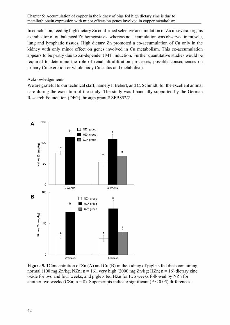
Chapter 5: Accumulation of copper in the kidney of pigs fed high dietary zinc is due to metallothionein expression with minor effects on genes involved in copper metabolism
In conclusion, feeding high dietary Zn confirmed selective accumulation of Zn in several organs as indicator of outbalanced Zn homeostasis, whereas no accumulation was observed in muscle, lung and lymphatic tissues. High dietary Zn promoted a co-accumulation of Cu only in the kidney with only minor effect on genes involved in Cu metabolism. This co-accumulation appears to be partly due to Zn-dependent MT induction. Further quantitative studies would be required to determine the role of renal ultrafiltration processes, possible consequences on urinary Cu excretion or whole body Cu status and metabolism. Acknowledgements We are grateful to our technical staff, namely I. Bebert, and C. Schmidt, for the excellent animal care during the execution of the study. The study was financially supported by the German Research Foundation (DFG) through grant # SFB852/2.
Figure 5. 1Concentration of Zn (A) and Cu (B) in the kidney of piglets fed diets containing normal (100 mg Zn/kg; NZn; n = 16), very high (2000 mg Zn/kg; HZn; n = 16) dietary zinc oxide for two and four weeks, and piglets fed HZn for two weeks followed by NZn for another two weeks (CZn; n = 8). Superscripts indicate significant (P < 0.05) differences.
42
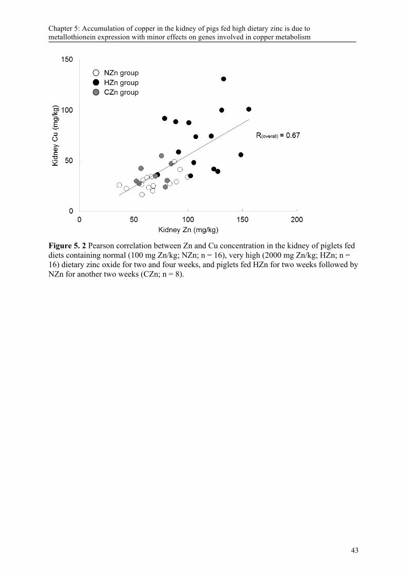
Chapter 5: Accumulation of copper in the kidney of pigs fed high dietary zinc is due to metallothionein expression with minor effects on genes involved in copper metabolism
Figure 5. 2 Pearson correlation between Zn and Cu concentration in the kidney of piglets fed diets containing normal (100 mg Zn/kg; NZn; n = 16), very high (2000 mg Zn/kg; HZn; n = 16) dietary zinc oxide for two and four weeks, and piglets fed HZn for two weeks followed by NZn for another two weeks (CZn; n = 8).
43

Chapter 5: Accumulation of copper in the kidney of pigs fed high dietary zinc is due to metallothionein expression with minor effects on genes involved in copper metabolism
Table 5. 1 Ingredients and chemical composition of the diets used in the study Ingredients g/kg diet Chemical composition NZn† HZn‡
Wheat 300 g/kg
Barley 200 Dry matter 892 891
Corn 230 Crude protein 188 192
Soybean meal 200 Crude fiber 30 32
Monocalcium phosphate 13 Ether extract 29 30
Limestone 14 Starch 424 445
Vitamine-mineral pre-mix* 15 Ash 51 52
Soy oil 10
Lysine-HCl 3.5 mg/kg
Tryptophan 1 Iron 183 156
Methionine 1 Manganese 96 113
Salt 2.5 Copper 18 19
Zinc oxide / Corn starch 10 Zinc 72 2103
*containing per kg: 600,000 IU vitamin, 120,000 IU Vitamin D3, 8,000 mg Vitamin E, 300 mg Vitamin K3, 250 mg Vitamin B2, 400 mg Vitamin B6, 2,000 µg Vitamin B12, 2,500 mg nicotine acid, 100 mg folic acid, 1,000 mg Pantothenic acid, 80,000 mg Choline chloride, 30 mg Cobalt, 45 mg Iodine, 35 mg Selenium, 6,000 mg Manganese, 1,000 mg Copper, 5,000 mg Iron † Normal dietary zinc group; ‡ High dietary zinc group
44
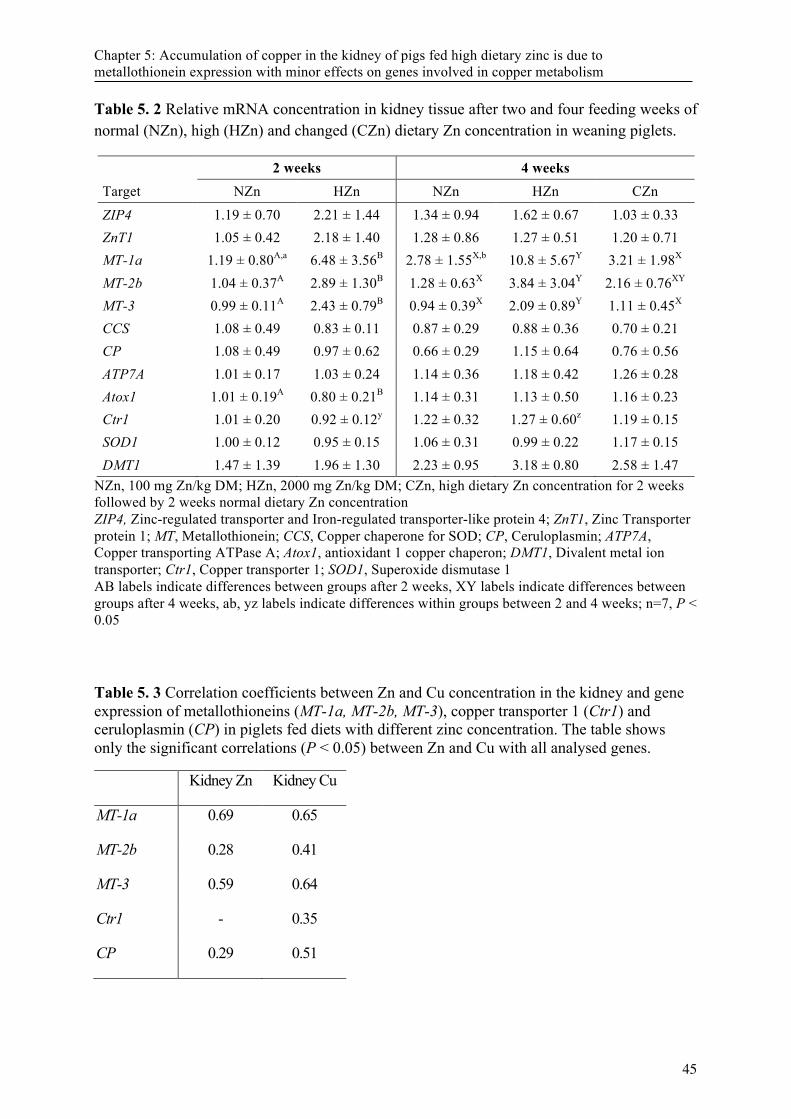
Chapter 5: Accumulation of copper in the kidney of pigs fed high dietary zinc is due to metallothionein expression with minor effects on genes involved in copper metabolism
Table 5. 2 Relative mRNA concentration in kidney tissue after two and four feeding weeks of normal (NZn), high (HZn) and changed (CZn) dietary Zn concentration in weaning piglets.
2 weeks 4 weeks Target NZn HZn NZn HZn CZn
ZIP4 1.19 ± 0.70 2.21 ± 1.44 1.34 ± 0.94 1.62 ± 0.67 1.03 ± 0.33 ZnT1 1.05 ± 0.42 2.18 ± 1.40 1.28 ± 0.86 1.27 ± 0.51 1.20 ± 0.71 MT-1a 1.19 ± 0.80A,a 6.48 ± 3.56B 2.78 ± 1.55X,b 10.8 ± 5.67Y 3.21 ± 1.98X
MT-2b 1.04 ± 0.37A 2.89 ± 1.30B 1.28 ± 0.63X 3.84 ± 3.04Y 2.16 ± 0.76XY
MT-3 0.99 ± 0.11A 2.43 ± 0.79B 0.94 ± 0.39X 2.09 ± 0.89Y 1.11 ± 0.45X
CCS 1.08 ± 0.49 0.83 ± 0.11 0.87 ± 0.29 0.88 ± 0.36 0.70 ± 0.21 CP 1.08 ± 0.49 0.97 ± 0.62 0.66 ± 0.29 1.15 ± 0.64 0.76 ± 0.56 ATP7A 1.01 ± 0.17 1.03 ± 0.24 1.14 ± 0.36 1.18 ± 0.42 1.26 ± 0.28 Atox1 1.01 ± 0.19A 0.80 ± 0.21B 1.14 ± 0.31 1.13 ± 0.50 1.16 ± 0.23 Ctr1 1.01 ± 0.20 0.92 ± 0.12y 1.22 ± 0.32 1.27 ± 0.60z 1.19 ± 0.15 SOD1 1.00 ± 0.12 0.95 ± 0.15 1.06 ± 0.31 0.99 ± 0.22 1.17 ± 0.15 DMT1 1.47 ± 1.39 1.96 ± 1.30 2.23 ± 0.95 3.18 ± 0.80 2.58 ± 1.47
NZn, 100 mg Zn/kg DM; HZn, 2000 mg Zn/kg DM; CZn, high dietary Zn concentration for 2 weeks followed by 2 weeks normal dietary Zn concentration ZIP4, Zinc-regulated transporter and Iron-regulated transporter-like protein 4; ZnT1, Zinc Transporter protein 1; MT, Metallothionein; CCS, Copper chaperone for SOD; CP, Ceruloplasmin; ATP7A, Copper transporting ATPase A; Atox1, antioxidant 1 copper chaperon; DMT1, Divalent metal ion transporter; Ctr1, Copper transporter 1; SOD1, Superoxide dismutase 1 AB labels indicate differences between groups after 2 weeks, XY labels indicate differences between groups after 4 weeks, ab, yz labels indicate differences within groups between 2 and 4 weeks; n=7, P < 0.05 Table 5. 3 Correlation coefficients between Zn and Cu concentration in the kidney and gene expression of metallothioneins (MT-1a, MT-2b, MT-3), copper transporter 1 (Ctr1) and ceruloplasmin (CP) in piglets fed diets with different zinc concentration. The table shows only the significant correlations (P < 0.05) between Zn and Cu with all analysed genes. Kidney Zn Kidney Cu
MT-1a 0.69 0.65
MT-2b 0.28 0.41
MT-3 0.59 0.64
Ctr1 - 0.35
CP 0.29 0.51
45
Chapter 5: Accumulation of copper in the kidney of pigs fed high dietary zinc is due to metallothionein expression with minor effects on genes involved in copper metabolism
Supplemental Table S 5. 1 Primes used in this study Target Sequence 5 ́to 3 ́ AT [°C]# Accession number PCR product size*
ZIP4 TGCTGAACTTGGCATCTGGG
CGCCACGTAGAGAAAGAGGC 60 AK393971.1 130
ZnT1 CCAGGGGAGCAGGGAACCGA
TCAGCCCGTTGGAGTTGCTGC 60 NM_001139470.1 84
MT-1a GCTTGGTCTCACCTGCCTC
CTCTTCTTGCAGGAGGTGCAT 60 NM_001001266.2 132
MT-2b GCCTGAAGTTGGGGAGACC TAGCAAACGGGTCAGGTTGTAT
60 XM_003355808.2 95
MT-3 CAAGTGCGAGGGATGCAAAT
TTACACACGCAATCCTTGGC 60 NM_214056.1 109
CCS GTCACTCTCTCTGTCCACCC CTGCACACTAGGTCTCCTGG
60 NM_001001866.1 86
CP GTGGCGCCCAAAGAAACATT
GACAGGATCTTTGTAAGTGGGC 60 NM_001267694.2 85
ATP7A TGTACCTCAAACTCTCCCTCCA
AGTCAGAGGCTGGCTCACTA 60 XM_003135200.2 81
Atox1 AGGTCTGCATTGACTCTGAGC
ACGGCCTTTCCTGTTTTCCC 60 NM_001167641.1 82
Ctr1 TGACAGAGAAGCGGATCGAG
CAGCAGAAGATTCTCCCCAGA 60 AF320815.2 84
SOD1 CTGAAGGGAGAGAAGACAGTGTTA
ATCTCCAAACTGATGGACATGGAA 60 GQ913661.1 100
DMT1 CGCGCTTCGCCCGAGTGAT
TGGAAGACGGCCACCAGCAGA 60 NM_001128440.1 78
* PCR Product size is presented in base pairs; # AT, Annealing temperature; ZIP4, Zinc–regulated transporter and Iron-regulated transporter-like protein 4; ZnT1, Zinc Transporter protein 1; MT, Metallothionein; CCS, Copper chaperone for SOD; CP, Ceruloplasmin; ATP7A, Copper transporting ATPase A; Atox-1, antioxidant 1 copper chaperon; Ctr1, Copper transporter 1; SOD1, Superoxide dismutase 1; DMT1, Divalent metal ion transporter 1
46
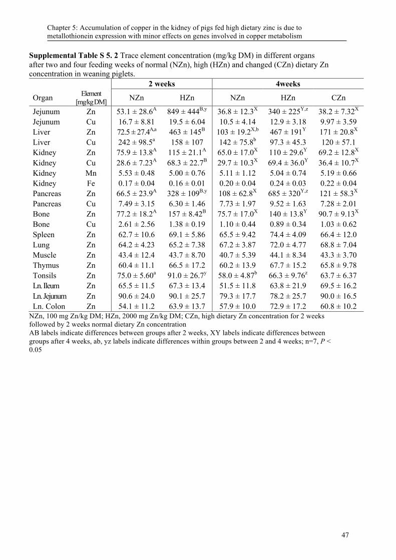
Chapter 5: Accumulation of copper in the kidney of pigs fed high dietary zinc is due to metallothionein expression with minor effects on genes involved in copper metabolism
Supplemental Table S 5. 2 Trace element concentration (mg/kg DM) in different organs after two and four feeding weeks of normal (NZn), high (HZn) and changed (CZn) dietary Zn concentration in weaning piglets. 2 weeks 4weeks
Organ Element [mg/kg DM] NZn HZn NZn HZn CZn
Jejunum Zn 53.1 ± 28.6A 849 ± 444B,y 36.8 ± 12.3X 340 ± 225Y,z 38.2 ± 7.32X
Jejunum Cu 16.7 ± 8.81 19.5 ± 6.04 10.5 ± 4.14 12.9 ± 3.18 9.97 ± 3.59 Liver Zn 72.5 ± 27.4A,a 463 ± 145B 103 ± 19.2X,b 467 ± 191Y 171 ± 20.8X
Liver Cu 242 ± 98.5a 158 ± 107 142 ± 75.8b 97.3 ± 45.3 120 ± 57.1 Kidney Zn 75.9 ± 13.8A 115 ± 21.1A 65.0 ± 17.0X 110 ± 29.6Y 69.2 ± 12.8X
Kidney Cu 28.6 ± 7.23A 68.3 ± 22.7B 29.7 ± 10.3X 69.4 ± 36.0Y 36.4 ± 10.7X
Kidney Mn 5.53 ± 0.48 5.00 ± 0.76 5.11 ± 1.12 5.04 ± 0.74 5.19 ± 0.66 Kidney Fe 0.17 ± 0.04 0.16 ± 0.01 0.20 ± 0.04 0.24 ± 0.03 0.22 ± 0.04 Pancreas Zn 66.5 ± 23.9A 328 ± 109B,y 108 ± 62.8X 685 ± 320Y,z 121 ± 58.3X
Pancreas Cu 7.49 ± 3.15 6.30 ± 1.46 7.73 ± 1.97 9.52 ± 1.63 7.28 ± 2.01 Bone Zn 77.2 ± 18.2A 157 ± 8.42B 75.7 ± 17.0X 140 ± 13.8Y 90.7 ± 9.13X
Bone Cu 2.61 ± 2.56 1.38 ± 0.19 1.10 ± 0.44 0.89 ± 0.34 1.03 ± 0.62 Spleen Zn 62.7 ± 10.6 69.1 ± 5.86 65.5 ± 9.42 74.4 ± 4.09 66.4 ± 12.0 Lung Zn 64.2 ± 4.23 65.2 ± 7.38 67.2 ± 3.87 72.0 ± 4.77 68.8 ± 7.04 Muscle Zn 43.4 ± 12.4 43.7 ± 8.70 40.7 ± 5.39 44.1 ± 8.34 43.3 ± 3.70 Thymus Zn 60.4 ± 11.1 66.5 ± 17.2 60.2 ± 13.9 67.7 ± 15.2 65.8 ± 9.78 Tonsils Zn 75.0 ± 5.60a 91.0 ± 26.7y 58.0 ± 4.87b 66.3 ± 9.76z 63.7 ± 6.37 Ln. Ileum Zn 65.5 ± 11.5 67.3 ± 13.4 51.5 ± 11.8 63.8 ± 21.9 69.5 ± 16.2 Ln. Jejunum Zn 90.6 ± 24.0 90.1 ± 25.7 79.3 ± 17.7 78.2 ± 25.7 90.0 ± 16.5 Ln. Colon Zn 54.1 ± 11.2 63.9 ± 13.7 57.9 ± 10.0 72.9 ± 17.2 60.8 ± 10.2
NZn, 100 mg Zn/kg DM; HZn, 2000 mg Zn/kg DM; CZn, high dietary Zn concentration for 2 weeks followed by 2 weeks normal dietary Zn concentration AB labels indicate differences between groups after 2 weeks, XY labels indicate differences between groups after 4 weeks, ab, yz labels indicate differences within groups between 2 and 4 weeks; n=7, P < 0.05
47
Chapter 5: Accumulation of copper in the kidney of pigs fed high dietary zinc is due to metallothionein expression with minor effects on genes involved in copper metabolism
References [1] Sales J. Effects of pharmacological concentrations of dietary zinc oxide on growth of
post-weaning pigs: a meta-analysis. Biological trace element research 2013; 152: 343-9.
[2] Bondzio A, Pieper R, Gabler C, Weise C, Schulze P, Zentek J, Einspanier R. Feeding low or pharmacological concentrations of zinc oxide changes the hepatic proteome profiles in weaned piglets. PloS one 2013; 8: e81202.
[3] Martin L, Lodemann U, Bondzio A, Gefeller EM, Vahjen W, Aschenbach JR, Zentek J, Pieper R. A high amount of dietary zinc changes the expression of zinc transporters and metallothionein in jejunal epithelial cells in vitro and in vivo but does not prevent zinc accumulation in jejunal tissue of piglets. The Journal of nutrition 2013; 143: 1205-10.
[4] Martin L, Pieper R, Schunter N, Vahjen W, Zentek J. Performance, organ zinc concentration, jejunal brush border membrane enzyme activities and mRNA expression in piglets fed with different levels of dietary zinc. Archives of animal nutrition 2013; 67: 248-61.
[5] Pieper R, Martin L, Schunter N, Villodre Tudela C, Weise C, Klopfleisch R, Zentek J, Einspanier R, Bondzio A. Impact of high dietary zinc on zinc accumulation, enzyme activity and proteomic profiles in the pancreas of piglets. J Trace Elem Med Biol 2015; 30: 30-6.
[6] Sutherland DE, Stillman MJ. The "magic numbers" of metallothionein. Metallomics : integrated biometal science 2011; 3: 444-63.
[7] Suzuki-Kurasaki M, Okabe M, Kurasaki M. Copper-metallothionein in the Kidney of Macular Mice: A Model for Menkes Disease. Journal of Histochemistry & Cytochemistry 1997; 45: 1493-501.
[8] Alscher DM, Braun N, Biegger D, Stuelten C, Gawronski K, Murdter TE, Kuhlmann U, Fritz P. Induction of metallothionein in proximal tubular cells by zinc and its potential as an endogenous antioxidant. Kidney & blood pressure research 2005; 28: 127-33.
[9] Ala A, Walker AP, Ashkan K, Dooley JS, Schilsky ML. Wilson's disease. Lancet 2007; 369: 397-408.
[10] Fieten H, Penning LC, Leegwater PA, Rothuizen J. New canine models of copper toxicosis: diagnosis, treatment, and genetics. Annals of the New York Academy of Sciences 2014; 1314: 42-8.
[11] Fosmire GJ. Zinc toxicity. Am J Clin Nutr 1990; 51: 225-7. [12] Suttle N. Mineral Nutrition of Livestock Editon ed. CABI Publishing: Wallingford
(UK), 2010. p. [13] Carlson MS, Hill GM, Link JE. Early- and traditionally weaned nursery pigs benefit
from phase-feeding pharmacological concentrations of zinc oxide: effect on metallothionein and mineral concentrations. Journal of animal science 1999; 77: 1199-207.
[14] Martinez MM, Hill GM, Link JE, Raney NE, Tempelman RJ, Ernst CW. Pharmacological zinc and phytase supplementation enhance metallothionein mRNA abundance and protein concentration in newly weaned pigs. The Journal of nutrition 2004; 134: 538-44.
[15] Naumann C, Bassler R. Die chemische Untersuchung von Futtermitteln. Methodenbuch, Editon ed. VDLUFA-Verlag: Darmstadt (Germany), 2004.
[16] Villodre Tudela C, Boudry C, Stumpff F, Aschenbach JR, Vahjen W, Zentek J, Pieper R. Down-regulation of monocarboxylate transporter 1 (MCT1) gene expression in the
48
Chapter 5: Accumulation of copper in the kidney of pigs fed high dietary zinc is due to metallothionein expression with minor effects on genes involved in copper metabolism
colon of piglets is linked to bacterial protein fermentation and pro-inflammatory cytokine-mediated signalling. The British journal of nutrition 2015; 113: 610-7.
[17] Zetzsche A, Pieper R, Zentek J. Influence of formula versus sow milk feeding on trace element status and expression of zinc-related genes in the jejunum, liver and pancreas of neonatal piglets. Archives of animal nutrition 2015; 69: 366-77.
[18] Pfaffl MW. A new mathematical model for relative quantification in real-time RT-PCR. Nucleic acids research 2001; 29: e45.
[19] Jensen-Waern M, Melin L, Lindberg R, Johannisson A, Petersson L, Wallgren P. Dietary zinc oxide in weaned pigs--effects on performance, tissue concentrations, morphology, neutrophil functions and faecal microflora. Research in veterinary science 1998; 64: 225-31.
[20] Janczyk P, Busing K, Dobenecker B, Nockler K, Zeyner A. Effect of high dietary zinc oxide on the caecal and faecal short-chain fatty acids and tissue zinc and copper concentration in pigs is reversible after withdrawal of the high zinc oxide from the diet. Journal of animal physiology and animal nutrition 2015; 99 Suppl S1: 13-22.
[21] Davin R, Manzanilla EG, Klasing KC, Perez JF. Effect of weaning and in-feed high doses of zinc oxide on zinc levels in different body compartments of piglets. Journal of animal physiology and animal nutrition 2013; 97 Suppl 1: 6-12.
[22] Wang Y, Tang JW, Ma WQ, Feng J. Dietary zinc glycine chelate on growth performance, tissue mineral concentrations, and serum enzyme activity in weanling piglets. Biological trace element research 2010; 133: 325-34.
[23] Buff CE, Bollinger DW, Ellersieck MR, Brommelsiek WA, Veum TL. Comparison of growth performance and zinc absorption, retention, and excretion in weanling pigs fed diets supplemented with zinc-polysaccharide or zinc oxide. Journal of animal science 2005; 83: 2380-6.
[24] Victery W, Smith JM, Vander AJ. Renal tubular handling of zinc in the dog. The American journal of physiology 1981; 241: F532-9.
[25] Davis SR, Cousins RJ. Metallothionein expression in animals: a physiological perspective on function. The Journal of nutrition 2000; 130: 1085-8.
[26] Kojima Y, Hamashima Y. Immunohistological Studies of Metallothionein .2. Its Detection in the Human-Fetal Kidney. Acta Histochem Cytoc 1980; 13: 277-86.
[27] Mididoddi S, McGuirt JP, Sens MA, Todd JH, Sens DA. Isoform-specific expression of metallothionein mRNA in the developing and adult human kidney. Toxicology letters 1996; 85: 17-27.
[28] Nartey NO, Banerjee D, Cherian MG. Immunohistochemical localization of metallothionein in cell nucleus and cytoplasm of fetal human liver and kidney and its changes during development. Pathology 1987; 19: 233-8.
[29] Laity JH, Andrews GK. Understanding the mechanisms of zinc-sensing by metal-response element binding transcription factor-1 (MTF-1). Archives of biochemistry and biophysics 2007; 463: 201-10.
[30] Blalock TL, Dunn MA, Cousins RJ. Metallothionein gene expression in rats: tissue-specific regulation by dietary copper and zinc. The Journal of nutrition 1988; 118: 222-8.
[31] Hoey JG, Garrett SH, Sens MA, Todd JH, Sens DA. Expression of MT-3 mRNA in human kidney, proximal tubule cell cultures, and renal cell carcinoma. Toxicology letters 1997; 92: 149-60.
[32] Banci L, Bertini I, Ciofi-Baffoni S, Kozyreva T, Zovo K, Palumaa P. Affinity gradients drive copper to cellular destinations. Nature 2010; 465: 645-8.
[33] Irato P, Santon A, Ossi E, Albergoni V. Interactions between metals in rat liver and kidney: localization of metallothionein. The Histochemical journal 2001; 33: 79-86.
49
Chapter 5: Accumulation of copper in the kidney of pigs fed high dietary zinc is due to metallothionein expression with minor effects on genes involved in copper metabolism
[34] Liuzzi JP, Cousins RJ. Mammalian zinc transporters. Annual review of nutrition 2004; 24: 151-72.
[35] Liuzzi JP, Blanchard RK, Cousins RJ. Differential regulation of zinc transporter 1, 2, and 4 mRNA expression by dietary zinc in rats. The Journal of nutrition 2001; 131: 46-52.
[36] Lichten LA, Cousins RJ. Mammalian zinc transporters: nutritional and physiologic regulation. Annual review of nutrition 2009; 29: 153-76.
[37] Lönnerdal B. Intestinal regulation of copper homeostasis: a developmental perspective. Am J Clin Nutr 2008; 88: 846S-50S.
[38] Canonne-Hergaux F, Gros P. Expression of the iron transporter DMT1 in kidney from normal and anemic mk mice. Kidney international 2002; 62: 147-56.
[39] De Romana DL, Olivares M, Uauy R, Araya M. Risks and benefits of copper in light of new insights of copper homeostasis. J Trace Elem Med Biol 2011; 25: 3-13.
[40] Asayama K, Sharp RA, Burr IM. Purification and radioimmunoassays for superoxide dismutases in the mouse: tissue concentrations in different strains. Int J Biochem 1985; 17: 1171-8.
[41] Prohaska JR, Broderius M, Brokate B. Metallochaperone for Cu,Zn-superoxide dismutase (CCS) protein but not mRNA is higher in organs from copper-deficient mice and rats. Archives of biochemistry and biophysics 2003; 417: 227-34.
[42] Hatori Y, Lutsenko S. An expanding range of functions for the copper chaperone/antioxidant protein Atox1. Antioxidants & redox signaling 2013; 19: 945-57.
50
Chapter 6: General discussion and conclusion
Chapter 6: General discussion and conclusion! The early period of piglets´ life is a stressful time and the adequate supply with nutrients is essential for animals´ health and consequently animal welfare, rare occurrence of infections, mortality and good farm economics. Furthermore, rising litter sizes with limited ability of sows´ milk production increased the need of artificial rearing systems and common research of the best composition of milk replacers (Foxcroft, 2012). Since growth, among others, dependent on trace minerals we focused on Fe, Mn, Cu and Zn supply. Thus, we conducted a trial examined suckling piglets´ mineral status and focused on gene expression of transporting and storage proteins. For a better understanding of the involvement of Zn- related genes in Zn accumulation and possibly Co-accumulation with further minerals, like they were described from Schell and Kornegay we conducted a further trial with different dietary Zn concentrations and feeding periods in weaned piglets (Schell and Kornegay, 1996). Formula For a productive animal production manifold factors are critical. Animal welfare, health, performance, fertility and sustainable production are only a part of these factors. Regarding the rising number of piglets per sow and farrow, already the suckling period can be a hurdle for some piglets and artificial breeding comes into a focus in pig production, too, like it is usual in several animal husbandries for decades. Because trace mineral supply has a great impact in piglets´ growth, considering common Fe injection or high-concentrated Zn diets during weaning period, we focused on trace minerals in formula (FO) milk and consequently in piglets´ organs. We hypothesised, that milk replacer would affect trace mineral status and gene regulation. Therefore, we determined trace mineral concentration in several tissues and relative mRNA contents of genes which are involved in homeostatic regulation from 16 piglets (n=8 per group, receiving either sow milk or formula milk) after an experimental time of 14 days.
After birth, the gut has to adapt to the new form of nutrition. This leads to a direct modification of the intestine and a lot of physiological and immunological responses (Lalles et al., 2007). The intricate problem is the question of the best way to ascertain the true requirements of trace elements. There are different ways to come to a result using the example of Zn (Hambidge and Krebs, 2001). The first, simple, but inaccurate method is the estimation of alimentary intakes in populations without signs of Zn deficiency. A better way could be the measurement of biomarkers to detect the Zn status. Unfortunately, we have a lack of adequate biomarkers for mild Zn deficiency, be it laboratory, clinical or functional. Neither plasma Zn concentration nor hair or activity of Zn-depended enzymes are useful. Also functional indices like haematocrit or concentration of circulating hormones are non-specific because they could causally change by changes of other trace minerals. Beyond this, Zn balance data were used to get a better result. For this, excreted Zn from faeces and urine is subtracted from dietary Zn. However, is has to be pointed to the failure of distinct endogenous from not absorbed Zn. A further strategy is the estimation of fractional approach. With the use of stable Zn isotopes,
51
Chapter 6: General discussion and conclusion
physiologic Zn requirements can be calculated since endogenous Zn can be detected with these isotopes. In addition, with the help of tracer isotopes measurements of exchangeable Zn pool sizes could be conducted, which seems to be an important research priority (Hambidge, 2003). On the other hand, MT seems to be further potential good biomarker. Especially MT mRNA in monocytes are fast responsible to several Zn levels (Hambidge, 2003).
In this study we decided to use relative MT mRNA concentration in several organs including jejunum as the organ of Zn uptake and adjustment, liver as a part of rapidly exchangeable Zn pool and pancreas as one of the tissues with the highest MT concentration (King, 2011; Pinna et al., 2001).
It is difficult to compare organ Zn concentration of suckling piglets to adult ones´ because growing individuals have a positive Zn balance regarding retention for new tissue requirements (Krebs and Hambidge, 1986). This is in good concordance to the decline concentration of Zn in milk during the lactation period. Certainly, it complicates the finding of the true requirement and rises the risk of an insidious Zn deficiency. In our two studies Zn concentration in liver, pancreas and jejunal tissue were measured and let us compare concentrations in piglets of different age. Numerically decreasing Zn concentrations could be recognized in jejunum with 79.6, 53.1 and 36.8 mg Zn/kg DM and liver with 188, 73.5 and 103 mg Zn/kg DM for 14 (sow milk suckling), 42 (receiving normal dietary Zn concentration) and 58 (receiving normal dietary Zn concentration) days of life, respectively (See Table 4.3 and supplemental table S5.2). This underlines the need of further researches concerning suckling piglets because we cannot compare them to weaned pigs. In the present study, the Zn concentration in FO milk is with 98 mg/kg DM nearly threefold higher than sow milk concentration (37 mg/kg DM), although used FO milk was in good concordance to commercial available milk replacers. Indeed, the significant disparity between liver Zn concentration of sow milk suckled (188 mg/kg DM) and FO fed piglets (569 mg/kg DM) rises the question whether the current commercially available formula milk contains too high Zn concentration or sow milk suckled piglets are undersupplied.
The research in human infants´ nutrition is much better in comparison to piglets´. On the one hand, dietary Zn is a limiting growth factor in young infants, regarding a combination of high fractional Zn absorption and efficient conservation of endogenous Zn (Krebs et al., 1996), but on the other hand, too high Zn contents in diets can result in Cu deficiency. Thus, in human milk the Zn:Cu ratio is about 4. And Cu deficiency of prepared cow milk fed infants is reported since in cows´ milk the ratio is higher (Widdowson et al., 1974). While in sow milk the Zn:Cu ratio was about 8, the ratio in FO milk was nearly 14. This suggest the assumption that the Zn concentration in used FO milk is likely too high, albeit Zn concentration only in liver tissue increased (threefold compared to sow milk fed piglets). However, no clinical signs of Cu deficiency were observed in our trial. Similar to the trial with older piglets, Cu concentration in liver tissue of FO fed piglets did not change, although relative expression of MT increased significantly two- to threefold. This is astonishing, because usually the binding affinity of MT is higher to Cu than to Zn so maybe this normally liver Cu concentration can indicate an incipient Cu deficiency. On the other hand, MT binding affinity in liver tissue seems to be different and no displacement of Zn through Cu occur (López-Alonso et al., 2012).
52
The current study did not include plasma sampling, because blood plasma is not a good indicator of Zn and Cu status. Indeed, Zn plasma as well as Zn binding capacity in plasma and activity of alkaline phosphatase were mentioned as a good marker for Zn deficiency conditions and the point of transition from deficient to sufficient Zn supply in piglets (Brugger et al., 2014). But to date, there are no investigations of markers in blood plasma for Zn status in general or for the high of oversupply in particular. Moreover, several biomarkers showed several results, so that a determination of Zn status with a combination of biomarkers including Zn plasma concentration and MT serum concentration is suggested, because biomarkers fluctuate to different points of life and reproductive cycles, suggesting several roles in Zn metabolism (van Riet et al., 2015).
Furthermore, Mn and Fe concentration increased in jejunum and pancreas tissue in FO group, whereas MT, analysed Zn transporter and DMT1 did not change in mentioned tissues, suggesting storage of surplus elements to protect other tissues in case of exceeding dietary elements, or inflammatory-induced sequestration in liver tissue. Inflammation seems to be an interesting and important point, considering increased relative cytokine mRNA concentration in jejunum and liver tissue of FO feed group (discussed detailed in chapter 4.4 Discussion).
Summarizing, FO feeding resulted in accumulation of Zn, Mn and Fe in jejunum and liver tissue and homeostatic counter regulation demonstrated by increased MT and decreased DMT1 mRNA expression were observed. Taken into account the manifold influences on Zn requirement including stress, health and stage of life, maybe several recommendations for different rearing systems (think of number of piglets per pen, separation from mother etc.), health status and age (considering decreasing sow milk Zn concentration during lactation period) have to be detected. Moreover, we have to keep Zn excretion and potential influence in antibiotic resistance (see below) in our mind and set us the goal to find the best mineral concentration for both, piglets and environment. Weaning piglets Weaning is a very stressful period in piglets´ life and the animals have to deal with manifold alterations, including separation from theirs mother, changed environmental temperature and microbiota, coming together with piglets from different litters and therefore hierarchic encounters, rotation from liquid to solid feed and its independent search, vaccination and consequently changing gut microbiota. High dietary Zn contents up to 3000 mg/kg DM are commonly used as alternative to antibiotic growth promoters in pigs. Although the mode of action is not completely understood, such high doses of Zn result in reduced diarrhoea and improved growth promotion, performance and gut health in the first two weeks after weaning (Hollis et al., 2005; Martin et al., 2013b). Some reports, however, showed no or even adverse effects of long-term (longer than two weeks) supplementation of high dietary Zn (Martin et al., 2013a; Martin et al., 2013b). The use of pharmacological high dietary Zn concentration for more than 14 days can also lead to increased abundance of antibiotic resistance genes including genes conferring resistance to sulfonamides and tetracyclines genes in pigs (Vahjen et al., 2015). In previous studies within SFB 852, among others, the impact of such high dietary Zn supply on tissue concentration, genes which were involved in Zn metabolism, composition of intestinal
53
Chapter 6: General discussion and conclusion
Chapter 6: General discussion and conclusion
bacteria population, proteomic profiles of pancreas and liver, jejunal morphology and immunology was examined (Bondzio et al., 2013; Starke et al., 2014; Liu et al., 2014; Pieper et al., 2015). Based on these researches, we conducted a study to get a deeper insight into kidney´s metabolism of Zn and Cu, since only in kidney tissue a Co-accumulation of Zn and Cu was observed. Besides, we conducted our study for two and for four weeks (n= 8 per group), to research the influence of time on metabolism. Additionally, we divided a third group, which received high concentrated Zn diet for the first two weeks after weaning and afterwards switched to normal dietary Zn supply for another two weeks, to study the impact on Zn-related genes and whether mineral concentration in tissues is reversible, too (see Chapter 5: Accumulation of copper in the kidney of pigs fed high dietary zinc is due to metallothionein expression with minor effects on genes involved in copper metabolism).
In weaned piglets of high dietary Zn group (HZn group), increased weight gains were observed only during the first two weeks. Afterwards no differences in performance parameters were observed, which is in good concordance to findings of Martin et al. (2014). Furthermore, no diarrhoea was observed, neither in control, nor in treated group.
In this trial we measured a positive correlation of Zn and Cu in kidney tissue of HZn group after two and four weeks (R=0.67; P<0.05) in addition to Zn accumulation in several tissues including jejunum, liver, pancreas, bone and tonsils. Although the absolute concentrations of Zn and Cu differ from each other, by nearly twofold, the tendency from the study of Janczyk et al. (2015) comes to the same result of Zn accumulation in mentioned tissues after several feeding periods. Furthermore, kidney tissue is the only tissue with a rise in Zn and Cu concentration, if piglets receive diets with pharmacological high Zn contents (see Supplemental Table S 5.2 and Janczyk et al., 2015). Zn is the only metal which can be found in all six enzyme classes and has mainly catalytic functions via binding and activating substrates (Andreini and Bertini, 2012; Vallee and Falchuk, 1993). Moreover, Zn plays a crucial role in gene regulation, since nearly half of the human transcriptional factors are so-called Zn finger proteins whereby Zn is important for the functions of more than 3000 transcription factors (Grattan and Freake, 2012; O'Geen et al., 2010). Furthermore, Zn have antioxidant properties by its induction of MT synthesis and SOD1 stabilization (Andrews, 2001). The changed expression of 15 liver proteins by changing of Zn supplementation in piglets´ diets, elucidate the influence of Zn intake on protein expression (Bondzio et al., 2015). These proteins included proteins with transport function, signal transduction, stress responds and metabolic function, which mirror the potential complexity of the global influence of Zn (Bondzio et al., 2015).
So it is hardly surprising that both, Zn surplus and Zn deficiency have a negative impact on the organism. If the homeostatic regulation failure, abnormal cellular Zn levels (in both directions: high and low) can result in apoptosis, while normal Zn levels inhibit apoptosis (Pal et al., 2004). Even autophagy, a form of programmed cell death, could be initiated by Zn deficiency to provide needed nutrients (Fraker, 2005). And nutrition plays an important role in the emergence of chronic diseases and Zn with it manifold functions comes into focus of cancer aetiology and outcome (Grattan and Freake, 2012). Supplementation of Zn leads to promotion of DNA synthesis, whereby depletion cause in inhibited DNA synthesis. Furthermore, all RNA polymerases are Zn metallo-enzymes (Paski and Xu, 2001). It is suggested, that Zn deficiency
54

can result in DNA damage and this is the start of cancer (Grattan and Freake, 2012). In contrast, rapidly dividing cancer cells necessarily needs Zn and developed mechanisms to reduce Zn efflux in case of reduced availability, likely through ZnT1 (Sankavaram and Freake, 2012). Indeed, different tissues show either increased (mamma) or decreased (prostate) Zn concentration in case of cancer (Grattan and Freake, 2012). Thus, Zn metabolism could be involved in tumour genesis so that the understanding of metabolism is very important.
As mentioned above, Zn influence MT induction and in current study relative MT content of three different MT isoforms were measured in kidney tissue, where all three isoforms increased two- to fourfold in HZn group in comparison to NZn group (see 5.3.3 Gene expression). We detected just mRNA levels, although post-translational changes can not be excluded. And while MT-detection by Western blot is difficult, a sensitive technique like high-performance liquid chromatography (HPLC) should be use to detect MT protein abundance in tissues. A further possibility is the use of micro RNA (miRNA) (Mizzen et al., 1996; Habibi et al., 2011). These nucleic acids are small and non coding. They reduce the translation process of some proteins by the interaction with “RNA-induced silencing complexes” and the detection of several miRNAs via qPCR is very sensitive and specific (Wang et al., 2013b). Thus, for the future, one of the suggested techniques should be used to get an insight into protein abundance. The overall MT concentration in pigs´ liver tissue is high, compared to other animals and is a possibly reason for relative tolerance of pigs to Cu and Zn poisoning. The small, cysteine-rich protein MT is involved in metabolism of trace minerals, detoxification of heavy metals and plays a role in several cell functions including protection against DNA damage and oxidative stress, angiogenesis and programmed cell death, so that MT expression is dysregulated in tumour cells. Moreover, MT is overexpressed in proliferating tissues (Beyersmann and Haase, 2001). Thus, during early differentiation state of cells, MT-1a expression is more dependent on Zn concentration in comparison to differentiated cells (Gefeller et al., 2015). Nevertheless, Zn is a strong inducer of MT abundance and consequently replacement of Zn through Cu in kidney tissue is conceivable. A further, not yet mentioned problem could be identified as the interaction of Zn and Cu with a further heavy metal – Cadmium (Cd). Cd contents of intensively and extensively produced pigs showed accumulation of Cd in kidney and liver tissue (López-Alonso et al., 2012). Cd can occupy binding sites of MT and have even stronger binding affinity in comparison to Zn and Cu. But it seems to appear tissue-specific differences in the ability of repressing Zn from MT and occupying binding sites. Cu-MT complexes increase with rising MT concentrations in kidney tissue, which is in contrary to liver tissue, were Cd and Cu cannot compete with Zn and the proportion of apparently occupied binding sites of Cu and Cd decrease with rising MT concentration in favour to Zn. However, the authors could not be sure, that this effect go along with high Zn or Cu intake or with high dietary Cu during weaning period and therefore increased Cd accumulation, which was described previously. And although the authors did not differentiate MT-isoforms the findings are in good concordance to our findings of Cu and Zn co-accumulation only in kidney tissue (López-Alonso et al., 2012).
In addition to storage and detoxification proteins, we determined mRNA contents of some transporters and Cu chaperons in kidney tissue. But with the exception of Atox-1 contents, it did not differ between both, time and group (extensive discussed in chapter 5.4 Discussion).
55
Chapter 6: General discussion and conclusion
Chapter 6: General discussion and conclusion
To summarize the findings, excessive dietary Zn supply resulted in accumulation of Zn in several tissues and a Co-accumulation of Cu only in kidney tissue, whereby neither Mn nor Fe showed such a Co-accumulation. Additionally, we determined increased MT mRNA expression (MT-1a, MT-2 and MT-3) in kidney tissue, whereas neither Cu, nor Zn-related transporter or chaperons adapted. Finally, the Zn and Cu concentration and MT expression decreased after withdrawal of high dietary Zn.
To develop further, someone could propose the question, if pet food consist also of too high Zn and Cu concentrations because of used kidney tissues and if thereby some negative effects in pet health could develop in turn through metal accumulation.
Although livestock manures are known as a good and important organic fertilizer source in agriculture for decades, excreted medicine residues, pathogens and heavy metals can trickle into soil and rise the question of the impact on food quantity and quality since trace elements possibly could be transferred into plants. Further on, such plants with high mineral concentration are potentially inappropriate for nutrition of livestock and man in regard to restricted contents in feed by the European union (Brugger and Windisch, 2015). Of course, discharge into water is conceivably and therefore groundwater protection is another important assignment. Though, suspected environmental pollution is one reason for the limitation of Zn oxide supplementation in pigs´ diets in the European union (Jondreville et al., 2003). A negative Zn balance even in diets with 150 ppm Zn is shown (Case and Carlson, 2002). Certainly, if pigs receive 3000 ppm Zn, excreted Zn content increased distinctly. The environmental pollution by Zn through slurry can be explained by the involvement in then prosthetic group of carbonic anhydrase (Keilin, 1939). Through this enzyme, all species including plants depend on Zn since carbonic anhydrase hydrate CO2 and therefore can transport CO2, regulate blood pH, build gastric fluid and is involved in photosynthesis (Vahjen et al., 2015). That is why it is alarming that the Zn concentration in pigs´ manure is fourfold higher than cattle manure (1200 mg/kg DM and 300 mg/kg DM, respectively) in Europe. Furthermore, Cu concentration is even eightfold higher in pigs´ manure, compared to cattle manure (400 mg/kg DM and 50 mg/kg DM, respectively). This demonstrate a fast need for action in Europe, where Zn application is limited by law and rises the question of concentration in further, non-limited, countries (Brugger and Windisch, 2015).
For the reduction of environmental pollution through Zn excretion at simultaneous positive effects of Zn supplementation like positive performance, organic Zn sources could be a good alternative and many investigations comparing organic to inorganic Zn sources exist. For example, Buff et al. (2005) used Zn-polysaccharide and showed that piglets fed 300 and 400 ppm Zn as Zn-polysaccharide had the same overall growth performance after 21 days of trial as pigs which received 2000 ppm Zn as ZnO. But the faecal excretion of piglets with Zn-polysaccharide diet (300 ppm) decreased by 76 % in comparison to ZnO (2000 ppm). Another possibility could be the use of microbial in-feed enzymes which reduce phytate contents and thereby increase Zn bioavailability of and reduce excreted Zn concentration consequently (Bikker et al., 2012).
56
Conclusion and perspective Dietary Zn in high concentration is a common feed additive to improve animal performance parameters. To achieve a deeper insight into Zn metabolism and influence on further trace minerals Cu, Mn and Fe, one animal trial with suckling and one with weaned piglets were conducted.
The formula feeding resulted in increased tissue concentration of Zn (liver), Mn (jejunum, liver) and Fe (jejunum, liver), although trace mineral concentration in formula milk meet the current recommendations for young, but weaned piglets and no recommendations for suckling piglets exists to date. Because of the requirement of specific recommendations for piglets´ milk replacer further dose-responded studies were needed.
Pharmacological high dietary Zn intake in weaned piglets resulted in an accumulation of Zn in several organs, including liver, pancreas, bone and jejunum, in addition to co-accumulation of Zn and Cu in kidney tissue and increased MT mRNA contents which switched to normal concentrations after return of feeding well-balanced dietary Zn. Further researches with the objective of body Cu status and influence on renal ultrafiltration were needed.
57
Chapter 6: General discussion and conclusion
Chapter 7: Summary / Zusammenfassung
Chapter 7: Summary / Zusammenfassung Zinc (Zn) is a trace mineral which has to supply to man an animal via nutrition. Through its participation in approximately 6000 proteins, 300 metallo-enzymes and its uniqueness of metals in involvement of all six enzyme classes the importance for organisms is demonstrated. However, it is an ambivalent metal, considering Zn surplus and undersupply have severe consequences (see chapters 2.3 Zinc deficiency and genetic disorder in zinc metabolism and 2.4 Zinc poisoning). Thus, Zn could be the limited growth factor and consequently the sufficient concentration in piglets´ feed is indispensable. Therefore, we conducted a feeding trial of suckling piglets to investigate the effect of trace elements in mineral organ concentrations and expression of Zn-related genes (see Chapter 4: Influence of formula versus sow milk feeding on trace element status and expression of zinc-related genes in the jejunum, liver and pancreas of neonatal piglets). Randomly selected neonatal piglets were divided into two groups (n=8 per group). The control group stayed by their mothers, whereby the second group were separated from the sows after uptake of colostrum, held in acryl boxes in sets of two and received milk replacer for a trial period of 14 days. After experimental time, BW of both groups did not differ significantly to each other (approximately 5.0 kg). To determine mineral concentration of Zn, Cu, Mn and Fe, tissues of jejunum, pancreas and liver were analysed by atomic absorption spectrometry. While Zn concentration in liver tissue of formula (FO) suckled piglets was approximately threefold higher compared to sow milk group (569 mg/kg DM and 188 mg/kg DM, respectively; P<0.01), they did not differ in pancreatic and jejunal tissue. Furthermore, Cu concentration in analysed tissues were comparable between both groups. Both, Fe and Mn concentration increased significantly in liver and jejunum in FO fed group (P<0.05). Following, we determined mRNA contents by using real-time-PCR and subsequent calculated relative mRNA abundance by the use of housekeeping genes (succinate dehydrogenase subunit A, ß-microglobulin and ß-actin), mean ct and PCR efficiency. This way, relative contents of Zn transporter (ZnT1 and 2 for reducing cellular Zn and ZIP4 for increasing cellular Zn concentration), metallothionein (MT-1A, MT-2B and MT-3 as cellular storage and detoxification proteins), divalent metal ion transporter (DMT1, transporting several divalent ions across the cellular membrane) and a pro-inflammatory cytokine (interleukin (IL)-6), involved in Fe metabolism in liver tissue) mRNA were determined. Neither in pancreas nor in jejunum changes occur. But in liver tissue all determined MT isoforms and IL-6 increased significantly in FO group. On the contrary, ZnT1 and DMT1 were significantly higher in sow milk suckled piglets. Finally, Spearman´s correlation was executed to get a deeper insight into correlations of minerals and gene expression in liver tissue (see Table 4.5). In conclusion, accumulation of diverse minerals and increased MT expression in tissues of FO fed piglets, although fed concentration satisfied recommendation of NRC for very young, but weaned, piglets, let us propose the question, whether requirements are possibly lower as to date expected. Considering the rising number of litter size per sow and limited ability of nourish the piglets, artificial rearing systems and therefore composition of milk replacer are an instant problem.
58

Chapter 7: Summary / Zusammenfassung
On the other hand, high dietary Zn concentration were used as antibiotic-free growth promoter in pig breeding for decades. Indeed, accumulation in different tissues and environmental pollution through excreted Zn were reported, dietary Zn up to the high of 3000 mg Zn/kg DM remains in weaning piglets´ nutrition (see chapter 2.5 Requirement and recommendation of zinc). Thus, we conducted an animal trial to examine the impact of Zn and Cu co-accumulation on mineral metabolism involved proteins in kidney tissue of weaned piglets after 14 and 28 days. Furthermore, we measured mineral concentrations of several tissues to evaluate whether prolonged pharmacological dietary Zn intake rise further on and if a regression of Zn tissue concentration occurs after 14 days of withdraw to required dietary Zn (see Chapter 5: Accumulation of copper in the kidney of pigs fed high dietary zinc is due to metallothionein expression with minor effects on genes involved in copper metabolism). Therefore, 40 weaned piglets were randomly divided into two groups (n=16 and 24, respectively) for the first two weeks and afterwards a third group was created and after further two weeks remaining 24 piglets were euthanized to take samples (n=8 per group). The tissue Zn concentration (measured by atomic absorption spectrometry) were higher in jejunum, liver, pancreas, and bone of high fed Zn group (HZn, 2100 mg Zn/kg DM) compared to normal dietary Zn group (NZn, 100 mg Zn/kg DM) after 14 and 28 experimental days, respectively. The group which switched from HZn to NZn after 14 days (CZn group) did not differ from NZn group, which indicated that Zn accumulation is reversible. Kidney tissue was the only of analysed tissues showing a Co-accumulation of Zn and Cu and a strong positive correlation to each other (R=0.67; P<0.05). Therefore, we determined relative mRNA concentration of Zn transporter (ZnT1, ZIP4), DMT1, MT-1a, MT-2b, MT-3 and genes which are involved in Cu metabolism (plasma membrane Cu transporter: Ctr1, Cu chaperons: Atox-1, CCS and ATP7A, Cu/Zn superoxide dismutase: SOD1 and a cytoplasmic binding protein ceruloplasmin: CP) in kidney tissue using real-time qPCR. Relative mRNA concentration of Cu metabolism involved proteins did not differ between time points and groups, apart from decreased Atox-1 after two weeks and increased Ctr1 over time in HZn group, respectively. Contrary, relative MT expression of all three measured iso-enzymes were higher in HZn group at both time points, compared to NZn and CZn group. Thereby, the most distinct effect was observed in MT-1a expression, followed by MT-2b and MT-3, possibly due to binding affinities or tempo of adaptation processes.
To conclude, despite usually carefully regulated Zn homeostasis, counter regulation in jejunal absorption processes during excessive high dietary Zn concentration for two and four weeks did not protect against Zn accumulation in several tissues, which is an indicator for outbalanced homeostasis. Furthermore, Zn induced MT expression resulted in Cu Co-accumulation in kidney, whereby Cu metabolism involved genes did not adapt on transcription level. Indeed, all mentioned changes were reversible by a change to normal dietary Zn intake for two weeks. Nevertheless, further quantitative studies were required to determine the reason for renal co-accumulation of Zn and Cu, consequences on urinary element emission and body Cu status of piglets.
59
Chapter 7: Summary / Zusammenfassung
Zusammenfassung Studien zum Einfluss von Zink auf den Spurenelementstatus neonataler Ferkel und die Kupferkonzentration in der Niere abgesetzter Ferkel, einschließlich zinkabhängiger Gene Zink (Zn) ist ein Spurenelement, das Menschen und Tieren über die Nahrung zugeführt werden muss. Seine außerordentliche Relevanz für den Organismus ist durch das Vorkommen in ca. 6000 Proteinen und 300 Metalloenzymen zu erkennen. Darüber hinaus kommt es als einziges Metall kommt es in allen sechs Enzymklassen vor. Allerdings ist es ein ambivalentes Metall, da sowohl Zn-Überschuss, als auch eine Unterversorgung mit Zn schwere Folgen haben können (siehe Kapitel 2.3 Zinc deficiency and genetic disorder in zinc metabolism and 2.4 Zinc poisoning). Dadurch kann Zn ein limitierender Wachstumsfaktor sein und eine bedarfsdeckende Versorgung über das Futter ist unerlässlich. Der Effekt von Mineralkonzentration in verschiedenen Organen und die Expression Zn-abhängiger Gene bei säugenden Ferkeln wurde in einem Fütterungsversuch untersucht (siehe Chapter 4: Influence of formula versus sow milk feeding on trace element status and expression of zinc-related genes in the jejunum, liver and pancreas of neonatal piglets). Dazu wurden zufällig ausgewählte, neonatale Ferkel in zwei Gruppen mit jeweils 8 Ferkeln eingeteilt. Die Kontrollgruppe blieb bei ihren Müttern, während die Tiere der zweiten Gruppe nach der Kolostrumaufnahme separiert wurden. Sie waren jeweils zu zweit untergebracht und bekamen für die Versuchszeit von 14 Tagen Milchaustauscher (MAT). Nach dieser Versuchszeit gab es zwischen beiden Versuchsgruppen keine signifikanten Unterschiede in der Körpermasse (ca. 5 kg). Die Mineralkonzentration im Jejunum-, Pankreas- und Lebergewebe wurde mittels Atomabsorptionsspektrometrie bestimmt und in mg/kg Trockenmasse (TM) angegeben. Während die Zn-Konzentration im Lebergewebe bei den mit MAT aufgezogenen Ferkeln ca. 3 Mal höher war als in der Kontrollgruppe (569 mg/kg TM bzw. 188 mg/kg TM; P<0.01), gab es keine Unterschiede in der Zn-Konzentration in Pankreas oder Jejunum. Allerdings fanden wir sowohl signifikant erhöhte Eisen- (Fe), als auch Mangan- (Mn) Konzentrationen in der Leber und dem Jejunum der MAT Gruppe (P<0,05). Anschließend bestimmten wir aus genannten Organen die mRNA unten genannter Gene mittels real-time-PCR und berechneten daraufhin relative mRNA-Gehalte, indem Haushaltsgene (Succinatdehydrogenase A, ß-Mikroglobulin uns ß-Aktin), mittlerer ct und PCR Effizienz verwendet wurden. So konnten wir relative Gehalte von Zn-Transportergenen (ZnT1 und 2 für die Reduktion der zellulären Zn-Konzentration und ZIP4 für die Erhöhung der zellulären Zn-Konzentration), Metallothioneine (MT-1a, MT-2b und MT-3 als zelluläre Speicher und Entgiftungsproteine), ein divalenter Metallionen Transporter (DMT1 als Transportprotein für verschiedene Ionen über die Zellmembran) und ein proinflammatorisches Cytokin (Interleukin 6, welches in den Fe-Metabolsimus involviert ist) bestimmen. Weder im Pankreas, noch im Jejunum wurden Veränderungen an genannten mRNA-Gehalten gemessen. In der Leber hingegen gab es in der MAT Gruppe einen signifikanten Anstieg von allen 3 bestimmten MT-Isoformen und Interleukin 6. Im Gegensatz dazu waren sowohl ZnT1-, als auch DMT1- mRNA Gehalte in der
60
Chapter 7: Summary / Zusammenfassung
Kontrollgruppe signifikant erhöht. Um einen tieferen Einblick in die Korrelation zwischen Mineralkonzentration und Genexpression im Lebergewebe zu erhalten, führten wir eine Korrelationsanalyse nach Spearman durch (siehe Tabelle 4.5).
Da es keine Empfehlungen für Ferkel-MAT gibt, jedoch einige auf dem Markt zu finden sind, hielten wir uns an die GfE- und NRC-Empfehlungen für sehr junge (allerdings abgesetzte) Ferkel und an die Zusammensetzung von MAT, die in früheren Studien genutzt wurden. Da dies zur Akkumulation verschiedener Mineralstoffe und einem signifikanten Anstieg der Expression von MT in Geweben der mit MAT ernährten Ferkel führte, wirft es die Frage auf, ob der Bedarf an Mineralstoffen niedriger ist, als derzeit angenommen. Durch die steigende Anzahl der Ferkel je Sau und Wurf und die limitierte Fähigkeit ihrer Ernährung durch die Sau, ist die Aufzucht mit Hilfe von MAT und folglich deren Zusammensetzung, ein dringendes Problem, das weitere Forschung bedarf. In einem weiteren Bereich der Schweineaufzucht, dem der abgesetzten Ferkel, werden Mineralstoffe, insbesondere hohe diätetische Zn-Zulagen bis zu 3000 mg Zn/kg Futter), seit Jahrzehnten als antibiotikafreie Wachstumsförderer eingesetzt. Allerdings kommt es dadurch sowohl zu massiver Umweltbelastung, durch ausgeschiedenes Zn, als auch zur Akkumulation in verschiedenen Geweben der Ferkel (siehe Kapitel 2.5 Requirement and recommendation of zinc). Um den Einfluss der Co-Akkumulation von Zn und Cu auf Proteine der Niere, die beim Mineralmetabolismus involviert sind, zu untersuchen, erfolgten Messungen bei abgesetzten Ferkeln nach 14 und 28 Tagen. Des Weiteren wurden auch in diesem Versuch die Konzentrationen verschiedener Mineralstoffe in unterschiedlichen Geweben mittels Atomabsorptionsspektrometrie gemessen, um zu evaluieren, ob pharmakologisch hohe Zn-Zulagen im Futter die Gehalte im Gewebe weiter ansteigen lassen und ob es nach 14 Tagen einen Rückgang der Zn-Konzentration in den Geweben gibt, wenn die Zn-Zulagen wieder verringert werden (siehe Chapter 5: Accumulation of copper in the kidney of pigs fed high dietary zinc is due to metallothionein expression with minor effects on genes involved in copper metabolism). Dazu wurden 40 abgesetzte Ferkel zufällig in zwei Gruppen eingeteilt (n=16 und n=24). Nach 14-tägiger Versuchsdauer wurden jeweils 8 Tiere beprobt und eine dritte Gruppe eröffnet, sodass nach weiteren 14 Tagen Proben von den verbleibenden Ferkeln (n=8 pro Gruppe) gewonnen wurden. Die Zn-Konzentration in der Gruppe mit hohen Zn-Zulage im Futter (HZn Gruppe, 2100 mg Zn/kg TM) war in Jejunum, Leber, Pankreas und Knochen im Vergleich zu bedarfsgedeckten Zn-Gruppe (NZn, 100 mg Zn/kg TM), sowohl nach 14, als auch nach 28 Tagen Versuchsdauer signifikant erhöht. Da sich der Zn-Gehalt in den genannten Geweben der Ferkel der angepassten Gruppe (CZn, 14 Tage HZn und anschließend 14 Tage NZn) sich nach 28 Tagen nicht von der NZn Gruppe unterschied, kann geschlussfolgert werden, dass diese Akkumulation reversibel ist. Des Weiteren war die Niere das einzige Gewebe, das eine Co-Akkumulation von Zn und Cu und eine starke positive Korrelation zueinander aufwies (R=0.67; P<0.05). Weiterhin bestimmten wir durch real-time qPCR die relativen mRNA Gehalte von Zn-Transportern (ZnT1, ZIP4, DMT1, MT-1a, MT-2b, MT-3) und Genen, die den Cu-Metabolismus beeinflussen (Cu Transporter der Plasmamembran: Ctr1; Cu-Chaperonen: Atox-
61
Chapter 7: Summary / Zusammenfassung
1, CCS, ATP7A; Cu/Zn-Superoxid Dismutase: SOD1 und cytoplasmatisches Bindungsprotein Ceruloplasmin: CP) im Nierengewebe der Ferkel. Abgesehen von Atox-1 und Ctr1, die jeweils in der HZn Gruppe anstiegen, gab es keine Veränderung der relativen mRNA Gehalte der am Cu-Metabolsimus beteiligten Gene. Im Gegensatz dazu stieg die relative mRNA Expression aller MT-Isoenzyme der HZn Gruppe im Vergleich zu NZn und CZn Gruppe an. Dabei war der größte Effekt bei der Expression von MT-1a zu beobachten, gefolgt von MT-2b und MT-3. Mögliche Ursachen sind entweder die Geschwindigkeit der Expressionsanpassung oder unterschiedliche Bindungsaffinitäten gegenüber den Mineralstoffen. Zusammenfassend ist festzustellen, dass trotz der äußert sensibel regulierten Homöostase von Zn, die Gegenregulation im Absorptionsprozess des Darms nicht ausreicht, um vor Zn-Akkumulation in verschiedenen Organen nach zwei- bzw. vierwöchiger exzessiv hohe Zn-Aufnahmen zu schützen, was ein Indikator für eine Störung der Homöostase ist. Darüber hinaus resultiert die erhöhte Zn-induzierte Expression von MT in einer Co-Akkumulation von Cu im Nierengewebe, wohingegen die Gene des Cu-Metabolismus auf dem Level der Transkription nicht beeinflusst werden. Anzumerken ist, dass alle genannten Prozesse nach 14 Tagen bedarfsdeckender Zn-Zugabe reversibel sind. Nichtsdestotrotz werden weitere, quantitative Studien benötigt, um die Gründe der ausschließlich renalen Co-Akkumulation und ihre Konsequenz für die eventuelle Ausscheidung der Mineralstoffe mit dem Urin und den Cu-Status von Schweinen zu beurteilen.
62
References
References Ackland, M.L.; Michalczyk, A., 2006. Zinc deficiency and its inherited disorders -a review. Genes Nutr 1, 41-49.
Adeola, O.; Lawrence, B.V.; Sutton, A.L.; Cline, T.R., 1995. Phytase-Induced Changes in Mineral Utilization in Zinc-Supplemented Diets for Pigs. J. Anim. Sci. 73, 3384- 3391.
Anderson, E.R.; Shah, Y.M., 2013. Iron homeostasis in the liver. Comprehensive Physiology 3, 315-330.
Andreini, C.; Bertini, I., 2012. A bioinformatics view of zinc enzymes. J Inorg Biochem 111, 150-156.
Andrews, G.K., 2001. Cellular zinc sensors: MTF-1 regulation of gene expression. Biometals 14, 223-237.
Annenkov, B.N., 1979. Mineral Metabolism in the digestive tract In: Mineral Nutrition of Animals : Studies in the Agricultural and Food Sciences Butterworths
Bagheri, S.; Gueguen, L., 1985. Effect of wheat bran and pectin on the absorption and retention of phosphorus, calcium, magnesium and zinc by the growing pig. Reprod Nutr. 25, 706- 716.
Baker, D.H.; Ammermann, C.B., 1995. Zinc bioavailability In: Bioavailability of Nutrients for Animals: Amino Acids, Minerals and Vitamins. Academic Press, San Diego.
Bal, W.; Christodoulou, J.; Sadler, P.J.; Tucker, A., 1998. Multi-metal binding site of serum albumin. J Inorg Biochem 70, 33-39.
Beauchemin, K.A.; Holthausen, L., 2001 Enzymes in Farm Animal Nutrition, 2nd ed. Cabi International
Bertinato, J.; Iskandar, M.; L'Abbe, M.R., 2003. Copper deficiency induces the upregulation of the copper chaperone for Cu/Zn superoxide dismutase in weanling male rats. J Nutr 133, 28-31.
Bertinato, J.; L'Abbe, M.R., 2004. Maintaining copper homeostasis: regulation of copper-trafficking proteins in response to copper deficiency or overload. J Nutr Biochem 15, 316-322.
Bettger, W.J.; O'Dell, B.L., 1981. A critical physiological role of zinc in the structure and function of biomembranes. Life Sci 28, 1425-1438.
Beyersmann, D.; Haase, H., 2001. Functions of zinc in signaling, proliferation and differentiation of mammalian cells. Biometals 14, 331-341.
Bikker, P.; Jongbloed, A.W.; Thissen, J.T., 2012. Meta-analysis of effects of microbial phytase on digestibility and bioavailability of copper and zinc in growing pigs. J Anim Sci 90 Suppl 4, 134-136.
63
References
Bondzio, A.; Pieper, R.; Gabler, C.; Weise, C.; Schulze, P.; Zentek, J.; Einspanier, R., 2013. Feeding low or pharmacological concentrations of zinc oxide changes the hepatic proteome profiles in weaned piglets. PLoS One 8, e81202.
Brink, E.J.; van den Berg, G. J.; van der Meer, R.; Wolterbeek, H. T.; Dekker, P. R.; Beynen, A. C., 1992. Inhibitory Effect of Soybean Protein vs. Casein on Apparent Absorption of Magnesium in Rats Is Due to Greater Excretion of Endogenous Magnesium. The Journal of Nutrition 122, 1910- 1916.
Brink, M.F.; Becker, D.E.; Terrill, S.W.; Jensen, A.H., 1959. Zinc Toxicity in the Weanling Pig. Journal of Animal Science 18, 836- 842.
Brugger, D., Buffler, M., Windisch, W., 2014. Development of an experimental model to assess the bioavailability of zinc in practical piglet diets. Arch Anim Nutr 68, 73-92.
Brugger, D., Windisch, W.M., 2015. Environmental responsibilities of livestock feeding using trace mineral supplements. Anim Nutr 1, 113-118.
Carlson, M.S., Hill, G.M., Link, J.E., 1999. Early- and traditionally weaned nursery pigs benefit from phase-feeding pharmacological concentrations of zinc oxide: effect on metallothionein and mineral concentrations. J Anim Sci 77, 1199-1207.
Case, C.L.; Carlson, M.S., 2002. Effect of feeding organic and inorganic sources of additional zinc on growth performance and zinc balance in nursery pigs. J Anim Sci 80, 1917-24.
Cater, M.A.; Mercer, J. F. B., 2005. Copper in mammals: mechanisms of homeostasis and pathophysiology Molecular Biology of Metal Homeostasis and Detoxification 14, 101- 129.
Cherian, M.G.; Jayasurya, A., Bay, B.H., 2003. Metallothioneins in human tumors and potential roles in carcinogenesis. Mutat Res 533, 201-209.
Chesters, J., 1997. Zinc, In: Handbook of Nutritionally Essential Mineral Elements. Marcel Dekker New York.
Chesters, J.K., 1982. Metabolism and biochemestry of zinc In: Clinical, Biochemical and Nutritional Aspects of Trace Elements Alan R. Liss 1982 New York pp. 221 - 238.
Chesters, J.K., 1983. Zinc metabolism in animals: pathology, immunology and genetics. J Inherit Metab Dis 6 Suppl 1, 34-38.
Chu, G.-M.; Komori, M.; Hattori, R.; Matsui, T., 2008. Dietary phytase increases the true absorption and endogenous fecal excretion of zinc in growing pigs given a corn-soybean meal based diet. Anim Sci J 80, 45- 51.
Comstock, S.S.; Reznikov, E.A.; Contractor, N.; Donovan, S.M., 2014. Dietary bovine lactoferrin alters mucosal and systemic immune cell responses in neonatal piglets. The Journal of nutrition 144, 525-532.
Cotzias, G.C.; Borg, D. C.; Selleck, B. , 1962. Specificity of zinc pathway through the body: Turnover of 65-Zn in the mouse. Amaerican Journal of Physiology 202, 359 - 363.
64
Cousins, R.J., 1985. Absorption, transport, and hepatic metabolism of copper and zinc: special reference to metallothionein and ceruloplasmin. Physiological Reviews 65, 238 - 309.
Cousins, R.J., 2010. Gastrointestinal factors influencing zinc absorption and homeostasis. Int J Vitam Nutr Res 80, 243-248.
Cousins, R.J.; Leinart, A.S., 1988. Tissue-specific regulation of zinc metabolism and metallothionein genes by interleukin 1. FASEB J 2, 2884-2890.
Cousins, R.J.; Swerdel, M.R., 1985. Ceruloplasmin and metallothionein induction by zinc and 13-cis-retinoic acid in rats with adjuvant inflammation. Proc Soc Exp Biol Med 179, 168-172.
Danbolt, N., 1979. Acrodermatitis enteropathica. Br J Dermatol 100, 37-40.
Danks, D.M., 1995. Disorders of copper transport, In: The metabolic and molecular basis of inherited disease 7th ed. ed. McGraw-Hill, New York pp. 2211- 2235.
Davin, R.; Manzanilla, E.G.; Klasing, K.C.; Perez, J.F., 2013. Effect of weaning and in-feed high doses of zinc oxide on zinc levels in different body compartments of piglets. Journal of animal physiology and animal nutrition 97 Suppl 1, 6-12.
de Romana, D.L.; Olivares, M.; Uauy, R.; Araya, M., 2011. Risks and benefits of copper in light of new insights of copper homeostasis. Journal of Trace Elements in Medicine and Biology 25, 3- 13.
Dillaha, C.J.; Lorincz, A.L.; Aavik, O.R., 1953. Acrodermatitis enteropathica; review of the literature and report of a case successfully treated with diodoquin. J Am Med Assoc 152, 509-512.
Dourmad, J. Y., 2015. Nutritional and growth-promoting levels of zinc in pigs: effects on performance and on the environment The First Animine Academy 2015 Paris
Edwards, H.M.; Baker, D.H., 2000. Zinc bioavailability in soybean meal. J Anim Sci 78, 1017-1021.
Eide, D.J., 2004. The SLC39 family of metal ion transporters. Pflugers Arch 447, 796-800.
Farmer, C., 2015. The Gestating and Lactating Sow. Wageningen Academic Publishers.
Fell, B.F.; Leigh, L.C.; Williams, R.B., 1973. The cytology of various organs in zinc-deficient rats with particular reference to the frequency of cell division. Res Vet Sci 14, 317-325.
Field, L.S.; Luk, E.; Culotta, V.C., 2002. Copper chaperones: personal escorts for metal ions. J Bioenerg Biomembr 34, 373-379.
Flanagan, P.R.; Haist, J.; Valberg, L. S., 1980. Comparative effects of iron deficiency induced by bleeding and low-iron diet on the intestinal absorptive interactions of iron, cobalt, manganese, zinc, lead and cadmium. J. Nutr. 110, 1754.
65
References

References
Fleet, J.C.; Golemboski, K.A.; Dietert, R.R.; Andrews, G.K.; McCormick, C.C., 1990. Induction of hepatic metallothionein by intraperitoneal metal injection: an associated inflammatory response. The American journal of physiology 258, G926-933.
Fleming, M.D.; Andrews, N.C., 1998. Mammalian iron transport: an unexpected link between metal homeostasis and host defense. The Journal of laboratory and clinical medicine 132, 464-468.
Forbes, R.M.; Parker, H.M.; Erdman, J.W. Jr., 1984. Effects of dietary phytate, calcium and magnesium levels on zinc bioavailability to rats. J Nutr 114, 1421-1425.
Fordyce, E.J.; Forbes, R. M.; Robins, K. R.; Erdmann, J. W., 1987. Phytate × Calcium/Zinc Molar Ratios: Are They Predictive of Zinc Bioavailability? J Food Sci 52, 440- 444.
Fosmire, G.J., 1990. Zinc toxicity. Am J Clin Nutr 51, 225-227.
Foxcroft, G.R., 2012. Reproduction in farm animals in an era of rapid genetic change: will genetic change outpace our knowledge of physiology? Reprod Domest Anim 47 Suppl 4, 313-319.
Fraker, P.J., 2005. Roles for cell death in zinc deficiency. J Nutr 135, 359-362.
Frassinetti, S.; Bronzetti, G.; Caltavuturo, L.; Cini, M.; Croce, C.D., 2006. The role of zinc in life: a review. J Environ Pathol Toxicol Oncol 25, 597-610.
Gabrielson, K.L.R., R. L.; Huso, D. L., 1996. Zinc Toxicity with Pancreatic Acinar Necrosis in Piglets Receiving Total Parenteral Nutrition. Vetrinary Pathology 33, 692 - 696.
Galin, M.A.; Nano, H. D.; Hall, T., 1962. Ocular zinc concentration Investigative ophthalmology & visual science 1, 142 - 148.
Ganz, T., Nemeth, E., 2012. Hepcidin and iron homeostasis. Biochimica et biophysica acta 1823, 1434-1443.
Garrick, M.D.; Dolan, K.G.; Horbinski, C.; Ghio, A.J.; Higgins, D.; Porubcin, M.; Moore, E.G.; Hainsworth, L.N.; Umbreit, J.N.; Conrad, M.E.; Feng, L.; Lis, A.; Roth, J.A.; Singleton, S.; Garrick, L.M., 2003. DMT1: a mammalian transporter for multiple metals. Biometals 16, 41-54.
Garrick, M.D.; Singleton, S.T.; Vargas, F.; Kuo, H.C.; Zhao, L.; Knopfel, M.; Davidson, T.; Costa, M.; Paradkar, P.; Roth, J.A.; Garrick, L.M., 2006. DMT1: which metals does it transport? Biol Res 39, 79-85.
Gefeller, E.M.; Martens, H.; Aschenbach, J.R.; Klingspor, S.; Twardziok, S.; Wrede, P.; Pieper, R.; Lodemann, U., 2015. Effects of age and zinc supplementation on transport properties in the jejunum of piglets. J Anim Physiol Anim Nutr 99, 542-552.
GfE, 2006. Empfehlungen zur Energie- und Nährstoffversorgung von Schweinen. Frankfurt, DLG Verlag.
Golden, M. H. N., 1989. The diagnosis of zinc deficiency. In Zinc in Human Biology (International Life Sciences Institute of Human Nutrition Reviews, pp. 324–333
66
Gorelick, F.; Jamieson, J.D., 2006. Structure–Function Relations in the Pancreatic Acinar Cell, In: Physiology of the Gastrointestinal Trac, 4th ed. ed, pp. 1313 - 1315.
Grattan, B.J.; Freake, H.C., 2012. Zinc and cancer: implications for LIV-1 in breast cancer. Nutrients 4, 648-675.
Grider, A.; Mouat, M.F.; Mauldin, E.A.; Casal, M.L., 2007. Analysis of the liver soluble proteome from bull terriers affected with inherited lethal acrodermatitis. Molecular genetics and metabolism 92, 249-257.
Gross, J.B.J.; Myers, B. M.; Kost, L. J.; Kuntz, S. M.; LaRusso, N. F., 1989. Biliary copper excretion by hepatocyte lysosomes in the rat. J Clin Invest 83, 30- 39
Gunshin, H.; Mackenzie, B.; Berger, U.V.; Gunshin, Y.; Romero, M.F.; Boron, W.F.; Nussberger, S.; Gollan, J.L.; Hediger, M.A., 1997. Cloning and characterization of a mammalian proton-coupled metal-ion transporter. Nature 388, 482-488.
Guo, L.; Lichten, L.A.; Ryu, M.S.; Liuzzi, J.P.; Wang, F.; Cousins, R.J., 2010. STAT5-glucocorticoid receptor interaction and MTF-1 regulate the expression of ZnT2 (Slc30a2) in pancreatic acinar cells. Proc Natl Acad Sci U S A 107, 2818-2823.
Guo, Y.; Smith, K.; Lee, J.; Thiele, D.J.; Petris, M.J., 2004. Identification of methionine-rich clusters that regulate copper-stimulated endocytosis of the human Ctr1 copper transporter. J Biol Chem 279, 17428-17433.
Habibi, M.H.; Askari, E.; Amirnasr, M.; Amiri, A.; Yamane, Y.; Suzuki, T., 2011. Syntheses, spectral, electrochemical and thermal studies of mononuclear manganese(III) complexes with ligands derived from 1,2-propanediamine and 2-hydroxy-3 or 5-methoxybenzaldehyde: self-assembled monolayer formation on nanostructure zinc oxide thin film. Spectrochim Acta A Mol Biomol Spectrosc 79, 666-671.
Hambidge, M.; Krebs, N.F., 2001. Interrelationships of key variables of human zinc homeostasis: relevance to dietary zinc requirements. Annu Rev Nutr 21, 429-452.
Hambidge, M.; Krebs, N., 2001. Zinc metabolism and requirements. Food and Nutrition Bulletin 22, 126 - 132.
Hambidge, M.C.; Casey, E.; Krebs, N. F., 1986. Zinc, In: Trace Elements in Human and Animal Nutrition 5th ed. Elsevier, Orlando pp. 1- 137.
Hambidge, M., 2003. Biomarkers of trace mineral intake and status. J. Nutr. 133, 948S- 955S.
Hansen, M.S.B.; Jensen, M.; Sörensen, S.S., 1997. Effect of Casein Phosphopeptides on Zinc and Calcium Absorption from Bread Meals J Trace Elem Med Biol 11, 143- 149
Hansen, S.L.; Trakooljul, N.; Liu, H.C.; Moeser, A.J.; Spears, J.W., 2009. Iron transporters are differentially regulated by dietary iron, and modifications are associated with changes in manganese metabolism in young pigs. The Journal of nutrition 139, 1474-1479.
67
References
References
Henkin, R.I.; Mueller, C.W.; Wolf, R.O., 1975. Estimation of zinc concentration of parotid saliva by flameless atomic absorption spectrophotometry in normal subjects and in patients with idiopathic hypogeusia. J Lab Clin Med 86, 175-180.
Hess, C.M.; Gasper, J.; Hoekstra, H.E.; Hill, C.E.; Edwards, S.V., 2000. MHC class II pseudogene and genomic signature of a 32-kb cosmid in the house finch (Carpodacus mexicanus). Genome Res 10, 613-623.
Hedges, J.D.; Kornegay, E.T.; Thomas, H.R., 1976, Comparison of dietary zinc levels for reproducing sows and the effect of dietary zinc and calcium on subsequent performance of their progeny. J Anim Sci 43, 453-463.
Heuchel, R.; Radtke, F.; Georgiev, O.; Stark, G.; Aguet, M.; Schaffner, W., 1994. The transcription factor MTF-1 is essential for basal and heavy metal-induced metallothionein gene expression. The EMBO journal 13, 2870-2875.
Hill, G.M.; Brewer, G.J.; Prasad, A.S.; Hydrick, C.R.; Hartmann, D.E., 1987. Treatment of Wilson's disease with zinc. I. Oral zinc therapy regimens. Hepatology 7, 522-528.
Hill, G.M.; Link, J.E., 2009. Transporters in the absorption and utilization of zinc and copper. J Anim Sci 87, E85-89.
Hill, G.M.; Mahan, D.C.; Jolliff, J.S., 2014. Comparison of organic and inorganic zinc sources to maximize growth and meet the zinc needs of the nursery pig. Journal of animal science 92, 1582-1594.
Hill, G.M.; Miller, E.R.; Ku, P.K., 1983a. Effect of dietary zinc levels on mineral concentration in milk. Journal of animal science 57, 123-129.
Hill, G.M.; Miller, E.R.; Whetter, P.A.; Ullrey, D.E., 1983b. Concentration of minerals in tissues of pigs from dams fed different levels of dietary zinc. Journal of animal science 57, 130-138.
Höhler, D.; Pallauf, J., 1994. Effekt einer abgestuften Zn-Zufuhr und Zulage von Citronensäure zu einer Mais-Soja-Diät auf Leistungsparameter und Mineralstoffverwertung beom Ferkel. J of Anim Nutr. 71, 181- 199.
Hollis, G.R.; Carter, S.D.; Cline, T.R.; Crenshaw, T.D.; Cromwell, G.L.; Hill, G.M.; Kim, S.W.; Lewis, A.J.; Mahan, D.C.; Miller, P.S.; Stein, H.H.; Veum, T.L., Nutrition, N.C.R.C.o.S., 2005. Effects of replacing pharmacological levels of dietary zinc oxide with lower dietary levels of various organic zinc sources for weanling pigs. J Anim Sci 83, 2123-2129.
Huang, L.; Gitschier, J., 1997. A novel gene involved in zinc transport is deficient in the lethal milk mouse. Nat Genet 17, 292-297.
Huang, L.; Kirschke, C. P.; Gitschier, J., 2002. Functional Characterization of a Novel Mammalian Zinc Transporter, ZnT6. Journal of biological chemistry 277, 26389-05.
Hurley, W.L., 2015. Composition of sow colostrum an milk, In: The gestating and lactating sow. Wageningen Academic Publishers
68
Starke, I.; Pieper, R.; Neumann, K.; Zentek, J; Vahjen, W., 2014. The impact of high dietary zinc oxide on the development of the intestinal microbiota in weaned piglets. FEMS Microbiology Ecolog 87, 416 - 427.
Iskandar, M.; Swist, E.; Trick, K.D.; Wang, B.; L'Abbe, M.R.; Bertinato, J., 2005. Copper chaperone for Cu/Zn superoxide dismutase is a sensitive biomarker of mild copper deficiency induced by moderately high intakes of zinc. Nutr J 4, 35.
Janczyk, P.; Busing, K.; Dobenecker, B.; Nockler, K.; Zeyner, A., Effect of high dietary zinc oxide on the caecal and faecal short-chain fatty acids and tissue zinc and copper concentration in pigs is reversible after withdrawal of the high zinc oxide from the diet. Journal of animal physiology and animal nutrition 2015; 99 Suppl S1: 13-22.
Jeroch, H.; Drochner, W.; Somin, O., 2008. Ernährung landwirtschaftlicher Nutztiere, 2nd ed. ed. Eugen Ulmer KG.
Jezyk, P.F.; Haskins, M.E.; MacKay-Smith, W.E.; Patterson, D.F., 1986. Lethal acrodermatitis in bull terriers. J Am Vet Med Assoc 188, 833-839.
Johnson, P.E., Hunt, C. D.; Milne, D. B.; Mullen, L. K., 1993. Homeostatic control of zinc metabolism in men: zinc excretion and balance in men fed diets low in zinc. The American Journal of Clinical Nutrition 57, 557- 565.
Jondreville, C.; Revy, P.S.; Dourmad, J.Y., 2003. Dietary means to better control the environmental impact of copper and zinc by pigs from weaning to slaughter. Livestock Production Science 84, 147- 156.
Kägi, J.H., 1991. Overview of metallothionein. Methods in enzymology 205, 613-626.
Kagi, J.H.; Valee, B.L., 1960. Metallothionein: a cadmium- and zinc-containing protein from equine renal cortex. J Biol Chem 235, 3460-3465.
Kaler, S.G., 2013. Inborn errors of copper metabolism. Handb Clin Neurol 113, 1745-1754.
Keilin, D.; Mann, T., 1939. Carbonic Anhydrase. Nature 144, 442- 443.
Kharfi, M.; El Fekih, N.; Aounallah-Skhiri, H.; Schmitt, S.; Fazaa, B.; Kury, S.; Kamoun, M.R., 2010. Acrodermatitis enteropathica: a review of 29 Tunisian cases. Int J Dermatol 49, 1038-1044.
King, J.C., 1990. Assessment of zinc status. J Nutr 120 Suppl 11, 1474-1479.
King, J.C., 2002. Enhanced zinc utilization during lactation may reduce maternal and infant zinc depletion1,2 . Am J Clin Nutr. 75, 2- 3.
King, J.C., 2011. Zinc: an essential but elusive nutrient. Am J Clin Nutr 94, 679S-684S.
King, J.C.; Keen, C. L., 1999. Zinc In: Modern Nutrition in Health and Disease 9th Edition ed, pp. 223 - 239
69
References
References
King, J.C.; Turnlund, J.R., 1989. Zinc in human biology Springer Verlag Berlin Heidelberg GmbH.
King, J.; Shames, D.M.; Woodhouse, L.R. , 2000. Zinc Homeostasis in Humans. The Journal of Nutrition 130, 1360 - 1366.
Kobayashi, K.; Kuroda, J.; Shibata, N.; Hasegawa, T.; Seko, Y.; Satoh, M.; Tohyama, C.; Takano, H.; Imura, N.; Sakabe, K.; Fujishiro, H.; Himeno, S., 2007. Induction of metallothionein by manganese is completely dependent on interleukin-6 production. The Journal of pharmacology and experimental therapeutics 320, 721-727.
Kordas, K.; Stoltzfus, R.J., 2004. New evidence of iron and zinc interplay at the enterocyte and neural tissues. The Journal of nutrition 134, 1295-1298.
Krämer, K.; Rimbach, G., 1994. Bioverfügbarkeit von Spurenelementen, Mineralstoffen und Vitaminen - Methode und Einflussfaktoren Ernährungsforschung 39, 127- 137
Krebs, N.F., 2000. Overview of zinc absorption and excretion in the human gastrointestinal tract. . Journal of Nutrition 130, 1374S– 1377S.
Krebs, N.F., 2013. Update on zinc defiency and excess in clinical pediatric practice. Annals of nutrition and metabolism 62, 19- 29.
Krebs, N.F.; Hambidge, K.M., 1986. Zinc requirements and zinc intakes of breast-fed infants. Am J Clin Nutr 43, 288-292.
Krebs, N.F.; Reidinger, C.J.; Miller, L.V.; Hambidge, K.M., 1996. Zinc Homeostasis in Brest-Fed Infants. Pediatr Res 39, 661- 665.
Lalles, J.P.; Bosi, P.; Smidt, H.; Stokes, C.R., 2007. Nutritional management of gut health in pigs around weaning. Proc Nutr Soc 66, 260-268.
Lee, D.Y.; Shay, N.F.; Cousins, R.J., 1992. Altered zinc metabolism occurs in murine lethal milk syndrome. J Nutr 122, 2233-2238.
Lee, H.H.; Prasad, A.S.; Brewer, G.J.; Owyang, C., 1989. Zinc absorption in human small intestine. The American journal of physiology 256, G87-91.
Lewis, P.K.; Hoekstra, W. G.; Grummer, R. H., 1957. Restricted Calcium Feeding versus Zinc supplementation for the control of parakeratosis in swine Journal of Animal Science 16, 578- 588.
Lichten, L.A., Cousins, R.J., 2009. Mammalian zinc transporters: nutritional and physiologic regulation. Annu Rev Nutr 29, 153-176.
Liu, P.; Pieper, R.; Tedin, L.; Martin, L.; Meyer, W.; Rieger, J.; Plendl, J.; Vahjen, W.; Zentek, J., 2014. Effect of dietary zinc oxide on jejunal morphological and immunological characteristics in weaned piglets. J Anim Sci 92, 5009-5018.
Liuzzi, J.P.; Blanchard, R.K.; Cousins, R.J., 2001. Differential regulation of zinc transporter 1, 2, and 4 mRNA expression by dietary zinc in rats. The Journal of nutrition 131, 46-52.
70
Liuzzi, J.P.; Cousins, R.J., 2004. Mammalian zinc transporters. Annu Rev Nutr 24, 151-172.
Liuzzi, J.P.; Guo, L.; Chang, S.M.; Cousins, R.J., 2009. Kruppel-like factor 4 regulates adaptive expression of the zinc transporter Zip4 in mouse small intestine. Am J Physiol Gastrointest Liver Physiol 296, G517-523.
Liuzzi, J.P.; Lichten, L.A.; Rivera, S.; Blanchard, R.K.; Aydemir, T.B.; Knutson, M.D.; Ganz, T.; Cousins, R.J., 2005. Interleukin-6 regulates the zinc transporter Zip14 in liver and contributes to the hypozincemia of the acute-phase response. Proceedings of the National Academy of Sciences of the United States of America 102, 6843-6848.
Lloyd, L.E.; McDonald, B.E.; Crampton, E.W. , 1978a. Fundamentals of Nutrition W. p. 260, H. Freeman and Company San Francisco.
Lloyd, L.E.; McDonald, B. E.; Crampton E. W., 1978b. Fundamentals of Nutrition, 2nd ed, San Francisco.
Lobo, J.C.; Aranha, L.N.; Moraes, C.; Brito, L.C.; Mafra, D., 2012. Linking zinc and leptin in chronic kidney disease: future directions. Biol Trace Elem Res 146, 1-5.
Lönnerdal, B., 2000. Dietary factors influencing zinc absorption The Journal of Nutrition 130, 1378S- 1383S.
Lönnerdal, B.; Sandberg, A.-S; Sandström, B.; Kunz, C., 1989. Inhibitory Effects of Phytic Acid and Other Inositol Phosphates on Zinc and Calcium Absorption in Suckling Rats. The Journal of Nutrition 119, 211- 214.
López-Alonso, M.; Benedito, J.L.; García-Vaquero, M.; Hernández, J.; Miranda, M., 2012. The involvement of metallothionein in hepatic and renal Cd, Cu and Zn accumulation in pigs. Livestock Science 150, 152- 158.
Machen, M.; Montgomery, T.; Holland, R.; Braselton, E.; Dunstan, R.; Brewer, G.; Yuzbasiyan-Gurkan, V., 1996. Bovine hereditary zinc deficiency: lethal trait A 46. J Vet Diagn Invest 8, 219-227.
Malhotra, C.L.; Prasad, K.; Dhalla, N.S.; Das, P.K., 1961. Effect of hersaponin and acorus oil on noradrenaline and 5-hydroxytryptamine content of rat brain. J Pharm Pharmacol 13, 447-448.
Maret, W., 2005. Zinc coordination environments in proteins determine zinc functions. J Trace Elem Med Biol 19, 7-12.
Maret, W., 2009. Molecular aspects of human cellular zinc homeostasis: redox control of zinc potentials and zinc signals. Biometals 22, 149-157.
Maret, W.; Sandstead, H.H., 2006. Zinc requirements and the risks and benefits of zinc supplementation. J Trace Elem Med Biol 20, 3-18.
Martin, L.; Lodemann, U.; Bondzio, A.; Gefeller, E.M.; Vahjen, W.; Aschenbach, J.R.; Zentek, J.; Pieper, R., 2013a. A high amount of dietary zinc changes the expression of zinc transporters and metallothionein in jejunal epithelial cells in vitro and in vivo but does not prevent zinc accumulation in jejunal tissue of piglets. J Nutr 143, 1205-1210.
71
References
References
Martin, L.; Pieper, R.; Schunter, N.; Vahjen, W.; Zentek, J., 2013b. Performance, organ zinc concentration, jejunal brush border membrane enzyme activities and mRNA expression in piglets fed with different levels of dietary zinc. Arch Anim Nutr 67, 248-261.
Martin, L.; Lodemann, U.; Bondzio, A.; Gefeller, E.-M.; Vahjen, W.; Aschenbach, J. R.; Zentek, J., Pieper, R., 2013. A High Amount of Dietary Zinc Changes the Expression of Zinc Transporters and Metallothionein in Jejunal Epithelial Cells in Vitro and in Vivo but Does Not Prevent Zinc Accumulation in Jejunal Tissue of Piglets. The Journal of Nutrition 143, 1205 - 1210.
Martinez, M.M.; Hill, G.M.; Link, J.E.; Raney, N.E.; Tempelman, R.J.; Ernst, C.W., 2004. Pharmacological zinc and phytase supplementation enhance metallothionein mRNA abundance and protein concentration in newly weaned pigs. J Nutr 134, 538-544.
Mason, K.E., 1979. A conspectus of research on copper metabolism and requirements of man. J Nutr 109, 1979-2066.
McDowell, L.R., 2003. Zinc, In: Minerals in Human and Animal Nutrition, 2nd ed. Elsvier Science, Amsterdam, p. 644.
McMahon, R.J.; Cousins, Robert J., 1998. Regulation of the zinc transporter ZnT-1 by dietary zinc. Proceedings of the National Academy of Sciences of the United States of America 95, 4841 - 4846.
Miller, E.R.; Ullrey, D.E.; Lewis, A.J., 1991. Swine Nutrition Butterworth-Heinemann
Miller, J.K.; Miller, W.J., 1962. Experimental zinc deficiency and recovery of calves. J Nutr 76, 467-474.
Miller, L.V.; Hambridge, K. M.; Naake, V. L.; Hong, Z.; Westcott, J. L.; Fennessey, P. V. , 1994. Size of the Zinc Pools That Exchange Rapidly with Plasma Zinc in Humans: Alternative Techniques for Measuring and Relation to Dietary Zinc Intake. The Journal of Nutrition 124, 2.
Mizzen, C.A.; Cartel, N.J.; Yu, W.H.; Fraser, P.E.; McLachlan, D.R., 1996. Sensitive detection of metallothioneins-1, -2 and -3 in tissue homogenates by immunoblotting: a method for enhanced membrane transfer and retention. J Biochem Biophys Methods 32, 77-83.
Nasi, J.M.; Helander, E. H.; Partanen, K.H., 1995. Availability for growing pigs of minerals and protein of a high phytate barley-rapeseed meal diet treated with Aspergillus niger phytase or soaked with whey. Anim. Feed Sci Tech 56, 83- 98.
Naumann, C.; Bassler, R., 2004. Die chemische Untersuchung von Futtermitteln. Methodenbuch. Darmstadt (Germany), VDLUFA-Verlag.
Nemeth, E.; Rivera, S.; Gabayan, V.; Keller, C.; Taudorf, S.; Pedersen, B.K.; Ganz, T., 2004. IL-6 mediates hypoferremia of inflammation by inducing the synthesis of the iron regulatory hormone hepcidin. The Journal of clinical investigation 113, 1271-1276.
Nitrayova, S.; Windisch, W.; von Heimendahl, E.; Müller, A.; Bartelt, J., 2012. Bioavailability of zinc from different sources in pigs. J Anim Sci 90, 185- 187.
72
NRC, 2012. Nutrient requirements of swine, 11th revised ed. ed. Washington (DC), National Research Council, National Academy Press.
O'Geen, H.; Frietze, S.; Farnham, P.J., 2010. Using ChIP-seq technology to identify targets of zinc finger transcription factors. Methods Mol Biol 649, 437-455.
O’Halloran, T.V., 1993. Transition metals in control of gene expression Science 261, 715- 725.
O´Dell, B.L., 1969. Effect of Dietary Components upon Zinc Availability: A Review with Original Data. The American Journal of Clinical Nutrition 22, 1315- 1322.
Oberleas, D., 1996. Mechanism of zinc homeostasis. Journal of inorganic biochemistry 62, 231-241.
Outten, C.E.; O'Halloran, T.V., 2001. Femtomolar sensitivity of metalloregulatory proteins controlling zinc homeostasis. Science 292, 2488-2492.
Pabón, M.; Lönnerdal, B., 1993. Effect of citrate on zinc bioavailability from milk, milk fractions and infant formulas, In: Nutrition Research pp. 103- 111.
Pabón, M.L.; Lönnerdal, B., 2000. Bioavailability of zinc and its binding to casein in milks and formulas. Journal of Trace Elements in Medicine and Biology 14, 146- 153.
Pal, S.; He, K.; Aizenman, E., 2004. Nitrosative stress and potassium channel-mediated neuronal apoptosis: is zinc the link? Pflugers Arch 448, 296-303.
Pallauf, J.; Kauer, C.; Most, E.; Habicht, S.D.; Moch, J., 2012. Impact of dietary manganese concentration on status criteria to determine manganese requirement in piglets. Journal of animal physiology and animal nutrition 96, 993-1002.
Pallauf, J.; Höhler, D.; Rimbach, G., 1992. Effekt einer Zulage an mikrobieller Phytase zu einer Mais-Soja-Diät auf die scheinbare Absorption von Mg, Fe, Cu, Mn und Zn sowie Parameter des Zinkstatus beim Ferkel. Anim Nutr 68, 1- 9.
Palmiter, R.D.; Huang, L., 2004. Efflux and compartmentalization of zinc by members of the SLC30 family of solute carriers. Pflugers Arch 447, 744-751.
Paski, S.C.; Xu, Z., 2001. Labile intracellular zinc is associated with 3T3 cell growth. J Nutr Biochem 12, 655-661.
Paulicks, B.R.; Ingenkamp, H.; Eder, K., 2011. Bioavailability of two organic forms of zinc in comparison to zinc sulphate for weaning pigs fed a diet composed mainly of wheat, barley and soybean meal. Archives of animal nutrition 65, 320-328.
Pyne, R.L.; Bidner, T.D.; Fakler, T.M.; Southern, L.L., 2006. Growth and intestinal morphology of pigs from sows fed two zinc sources during gestation and lactation. J Anim Sci 84, 2141-49.
Pechanova, O.; Hsu, C.Y.; Adams, J.P.; Pechan, T.; Vandervelde, L.; Drnevich, J.; Jawdy, S.; Adeli, A.; Suttle, J.C.; Lawrence, A.M.; Tschaplinski, T.J.; Seguin, A.; Yuceer, C., 2010. Apoplast proteome reveals that extracellular matrix contributes to multistress response in poplar. BMC Genomics 11, 674.
73
References
References
Perafan-Riveros, C.; Franca, L.F.; Alves, A.C.; Sanches, J.A. Jr., 2002. Acrodermatitis enteropathica: case report and review of the literature. Pediatr Dermatol 19, 426-431.
Petris, M.J.; Smith, K.; Lee, J.; Thiele, D.J., 2003. Copper-stimulated endocytosis and degradation of the human copper transporter, hCtr1. J Biol Chem 278, 9639-9646.
Pfaffl, M.W., 2001. A new mathematical model for relative quantification in real-time RT-PCR. Nucleic acids research 29, e45.
Pieper, R.; Martin, L.; Schunter, N.; Villodre Tudela, C.; Weise, C.; Klopfleisch, R.; Zentek, J.; Einspanier, R.; Bondzio, A., 2015. Impact of high dietary zinc on zinc accumulation, enzyme activity and proteomic profiles in the pancreas of piglets. J Trace Elem Med Biol 30, 30-36.
Pieper, R.; Martin, L.; Schunter, N.; Villodre Tudela, C.; Weise, C.; Klopfleisch, R.; Zentek, J.; Einspanier, R.; Bondsio, A., 2015. Impact of high dietary zinc on zinc accumulation, enzyme activity and proteomic profiles in the pancreas of piglets. Journal of Trace Elements in Medicine and Biology 30, 30 - 36.
Pinna, K.; Woodhouse, L.R.; Sutherland, B.; Shames, D.M.; King, J.C., 2001. Exchangeable zinc pool masses and turnover are maintained in healthy men with low zinc intakes. J Nutr 131, 2288-2294.
Plumlee, K., 2003. Clinical Veterinary Toxicology. 1st Ed., p. 221, Mosby.
Prasad, A.S., 2012. Discovery of human zinc deficiency: 50 years later. J Trace Elem Med Biol 26, 66-69.
Prasad, A.S.; Bao, B.; Beck, F.W.; Kucuk, O.; Sarkar, F.H., 2004. Antioxidant effect of zinc in humans. Free Radic Biol Med 37, 1182-1190.
Prasad, A.S.; Halsted, J.A.; Nadimi, M., 1961. Syndrome of iron deficiency anemia, hepatosplenomegaly, hypogonadism, dwarfism and geophagia. Am J Med 31, 532-546.
Pritchard, G.C.; Lewis, G.; Wells, G.A.; Stopforth, A., 1985. Zinc toxicity, copper deficiency and anaemia in swill-fed pigs. Vet Rec 117, 545-548.
Raulin, J., 1869. Études cliniques sur la végétation. Ann Sci Nat Bot Biol Veg.
Revy, P.S.; Jondreville, C.; Dourmad, J.Y.; Nys, Y., 2006. Assessment of dietary zinc requirement of weaned piglets fed diets with or without microbial phytase. Journal of animal physiology and animal nutrition 90, 50-59.
Revy, P.S.; Jondreville, C.; Dourmad, J.Y.; Nys, Y., 2006. Assessment of dietary zinc requirement of weaned piglets fed diets with or without microbial phytase. J Anim Physiol Anim Nutr 90, 50- 59.
Reyes, J.G., 1996. Zinc transport in mammalian cells. The American journal of physiology 270, C401-410.
Rimbach, G.M.; A.; Pallauf, J.; Krämer, K., 1996. Zink - Update eine essentiellen Spurenelements Zeitschrift für Ernährungswissenschaft 35, 123- 142.
74
Rincker, M.J.; Hill, G.M.; Link, J. E.; Rowntree, J. E., 2004. Effects of dietary iron supplementation on growth performance, hematological status and whole-body mineral concentration of nursery pigs Journal of Animal Science 82, 3189 - 3197.
Ritchie, H.D.; Luekce, R.W.; Baltzer, B.W.; Miller, E.R.; Ullrey, D.E.; Hoefer, J.A., 1963. Copper and Zinc interrelationship in the pig J Nutr. 79, 117.
Romanucci, M.; Bongiovanni, L.; Russo, A.; Capuccini, S.; Mechelli, L.; Ordeix, L.; Della Salda, L., 2011. Oxidative stress in the pathogenesis of canine zinc-responsive dermatosis. Vet Dermatol 22, 31-38.
Romeo, A.; Vacchina, V.; Legros, S.; Doelsch, E., 2014. Zinc fate in animal husbandry systems. Metallomics 6, 1999-2009.
Ronis, M.J.; Miousse, I.R.; Mason, A.Z.; Sharma, N.; Blackburn, M.L.; Badger, T.M., 2015. Trace element status and zinc homeostasis differ in breast and formula-fed piglets. Exp Biol Med (Maywood) 240, 58-66.
Roohani, N.; Hurrell R.; Kelishadi, R.; Schulin, R., 2013. Zinc and its importance for human health: An integrative review, Nazanin Roohani, Richard Hurrell,1 Roya Kelishadi,2 and Rainer Schulin). . Journall of Research in Medical Sciences 18, 144-157.
Ruttkay-Nedecky, B.; Nejdl, L.; Gumulec, J.; Zitka, O.; Masarik, M.; Eckschlager, T.; Stiborova, M.; Adam, V.; Kizek, R., 2013. The role of metallothionein in oxidative stress. Int J Mol Sci 14, 6044-6066.
Sandberg, A.-S.; Ahderinne, R. , 1986. HPLC method for determination of inositol tri-, tetra-, penta-, and hexaphosphates in foods and intestinal contents. J. Food Sci. 51, 547- 550.
Sandberg, A.S.; Ahderinne, R.; Andersson, H.; Hallgren, B.; Hulten, L., 1983. The effect of citrus pectin on the absorption of nutrients in the small intestine. Hum Nutr Clin Nutr 37, 171-183.
Sandstrom, B.; Keen, C.L.; Lonnerdal, B., 1983. An experimental model for studies of zinc bioavailability from milk and infant formulas using extrinsic labeling. Am J Clin Nutr 38, 420-428.
Sandström, B.; Cederblad, A., 1980. Zinc absorption from composite meals II. Influence of the main protein source. The American Journal of Clinical Nutrition 33, 1778- 1783.
Sandström, B.; Fairweather-Tait, S.; Hurrell, R.; Van Dokkum, W. , 1993. Methods for studying mineral and trace element absorption in HUmans using stable isotopes Nutr. Res. Revs. 6, 71- 95.
Sandström, B., Sandberg, A.S., 1992. Inhibitory effects of isolated inositol phosphates on zinc absorption in humans. Journal of Trace Elements and Elextrolytes in Health and Disease 6, 99- 103.
Sankavaram, K.; Freake, H.C., 2012. The effects of transformation and ZnT-1 silencing on zinc homeostasis in cultured cells. J Nutr Biochem 23, 629-634.
75
References
References
Schell, T.C.; Kornegay, E.T., 1996. Zinc concentration in tissues and performance of weanling pigs fed pharmacological levels of zinc from ZnO, Zn-methionine, Zn-lysine, or ZnSO4. J Anim Sci 74, 1584-1593.
Schlegel, P.; Sauvant, D.; Jondreville, C., 2013. Bioavailability of zinc sources and their interaction with phytates in broilers and piglets. Animal : an international journal of animal bioscience 7, 47-59.
Schlemmer, U.; Frølich W.; Prieto R.M.; Grases F., 2009. Phytate in foods and signifocance for humans: food source, intake, processing, bioavailability, protective role and analysis. Mol Nutr Food Red 53, S330- 375.
Scholmerich, J.; Freudemann, A.; Kottgen, E.; Wietholtz, H.; Steiert, B.; Lohle, E.; Haussinger, D.; Gerok, W., 1987. Bioavailability of zinc from zinc-histidine complexes. I. Comparison with zinc sulfate in healthy men. Am J Clin Nutr 45, 1480-1486.
Schwarz, V.F.; Kirchgessner, M., 1975. The effect of organic ligands on zinc absorption in vitro. Z Tierphysiol Tierernahr Futtermittelkd 35, 257-266.
Sekler, I.; Sensi, S.L.; Hershfinkel, M.; Silverman, W.F., 2007. Mechanism and regulation of cellular zinc transport. Mol Med 13, 337-343.
Shanklin, S.H.; Miller, E.R.; Ullrey, D.E.; Hoefer, J.A.; Luecke, R.W., 1968. Zinc requirement of baby pigs on casein diet The Journal of nutrition 96, 101-108.
Shrader, R.E.; Hurley, L.S., 1972. Enzyme histochemistry of peripheral blood and bone marrow in zinc-deficient rats. Lab Invest 26, 566-571.
Solomons, N.W.; Cousins, R. J., 1984. In: Absorption and Malabsorption of mineral nutirients Alan R. Liss., New York
Starke, I.C.; Pieper, R.; Neumann, K.; Zentek, J.; Vahjen, W., 2014. The impact of high dietary zinc oxide on the development of the intestinal microbiota in weaned piglets. FEMS Microbiol Ecol 87, 416-427.
Stefanidou, M.; Maravelias, C.; Dona, A.; Spiliopoulou, C. 2016. Zinc: a multipurpose trace element Archive of toxicology 80 (1):1 - 9.
Sullivan, J.F.; Williams, R.V.; Wisecarver, J.; Etzel, K.; Jetton, M.M.; Magee, D.F., 1981. The zinc content of bile and pancreatic juice in zinc-deficient swine. Proc Soc Exp Biol Med 166, 39-43.
Suttle, N., 2010a. Mineral Nutrition of Livestock 4th ed. ed., p. 426- 458, Wallingford (UK), CABI Publishing.
Suttle, N., 2010b. Mineral Nutrition of Livestock. CABI Publishing, Wallingford, UK.
Suzuki, H.; Asakawa, A.; Li, J.B.; Tsai, M.; Amitani, H.; Ohinata, K.; Komai, M.; Inui, A., 2011. Zinc as an appetite stimulator - the possible role of zinc in the progression of diseases such as cachexia and sarcopenia. Recent Pat Food Nutr Agric 3, 226-231.
76

Taylor, K.M.; Morgan, H.E.; Johnson, A.; Nicholson, R.I., 2005. Structure-function analysis of a novel member of the LIV-1 subfamily of zinc transporters, ZIP14. FEBS Lett 579, 427-432.
Thirumoorthy, N.; Shyam Sunder, A.; Manisenthil Kumar, K.; Senthil Kumar, M.; Ganesh, G.; Chatterjee, M., 2011. A review of metallothionein isoforms and their role in pathophysiology. World J Surg Oncol 9, 54.
Thymann, T.; Burrin, D.G.; Tappenden, K.A.; Bjornvad, C.R.; Jensen, S.K.; Sangild, P.T., 2006. Formula-feeding reduces lactose digestive capacity in neonatal pigs. The British journal of nutrition 95, 1075-1081.
Todd, W.; Elvehjem, C.; Hart, E., 1934. Zinc in the nutrition of the rat The American journal of physiology 107, 146 - 156.
Tumer, Z.; Moller, L.B., 2010. Menkes disease. Eur J Hum Genet 18, 511-518.
Turnbull, A.J.; Blakeborough, P.; Thompson, R.P., 1990. The effects of dietary ligands on zinc uptake at the porcine intestinal brush-border membrane. Br J Nutr. 64, 733- 741.
Turnlund, J.R.; Michel, M.C.; Keyes, W.R.; King, J.C.; Margen, S., 1982. Use of enriched stable isotopes to determine zinc and iron absorption in elderly men. Am J Clin Nutr 35, 1033-1040.
Turnlund, J.R.; King, J. C.; Keyes, W. R.; Gong, B.; Michel, M. C., 1984. A stable isotope study of zinc absorption in young men: effects of phytate and alpha-cellulose. Am J Clin Nutr. 40, 1071- 1077.
Underwood, E.J., 1977. Trace Elements in Human and Animal Nutrition, 4th Edition ed.
Vahjen, W.; Pietruszynska, D.; Starke, I.C.; Zentek, J., 2015. High dietary zinc supplementation increases the occurrence of tetracycline and sulfonamide resistance genes in the intestine of weaned pigs. Gut Pathog 7, 23.
Vallberg, L.S.; Flanagan, P.R.; Brennan, J.; Chamberlain, M. J. , 1984. Effect of Iron, tin and copper on zinc absorption in humans Am J Clin Nutr. 40, 536- 541.
Vallee, B.L.; Falchuk, K.H., 1993. The biochemical basis of zinc physiology. Physiol Rev 73, 79-118.
Van Campen, D.R., 1969. Copper Interference with the Intestinal Absorption of Zinc-65 by Rats. The Journal of Nutrition 97, 104- 108.
van Heugten, E.; Spears, J.W.; Kegley, E. B.; Ward, J. D.; Qureshi, M. A., 2003. Effects of organic forms of zinc on growth performance, tissue zinc distribution, and immune response of weanling pigs. J Anim Sci 81, 2063- 20171.
van Riet, M.M.; Millet, S.; Nalon, E.; Langendries, K.C.; Cools, A.; Ampe, B.; Du Laing, G.; Tuyttens, F.A.; Maes, D.; Janssens, G.P., 2015. Fluctuation of potential zinc status biomarkers throughout a reproductive cycle of primiparous and multiparous sows. Br J Nutr 114, 544-552.
Villodre Tudela, C.; Boudry, C.; Stumpff, F.; Aschenbach, J.R.; Vahjen, W.; Zentek, J.; Pieper, R., 2015. Down-regulation of monocarboxylate transporter 1 (MCT1) gene expression in the
77
References
References
colon of piglets is linked to bacterial protein fermentation and pro-inflammatory cytokine-mediated signalling. The British journal of nutrition 113, 610-617.
Vohra, P.; Kratzer, H., 1964. Influence of Various Chelating Agents on the Avaliability of Zinc. J Nutr 82, 249-256.
Wang, F.; Kim, B.E.; Petris, M.J.; Eide, D.J., 2004. The mammalian Zip5 protein is a zinc transporter that localizes to the basolateral surface of polarized cells. J Biol Chem 279, 51433-51441.
Wang, M.; Radlowski, E.C.; Monaco, M.H.; Fahey, G.C., Jr.; Gaskins, H.R.; Donovan, S.M., 2013a. Mode of delivery and early nutrition modulate microbial colonization and fermentation products in neonatal piglets. The Journal of nutrition 143, 795-803.
Wang, X.; Gu, Z.; Jiang, H., 2013b. MicroRNAs in farm animals. Animal 7, 1567-1575.
Wang, X.; Zhou, B., 2010. Dietary zinc absorption: A play of Zips and ZnTs in the gut. IUBMB Life 62, 176-182.
Wang, Y.; Tang, J.W.; Ma, W. Q.; Feng, J.; Feng, J. , 2010 Dietary zinc glycine chelate on growth performance, tissue mineral concentrations, and serum enzyme activity in weanling piglets. Biol Trace Elem Res 133, 325 - 324
Weaver, B.P.; Dufner-Beattie, J.; Kambe, T.; Andrews, G.K., 2007. Novel zinc-responsive post-transcriptional mechanisms reciprocally regulate expression of the mouse Slc39a4 and Slc39a5 zinc transporters (Zip4 and Zip5). Biol Chem 388, 1301-1312.
Wedekind, K.J.; Lewis, A. J.; Giesemann, M. A.; Miller, P. S., 1994. Bioavailability of zinc from inorganic and organic sources for pigs fed corn-soybean meal diets. J Anim Sci. 72, 2681- 2689.
Weigand, E.; Kirchgessner, M., 1980. Total true efficiency of zinc utilization: determination and homeostatic dependence upon the zinc supply status in young rats. The Journal of nutrition 110, 469-480.
Weigand E.; Kirchgessner, M., 1978. Homeostatic adjustments in zinc digestion to widely varying dietary zinc intake. Nutr Metab. 22, 101- 112.
Weigand, E.; Kirchgessner, M., 1979. Change in apparent and true absorption and retention of dietary zinc with age in rats. Biological Trace Element Research 1, 347- 358.
Weismann, K.; Flagstad, T., 1976. Hereditary zinc deficiency (Adema disease) in cattle, an animal parallel to acrodermatitis enteropathica. Acta Derm Venereol 56, 151-154.
West, A.R.; Oates, P.S., 2008. Subcellular location of heme oxygenase 1 and 2 and divalent metal transporter 1 in relation to endocytotic markers during heme iron absorption. Journal of gastroenterology and hepatology 23, 150-158.
Widdowson, E.M.; Dauncey, J.; Shaw, J. C. L., 1974. Trace elements in foetal and early postnatal development Porc. Nutr. Soc 33, 275- 284.
78

Williams, R.B.; Mills, C.F., 1970. The experimental production of zinc deficiency in the rat. Br J Nutr 24, 989-1003.
Windisch, W., 1996a. Zum Effekt von Phytase auf die scheinbare Verdaulichkeit und Gesamtverwertung von Eisen, Kupfer, Zink und Mangan bei abgestufter Ca-Versorgung in der Ferkelaufzucht und in der Broilermast. Agribiological Research: Zeitschrift für Agrarbiologie-Agrikulturchemie-Ökologie 49, 23- 29.
Windisch, W.K., 1996b. Zum Effekt von Phytase auf die scheinbare Verdaulichkeit und Gesamtverwertung von Eisen, Kupfer, Zink und Mangan bei abgestufter Ca-versorgung in der Ferkelaufzucht und in der Broilermast. Agribiological Research: Zeitschrift für Agrarbiologie-Agrikulturchemie-Ökologie 49, 23- 29.
Yamaji, S.; Tennant, J.; Tandy, S.; Williams, M.; Singh Srai, S.K.; Sharp, P., 2001. Zinc regulates the function and expression of the iron transporters DMT1 and IREG1 in human intestinal Caco-2 cells. FEBS Lett 507, 137-141.
Yang, Y.; Maret, W.; Vallee, B.L., 2001. Differential fluorescence labeling of cysteinyl clusters uncovers high tissue levels of thionein. Proc Natl Acad Sci U S A 98, 5556-5559.
Lei, X.; Ku, K.P.; Miller, E.R.; Ullrey, D.E.; Yokoyama, M.T., 1993. Supplemental Microbial Phytase Improves Bioavailability of Dietary Zinc to Weanling Pigs The Journal of Nutrition 123, 1117- 1123.
Yoshida, K.; Kawano, N.; Yoshiike, M.; Yoshida, M.; Iwamoto, T.; Morisawa, M., 2008. Physiological roles of semenogelin I and zinc in sperm motility and semen coagulation on ejaculation in humans. Mol Hum Reprod 14, 151-156.
Yuzbasiyan-Gurkan, V.; Bartlett, E., 2006. Identification of a unique splice site variant in SLC39A4 in bovine hereditary zinc deficiency, lethal trait A46: An animal model of acrodermatitis enteropathica. Genomics 88, 521-526.
Zhang, B.K.; Guo, Y.M., 2007. Beneficial effects of tetrabasic zinc chloride for weanling piglets and the bioavailability of zinc in tetrabasic form relative to ZnO. Anim Feed Sci. Tech. 135, 75- 85.
79
References
Publication list
Publication list Publications (peer reviewed) 2015 Alina Zetzsche, Robert Pieper & Jürgen Zentek, Influence of formula versus sow milk feeding on trace element status and expression of zinc-related genes in the jejunum, liver and pancreas of neonatal piglets 2016 Zetzsche A, Schunter N, Zentek J, Pieper R. Accumulation of copper in the kidney of pigs fed high dietary zinc is due to metallothionein expression with minor effects on genes involved in copper metabolism. R. Pieper, L. Scharek-Tedin, A. Zetzsche, I. Röhe, S. Kröger, W. Vahjen and J. Zentek Bovine milk–based formula leads to early maturation-like morphological, immunological, and functional changes in the jejunum of neonatal piglets Abstracts in proceedings & participation in conferences 2015 Zetzsche, Alina; Schunter, Nadine; Rieger, Juliane; Vahjen, Wilfried; Zentek, Jürgen; Pieper, Robert Influence of high levels of dietary zinc oxide on expression of genes involved in zinc and copper metabolism in the kidney of weaned piglets in: Gesellschaft für Ernährungsphysiologie: Proceedings of the Society of Nutrition Physiology = Berichte der Gesellschaft für Ernaährungsphysiologie. – 24 (2015), S. 159 Zetzsche, A.; Schunter, N.; Rieger, J.; Vahjen, W.; Zentek, J.; Pieper, R. (2015): Influence of high dietary zinc oxide on zinc and copper accumulation and expression of related genes in the kidney of piglets. 13th Digestive Physiology of Pigs Kliczkow/Polen – 19.05.-21.05.2015. Alina Zetzsche, Robert Pieper, Jürgen Zentek, Institute of Animal Nutrition Freie Universität Berlin�Germany Effect of formula feeding on zinc contents and zinc-related genes in liver, jejunal and pancreatic tissue of neonatal piglets 19th congress of the European Society of Veterinary and Comparative Nutrition (ESVCN), Toulouse, 17.-19.9.2015 A. Zetzsche, R. Pieper, J. Zentek (Freie Universität Berlin) Effect of formula feeding on mineral concentration and gene expression in liver tissue of neonatal piglets Jahrestagung der Gesellschaft für Mineralstoffe und Spurenelemente, 15.-17.10.2015, Potsdam
80
Danksagung Prof. Dr. Jürgen Zentek – An Sie möchte ich meinen Dank zu erst adressieren. Sie haben mir die Möglichkeit der Promotion gegeben, haben mich durch so manch steinigen Weg begleitet und waren stets schier unendlich geduldig mit mir. Tausend Dank für diese wunderbare Zeit und die Erfahrungen, die ich sammeln dürfte. Robert Pieper - Auch ohne dich würde es diese Arbeit nicht geben. An den zahlreichen Momenten, bei denen ich schon beinahe aufgeben wollte, hast du den Glauben an mich nicht verloren, mir Zeit gelassen, mich aufgebaut und mir geholfen, alle kleinen und großen Probleme zu bewältigen! Mein unendlich großer Dank für dein Vertrauen, die oft stundenlangen Diskussionen über die Ergebnisse, Formulierungen und weiteren Aufgaben und die immer offene Tür, wenn sie auch manchmal geschlossen war! Du hast es so toll geschafft, mir mal Druck zu machen und einige Tatsachen auch in stoischer Ruhe zu ertragen. Ich kann dir nicht genug danken und mich Lena nur anschließen #bester Betreuer Berlins Wilfried Vahjen – Ich danke dir für die unzähligen Momente, in denen du mir bei Auswertungen von PCR-Ergebnissen und Statistik geholfen hast. Marita Eitinger – Ohne unsere Laborfee wären so manche Ergebnisse nicht möglich gewesen. Auch meine fehlende Erfahrung hat dich nicht abgeschreckt und so manches Mal hast du mich auf drohende Fehler aufmerksam gemacht. Luisa, Katharina, Kirstie – Im Winter wie im Sommer, die Proben werden von keinem andere Team so gut aufbereitet ;-) Sara und Lisa – Die allerallerallerbesten Büropartner der Welt. Was haben wir gemeinsam gelacht, geweint, getobt vor Empörung, uns aufgebaut, uns im Labor die Klinke in die Hand gegeben, gemeinsam Cappuchino getrunken, genascht, gekocht und diskutiert. Ihr habt mir als „die Erfahrenen“ den Start super leichtgemacht und mir durch die ganze Zeit geholfen (bis zur letzten Sekunde). Ich bin sehr froh, dass ich euch habe. Lena – Wie froh ich bin, dass wir uns kennengelernt haben. Als quasi deine Nachfolgerin wurde ich an dir und deiner unfassbar hohen Messlatte gemessen (Natürlich kann ich die nicht erreichen, aber etwas von deiner positiven Energie konnte ich hoffentlich eine Weile im Institut lassen). Dein Lachen, deine Vorbereitungen und die seltenen, aber immer inspirierenden Telefonate mit dir genieße so sehr. Susan- vielen vielen lieben Dank. Deine ruhige Art hat mir so oft geholfen, dein Fleiß war so oft Vorbild und dein stets offenes Ohr ein häufiger und wunderbarer Begleiter. Anne – Meine liebe Anne, was wäre ich ohne dich? Schon durch Studium haben wir uns geholfen, geschoben, gelacht. Nun ist auch dieser Abschnitt unseres Lebens geschafft und wir sind gespannt, was als nächstes kommt ;-) Erik – Wo soll ich da anfangen? Bei deiner Unerschrockenheit, deinem nicht enden wollendem Optimismus, deiner einzigartigen Fähigkeit, mir den Rücken freizuhalten und mich wieder aufzubauen, den unzähligen Momenten, in denen du mich zum Lachen gebracht hast, dem unerklärlichen und unerschütterlichem Interesse an meinen Tätigkeiten, endlosen Spaziergängen und gemeinsam Joggingstunden, dem Gespür für den richtigen Zeitpunkt der
81

nächsten Schoki, den richtigen Worten, der Hilfe durch so schwierige Zeiten oder das gemeinsame Erleben der schönsten ...? Alles zusammen und noch viel mehr. Meine wundervollen Kinder – Ihr seid die Sonne, die immer da ist. Meine Familie – Niemals wäre ich da, wo ich jetzt bin, wenn ihr nicht wärt. Ich bin jeden Tag dankbar, so eine tolle Familie zu haben. Das ist nicht der Regelfall und wenn es auch manchmal Energie kostet so ein enges Band aufrecht zu halten, kann keiner jemals die Kraft ersetzen, die man aus einer so einzigartigen Familie ziehen kann! Mama, Papa, Till, Theo - ♥ !
82
Eidesstattliche Erklärung Hiermit bestätige ich, dass ich die vorliegende Arbeit selbstständig angefertigt habe. Ich versichere, dass ich ausschließlich die angegebenen Quellen und Hilfen in Anspruch genommen habe. Berlin, 08.11.2019
Alina Zetzsche
83