Embed Size (px)
Citation preview
JK SCIENCE
76 www.jkscience.org Vol. 18 No. 2, April - June 2016
ORIGINAL ARTICLE
Distal Femur Fractures and its Treatment with DistalFemur Locking Plate
Kanav Padha, Sandeep Singh, Abdul Ghani, Harish Dang
The incidence of distal femur fractures is around 37/100,000 patients per year. If the fractures around hip areexcluded, 31% femoral fractures involve distal portion(1). Fractures of the distal femur whether supracondylaror intercondylar have been historically difficult to treatbecause of their unstable nature and degree ofcomminution. The proximity of these fractures to kneejoint further makes full range of motion and functiondifficult. The incidence of malunion, nonunion, andinfection is also high. Anatomical reduction of the articularsurface, restoration of limb alignment and early mobilizationhas shown to be effective ways of managing most distalfemoral fractures. Despite the advances in techniquesand the improvement in surgical implants, treatment ofdistal femoral fractures remains a challenge. Long termdisability can occur in patients with extensive articularcartilage damage, marked bone comminution and severesoft tissue injury (2). Many treatment modalities havebeen used for the management of these fractures.Currently popular devices are Dynamic condylar screw(DCS), GSH (Green Seligson Henry) nail, and fixed anglelocking plates.
As the complexity of fractures needing treatment haschanged from simple extra articular supracondylar typesto intercondylar communited types, DCS or nail may notbe ideal. So there is a need for an implant whichovercomes the potential limitations of these earlierimplants. A newer implant distal femoral-lockingcompression plate has been designed that combinesbiologically friendly minimally invasive submuscular plateplacement with screws that lock into the plate to createfixed angle contact. The plate is anatomically preconturedto match lateral side of femur. So we undertook a studyto determine the treatment outcomes of distal femurfractures with distal femoral locking plate.Material and Methods
This prospective study was conducted in the PostGraduate Department of Orthopaedics, GovernmentMedical College, Jammu with patients having followedup for one year. All the adult patients, of either sex withdistal femoral fractures (lower 9 to 15 cm of femur)attending the emergency services were taken up for thestudy. Old and fresh cases and, simple or compoundfractures were taken up. Fifty consecutive patients were
Introduction
AbstractIn this prospective study, 50 patients with distal femur fracture were treated using distal femur lockingplate. Extra-articular fractures were fixed with minimal invasive technique without exposing the fracturesite and intra-articular fractures were treated by open technique. Schatzker and Lambert (1979) criteria isused for functional assessment. In our series majority of the patients were males (70%), predominantlywith AO type C fracture. RTA was the major mode of trauma (80%). Average union time was 14.2 weeksand average range of motion was 109.50. According to Schatzker and Lambert's criteria 22 patients hadexcellent results, 16 patients had good results, 8 patients had fair results and 4 patients had failure. Weconclude that this implant should be used in distal femur fractures especially in, fractures with articularextension and comminution. Locking compression plate allows early weight bearing which is an additionaladvantage for good vocational, mental, social and physical health.
Key WordsExtra-articular fractures, Distal Femur Fracture, Femur Locking Plate, Intraarticular Fractures
From the Deptt. of Orthopedics, Govt. Medical College Jammu- J&K IndiaCorrespondence to : Dr. Kanav Padha Senior Resident Postgraduate Deptt. of Orthopedics, Govt. Medical College, Jammu J&K India
JK SCIENCE
Vol. 18 No. 2, April - June 2016 www.jkscience.org 77
included in the study. There were 30 males and 15females. The average age at time of surgery was 47.47yrs. All patients were initially managed by ATLS protocol.The anterio-posterior (AP) and lateral x-rays of the distalfemur with knee joint were performed. CT scan of theknee joint was performed in all intra-articular fractures.Surgical Technique:The patients were positioned supineon a radiolucent table that allowed unimpededfluoroscopic imaging in both planes. A small bump wasplaced beneath the ipsilateral hip to ensure that the femurremained in neutral rotation. The knee was placed in slightflexion over a custom ramp with an additional small rolledbump at the fracture site. This position facilitatesintraoperative, lateral fluoroscopic imaging of the proximalthigh without obstruction from the contralateralextremityand also relaxes gastro-soleus muscle. A steriletourniquet was applied proximally if desired.In extra-articular fractures minimally invasive approach to thedistal femur was used. Two longitudinal 4- 5 cms incisions,one at the articular end of femur and other at desiredlength depending on the length of plate to be used wasgiven. A submuscular tunnel under vastus lateralis wasformed with epiperiosteal elevator. Care was taken notto disrupt the periosteum. Indirect reduction using manualtraction, reduction clamps, Homen retractors or
with touch-down weight bearing under close supervisionof a physical therapist. After attaining sufficientquadriceps power, patient was mobilized with the help ofwalker with instruction of partial weight bearing. Onlyafter three months post operative or radiological evidenceof fracture union full weight bearing was allowed. Clinicaland radiographic studies were performed on 6, 12, 24and 36 weeks respectively. In case of delayed or nonunion, further follow-up was performed. Patients withfollow up to one year are included in study.
Osseous healing was defined radiographically as thepresence of at least three of four healed cortices, withbridging callus formation and crossing trabeculae on APand lateral radiographs. Clinical healing was defined asthe absence of functional pain and local tenderness atthe previous fracture site. Assessment of result was donewith criteria laid down by Schatzker and Lambert (3)for supracondylar fractures which is given below .
Excellent: Full extension, No varus, valgus or rotationaldeformity, No pain, Perfect joint congruency
Good: Not more than one of the following. Loss oflength not more than 1.2 cm,Less than 100 valgus orvarus deformity, Flexion loss more than 200,Minimal pain
Fair: Any of two criteria in good category.Failure: Flexion to 900 or less,Varus or valgus
deformity more than 150,Joint incongruency, Disablingpain no matter how perfect the X- ray.Results
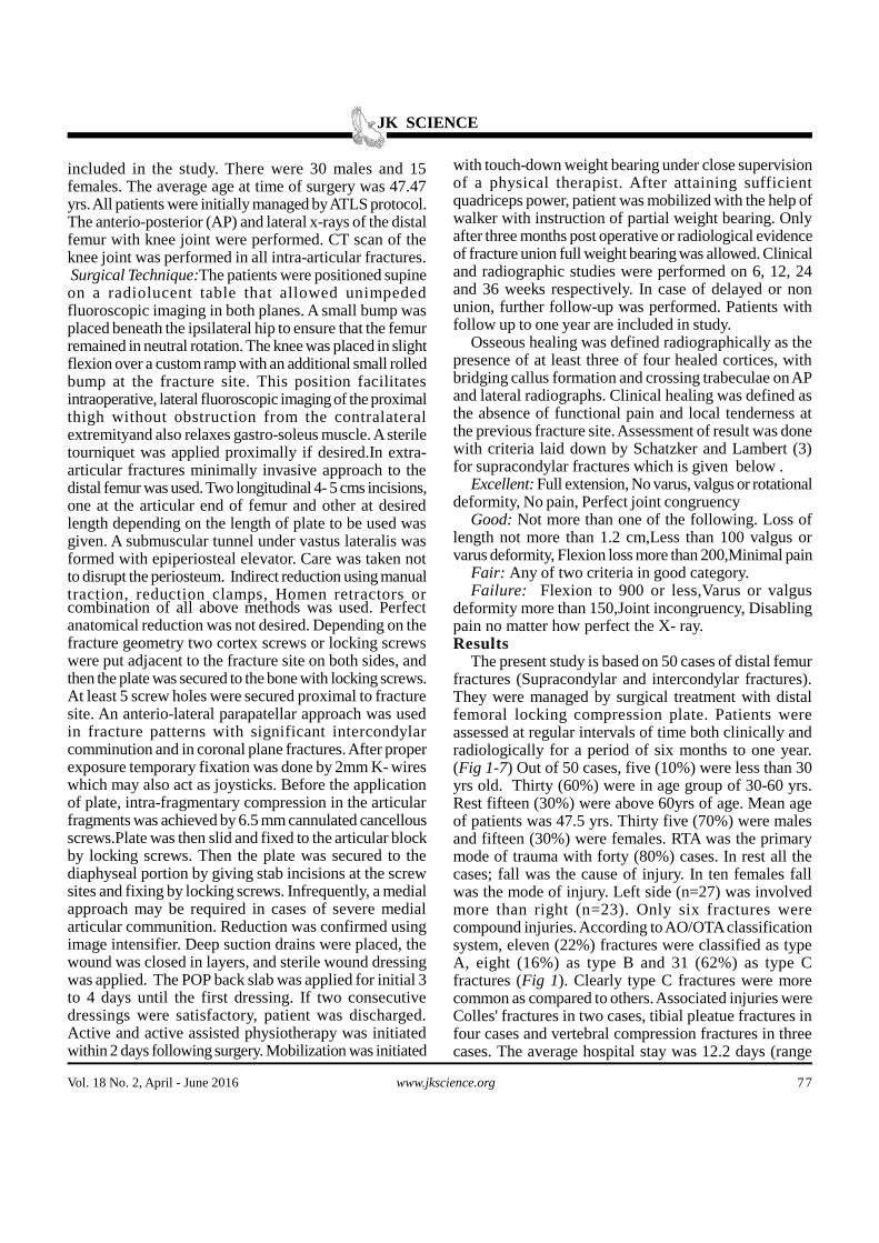
The present study is based on 50 cases of distal femurfractures (Supracondylar and intercondylar fractures).They were managed by surgical treatment with distalfemoral locking compression plate. Patients wereassessed at regular intervals of time both clinically andradiologically for a period of six months to one year.(Fig 1-7) Out of 50 cases, five (10%) were less than 30yrs old. Thirty (60%) were in age group of 30-60 yrs.Rest fifteen (30%) were above 60yrs of age. Mean ageof patients was 47.5 yrs. Thirty five (70%) were malesand fifteen (30%) were females. RTA was the primarymode of trauma with forty (80%) cases. In rest all thecases; fall was the cause of injury. In ten females fallwas the mode of injury. Left side (n=27) was involvedmore than right (n=23). Only six fractures werecompound injuries. According to AO/OTA classificationsystem, eleven (22%) fractures were classified as typeA, eight (16%) as type B and 31 (62%) as type Cfractures (Fig 1). Clearly type C fractures were morecommon as compared to others. Associated injuries wereColles' fractures in two cases, tibial pleatue fractures infour cases and vertebral compression fractures in threecases. The average hospital stay was 12.2 days (range
combination of all above methods was used. Perfectanatomical reduction was not desired. Depending on thefracture geometry two cortex screws or locking screwswere put adjacent to the fracture site on both sides, andthen the plate was secured to the bone with locking screws.At least 5 screw holes were secured proximal to fracturesite. An anterio-lateral parapatellar approach was usedin fracture patterns with significant intercondylarcomminution and in coronal plane fractures. After properexposure temporary fixation was done by 2mm K- wireswhich may also act as joysticks. Before the applicationof plate, intra-fragmentary compression in the articularfragments was achieved by 6.5 mm cannulated cancellousscrews.Plate was then slid and fixed to the articular blockby locking screws. Then the plate was secured to thediaphyseal portion by giving stab incisions at the screwsites and fixing by locking screws. Infrequently, a medialapproach may be required in cases of severe medialarticular communition. Reduction was confirmed usingimage intensifier. Deep suction drains were placed, thewound was closed in layers, and sterile wound dressingwas applied. The POP back slab was applied for initial 3to 4 days until the first dressing. If two consecutivedressings were satisfactory, patient was discharged.Active and active assisted physiotherapy was initiatedwithin 2 days following surgery. Mobilization was initiated
JK SCIENCE
78 www.jkscience.org Vol. 18 No. 2, April - June 2016
Fig 1. Epidemiology of Distal Femur Fractures
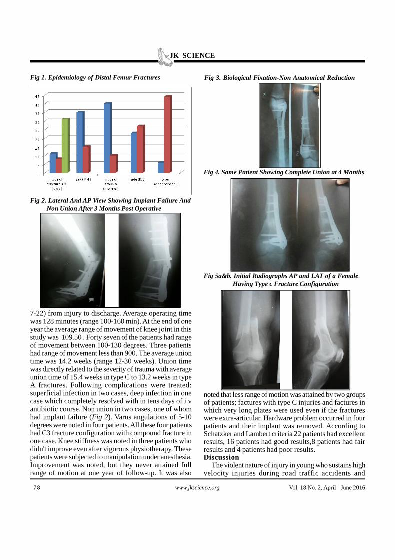
Fig 2. Lateral And AP View Showing Implant Failure And Non Union After 3 Months Post Operative
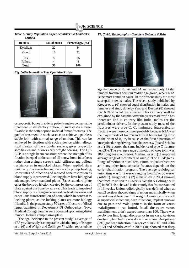
Fig 3. Biological Fixation-Non Anatomical Reduction
Fig 4. Same Patient Showing Complete Union at 4 Months
Fig 5a&b. Initial Radiographs AP and LAT of a Female Having Type c Fracture Configuration
7-22) from injury to discharge. Average operating timewas 128 minutes (range 100-160 min). At the end of oneyear the average range of movement of knee joint in thisstudy was 109.50 . Forty seven of the patients had rangeof movement between 100-130 degrees. Three patientshad range of movement less than 900. The average uniontime was 14.2 weeks (range 12-30 weeks). Union timewas directly related to the severity of trauma with averageunion time of 15.4 weeks in type C to 13.2 weeks in typeA fractures. Following complications were treated:superficial infection in two cases, deep infection in onecase which completely resolved with in tens days of i.vantibiotic course. Non union in two cases, one of whomhad implant failure (Fig 2). Varus angulations of 5-10degrees were noted in four patients. All these four patientshad C3 fracture configuration with compound fracture inone case. Knee stiffness was noted in three patients whodidn't improve even after vigorous physiotherapy. Thesepatients were subjected to manipulation under anesthesia.Improvement was noted, but they never attained fullrange of motion at one year of follow-up. It was also
noted that less range of motion was attained by two groupsof patients; factures with type C injuries and factures inwhich very long plates were used even if the fractureswere extra-articular. Hardware problem occurred in fourpatients and their implant was removed. According toSchatzker and Lambert criteria 22 patients had excellentresults, 16 patients had good results,8 patients had fairresults and 4 patients had poor results.Discussion
The violent nature of injury in young who sustains highvelocity injuries during road traffic accidents and
JK SCIENCE
Vol. 18 No. 2, April - June 2016 www.jkscience.org 79
Results. No. of cases Percentage. (%)
Excellent. 22 44
Good. 16 32
Fair. 8 16Failure. 4 8Total. 50 100
Table 1. Study Population as per Schatzker's &Lambert's Criteria
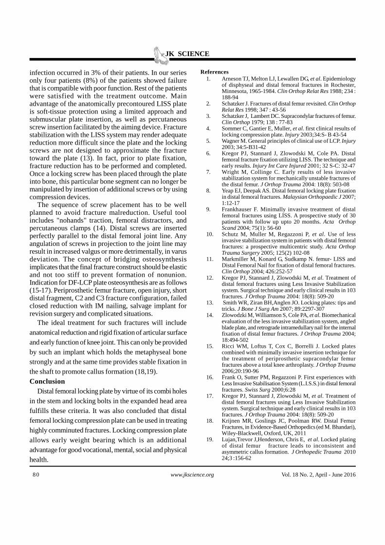
Fig. 6a&b Immediate Post Operative X rays
Fig 7a&b. Radiographs - Complete Union at 6 Mths
osteoporotic bones in elderly patients makes conservativetreatment unsatisfactory option, in such cases internalfixation is the better option in distal femur fractures. Thegoal of treatment in such cases is to achieve a painlessstable joint with normal range of motion. This can beachieved by fixation with such a device which allowsrigid fixation of the articular surface, gives respect tosoft tissues and allows early weight bearing. The DF-LCP is a single beam construct where the strength of itsfixation is equal to the sum of all screw-bone interfacesrather than a single screw's axial stiffness and pulloutresistance as in unlocked plates. When applied via aminimally invasive technique, it allows for prompt healing,lower rates of infection and reduced bone resorption asblood supply is preserved. Locking plates have biologicaladvantages over standard plates (5). A standard plategrips the bone by friction created by the compression ofplate against the bone by screws. This leads to impairedblood supply resulting in decreased cortical thickness andcancellous transformation of bone which in contrast tolocking plates, as the locking plates are more biologyfriendly. In the present study 50 cases of fracture of distalfemur admitted in Department Of Orthopedics, Govt.Medical College Jammu were operated upon using distalfemoral locking compression plate.
The age incidence in the present study is average of47.5 yrs. Our study is comparable with the study of Kregoret al (6) and Wright and Collinge (7) which reported the
age incidence of 49 yrs and 44 yrs respectively. Distalfemoral fractures occur in middle age group, where RTAis the most common cause. In the present study the mostsusceptible sex is males. The recent study published byKregor et al (6) showed equal distribution in males andfemales and study done by Yeap and Deepak (8) showedthat 63% affected were males. This can very well beexplained by the fact that over the years road traffic hasincreased and in country like India, males are thepredominant drivers. In the present study most of thefractures were type C. Comminuted intra-articularfracture were more common probably because RTA wasthe major mode of trauma and distal femur taking mostof the brunt of injury because of the flexed position ofknee joint during driving. Frankhauser et al (9) and Schultzet al (10) reported the same incidence of type C fracturei.e. 63%. The average range of motion of knee joint was109.5 degrees in our series. Markmiller et al (11) reportedaverage range of movement of knee joint of 110 degrees.Range of motion in distal femur intra-articular fracturesas in any other intra-articular fractues depends on theearly rehabilitation program. The average radiologicalunion time was 14.2 weeks ranging from 12 to 30 weeks(Table 1). Kregor et al (12) in his study in 2004 showedthat fracture united in 12 weeks. Wright & Collinge et al(7) in 2004 also showed in their study that fractures unitedin 13 weeks. Union radiologically was defined when atleast 3 cortices showed signs of union and clinically whenpatient was able to bear full weight. Complications suchas superficial infections, deep infections, implant removaldue to pain and malalignment in the form of varusmalalignment was found. In all the cases varusmalalignment didn't exceed more than 100. There wasno obvious limb length discrepancy in any case. Revisiondue to implant failure was done in one case. One patient(2%) got deep infection. Kregor et al in 2001 and 2004(6,12) and Schultz et al in 2005 (10) showed that deep
JK SCIENCE
80 www.jkscience.org Vol. 18 No. 2, April - June 2016
1. Arneson TJ, Melton LJ, Lewallen DG, et al. Epidemiologyof disphyseal and distal femoral fractures in Rochester,Minnesota, 1965-1984. Clin Orthop Relat Res 1988; 234 :188-94
2. Schatzker J. Fractures of distal femur revisited. Clin OrthopRelat Res 1998; 347 : 43-56
3. Schatzker J, Lambert DC. Supracondylar fractures of femur.Clin Orthop 1979; 138 : 77-83
4. Sommer C, Gantier E, Muller, et al. first clinical results oflocking compression plate. Injury 2003;34:S- B 43-54
5. Wagner M. General principles of clinical use of LCP. Injury2003; 34:5-B31-42
6. Kregor PJ, Stannard J, Zlowodski M, Cole PA. Distalfemoral fracture fixation utilizing LISS. The technique andearly results. Injury Int Care Injured 2001; 32 S-C: 32-47
7. Wright M, Collinge C. Early results of less invasivestabilization system for mechanically unstable fractures ofthe distal femur. J Orthop Trauma 2004: 18(8): 503-08
8. Yeap EJ, Deepak AS. Distal femoral locking plate fixationin distal femoral fractures. Malaysian Orthopaedic J 2007;1:12-17
9. Frankhauser F. Minimally invasive treatment of distalfemoral fractures using LISS. A prospective study of 30patients with follow up upto 20 months. Acta OrthopScand 2004; 75(1): 56-60
10. Schutz M, Muller M, Regazzoni P, et al. Use of lessinvasive stabilization system in patients with distal femoralfractures: a prospective multicentric study. Acta OrthopTrauma Surgery 2005; 125(2) 102-08
11. Markmiller M, Konard G, Sudkamp N. femur- LISS andDistal Femoral Nail for fixation of distal femoral fractures.Clin Orthop 2004; 426:252-57
12. Kregor PJ, Stannard J, Zlowodski M, et al. Treatment ofdistal femoral fractures using Less Invasive Stabilizationsystem. Surgical technique and early clinical results in 103fractures. J Orthop Trauma 2004: 18(8): 509-20
13. Smith WR, Ziran BH,Anglen JO. Locking plates: tips andtricks. J Bone J Surg Am 2007; 89:2297-307
14. Zlowodzki M, Williamson S, Cole PA, et al. Biomechanicalevaluation of the less invasive stabilization system, angledblade plate, and retrograde intramedullary nail for the internalfixation of distal femur fractures. J Orthop Trauma 2004;18:494-502
15. Ricci WM, Loftus T, Cox C, Borrelli J. Locked platescombined with minimally invasive insertion technique forthe treatment of periprosthetic supracondylar femurfractures above a total knee arthroplasty. J Orthop Trauma2006;20:190-96
16. Frank O, Sutter PM, Regazzoni P. First experiences withLess Invasive Stabilisation System (L.I.S.S.) in distal femoralfractures. Swiss Surg 2000;6:28
17. Kregor PJ, Stannard J, Zlowodski M, et al. Treatment ofdistal femoral fractures using Less Invasive Stabilizationsystem. Surgical technique and early clinical results in 103fractures. J Orthop Trauma 2004: 18(8): 509-20
18. Krijnen MR, Goslings JC, Poolman RW. Distal FemurFractures, in Evidence-Based Orthopedics (ed M. Bhandari),Wiley-Blackwell, Oxford, UK, 2011
19. Lujan,Trevor J,Henderson, Chris E, et al. Locked platingof distal femur fracture leads to inconsistent andasymmetric callus formation. J Orthopedic Trauma 201024;3 :156-62
Referencesinfection occurred in 3% of their patients. In our seriesonly four patients (8%) of the patients showed failurethat is compatible with poor function. Rest of the patientswere satisfied with the treatment outcome. Mainadvantage of the anatomically precontoured LISS plateis soft-tissue protection using a limited approach andsubmuscular plate insertion, as well as percutaneousscrew insertion facilitated by the aiming device. Fracturestabilization with the LISS system may render adequatereduction more difficult since the plate and the lockingscrews are not designed to approximate the fracturetoward the plate (13). In fact, prior to plate fixation,fracture reduction has to be performed and completed.Once a locking screw has been placed through the plateinto bone, this particular bone segment can no longer bemanipulated by insertion of additional screws or by usingcompression devices.
The sequence of screw placement has to be wellplanned to avoid fracture malreduction. Useful toolincludes "nohands" traction, femoral distractors, andpercutaneous clamps (14). Distal screws are insertedperfectly parallel to the distal femoral joint line. Anyangulation of screws in projection to the joint line mayresult in increased valgus or more detrimentally, in varusdeviation. The concept of bridging osteosynthesisimplicates that the final fracture construct should be elasticand not too stiff to prevent formation of nonunion.Indication for DF-LCP plate osteosynthesis are as follows(15-17). Periprosthetic femur fracture, open injury, shortdistal fragment, C2 and C3 fracture configuration, failedclosed reduction with IM nailing, salvage implant forrevision surgery and complicated situations.
The ideal treatment for such fractures will includeanatomical reduction and rigid fixation of articular surfaceand early function of knee joint. This can only be providedby such an implant which holds the metaphyseal bonestrongly and at the same time provides stable fixation inthe shaft to promote callus formation (18,19).Conclusion
Distal femoral locking plate by virtue of its combi holesin the stem and locking bolts in the expanded head areafulfills these criteria. It was also concluded that distalfemoral locking compression plate can be used in treatinghighly comminuted fractures. Locking compression plateallows early weight bearing which is an additionaladvantage for good vocational, mental, social and physicalhealth.