Embed Size (px)
Citation preview

Distinct Biological Roles for the Notch Ligands Jagged-1and Jagged-2*□S
Received for publication, April 2, 2009 Published, JBC Papers in Press, April 27, 2009, DOI 10.1074/jbc.M109.003111
Kuicheon Choi‡1, Young-Ho Ahn‡1, Don L. Gibbons‡, Hai T. Tran‡, Chad J. Creighton§, Luc Girard¶, John D. Minna¶,F. Xiao-Feng Qin�, and Jonathan M. Kurie‡2
From the ‡Department of Thoracic/Head and Neck Medical Oncology, University of Texas M. D. Anderson Cancer Center,Houston, Texas 77030, the §Dan L. Duncan Cancer Center, Baylor College of Medicine, Houston, Texas 77030, the ¶Hamon Centerfor Therapeutic Oncology Research, Simmons Cancer Center, University of Texas Southwestern Medical Center at Dallas, Dallas,Texas 75390, and the �Department of Immunology, University of Texas M. D. Anderson Cancer Center, Houston, Texas 77030
Notch signaling is activated in a subset of non-small cell lungcancer cells because of overexpression ofNotch3, but the role ofNotch ligands has not been fully defined. On the basis of geneexpression profiling of a panel of non-small cell lung cancer celllines, we found that the predominant Notch ligands were JAG1,JAG2, DLL1, and DLL3. Given that Notch ligands reportedlyhave overlapping receptor binding specificities, we postulatedthat they have redundant biological roles. Arguing against thishypothesis, we found that JAG1 and JAG2 were differentiallyregulated; JAG1 expression was dependent upon epidermalgrowth factor receptor (EGFR) activation in HCC827 cells,which require EGFR for survival, whereas JAG2 expression wasEGFR-independent in these cells. Furthermore, HCC827 cellsunderwent apoptosis following depletion of JAG1 but not JAG2,whereas co-culture experiments revealed that depletion ofJAG2, but not JAG1, enhanced the ability of HCC827 cells tochemoattract THP-1 human monocytes. JAG2-depletedHCC827 cells expressed high levels of inflammation-relatedgenes, including interleukin 1 (IL1) and a broad range of IL1-regulated cytokines, which was attenuated by inhibition ofIL1 receptor (IL1R). Our findings suggest that JAG1 and JAG2have distinct biological roles including a previously undis-covered role for JAG2 in regulating the expression of cyto-kines that can promote antitumor immunity.
In mammals, there are four Notch homologues (Notch1–4)and five ligands (three Delta-like and two Jagged/Serrate) (1).Receptor and ligand are typically presented on neighboringcells; hence, ligand binding is a means of cell-cell communica-tion. Notch participates in key aspects of organogenesis (lateralinhibition, lineage specification, and boundary formation) inthe developing embryo, maintains stem cell viability andrenewal in the adult, and has been implicated in multiplehuman cancers (1). In the developing lung,Notch receptors andligands are expressed in a cell type-specific manner, increase in
abundance from embryonic day 11.5 into adulthood, and con-tribute to cell lineage specification (2).A growing body of evidence supports a role for Notch in
tumorigenesis. Notch1 was first identified in humans as theTan1 oncogene, fusing the control region of the T-cell receptorB gene to a truncated, active form of Notch1 in T-cell acutelymphoblastic leukemia (3). Subsequently, oncogenic forms ofNotch2 were discovered in feline thymic lymphomas, andNotch4 was found to be an insertion site for mouse mammarytumor virus (4, 5). In NSCLC3 cell lines, the Notch3 gene onchromosome 19 is involved in balanced translocations withmultiple other chromosomes, leading to Notch3 overexpres-sion, and pharmacologic or genetic inhibition of Notch3 sup-presses the proliferation of these cells (6–8). Although otherexamples of activation of Notch receptors by genomic rear-rangement appear to be rare in human tumors, overexpressionis common in a variety of solid tumors including pancreas, cer-vix, breast, and prostate (9–12).Unlike the solid body of evidence supporting a role forNotch,
less is known about the importance of Notch ligands in cancer.Notch ligands function as Notch signaling agonists throughintercellular interactions (trans-interactions) and as Notch sig-naling antagonists through intracellular interactions (cis-inter-actions) (13). In mammalian systems, Notch ligands bind toNotch family members non-selectively; for example, Jagged-1and Jagged-2 can both bind to Notch1, Notch2, and probablyNotch3 (14, 15), suggesting a high degree of redundancy inmammalian cells to maintain Notch activity. However, micethat are null for the genes encoding Jagged-1 (Jag1), Jagged-2(Jag2), or Delta-like ligand4 (Dll4) exhibit distinct embryonicdefects (13), suggesting that these ligands exert unique actionsthat cannot be explained entirely by their receptor bindingactivities. Indeed, these ligands have intrinsic ligand signalingactivity independent of Notch and undergo multiple post-translational modifications, proteolytic processing, endocyto-sis, and membrane trafficking (13), all of which may contributeto the multifunctionality of Notch ligands.In this study, we examined Notch ligand expression in a
panel of NSCLC cell lines and found that they co-expressed* This work was supported, in whole or in part, by National Institutes of Health
Grants P50 CA70907 and CA117965 (to J. M. K.).□S The on-line version of this article (available at http://www.jbc.org) contains
supplemental Tables S1 and S2.1 Both authors contributed equally to this work.2 To whom correspondence should be addressed: Dept. of Thoracic/Head
and Neck Medical Oncology, M. D. Anderson Cancer Center, 1515 Hol-combe Blvd., Houston, TX 77030. Fax: 713-792-1220; E-mail: [email protected].
3 The abbreviations used are: NSCLC, non-small cell lung cancer; EGF, epider-mal growth factor; EGFR, epidermal growth factor receptor; IL1, interleu-kin-1; IL1R, IL1 receptor; IL1RA, interleukin-1 receptor antagonist; siRNA,short interfering RNA; TNF, tumor necrosis factor; PARP, poly(ADP-ribose)polymerase; DMSO, dimethyl sulfoxide.
THE JOURNAL OF BIOLOGICAL CHEMISTRY VOL. 284, NO. 26, pp. 17766 –17774, June 26, 2009© 2009 by The American Society for Biochemistry and Molecular Biology, Inc. Printed in the U.S.A.
17766 JOURNAL OF BIOLOGICAL CHEMISTRY VOLUME 284 • NUMBER 26 • JUNE 26, 2009
by guest on July 14, 2018http://w
ww
.jbc.org/D
ownloaded from

multiple ligands, including JAG1, JAG2, DLL1, and DLL3. Fur-ther analysis revealed that the expression of JAG1 and JAG2wasregulated independently, and these ligands had distinct biolog-ical roles, including a novel finding that JAG2 regulated theexpression of proinflammatory cytokines. Thus, JAG1 andJAG2 have non-redundant functions in NSCLC cells.
EXPERIMENTAL PROCEDURES
Reagent—Gefitinibwas a gift (AstraZeneca Pharmaceuticals,Wilmington, DE).We purchased a recombinant human IL1RA(Santa Cruz Biotechnology, Santa Cruz, CA), Hoechst 33342(Invitrogen), polyclonal antibodies derived in goat against Jag1(Santa Cruz Biotechnology) and in rabbits against Jag2 (SantaCruz Biotechnology), a horseradish peroxidase-linked anti-mouse (Cell Signaling Technology, Danvers, MA), anti-goatsecondary antibodies (Santa Cruz Biotechnology), and an anti-body against �-actin (Sigma-Aldrich).Cell Line—TheNSCLC cell lines used in this studywere from
theHamonCenter Repository or purchased from theAmericanType Culture Collection (Manassas, VA) and were grown in 5%CO2 at 37 °C in RPMI 1640 medium with high glucose (4.5g/liter; Invitrogen), supplemented with 10% fetal bovine serum(HyClone, Logan,UT). THP-1 cells were cultured in RPMI1640medium supplemented with 10% fetal bovine serum, 10 mMHEPES, 1.5 g/liter sodium bicarbonate, 1 mM sodium pyruvate,and 2 mM glutamine.Transient Transfection of Cells with siRNA—The siRNA con-
trol (siCTL nontargeting pool) and the siRNA pooled oligonu-cleotides against human JAG1 and JAG2 were purchased(Dharmacon, Denver, CO). Cells were transfected at 50% con-fluency using 10 nM of each construct over a 16-h period usingDharmaFECT 1 (Dharmacon).Stable Transfection of Cells with JAG2 Short Hairpin RNA—
The short hairpin RNA plasmid constructs against humanJAG2 and corresponding empty vector were purchased (Ori-gene, Rockville, MD). Cells were transfected at 50% confluencyusing 3 �g of each construct over a 4-h period with TransPassD1 transfection reagent (New England Biolabs, Ipswich, MA)according to the manufacturer’s protocol. After 48 h, mediumwas replaced with medium containing 1 �g/ml puromycin.After 14 days, the cells were trypsinized, seeded on 10-cmplatesat low density, and single colonies were selected and expanded.Expression Profiling Using Illumina Array—RNAs were
labeled and hybridized to the Illumina expression arrayWG6-V2 according to the manufacturer’s protocol. The arraycontains 48,701 probes, which correspond to 26,390 distinctUnigene IDs. Array data were background-corrected using theMBCB R package (16) and quantile-normalized. All genes onthe array were BLAST-verified and annotated using recent ver-sions of public National Center for Biotechnology Information(NCBI) databases, and all cell lines were mycoplasma-testedand DNA-fingerprinted. Heat maps were generated usingJavaTreeView (17) of absolute signal intensities using the equa-tion log2(x � 50) � log2(100).Quantitative PCR Array Analysis—RNA was isolated from
the cells transfectedwith siRNAs against JAG1, JAG2, or scram-bled controls by using TRIzol (Invitrogen), and 2 �g of totalRNAwas converted into cDNA. Expression profiling of inflam-
matory cytokines and their receptors was performed withhuman inflammatory cytokines and receptor RT2 Profiler PCRarray (SABioscience, Frederick, MD) according to the manu-facturer’s instructions. PCR was performed with the ABI Prism7500 fast real-time PCR system (Applied Biosystems, FosterCity, CA). The��Ctmethodwas used to analyze the expressionlevel of each gene. After PCR, the dissociation curve for eachgene was examined to exclude ones with nonspecific amplifica-tion or with undetectable expression. The expression profilingof each gene was displayed as a heat map made by using MeVMultiExperiment Viewer 4.1 (18).Quantitative PCR Using SYBR Green—The level of mRNA
for each gene was measured with SYBR Green-based real-timePCR. The primers used for real-time PCR were designed byusing Primer Express (Applied Biosystems). The primersequences used are listed in supplemental Table 1. Each cDNAsample (7 �l after 1:10 dilution with water) was amplified byusing SYBR Green PCRMaster Mix according to the manufac-turer’s instructions. The PCR products and their dissociationcurves were detected with the ABI Prism 7500 fast real-timePCR system. The level of the housekeeping gene L32 ribosomalgene (Rpl32) was used as an internal control. Quantitative PCRresults from triplicate RNA samples were used for calculationof mean expression values for each gene.HiMAP Interactome Analysis—Gene lists were imported
into the HiMAP program (19) for protein-protein interactionnetwork analysis. HiMAP includes both experimentally vali-dated protein-protein interactions (as cataloged in the HumanProtein Reference Database, or HPRD) and predicted protein-protein interactions based on a probabilistic model integratingmultiple factors, including interactome data from the Databaseof Interacting Proteins, protein domain data, genome-wideexpression data, and functional annotation data from the GeneOntology Project (GO).Western Blotting—Cells were lysed with M-PER mammalian
protein extraction reagent (Pierce). Lysates were cleared bycentrifugation, and protein concentrations were quantifiedwith 1� Quick Start Bradford dye reagent (Bio-Rad) so thatequal amounts of protein (40 �g) could be resolved on 10%SDS-polyacrylamide gels. After transfer to membranes, sam-ples were processed and visualized with ECL Western blot-ting reagents (Amersham Biosciences). All of the Westernblotting data were representative of at least three independ-ent experiments.Hoechst 33342 Staining—Cells with nuclear fragmenta-
tion were quantified by staining with 10 �g/ml Hoechst33342 for 15 min at room temperature and counting underfluorescence microscopy. Mean values for each conditionwere calculated based on results from 12 replicates (threeindependent experiments with quadruplicate samples ineach experiment).Cell Migration Assay—In vitro migration assays were per-
formed using a 24-well Transwell unit (BD Biosciences) withpolycarbonate filters. HCC827 cells transfected with siRNAfor Jag1, Jag2, or control were seeded on the lower compart-ments of the unit in the complete medium for 16 h. Justbefore the migration assay, the cells on the lower compart-ments were washed with phosphate-buffered saline, and cul-
Distinct Roles for Jagged-1 and Jagged-2
JUNE 26, 2009 • VOLUME 284 • NUMBER 26 JOURNAL OF BIOLOGICAL CHEMISTRY 17767
by guest on July 14, 2018http://w
ww
.jbc.org/D
ownloaded from

ture medium was changed to serum-free medium with treat-ment. THP-1 cells were placed in the upper compartments inserum-free medium and incubated for 24 h. The mediumwas aspirated after the incubation, and the cells wereremoved from the upper compartment by scrubbing with acotton swab. Cells on the underside of the membrane(migrated cells) were stained with SureStain Wright (Fisher)and then washed with water. Cells were counted by micros-copy at �20 magnification. Mean values were calculatedfrom cell counts in five random fields for each filter. Meanvalues for each condition were calculated based on resultsfrom eight replicates (two independent experiments withquadruplicate samples in each experiment).Statistical Methodology—For analysis of treatment effects
and differences between genetically modified cells, the Stu-dent’s t test was used, and p values �0.05 were considered sta-tistically significant.
RESULTS
Expression Profiling of NotchLigands and Family Members inNSCLC Cells—We profiled geneexpression in a panel of 70 NSCLCcell lines by using Illumina arrays.Of the five Notch ligands, JAG1,JAG2, DLL1, and DLL3 wereexpressed at levels that varied by upto 15-fold between cell lines,whereas DLL4 expression was uni-formly low in all cell lines (Fig. 1).The majority of the cell linesexpressed at least two ligands, andsome (HCC95, HCC4018, H1755,H1666, and HCC1833) expressedfour ligands.Quantitative PCRanal-ysis of JAG1, JAG2, and Notch1–4was performed on a subset of thecell lines, which confirmed the rela-tive expression levels of JAG1 andJAG2 observed in the expressionarray studies performed on thosecells and revealed that most celllines expressedmultipleNotch fam-ily members (Fig. 2).JAG1 and JAG2 Are Regulated
through Distinct Mechanisms—Onthe basis of reports that Notchligands exhibit overlapping Notchbinding activities (14, 15), we postu-lated that they have redundant bio-logical functions in NSCLC cellsand used pharmacologic andgenetic approaches to test thishypothesis. The cell lines in Fig. 2differ with respect to somatic muta-tions that constitutively activateEGFR (HCC827, HCC2279, H4006,H3255, and H1975) or N-Ras(H1299) or have neither mutation
(H1819) (20). Comparison of their expression levels deter-mined by quantitative PCR analyses revealed that JAG1,NOTCH1, and NOTCH2 were more highly expressed in EGFRmutant cells (p values � 0.025, 0.013, and 0.03, respectively)(Fig. 2A), whereas NOTCH3, NOTCH4, and JAG2 were notdifferentially expressed (Fig. 2B). Because increased JAG1expression correlated with the presence of mutant EGFR, weexamined whether JAG1 was regulated by EGFR. JAG1 RNAand protein levels decreased sharply in HCC827 cells andH4006 cells treated with the EGFR-specific tyrosine kinaseinhibitor gefitinib (Fig. 3, A and B). Gefitinib treatment hadno effect on JAG1 levels in H1975 cells, which have a resist-ance mutation (T790M) (18) that inhibits EGFR binding togefitinib (Fig. 3, A and B), indicating that the decrease inJAG1 levels by gefitinib required EGFR inhibition. In con-trast, gefitinib had no effect on JAG2 levels in these cells (Fig.
FIGURE 1. Expression of Notch ligands in a panel of 70 NSCLC cell lines. A heat map representation ofabsolute signal intensities in Illumina expression arrays is shown. * indicates cell lines studied in Fig. 2.
FIGURE 2. Differential expression of Notch ligands and family members based on EGFR mutational sta-tus. Included are NSCLC cell lines with wild-type (black bars) or mutant (gray bars) EGFR. Quantitative PCRanalysis of basal gene expression revealed genes that were (A) or were not (B) differentially expressed based onEGFR mutational status. The values represent the means of replicate samples and are normalized to the L32ribosomal RNA gene.
Distinct Roles for Jagged-1 and Jagged-2
17768 JOURNAL OF BIOLOGICAL CHEMISTRY VOLUME 284 • NUMBER 26 • JUNE 26, 2009
by guest on July 14, 2018http://w
ww
.jbc.org/D
ownloaded from
3A). To examine the role of EGFR using a different approach,EGFR-wild-type H1299 cells, which expressed low levels ofJAG1 and JAG2 (Fig. 2), were treated with EGF or transfectedwith wild-type EGFR, both of which increased the expressionof JAG1 (Fig. 3C) but not JAG2 (data not shown), andgefitinib treatment abrogated EGFR-induced JAG1 expres-sion in H1299 cells, indicating that this effect was EGFRkinase-dependent. Collectively, these findings suggest thatEGFR regulates the expression of JAG1 but not JAG2.JAG1 and JAG2 Levels Are Regulated Inversely—We next
sought to examine the biological roles of JAG1 and JAG2 by
transfecting siRNAs to selectivelydeplete JAG1 or JAG2 from culturedcells. Transfection of HCC827NSCLC cells with JAG1- or JAG2-specific siRNAs achieved greaterthan 90% reductions in Jagged-1and Jagged-2, respectively (Fig. 4A).Of note, Jagged-2 protein levelsincreased in JAG1 siRNA-trans-fected cells, and Jagged-1 proteinlevels increased in JAG2 siRNA-transfected cells (Fig. 4A), indicat-ing that JAG1 and JAG2 levels wereregulated reciprocally.JAG1 Maintains the Survival of
HCC827 Cells—Relative to the con-trol transfectants, the JAG1 siRNA-transfected HCC827 cells exhibiteda reduction in cell density over time(Fig. 4B) and an increase in nuclearfragmentation and cleavage of poly-(ADP-ribose) polymerase (PARP)(Fig. 4C), which is consistent with
apoptotic cell death. In contrast, JAG2 siRNA-transfected cellsexhibitedminimal evidence of apoptotic cell death based on theabsence of PARP cleavage (Fig. 4C) and nuclear fragmentation(data not shown). Collectively, these findings suggest thatHCC827 cell survival was dependent upon JAG1 but not JAG2.JAG2 Inhibits the Ability of HCC827 Cells to Recruit
Monocytes—In addition to their ability to proliferate in anuncontrolled manner, NSCLC cells recruit inflammatory cells,fibroblasts, and endothelial cells, which constitute the tumorstroma (21). These cells are required for tumor growth and
FIGURE 3. Distinct regulation of JAG1 and JAG2 by EGFR. A, quantitative PCR analysis of cells treated with gefitinib (1 �M) or vehicle (DMSO). The valuesrepresent the means of replicate samples and are normalized to the L32 ribosomal RNA gene. *, p � 0.05. B, Jagged-1 Western blot in HCC827 and H1975 cellstreated with gefitinib. C, quantitative PCR analysis of gene expression in H1299 cells treated with EGF (100 ng/ml) or transfected with wild-type EGFR or emptyvector (EV). Relative EGFR expression in the transfectants was quantified by quantitative PCR and Western analysis (inset). The quantitative PCR values representthe means of replicate samples and are normalized to the L32 ribosomal RNA gene. *, empty vector versus EFGR (DMSO treatment), p � 0.05; �, DMSO versusgefitinib (EGFR transfectants), p � 0.05.
FIGURE 4. JAG1 and JAG2 are mutually suppressive, and JAG1 maintains the survival of HCC827 cells.A, Western analysis of HCC827 cells transfected with JAG1, JAG2, or scrambled control (SCR) siRNA. B, cellnumbers of scrambled (SCR) or JAG1 siRNA transfectants over time. *, p � 0.05, **, p � 0.01. C, percentages ofapoptotic cells (bar graph; *, p � 0.05) as determined by Hoechst 33342 staining (images) to identify frag-mented nuclei (arrows). Results are the means of replicate samples. The Western blot shows cleaved PARP(arrow) in JAG1 siRNA transfectant.
Distinct Roles for Jagged-1 and Jagged-2
JUNE 26, 2009 • VOLUME 284 • NUMBER 26 JOURNAL OF BIOLOGICAL CHEMISTRY 17769
by guest on July 14, 2018http://w
ww
.jbc.org/D
ownloaded from
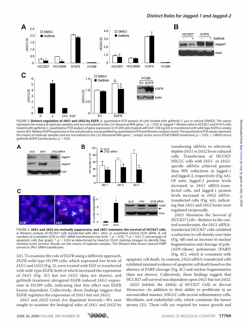
metastasis (22). To determine whether JAG1 and JAG2 areinvolved in this process, we examined the chemoattraction ofTHP-1, a human monocytic cell line (23), by HCC827 cells fol-lowing transfection with JAG1 or JAG2 siRNAs. HCC827 cellsand THP-1 cells were seeded into the lower and upper cham-bers, respectively, of Transwell plates. These chambers wereseparated by a porous membrane, allowing bidirectional diffu-sion of secreted, soluble mediators. Following 24 h of incuba-tion, the numbers of THP-1 cells that had migrated across theporous membrane were counted. Relative to control siRNA,JAG2 siRNA transfection enhanced the ability of NSCLC cellsto recruit THP-1 cells, whereas JAG1 siRNA had no effect (Fig.5). Thus, JAG2 inhibited monocytic recruitment by HCC827cells.JAG2 Inhibits the Expression of Inflammation-related
Genes in an IL1R-dependent Manner—To explore the mech-anism by which JAG2 inhibits monocyte recruitment, JAG1and JAG2 siRNA-transfected HCC827 cells were transcrip-tionally profiled using a quantitative PCR expression array(RT2 Profiler array, SABiosciences) containing 84 inflamma-tion-related genes (gene lists are in supplemental Table 2).
Of these, 33 genes were measurable in all three samples (Fig.6A); the other genes either were undetectable or did notachieve a technically satisfactory amplification in all sam-ples. JAG1 and JAG2 siRNA transfectants exhibited strikingdifferences; relative to the control transfectants, JAG2siRNA enhanced the expression of the majority of the meas-urable genes, whereas JAG1 siRNA reduced the expressionof a smaller subset (Fig. 6A). Those increased by JAG2 siRNAinclude, among others, a broad spectrum of CXC chemo-kines (CXCL1, CXCL3, CXCL5, CXCL6, and CXCL9), CCchemokines (CCL2, CCL5, CCL13, CCL20, and CCL24),interleukins (IL1�, IL1�, IL1F8, IL1F9, and IL17c) and theirreceptors (IL1RN, IL10R�, IL13R�, CCR1, CCR8, andCXCR1). To validate these findings, quantitative PCR wasperformed on RNA samples from an independent experi-ment using different primers from those on the array, whichconfirmed that JAG2 siRNA enhanced the expression of allfour genes tested (CCL20, IL1�, IL1�, and CCL5), whereasJAG1 siRNA had no detectable effect on any of them (Fig.6B). Thus, JAG2 was unique in its ability to regulate theexpression of inflammation-related genes.
FIGURE 5. JAG2 depletion enhances the ability of HCC827 cells to recruit THP-1 monocytic cells. Shown is quantification of migrated THP-1 cells inco-culture with JAG1, JAG2, or scrambled (SCR) siRNA-transfected HCC827 cells. Results represent the means of replicate wells. Images illustrate stained,migrated cells on membrane (encircled). *, p � 0.05.
Distinct Roles for Jagged-1 and Jagged-2
17770 JOURNAL OF BIOLOGICAL CHEMISTRY VOLUME 284 • NUMBER 26 • JUNE 26, 2009
by guest on July 14, 2018http://w
ww
.jbc.org/D
ownloaded from

Given the widespread nature of the gene expression changesinduced by JAG2 siRNA, we postulated that JAG2 regulates oneor more genes that are central nodes in the inflammatory proc-ess. To test this, we analyzed their positions within known orpredicted global protein interaction networks (interactomes)by using theHiMAP software program. Interactomes identifiedby this approach are organized into a series of modular struc-tures characterized by centrally located nodes (called hubs) thathave multiple connections with other proteins (18). Althoughthis approach is purely exploratory and carries no statisticalweight, findings in yeast show that centrality within a proteininteractome predicts the biological importance of a protein(24).Of the 33 measurable inflammation-related genes from
the RT2 Profiler PCR array, 28 mapped within a single inter-actome based on HiMAP analysis (Fig. 7). The hubs withinthe network with the highest number of links (�10) includedTNF�, IL1�, IL1�, IL1R, CCL2, and CCL5, all of which wereprominently up-regulated by JAG2 siRNA (Fig. 6A). Basedon the centrality of IL1R and its ligands within the network,we postulated that IL1R mediates JAG2-induced suppres-
sion of inflammation-relatedgenes. To test this, we examinedthe effects of treatment with IL1Rantagonist (IL1RA), a naturallyoccurring, physiological inhibitorof IL1R (25). In co-culture assays,IL1RA treatment abrogated therecruitment of THP-1 cells byJAG2 siRNA-transfected HCC827cells (Fig. 8A). Furthermore,IL1RA treatment attenuated theeffects of JAG2 siRNA on three(CCL20, IL1�, andTNF�) of the 10inflammation-related genes ana-lyzed (IL1R, CXCL3, CXCL5,CXCL1, IL1�, IL1�, CCL2, CCL5,CCL20, and TNF�) (Fig. 8B). Col-lectively, these findings suggestthat IL1R had a pivotal role in thebiological effects of JAG2 inHCC827 cells.
DISCUSSION
Notch signaling has been impli-cated in multiple facets of cancerbiology, including, among others,stem cell renewal, cancer cell pro-liferation, tumor angiogenesis,and metastasis (1). The findingspresented in the current studyadvance our understanding of therole of Notch in cancer by demon-strating that the roles of JAG1 andJAG2 in NSCLC cells are quite dis-tinct, including regulation ofdiverse proinflammatory cyto-kines, a biological property of
Notch signaling that, to our knowledge, has heretofore notbeen reported.Given the reports that Jagged-1 and Jagged-2 bind to
Notch family members with similar specificities (14, 15), thefinding that they had distinct biological functions inHCC827 cells raises the possibility that they mediate theiractions in part through Notch-independent mechanisms.Providing further support for this possibility was evidencethat treatment with a �-secretase inhibitor N-(N-(3,5-diflu-orophenacetyl-L-alanyl))-S-phenylglycine t-butyl ester,which reportedly inhibits Notch activity (26), only minimallydecreased HCC827 cell density (data not shown). All of theNotch ligands, with the exception of Delta-like ligand-3 andJagged-2, have PDZ-binding motifs at their extreme C ter-mini (27). These motifs are dispensable for ligand activationand Notch inhibition (28–31), but they are required forNotch ligands to affect oncogenic transformation (28).Although it is unclear whether such interactions might havecaused the changes in cytokine expression observed here,some PDZ domain proteins such as calcium/calmodulin-de-pendent serine protein kinase (CASK), Bridge-1, and gluta-
FIGURE 6. JAG1 and JAG2 siRNA-induced changes in inflammation-related genes. A, a heat map represen-tation of gene expression in HCC827 cells transfected with JAG1 or JAG2 siRNA centered on that of scrambled(SCR) siRNA transfectants. B, quantitative PCR validation of findings from expression arrays in A using RNAsamples from an independent experiment. The values represent the means of replicate samples and arenormalized to the L32 ribosomal RNA gene. *, p � 0.05.
Distinct Roles for Jagged-1 and Jagged-2
JUNE 26, 2009 • VOLUME 284 • NUMBER 26 JOURNAL OF BIOLOGICAL CHEMISTRY 17771
by guest on July 14, 2018http://w
ww
.jbc.org/D
ownloaded from

mate receptor interacting protein 1-� (GRIP1-�) act as tran-scriptional activators (32–34), whereas others, such asafadin/A6 and Acvrinp1, interact with Ras and Smad3 (28,35–37), respectively, which regulate diverse transcriptionalprograms.We observed that JAG1 and JAG2were regulated inversely in
HCC827 cells. Notch signaling interacts with a number of dif-ferent signaling systems, and many of these affect Notch ligandexpression. In particular, fibroblast growth factor, platelet-de-rived growth factor, transforming growth factor-�, vascularendothelial growth factor, hedgehog, andWnt have been foundto modulate Notch ligand expression (13), any of which mighthavemediated themutual suppression between JAG1 and JAG2inHCC827 cells. Equally intriguing is the possibility of aNotch-mediated feed-forward loop that sensed the loss of Notch
ligand through the disengagement of Notch trans-interactions,to which it responded by increasing the expression of otherNotch ligands.Findings presented here suggest that following JAG2 inhibi-
tion, HCC827 cells can induce THP-1 monocyte recruitment.Because the cells in this study were co-cultured in separatecompartments and communicated through a porous barrier,we conclude that these interactions were mediated by secretedfactors and did not require cell-cell contact. In fact, JAG2 deple-tion dramatically increased the expression of a broad spectrumof inflammatory mediators. Within the interactome of inflam-mation-related genes that were regulated by JAG2 depletion,IL1 receptor occupied a central position, and treatment withIL1RA abrogated the recruitment of monocytic cells and theexpression of certain inflammatory mediators. Consistent with
FIGURE 7. Centrality of IL1R and its ligands in the network of inflammation-related genes regulated by JAG2 siRNA. An interactome of inflammation-related genes illustrates theoretical protein-protein physical and functional interactions; it was drawn using the HiMAP software. Genes from the expressionarray are in red.
Distinct Roles for Jagged-1 and Jagged-2
17772 JOURNAL OF BIOLOGICAL CHEMISTRY VOLUME 284 • NUMBER 26 • JUNE 26, 2009
by guest on July 14, 2018http://w
ww
.jbc.org/D
ownloaded from

these findings, IL1 has been reported to regulate the expressionof a vast array of proinflammatory cytokines and chemokines(38). Collectively, these findings suggest that IL1 is a criticalmediator of inflammation by JAG2, adding to the complexity ofcell-cell interactions known to regulate macrophage function(14), and these findings constitute the first report, to our knowl-edge, that Notch pathways can regulate tumor cell-inducedinflammation.NSCLC remains the primary cause of cancer-related death
in Western countries, which is largely because of the factthat once the disease has metastasized, NSCLC cells areresistant to the current treatment options. Even in the set-ting of tumors with activating EGFR somatic mutations,which confer a unique sensitivity to treatment with EGFRtyrosine kinase inhibitors that leads to rapid and often sus-tained tumor shrinkage (39–41), the initial tumor responseis typically followed by disease recurrence. The problem ofdisease recurrence has not been obviated by the addition ofstandard chemotherapeutic agents to EGFR tyrosine kinase
inhibitors. Given the importance of Notch signaling in thedevelopment of a variety of malignancies, pharmacologicstrategies are under development to inhibit the Notch sig-naling pathway in cancer patients. The findings presentedhere suggest that such strategies may benefit NSCLCpatients by inhibiting cancer cell viability and possibly byenhancing antitumor immunity.
Acknowledgment—We thank Dror Berel for technical assistance.
REFERENCES1. Fiuza, U. M., and Arias, A. M. (2007) J. Endocrinol. 194, 459–4742. Collins, B. J., Kleeberger,W., andBall, D.W. (2004) Semin. Cancer Biol.14,
357–3643. Ellisen, L. W., Bird, J., West, D. C., Soreng, A. L., Reynolds, T. C., Smith,
S. D., and Sklar, J. (1991) Cell 66, 649–6614. Rohn, J. L., Lauring, A. S., Linenberger, M. L., and Overbaugh, J. (1996)
J. Virol. 70, 8071–80805. Gallahan, D., and Callahan, R. (1997) Oncogene 14, 1883–1890
FIGURE 8. IL1RA treatment inhibits the biological effects of JAG2 in HCC827 cells. A, quantification of migrated THP-1 cells in co-culture with JAG2siRNA-transfected HCC827 cells treated with IL1RA or bovine serum albumin (BSA) control. Results represent the means of replicate wells. Imagesillustrate stained, migrated cells on membrane (encircled). SCR, scrambled. *, p � 0.05. B, HCC827 cells transfected with the indicated siRNAs were treatedwith IL1RA or BSA control. RNA isolated from these cells was subjected to quantitative PCR assays. The values represent the means of replicate samplesand are normalized to the L32 ribosomal RNA gene. *, JAG2 versus scrambled siRNA transfectants (BSA treatment), p � 0.05; †, IL1RA versus BSA (JAG2siRNA transfectants), p � 0.05.
Distinct Roles for Jagged-1 and Jagged-2
JUNE 26, 2009 • VOLUME 284 • NUMBER 26 JOURNAL OF BIOLOGICAL CHEMISTRY 17773
by guest on July 14, 2018http://w
ww
.jbc.org/D
ownloaded from

6. Dang, T. P., Gazdar, A. F., Virmani, A. K., Sepetavec, T., Hande, K. R.,Minna, J. D., Roberts, J. R., and Carbone, D. P. (2000) J. Natl. Cancer Inst.92, 1355–1357
7. Haruki, N., Kawaguchi, K. S., Eichenberger, S., Massion, P. P., Olson, S.,Gonzalez, A., Carbone, D. P., and Dang, T. P. (2005) Cancer Res. 65,3555–3561
8. Konishi, J., Kawaguchi, K. S., Vo, H., Haruki, N., Gonzalez, A., Carbone,D. P., and Dang, T. P. (2007) Cancer Res. 67, 8051–8057
9. Miyamoto, Y., Maitra, A., Ghosh, B., Zechner, U., Argani, P., Iacobuzio-Donahue, C. A., Sriuranpong, V., Iso, T., Meszoely, I. M., Wolfe, M. S.,Hruban, R. H., Ball, D. W., Schmid, R. M., and Leach, S. D. (2003) CancerCell 3, 565–576
10. Gray, G. E., Mann, R. S., Mitsiadis, E., Henrique, D., Carcangiu, M. L.,Banks, A., Leiman, J., Ward, D., Ish-Horowitz, D., and Artavanis-Tsako-nas, S. (1999) Am. J. Pathol. 154, 785–794
11. Weijzen, S., Rizzo, P., Braid, M., Vaishnav, R., Jonkheer, S. M., Zlobin, A.,Osborne, B. A., Gottipati, S., Aster, J. C., Hahn, W. C., Rudolf, M., Siz-iopikou, K., Kast, W. M., and Miele, L. (2002) Nat. Med. 8, 979–986
12. Zayzafoon, M., Abdulkadir, S. A., and McDonald, J. M. (2004) J. Biol.Chem. 279, 3662–3670
13. D’Souza, B., Miyamoto, A., and Weinmaster, G. (2008) Oncogene 27,5148–5167
14. Shimizu, K., Chiba, S., Kumano, K., Hosoya, N., Takahashi, T., Kanda, Y.,Hamada, Y., Yazaki, Y., and Hirai, H. (1999) J. Biol. Chem. 274,32961–32969
15. Shimizu, K., Chiba, S., Hosoya, N., Kumano, K., Saito, T., Kurokawa, M.,Kanda, Y., Hamada, Y., and Hirai, H. (2000)Mol. Cell Biol. 20, 6913–6922
16. Ding, L. H., Xie, Y., Park, S., Xiao, G., and Story,M.D. (2008)Nucleic AcidsRes. 36, e58
17. Saldanha, A. J. (2004) Bioinformatics 20, 3246–324818. Rhodes, D. R., Tomlins, S. A., Varambally, S., Mahavisno, V., Barrette, T.,
Kalyana-Sundaram, S., Ghosh, D., Pandey, A., and Chinnaiyan, A. M.(2005) Nat. Biotechnol. 23, 951–959
19. Saeed, A. I., Sharov, V., White, J., Li, J., Liang, W., Bhagabati, N., Braisted,J., Klapa, M., Currier, T., Thiagarajan, M., Sturn, A., Snuffin, M., Rezant-sev, A., Popov, D., Ryltsov, A., Kostukovich, E., Borisovsky, I., Liu, Z.,Vinsavich, A., Trush, V., and Quackenbush, J. (2003) BioTechniques 34,374–378
20. Fujimoto, N., Wislez, M., Zhang, J., Iwanaga, K., Dackor, J., Hanna, A. E.,Kalyankrishna, S., Cody, D. D., Price, R. E., Sato, M., Shay, J. W., Minna,J. D., Peyton, M., Tang, X., Massarelli, E., Herbst, R., Threadgill, D. W.,Wistuba, I. I., and Kurie, J. M. (2005) Cancer Res. 65, 11478–11485
21. Wislez, M., Antoine, M., Rabbe, N., Gounant, V., Poulot, V., Lavole, A.,Fleury-Feith, J., and Cadranel, J. (2007) Clin. Cancer Res. 13, 3518–3527
22. Tlsty, T. D., and Coussens, L. M. (2006) Annu. Rev. Pathol. 1, 119–150
23. Tsuchiya, S., Yamabe, M., Yamaguchi, Y., Kobayashi, Y., Konno, T., andTada, K. (1980) Int. J. Cancer 26, 171–176
24. Jeong, H., Mason, S. P., Barabasi, A. L., and Oltvai, Z. N. (2001) Nature411, 41–42
25. Liao, Z., Grimshaw, R. S., and Rosenstreich, D. L. (1984) J. Exp. Med. 159,126–136
26. Geling, A., Steiner, H., Willem, M., Bally-Cuif, L., and Haass, C. (2002)Embo. Rep. 3, 688–694
27. Pintar, A., De Biasio, A., Popovic, M., Ivanova, N., and Pongor, S. (2007)Biol. Direct. 2, 19
28. Ascano, J.M., Beverly, L. J., andCapobianco, A. J. (2003) J. Biol. Chem. 278,8771–8779
29. Six, E. M., Ndiaye, D., Sauer, G., Laabi, Y., Athman, R., Cumano, A., Brou,C., Israel, A., and Logeat, F. (2004) J. Biol. Chem. 279, 55818–55826
30. Wright, G. J., Leslie, J. D., Ariza-McNaughton, L., and Lewis, J. (2004)Development 131, 5659–5669
31. Mizuhara, E., Nakatani, T., Minaki, Y., Sakamoto, Y., Ono, Y., and Takai,Y. (2005) J. Biol. Chem. 280, 26499–26507
32. Hsueh, Y. P., Wang, T. F., Yang, F. C., and Sheng, M. (2000) Nature 404,298–302
33. Nakata, A., Ito, T., Nagata, M., Hori, S., and Sekimizu, K. (2004) GenesCells 9, 1125–1135
34. Lee, J. H., Volinic, J. L., Banz, C., Yao, K. M., and Thomas, M. K. (2005) J.Endocrinol. 187, 283–292
35. Pfister, S., Przemeck, G. K., Gerber, J. K., Beckers, J., Adamski, J., andHrabe de Angelis, M. (2003) J. Mol. Biol. 333, 229–235
36. Shoji, H., Tsuchida, K., Kishi, H., Yamakawa, N., Matsuzaki, T., Liu, Z.,Nakamura, T., and Sugino, H. (2000) J. Biol. Chem. 275, 5485–5492
37. Quilliam, L. A., Castro, A. F., Rogers-Graham, K. S., Martin, C. B., Der,C. J., and Bi, C. (1999) J. Biol. Chem. 274, 23850–23857
38. Allan, S. M., Tyrrell, P. J., and Rothwell, N. J. (2005)Nat. Rev. Immunol. 5,629–640
39. Lynch, T. J., Bell, D.W., Sordella, R., Gurubhagavatula, S., Okimoto, R. A.,Brannigan, B.W., Harris, P. L., Haserlat, S. M., Supko, J. G., Haluska, F. G.,Louis, D. N., Christiani, D. C., Settleman, J., and Haber, D. A. (2004)N. Engl. J. Med. 350, 2129–2139
40. Paez, J. G., Janne, P. A., Lee, J. C., Tracy, S., Greulich, H., Gabriel, S.,Herman, P., Kaye, F. J., Lindeman, N., Boggon, T. J., Naoki, K., Sasaki, H.,Fujii, Y., Eck, M. J., Sellers, W. R., Johnson, B. E., andMeyerson, M. (2004)Science 304, 1497–1500
41. Pao,W., Miller, V., Zakowski, M., Doherty, J., Politi, K., Sarkaria, I., Singh,B., Heelan, R., Rusch, V., Fulton, L.,Mardis, E., Kupfer, D.,Wilson, R., Kris,M., and Varmus, H. (2004) Proc. Natl. Acad. Sci. U. S. A. 101,13306–13311
Distinct Roles for Jagged-1 and Jagged-2
17774 JOURNAL OF BIOLOGICAL CHEMISTRY VOLUME 284 • NUMBER 26 • JUNE 26, 2009
by guest on July 14, 2018http://w
ww
.jbc.org/D
ownloaded from

Girard, John D. Minna, F. Xiao-Feng Qin and Jonathan M. KurieKuicheon Choi, Young-Ho Ahn, Don L. Gibbons, Hai T. Tran, Chad J. Creighton, Luc
Distinct Biological Roles for the Notch Ligands Jagged-1 and Jagged-2
doi: 10.1074/jbc.M109.003111 originally published online April 27, 20092009, 284:17766-17774.J. Biol. Chem.
10.1074/jbc.M109.003111Access the most updated version of this article at doi:
Alerts:
When a correction for this article is posted•
When this article is cited•
to choose from all of JBC's e-mail alertsClick here
Supplemental material:
http://www.jbc.org/content/suppl/2009/04/27/M109.003111.DC1
http://www.jbc.org/content/284/26/17766.full.html#ref-list-1
This article cites 41 references, 20 of which can be accessed free at
by guest on July 14, 2018http://w
ww
.jbc.org/D
ownloaded from