Embed Size (px)
Citation preview
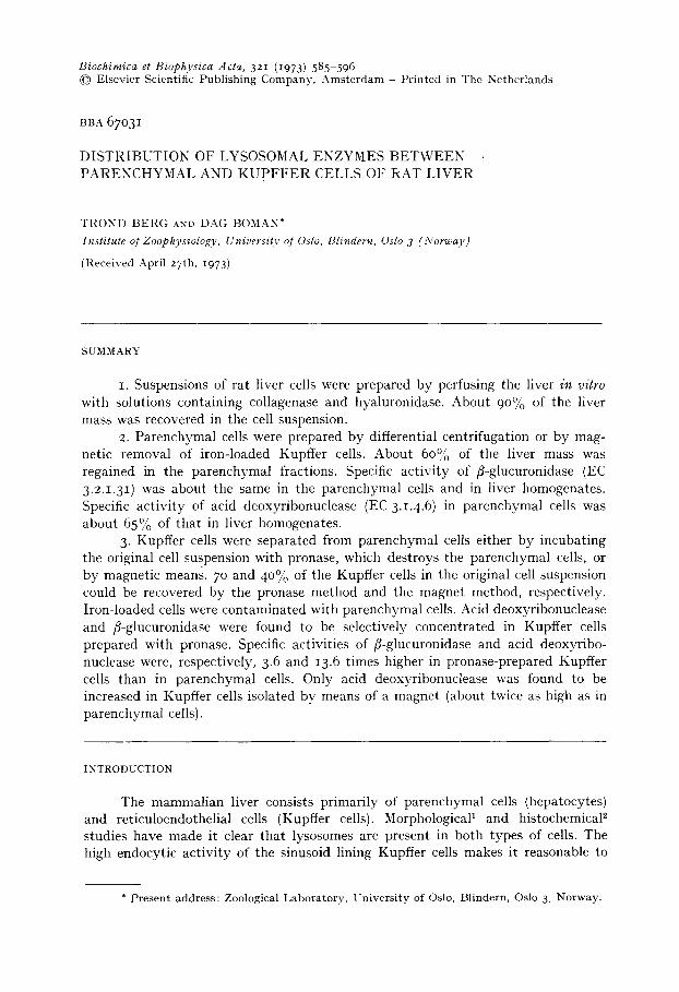
Biochimica et Biophysica Acta, 321 (1973) 585-596 © Elsevier Scientific Publishing Company, Amsterdam Printed in The Netherlands
BBA 67O31
DISTRIBUTION OF LYSOSOMAL ENZYMES BETWEEN PARENCHYMAL AND K U P F F E R CELLS OF RAT LIVER
TROND BERG AND DAG BOMAN*
Institute of Zoophysiology, University of Oslo, Blindern, Oslo 3 (Norway)
(Received April 27th, I973)
SUMMARY
I. Suspensions of rat liver cells were prepared by perfusing the liver in vitro with solutions containing collagenase and hyaluronidase. About 90% of the liver mass was recovered in the cell suspension.
2. Parenchymal cells were prepared by differential centrifugation or by mag- netic removal of iron-loaded Kupffer cells. About 60°/'0 of the liver mass was regained in the parenchymal fractions. Specific activity of fl-glucuronidase (EC 3.2.1.31) was about the same in the parenchymal cells and in liver homogenates. Specific activity of acid deoxyribonuclease (EC 3.1.4.6 ) in parenchymal cells was about 65% of that in liver homogenates.
3. Kupffer cells were separated from parenchymal cells either by incubating the original cell suspension with pronase, which destroys the parenchymal cells, or by magnetic means. 7 ° and 40% of the Kupffer cells in the original cell suspension could be recovered by the pronase method and the magnet method, respectively. Iron-loaded cells were contaminated with parenchymal cells. Acid deoxyribonuclease and fl-glucuronidase were found to be selectively concentrated in Kupffer cells prepared with pronase. Specific activities of fl-glucuronidase and acid deoxyribo- nuclease were, respectively, 3.6 and 13.6 times higher in pronase-prepared Kupffer cells than in parenchymal cells. Only acid deoxyribonuclease was found to be increased in Kupffer cells isolated by means of a magnet (about twice as high as in parenchymal ceils).
INTRODUCTION
The mammalian liver consists primarily of parenchymal cells (hepatocytes) and reticuloendothelial cells (Kupffer cells). Morphological 1 and histochemical ~ studies have made it clear that lysosomes are present in both types of cells. The high endocytic activity of the sinusoid lining Kupffer cells makes it reasonable to
* Present address: Zoological Laboratory, University of Oslo, Blindern, Oslo 3, Norway.

586 T. BERG, D. BOMAN
assume that these cells have a high content of acid hydrolases, since endocytosed compounds are brought to the lysosomes for intracellular digestion a. Earlier reports ~ have indicated that deoxyribonuclease, ribonuclease and cathepsin are slightly con- centrated in reticuloendothelial cells, but the specific activity of other acid hydrolases have been found to be about the same in the two types of cells 5. Studies of the distribution of lysosomes in liver cells have been hampered by the lack of adequate methods for the isolation of the cells. Mechanically prepared liver cells have been shown to have a damaged membrane leading to leakage of enzymes 6, and the recovery of reticuloendothelial cells is notoriously low 7. Particularly the leakage of cellular components makes it difficult to draw valid conclusions about the composition of the cells in vitro.
Recently, methods have been worked out for enzymatic dispersion of rat liver cellsS, 9. Enzymatieally prepared parenchymal cells seem to have an intact membrane by several criteria: the cells resist the uptake of dyes 9, and leakage of cytoplasmic enzymes is negligibleg, 1°. The isolated cells have maintained several liver-specific functions, i.g. glucagon-sensitive adenyl cyelase n, glucocorticoid-inducible t ryptophan oxygenase 1° and tyrosine aminotransferase 12 and the synthesis of albumin ~3. The yield of cells varies with the method used. From 5 to 6000 of the original tissue may be recovered as isolated cells 8,1°.
in the present study an a t tempt has been made to determine the activity of two lyscsomal enzymes, /~-glucuronidase (EC 3.2.1.31 ) and acid deoxyribonuclease (EC 3.i.4.6), in enzymatically-prepared Kupffer and parenchymal cells. Watt iaux et al. 4, using mechanically-prepared cells, found that acid deoxyribonuclease, but not fl- glucuronidase, was concentrated in iron-loaded Kupffer cells. We have in the present study purified Kupffer cells either by destroying parenchymal cells selestively by pronase 14, or by means of a magnet after injection of particulate iron intravenously. Purified parenchymal cells have been obtained by differential centrifugation of a mixed cell population or by magnetic removal of iron-loaded Kupffer cells.
MATERIALS AND METHODS
Animals and materials Male Wistar rats, 19o-23o g, were used. Collagenase (Type I) and hyaluronidase
(Type I) were obtained from Sigma Chemical Co. Pronase (B grade) was obtained from Calbiochem. Enzyme substrates were obtained from Sigma Chemical Co.
Preparation of a mixed cell suspension Liver cell suspensions were obtained by enzymatic t reatment of the liver per-
fused in vitro 1°, essentially by the method of Berry and Friend 9. The liver was first perfused without recirculation with calcium-free Hanks ' solution for about 3 min. During this period liver samples were removed for the preparation of liver homogenates. CaC12 (4 raM) was included in the enzyme perfusion medium (0.05°.0 eollagenase and o . Io% hyaluronidase in Hanks ' solution) since this has been shown to reduce the perfusion time15,16. About 5 ° ml of medium were recirculated at a con- stant flow of about 6 ml/min per g liver. The medium was gassed with 95% O z and 5% CO z. pH was kept at 7-45 throughout the perfusion by the addition of NaHCO a. pO 2 was kept at 45 ° mm Hg in the influent medium by vigorous bubbling of gas in a

LYSOSOMES IN L I V E R CELLS 587
cylinder (5 ° ml) through which the medium was recirculated. After about 7 min of enzyme treatment, the liver cells started to fall apart and the liver was then trans- ferred to a petri dish containing ice-cold Hanks ' solution with IO mM H E P E S (N-2- hydroxyethylpiperazine-N'-2-ethanesulfonic acid), pH was adjusted to 7.45 by the addition of i M NaOH. The osmolality of the medium was 3o5 mosM. The liver capsule was removed and a cell suspension was obtained by shaking the liver gently with a pair of forceps for about I min. Connective tissue strands were removed. The cell suspension at this point is later referred to as " total cell suspension".
Separation of parenchymal cells and Kupffer cells The total cell suspension was used as starting material for the preparation of
parenchymal cells and Kupffer cells. The following methods were tested: (i) Differential centrifugation. The total cell suspension was filtered through
one layer of nylon mesh (Nytal) with a pore size of 45 #m to remove aggregates and then incubated for 20 rain at 37 °C. The cells were finally centrifuged 3 times at 50 x g for I min. The final pellet was resuspended in 0.25 M sucrose.
(2) Removal of Kupffer cells by a magnet. Rats were injected intravenously through one of the tail veins I h prior to sacrifice with IOO mg of iron carbonyl. A magnetic chuck (Eclipse, AX 51o, E. J. Neill and Co., Sheffield) was used to remove iron-filled Kupffer cells. The external magnetic flux of the chuck could be varied. The chuck was covered with a sheet of laboratory film (parafilm) which was formed as a trough. 25 ml of total cell suspension (about 4" lO6 cells/ml) were poured on to the film. Cells that did not at tach to the magnet were poured off into a beaker and the remaining cells resuspended in new medium. The procedure was repeated twice. The cells that were poured off as well as those left on the chuck were washed twice by centrifugation (50 × g in 5 rain).
(3) Removal of parenchymal cells with pronase. This method was based on that described by Mills and Zucker-Franklin 14. 25-ml portions of total cell suspension (4" IOe cells/ml) were incubated with shaking (75 rev./min) at 37 °C in the presence of 0.25% pronase. Control suspensions were incubated without the enzyme. All parenchymal cells were destroyed after about I h. The suspension was then centrifuged 3 times at 500 × g for 3.5 min in a Sorvall centrifuge (HB- 4 rotor). The final pellet which consisted primarily of nonparenchymal cells was resuspended in 5 ml of 0.25 M sucrose.
Cell yield and assessment of viability Cell counts were determined with a Biirker chamber. Cell viability was
estimated by exclusion of 0.4% trypan blue. Kupfler cells were identified by their uptake of colloidal carbon injected 2 h prior to sacrifice.
Assay procedures Cells or tissues were homogenized in 0.25 M sucrose in a Potter-Elvehjem
homogenizer with a teflon pestle. Homogenization was continued until no intact cells were revealed by light microscopy, fl-Glucuronidase (EC3.2.I .3I) 17 and acid deoxyribonuclease (EC 3.1.4.6) TM were measured in the presence of o.Io M acetate buffer, pH 5.0. Bovine testes DNA (Sigma) and phenolphthalein glucuronic acid (Sigma) served as substrates for acid deoxyribonuclease and fl-glucuronidase,

588 T. BERG, D. BOMAN
respectively. O.lO% (v/v) Triton X-Ioo (Sigma) was included in the homogenates to assure that total activity of the enzymes was measured. Enzyme activities were flmnd to be linear both with respect to duration of incubation and homogenate concentration under the conditions used.
Protein was determined according to Lowry el al. ~9.
Tests for adsorption and e,zdoQ, tosis of lysosomal enzymes by Kupffer cells during pronase trealme~,t
During incubation with pronase, Kupffer cells are exposed to increasing amounts of lysosomal enzymes from ruptured parenchymal cells. To test whether or not Kupffer cells had received lysosomal enzymes by endocytosis or by adsorption to the cell surface, the following experiments were performed: (I) The lysosomal enzymes were assayed both with intact and homogenized Kupffer cells in the assay mixture. 0.25 M sucrose was included in the assay mixture for osmotic protection of the cells, and the incubation times were kept short (io min) to prevent leakage of enzymes during incubation at pH 5. If the Kupffer cells do not leak enzymes during incubation with substrate, the enzyme activity measured in intact cells as the percentage of that flmnd with cell homogenates, should give an indication of the amount of enzyme adsorbed to the surface of the cells (and accessible for enzyme substrate). (2) The amount of enzyme that may have been endocytosed during incu- bation with pronase is probably dependent both on the incubation time and the amount of parenchymal cells present initially in the total cell suspension. The activities of/5-glucuronidase and acid deoxyribonuclease were, therefore, measured in Kupffer cells that were prepared after pronase treatment for I h and 3 h and also in Kupffer fractions that were prepared from total cell suspensions with varying cell concentrations (initial cell concentration of parenchymal cells: IO 5 cells/ml, 4" I°s cells/ml and 4.1o G cells/ml). We also, in addition, prepared Kupffer cells from supernatants after low speed centrifugation (50 x g for I rain) of a total cell suspen- sion. More than 50°,.0 of the Kupffer cells and about IO°o of the parenchymal cells in the initial cell suspension is recovered in the supernatant after centrifuging the total cell suspension for I rain at 50 X g.
RESULTS
Pr@erties of the total cell suspcmio~ The total cell suspension consists of isolated parenchymal cells and Kupffer
cells along with cell debris and a few cell aggregates. Very few erythrocytes were seen. More than 90% of the total liver protein could be recovered in the total cell suspension (Table I, A). The specific activities of the lysosomal enzymes fl-glucuroni- dase and acid deoxyribonuclease are as high in the cells as in the liver, indicating no selective loss of these enzymes during the preparation procedure (Table II). None of these enzymes could be detected in the perfusion fluid. Usually more than 90% of the parenchymal cells and more than 95°,"0 of the Kupffer cells resisted t rypan blue up- take. By injecting collodial carbon 2 tl prior to sacrifice it was found that carbon- filled Kupffer cells represented about 3O~o of the cell populations prepared from untreated animals. About 2o}~ of the cells contained iron after iron carbonyl injection (Table I, A).

LYSOSOMES IN LIVER CELLS 589
TABLE I
P R O T E I N C O N T E N T , Y I E L D A N D V I A B I L I T Y OF L I V E R C E L L S
Values are means ± S.E. Numbers in parentheses represent the n u m b e r of individual prepara- tions. Protein content and cell yield of purified parenehymal cells and Kupffer cells are given as °/o. of the values found for the total cell suspensions. Viability. of cells was est imated by. 0. 4'~, t rypan blue staining in Hanks ' solution. The t rypan blue-exclusion test was found to be un- reliable in the case of iron-filled Kupffer cells. Parenchymal cells purified by differential centrifuga- tion and t(upffer cells prepared by pronase t r ea tmen t were obtained fronl the same total cell sus- pensions. A. Total ceil suspensions. (i) Suspensions used for differential centr ifugation or pronase t rea tment . (z) Suspension after iron carbonyl injection in vivo. Protein content is also given as ~'o of whole liver.
Protein Cell yield (lo ~ cells) Viability (%)
(rag) (% qf liver) Parenchymal Kupffer
I. (io) I644 ± 36 91.5 756 ± 29 299 ± 34 91.9 ± 0.8 2. (6) 1437 ± 47 77 .o 53 ° ± 28 96 :c. 6 84.4 ± 0.9
B. Purified fractions of pa renchymal cells and Kupffer cells. Diff. centr.: purified by differential centrifugation. Pronase: purified by t r ea tmen t with pronase. Magnet: purified by means of a magnet .
Protein (% of total Cell ~'ield (% of total cell Viability cell suspension) sz~spension ) (%)
Parenchymal Kupffer
Parenchymal Diff. centr. (io) 71.7 ± 6. 4 73.5 ± 3 .8 2.1 ± o.i 95.3 ± 0.4 Magnet (6) 87.9 i 7.7 98.o ± 7.5 o 80.6 ± 1. 7
Kupffer Pronase (IO) 1.9 ± 0.2 o 7o.6 ± 5.6 97-4 ± 1.6 Magnet (6) lO.2 ± 1. 4 i2.o ~ 2.6 4o.o ± 6.6
Separation of Kupffer and parenehymal cells (I) Differential centrifugation. When the total cell suspension was filtered
through nylon mesh and incubated for 20 min at 37 °C, Kupffer cells together with damaged cells and debris were effectively removed by low-speed centrifugation (Table I, B). Our data indicate that more than 70°% of the liver mass may be recovered as parenchvmal~ ceils in this way. About 2°{~/of the cells in the final pellet are Kupffer ceils (Table I, B). About 95% of the parenchymal cells in the final pellet are viable by the trypan blue-exclusion test.
(2) The magnet method. Iron-filled Kupffer cells were removed very efficiently by the magnet method, and most of the parenchymal cells in the total cell suspension were recovered. About 8o%; of the parenchymal cells were viable by the trypan blue- exclusion test (Table I, B). Some of the parenchymal cells adhere to the Kupffer cells, and the Kupffer fraction was, therefore, somewhat contaminated with parenchymal cells. Iron particles could be observed outside the cells, indicating that some of the Kupffer cells had ruptured during the preparation. The rupture occurred during exposure to the magnet, and it was found advantageous to use as low a magnetic strength as possible to avoid excessive cell damage.

5 9 0 T. BERG, D. BOMAN
T A B L E II
L Y S O S O M A L E N Z Y M E A C T I V I T Y I N C E L L S A N D H O M O G E N A T E S F R O M R A T L I V E R
Values are m e a n s ± S.E. N u m b e r in p a r e n t h e s e s r ep resen t the n u m b e r of ind iv idua l p repara t ions . [3-Glucuronidase: nmoles p h e n o l p h t h a l e i n / m i n per m g protein. Acid deoxyr ibonuc lease : nmoles mononuc leo t ide e q u i v a l e n t s / m i n per m g protein. Recover ies : the s u m of e n z y m e act ivi t ies in purif ied f ract ions , s u p e r n a t a n t s and f i l t rates as ~o of ac t iv i ty in to ta l cell suspens ions . Super- n a t a n t s : e n z y m e ac t iv i ty in cell free s u p e r n a t a n t s as °/o of ac t iv i ty in to ta l cell suspens ions .
[3-Glucuronidase A cid deoxyribonuclease
Differential cen t r i fuga t ion Liver h o m o g e n a t e (7) IO. I4 • 0.40 5.35 ± o,4o Tota l cell suspens ion (7) i i . oo ± i .oo 5.3 ° ~= o.72 P a r e n c h y m a l f rac t ion (7) 9.1o ___ o.6o 3.37 ± o.25 Recovery (%) (7) 92.3 ± 5.3 85.8 ~ 2.2 S u p e r n a t a n t s (%) (7) 6. 3 ± o. 7 i i . o ± 1. 7
I ron loading Liver h o m o g e n a t e (5) 11-91 i o.31 5-27 ± o,72 Tota l cell suspens ion (5) I1.6o ± o.59 5.12 ± o.85 P a r e n c h y m a l f rac t ion (5) 9.3 ° ± o.29 4.o2 ± o.8o Kupffer f rac t ion (5) lO.68 i 1.22 8.32 ± o.67 Recovery (%) (5) 94.9 ± 8.2 91.9 i 1.5 S u p e r n a t a n t s (°/o) (5) lO.2 ± 4.3 12. 9 4- 3.0
P ronase t r e a t m e n t Liver h o m o g e n a t e (6) lO. 3 ± 0.2 5.0 ~ 0. 4 To ta l cell suspens ion (6) lO.9 ± i .o 5.3 ± 0.2 Kupf fe r f rac t ion (6) 32.8 ± o.9 47.7 ± 3.2 Recove ry (~o) (4) l °3 .9 ± 9.1 lO9.6 ± I i . i S u p e r n a t a n t s (%) (4) 93 .1 ± 6.2 85.2 ± 7.1
(3) Selective destruction of parenchymal cells with pronase. No intact paren- chymal cells were left after about 60 min of incubation in the presence ofo.25% pronase (Fig. I). The number as well as the viability of the Kupffer cells remained constant during incubation. In control suspensions (without pronase present) there is practi- cally no decrease in the percentage of intact parenchymal cells (Fig. I). More vigorous
100'
8O
6C
4c _c
2C
0 15 30 45 60 75 Time (min)
Fig. 1. Selective dest ruc t ion o f parenehymal cells in the presence o f 0.25 % pronase. 25-ml por t ions of cell su spens ions (4" lO6 cells/ml) were i ncuba t ed wi th shak ing a t 37 °C in t he presence or absence of pronase . II- -II , i n t a c t p a r e n c h y m a l cells as °/o of init ial value, in t he presence of pronase . [ ] - - - • , % in t ac t p a r e n c h y m a l cells in the absence of pronase . O---O, ~o i n t ac t Kupf fe r ceils,
in t he p resence or absence of pronase .

LYSOSOMES IN LIVER CELLS 591
Fig. 2. Light microphotography of purified rat liver parenchymal cells and Kupffer cells (inset). Magnification, 5o0 ×. Kupffer cells were purified by centrifugation after pronase treatment. Parenchymal cells were purified by differential centrifugation.
mechanical treatment (shaking speed above 2oo rev./min, the use of magnetic stirrers or "Whirli-mixers") decreased the time needed to destroy the parenchymal cells, but the yield of Kupffer cells was also reduced drastically. When the animals were injected with colloidal carbon 2 h prior to sacrifice, most of the cells remaining after pronase treatment contained carbon, indicating that these cells are indeed Kupffer cells. 95-1oo% of these cells resist t rypan blue uptake. The cells appear rounded (Fig. 2), and some of the cells had small blebs on their surface. Our data indicate that about 70% of the reticuloendothelial cells in the total cell suspensiola may be recovered after pronase treatment (Table I, B). The isolated Kupffer cells are much smaller than the parenchymal cells. A rough estimate of the cell volumes was obtained by measuring the diameters of the cells in a light microscope. The results indicated that the average volume of the parenchymal cells is about 20 times that of the Kupffer cells. The protein content of the parenchymal cells is in comparison about 15 times that of the Kupffer cells (o.138 mg protein/Io 6 Kupffer cells, 2.125 nag protein/Io 6 parenchymal cells).
Distribution of fl-glucuronidase and acid deoxyribonuclease activity between Kupffer and parenchymal liver cells
There is no significant difference in the specific activity of/%glucuronidase and acid deoxyribonuclease in parenchymal cells purified by differential centrifugation or

592 T. BERG, D. BOMAN
by magnetic means (Table II). The fl-glucuronidase activity is approximately the same in purified parenchymal cells, in the total cell suspension and in liver homogenates. The specific activity of deoxyribonuclease in the parenchymal cells is, however, significantly lower than in the total cell suspension or in the liver homogenate (Table II).
There is a considerably higher specific activity of both fl-glucuronidase and acid deoxyribonuclease in Kupffer cells prepared by the pronase method than in those purified by means of the magnet (Table II). Comparisons are, however, rendered dif- ficult because of the contamination of parenchymal cells among the iron-loaded Kupffer cells (Table. I, B). Pronase-prepared Kupffer cells show also a markedly higher specific activity of both fi-glucuronidase and acid deoxyribonuclease than parenchymal cells. The data presented in Table I I indicate that the specific activity of fl-glucuronidase is 3.6 times, and that of acid deoxyribonuclease I3.7 times higher in Kupffer cells than in parenchymal cells. When Kupffer cells and parenchymal cells are compared it is also evident that acid deoxyribonuclease is relatively more con- centrated in Kupffer cells than fl-glucuronidase. The relatively high deoxyribo- nuclease activity is also evident in Kupffer cells purified by the iron-loading technique (Table II).
The acid deoxyribonuclease activity per lO 6 cells is about the same for parencbymal cells and Kupffer cells (prepared by pronase treatment), the fl-glucuro- nidase activity is, on the other hand, about 3 times higher in parenchymal cells than in Kupffer cells (Table III) .
"FABLE II[
PROTEIN AND LYSOSOMAL ENZYME ACTIVITIES PER 10 6 CELLS Values are means ± S.E. Number in parentheses represent the number of individual prepa- rations, fl-Glucuronidase: nmoles phenolphthalein/min per IO 6 cells. Acid deoxyribonuclease; nmoles mononucleotide equivalents/min per lO s cells. Parenchymal cells purified by differential centrifugation (A). Parenchymal cells purified by magnetic removal of Kupffer cells (B). Kupffer cells were prepared by the pronase method.
Protein (3-Glucuronidase Acid ( mg/ z o" cells) deo x yribonuclease
Parenchvmal cells A (IO) 2.I25 ± 0.074 (7) I9.6 ~ 0-9 (7) 7"4 ± 0.6 B (6) 2.43o 3_ o. I25 (6) 15. 9 :Z 1.2 (4) 7.7 m 1.5
l£upffer cells (5) o, I48 ± 0.009 (5) 4,9 ~ 0.2 (5) 7 .2 ± 0'7
Table I I also shows leakage of lysosomal enzymes from the ceils during the preparation procedures. The supernatants after repeating washing of the parenchymal cells prepared by the magnet method contain lO-13% of the enzyme activities in the total cell suspension. These values correspond quite well with the percentage of cells taking up t rypan blue. When parenchymal cells are purified by low-speed centrifuga- tion a considerable amount of the enzymes is lost in the supernatants and the filtrates. I f the combined supernatants and the filtrates are centrifuged to sediment all cells (IOOO × g for IO min) it is evident from Table I I that about 5-IO% of the total activity of the enzymes in the initial cell suspension is unsedimentable. Again, this compares fairly well with the t rypan blue-exclusion test.

LYSOSOMES IN LIVER CELLS 593
The sum of activities of both fl-glucuronidase and acid deoxyribonuclease in the supernatants, the filtrates and the pellet after differential centrifugation, is only slightly lower than that of the total cell suspension (92.3 and 85.8% of the activity in the total cell suspension for fi-glucuronidase and acid deoxyribonuclease, respectively). A similar recovery is observed with the magnet method when the activity in the Kupffer fraction, the parenchymal cells and the supernatants are added (Table II). Pronase does not affect the total activity of the lysosomal enzymes when added to a total cell suspension. The sum of activities in the Kupffer cell frac- tion and the combined supernatants which consist primarily of destroyed parenchymal cells is not significantly different from that of the cell suspension from which the Kupffer cells were prepared (Table II). It may be concluded that none of the preparation procedures described involve inactivation of the two selected lysosomal enzymes.
The activities of fl-glucuronidase and acid deoxyribonuclease found with whole Kupffer cells in the enzyme assay mixture were only about 5% of that found with cell homogenates (4 .2~i .5 (S.E.) for fl-glucuronidase and 5.6±2.0 (S.E.) for acid deoxy- ribonuclease in 3 separate experiments). This high latency of the enzyme activity with intact cells suggests that little, if any, of the enzymes is adsorbed to the cells during incubation with pronase. Adsorption artefacts are also unlikely since none of the enzymes could be detected in the supernatants when the Kupffer fraction was washed more than 3 times by centrifugation.
Table IV shows the effect of incubation time with pronase on the activity of/5- glucuronidase and acid deoxyribonuclease in the Kupffer fraction. There is no sig- nificant change in enzyme activities when Kupffer cells prepared after 3 h of pronase treatment are compared to those prepared after I h. Table IV further indicates that the concentration of cells during incubation with pronase is without significant effect on the activity of the enzymes studied. Thus, similar data were obtained when Kupffer cells were prepared from total cell susenpsions with IO 5, 4 " 1 0 5 and 4' lO6 parenchymal cells/ml. If lysosomal enzymes from ruptured parenchymal cells are taken up by endocytosis in the Kupffer cells during incubation with pronase, one would expect an increase in enzyme activity with the duration of the pronase treat- ment. It is also likely that the enzyme activities would increase with increasing con-
T A B L E IV
EFFECTS OF INCUBATION TIME AND CELL CONCENTRATION ON THE ACTIVITY OF LYSOSOMAL EN- ZYMES IN KUPFFER CELL FRACTIONS AFTER PRONASE TREATMENT
F r o m a t o t a l cel l s u s p e n s i o n , s u s p e n s i o n s c o n t a i n i n g 4" Io6 a n d i " IO 5 p a r e n c h y m a l ce l l s p e r ml , as we l l as a low s p e e d s u p e r n a t a n t (4" l °5 p a r e n c h y m a l ce i l s p e r ml) w e r e p r e p a r e d b y c e n t r i f u - g a t i o n a t 5 ° × g for I m i n . One s u s p e n s i o n w i t h 4" I°6 p a r e n c h y m a l ce l l s p e r ml w a s i n c u b a t e d w i t h p r o n a s e for 3 h, t h e o t h e r s u s p e n s i o n s w e r e t r e a t e d w i t h p r o n a s e for i h. 3 s e p a r a t e e x p e r i - m e n t s w e r e done . R e s u l t s a re g i v e n as m e a n ~ S .E. E n z y m e a c t i v i t i e s a re e x p r e s s e d as in T a b l e 11 a n d T a b l e I I I . E n z y m e a c t i v i t y p e r IO 5 ce l l s is g i v e n in p a r e n t h e s e s .
Incubation time Cell ~-Glucuronidase A cid deoxyribonuclease with pronase (h) concn/ml activity~rag protein activity~rag protein
I 4 ' 1 o 6 3o.o ± 2.2 (4.7 4- o.4) 45 .I ± 3.3 (6.5 ± o.7) I* 4" 1°5 35.5 ± 3.3 (4.9 ~ o.5) 51 . ° ± 4 .0 (6.4 ± o.3) I 1"1o 5 35.0 ± 3.1 (4.9 ~- 0.5) 49.0 ~ 3.3 (6.7 ~- o.7) 3 4 ' lO6 32-7 ± 4 .0 (4.7 ! 0.4) 50.4 ± 5.6 (6.6 ± 0.6)
* S u p e r n a t a n t a f t e r c e n t r i f u g a t i o n of a t o t a l cel l s u s p e n s i o n a t 5 ° × g for I min .

594 T. BERG, D. BOMAN
centration of parenchymal cells in the incubation mixture. The lack of effects of both cell concentration and time of incubation on the activity of the lysosomal enzymes thus suggests that pronase-prepared Kupffer cells have not received significant amounts of lysosomal enzymes from hepatocytes by endocytosis. This is also what one would expect. The lysosomal enzymes in the incubation mixture represent an extremely small fraction of the total amount of protein present. An increase in specific activity of lysosomal enzymes in the Kupffer cells would, therefore, require a particularly high affinity of these cells for lysosomal enzymes in the medium.
DISCUSSION
Reliable information about the distribution of lysosomal enzymes in different cell types of the liver is dependent on a cell preparation procedure which does not involve selective loss of enzymes from the cells and at the same time gives the dif- ferent cell types in high yield. Our results indicate, in accordance with previous reports 9,1°, that about 60% of the liver mass may be regained as parenchymal cells after magnetic removal of iron-loaded Kupffer cells or after simple differential centri- fugation of an enzymatically prepared mixed cell suspension. The parenchymal cells do not seem to have lost significant amounts of /5-glucuronidase or acid deoxy- ribonuclease, since little enzyme is found in the perfusion fluid and the cell-free supernatants. Significant damage to the cell membrane has not occurred as evi- denced by the ability to exclude t rypan blue. As reported earlier% 1°, there is practically no loss of the cytoplasmic enzymes, lactate dehydrogenase and t ryptophan oxygenase, during the preparation procedure, the specific activities of the enzymes being the same in the liver, the initial cell suspension and in the purified parenchymal cells. We have also found that adenyl cyclase can be activated to the same extent by glucagon in the parenchymal cells as in the liver from which the cells were derived n. This observation indicates that the cell membrane has maintained its integrity during the preparation procedure.
The percentage of non-parenchymal cells found in the total cell suspension (about 30%) is in good agreement with data obtained on the whole liver 2°. I t is, therefore, unlikely that the perfusion technique involves selective loss of Kupffer cells. Pronase seems to destroy parenchymal cells surprisingly selectively 14. Contrary to earlier reports 1~ we found that vigorous mechanical t reatment did not improve the yield of Kupffer cells. This may be due to the fact that we have treated mixed cell suspensions rather than tissue slices 14,21 with pronase. Our method probably also makes possible higher yields of Kupffer cells. From the data in Table I it may be suggested that the yield of Kupffer cells per g liver is about 3o" Io 6 cells (2Io- lO 8 cells divided by a mean liver weight of about 7 g)- Earlier reports have in comparison obtained about 6. Io 6 cells per g liver 5. I t is not possible to determine the amount of enzyme lost from the Kupffer cells during the preparation procedure. The cells exclude t rypan blue almost completely, and this indicates that membrane damage has not occurred to any significant extent. Previous experiments ~° have indicated a close correlation between t rypan blue uptake and release of cytoplasmic enzymes from hepatocytes.
Kupffer cells prepared by the pronase method show apparently much higher specific activity of the lysosomal enzymes fi-glucuronidase and acid deoxyribo-

LYSOSOMES IN LIVER CELLS 595
nuelease than those prepared by iron loading. The difference may partly be due to the presence of parenchymal cells among the iron-loaded Kupffer cells, and partly to release of enzymes from the Kupffer cells that are damaged on exposure to the magnet. It has been observed earlier that iron-loaded Kupffer cells are liable for destruction when treated with an electromagnet 4.
When lysosomal enzyme activities in Kupffer cells (prepared by pronase treatment) are compared to those of parenchymal cells, it is evident that both acid deoxyribonuclease, and, to a lesser extent, $-glucuronidase are concentrated in the Kupffer cells. In good accordance with this is the decreased deoxyribonuclease activity of the parenchymal cells relative to the liver (Table II). Earlier studies have indicated specific activities of lysosomal enzymes in Kupffer cells and parenchymal cells similar to what we found in cells separated by means of a magnet, i .e. about the same specific activity of fl-glucuronidase in the two cell types 5, and a somewhat higher specific acid deoxyribonuclease activity in the Kupffer cells 4. Our data with Kupffer cells prepared with the pronase method are, however, in good accordance with morphological and histochemical studies that have demonstrated that macro- phages in the sinusoids of the liver have more numerous lysosomes and higher levels of acid phosphatase than the hepatocytes2, 22. This is also what one would expect when the high endocytic activity of the reticuloendothelial cells is considered.
Tissue fractionation studies have repeatedly indicated that hepatic lysosomes are heterogeneous with respect to enzyme composition 2~,24. The present investigation suggests that one source of the heterogeneity is differences in the enzyme composition of the lysosomes from Kupffer cells and parenchymal cells. Thus, the ratio of specific acid deoxyribonuclease activity to specific fl-glucuronidase activity in parenchymal cells is about 0.40 as compared to about 1.45 for Kupffer cells. If 30% of the liver cells are Kupffer cells, and the acid deoxyribonuclease activity per cell is the same for Kupffer and parenchymal cells (Table III) about 3O~o of the total liver acid deoxyribonuclease activity is in the Kupffer cells. If Kupffer cell lysosomes differ from those of the parenchymal cells in physical parameters (size, density), it is obvious that the distribution of acid deoxyribonuclease in subcellular liver fractions would be influenced considerably by the presence of Kupffer cell lysosomes.
ACKNOWLEDGEMENTS
We wish to acknowledge the excellent technical assistance of Mrs Kera Ahme- dova. These investigations were supported by the Norwegian Council for Science and the Humanities.
REFERENCES
I Benacerraf, B. (1963) in The Liver (Rouiller, C. H., ed.), Vol. 2, pp. 37-62, Academic Press, New York
2 Thorbecke, G., Old, L., Benacerraf, B. and Clark, D. (1962) J. Histochem. Cytochem. 9, 392-399 3 De Duve, C. and Wat t iaux , R. (1966) Annu. Rev. Physiol. 28, 435-492 4 Wat t iaux , R., Baudhuin, P., Berleur, A. and de Duve, C. (1956) Biochem. J. 63, 6o8-612 5 Lentz, P. E. and DiLuzio, N. R. (1971) Exp. Cell Res. 67, 17-26 6 Berry, M. N. (1962) J. Biol. Chem. 15, i - i o 7 Pertoft, H. (1969) Exp. Cell Res. 57, 338-35 ° 8 Howard, R. B. and Pesch, L. A. (1968) J. Biol. Chem. 243, 31o5-31o9

596 T. BERG, I). BOMAN
9 Berry, M. N. and Friend, D. S. (I969) J. Cell Biol. 43, 506-520 io Berg, T., Boman, D. and Seglen, P. O. 11972) Exp. Cell Res. 72, 571-574 11 Christoffersen, T., Morland, J., (/snes, J. B., Berg, T., Boman, D. and Seglen, P. (). 11972)
Arch. Biochem. Biophys. I5o, 807-809 12 Haung, Y. L. and Ebner , K. E. 11969) Biochim. Biophvs. Acla 191, i0 i 163 13 Schreiber, G., Ro te rmund , H. M., Maeno, H., Weigand, K. and Lesch, R. (L909) E~o' . . ] .
Biochem. io, 355-361 14 Mills, D. N. and Zucker-Frankl in , D. (1969) Am. .[ . PalhoL 54, 147 155 15 Seglen, P. O. (1972) Exp. Cell Res. 74, 45o-454 i0 Seglen, P. O. 11973) Exp. Cell Res. 76, 25 3 ° 17 Gianet to, R. and de Duve, C. (1955) Biochem. J. 59, 433-438 18 De Duve, C., Pressman, B. C., Gianet to, R., Wat t i aux , R. and Appelnlans, F. (1955) Biochem.
J . 60, 0o4-617 19 Lowry, O. H., Rosebrough, N. J., Farr, A. L. and Randall , IC J. (1951) J. Biol. Chem. I93,
265-275 2o Daoust , R. (1958) in Liver Funclion (Brauer, R. W., ell.), Publ icat ion 4, PP. 3 io, American
Ins t i tu te Biological Sciences 21 Van Berkel, J. C., Koster, J. F. and H~lsnlann, W. C. 11972) Biochem. Biophys. Acta 276,
425-429 22 Novikoff, A. 11901 ) in The Cell (Brachet, J. and Mirsky, A. E., eds), Vol. 2, pp. 423-488,
Academic Press, New York 23 Beaufay, H., Bendall, D. S., Baudhuin, P., Wat t iaux , R. and de Duve, C. (1959) Biochem. J.
73, 028-637 24 Futai , M., Tsung, P. N. and Mizuno, D. (1972) Biochim. t3iophys. Acta 261, 5o8-516