Embed Size (px)
Citation preview
JOURNAL OF VIROLOGY,0022-538X/98/$04.0010
Feb. 1998, p. 1219–1223 Vol. 72, No. 2
Copyright © 1998, American Society for Microbiology
Distribution of Mouse Adenovirus Type 1 in Intraperitoneallyand Intranasally Infected Adult Outbred Mice
ADRIANA E. KAJON,1 CORRIE C. BROWN,2 AND KATHERINE R. SPINDLER1*
Department of Genetics, Franklin College of Arts and Sciences,1 and Department of Veterinary Pathology,College of Veterinary Medicine,2 University of Georgia, Athens, Georgia
Received 2 September 1997/Accepted 14 October 1997
In situ nucleic acid hybridization and immunohistochemistry were used to determine the histological local-ization of mouse adenovirus type 1 (MAV-1) during acute infection of adult mice infected either intraperitoneal-ly or intranasally with 1,000 PFU of wild-type virus. Organ samples were collected from days 1 to 17 postin-fection for the intraperitoneally infected mice and from days 1 to 13 for the intranasally infected mice.Endothelial cells of the brain and spinal cord showed extensive evidence of MAV-1 infection. Endothelial cellsin lungs, kidneys, and other organs were also positive for MAV-1, indicating a widespread involvement of thesystemic circulation. The presence of viral nucleic acid and/or antigen was also demonstrated in lymphoid tis-sue. The spleens, Peyer’s patches, and peripheral lymph nodes showed positive staining at various times post-infection in mice infected by either route. Virus-infected cells in the spleen exhibited a stellate shape and werelocalized to the red pulp and germinal centers, suggesting that they are cells of the mononuclear phagocytic system.
Mouse adenovirus type 1 (MAV-1), first isolated by Hartleyand Rowe in 1960 (11), is the best characterized of the twoknown serotypes of adenovirus that infect the house mouse.MAV-1 infection of laboratory mice provides an ideal modelsystem for the study of adenoviral pathogenesis and virus-hostinteractions in vivo. The experimental infection of adult andnewborn mice has been studied by several groups. MAV-1 hasbeen shown to establish a systemic infection involving thespleen, heart, adrenals, intestine, lung, liver, kidney, brain, andspinal cord (20). Whereas inoculation of suckling mice at lowdoses usually results in fatal disease, among adult immuno-competent mice, mortality is low and a subclinical persistentinfection with a prolonged viruria is established (24, 25). How-ever, inoculation of adults with high doses of MAV-1 results inclinical disease and death (10, 18, 26). Although reports areinconsistent, signs associated with MAV-1 infection usuallyinclude ruffled coat, hunched posture, lethargy, and a wastingdisease. Clinical and pathologic evidence of central nervoussystem compromise following intraperitoneal (i.p.) infectionhas also been recently reported (10, 18).
Despite a number of studies of experimental MAV-1 infec-tion of mice, the precise identity of the cell types that supportMAV-1 replication during acute infection and the major site ofvirus persistence are still unclear. A tropism for endothelialcells was noted in several studies examining MAV-1-inducedpathology in the adrenal glands, respiratory tract, and heartvalves (4, 5, 12, 13, 26). Other cell types described as harboringvirus include Purkinje cells of the cerebellum, adrenal paren-chymal cells, fibroblasts of heart valves, and myocytes of myo-cardium (4, 5, 13, 24). Leukocyte-associated viremia has beendemonstrated, although the infected cell types were not iden-tified (24). As suggested by the prolonged urinary excretion ofinfectious virus (24, 25) and the detection of viral intranuclearinclusion bodies in tubular epithelial cells up to 70 days postin-fection (dpi) (7), the kidney may be a site of persistence.
With the aim of achieving a more detailed definition of
MAV-1 tropism in the natural host, in situ hybridization andimmunohistochemistry were used to investigate viral infectionof organs previously shown to support replication during thecourse of in vivo infection. Our results indicate that the vas-cular endothelium and lymphoid tissue are major sites of in-fection and replication in acutely infected mice.
MATERIALS AND METHODS
Mice. Four-week-old NIH Swiss outbred mice were purchased from HarlanSprague Dawley, Inc., and used for experimental infection within 1 week ofarrival. Animals were kept in microisolator cages in groups of two to fourindividuals and allowed access to food and water ad libitum.
Experimental infections. Mice were inoculated either intraperitoneally (i.p.)(n 5 19) or intranasally (i.n.) (n 5 20) with 103 PFU of our standard wild-typestrain of MAV-1 (2). Control mice were inoculated with an equivalent volume ofconditioned cell culture medium. One mouse served as a control for the i.p.infection, and two mice served as controls for the i.n. infection. Organ sampleswere collected at days 1, 3, 5, 7, 9, 11, 13, and 17 postinfection (p.i.) for thei.p.-infected mice and at days 1, 2, 3, 4, 5, 7, 9, 11, and 13 p.i. for the i.n.-infectedmice. Animals were euthanized by inhalation of CO2 and bled from the heartimmediately postmortem and just prior to necropsy.
Histopathology. The following organs were harvested and fixed in 10% for-malin: spleen, lung, heart, small and large intestine, prefemoral and mandibularlymph nodes, liver, kidney, heart, brain, and spinal cord (the latter only ini.n.-infected mice). After 24 h in formalin, tissues were embedded in paraffin. Ina few cases where embedding was delayed, tissues were transferred from forma-lin to phosphate-buffered saline after 24 h of fixation. Once embedded in par-affin, 3-mm sections were cut for histopathology, in situ hybridization, and im-munohistochemistry. For histopathology, sections were stained routinely withhematoxylin and eosin.
In situ hybridization. An antisense digoxigenin riboprobe was prepared bytranscribing a segment of the E3 region from a class 1 cDNA inserted intopBluescript SK2 vector (3). The resulting labeled product was 714 nucleotides inlength. The transcript concentration was determined by dot blot comparison witha known standard digoxigenin-labeled RNA. Paraffin sections were deparaf-finized, rehydrated, and digested with proteinase K (5 mg/ml) for 15 min at 37°C.Approximately 25 ng of probe was used per slide. Hybridization occurred over-night at 52°C in a solution consisting of 50% formamide, 53 SSC (13 SSC is 0.15M NaCl plus 0.015 M sodium citrate), 5% blocking reagent (Boehringer Mann-heim, Indianapolis, Ind.), 1% N-lauroylsarcosine, and 0.02% sodium dodecylsulfate (SDS). The following day, stringent washes were done at 55°C and roomtemperature with decreasing concentrations of SSC and SDS. Slides were thenincubated with anti-digoxigenin-alkaline phosphatase (Boehringer Mannheim)for 2 h at 37°C. The substrate was nitroblue tetrazolium and 5-bromo-4-chloro-3-indolylphosphate (Boehringer Mannheim). Color development progressed for1 to 3 h. Slides were counterstained lightly with hematoxylin and coverslippedwith Permount for a permanent record.
Immunohistochemistry. Sections were deparaffinized, rehydrated, and di-gested with 0.01% trypsin for 20 min at 37°C. After blocking, sections were
* Corresponding author. Mailing address: Department of Genetics,University of Georgia, Life Sciences Bldg., Athens, GA 30602-7223.Phone: (706) 542-8395. Fax: (706) 542-3910. E-mail: [email protected].
1219
on July 7, 2018 by guesthttp://jvi.asm
.org/D
ownloaded from
incubated at 37°C for 2 h with a primary antiserum to viral structural proteins(AKO-1-68, a rabbit polyclonal antiserum to MAV-1 purified virions [see be-low]), factor VIII-related antigen (polyclonal antibody 016P; Bio Genex, SanRamon, Calif.), or T cell-CD3 antigen (DAKO A/S, Glostrup, Denmark). TheAKO-1-68 antivirion antiserum was prepared as follows. Virions were purified byCsCl centrifugation (2), dialyzed against 4 M urea in TE buffer (10 mM Tris [pH8.0], 1 mM EDTA) for 20 min, and then dialyzed against TE buffer overnight.Protein concentration was determined by the Bradford assay (Bio-Rad). Thesample was adjusted to 0.1% SDS, dialyzed against 8 M urea for 1 h, and thendialyzed against phosphate-buffered saline overnight. Doses of 80 mg were usedto immunize rabbits, and subsequent boosts were 40 mg. Serum was adsorbedwith an acetone powder of 3T6 cells and mouse mononuclear blood cells.
Incubation of tissue sections with biotinylated secondary anti-rabbit antibody(Vector Laboratories, Burlingame, Calif.) was followed by avidin-peroxidase oravidin-alkaline phosphatase (Vector Laboratories). The substrate was diamino-benzidine for peroxidase and Vector Red (Vector Laboratories) for alkalinephosphatase. Sections were counterstained lightly with hematoxylin and cover-slipped with Permount for a permanent record.
RESULTS
Clinical disease. In the group inoculated i.p., signs of diseasewere recorded in three mice as early as day 5 p.i. in the formof hunched posture, ruffled coat, and lethargy. Paralysis wasnoted in two individuals at days 7 and 8 p.i. One of these twomice was not available for histopathological studies. Amongthe i.n.-infected mice, overt signs of disease (hunched postureand ruffled fur) were recorded in only one individual, at 5 dpi.
Gross pathology and histopathology. Two of the mice i.p.-inoculated and sacrificed at days 3 and 7 p.i. had enlargedspleens. No other gross pathological signs were seen in thei.p.-inoculated mice or in any i.n.-infected mice. Histologically,there was a surprising lack of observable lesions, and inflam-matory infiltrates were minimal to nonexistent. Many of themice had multifocal intra-alveolar hemorrhage without anyaccompanying inflammatory changes; these hemorrhages wereattributable to the method of euthanasia. In the i.p.-infectedmice, there was a modest karyorrhexis in lymphoid areas of thespleen at 9 dpi and numerous and prominent germinal centersin the spleen by 13 dpi. In the brain, there was acute degen-eration of groups of neurons at 5 and 7 dpi and a focus ofgliosis in the cerebellum in one mouse at 13 dpi. Minor foci ofmicrohemorrhages were also observed in the hematoxylin-eo-sin-stained sections. Rare intranuclear inclusion bodies werenoted in capillaries of lung (one mouse at 7 dpi) and smallintestine (one mouse at 7 dpi). In the i.n.-infected mice, ger-minal center formation was noted in the spleen at 7, 9, 11, and13 dpi. Mandibular lymph nodes appeared reactive at 9 dpi.
Immunohistochemistry and in situ hybridization. No majordifferences were found between the i.n.- and i.p.-infected micein tissue distribution or cell tropism of the virus. The mostintensely infected organs after infection were the spleen, brain,spinal cord, lung, and kidney.
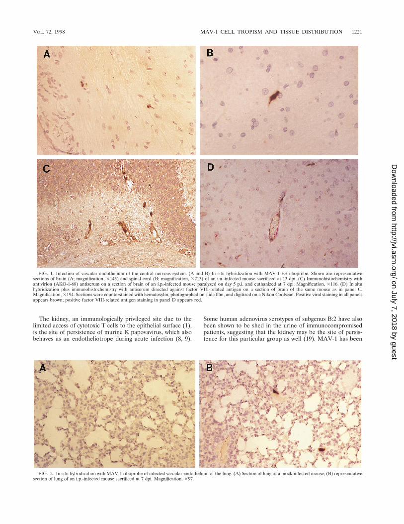
Infection of vascular endothelium. Evidence of MAV-1DNA and antigens was seen in endothelial cells, particularly inthe brain and spinal cord (Fig. 1). Immunohistochemistry withan antibody to factor VIII-related antigen, a specific compo-nent of endothelial cells, was used in double-staining experi-ments to confirm that the nucleic acid detected by in situhybridization was within vascular endothelium (Fig. 1D). En-dothelial cells in the lung, kidney, spleen, liver, and otherorgans also stained positive for MAV-1, indicating a wide-spread involvement of the systemic circulation (Fig. 2 and 3A).
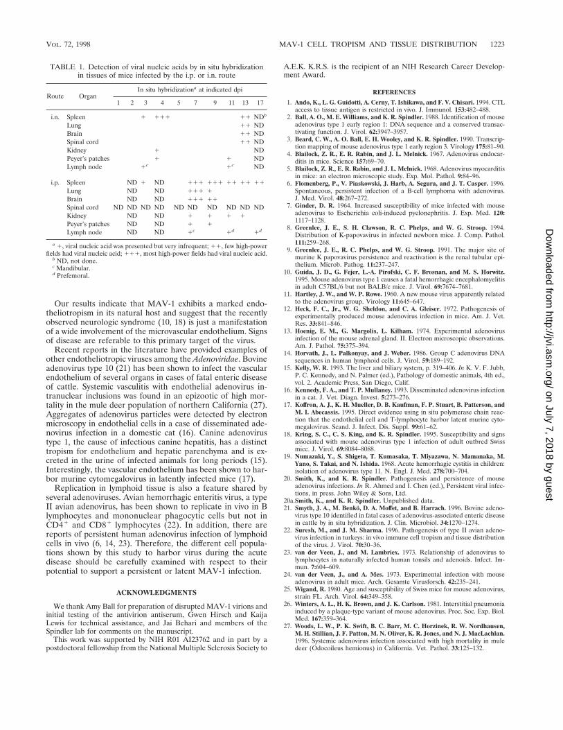
Infection of lymphoid tissue. The presence of viral nucleicacid and/or antigen was demonstrated by in situ hybridizationand immunohistochemistry in the spleens, Peyer’s patches, andmandibular and prefemoral lymph nodes in mice infected byeither route. Virus-infected cells in the spleen were localized tothe red pulp and exhibited a stellate shape strongly suggestive
of macrophages (Fig. 4A and B). Between days 13 and 17 p.i.,virus could also be detected in the germinal centers. In Peyer’spatches and lymph nodes, infected cells were localized primar-ily in germinal centers of follicles (Fig. 4C). Spatially, thevirus-positive cells were not located in B-cell areas. The occur-rence of staining in cytoplasmic processes of large stellate cellssuggested that the infected elements might be follicular den-dritic cells. By combining in situ hybridization and immunohis-tochemistry protocols, viral infection was found not to involvethe T-cell population (Fig. 4D).
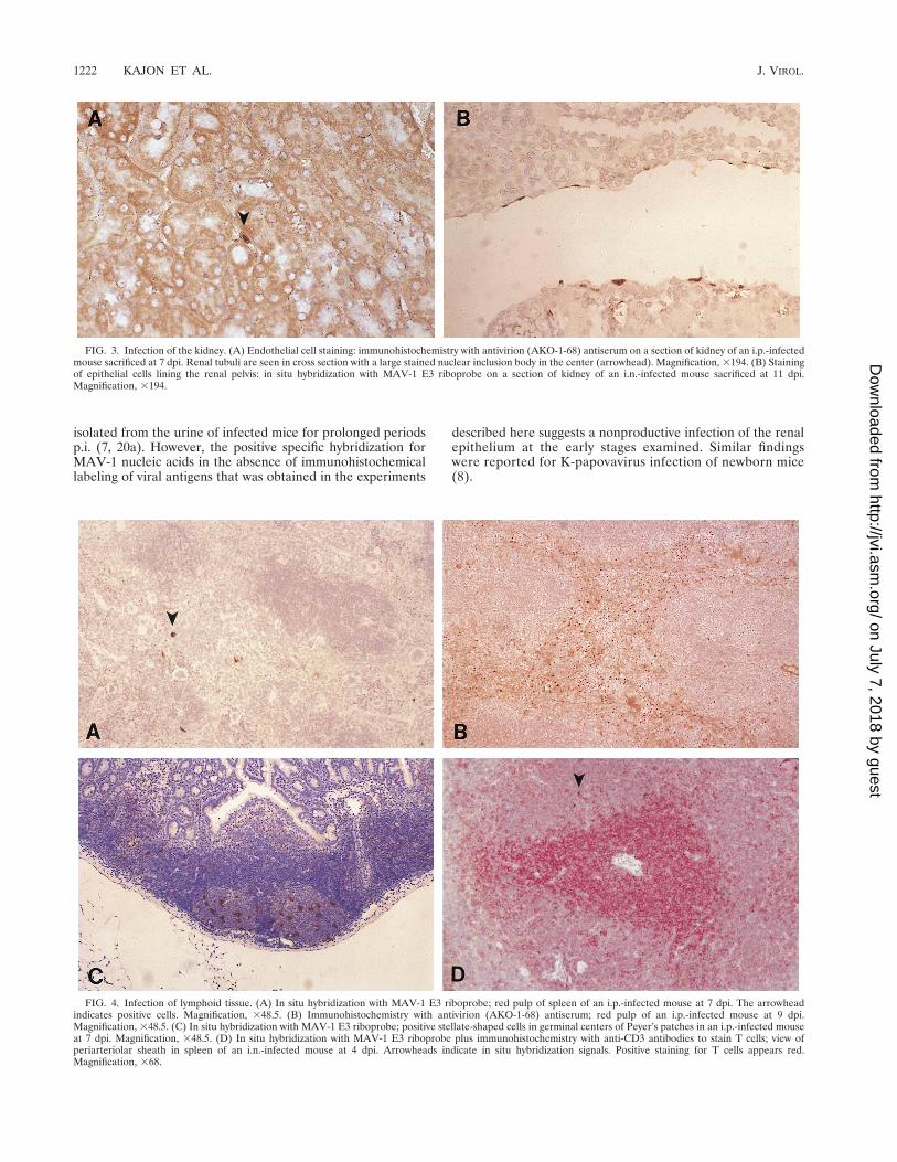
Infection of epithelium. Rare staining was found in epithelialcells of the distal tubules of the kidney in one i.n.-infectedmouse at 4 dpi. As shown in Fig. 3B, viral nucleic acid wasfound in the epithelial cells lining the renal pelvis in one i.p.-infected mouse sacrificed at 11 dpi. Viral antigen was notdetected in these same sections, as assayed by staining withantivirion antibody.
Spread of MAV-1 after i.p. and i.n. infection. The timecourse of infection differed between routes of inoculation (Ta-ble 1). However, in both groups, the first signs of presence ofviral nucleic acids were found at 3 dpi in the spleen. In thei.n.-infected mice, viral nucleic acid was also found in themandibular lymph node at 3 dpi. Subsequently there was anextensive positivity in endothelial cells throughout the body. Inthe i.p.-infected mice, there was more extensive and wide-spread involvement of endothelium at 7 dpi. In contrast, in thei.n.-infected mice, this widespread vascular involvement wasseen later, at 13 dpi.
DISCUSSION
The major site of MAV-1 infection and replication in out-bred adult mice infected either i.p. or i.n. with 1,000 PFU ofvirus and studied over a 13- to 17-day period was found to bethe vascular endothelium, especially that in the brain and spi-nal cord. The presence of viral nucleic acids and/or antigenswas also demonstrated in endothelial cells of the lung, kidney,liver, and spleen at different time points p.i. The absence ofsignificant staining in the heart and adrenal glands is notewor-thy and represents a major difference from findings reportedby other authors (4, 5, 13, 24). In our hands, staining in thesetissues was strictly circumscribed to endothelium.
MAV-1 nucleic acids and antigens were also detected inlymphoid tissue during the course of acute infection. The iden-tity of MAV-1-positive lymphoid cells was investigated by usingimmunohistochemistry targeting specific cell types. The spatialdistribution and morphoplogy of the stained elements suggestthat the cell type harboring MAV-1 in lymphoid tissue is amember of the mononuclear phagocytic system and that theinitial replication after infection may take place in these cells.In the case of i.p. infection, there would be a large bed ofperitoneal macrophages in which virus could replicate, withsubsequent early spread of virus to the spleen. In the i.n.-infected mice, the primary site of replication is unknown, butthe presence of large mononuclear cells staining in the man-dibular lymph node suggests initial replication in macrophageswithin lymphoid tissue. It is therefore likely that peripheralblood monocytes or dendritic cell precursors are the primaryvehicles for virus dissemination during acute infection. Thisissue must be further investigated by use of additional cell-typing reagents.
Staining of epithelial cells was noted only in the kidney.Infection of the renal tubular epithelium was detected as earlyas 4 dpi, suggesting that it is an early event during acuteinfection. At day 11 p.i., viral nucleic acid was also detected inthe epithelium lining the renal pelvis.
1220 KAJON ET AL. J. VIROL.
on July 7, 2018 by guesthttp://jvi.asm
.org/D
ownloaded from
The kidney, an immunologically privileged site due to thelimited access of cytotoxic T cells to the epithelial surface (1),is the site of persistence of murine K papovavirus, which alsobehaves as an endotheliotrope during acute infection (8, 9).
Some human adenovirus serotypes of subgenus B:2 have alsobeen shown to be shed in the urine of immunocompromisedpatients, suggesting that the kidney may be the site of persis-tence for this particular group as well (19). MAV-1 has been
FIG. 1. Infection of vascular endothelium of the central nervous system. (A and B) In situ hybridization with MAV-1 E3 riboprobe. Shown are representativesections of brain (A; magnification, 3145) and spinal cord (B; magnification, 3213) of an i.n.-infected mouse sacrificed at 13 dpi. (C) Immunohistochemistry withantivirion (AKO-1-68) antiserum on a section of brain of an i.p.-infected mouse paralyzed on day 5 p.i. and euthanized at 7 dpi. Magnification, 3116. (D) In situhybridization plus immunohistochemistry with antiserum directed against factor VIII-related antigen on a section of brain of the same mouse as in panel C.Magnification, 3194. Sections were counterstained with hematoxylin, photographed on slide film, and digitized on a Nikon Coolscan. Positive viral staining in all panelsappears brown; positive factor VIII-related antigen staining in panel D appears red.
FIG. 2. In situ hybridization with MAV-1 riboprobe of infected vascular endothelium of the lung. (A) Section of lung of a mock-infected mouse; (B) representativesection of lung of an i.p.-infected mouse sacrificed at 7 dpi. Magnification, 397.
VOL. 72, 1998 MAV-1 CELL TROPISM AND TISSUE DISTRIBUTION 1221
on July 7, 2018 by guesthttp://jvi.asm
.org/D
ownloaded from
isolated from the urine of infected mice for prolonged periodsp.i. (7, 20a). However, the positive specific hybridization forMAV-1 nucleic acids in the absence of immunohistochemicallabeling of viral antigens that was obtained in the experiments
described here suggests a nonproductive infection of the renalepithelium at the early stages examined. Similar findingswere reported for K-papovavirus infection of newborn mice(8).
FIG. 3. Infection of the kidney. (A) Endothelial cell staining: immunohistochemistry with antivirion (AKO-1-68) antiserum on a section of kidney of an i.p.-infectedmouse sacrificed at 7 dpi. Renal tubuli are seen in cross section with a large stained nuclear inclusion body in the center (arrowhead). Magnification, 3194. (B) Stainingof epithelial cells lining the renal pelvis: in situ hybridization with MAV-1 E3 riboprobe on a section of kidney of an i.n.-infected mouse sacrificed at 11 dpi.Magnification, 3194.
FIG. 4. Infection of lymphoid tissue. (A) In situ hybridization with MAV-1 E3 riboprobe; red pulp of spleen of an i.p.-infected mouse at 7 dpi. The arrowheadindicates positive cells. Magnification, 348.5. (B) Immunohistochemistry with antivirion (AKO-1-68) antiserum; red pulp of an i.p.-infected mouse at 9 dpi.Magnification, 348.5. (C) In situ hybridization with MAV-1 E3 riboprobe; positive stellate-shaped cells in germinal centers of Peyer’s patches in an i.p.-infected mouseat 7 dpi. Magnification, 348.5. (D) In situ hybridization with MAV-1 E3 riboprobe plus immunohistochemistry with anti-CD3 antibodies to stain T cells; view ofperiarteriolar sheath in spleen of an i.n.-infected mouse at 4 dpi. Arrowheads indicate in situ hybridization signals. Positive staining for T cells appears red.Magnification, 368.
1222 KAJON ET AL. J. VIROL.
on July 7, 2018 by guesthttp://jvi.asm
.org/D
ownloaded from
Our results indicate that MAV-1 exhibits a marked endo-theliotropism in its natural host and suggest that the recentlyobserved neurologic syndrome (10, 18) is just a manifestationof a wide involvement of the microvascular endothelium. Signsof disease are referable to this primary target of the virus.
Recent reports in the literature have provided examples ofother endotheliotropic viruses among the Adenoviridae. Bovineadenovirus type 10 (21) has been shown to infect the vascularendothelium of several organs in cases of fatal enteric diseaseof cattle. Systemic vasculitis with endothelial adenovirus in-tranuclear inclusions was found in an epizootic of high mor-tality in the mule deer population of northern California (27).Aggregates of adenovirus particles were detected by electronmicroscopy in endothelial cells in a case of disseminated ade-novirus infection in a domestic cat (16). Canine adenovirustype 1, the cause of infectious canine hepatitis, has a distincttropism for endothelium and hepatic parenchyma and is ex-creted in the urine of infected animals for long periods (15).Interestingly, the vascular endothelium has been shown to har-bor murine cytomegalovirus in latently infected mice (17).
Replication in lymphoid tissue is also a feature shared byseveral adenoviruses. Avian hemorrhagic enteritis virus, a typeII avian adenovirus, has been shown to replicate in vivo in Blymphocytes and mononuclear phagocytic cells but not inCD41 and CD81 lymphocytes (22). In addition, there arereports of persistent human adenovirus infection of lymphoidcells in vivo (6, 14, 23). Therefore, the different cell popula-tions shown by this study to harbor virus during the acutedisease should be carefully examined with respect to theirpotential to support a persistent or latent MAV-1 infection.
ACKNOWLEDGMENTS
We thank Amy Ball for preparation of disrupted MAV-1 virions andinitial testing of the antivirion antiserum, Gwen Hirsch and KaijaLewis for technical assistance, and Jai Behari and members of theSpindler lab for comments on the manuscript.
This work was supported by NIH R01 AI23762 and in part by apostdoctoral fellowship from the National Multiple Sclerosis Society to
A.E.K. K.R.S. is the recipient of an NIH Research Career Develop-ment Award.
REFERENCES
1. Ando, K., L. G. Guidotti, A. Cerny, T. Ishikawa, and F. V. Chisari. 1994. CTLaccess to tissue antigen is restricted in vivo. J. Immunol. 153:482–488.
2. Ball, A. O., M. E. Williams, and K. R. Spindler. 1988. Identification of mouseadenovirus type 1 early region 1: DNA sequence and a conserved transac-tivating function. J. Virol. 62:3947–3957.
3. Beard, C. W., A. O. Ball, E. H. Wooley, and K. R. Spindler. 1990. Transcrip-tion mapping of mouse adenovirus type 1 early region 3. Virology 175:81–90.
4. Blailock, Z. R., E. R. Rabin, and J. L. Melnick. 1967. Adenovirus endocar-ditis in mice. Science 157:69–70.
5. Blailock, Z. R., E. R. Rabin, and J. L. Melnick. 1968. Adenovirus myocarditisin mice: an electron microscopic study. Exp. Mol. Pathol. 9:84–96.
6. Flomenberg, P., V. Piaskowski, J. Harb, A. Segura, and J. T. Casper. 1996.Spontaneous, persistent infection of a B-cell lymphoma with adenovirus.J. Med. Virol. 48:267–272.
7. Ginder, D. R. 1964. Increased susceptibility of mice infected with mouseadenovirus to Escherichia coli-induced pyelonephritis. J. Exp. Med. 120:1117–1128.
8. Greenlee, J. E., S. H. Clawson, R. C. Phelps, and W. G. Stroop. 1994.Distribution of K-papovavirus in infected newborn mice. J. Comp. Pathol.111:259–268.
9. Greenlee, J. E., R. C. Phelps, and W. G. Stroop. 1991. The major site ofmurine K papovavirus persistence and reactivation is the renal tubular epi-thelium. Microb. Pathog. 11:237–247.
10. Guida, J. D., G. Fejer, L.-A. Pirofski, C. F. Brosnan, and M. S. Horwitz.1995. Mouse adenovirus type 1 causes a fatal hemorrhagic encephalomyelitisin adult C57BL/6 but not BALB/c mice. J. Virol. 69:7674–7681.
11. Hartley, J. W., and W. P. Rowe. 1960. A new mouse virus apparently relatedto the adenovirus group. Virology 11:645–647.
12. Heck, F. C., Jr., W. G. Sheldon, and C. A. Gleiser. 1972. Pathogenesis ofexperimentally produced mouse adenovirus infection in mice. Am. J. Vet.Res. 33:841–846.
13. Hoenig, E. M., G. Margolis, L. Kilham. 1974. Experimental adenovirusinfection of the mouse adrenal gland. II. Electron microscopic observations.Am. J. Pathol. 75:375–394.
14. Horvath, J., L. Palkonyay, and J. Weber. 1986. Group C adenovirus DNAsequences in human lymphoid cells. J. Virol. 59:189–192.
15. Kelly, W. R. 1993. The liver and biliary system, p. 319–406. In K. V. F. Jubb,P. C. Kennedy, and N. Palmer (ed.), Pathology of domestic animals, 4th ed.,vol. 2. Academic Press, San Diego, Calif.
16. Kennedy, F. A., and T. P. Mullaney. 1993. Disseminated adenovirus infectionin a cat. J. Vet. Diagn. Invest. 5:273–276.
17. Koffron, A. J., K. H. Mueller, D. B. Kaufman, F. P. Stuart, B. Patterson, andM. I. Abecassis. 1995. Direct evidence using in situ polymerase chain reac-tion that the endothelial cell and T-lymphocyte harbor latent murine cyto-megalovirus. Scand. J. Infect. Dis. Suppl. 99:61–62.
18. Kring, S. C., C. S. King, and K. R. Spindler. 1995. Susceptibility and signsassociated with mouse adenovirus type 1 infection of adult outbred Swissmice. J. Virol. 69:8084–8088.
19. Numazaki, Y., S. Shigeta, T. Kumasaka, T. Miyazawa, N. Mamanaka, M.Yano, S. Takai, and N. Ishida. 1968. Acute hemorrhagic cystitis in children:isolation of adenovirus type 11. N. Engl. J. Med. 278:700–704.
20. Smith, K., and K. R. Spindler. Pathogenesis and persistence of mouseadenovirus infections. In R. Ahmed and I. Chen (ed.), Persistent viral infec-tions, in press. John Wiley & Sons, Ltd.
20a.Smith, K., and K. R. Spindler. Unpublished data.21. Smyth, J. A., M. Benko, D. A. Moffet, and B. Harrach. 1996. Bovine adeno-
virus type 10 identified in fatal cases of adenovirus-associated enteric diseasein cattle by in situ hybridization. J. Clin. Microbiol. 34:1270–1274.
22. Suresh, M., and J. M. Sharma. 1996. Pathogenesis of type II avian adeno-virus infection in turkeys: in vivo immune cell tropism and tissue distributionof the virus. J. Virol. 70:30–36.
23. van der Veen, J., and M. Lambriex. 1973. Relationship of adenovirus tolymphocytes in naturally infected human tonsils and adenoids. Infect. Im-mun. 7:604–609.
24. van der Veen, J., and A. Mes. 1973. Experimental infection with mouseadenovirus in adult mice. Arch. Gesamte Virusforsch. 42:235–241.
25. Wigand, R. 1980. Age and susceptibility of Swiss mice for mouse adenovirus,strain FL. Arch. Virol. 64:349–358.
26. Winters, A. L., H. K. Brown, and J. K. Carlson. 1981. Interstitial pneumoniainduced by a plaque-type variant of mouse adenovirus. Proc. Soc. Exp. Biol.Med. 167:359–364.
27. Woods, L. W., P. K. Swift, B. C. Barr, M. C. Horzinek, R. W. Nordhausen,M. H. Stillian, J. F. Patton, M. N. Oliver, K. R. Jones, and N. J. MacLachlan.1996. Systemic adenovirus infection associated with high mortality in muledeer (Odocoileus hemionus) in California. Vet. Pathol. 33:125–132.
TABLE 1. Detection of viral nucleic acids by in situ hybridizationin tissues of mice infected by the i.p. or i.n. route
Route OrganIn situ hybridizationa at indicated dpi
1 2 3 4 5 7 9 11 13 17
i.n. Spleen 1 111 11 NDb
Lung 11 NDBrain 11 NDSpinal cord 11 NDKidney 1 NDPeyer’s patches 1 1 NDLymph node 1c 1c ND
i.p. Spleen ND 1 ND 111 111 11 11 11Lung ND ND 111 1Brain ND ND 111 11Spinal cord ND ND ND ND ND ND ND ND ND NDKidney ND ND 1 1 1 1Peyer’s patches ND ND 1 1Lymph node ND ND 1c 1d 1d
a 1, viral nucleic acid was presented but very infrequent; 11, few high-powerfields had viral nucleic acid; 111, most high-power fields had viral nucleic acid.
b ND, not done.c Mandibular.d Prefemoral.
VOL. 72, 1998 MAV-1 CELL TROPISM AND TISSUE DISTRIBUTION 1223
on July 7, 2018 by guesthttp://jvi.asm
.org/D
ownloaded from