Embed Size (px)
Citation preview

Diverse functions of myosin VI elucidated by an isoform-specific -helix domain.
Item type Article
Authors Wollscheid, Hans-Peter; Biancospino, Matteo; He, Fahu;Magistrati, Elisa; Molteni, Erika; Lupia, Michela;Soffientini, Paolo; Rottner, Klemens; Cavallaro, Ugo;Pozzoli, Uberto; Mapelli, Marina; Walters, Kylie J; Polo,Simona
Citation Diverse functions of myosin VI elucidated by an isoform-specific -helix domain. 2016, 23 (4):300-308 Nat. Struct.Mol. Biol.
DOI 10.1038/nsmb.3187
Journal Nature structural & molecular biology
Downloaded 9-Apr-2018 18:35:11
Item License http://creativecommons.org/licenses/by-nc-sa/4.0/
Link to item http://hdl.handle.net/10033/621298
Diverse functions of myosin VI elucidated by an isoform-specific α-helix domain
Hans-Peter Wollscheid#1,9, Matteo Biancospino#1, Fahu He#2, Elisa Magistrati1, Erika Molteni3, Michela Lupia4, Paolo Soffientini1, Klemens Rottner5,6, Ugo Cavallaro4, Uberto Pozzoli3, Marina Mapelli7, Kylie J. Walters2, and Simona Polo1,8
1Fondazione Istituto FIRC di Oncologia Molecolare (IFOM), Milan 20139, Italy
2Center for Cancer Research, National Cancer Institute, Frederick, MD 21702, USA
3Computational Biology, Scientific Institute IRCCS E.MEDEA, Bosisio Parini 23842, Italy
4Molecular Medicine Program, European Institute of Oncology, Milan 20141, Italy
5Helmholtz Centre for Infection Research, 38124 Braunschweig, Germany
6Braunschweig University of Technology, 38106 Braunschweig, Germany
7Department of Experimental Oncology, European Institute of Oncology, Milan 20139, Italy
8Dipartimento di oncologia ed emato-oncologia (DIPO), Universita’ degli Studi di Milano, Milan 20122, Italy
# These authors contributed equally to this work.
Abstract
Myosin VI functions in endocytosis and cell motility. Alternative splicing of myosin VI mRNA
generates two distinct isoform types, myosin VIshort and myosin VIlong, which differ in the C-
terminal region. Their physiological and pathological role remains unknown. Here we identified an
isoform-specific regulatory helix, named α2-linker that defines specific conformations and hence
determines the target selectivity of human myosin VI. The presence of the α2-linker structurally
defines a novel clathrin-binding domain that is unique to myosin VIlong and masks the known RRL
interaction motif. This finding is relevant to ovarian cancer, where alternative myosin VI splicing
Users may view, print, copy, and download text and data-mine the content in such documents, for the purposes of academic research, subject always to the full Conditions of use:http://www.nature.com/authors/editorial_policies/license.html#terms
Correspondence to: Simona Polo, Tel: +39 02 574303242; Fax: +39 02 574303231; [email protected] address: Institute of Molecular Biology (IMB), Mainz, Germany
Accession numbersThe structural coordinates and chemical shift data have been deposited in the Protein Data Bank (PDB) and Biological Magnetic Resonance Data Bank (BMRB) with respective accession codes 2N12 and 25544. The proteomic data as raw files, total proteins and peptides identified with relative intensities and search parameters have been deposited on Peptide Atlas repository (accession number PASS00591).
Author contributionsH-P.W., M.B and El.M. designed and performed the experiments and analyzed the data; F.H. and K.J.W. designed and interpreted CD and NMR experiments, which F.H. performed and analyzed; E.V. carried out FP analysis; P.S. carried out MS analysis; M.L. and U.C. generated primary cells from high-grade ovarian cancer; Er.M and U.P. conducted exons analysis; K.R. participated in setting up and interpreting the migration assays; M.M. participated in the experimental design and data analysis; S.P. conceived the project, interpreted the results and wrote the paper with the contribution of all authors.
Europe PMC Funders GroupAuthor ManuscriptNat Struct Mol Biol. Author manuscript; available in PMC 2016 September 22.
Published in final edited form as:Nat Struct Mol Biol. 2016 April ; 23(4): 300–308. doi:10.1038/nsmb.3187.
Europe PM
C Funders A
uthor Manuscripts
Europe PM
C Funders A
uthor Manuscripts

is aberrantly regulated, and exon skipping dictates cell addiction to myosin VIshort for tumor cell
migration. The RRL interactor optineurin contributes to this process by selectively binding myosin
VIshort. Thus the α2-linker acts like a molecular switch that assigns myosin VI to distinct
endocytic (myosin VIlong) or migratory (myosin VIshort) functional roles.
Molecular motors exert critical roles in virtually all cell processes. Myosin VI belongs to a
superfamily of more than 20 different classes of actin-based motor proteins1, 2 that
hydrolyze ATP for mechanical work and movement of cargos along actin tracks. The ability
to walk towards the minus end of actin filaments makes myosin VI unique among the
myosin family3. This unusual property appears to bestow on myosin VI a number of specific
cellular functions, such as clathrin-mediated endocytosis and vesicular transport4, 5. Myosin
VI was originally discovered in Drosophila, where it participates in cell migration during
ovary development6, spermatogenesis7, 8 and neuroblast asymmetric division9, 10. In
mammalian cells, myosin VI localizes to the Golgi complex, membrane ruffles at the
leading edges of migrating cells, clathrin coated pits at plasma membranes, APPL1-positive
early endosomes and autophagosomes (reviewed in11). Detailed mechanisms underlying the
function of myosin VI in these diverse processes are currently unclear.
At a molecular scale, myosin VI consists of two functional domains (Fig. 1a), a motor–IQ
domain sufficient for pointed-end (minus-end) directed movement, and a tail domain, which
can be further divided into a long helical region, reported as a coiled-coil domain, and a C-
terminal globular region, known as a cargo-binding domain (CBD). A MIU (Motif
Interacting with Ubiquitin)12 is located at the boundaries. The CBD contains two myosin VI
ligand interaction surfaces, the “RRL” and “WWY” motifs11. The RRL triplet is
responsible for myosin VI binding to endocytic adaptors, including GAIP-interacting protein
C-terminus (GIPC)13, 14, and autophagy receptors, such as optineurin15, Nuclear Dot
Protein 52 (NDP52)16, and Traf6-binding protein (T6BP)16.
An intrinsic level of complexity in deciphering the functions of myosin VI comes from the
presence of different isoforms. Two regions are alternatively spliced within the tail (Fig. 1a),
leading to an insertion between the helical domain and the CBD (large insert, LI)17, 18, as
well as additional residues at the C-terminal (small insert, SI)19. Although myosin VI is
widely expressed in most tissues, isoforms containing the LI are specifically found in
polarized epithelial cells with well-developed apical microvilli17, 20. How the presence of
LI in the tail influences the functions and intracellular targeting of myosin VI is not known.
Up to now, isoforms lacking the LI present no specific expression and localization but are
required for polarized transport of tyrosine motif containing basolateral membrane
proteins20 and for maintaining an active pool of secretory granules near the plasma
membrane of neurosecretory cells19.
The role of myosin VI in cell migration has been linked to ovarian21 and prostate
cancers22–24, where myosin VI overexpression correlates with clinically aggressive
behavior. Notably, silencing of myosin VI expression decreases the migratory potential and
the malignant properties of ovarian21 and prostate cancer cell lines21, 22.
Wollscheid et al. Page 2
Nat Struct Mol Biol. Author manuscript; available in PMC 2016 September 22.
Europe PM
C Funders A
uthor Manuscripts
Europe PM
C Funders A
uthor Manuscripts

In this study, we set out to investigate the major splicing event occurring in myosin VI from
a molecular and a functional perspective. By analyzing the interactomes and the
conformational structure of myosin VI isoforms, we provided mechanistic insights into how
myosin VI function becomes pathological in human cancers and demonstrated the
importance of an isoform-specific helix in assigning myosin VI to distinct functional roles.
Results
Selective expression of myosin VIshort in ovarian cancer
Several studies report overexpression of myosin VI in prostate22–24 and ovarian cancers21,
which positively correlates with their grade and metastatic potential. No information is
available concerning specificity of isoform expression. To shed light on this issue, we
analyzed human myosin VI transcripts in detail. The gene consists of 36 coding exons, three
of which (exons 29, 30 and 31) generate the LI (Supplementary Fig. 1a). We focused on the
main myosin VI isoforms expressed in human cell lines17, 18, 20, namely isoforms 1 and 3,
which have different LI lengths (23- or 32-residues long, respectively, due to the presence or
absence of exon 29, Supplementary Fig. 1a), and isoform 2, which lacks the LI entirely
(exons 29, 30 and 31 are absent, Supplementary Fig. 1a). Interestingly, these alternative
exons are conserved throughout evolution (Supplementary Fig. 1b), suggesting a relevant
function for the regulated splicing in myosin VI.
To gain insight into possible differential expression of myosin VI isoforms in tumors, we
analyzed their expression by RT-PCR using primers positioned in exon 28 and 33 (Fig. 1b).
The PCR reaction was able to discriminate between the different isoforms, as demonstrated
in the control lanes (Fig. 1c). RT-PCR was performed using RNA extracted from high-grade
primary ovarian cancer cells cultured in vitro for two passages. In contrast to their normal
counterpart (ovarian surface epithelium, OSE), ten out of ten tumors expressed almost
exclusively isoform 2, suggesting a positive selection for this isoform in tumor progression
and metastasis (Fig. 1c). We then examined a number of ovarian cancer cell lines for myosin
VI isoform expression in order to identify possible cellular models for in vitro studies.
OVCAR-5 is unique among them as it expresses all myosin VI isoforms, while the
remaining 6 cell lines tested selectively expressed isoform 2 (Fig. 1d).
Knocking-down myosin VI substantially inhibits migration in vitro and reduces
dissemination of ovarian tumor cells propagated in nude mice21. To gain insight into the
possible advantages derived from overexpression of myosin VI isoform 2 in cancer cells, we
looked at cell migration as an important feature of cancer metastasis. We analyzed the effect
of myosin VI depletion in SK-OV-3, HEY and OVCAR-5 cell lines in wound-healing assay.
Depletion of myosin VI was complete and comparable in all cell lines (Fig. 1e). In the
absence of myosin VI, SK-OV-3 and HEY cells were severely impaired in their ability to
migrate and to close wounds, whereas the migration efficiency of OVCAR-5 cells was
unaffected (Fig. 1e and Supplementary movies 1-3). Comparable results were obtained with
different oligos (Supplementary Fig. 2a,b). Hence, only ovarian cancer cells that selectively
express myosin VI isoform 2 were deficient in cell migration upon myosin VI depletion.
Wollscheid et al. Page 3
Nat Struct Mol Biol. Author manuscript; available in PMC 2016 September 22.
Europe PM
C Funders A
uthor Manuscripts
Europe PM
C Funders A
uthor Manuscripts
Next, we looked at possible partners of myosin VI in this process. In A549 cells, ablation of
myosin VI or its interactor optineurin15 inhibits polarized delivery of the epidermal growth
factor receptor into the leading edge and leads to profound defects in lamellipodia
formation25. This prompted us to test the effect of optineurin depletion in our model system.
Optineurin knockdown reduced the migration ability of SK-OV-3 but did not further
decrease cell speed in myosin VI-depleted cells (Fig. 2a,b and Supplementary movie 4). The
lack of additive effects suggests that the two proteins are acting in the same axis.
Our functional data suggested that optineurin may specifically interact with myosin VI
isoform 2. Optineurin binding requires the RRL motif15 of myosin VI which lies outside of
the splicing region (Fig. 2c). Nonetheless, we tested its binding to the different isoforms. A
sequence alignment of the utilized region of myosin VI (myosin VI998–1131, amino acid
numbering from isoform 3) together with its features is shown in Figure 2c. As shown in
Figure 2d, optineurin binds specifically to isoform 2 and to a shorter construct lacking the
alternatively spliced region (1080–1131). This prompted us to test for a possible preference
of the other known RRL interactors; GIPC13, NDP52 (ref.16) and T6BP16. Surprisingly, all
three interactors showed a clear preference for isoforms 2, which lacks the large insert (Fig.
2c).
Myosin VI isoforms display different interaction partners
Distinct functions for myosin VI isoforms are described but no molecular rationale for such
differences is provided17, 20. The results obtained with the RRL interactors prompted us to
hypothesize that the various myosin VI isoforms may have different interactomes. Thus, we
performed a mass spectrometry-based proteomic screen, aimed at comparing the interaction
profiles of the various isoforms. GST-tagged myosin VI998–1131 constructs were used in
pull-down assays with HEK293T cellular lysates (Fig. 2e). A label-free quantitative
experiment was performed, and resulting data were analysed by MaxQuant software.
Inspection of the gel (Fig. 2e), confirmed by data presented in Table 1, indicated that
isoforms 1 and 3 have almost identical binding partners. Most of these binding partners are
implicated in endocytosis and vesicular trafficking and are not shared by isoform 2. Isoform
2-specific interactors were more heterogeneous and comprised nuclear factors, an E3 ligase
(DDB1–DCAF1) and few GEFs, among which the previously identified myosin VI binding
partner DOCK7 (ref.26). Few interactors common to all three isoforms (e.g. COPB2) were
also identified. Table 1 reported examples of the top hits for each class with their total
peptide counts. None of the previously characterized RRL interactors were present. Since we
cut the gel higher than GST fusion proteins, GIPC was automatically excluded from the
analysis whereas NDP52 and T6BP were identified with a single peptide and thus filtered
out. Supplementary Table 1 reported the complete list of proteins that differentially interact
with isoform 1, isoform 2 and 3 vs GST control confirmed by additional proteomics
experiments.
Experimental validation confirmed the most abundant identified interactors (5 out of 5
tested) and their specificity (Fig. 2f). Since isoforms 1 and 3 showed no difference in terms
of interaction networks, we referred to them collectively as myosin VIlong, while using
myosin VIshort to indicate isoform 2.
Wollscheid et al. Page 4
Nat Struct Mol Biol. Author manuscript; available in PMC 2016 September 22.
Europe PM
C Funders A
uthor Manuscripts
Europe PM
C Funders A
uthor Manuscripts

Structure of Myosin VIlong clathrin-binding domain
The interactome analysis identified clathrin heavy chain (CLTC) as a major and specific
binding partner of myosin VIlong (Table 1 and Fig. 2f). It is well-known that myosin VI and
clathrin form a ternary complex with the Dab-2 adaptor27, 28. However, neither the WWY
motif of myosin VI (ref.27), or the larger surfaces implicated in binding to Dab-2 (ref.28)
were present in the constructs used for the proteomic screening. Thus, we inferred that
myosin VI could also interact with clathrin directly.
To map this novel interaction surface, we generated a panel of GST-tagged deletion
constructs and tested their interaction with endogenous clathrin starting from isoform 3
myosin VI998–1131, which was used for the proteomic screen (Supplementary Fig. 3a-c).
Regions deleted from the C-terminal end did not show any clathrin binding (Supplementary
Fig. 3a), whereas fragments deleted at the N-terminal end retained clathrin interaction
(Supplementary Fig. 3b). The minimal binding region appeared to be 1055–1131, which
includes the isoform-specific LI region (Fig. 2c) and the region directly following it. Further
N-terminal shortening, or even small internal deletion disrupted binding (Supplementary
Fig. 3b,c).
We used NMR techniques to solve the structure of the clathrin binding surface (Fig. 3a). The
twenty calculated NMR structures with lowest energy from 100 starting structures converged
well (stereoview showed in Supplementary Fig. 3d) with a backbone root mean square
deviation (RMSD) of 0.18 Å, following the exclusion of an 11-amino acid flexible loop that
directly follows the LI (Supplementary Fig. 3d and Table 2). In the isoform-specific LI
region, residues 1055–1068 form an amphipathic α-helix (α2-linker, Fig. 2c and
Supplementary Fig. 3e) that packs against two other helices present in all isoforms (Fig. 3a),
named α3 and α4. Interaction between the isoform-specific α2-linker and the common
helices α3 and α4 (α3/α4) is defined by 106 NOE interactions, as exemplified for L1118
(Supplementary Fig. 3f). A hydrophobic surface of the α2-linker formed by M1058, M1062,
and L1066 inserts into a cleft formed by the α3/α4 interface and interacts with hydrophobic
residues Y1091, F1114, L1118, and Y1121 (Fig. 3a and 3c). In addition, a salt bridge is
formed between α2-linker E1061 and α3 R1095 (Fig. 3b).
Mutually exclusive association of clathrin and RRL binders
The RRL motif is embedded in helix α4 of the clathrin-binding domain (Fig. 2c). As visible
in the structure, two residues of the RRL motif were not accessible for binding with
interactors (Fig. 3d), thus explaining their specificity for binding only myosin VIshort (Fig.
2d). L1118 is buried in the interface between α2-linker and α3/α4 (Fig. 3c and 3d) while
R1117 formed a salt bridge with S1087 of the α3 (Fig. 3d). We generated single point
mutations of the RRL motif as well as the triple AAA mutation previously used to validate
interactions with known myosin VI binders13, 16 in the context of a myosin VIlong
fragment. As shown in Figure 3e, R1117A (RAL), L1118A (RRA) and the triple AAA
mutation abrogated the ability of myosin VI998–1131 to pull-down clathrin from cell lysates,
whereas the R1116A (ARL) mutant behaved as wild type. Circular dichroism data of the
R1117A-containing fragment (Supplementary Fig. 3g) revealed that R1117 is required for
structural integrity of this myosin VI region.
Wollscheid et al. Page 5
Nat Struct Mol Biol. Author manuscript; available in PMC 2016 September 22.
Europe PM
C Funders A
uthor Manuscripts
Europe PM
C Funders A
uthor Manuscripts

The structural data provided an explanation for the myosin VIshort selective interaction of
RRL binders (Fig. 2d) but raised an issue related the triple AAA mutation. Thus, we probed
the ability of all RRL interactors to interact with a myosin VI fragment carrying mutations in
the RRL motif. We used a minimal fragment (G1080–R1131) that includes α3/α4 but lacks
the α2-linker and retains binding (Fig. 2d). As for clathrin, and consistent with the structural
data, the destabilizing R1117A mutation either alone or in the context of the AAA mutant
impaired binding to partners (Fig. 3f). Analyses performed with the other single point
mutants demonstrated that R1116 and L1118 are critical for GIPC and optineurin binding,
whereas T6BP and NDP52 associate with myosin VI1080–1131 in an RRL-independent
fashion (Fig. 3f).
Taken together, these data implied that packing of the α2-linker against the α3/α4 helices in
myosin VIlong determined: i) its exquisite clathrin-binding ability and ii) its impairment in
RRL-mediated binding (i.e. optineurin). We endeavored to validate the functional impact of
the myosin VIlong conformation by testing point mutation(s) capable of disrupting
interaction between α3/α4 and the α2-linker. We replaced M1062, which is sandwiched
between α3 and α4 (Fig. 4a), with glutamine, and evaluated this variant together with the
previously characterized L1118A mutant (Fig. 3e), for binding to RRL-specific interactors
by pull-down assays. As shown in Fig. 4b, M1062Q was impaired in clathrin binding,
similarly to L1118A. Strikingly, GIPC and optineurin gained binding to M1062Q but not to
L1118A (Fig. 4b), consistent with the evidence that L1118 belongs to their interaction
surface (Fig. 3f). Finally, NDP52 and T6BP interacted with both mutants at levels similar to
myosin VIshort (Fig. 4b). Thus, these point mutations were able to induce a conformational
change that converts a myosin VIlong into a myosin VIshort-like protein. NMR data of the
L1118A mutant further validated this conclusion (Supplementary Fig. 3h).
These results confirmed the conformational differences between the myosin VI isoforms
induced by the presence of the α2-linker and established their mutually exclusive
association with clathrin and RRL interactors (i.e. optineurin) (Fig. 4c).
Functional analysis of Myosin VIlong–clathrin interaction
To functionally evaluate these findings, we generated GFP-tagged versions of full-length
myosin VIlong wild type, L1118A and M1062Q mutants. As controls, we used myosin
VIshort and a myosin VIlong mutated in the WWY motif (W1192L) and impaired for Dab-2
binding13. We transfected the constructs in HeLa cells where we knocked-down endogenous
myosin VI to avoid confounding effects due to the presence of the endogenous protein (e.g.,
dimerization). Results showed that endogenous clathrin was immunoprecipitated by myosin
VIlong but not by myosin VIshort and that L1118A and M0162Q mutations were sufficient to
abrogate this interaction (Fig. 4d). In this context, the W1192L mutant showed binding
comparable to that of wild type myosin VIlong (Fig. 4d), suggesting that Dab-2 is
dispensable for a stable interaction with clathrin. We also analyzed the behavior of the
isoforms by immunofluorescence analysis. As previously reported17, myosin VIlong strongly
co-localized with clathrin, whereas myosin VIshort exhibited no significant co-localization
(Fig. 4e,f). Notably, W1192L, L1118A and M1062Q mutants failed to co-localize with
clathrin, similarly to myosin VIshort (Fig. 4e,f). Similar results were obtained using myosin
Wollscheid et al. Page 6
Nat Struct Mol Biol. Author manuscript; available in PMC 2016 September 22.
Europe PM
C Funders A
uthor Manuscripts
Europe PM
C Funders A
uthor Manuscripts

VI tails (Supplementary Fig. 4), which recapitulate myosin VI interaction and
localization17. Taken together, these findings led us to conclude that, in addition to the
known Dab-2-binding motif, a second isoform-specific binding surface drives myosin VIlong
interaction with clathrin.
Exon skipping is a common event in cancer
Our findings revealed that the isoform-specific α2-linker defines specific conformation (Fig.
3a), interaction (Fig. 4d) and localization (Fig. 4e,f) of myosin VI isoforms. During in vitro epithelial cell polarization a myosin VI isoform switch occurs towards longer isoforms17,
18, suggesting a need of myosin VIlong for clathrin-mediated endocytosis in fully polarized
cells.
As loss of cell polarity is a hallmark of cancer, we decided to extend our analyses of myosin
VI isoform expression to other cancer types using an in silico approach. We evaluated the
relative expression of the isoforms by exploiting the presence of exon 31, which encodes for
the α2-linker (outlined in Supplementary Figure 1a) as a discriminating marker of myosin
VIlong. As a source of data, we used The Cancer Genome Atlas (TCGA) database and
included all experiments performed using RNAseq technology. The resulting dataset
consisted of 293 control and 1646 tumor samples, belonging to 14 different tumor types. The
relative abundance of exon 31 (E31RA) was obtained as the ratio of its RPKM (Reads Per
Kilobase of Exon Model per Million Mapped Reads, see Materials and Methods for details)
and the average RPKM values of the four flanking constitutive exons (namely E27, E28,
E32 and E33, Supplementary Fig. 1b).
The box plot relative to the overall analysis (Fig. 5a) showed that exon 31 skipping is a
common event occurring with significantly increased frequency in tumor compared to
normal samples. Thus, myosin VIshort is selectively expressed in tumors. The analysis
conducted at the single cancer type level showed that exon skipping is highly frequent
among certain tumors, but not others (Supplementary Fig. 5), and suggested that the function
of myosin VIshort may be critical and positively selected only in specific types of cancers. Of
note, in the case of ovarian cancer, no ‘normal’ controls were present in the database, but
compared to the average of normal samples, exon E31RA is barely measurable (Fig. 5b),
further confirming the data obtained in primary tumors (Fig. 1c). As proof of principle, we
selected two breast cell lines, MCF10A and MDA-MB-231. This latter cancer cell line
showed high levels of myosin VIshort-only in both sparse and confluent conditions (Fig. 5c).
Upon myosin VI depletion, MDA-MB-231 cells displayed a clear impairment in wound
closure (Fig. 5d and Supplementary movie 5), while MCF10A cells were unaffected (Fig. 5e
and Supplementary movie 6). Thus, cancer cells that selectively express myosin VIshort-only
are addicted to myosin VI for cell migration.
Discussion
The presence of myosin VI splice variants lead to the suggestion that they are directly
responsible for disparate myosin VI functions, but no data confirm this hypothesis11, 29.
Dab-2 is considered as the adaptor that links myosin VI to clathrin-mediated function13, 27,
30. The Dab-2-myosin VI interaction, however, fails to explain the myosin VIlong-specific
Wollscheid et al. Page 7
Nat Struct Mol Biol. Author manuscript; available in PMC 2016 September 22.
Europe PM
C Funders A
uthor Manuscripts
Europe PM
C Funders A
uthor Manuscripts

interaction with clathrin17, as the Dab-2 binding region lies outside of the alternatively
spliced regions13, 27. We provided for the first time a rationale for clathrin selectivity, as the
presence of the isoform-specific α2-linker, together with the following region defines a
novel clathrin-binding domain and dictates isoform-specific interactions. This finding has
important functional implications. During epithelial cell polarization, an isoform switch of
myosin VI occurs17, 20. In this context, cells acquire myosin VIlong that is recruited,
possibly via clathrin interaction, to the apical part of polarized cells17. Here, myosin VIlong
could provide the coated vesicles with the required mechanical force to pass through the
intricate meshwork of cortical actin29.
Our study also offers a new perspective on the molecular mechanism that regulates the
monomer-dimer conversion of this anchor-motor protein31, 32. It is tempting to speculate
that the existence of distinct interaction surfaces for clathrin and Dab-2, both of which are
required for co-localization with clathrin-coated pits (Fig. 4e,f) may provide critical synergy
that regulates the myosin VI motor ability. Clathrin binding may recruit and concentrate
myosin VI at clathrin-coated pits where Dab2 could tether it into a stable and active dimer,
crucial to convert myosin VI into a cellular transporter28.
Unexpectedly, our data showed a clear preference of RRL interactors towards myosin
VIshort. In the case of optineurin and GIPC, structural data elucidated this specificity as
R1116 and L1118, which belong to their interaction surface, are masked in the myosin
VIlong conformation. A similar explanation could also apply for NDP52 and T6BP, although
their precise interaction surface remains to be established. Whatever the case, our data
suggested that myosin VIshort is the isoform specifically involved in the functional
interaction with these adaptor proteins that have an established role in autophagy33.
Myosin VI is implicated in cell migration both in Drosophila and mammals (reviewed in
34). However published data lead to an intriguing conundrum. On the one hand, myosin VI
depletion from epithelial cells leads to loss of vinculin from adhesion sites and weakens E-
cadherin complexes35. Such rearrangement results in loss of cohesive forces in epithelial
layers, a well-known prerequisite for invasive cancer cell migration. On the other hand,
myosin VI is remarkably upregulated in prostate and ovarian cancer cells, where its
overexpression correlates with aggressive clinical behavior21, 22. In this context, silencing
of myosin VI expression in ovarian and prostate cancer cell lines decreases the migratory
potential of these cells in vitro and ovarian tumor dissemination in vivo21, 22. Our results
provided a molecular explanation for this conundrum, ascribing the cohesive and migratory
defects to the two different myosin VI isoforms. We found that myosin VIshort is
overexpressed in ovarian and other invasive cancers, whereas myosin VIlong is expressed in
polarized epithelial cells, in which it likely promotes E-cadherin stabilization at cell-cell
junctions.
In Drosophila, the border cell migration occurring during oogenesis is reminiscent of tumor
cell invasion21. Initially, the border cells are polarized epithelial cells with strong cell-cell
contacts. For migration to begin, cell polarity changes from apico-basal to front-rear, and
proteins found at cell-cell junctions reorganize at the invasive edge of the cell to facilitate
motility and invasion36. During these events, myosin VI changes localization, from the
Wollscheid et al. Page 8
Nat Struct Mol Biol. Author manuscript; available in PMC 2016 September 22.
Europe PM
C Funders A
uthor Manuscripts
Europe PM
C Funders A
uthor Manuscripts

baso-lateral membrane to membrane ruffles at the front edge of moving cells36. An
interesting possibility is that at the onset of border cell migration, an isoform switch towards
the shorter myosin VI isoform occurs. Nothing is known about the differential expression
and functions of myosin VI isoforms in Drosophila. However, we noticed that different
splicing variants also exist in the fly gene. Secondary structure prediction analysis suggested
that, even if the primary sequence is not conserved, the exon that is skipped in the shortest
isoforms encodes an alpha helix with length and position similar to the α2-linker in
mammals (Supplementary Fig. 6).
Our data suggest that cancer cells positively select myosin VIshort because it confers them
with a migratory advantage. The exact molecular mechanism through which myosin VIshort
modulates cell migration remains to be established. Here, we substantiated previous
findings25, providing genetic evidence that the autophagic RRL binder optineurin acts in the
same pathway (Fig. 2a,b). In addition, we have demonstrated that myosin VI has a novel
ubiquitin-binding domain37 that most likely contributes to optinerurin interaction and to
myosin VI short migratory function.
The contribution of alternative splicing in human diseases is widely recognized38.
Unbalanced expression of splicing variants or failure to properly express the correct isoform
is clearly part of cancer cell biology39. Here, we found that exon skipping determines the
expression of myosin VIshort-only in in several types of tumors. Cancer-associated
alternative splicing variants are considered new tools for the diagnosis and classification of
cancers and could become targets for innovative therapeutic interventions based on highly
specific splicing correction approaches. The selective expression of myosin VIshort in cancer
may very well fit in this context. This unexpected finding paves the way for defining the
splicing mechanism of myosin VI and provides an exciting starting point to understand and
therapeutically exploit novel key events in pathological cell migration.
Online Methods
Cell lines, Media and Constructs
Cell line Source Medium
HEK293T ICLC DMEM (Lonza), 10% FBS S.A. (Biowest), 2 mM L-glutamine
HEY CELLution biosystems DMEM, 10% FBS S.A., 2 mM L-glutamine
MCF10A ATCC DMEM/Ham’s F12 1:1 (Gibco), 5% horse serum (Thermo Fisher Scientific), hydrocortisone (0.5 μg/ml), insulin (10 μg/ml), cholera toxin (50 ng/ml), EGF (20 ng/ml)
OVCAR-3 NCI-60 RPMI-1640 (Lonza), 10% FBS N.A. (SIGMA), 2 mM L-glutamine
OVCAR-4 NCI-60 RPMI-1640, 10% FBS N.A., 2 mM L-glutamine
OVCAR-5 NCI-60 RPMI-1640, 10% FBS N.A., 2 mM L-glutamine
OVCAR-8 NCI-60 RPMI-1640, 10% FBS N.A., 2 mM L-glutamine
MDA-MB-231 ATCC RPMI-1640, 10% FBS N.A., 2 mM L-glutamine
COLO-704 DSMZ RPMI-1640, 10% FBS N.A., 2 mM L-glutamine
SK-OV-3 NCI-60 McCoy’s 5A (Gibco), 10% FBS S.A.
Wollscheid et al. Page 9
Nat Struct Mol Biol. Author manuscript; available in PMC 2016 September 22.
Europe PM
C Funders A
uthor Manuscripts
Europe PM
C Funders A
uthor Manuscripts

Cell line Source Medium
HeLa ATCC MEM (Gibco), 10% FBS S.A., 0.1 mM non-essential amino acids, 2 mM L-glutamine, 1 mM Na-Pyruvate
At each batch freezing all cell lines were authenticated by STR profiling (StemElite ID
System, Promega) and tested for mycoplasma using PCR and biochemical test (MycoAlert,
Lonza).
Human myosin VI cDNA KIAA0389 (isoform 1) was obtained from Kazusa DNA Research
Institute (Kisarazu, Japan); PCR amplified and cloned into pGEX 6P1 (Amersham
Pharmacia Biotech), FLAG-pcDNA3.1 (Invitrogen) and pEGFP-C2 (Clontech). Myosin VI
isoform 2 and isoform 3 were generated by mutagenesis (deletion and insertion,
respectively) using isoform 1 as template and the primers shown in Supplementary Table 2.
All other constructs described were engineered by site-directed mutagenesis or recombinant
PCR and sequence verified. Details are available upon request.
GFP-PI3KC2α was kindly provided by Emilio Hirsch, SFB-DDB1 by Maddika Subba
Reddy, GFP-optineurin by Alain Israel, FLAG-T6BP by Folma Buss, His-GIPC by Guido
Serini, FLAG-NDP52 by Felix Randow.
Antibodies
Antibody Species Supplier Code WB IF
anti-FLAG M2 Mouse Sigma F3165 1:10000
anti-HA Mouse Babco 16B12 1:1000
anti-His Mouse GE Healthcare 27-4710-01 1:3000
anti-GFP Rabbit Sigma G1544 1:5000
anti-GFP Rabbitgenerated by EUROGENTEC S.A., purified by Cogentech SI470 1:400
anti-myosin VI Mouse Sigma MUD-19 1:1000
anti-myosin VI Rabbitgenerated by EUROGENTEC S.A., purified by Cogentech 1295 1:2000
anti-CLCT, clone X22 Mouse Pierce MA1-065 1:2000
anti-CLTC, clone 23 Mouse BD bioscience 610499 1:1000
anti-CLINT1 Rabbit Novus biological 05991 1:2000
anti-COPB2 Goat Santa Cruz Biotechnologies sc13332 1:1000
anti-hVinculin Mouse Sigma V9131 1:5000
anti-optineurin Rabbit AbCam ab-23666 1:1000
anti-Dab-2 Mouse BD bioscience 610464 1:1000
anti-Mouse IgG HRP Goat Bio-Rad 1721011 1:5000
anti-Rabbit IgG HRP Goat Bio-Rad 1706515 1:5000
anti-Goat IgG HRP Rabbit Dako P044902 1:10000
anti-mouse A647 Donkey LifeTechnologies A31571 1:400
anti-rabbit A488 Donkey LifeTechnologies A21206 1:400
Wollscheid et al. Page 10
Nat Struct Mol Biol. Author manuscript; available in PMC 2016 September 22.
Europe PM
C Funders A
uthor Manuscripts
Europe PM
C Funders A
uthor Manuscripts

Commercial antibodies were validated by the manufacturer. The home-made anti-myosinVI
and anti-GFP rabbit polyclonal antibodies were produced by Eurogentech S.A., affinity
purified by Cogentech and validated by immunoblot (Fig. 1e, right panels) or by
immunofluorescence (Supplementary Fig. 4a), respectively.
Myosin VI knockdown
Transient myosin VI and optineurin knock-down were performed using Stealth siRNA
oligonucleotides (ThermoFisher Scientific). Cells were transfected using RNAiMax
(Invitrogen) at 8nM final concentration according to manufacturer’s instruction. For double
knock-down single oligos were used at a final 4nM each. Two oligonucleotides (shown in
Supplementary Table 2) were used with comparable results. HeLa stably knocked down for
Myosin VI were generated using a pSUPER vector carrying the shRNA shown in
Supplementary Table 2, targeting a sequence in the motor domain.
Immunofluorescence and Co-localization experiments
For immunofluorescence analysis, cells were fixed with 4% paraformaldehyde for 10
minutes and permeabilized at room temperature with 0.1% Triton-X100. Cells were
incubated with primary antibodies for 1 hour followed by secondary antibodies for 30
minutes, at room temperature. Coverslips were mounted in a glycerol solution (20%
glycerol, 50 mM Tris pH=8.4) to avoid mechanical deformation of the sample. Images were
captured using a Leica inverted SP2 microscope with a laser scanning confocal system.
Analysis was performed with ImageJ (http://imagej.nih.gov/ij/).
For co-localization experiments in HeLa cells, to remove soluble cellular proteins, cells were
extracted with 0.03% saponin in cytosolic buffer (25 mM Hepes-KOH, pH 7.4, 25 mM KCl,
2.5 mM magnesium acetate, 5 mM EGTA, 150 mM K-glutamate) for 1 minute prior to
fixation. For co-localization analysis ROIs were drawn around individual cells (n=15 from 2
independent experiments) were analyzed. The Pearson’s and Manders’ coefficients obtained
using JACoP plugin and processed for statistical analysis with Prism (GraphPad software).
Statistical significance was determined by non-parametric two-tailed t-tests. Sample size was
chosen arbitrary with no inclusion and exclusion criteria. The investigators were not blinded
to the group allocation during the experiments and data analysis.
Isoform detection by PCR
Expression of myosinVI isoforms in various cell lines was assessed by PCR. Messenger
RNA was isolated from cells grown on plastic dishes using TRIzol reagent (Invitrogen) and
RNeasy Mini Kit (Qiagen) according to the manufacturer’s protocols. Genomic DNA and
RNA retro-transcription was performed with QuantiTect Reverse Transcription Kit (Qiagen).
cDNA obtained was used in PCR reactions with primers flanking the spliced region
(Supplementary Table 2).
Original images of gels showed in this study can be found in Supplementary Data Set 1.
Wollscheid et al. Page 11
Nat Struct Mol Biol. Author manuscript; available in PMC 2016 September 22.
Europe PM
C Funders A
uthor Manuscripts
Europe PM
C Funders A
uthor Manuscripts

Wound-healing assay
3x10⁴ cells were plated into each chamber of a Culture–Insert for wound healing (Ibidi)
48-72 hours before the beginning of the experiment. After insert removal, cells pictures were
acquired every 5 minutes for 24 hours. Live-imaging was performed using an ORCA-ER
camera (Hamamatsu) on an Olympus IX81 automatic microscope equipped with closed
heating and CO2 perfusion devices. Data analysis was performed automatically using
ImageJ (http://rsb.info.nih.gov/ij/), and a script designed in order to distinguish and measure
the wound area from the area covered by cells. Wound closure rate values correspond to
normalized slopes of linear equations derived from regression analyses of the area over time
plots. Statistical significance was determined by non-parametric two-tailed t-tests. Sample
size was chosen arbitrary with no inclusion and exclusion criteria. The investigators were not
blinded to the group allocation during the experiments and data analysis.
Protein expression and purification
GST fusion proteins were expressed in E.coli Bl21 (DE3) Rosetta at 18°C for 16 hours after
induction with 1 mM IPTG at an OD600 of 0.5. Cell pellets were resuspended in lysis buffer
(50 mM Na-HEPES pH 7.5, 150 mM NaCl, 1 mM EDTA, 5% Glycerol, 0.1% NP40,
Protease Inhibitor Cocktail set III, Calbiochem). Sonicated lysates were cleared by
centrifugation at 20,000 rpm for 30 minutes. Supernatants were incubated with 1 ml of
glutathione-sepharose-beads (GE Healthcare) per liter of bacterial culture. After 2 hours at
4°C, the beads were washed with PBS/0.1% Triton and equilibrated in storage buffer (50
mM Tris pH 7.4, 100 mM NaCl, 1 mM EDTA, 1 mM DTT, 10% glycerol).
Biochemical Experiments
For pull-down experiments, 2 μM of GST-fusion proteins immobilized onto GSH beads
were incubated for 1 hour at 4°C in JS buffer (50 mM Hepes pH 7.5, 50 mM NaCl, 1.5mM
MgCl2, 5mM EGTA, 5% glycerol and 1% Triton X-100) with either 1 mg of HEK293T
cellular lysate or 200 µg of transfected HEK293T cellular lysate. Detection was performed
by immunoblotting using specific antibody. Ponceau-stained membrane was used to show
loading of GST-fusion proteins.
For co-immunoprecipitation (co-IP) experiments, HeLa cells stably knocked-down for
myosin VI were transfected using Lipofectamine 2000 (Life Technologies) according to
manufacturer’s instruction and IP was performed using 500 μg of lysate and GFP-Trap
(Chromotek) in JS buffer.
Original images of blots showed in this study can be found in Supplementary Data Set 1.
Liquid chromatography–tandem MS (LC–MS/MS) analysis
Proteins were resolved by SDS-PAGE on a gradient gel (4-12 % TGX Precast Gel, Biorad)
and stained with Colloidal Blue (Colloidal Blue Staining Kit, Invitrogen).
Each lane was divided into 5 slices, higher than GST fusion proteins (Fig. 2e) and digested
with trypsin. Proteins with a molecular mass lower than 50 kDa were automatically excluded
from the analysis (i.e. the RRL interactor GIPC, 36 kDa). Briefly, samples were subjected to
Wollscheid et al. Page 12
Nat Struct Mol Biol. Author manuscript; available in PMC 2016 September 22.
Europe PM
C Funders A
uthor Manuscripts
Europe PM
C Funders A
uthor Manuscripts

reduction in 10 mM DTT for 1 hour at 56ºC. Digestion was carried out saturating the gel
with 12.5 ng/μL sequencing grade modified trypsin (Promega) in 50 mM ammonium
bicarbonate overnight. Peptide mixtures were acidified with tri-fluoro acetic acid (TFA, final
concentration 3%), extracted from gel slices with 30% acetonitrile (ACN)/ 3% TFA, dried in
a Speed-Vac and resuspended in 20 µL of 0.1% FA. Three technical replicates of 5 µL
injected for each sample were analyzed on a Fourier transformed-LTQ mass spectrometer
(Thermo Electron, San Jose, CA). Peptides separation was achieved by a linear LC gradient
from 100% solvent A (5 % ACN, 0.1% formic acid) to 20% solvent B (ACN, 0.1% formic
acid) over 33 minutes and from 20% to 80% solvent B in 4 minutes at a constant flow rate of
0.3µL/minutes on Agilent chromatographic separation system 1100 (Agilent Technologies,
Waldbronn, Germany) equipped with a 15 cm fused-silica emitter of 75 µm inner diameter
(New Objective, Inc. Woburn, MA USA), packed in-house with ReproSil-Pur C18-AQ 3 µm
beads (Dr. Maisch GmbH, Ammerbuch, Germany) using a high-pressure bomb loader
(Proxeon, Odense, Denmark). Survey MS scans were acquired in the FT from m/z 350-1650
with 100,000 resolution. The five most intense doubly and triply charged ions were
automatically selected for fragmentation. Target ions already selected for the MS/MS were
dynamically excluded for 60s.
Data processing and protein quantitation analysis
Raw MS files were converted into peaklist (.msm files); all MS/MS samples were analyzed
using Mascot (Matrix Science, London, UK; version 2.3.02) set up with the following
parameters: UniProt_CP_Human_20140416 database (88708 entries), Taxonomy Homo
sapiens, enzyme Trypsin, maximum missed cleavages 2, fixed modification
carbamidomethyl cysteine, variable modifications methionine oxidation and acetyl (protein
N-terminus), peptide tolerance 10 ppm, MS/MS tolerance 0.5 Da, instrument ESI-TRAP.
Interactomics results were generated with Scaffold_4.3.4 (Proteome Software Inc., Portland,
OR) and protein quantitation was displayed as Total Spectral Count (best candidates are
reported in Table 1). Peptide identifications were accepted if they could be established at
greater than 95.0% probability by the Peptide Prophet algorithm with Scaffold delta-mass
correction40. Protein identifications were accepted if they could be established at greater
than 99.0% probability and contained at least 2 identified peptides. Two RRL interactors,
T6BP and NDP52, were excluded at this step due to single peptide identification. Protein
probabilities were assigned by the Protein Prophet algorithm41. Proteins that contained
similar peptides and could not be differentiated based on MS/MS analysis alone were
grouped to satisfy the principles of parsimony. Proteins sharing marked peptide evidence
were grouped into clusters.
Label-free quantification was obtained using MaxQuant software (1.4.0.5). MS/MS peak
lists were searched against the Uniprot_cp_human_2013_07 in which trypsin specificity was
used with up to two missed cleavages allowed. Searches were performed selecting alkylation
of cysteine by carbamidomethylation as fixed modification, and oxidation of methionine and
N-terminal acetylation as variable modifications. Mass tolerance was set to 20 ppm and 0.5
Da for parent and fragment ions, respectively. The false discovery rate for both peptides and
proteins was set at 0.01. Additionally, we required at least two peptide identifications per
Wollscheid et al. Page 13
Nat Struct Mol Biol. Author manuscript; available in PMC 2016 September 22.
Europe PM
C Funders A
uthor Manuscripts
Europe PM
C Funders A
uthor Manuscripts

protein, of which at least one unique. “LFQ intensities”, which are the intensity values
normalized across the entire dataset, were used for quantification based on unique plus razor
peptides. Statistical analysis was performed using Perseus. Proteins were filtered applying
Benjamini Hochberg and t-test. Proteins that differentially interact with isoform 1, isoform 2
and 3 vs GST control are reported in Supplementary Table 1. The proteomic data as raw
files, total proteins and peptides identified with relative intensities and search parameters
have been deposited on Peptide Atlas repository (accession number PASS00591).
NMR spectroscopy
The NMR experiments were acquired at 10 °C on Bruker 700, 800, and 850 MHz
spectrometers equipped with gradient cryoprobes. 15N- or 13C,15N-labeled samples used for
titration experiments ranged from 0.1 to 0.2 mM, whereas experiments acquired for structure
determination were performed with 0.5 to 0.8 mM samples; all experiments were conducted
in 20 mM sodium phosphate buffer (pH 6.5), 50 mM sodium chloride, 2 mM DTT, 0.01 %
NaN3, and 10 % 2H2O/90 % 1H2O. 2D [1H, 15N]-HSQC and 3D HNCO, HN(CA)CO,
HNCA, HN(CO)CA, HNCACB and CBCA(CO)NH spectra were used for
backbone 1H, 15N, and 13C assignments. Side chain 1H and 13C assignments were obtained
by 2D [1H, 13C]-HSQC and 3D HBHA(CO)NH, H(CCCO)NH, (H)CC(CO)NH, HCCH-
COSY, HCCH-TOCSY and (H)CCH-TOCSY spectra. Assignments were checked for
consistency with 3D 15N/13C –NOESY-HSQC spectra recorded with mixing times of 120 or
100 ms.
Structure determination
Statistics for the structure calculations for myosin VI1050-1131 are displayed in Table 2. The
regions for Ramachandran plot statistics and RMSD calculations include T1054-R1068 and
Y1084-S1126. NMR spectra were processed with NMRPipe and analyzed with KUJIRA42
and SPARKY. CYANA2.1 (ref.43) was used for automated NOE assignment and to calculate
the structures by torsion angle dynamics; each NOE assignment was manually inspected and
confirmed or corrected. Dihedral angle restraints were derived by TALOS44. A total of 100
structures were independently calculated and the twenty conformers with the lowest target-
function values were selected for refinement with Xplor-NIH. Structures were evaluated
with PROCHECK-NMR45, visualized with MOLMOL46, and figures generated by
PYMOL (The PyMOL Molecular Graphics System, http://www.pymol.org/). The quality of
the structures is also reflected by the distribution of 94.6% and 5.4% of the backbone torsion
angles in the most favored and additionally allowed regions of the Ramachandran plot,
respectively, as calculated by PROCHECK_NMR.
Isolation, culture and processing of primary epithelial ovarian cancer cells
Patient-derived tissue samples were collected with the approval of the Ethical Committee of
the European Institute of Oncology. Fresh biopsies of ovarian cancer were obtained from
patients with high-grade epithelial ovarian serous carcinoma who underwent surgical tumor
debulking. Fresh biopsies of normal ovaries were obtained upon informed consent from
patients undergoing adnexectomy for non-ovarian gynecological pathologies.
Wollscheid et al. Page 14
Nat Struct Mol Biol. Author manuscript; available in PMC 2016 September 22.
Europe PM
C Funders A
uthor Manuscripts
Europe PM
C Funders A
uthor Manuscripts

All tumors were digested in DMEM/F12 medium (Gibco) containing 2 mM glutamine, 1%
Penicillin/Streptomycin, 200 U/ml collagenase IA and 100 U/ml hyaluronidase. Normal
ovaries were digested in 5U/ml Dispase for 30 minutes at 37°C and then the organ surfaces
were scraped to isolate the epithelial cells. The derived primary epithelial cells were
maintained in monolayer adherent cultures in collagen I Cellware coated flask (Corning) in
DMEM/F12 medium (Gibco) containing 1% fetal bovine serum, 2 mM glutamine, 1%
Penicillin/Streptomycin, 0.2% gentamicin, 0.2% amphotericin, 10 mg/ml transferrin, 1
mg/ml insulin, 1 mg/ml hydrocortisone, 10 mM HEPES pH 7.5, 50 mM ascorbic acid, 15
nM sodium selenite, 50 ng/ml cholera toxin, 10 nM EGF, 35 mg/ml bovine pituary extract,
10 nM T3 and 10 nM β-estradiol. For RNA extraction, 5x10⁶ primary cells were washed in
PBS and the cell pellets were snap frozen in dry ice.
RNA-Seq Analysis
RNA-Seq exons quantification data were downloaded from 'The Cancer Genome Atlas' Data
Portal (https://tcga-data.nci.nih.gov/tcga/dataAccessMatrix.htm). Downloaded files included
experiments which were analyzed with the pipeline named RNAseq and conducted on
neoplastic cells belonging to ten different tissues: bladder urothelial carcinoma (BLCA),
breast invasive carcinoma (BRCA), esophageal carcinoma (ESCA), head and neck
squamous cell carcinoma (HNSC), kidney renal clear cell carcinoma (KIRC), liver
hepatocellular carcinoma (LIHC), lung adenocarcinoma (LUAD), lung squamous cell
carcinoma (LUSC), stomach adenocarcinoma (STAD) and uterine corpus endometrial
carcinoma (UCEC). Control experiments were also downloaded for each neoplastic
counterpart. Additionally, experiment files relative to four additional neoplasic tissues were
considered: colon adenocarcinoma (COAD), kidney renal papillary cell carcinoma (KIRP),
serous cystadenocarcinoma (OV) and rectum adenocarcinoma (READ). For these last four
sets, control counterparts were not available. Despite the availability of RNA-Seq data from
acute myeloid leukemia (LAML), such files were discarded, due to peculiar non-comparable
exon notation. The resulting dataset consisted of 293 control and 1646 tumor samples,
belonging to 14 different tumor types. For each experiment, the exon quantification file was
considered. The 'raw counts' (RC) and 'Reads Per Kilobase of Exon Model per Million
Mapped Reads’ (RPKM) were extracted 47 for the genomic positions corresponding to
exons 27 to 33 of the MYO VI gene, which follow
E27: chr6:76600945-76601023:+
E28: chr6:76602247-76602407:+
E31: chr6:76608090-76608128:+
E32: chr6:76617322-76617425:+
E33: chr6:76618213-76618344:+
All positions are relative to the hg19 genome assembly. Exons E29 and E30 were not
individually annotated due to the presence of a confounding overlapping first exon
(chr6:76603648-76604977:+). A detailed representation of the isoforms and the considered
exons is reported in Supplementary Figure 2a. Exon expression changes between cancer and
Wollscheid et al. Page 15
Nat Struct Mol Biol. Author manuscript; available in PMC 2016 September 22.
Europe PM
C Funders A
uthor Manuscripts
Europe PM
C Funders A
uthor Manuscripts

normal samples were estimated using RPKM, a measure of expression that reflects the
molar concentration of a transcript in the sample by normalizing read counts for mRNA
length and for the total read number in the sample. Exon E31 relative abundances (E31RA)
were then obtained for each sample as the ratio of their RPKM and the average RPKM
values of the four flanking constitutive exons (namely E27, E28, E32 and E33). E31
Normalized Relative Abundances (E31NRA) were obtained for each cancer type as the ratio
between each sample E31RA and the median E31RA value of the corresponding control
samples. These normalized values can be compared among different cancer types. For
cancer types with no matched control set, we obtained E31NRA as the ratio between each
sample E31RA and the median E31RA value of all the control samples. Wilcoxon test48 has
been performed by using the R-package (http://www.R-project.org/.) to verify the hypothesis
that the logRatio between tumors and control samples E31NRA values is negative (i.e. the
median E31NRA is less in tumors than in controls). This analysis has been performed for
cancer types for which a control set was available as well as overall among all cancer types.
Supplementary Material
Refer to Web version on PubMed Central for supplementary material.
Acknowledgments
We thank Folma Buss for critically reading the manuscript and for helpful discussions and advice, Markus Ladwein for performing initial wound–healing experiments. We also thank Emilio Hirsch (Università di Torino), Maddika Subba Reddy (Centre for DNA Fingerprinting and Diagnostics), Alain Israel (Institute Pasteur), Folma Buss (Cambridge Institute for Medical Research), Guido Serini (Università di Torino) and Felix Randow (Medical Research Council) for DNA constructs. This work was supported by the Association for International Cancer Research (AICR), grant 11-0051 (S.P.); from Italian Ministry of Education, Universities and Research, grant PRIN 20108MXN2J (S.P.) and by the Intramural Research Program of the US National Cancer Institute (K.J.W.). H-P.W. was supported by fellowship from the Associazione Italiana per la Ricerca sul Cancro (AIRC) co-funded by Marie Curie Actions. M.B was supported by fellowship from Fondazione Umberto Veronesi (FUV).
References
1. Foth BJ, Goedecke MC, Soldati D. New insights into myosin evolution and classification. Proceedings of the National Academy of Sciences of the United States of America. 2006; 103:3681–3686. [PubMed: 16505385]
2. Richards TA, Cavalier-Smith T. Myosin domain evolution and the primary divergence of eukaryotes. Nature. 2005; 436:1113–1118. [PubMed: 16121172]
3. Wells AL, et al. Myosin VI is an actin-based motor that moves backwards. Nature. 1999; 401:505–508. [PubMed: 10519557]
4. Buss F, Spudich G, Kendrick-Jones J. Myosin VI: cellular functions and motor properties. Annual review of cell and developmental biology. 2004; 20:649–676.
5. Hasson T. Myosin VI: two distinct roles in endocytosis. Journal of cell science. 2003; 116:3453–3461. [PubMed: 12893809]
6. Geisbrecht ER, Montell DJ. Myosin VI is required for E-cadherin-mediated border cell migration. Nature cell biology. 2002; 4:616–620. [PubMed: 12134162]
7. Hicks JL, Deng WM, Rogat AD, Miller KG, Bownes M. Class VI unconventional myosin is required for spermatogenesis in Drosophila. Molecular biology of the cell. 1999; 10:4341–4353. [PubMed: 10588662]
8. Rogat AD, Miller KG. A role for myosin VI in actin dynamics at sites of membrane remodeling during Drosophila spermatogenesis. Journal of cell science. 2002; 115:4855–4865. [PubMed: 12432073]
Wollscheid et al. Page 16
Nat Struct Mol Biol. Author manuscript; available in PMC 2016 September 22.
Europe PM
C Funders A
uthor Manuscripts
Europe PM
C Funders A
uthor Manuscripts

9. Petritsch C, Tavosanis G, Turck CW, Jan LY, Jan YN. The Drosophila myosin VI Jaguar is required for basal protein targeting and correct spindle orientation in mitotic neuroblasts. Developmental cell. 2003; 4:273–281. [PubMed: 12586070]
10. Erben V, et al. Asymmetric localization of the adaptor protein Miranda in neuroblasts is achieved by diffusion and sequential interaction of Myosin II and VI. Journal of cell science. 2008; 121:1403–1414. [PubMed: 18398000]
11. Tumbarello DA, Kendrick-Jones J, Buss F. Myosin VI and its cargo adaptors - linking endocytosis and autophagy. Journal of cell science. 2013; 126:2561–2570. [PubMed: 23781020]
12. Penengo L, et al. Crystal structure of the ubiquitin binding domains of rabex-5 reveals two modes of interaction with ubiquitin. Cell. 2006; 124:1183–1195. [PubMed: 16499958]
13. Spudich G, et al. Myosin VI targeting to clathrin-coated structures and dimerization is mediated by binding to Disabled-2 and PtdIns(4,5)P2. Nature cell biology. 2007; 9:176–183. [PubMed: 17187061]
14. Bunn RC, Jensen MA, Reed BC. Protein interactions with the glucose transporter binding protein GLUT1CBP that provide a link between GLUT1 and the cytoskeleton. Molecular biology of the cell. 1999; 10:819–832. [PubMed: 10198040]
15. Sahlender DA, et al. Optineurin links myosin VI to the Golgi complex and is involved in Golgi organization and exocytosis. The Journal of cell biology. 2005; 169:285–295. [PubMed: 15837803]
16. Morriswood B, et al. T6BP and NDP52 are myosin VI binding partners with potential roles in cytokine signalling and cell adhesion. Journal of cell science. 2007; 120:2574–2585. [PubMed: 17635994]
17. Buss F, Arden SD, Lindsay M, Luzio JP, Kendrick-Jones J. Myosin VI isoform localized to clathrin-coated vesicles with a role in clathrin-mediated endocytosis. The EMBO journal. 2001; 20:3676–3684. [PubMed: 11447109]
18. Dance AL, et al. Regulation of myosin-VI targeting to endocytic compartments. Traffic. 2004; 5:798–813. [PubMed: 15355515]
19. Tomatis VM, et al. Myosin VI small insert isoform maintains exocytosis by tethering secretory granules to the cortical actin. The Journal of cell biology. 2013; 200:301–320. [PubMed: 23382463]
20. Au JS, Puri C, Ihrke G, Kendrick-Jones J, Buss F. Myosin VI is required for sorting of AP-1B-dependent cargo to the basolateral domain in polarized MDCK cells. The Journal of cell biology. 2007; 177:103–114. [PubMed: 17403927]
21. Yoshida H, et al. Lessons from border cell migration in the Drosophila ovary: A role for myosin VI in dissemination of human ovarian cancer. Proceedings of the National Academy of Sciences of the United States of America. 2004; 101:8144–8149. [PubMed: 15146066]
22. Dunn TA, et al. A novel role of myosin VI in human prostate cancer. The American journal of pathology. 2006; 169:1843–1854. [PubMed: 17071605]
23. Puri C, et al. Overexpression of myosin VI in prostate cancer cells enhances PSA and VEGF secretion, but has no effect on endocytosis. Oncogene. 2010; 29:188–200. [PubMed: 19855435]
24. Szczyrba J, et al. The microRNA profile of prostate carcinoma obtained by deep sequencing. Mol Cancer Res. 2010; 8:529–538. [PubMed: 20353999]
25. Chibalina MV, Poliakov A, Kendrick-Jones J, Buss F. Myosin VI and optineurin are required for polarized EGFR delivery and directed migration. Traffic. 2010; 11:1290–1303. [PubMed: 20604900]
26. Majewski L, Sobczak M, Havrylov S, Jozwiak J, Redowicz MJ. Dock7: a GEF for Rho-family GTPases and a novel myosin VI-binding partner in neuronal PC12 cells. Biochemistry and cell biology = Biochimie et biologie cellulaire. 2012; 90:565–574. [PubMed: 22475431]
27. Morris SM, et al. Myosin VI binds to and localises with Dab2, potentially linking receptor-mediated endocytosis and the actin cytoskeleton. Traffic. 2002; 3:331–341. [PubMed: 11967127]
28. Yu C, et al. Myosin VI undergoes cargo-mediated dimerization. Cell. 2009; 138:537–548. [PubMed: 19665975]
29. Buss F, Kendrick-Jones J. How are the cellular functions of myosin VI regulated within the cell? Biochemical and biophysical research communications. 2008; 369:165–175. [PubMed: 18068125]
Wollscheid et al. Page 17
Nat Struct Mol Biol. Author manuscript; available in PMC 2016 September 22.
Europe PM
C Funders A
uthor Manuscripts
Europe PM
C Funders A
uthor Manuscripts

30. Inoue A, Sato O, Homma K, Ikebe M. DOC-2/DAB2 is the binding partner of myosin VI. Biochemical and biophysical research communications. 2002; 292:300–307. [PubMed: 11906161]
31. Lu Q, Li J, Zhang M. Cargo recognition and cargo-mediated regulation of unconventional myosins. Accounts of chemical research. 2014; 47:3061–3070. [PubMed: 25230296]
32. Sweeney HL, Houdusse A. Myosin VI rewrites the rules for myosin motors. Cell. 2010; 141:573–582. [PubMed: 20478251]
33. Tumbarello DA, et al. Autophagy receptors link myosin VI to autophagosomes to mediate Tom1-dependent autophagosome maturation and fusion with the lysosome. Nature cell biology. 2012; 14:1024–1035. [PubMed: 23023224]
34. Chibalina MV, Puri C, Kendrick-Jones J, Buss F. Potential roles of myosin VI in cell motility. Biochemical Society transactions. 2009; 37:966–970. [PubMed: 19754433]
35. Maddugoda MP, Crampton MS, Shewan AM, Yap AS. Myosin VI and vinculin cooperate during the morphogenesis of cadherin cell cell contacts in mammalian epithelial cells. The Journal of cell biology. 2007; 178:529–540. [PubMed: 17664339]
36. Pinheiro EM, Montell DJ. Requirement for Par-6 and Bazooka in Drosophila border cell migration. Development. 2004; 131:5243–5251. [PubMed: 15456726]
37. He F, et al. Myosin VI contains a structural motif that binds to ubiquitin chains. Cell reports. 2016
38. Singh RK, Cooper TA. Pre-mRNA splicing in disease and therapeutics. Trends in molecular medicine. 2012; 18:472–482. [PubMed: 22819011]
39. Biamonti G, Catillo M, Pignataro D, Montecucco A, Ghigna C. The alternative splicing side of cancer. Seminars in cell & developmental biology. 2014; 32:30–36. [PubMed: 24657195]
40. Keller A, Nesvizhskii AI, Kolker E, Aebersold R. Empirical statistical model to estimate the accuracy of peptide identifications made by MS/MS and database search. Analytical chemistry. 2002; 74:5383–5392. [PubMed: 12403597]
41. Nesvizhskii AI, Keller A, Kolker E, Aebersold R. A statistical model for identifying proteins by tandem mass spectrometry. Analytical chemistry. 2003; 75:4646–4658. [PubMed: 14632076]
42. Kobayashi N, et al. KUJIRA, a package of integrated modules for systematic and interactive analysis of NMR data directed to high-throughput NMR structure studies. Journal of biomolecular NMR. 2007; 39:31–52. [PubMed: 17636449]
43. Guntert P. Automated NMR structure calculation with CYANA. Methods Mol Biol. 2004; 278:353–378. [PubMed: 15318003]
44. Cornilescu G, Delaglio F, Bax A. Protein backbone angle restraints from searching a database for chemical shift and sequence homology. Journal of biomolecular NMR. 1999; 13:289–302. [PubMed: 10212987]
45. Laskowski RA, Rullmannn JA, MacArthur MW, Kaptein R, Thornton JM. AQUA and PROCHECK-NMR: programs for checking the quality of protein structures solved by NMR. Journal of biomolecular NMR. 1996; 8:477–486. [PubMed: 9008363]
46. Koradi R, Billeter M, Wuthrich K. MOLMOL: a program for display and analysis of macromolecular structures. J Mol Graph. 1996; 14:51–55. 29–32. [PubMed: 8744573]
47. Mortazavi A, Williams BA, McCue K, Schaeffer L, Wold B. Mapping and quantifying mammalian transcriptomes by RNA-Seq. Nature methods. 2008; 5:621–628. [PubMed: 18516045]
48. Bauer DF. Constructing confidence sets using rank statistics. Journal of the American Statistical Association. 1972; 67:687–690.
Wollscheid et al. Page 18
Nat Struct Mol Biol. Author manuscript; available in PMC 2016 September 22.
Europe PM
C Funders A
uthor Manuscripts
Europe PM
C Funders A
uthor Manuscripts

Figure 1. Myosin VIshort is selectively expressed in ovarian cancer cells and is critical for cell migration.(a) Myosin VI domain structure. (b) Schematic representation of the region amplified by RT-
PCR. Coding exons are represented by grey boxes. Alternative splicing events are depicted.
Oligos used for the PCR mapped in Exon 28 and Exon 33 and are indicated by arrows. (c,d)
RT-PCR from cDNA prepared from the indicated primary cells (c) and cell lines (d).
Controls are from plasmids carrying the tails of the different isoforms. (e) Wound healing
assay. The indicated cell lines were knocked down for myosin VI (KD) or mock treated. Left
Wollscheid et al. Page 19
Nat Struct Mol Biol. Author manuscript; available in PMC 2016 September 22.
Europe PM
C Funders A
uthor Manuscripts
Europe PM
C Funders A
uthor Manuscripts

panel, sample images: T0 first frame, T1 and T2 arbitrary points identical for control and KD
of the same cell line. Scale bars, 200μm. Central panel: quantification of the wound closure
speed relative to control. Error bars, s.d. (n=6 movies for SK-OV-3 and HEY, n=8 movies for
OVCAR-5. Data are from three independent experiments). **** P<0.0001; ns, no significant
difference by two-tailed T-test. Right panel: anti-myosin VI immunoblotting (IB) performed
at T0.
Wollscheid et al. Page 20
Nat Struct Mol Biol. Author manuscript; available in PMC 2016 September 22.
Europe PM
C Funders A
uthor Manuscripts
Europe PM
C Funders A
uthor Manuscripts

Figure 2. The RRL binder optineurin affects cell migration and selectively binds myosin VIshort.(a) Wound healing assay. SK-OV-3 cells were mock treated or KD for myosin VI,
optineurin, or for both. Left panel, sample images: T0 first frame, T1 and T2 arbitrary points
identical for control and KD cells. Scale bars, 200μm. Bottom panel: anti-myosin VI and
anti-optineurin IBs performed at T0. (b) Quantification of the wound closure speed relative
to control. Error bars, s.d. (n=36 movies for mock and myosin VI KD, n=39 movies for
optineurin and n=25 for double KD. Data are from three independent experiments). ****
P<0.0001; *** P<0.001; ns, no significant difference by two-tailed T-test. (c) Amino acid
Wollscheid et al. Page 21
Nat Struct Mol Biol. Author manuscript; available in PMC 2016 September 22.
Europe PM
C Funders A
uthor Manuscripts
Europe PM
C Funders A
uthor Manuscripts

sequence alignment covering the region amplified by RT-PCR. The RRL motif is highlighted
in red. Secondary structure elements, predicted using http://www.predictprotein.org and
confirmed by structural data are depicted above the sequence. The large insert (LI)
corresponds to the linker region that varies between the short (iso 2) and the long (iso 1,3)
isoforms, and is boxed in orange. Numbering on top of the sequence alignment refers to
isoform 3. (d) GST pull-down assay using the indicated myosin VI998–1131 constructs and
lysates from HEK293T cells transfected with GFP-optineurin, Flag-NDP52, His-GIPC,
Flag-T6BP1 constructs. IB was performed with the respective anti-TAG antibodies. Ponceau
as indicated. (e) Colloidal-blue stained SDS-PAGE of the pull-down performed with GST-
myosin VI998–1131 of isoform variants. Numbering and lines refer to the slices cut for the
mass spectrometry identification. Asterisks indicate clathrin heavy chain bands. (f) Validation analysis performed as in d. using lysate from HEK293T (clathrin and PI3KC2α),
or from HEK293T transfected with tagged constructs.
Wollscheid et al. Page 22
Nat Struct Mol Biol. Author manuscript; available in PMC 2016 September 22.
Europe PM
C Funders A
uthor Manuscripts
Europe PM
C Funders A
uthor Manuscripts

Figure 3. Clathrin interacts specifically with myosin VIlong.(a) Ribbon representation of the myosin VIlong (R1050–R1131) structure. The isoform-
specific α2-linker is in orange while the α3 and α4 helices are displayed in blue.
Hydrophobic interactions between the three helices are highlighted. (b) Expanded view of a that illustrates the salt bridge formed between α2-linker E1061 and α4 R1095. (c) Expanded
view of a with α2-linker displayed on an electrostatic surface representation of the α3 and
α4 helices. (d) Expanded view of a that illustrates the position of the RRL motif residues.
L1118 is directed towards α2-linker while R1117 forms a hydrogen bond with S1087. (e,f)
Wollscheid et al. Page 23
Nat Struct Mol Biol. Author manuscript; available in PMC 2016 September 22.
Europe PM
C Funders A
uthor Manuscripts
Europe PM
C Funders A
uthor Manuscripts

GST pull-down assay with myosin VI998–1131 (e) and myosin VI1080–1131 (f) iso3 constructs
carrying the indicated mutations in the RRL motif. IB and ponceau as indicated. Except for
clathrin (e), tagged constructs were transiently transfected in HEK293T cells and IB were
performed with anti-tag antibody.
Wollscheid et al. Page 24
Nat Struct Mol Biol. Author manuscript; available in PMC 2016 September 22.
Europe PM
C Funders A
uthor Manuscripts
Europe PM
C Funders A
uthor Manuscripts

Figure 4. Myosin VI isoforms have mutually exclusive interactors.(a) Expanded view of the myosin VI R1050–R1131 structure that illustrates the interaction
surface between α2-linker and α3/α4 and highlights M1062 and L1118. (b) GST pull-down
assay with the myosin VI1080–1131 and lysates from HEK293T cells transfected with GFP-
optineurin, Flag-NDP52, His-GIPC, Flag-T6BP1 constructs. Ponceau as indicated. (c)
Model representation of myosin VIlong–clathrin (left panel) and myosin VIshort–optineurin
(right panel) interaction. (d) Immunoprecipitation (IP) analysis of HeLa cells stably knocked
down for myosin VI transfected with the indicated GFP-myosin VI full-length constructs. IP
Wollscheid et al. Page 25
Nat Struct Mol Biol. Author manuscript; available in PMC 2016 September 22.
Europe PM
C Funders A
uthor Manuscripts
Europe PM
C Funders A
uthor Manuscripts

and IB as indicated. (e) Immunofluorescence analysis of HeLa cells stably knocked down
for myosin VI transfected as in d. Endogenous CLTC is in red. Scale bars, 20µm, 5 µm for
magnifications. Right panels show co-localization generated by ImageJ plugin colocalization
highlighter (pseudocolored in grey). (f) Quantification of the co-localization using Pearson’s
and Manders’ coefficients. Error bars, s.d. (n= 15 cells from two independent experiments)
**** P<0.0001 by two-tailed T-test.
Wollscheid et al. Page 26
Nat Struct Mol Biol. Author manuscript; available in PMC 2016 September 22.
Europe PM
C Funders A
uthor Manuscripts
Europe PM
C Funders A
uthor Manuscripts
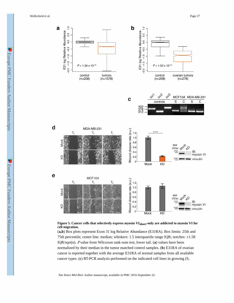
Figure 5. Cancer cells that selectively express myosin VIshort-only are addicted to myosin VI for cell migration.(a,b) Box plots represent Exon 31 log Relative Abundance (E31RA). Box limits: 25th and
75th percentile; center line: median; whiskers: 1.5 interquartile range IQR; notches: ±1.58
IQR/sqrt(n). P value from Wilcoxon rank-sum test, lower tail. (a) values have been
normalized by their median in the tumor matched control samples. (b) E31RA of ovarian
cancer is reported together with the average E31RA of normal samples from all available
cancer types. (c) RT-PCR analysis performed on the indicated cell lines in growing (S,
Wollscheid et al. Page 27
Nat Struct Mol Biol. Author manuscript; available in PMC 2016 September 22.
Europe PM
C Funders A
uthor Manuscripts
Europe PM
C Funders A
uthor Manuscripts

sparse) or confluent (C, confluent) condition. (d,e) Wound healing assay. The indicated cell
lines were KD for myosin VI or mock treated. Left panel, sample images: T0 first frame, T1
and T2 arbitrary points identical for control and KD of the same cell line. Scale bars, 200μm.
Central panel: quantification of the wound closure speed relative to control. Error bars, s.d.
(n=6 movies for MDA-MB-231, n=13 movies for MCF10A from three independent
experiments). **** P<0.0001; ns, no significant difference by two-tailed T-test. Right panel:
anti-myosin VI IB performed at T0.
Wollscheid et al. Page 28
Nat Struct Mol Biol. Author manuscript; available in PMC 2016 September 22.
Europe PM
C Funders A
uthor Manuscripts
Europe PM
C Funders A
uthor Manuscripts

Europe PM
C Funders A
uthor Manuscripts
Europe PM
C Funders A
uthor Manuscripts
Wollscheid et al. Page 29
Table 1List of the top hits of interactors identified by label-free quantitative mass spectrometry.
Gene name, description and total peptide count (TPC) is reported for each protein. *The same total peptide
count was reported for the control (GST alone), see also Supplementary Table 1.
Gene name Iso1 (TPC) Iso2 (TPC) Iso3 (TPC)
CLTC clathrin, heavy chain (CHC) 1050 *153 1068
SEC16A SEC16 homolog A (S. cerevisiae) 175 0 145
AP2B1 adaptor-related protein complex 2, beta 1 subunit 91 0 107
AP2A2 adaptor-related protein complex 2, alpha 2 subunit 87 0 89
AP2A1 adaptor-related protein complex 2, alpha 1 subunit 85 0 90
AP1G1 adaptor-related protein complex 1, gamma 1 subunit 28 0 32
AP2M1 AP-2 complex subunit mu 35 0 51
CLINT1 clathrin interactor 1 (EPSIN4) 138 0 146
PIK3C2A Class 2 phosphatidylinositol-4-phosphate 3-kinase 73 0 90
PICALM Phosphatidylinositol-binding clathrin assembly protein 15 0 19
EPS15 Epidermal growth factor receptor substrate 15 6 0 5
REPS RalBP1-associated Eps domain-containing protein 1 10 0 5
COPB2 Coatomer subunit beta' 45 50 57
DDB1 DNA damage-binding protein 1 137 251 123
EPRS Bifunctional glutamate, proline, tRNA ligase 206 686 183
VPRBP Vpr (HIV-1) binding protein (DCAF) 0 140 0
DCTN1 dynactin 1 0 105 0
GTPBP4 GTP binding protein 4 5 44 0
ARHGEF12 Rho guanine nucleotide exchange factor 12 6 64 0
YTHDC2 Probable ATP-dependent RNA helicase 4 37 2
DCTN2 Dynactin subunit 2 2 37 0
DOCK7 Dedicator of cytokinesis protein 7 2 32 4
Nat Struct Mol Biol. Author manuscript; available in PMC 2016 September 22.

Europe PM
C Funders A
uthor Manuscripts
Europe PM
C Funders A
uthor Manuscripts
Wollscheid et al. Page 30
Table 2Structural statistics for myosin VI1050–1131.
Protein
NMR distance and dihedral constraints
Distance constraints
Total NOE 1846
Intraresidue 483
Inter-residue
Sequential (|i – j| = 1) 537
Medium range (2 ≤ |i – j| ≤ 4) 566
Long range (|i – j| ≥ 5) 260
Intermolecular
Hydrogen bonds 42
Total dihedral-angle restraints
ϕ 51
ψ 51
Structure statistics
Violations (mean ± s.d.)
Distance constraints (Å) 0.0077±0.0010
Dihedral-angle constraints (°) 0.12±0.041
Max. dihedral-angle violation (°) 1.13
Max. distance-constraint violation (Å) 0.22
Deviations from idealized geometry
Bond lengths (Å) 0.0016±0.000073
Bond angles (°) 0.39±0.0042
Impropers (°) 0.23±0.0067
Average pairwise r.m.s. deviation (Å)a
Heavy 0.68±0.06
Backbone 0.18±0.05
aValues were calculated for residues T1054-R1068 and Y1084-S1126 from the twenty lowest energy structures.
Nat Struct Mol Biol. Author manuscript; available in PMC 2016 September 22.