Embed Size (px)
Citation preview

ORIGINAL ARTICLE
Diversity and antimicrobial resistance of Enterococcus fromthe Upper Oconee Watershed, GeorgiaS. Cho1, L.M. Hiott2, J.M. McDonald3, J.B. Barrett2, E.A. McMillan1, S.L. House2, E.S. Adams2,J.G. Frye2 and C.R. Jackson2
1 Department of Microbiology, University of Georgia, Athens, GA, USA
2 Bacterial Epidemiology and Antimicrobial Resistance Research Unit, USDA-ARS Russell Research Center, Athens, GA, USA
3 Lewis F. Rogers Institute for Environmental and Spatial Analysis, University of North Georgia, Oakwood, GA, USA
Keywords
antimicrobial resistance, diversity,
Enterococcus, prevalence, water.
Correspondence
Charlene R. Jackson, Richard B. Russell
Research Center, 950 College Station Road,
Athens, GA 30605, USA.
E-mail: [email protected]
J.G.F. and C.R.J. are joint senior authors on
this work.
2019/2072: received 26 March 2019, revised
15 November 2019 and accepted 3 Decem-
ber 2019
doi:10.1111/jam.14550
Abstract
Aim: It is well-known that enterococci are abundant in the environment;
however, the role of surface water as a reservoir of antimicrobial-resistant
enterococci remains largely undefined. In this study, surface water samples
were collected over a 2-year period from the Upper Oconee watershed, Athens,
GA to examine enterococci and their antimicrobial resistance.
Methods and Results: Approximately 97% (445/458) of the samples were
positive for enterococci and a total of 637 enterococci were isolated. The
predominant species were Enterococcus casseliflavus (33�6%) followed by
Enterococcus faecalis (26�5%) and Enterococcus hirae (13�2%). Regardless of
species, the highest levels of resistance were to lincomycin (88�5%) and
tetracycline (13%); isolates also exhibited resistance to newer antimicrobials,
daptomycin (8�9%) and tigecycline (6�4%). Multidrug resistance (resistance ≥3antimicrobial classes) was observed to as many as five classes of antimicrobials.
Resistant enterococci appeared to be randomly dispersed over the seasons
rather than clustered by species or antimicrobial resistance.
Conclusions: This study demonstrated that surface waters contain a large
population of diverse species of antimicrobial-resistant enterococci, including
resistance to new antimicrobials.
Significance and Impact of the Study: These results may indicate the potential
of human intestinal illness and/or colonization of the human gut with resistant
enterococci as enterococci correlate with increased disease risk to humans
during recreational exposure to water.
Introduction
Enterococci are a diverse group of bacteria with impor-
tance in several areas including clinical medicine, food-
borne illness, food processing and microbial risk
assessment. In clinical medicine, they are a leading cause
of nosocomial infections and have been implicated in
bacteraemia, endocarditis and urinary tract infections
(Murray 1990; Huycke et al. 1991; Jett et al. 1994). Treat-
ment of enterococcal infections in clinical medicine is
often complicated due to intrinsic and acquired resistance
in the bacteria (Murray 1990; Facklam et al. 2002; Malani
et al. 2002; Huijbers et al. 2015)). Enterococcal foodborne
illness is less severe causing symptoms such as vomiting
and headaches as a result of production of biogenic ami-
nes from ingestion of enterococci in fermented foods
(Tham et al. 1990; Gardin et al. 2001; Giraffa 2002). On
the other hand, enterococci have beneficial qualities that
have made them useful as probiotics and as indicators of
faecal contamination in water bodies (Foulquie Moreno
et al. 2006; Byappanahalli et al. 2012). As natural inhabi-
tants of the gastrointestinal tract of humans and animals,
enterococci have been used as indicators of faecal pollu-
tion along with Escherichia coli (Roslev and Bukh 2011).
Of the two bacteria, enterococci are thought to be better
indicators of the possible presence of bacterial pathogens
Journal of Applied Microbiology 128, 1221--1233 © 2019 The Society for Applied Microbiology 1221
Journal of Applied Microbiology ISSN 1364-5072

in surface water as their occurrence, particularly at high
levels, is associated with the risk of contracting human
gastroenteritis during swimming-related activities (Tur-
bow et al. 2003).
As enterococci are generally not considered important
pathogens outside the hospitals and healthcare settings,
studies reporting enterococci from the environment are
scarce. Most studies on water sources are focused either
on water quality, human-specific species, Enterococcus fae-
calis and E. faecium, or on polluted water environments
such as hospital-impacted wastewater and agricultural
run-off (Sadowy and Luczkiewicz 2014; Kapoor et al.
2015; Nishiyama et al. 2015). Less attention has been
given to enterococci in natural water environments and
on different species of Enterococcus. Resistant enterococci
present in water sources can potentially transfer resistance
genes to bacterial pathogens in the environment or to
bacteria in the human gastrointestinal tract after ingestion
of contaminated water during recreational activities
(Murray 1990; Facklam et al. 2002; Malani et al. 2002;
Huijbers et al. 2015). Since resistance in enterococci may
vary according to species, species identification is an
important factor to consider when characterizing antimi-
crobial resistance.
Bacterial diversity and prevalence in surface water are
subject to change due to both point and nonpoint (e.g.
agricultural run-off, storm water routes) sources as well
as seasonal influences such as water temperature and
rainfall (Singer et al. 2006; Lanthier et al. 2011). In
order to better understand human health risks associ-
ated with the presence of enterococci in surface water
impacted by nonpoint source contamination, studies are
needed to identify the differences in enterococcal species
composition, antimicrobial resistance of those species
and changes in the composition over time. In this
study, the occurrence, species distribution and antimi-
crobial susceptibility of enterococci from surface water
from the Upper Oconee watershed in northeastern
Georgia were evaluated. The Oconee River is formed by
the convergence of the North Oconee River and the
Middle Oconee River just south of Athens, Georgia, and
is used for municipal, industrial, agricultural and recre-
ational purposes (Environmental Protection Division
1998). Samples were collected quarterly, once each sea-
son, at different locations in the Upper Oconee water-
shed, for 2 years by the Upper Oconee Watershed
Network (UOWN) (http://www.uown.org). Enterococci
were isolated from the samples, identified to species and
tested for resistance to antimicrobials used in clinical
medicine and agriculture. The results of this study will
improve the understanding of the dynamics of entero-
cocci in a surface water system which may impact
human health.
Materials and methods
Sampling of river water
The rivers and streams sampled in this study were located
in the Upper Oconee watershed, Georgia, USA (USGS
Cataloging unit 03070101). The sampling sites were
located in the Middle Oconee River, North Oconee River
and the tributaries that drain into these rivers (Fig. 1).
Sample sites were selected to represent a range of land
uses, such as forested, agricultural, residential, recre-
ational and industrial. One litre water samples from each
site were collected seasonally four times a year with the
assistance of the UOWN. Since 2008, UOWN has assem-
bled a team of volunteers to collect water samples from
the Upper Oconee watershed on a quarterly basis. The
number of locations sampled varied from 80 to 100 dur-
ing the Spring collection, which is the UOWN’s largest
collection event, River Rendezvous. The annual River
Rendezvous event enlists over 100 volunteers to sample
approximately 100 locations in the watershed to evaluate
the health of the watershed through chemical and biolog-
ical sample analysis. During the remaining quarterly col-
lections, 30 to 60 water samples were collected depending
on available volunteers and access to the sampling sites.
A total of 29 locations were routinely sampled to serve as
controls with nine sites sampled all eight times, eight sites
sampled seven times and 12 sites sampled six times.
Isolation and identification of bacteria
Filtration and enrichment of water samples were per-
formed as previously described (Cho et al. 2018). Briefly,
0�5 g of cellulose filter powder (Aqua DewTM, Lahore,
Pakistan) was added to water samples (1 L), which was
then filtered onto 47-mm glass fibre filters (Pall Corpora-
tion, Ann Arbor, MI). The glass fibre filter and cellulose
filter powder were incubated in 25 ml of buffered pep-
tone (BP; BD DifcoTM, Franklin Lakes, NJ) water for 24 h
at 37°C. For Enterococcus isolation, 0�1 ml of BP enrich-
ment was streaked on selective agar plates and incubated
for 24 h at 37°C. In order to test for the efficacy of the
media, multiple media were used until Enterococcosel
(BD Difco) was chosen as the media of use from 2015
Fall onward. For 2015 Winter, Enterococcosel and mE
agar (BD Difco) were used; for 2015 Spring, mE agar was
used; for 2015 Summer, Enterococcosel, mE agar and
CHROMagar Enterococcus (not on market, CHROMagar
Microbiology, Paris, France) were used. One colony hav-
ing the typical appearance of Enterococcus from each pos-
itive plate was confirmed and its species was determined
using multiplex PCR as previously described (Jackson
et al. 2004). An exception was the Summer of 2015 when
Journal of Applied Microbiology 128, 1221--1233 © 2019 The Society for Applied Microbiology1222
Diversity of enterococci in water S. Cho et al.

multiple suspect colonies were selected per positive plate
and tested for a media comparison purpose. A variant of
Enterococcus casseliflavus was detected by PCR using the
same forward primer (CA1-E. casseliflavus) with a differ-
ent reverse primer 50-CGATTAAACGGTAGAAAGTGC-30
(designated CA3). The same PCR condition was used as
described by Jackson et al. (2004).
Antimicrobial susceptibility testing
Minimum inhibitory concentrations (MIC; µg ml�1) for
enterococci were determined by broth microdilution using
the SensititreTM semiautomated antimicrobial susceptibility
system (Trek Diagnostic Systems Inc., Cleveland, OH) and
the Sensititre Gram-Positive Custom Plate CMV3AGPF
according to the manufacturer’s directions. Results were
interpreted according to Clinical and Laboratory Standards
Institute (CLSI) guidelines when defined (CLSI 2018). No
CLSI interpretive criteria have been defined for kanamycin,
lincomycin and tylosin, and only susceptible breakpoints
have been established for daptomycin (≤4 µg ml�1) and
tigecycline (≤0�25 µg ml�1). Breakpoints for daptomycin,
kanamycin, lincomycin, tigecycline and tylosin were those
defined by the National Antimicrobial Resistance Monitor-
ing System (NARMS) (https://www.ars.usda.gov/ARSUse
rFiles/60400520/NARMS/ABXEntero.pdf). The panel of
antimicrobials and breakpoints for classification as resis-
tant were as follows: chloramphenicol (≥32 µg ml�1),
ciprofloxacin (≥4 µg ml�1), daptomycin (≥8 µg ml�1),
erythromycin (≥8 µg ml�1), gentamicin (≥500 µg ml�1),
kanamycin (≥1024 µg ml�1), lincomycin (≥8 µg ml�1),
linezolid (≥8 µg ml�1), nitrofurantoin (≥128 µg ml�1),
penicillin (≥16 µg ml�1), streptomycin (≥1000 µg ml�1),
Synercid (Quinupristin/Dalfopristin; Q/D) (≥4 µg ml�1),
tetracycline (≥16 µg ml�1), tigecycline (≥0�5 µg ml�1),
tylosin (≥32 µg ml�1) and vancomycin (≥32 µg ml�1).
TN
ALGA
FL
0 100 200 mi
SC
NCResistance
N
0 2·5 5 km
5 mi2·50
MDR
Non-MDR
Figure 1 Map of water sampling sites in the Upper Oconee Watershed near Athens, GA. Sampling sites where multidrug-resistant (MDR) Entero-
coccus were isolated are labelled and in red. Other sites, where non-MDR Enterococcus were isolated, are in blue. Inset map shows the area cov-
ered by the Upper Oconee Watershed in grey and Athens, GA as a black triangle. [Colour figure can be viewed at wileyonlinelibrary.com]
Journal of Applied Microbiology 128, 1221--1233 © 2019 The Society for Applied Microbiology 1223
S. Cho et al. Diversity of enterococci in water

Enterococcus faecalis ATCC 29212, Staphylococcus aureus
ATCC 29213, E. coli ATCC 25922 and Pseudomonas aerug-
inosa ATCC 27853 were quality controls for the determi-
nation of MIC.
Statistical methods
Data were analysed using either Microsoft Excel or
GraphPad. Student’s t-test and Pearson correlation coeffi-
cients were performed. P values of ≤0�05 were considered
significant.
Results
Prevalence and seasonal diversity
Over 450 samples were collected in the Upper Oconee
watershed (Fig. 1) during eight seasons in 2015 and 2016.
The number of sites sampled varied for each of the sea-
sons depending on several factors including the number
of volunteers, lack of rainfall resulting in a dry channel
and failure to obtain a sample, access to the sampling
sites and available resources. The number of sites sampled
ranged from 27 in Summer 2016 to 100 in Spring 2015
(Table 1). During the sampling period, enterococci were
detected in over 90% of the sites with the percent of pos-
itive sites ranging from 93 to 100%; in four seasons
(Summer 2015 and Winter, Spring and Summer 2016) all
sites were positive for the presence of enterococci.
At least 10 different enterococcal species were detected
during the sampling period (Table 2). Typically, more
than one colony morphology was present on each sample
plate, but only one colony was randomly selected for fur-
ther study. A higher number of isolates than the number
of sites was obtained during Winter and Summer of 2015
due to the use of multiple media. Additionally, more
than one Enterococcus was isolated per positive plate for
Table 1 Prevalence of Enterococcus among surface water sampling
sites
Sampling season
No. sampling
sites
No. positive
sampling sites (%)
2015
Winter 30 29 (96�7)Spring 100 93 (93)
Summer 33 33 (100)
Fall 59 58 (98�3)2016
Winter 41 41 (100)
Spring 87 87 (100)
Summer 27 27 (100)
Fall 81 77 (95�1)
Table
2Seasonal
distributionofen
terococcifrom
water
samples
Season
No.ofisolates(%
)
E.avium
(n=3)
E.casseliflavus
(n=214)
E.casseliflavusvar.
(n=71)
E.durans
(n=1)
E.faecalis
(n=169)
E.faecium
(n=33)
E.gallinarum
(n=50)
E.hirae
(n=84)
E.mundtii
(n=8)
E.pallens
(n=1)
E.species
(n=3)
2015
Winter(n
=58)
1(1�7)
11(19)
0(0)
0(0)
13(22�4)
12(20�7)
5(8�6)
15(25�9)
1(1�7)
0(0)
0(0)
Spring(n
=93)
0(0)
61(65�5)
11(11�8)
0(0)
13(14)
0(0)
7(7�5)
1(1�2)
0(0)
0(0)
0(0)
Summer
(n=196)
1(0�5)
40(20�4)
0(0)
0(0)
77(39�3)
14(7�1)
26(13�3)
32(16�3)
3(1�5)
1(0�5)
2(1)
Fall(n
=58)
0(0)
25(43�1)
27(46� 6)
0(0)
1(1�7)
1(1�7)
3(5�2)
1(1�7)
0(0)
0(0)
0(0)
2016
Winter(n
=41)
0(0)
5(12�2)
12(29�3)
1(2�4)
4(9�8)
5(12�2)
0(0)
14(34�1)
0(0)
0(0)
0(0)
Spring(n
=87)
0(0)
31(35�6)
10(11�5)
0(0)
28(32�2)
1(1�1)
6(6�9)
11(12�6)
0(0)
0(0)
0(0)
Summer
(n=27)
0(0)
7(25�9)
8(29�6)
0(0)
12(44�4)
0(0)
0(0)
0(0)
0(0)
0(0)
0(0)
Fall(n
=77)
1(1�3)
34(44�2)
3(3�9)
0(0)
21(27�3)
0(0)
3(3�9)
10(13)
4(5�2)
0(0)
1(1�3)
Journal of Applied Microbiology 128, 1221--1233 © 2019 The Society for Applied Microbiology1224
Diversity of enterococci in water S. Cho et al.
Summer of 2015 as different colony morphologies were
selected for species identification. This was done in order
to test the efficacy of different media for Enterococcus iso-
lation, the comparison results of which will be discussed
elsewhere. Accordingly, although 33 sampling sites were
positive for enterococci in Summer 2015, 196 enterococci
were selected and confirmed as enterococci. Notably,
Summer 2015 was the season with the most diverse ente-
rococcal species isolated and the only season in which E.
pallens was identified; it was also one of only two seasons
in which unidentified enterococci were isolated (Table 2).
In contrast to Summer 2015, Summer 2016 had the few-
est number of species identified (n = 3) although all 27
sites sampled were positive for enterococci. Not surpris-
ingly, the season with the highest number of isolates
(Summer 2015; n = 196) and the lowest number (Sum-
mer 2016; n = 27) had the highest and lowest diversity of
species, eight and three different species respectively. A
significantly positive correlation was observed between
the number of samples obtained in each season and the
number of different species obtained (r = 0�585, P < 0�5),but the moderate correlation suggests that other factors
may contribute to the diversity of Enterococcus in surface
water. On average, six different species were identified
each sampling period.
Prevalence and seasonal occurrence of Enterococcus iso-
lates appeared to relate; as the number of isolates
increased, so did the number of seasons in which they
were observed (r = 0�870, P < 0�05). For example the
most prevalent species isolated was E. casseliflavus
(n = 214) and E. faecalis (n = 169) (Table 2). These two
species were also the only ones isolated from all eight
sampling events. Fewer E. hirae (n = 84), E. casseliflavus
variant (n = 71) and E. gallinarum (n = 50) were isolated
and were distributed over fewer sampling seasons (seven
seasons for E. hirae and six seasons each for E. cas-
seliflavus variant and E. gallinarum). Only 33 E. faecium
were isolated in five of the eight sampling events
(Table 2). Although E. avium (n = 3) and E. mundtii
(n = 8) were low in numbers, all isolates of these species
were seen in the same three sampling seasons (Winter
and Summer 2015 and Fall 2016).
Antimicrobial resistance
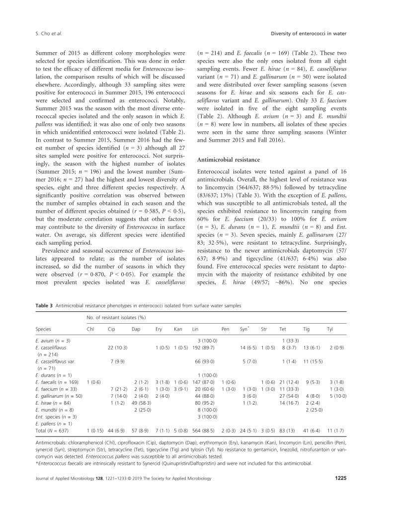
Enterococcal isolates were tested against a panel of 16
antimicrobials. Overall, the highest level of resistance was
to lincomycin (564/637; 88�5%) followed by tetracycline
(83/637; 13%) (Table 3). With the exception of E. pallens,
which was susceptible to all antimicrobials tested, all the
species exhibited resistance to lincomycin ranging from
60% for E. faecium (20/33) to 100% for E. avium
(n = 3), E. durans (n = 1), E. mundtii (n = 8) and Ent.
species (n = 3). Seven species, mainly E. gallinarum (27/
83; 32�5%), were resistant to tetracycline. Surprisingly,
resistance to the newer antimicrobials daptomycin (57/
637; 8�9%) and tigecycline (41/637; 6�4%) was also
found. Five enterococcal species were resistant to dapto-
mycin with the majority of resistance exhibited by one
species, E. hirae (49/57; ~86%). No one species
Table 3 Antimicrobial resistance phenotypes in enterococci isolated from surface water samples
Species
No. of resistant isolates (%)
Chl Cip Dap Ery Kan Lin Pen Syn* Str Tet Tig Tyl
E. avium (n = 3) 3 (100�0) 1 (33�3)E. casseliflavus
(n = 214)
22 (10�3) 1 (0�5) 1 (0�5) 192 (89�7) 14 (6�5) 1 (0�5) 8 (3�7) 13 (6�1) 2 (0�9)
E. casseliflavus var.
(n = 71)
7 (9�9) 66 (93�0) 5 (7�0) 1 (1�4) 11 (15�5)
E. durans (n = 1) 1 (100�0)E. faecalis (n = 169) 1 (0�6) 2 (1�2) 3 (1�8) 1 (0�6) 147 (87�0) 1 (0�6) 1 (0�6) 21 (12�4) 9 (5�3) 3 (1�8)E. faecium (n = 33) 7 (21�2) 2 (6�1) 1 (3�0) 3 (9�1) 20 (60�6) 1 (3�0) 1 (3�0) 1 (3�0) 11 (33�3) 1 (3�0)E. gallinarum (n = 50) 7 (14�0) 2 (4�0) 2 (4�0) 44 (88�0) 3 (6�0) 27 (54�0) 4 (8�0) 5 (10�0)E. hirae (n = 84) 1 (1�2) 49 (58�3) 80 (95�2) 1 (1�2) 14 (16�7) 2 (2�4)E. mundtii (n = 8) 2 (25�0) 8 (100�0) 2 (25�0)Ent. species (n = 3) 3 (100�0)E. pallens (n = 1)
Total (N = 637) 1 (0�15) 44 (6�9) 57 (8�9) 7 (1�1) 5 (0�8) 564 (88�5) 2 (0�3) 24 (5�1) 3 (0�5) 83 (13) 41 (6�4) 11 (1�7)
Antimicrobials: chloramphenicol (Chl), ciprofloxacin (Cip), daptomycin (Dap), erythromycin (Ery), kanamycin (Kan), lincomycin (Lin), penicillin (Pen),
synercid (Syn), streptomycin (Str), tetracycline (Tet), tigecycline (Tig) and tylosin (Tyl). No resistance to gentamicin, linezolid, nitrofurantoin or van-
comycin was detected. Enterococcus pallens was susceptible to all antimicrobials tested.
*Enterococcus faecalis are intrinsically resistant to Synercid (Quinupristin/Dalfopristin) and were not included for this antimicrobial.
Journal of Applied Microbiology 128, 1221--1233 © 2019 The Society for Applied Microbiology 1225
S. Cho et al. Diversity of enterococci in water

contributed primarily to tigecycline resistance as six dif-
ferent species were resistant at levels of <25% each. Low
resistance was observed for two of the aminoglycosides,
kanamycin (5/637; 0�8%) and streptomycin (3/637;
0�5%), whereas none of the isolates were resistant to gen-
tamicin (Table 3). The same three species (E. casseliflavus,
E. faecalis and E. faecium) were resistant to both kanamy-
cin and streptomycin although two additional E. faecium
were resistant to kanamycin, but not to streptomycin.
The same five species of isolates (E. casseliflavus, E. fae-
calis, E. faecium and E. gallinarum) were resistant at simi-
lar levels for the macrolides, erythromycin and tylosin,
1�1% (7/637) and 1�7% (11/637) respectively. In addition
to gentamicin, none of the isolates were resistant to line-
zolid, nitrofurantoin or vancomycin. Statistical analysis
revealed a significant correlation in the distribution of
certain AR among different Enterococcus species. Very
strong positive correlations were observed between dapto-
mycin and E. hirae as well as tetracycline and E. galli-
narum, E. faecalis and E. faecium (P < 0�05).For some species, higher numbers of isolates resulted
in resistance to a greater number of antimicrobials. E.
casseliflavus was the predominant species isolated and was
resistant to nine antimicrobials, whereas E. faecalis, with
the second highest number of isolates, was resistant to 10
of 16 antimicrobials tested including the only resistance
detected to chloramphenicol (Table 3). However, 33 E.
faecium isolates were also resistant to 10 antimicrobials;
21% (7/33) of those isolates were resistant to ciprofloxa-
cin, whereas none of the E. faecalis were ciprofloxacin
resistant. Compared to E. casseliflavus, the E. casseliflavus
variants were resistant to only five antimicrobials (cipro-
floxacin, lincomycin, Q/D, tetracycline and tigecycline),
four fewer than E. casseliflavus. The differences between
the two groups were primarily due to susceptibility to the
aminoglycoside and macrolide classes for the E. cas-
seliflavus variant. Other enterococcal species with resistant
isolates distributed over a range of antimicrobials
included E. gallinarum (n = 50) and E. hirae (n = 84)
exhibiting resistance to eight and six antimicrobials
respectively (Table 3).
Multidrug resistance (MDR), defined as resistance to
three or more antimicrobial classes and resistance to mul-
tiple antimicrobials is shown in Table 4. Isolates were
resistant to three to six different antimicrobials and up to
five antimicrobial classes. One isolate, an E. faecalis, was
resistant to six antimicrobials (ChlDapEryLinTetTyl) and
five antimicrobial classes. Eighteen different patterns were
observed among 51 MDR enterococcal isolates. The most
common MDR group by drug class was the three drug
combination which was composed of 11 different resis-
tance patterns (Table 4), whereas the fewest different pat-
terns (n = 1) was for the six drug combination. Due to
the prevalence of resistance to lincomycin in the isolates,
lincomycin was found in all resistance patterns with the
exception of CipDapTet which was represented by E. fae-
cium (Table 4).
Analysis of distribution of resistant enterococci from
the sampling events did not reveal the differences in spe-
cies, the clustering of species or the antimicrobial resis-
tance over the seasons. Only two sampling events (Fall
2015 and Spring 2016) had identical groups of species (E.
casseliflavus, E. casseliflavus variant, E. faecalis, E. faecium,
E. gallinarum and E. hirae), but those isolates were resis-
tant to different antimicrobials (Table 5). Resistance to
the fewest number of antimicrobials (n = 3) also
occurred during Summer 2016 with the lowest number
Table 4 Multidrug resistance patterns in enterococci from surface
water
Pattern*No.
resistances
No.
classes
No.
isolates Species (no.)
CipDapTet 3 3 2 E. faecium (2)
CipLinSyn 3 3 5 E. casseliflavus (4)
E. hirae (1)
CipLinTet 3 3 8 E. casseliflavus (1)
E. faecium (3)
E. gallinarum (4)
CipLinTig 3 3 5 E. casseliflavus (1)
E. casseliflavus
var. (4)
DapLinTet 3 3 6 E. hirae (6)
DapLinTig 3 3 2 E. hirae (1)
E. mundtii (1)
KanLinTet 3 3 1 E. faecalis (1)
LinPenTet 3 3 1 E. faecalis (1)
LinStrTet 3 3 2 E. faecalis (1)
E. faecium (1)
LinSynTig 3 3 5 E. casseliflavus
var. (3)
E. gallinarum (2)
LinTetTig 3 3 2 E. faecalis (2)
CipLinSynTig 4 4 1 E. casseliflavus (1)
CipLinTetTig 4 4 3 E. casseliflavus (1)
E. gallinarum (2)
EryLinTetTyl 4 3 4 E. casseliflavus (1)
E. faecalis (2),
E. gallinarum (1)
CipEryLinTetTyl 5 4 1 E. faecium (1)
EryLinSynTetTyl 5 4 1 E. gallinarum (1)
KanLinStrTetTyl 5 4 1 E. casseliflavus (1)
ChlDapEryLinTetTyl 6 5 1 E. faecalis (1)
Antimicrobials: chloramphenicol (Chl), ciprofloxacin (Cip), daptomycin
(Dap), erythromycin (Ery), kanamycin (Kan), lincomycin (Lin), Penicillin
(Pen), Synercid (Syn), streptomycin (Str), tetracycline (Tet), tigecycline
(Tig) and tylosin (Tyl).
*Enterococcus faecalis are intrinsically resistant to Synercid (Quin-
upristin/Dalfopristin) and were not included for this antimicrobial.
Journal of Applied Microbiology 128, 1221--1233 © 2019 The Society for Applied Microbiology1226
Diversity of enterococci in water S. Cho et al.

Table
5Seasonal
distributionofan
timicrobialresistan
cephen
otypes
inen
terococciisolatedfrom
surfacewater
samples
Season(sam
ple
no.)
Species
No.ofresistan
tisolates(%
)*
Chl
Cip
Dap
Ery
Kan
Lin
Pen
Syn†
Str
Tet
Tig
Tyl
2015
Winter(n
=58)
E.avium
(n=1)
1(100�0)
Enterococcuscasseliflavus(n
=11)
4(36�4)
11(100�0)
5(45�5)
E.faecalis(n
=13)
1(7�7)
13(100�0)
3(23�1)
E.faecium
(n=12)
6(50�0)
2(16�7)
1(8�3)
1(8�3)
7(58�3)
1(8�3)
1(8�3)
6(50�0)
1(8�3)
E.gallinarum
(n=5)
1(20�0)
5(100�0)
1(20�0)
3(60�0)
E.hirae
(n=15)
11(73�3)
15(100�0)
1(6�7)
E.mundtii(n
=1)
Spring(n
=93)
E.casseliflavus(n
=61)
3(4�9)
1(1�6)
59(96�7)
4(6�6)
1(1�6)
2(3�3)
4(6�6)
1(1�6)
E.casseliflavusvar.(n
=11)
10(90�9)
2(18�2)
E.faecalis(n
=13)
1(7�7)
1(7�7)
1(7�7)
12(92�3)
3(23�1)
2(15�4)
1(7�7)
E.gallinarum
(n=7)
1(14�3)
7(100�0)
3(42�9)
E.hirae
(n=1)
1(100�0)
Summer
(n=196)
E.avium
(n=1)
1(100�0)
E.casseliflavus(n
=40)
1(2�5)
24(60�0)
1(2�5)
E.faecalis(n
=77)
2(2�6)
72(93�5)
1(1�3)
8(10�4)
2(2�6)
E.faecium
(n=14)
2(14�3)
8(57�1)
1(7�1)
2(14�3)
E.gallinarum
(n=26)
2(7�7)
2(7�7)
2(7�7)
26(100�0)
1(3�8)
19(73�1)
2(7�7)
E.hirae
(n=32)
22(68�8)
31(96�9)
5(15�6)
E.mundtii(n
=3)
3(100�0)
†
Entspecies(n
=2)
2(100�0)
Fall(n
=58)
E.casseliflavus(n
=25)
4(16�0)
24(96)
2(8�0)
5(20�0)
E.casseliflavusvar.(n
=27)
6(22�2)
27(100�0)
3(11�1)
1(3�7)
11(40�7)
E.faecalis(n
=1)
E.faecium
(n=1)
E.gallinarum
(n=3)
3(100�0)
2(66�7)
2(66�7)
E.hirae
(n=1)
1(100)
2016
Winter(n
=41)
E.casseliflavus(n
=5)
5(100�0)
E.casseliflavusvar.(n
=12)
8(66�7)
E.durans(n
=1)
1(100�0)
E.faecalis(n
=4)
4(100�0)
1(25�0)
1(25�0)
E.faecium
(n=5)
1(20�0)
4(80�0)
2(40�0)
E.hirae
(n=14)
7(50�0)
12(85�7)
3(21�4)
(Continued
)
Journal of Applied Microbiology 128, 1221--1233 © 2019 The Society for Applied Microbiology 1227
S. Cho et al. Diversity of enterococci in water

Table
5(Continued
)
Season(sam
ple
no.)
Species
No.ofresistan
tisolates(%
)*
Chl
Cip
Dap
Ery
Kan
Lin
Pen
Syn†
Str
Tet
Tig
Tyl
Spring(n
=87)
E.casseliflavus(n
=31)
5(16�1)
1(3�2)
31(100�0)
2(6�5)
2(6�5)
1(3�2)
E.casseliflavusvar.(n
=10)
1(10�0)
10(100�0)
E.faecalis(n
=28)
27(96�4)
1(3�6)
E.faecium
(n=1)
1(100�0)
1(100�0)
E.gallinarum
(n=6)
1(16�7)
1(16�7)
E.hirae
(n=11)
4(36�4)
10(90�9)
3(27�3)
Summer
(n=27)
E.casseliflavus(n
=7)
2(28�6)
7(100�0)
1(14�3)
E.casseliflavusvar.(n
=8)
8(100�0)
E.faecalis(n
=12)
1(8�3)
Fall(n
=77)
E.avium
(n=1)
1(100�0)
1(100� 0)
E.casseliflavus(n
=34)
3(8�8)
31(91�2)
3(8�8)
4(11�8)
E.casseliflavusvar.(n
=3)
3(100�0)
E.faecalis(n
=21)
1(4�8)
19(90�5)
4(19�0)
7(33�3)
E.gallinarum
(n=3)
2(66�7)
3(100�0)
3(100�0)
2(66�7)
E.hirae
(n=10)
1(10�0)
5(50�0)
10(100�0)
1(10�0)
2(20�0)
2(20�0)
E.mundtii(n
=4)
2(50�0)
4(100�0)
2(50�0)
Entspecies(n
=1)
1(100�0)
Total(N
=637)
144
57
75
564
224
383
41
11
Antimicrobials:Chloramphen
icol(Chl),
Ciprofloxacin(Cip),Dap
tomycin
(Dap
),Erythromycin
(Ery),Kan
amycin
(Kan
),Lincomycin
(Lin),Penicillin
(Pen
),Synercid(Syn),Streptomycin
(Str),Tetracycline
(Tet),Tigecycline(Tig)an
dTylosin(Tyl).Noresistan
ceto
gen
timicin,linezolid,nitrofurantoin
orvancomycin
was
detected.
*Percen
tagebyspecies.
†E.
faecalisareintrinsically
resistan
tto
Synercid(Quinupristin/Dalfopristin)an
dwerenotincluded
forthisan
timicrobial.
Journal of Applied Microbiology 128, 1221--1233 © 2019 The Society for Applied Microbiology1228
Diversity of enterococci in water S. Cho et al.

of isolates. Spring 2015, with lower number of isolates
analysed (n = 93) than Summer 2015, during which the
highest number of isolates were obtained, had a low
diversity of species (n = 4) but the most resistance
detected (i.e. resistance to the highest number of antimi-
crobials; n = 11) (Table 5). Like the prevalence of lin-
comycin resistance among MDR patterns, lincomycin
resistance was detected in enterococci isolated during all
seasons in both 2015 and 2016. In contrast, although
lower numbers of isolates resistant to ciprofloxacin
(n = 44) and tetracycline (n = 83) were detected when
compared to lincomycin (n = 564), resistance to both
ciprofloxacin and tetracycline was found in all seasons as
well.
Discussion
The Upper Oconee watershed, located in the Piedmont
region of northeast Georgia, is one of 14 river basins in
the state (Meyer and Loeffler 2018). The headwaters of
the river originate in an area dominated by forest, but
forested areas decrease as areas of agriculture increase
along the river. Agricultural use of the land has greatly
affected the quality of the water within the watershed.
Additionally, the watershed has also been impacted by
other factors including pollutants from urban run-off,
municipal sources, storm and combined sewer systems.
As a mixed-use watershed that is utilized for numerous
purposes including recreational activity as well as for
municipal, commercial and industrial purposes, assess-
ment of the condition of the watershed is important. In
2005, the Upper Oconee watershed was surveyed to
obtain a general description (prevalence, antimicrobial
resistance) of the bacterial composition focusing primar-
ily on enteric bacteria, including Enterococcus (Meiners-
mann et al. 2008). Although results from the study
provided much needed information, no data were gener-
ated on possible seasonal changes in the occurrence and
diversity of Enterococcus in the watershed. This study
aimed to provide a description of prevalence, species dis-
tribution and antimicrobial resistance profile of entero-
cocci over different seasons and years to more clearly
assess this population of bacteria in the Upper Oconee
watershed, which may indicate potentially negative
impact on the communities utilizing the water.
As enterococci are ubiquitous in the environment, the
high number of positive sampling sites across seasons was
not unexpected. Concentrations of enterococci were not
recorded in this study, but peak numbers of enterococci
have been reported to be frequently detected in late Win-
ter and early Spring in coastal waters (Turbow et al.
2003). In this study, no significant difference was
observed in the number of Enterococcus-positive sites
based on season (P > 0�05), with all seasons exhibiting a
high prevalence of enterococci. The lowest percent posi-
tive (93%) was higher than that from the 2005 study in
which 85�5% (71/83) sites tested were positive for entero-
cocci using the same isolation method (Meinersmann
et al. 2008). While the two studies of the Upper Oconee
watershed occurred 10 years apart, the average tempera-
ture varied very little between those two sampling months
with 60°F and 65�1°F recorded in April 2005 and April
2015 respectively (https://www.usclimatedata.com/clima
te/athens/georgia/united-states/usga0027/2016/11). How-
ever, the differences in precipitation may have affected
the detected enterococcal population. April 2005 had sig-
nificantly less total precipitation recorded (0�22 inches)
than April 2015 (8�01 inches) or the lowest amount of
any sampling month of this study (2�24 inches recorded
for November, 2016).
Previous studies have shown that surface water con-
tains a wide variety of enterococcal species (Svec and
Sedlacek 1999; Svec et al. 2001; Niemi et al. 2012). The
spectrum of species isolated may be influenced by the
surrounding environment or human/animal contact with
the water as many sources of enterococci exist. Com-
monly isolated enterococcal species include E. faecalis and
E. faecium as well as E. casseliflavus, E. durans, E. galli-
narum, E. hirae and E. mundtii. Of the 10 species of ente-
rococci identified in this study, E. casseliflavus was the
predominant species paralleling the results from the pre-
vious study of enterococci in the Upper Oconee water-
shed (Meinersmann et al. 2008). A variant of E.
casseliflavus was also identified. Sequencing of the sodA
gene has shown that the variant had the same nucleotide
changes in the superoxide dismutase gene (C377T,
T381C, C384T and T390G) as an Enterococcus isolate
obtained from a park in Athens, GA from a previous
study at the USDA-ARS conducted in 2001 (P. J.
Fedorka-Cray, C.R. Jackso, & S.L. House, unpublished
data), indicating the prevalence and persistence of this
particular E. casseliflavus variant in the environment in
this area. The detection of high numbers of E. cas-
seliflavus in this surface water is similar to findings in
some studies but differs from most other studies, as
either E. faecalis or E. faecium was reported as the domi-
nant species from surface or other environmental waters
(Pinto et al. 1999; Moore et al. 2008; Lata et al. 2009;
Luczkiewicz et al. 2010; Lanthier et al. 2011; de et al.
2013). This difference in species distribution could be
due to a difference in the isolation method as studies
have shown that difference in media and incubation tem-
peratures could select for certain Enterococcus species
(Jackson et al. 2005; Ferguson et al. 2013). The difference
in species distribution could also be the result of the
regional difference as Moore et al. (2008) detected
Journal of Applied Microbiology 128, 1221--1233 © 2019 The Society for Applied Microbiology 1229
S. Cho et al. Diversity of enterococci in water

different Enterococcus species distribution at separate loca-
tions based on the structure of the waterbody and the
presence of potential contamination sources. E. faecalis
and E. faecium have been documented to be more resis-
tant to stress caused by environmental conditions and
thus may survive longer in harsh environments, such as
water, where nutrients are scarce (Leclerc et al. 1996).
Definitive contributing sources of Enterococcus in the
environment are still unresolved. For example some stud-
ies attribute the presence of E. faecalis to nonhuman
sources (livestock, poultry, wildlife) (Lanthier et al.
2011), whereas other reports point to clinical specimens,
hospital patients and hospital sewage (Kuhn et al. 2003).
Contamination of water with E. faecium usually suggests
human or wastewater sources (Niemi et al. 1993; Lanthier
et al. 2011). Both of these scenarios are plausible, as E.
faecalis has historically accounted for more human infec-
tions in the community and hospital settings, whereas E.
faecium is associated with increased healthcare-acquired
infections (Hidron et al. 2008). On the other hand, E.
casseliflavus has been primarily associated with plants
and, to a lesser degree, as an animal-derived or food-as-
sociated species (Luczkiewicz et al. 2010).
The clinical importance of E. faecalis and E. faecium is
well-known; however, other enterococcal species can
cause disease in humans, including E. casseliflavus. E. cas-
seliflavus has been known to cause invasive infection in
humans, but the true clinical significance of this species
has not been fully determined (Reid et al. 2001). One
other enterococcal species identified in this study was E.
pallens. This species has not been previously found in
surface water but has primarily been described as a cause
of peritonitis in humans (Tyrrell et al. 2002; Levesque
et al. 2016). The significance of detecting this species in
surface water remains unknown, but could be a possible
source for human infections.
Antimicrobial resistance in enterococci from surface
water varies with the geographical area under study and
is most likely due to the type of polluting source the
water is receiving as antimicrobial use in humans and
animals differ. Resistance to aminoglycosides, b-lactams,
fluoroquinolones, lincosamides, macrolides, phenicols
and tetracycline has been described with erythromycin,
lincomycin and tetracycline resistance observed most
often (Moore et al. 2008; Servais and Passerat 2009; Lucz-
kiewicz et al. 2010; Lanthier et al. 2011; de et al. 2013).
Those same patterns of resistance were seen in this study
with a vast majority of isolates displaying resistance to
lincomycin; at least one of every species exhibited resis-
tance to this drug. None of the isolates were resistant to
vancomycin, although other studies have detected van-
comycin resistance in enterococci from surface water
(Pinto et al. 1999; Luczkiewicz et al. 2010; Lanthier et al.
2011; Santiago-Rodriguez et al. 2013; Molale and
Bezuidenhout 2016). Schwartz et al. (2003) identified
vancomycin-resistant E. faecium with an identified van-
comycin resistance gene, but only from hospital wastewa-
ter biofilms. Enterococcus casseliflavus and E. gallinarum
are known to be intrinsically resistant to vancomycin as
they carry vanC genes; however, their level of resistance is
lower than the CLSI breakpoint of 32 µg ml�1and not
considered clinically relevant (Gold 2001). Because the
NARMS susceptibility panel was used to phenotype the
isolates, estimation of resistance to the newer drugs (dap-
tomycin and tigecycline) used to treat human infections
was possible. Neither those drugs nor their analogs are
used in food animal production in the US suggesting that
those isolates originated from a human source. Alterna-
tively, those isolates could also either be intrinsically
resistant, especially as E. hirae is highly associated with
resistance to daptomycin, or have acquired resistance to
the antimicrobials. Nevertheless, resistance in environ-
mental isolates to drugs used in human medicine is a
cause for concern due to the potential for horizontal
transfer of genes responsible for these resistances to clini-
cal isolates.
The high number of MDR profiles detected in this
study was notable; 18 different MDR profiles were
observed for the enterococci with resistance to a maxi-
mum of five classes of antimicrobials. Direct comparison
of MDR pattern prevalence between studies may be diffi-
cult due to the differences in antimicrobials and antimi-
crobial classes tested as well as the susceptibility testing
method used (Servais and Passerat 2009). However, a
high number of MDR profiles was also reported in a
study by Carvalho et al. (2014) in which 24 MDR profiles
with resistance to six classes of antimicrobials in entero-
cocci from a marine outfall in Brazil were seen. Resis-
tance genes carried on plasmids accounted for some of
the MDR phenotypes in that study. Although analysis of
plasmids was not performed in this study, MDR profiles
in isolates from this study could also be due to the pres-
ence of mobile genetic elements harbouring antimicrobial
resistance genes.
A total of 51 MDR Enterococcus isolates were isolated
from 35 sampling sites. The site locations where MDR
enterococci were recovered are indicated in Fig. 1 and the
exact locations of the sites, along with the GPS coordi-
nates, are in Table S1. Interestingly, a complimentary
study conducted on the same sampling sites showed that
Enteropathogenic E. coli and AR E. coli isolates were
recovered from 22 of the 35 MDR Enterococcus-positive
sites (Cho et al. 2018). This finding shows that there are
sites within the Upper Oconee watershed that contribute
towards the spread of pathogenic and AR bacteria. Two
or more MDR isolates were recovered from 14 of 35 sites,
Journal of Applied Microbiology 128, 1221--1233 © 2019 The Society for Applied Microbiology1230
Diversity of enterococci in water S. Cho et al.

most of which were residential areas, just as AR E. coli
were mostly isolated from sites draining residential areas
(Cho et al. 2018). Additionally, McNutt Creek, a creek
previously identified as a source of AR E. coli, served as
the source of six MDR Enterococcus isolates at three dis-
tinct locations along the creek.
Seasonal effects on species distribution and antimicro-
bial resistance have been examined previously (Lanthier
et al. 2011). In that study, significant differences in species
were found between seasons with E. faecalis having a more
dominant presence in Summer and Fall, whereas E. fae-
cium was more frequently identified in Winter and Spring.
That study also found a higher proportion of antimicrobial
susceptible isolates in Fall compared to Winter and Spring,
but not to Summer. Data from this study differed from
that of Lanthier et al. as E. faecalis seemed to be predomi-
nant in Summer and E. faecium in Winter; however, none
of these differences were significant (P > 0�05). On the
other hand, the frequency of E. casseliflavus was signifi-
cantly higher during Fall than during Winter and Summer
(P < 0�05). Unknown environmental variations between
studies may exist, rendering significant assumptions about
species diversity and antimicrobial resistance very difficult
to interpret. Additional collections over time need to be
done in order to provide statistically sound data for sea-
sonal diversity and antimicrobial resistance of enterococci
from surface water.
For this study, one typical, well-isolated colony was
randomly selected per positive plate, according to
NARMS methodology (https://www.fda.gov/animal-vete
rinary/national-antimicrobial-resistance-monitoring-syste
m/resources), in order to represent the enterococcal spe-
cies distribution of the watershed as a whole and not of
individual water samples. Furthermore, different selective
media were used for the first few samplings for Enterococ-
cus isolation in order to test for the efficacy of the media
used (S. Cho, L.M. Hiott, T.A. Woodley, H. Ramadan,
J.G. Frye, & C.R. Jackson, manuscript in preparation). As
the purpose of this study was to examine the different
enterococcal species composition of the watershed and
their antimicrobial resistance, isolates recovered from all
the media were included in this study.
This study has demonstrated that surface waters of the
Upper Oconee watershed contain a high population of
diverse species of enterococci. The detection of antimi-
crobial resistance and MDR in the isolates, especially to
newer drugs used to treat human infections, was concern-
ing and may have resulted from human impacts in the
area, including excretion of antibiotics, as these antibi-
otics are only used in human medicine and not in food
animals. Resistant enterococcal species were consistently
isolated over the sampling period indicating that the
results were not a transient observation, but that those
isolates maintain a presence in surface water of the Upper
Oconee watershed. As some of the resistance phenotypes
were present in species of enterococci implicated as lead-
ing causes of nosocomial infections, those isolates may be
an additional source of contact for humans during recre-
ational and other surface water use. Colonization of the
human gut with resistant enterococci could limit thera-
peutic options during human infections posing a risk to
human health.
Acknowledgements
The authors gratefully acknowledge the volunteers of the
Upper Oconee Watershed Network for their assistance in
collecting the water samples. This work was supported by
the U.S. Department of Agriculture (6040-32000-009-00-
D); and the Centers for Disease Control and Prevention
(Broad Agency Announcement to address antibiotic resis-
tance, Agricultural Research Service Sub-Project Number:
6040-32000-009-08-R). The funders had no role in study
design, data collection and interpretation or the decision
to submit the work for publication.
Conflict of Interest
No conflict of interest declared.
Disclaimer
The mention of trade names or commercial products in
this manuscript is solely for the purpose of providing
specific information and does not imply recommendation
or endorsement by the U.S. Department of Agriculture.
USDA is an equal opportunity provider and employer.
References
Byappanahalli, M.N., Nevers, M.B., Korajkic, A., Staley, Z.R.
and Harwood, V.J. (2012) Enterococci in the
environment. Microbiol Mol Biol Rev 76, 685–706.Carvalho, E.M.R., Costa, R.A., Araujo, A.J.G., Carvalho,
F.C.T., Pereira, S.P., Sousa, O.V. and Vieira, R.H.S.F.
(2014) Multiple antibiotic-resistance of Enterococcus
isolated from coastal water near an outfall in Brazil.
African J Microbiol 8, 1825–1831.Cho, S., Hiott, L.M., Barrett, J.B., McMillan, E.A., House, S.L.,
Humayoun, S.B., Adams, E.S., Jackson, C.R. et al. (2018)
Prevalence and characterization of Escherichia coli isolated
from the Upper Oconee Watershed in Northeast Georgia.
PLoS ONE 13, e0197005.
CLSI (2018) Performance Standards for Antimicrobial
Susceptibility Testing. Wayne, PA: Clinical and Laboratory
Standards Institute.
Journal of Applied Microbiology 128, 1221--1233 © 2019 The Society for Applied Microbiology 1231
S. Cho et al. Diversity of enterococci in water

De Niederh€ausern, S., Bondi, M., Anacarso, I., Iseppi, R.,
Sabia, C., Bitonte, F. and Messi, P. (2013) Antibiotics and
heavy metals resistance and other biological characters in
enterococci isolated from surface water of Monte Cotugno
Lake (Italy). J Environ Sci Health A Tox Hazard Subst
Environ Eng 48, 939–946.Environmental Protection Division (1998) Oconee River Basin
Management Plan. Atlanta, GA: Environmental Protection
Division.
Facklam, R.R., Carvalho, M.G.S. and Teixeiram, L.M. (2002)
History, taxonomy, biochemical characteristics, and
antibiotic susceptibility testing of enterococci. In The
Enterococci: Pathogenesis, Molecular Biology, and Antibiotic
Resistance ed. Gilmore, M.S., Clewell, D.B., Courvalin, P.,
Dunny, G.M., Murray, B.E. and Rice, L.B. pp. 1–54.Washington, DC: ASM Press.
Ferguson, D.M., Griffith, J.F., McGee, C.D., Weisberg, S.B. and
Hagedorn, C. (2013) Comparison of Enterococcus species
diversity in marine water and wastewater using Enterolert
and EPA Method 1600. J Environ Public Health 2013,
848049.
Foulquie Moreno, M.R., Sarantinopoulos, P., Tsakalidou, E.
and De Vuyst, L. (2006) The role and application of
enterococci in food and health. Int J Food Microbiol 106,
1–24.Gardin, F., Martuscelli, M., Caruso, M.C., Galgano, F.,
Crudele, M.A., Favati, F., Guerzoni, M.E. and Suzzi, G.
(2001) Effects of pH, temperature and NaCl concentration
on the growth kinetics, proteolytic activity and biogenic
amine production of Enterococcus faecalis. Int J Food
Microbiol 64, 105–117.Giraffa, G. (2002) Enterococci from foods. FEMS Microbiol
Rev 26, 163–171.Gold, H.S. (2001) Vancomycin-resistant enterococci:
mechanisms and clinical observations. Clin Infect Dis 33,
210–219.Hidron, A.I., Edwards, J.R., Patel, J., Horan, T.C., Sievert,
D.M., Pollock, D.A. and Fridkin, S.K. (2008) NHSN
annual update: antimicrobial-resistant pathogens
associated with healthcare-associated infections: annual
summary of data reported to the National Healthcare
Safety Network at the Centers for Disease Control and
Prevention, 2006–2007. Infect Control Hosp Epidemiol 29,
996–1011.Huijbers, P.M., Blaak, H., de Jong, M.C., Graat, E.A.,
Vandenbroucke-Grauls, C.M. and de Roda Husman, A.M.
(2015) Role of the environment in the transmission of
antimicrobial resistance to humans: a review. Environ Sci
Technol 49, 11993–12004.Huycke, M.M., Spiegel, C.A. and Gilmore, M.S. (1991)
Bacteremia caused by hemolytic, high-level gentamicin-
resistant Enterococcus faecalis. Antimicrob Agents
Chemother 35, 1626–1634.Jackson, C.R., Fedorka-Cray, P.J. and Barrett, J.B. (2004) Use
of a genus- and species-specific multiplex PCR for
identification of enterococci. J Clin Microbiol 42, 3558–3565.
Jackson, C.R., Fedorka-Cray, P.J., Jackson-Hall, M.C. and
Hiott, L.M. (2005) Effect of media, temperature and
culture conditions on the species population and
antibiotic resistance of enterococci from broiler chickens.
Lett Appl Microbiol 41, 262–268.Jett, B.D., Huycke, M.M. and Gilmore, M.S. (1994) Virulence
of enterococci. Clin Microbiol Rev 7, 462–478.Kapoor, V., Pitkanen, T., Ryu, H., Elk, M., Wendell, D. and
Santo Domingo, J.W. (2015) Distribution of human-
specific bacteroidales and fecal indicator bacteria in an
urban watershed impacted by sewage pollution,
determined using RNA- and DNA-based quantitative PCR
assays. Appl Environ Microbiol 81, 91–99.Kuhn, I., Iversen, A., Burman, L.G., Olsson-Liljequist, B.,
Franklin, A., Finn, M., Aarestrup, F., Seyfarth, A.M. et al.
(2003) Comparison of enterococcal populations in
animals, humans, and the environment–a European study.
Int J Food Microbiol 88, 133–145.Lanthier, M., Scott, A., Zhang, Y., Cloutier, M., Durie, D.,
Henderson, V.C., Wilkes, G., Lapen, D.R. et al. (2011)
Distribution of selected virulence genes and antibiotic
resistance in Enterococcus species isolated from the South
Nation River drainage basin, Ontario, Canada. J Appl
Microbiol 110, 407–421.Lata, P., Ram, S., Agrawal, M. and Shanker, R. (2009)
Enterococci in river Ganga surface waters: propensity of
species distribution, dissemination of antimicrobial-
resistance and virulence-markers among species along
landscape. BMC Microbiol 9, 140.
Leclerc, H., Devriese, L.A. and Mossel, D.A. (1996)
Taxonomical changes in intestinal (faecal) enterococci and
streptococci: consequences on their use as indicators of
faecal contamination in drinking water. J Appl Bacteriol
81, 459–466.Levesque, S., Longtin, Y., Domingo, M.C., Masse, C.,
Bernatchez, H., Gaudreau, C. and Tremblay, C. (2016)
Enteroccocus pallens as a potential novel human pathogen:
three cases of spontaneous bacterial peritonitis. JMM Case
Rep 3, e005024.
Luczkiewicz, A., Jankowska, K., Kurlenda, J. and Olanczuk-
Neyman, K. (2010) Identification and antimicrobial
resistance of Enterococcus spp. isolated from surface water.
Water Sci Technol 62, 466–473.Malani, P.N., Kauffman, C.A. and Zervos, M.J. (2002)
Enterococcal disease, epidemiology, and treatment. In The
Enterococci: Pathogenesis, Molecular Biology, and Antibiotic
Resistance ed. Gilmore, M.S., Clewell, D.B., Courvalin, P.,
Dunny, G.M., Murray, B.E. and Rice, L.B. pp. 385–408.Washington, DC: ASM Press.
Meinersmann, R.J., Berrang, M.E., Jackson, C.R., Fedorka-
Cray, P., Ladely, S., Little, E., Frye, J.G. and Mattsson, B.
(2008) Salmonella, Campylobacter and Enterococcus spp.:
their antimicrobial resistance profiles and their spatial
Journal of Applied Microbiology 128, 1221--1233 © 2019 The Society for Applied Microbiology1232
Diversity of enterococci in water S. Cho et al.

relationships in a synoptic study of the Upper Oconee
River basin. Microb Ecol 55, 444–452.Meyer, J.L. and Loeffler, G.L. (2018) River basins. In New
Georgia Encyclopedia. Available at georgiaencyclopedia.org/
articles/geography-environment/river-basins. Accessed May
29, 2018.
Molale, L.G. and Bezuidenhout, C.C. (2016) Antibiotic
resistance, efflux pump genes and virulence determinants
in Enterococcus spp. from surface water systems. Environ
Sci Pollut Res Int 23, 21501–21510.Moore, D.F., Guzman, J.A. and McGee, C. (2008) Species
distribution and antimicrobial resistance of enterococci
isolated from surface and ocean water. J Appl Microbiol
105, 1017–1025.Murray, B.E. (1990) The life and times of the Enterococcus.
Clin Microbiol Rev 3, 46–65.Niemi, R.M., Niemela, S.I., Bamford, D.H., Hantula, J.,
Hyvarinen, T., Forsten, T. and Raateland, A. (1993)
Presumptive fecal streptococci in environmental samples
characterized by one-dimensional sodium dodecyl sulfate-
polyacrylamide gel electrophoresis. Appl Environ Microbiol
59, 2190–2196.Niemi, R.M., Ollinkangas, T., Paulin, L., Svec, P., Vandamme,
P., Karkman, A., Kosina, M. and Lindstrom, K. (2012)
Enterococcus rivorum sp. nov., from water of pristine
brooks. Int J Syst Evol Microbiol 62, 2169–2173.Nishiyama, M., Iguchi, A. and Suzuki, Y. (2015) Identification
of Enterococcus faecium and Enterococcus faecalis as vanC-
type Vancomycin-Resistant Enterococci (VRE) from
sewage and river water in the provincial city of Miyazaki,
Japan. J Environ Sci Health A Tox Hazard Subst Environ
Eng 50, 16–25.Pinto, B., Pierotti, R., Canale, G. and Reali, D. (1999)
Characterization of ’faecal streptococci’ as indicators of
faecal pollution and distribution in the environment. Lett
Appl Microbiol 29, 258–263.Reid, K.C., Cockerill, I.F. and Patel, R. (2001) Clinical and
epidemiological features of Enterococcus casseliflavus/
flavescens and Enterococcus gallinarum bacteremia: a report
of 20 cases. Clin Infect Dis 32, 1540–1546.Roslev, P. and Bukh, A.S. (2011) State of the art molecular
markers for fecal pollution source tracking in water. Appl
Microbiol Biotechnol 89, 1341–1355.Sadowy, E. and Luczkiewicz, A. (2014) Drug-resistant and
hospital-associated Enterococcus faecium from wastewater,
riverine estuary and anthropogenically impacted marine
catchment basin. BMC Microbiol 14, 66.
Santiago-Rodriguez, T.M., Rivera, J.I., Coradin, M. and
Toranzos, G.A. (2013) Antibiotic-resistance and virulence
genes in Enterococcus isolated from tropical recreational
waters. J Water Health 11, 387–396.Schwartz, T., Kohnen, W., Jansen, B. and Obst, U. (2003)
Detection of antibiotic-resistant bacteria and their
resistance genes in wastewater, surface water, and drinking
water biofilms. FEMS Microbiol Ecol 43, 325–335.Servais, P. and Passerat, J. (2009) Antimicrobial resistance of
fecal bacteria in waters of the Seine river watershed
(France). Sci Total Environ 408, 365–372.Singer, R.S., Ward, M.P. and Maldonado, G. (2006) Can
landscape ecology untangle the complexity of antibiotic
resistance? Nat Rev Microbiol 4, 943–952.Svec, P. and Sedlacek, I. (1999) Occurrence of Enterococcus
spp. in waters. Folia Microbiol (Praha) 44, 3–10.Svec, P., Devriese, L.A., Sedlacek, I., Baele, M., Vancanneyt,
M., Haesebrouck, F., Swings, J. and Doskar, J. (2001)
Enterococcus haemoperoxidus sp. nov. and Enterococcus
moraviensis sp. nov., isolated from water. Int J Syst Evol
Microbiol 51, 1567–1574.Tham, W., Karp, G. and Danielsson-Tham, M.-L. (1990)
Histamine formation by enterococci in goat cheese. Int J
Food Microbiol 11, 225–229.Turbow, D.J., Osgood, N.D. and Jiang, S.C. (2003) Evaluation
of recreational health risk in coastal waters based on
Enterococcus densities and bathing patterns. Environ Health
Perspect 111, 598–603.Tyrrell, G.J., Turnbull, L., Teixeira, L.M., Lefebvre, J., Carvalho
Mda, G., Facklam, R.R. and Lovgren, M. (2002)
Enterococcus gilvus sp. nov. and Enterococcus pallens sp.
nov. isolated from human clinical specimens. J Clin
Microbiol 40, 1140–1145.
Supporting Information
Additional Supporting Information may be found in the
online version of this article:
Table S1. Masterfile of multidrug resistant enterococci
from surface water.
Journal of Applied Microbiology 128, 1221--1233 © 2019 The Society for Applied Microbiology 1233
S. Cho et al. Diversity of enterococci in water