Embed Size (px)
Citation preview

Dkk-3 is elevated in cerebrospinal fluid and plasma of Alzheimer’s
disease patients
Christoph Zenzmaiera, Josef Marksteinerb, Andreas Kieferc, Peter Bergera,* and
Christian Humpeld
a Institute for Biomedical Aging Research, Austrian Academy of Sciences, Rennweg
10, A-6020 Innsbruck, Austria
bDepartment of Psychiatry and Psychotherapy, Landeskrankenhaus Klagenfurt, St.
Veiter Str. 47, A-9020 Klagenfurt, Austria
cLandeskrankenhaus Klagenfurt, St. Veiter Str. 47, A-9020 Klagenfurt, Austria
dLaboratory of Psychiatry and Exp. Alzheimer's Research, Department of Psychiatry,
Innsbruck Medical University, Anichstr. 35, A-6020 Innsbruck, Austria
*Corresponding author:Peter Berger, Ph.D.Institute for Biomedical Aging Research Austrian Academy of SciencesRennweg 10Innsbruck, A-6020AUSTRIAPhone: +43-512-583919-24Fax: +43-512-583919-8e-mail: [email protected]
Abbreviations: AUC, area under the ROC curve; AD, Alzheimer’s Disease; CSF,
cerebrospinal fluid; Dkk, Dickkopf homolog; FTLD, frontotemporal lobe dementia;
IEMA, immunoenzymometric assay; IHC, immunohistochemistry; HRP, horseradish
peroxidase; LBD, Lewy Body dementia; mAb, monoclonal antibody; MCI, mild
cognitive impairment; MS, mass spectrometry; recDkk-3, recombinant human Dkk-3;
RT, room temperature; ROC, receiver operating characteristics; SDS-PAGE, sodium
dodecyl sulfate polyacrylamide gel electrophoresis; vaD, vascular dementia.

2
Abstract
Biomarkers in cerebrospinal fluid (CSF) can offer improved diagnostic accuracy for
Alzheimer’s disease (AD). The present study investigated whether the glycoprotein
and putative tumor suppressor Dkk-3 is secreted into CSF and evaluated its
applicability as a diagnostic marker for AD. Using our highly specific IEMA Dkk-3
levels were measured in plasma and/or CSF of patients suffering from depression,
mild cognitive impairment (MCI) or AD and compared with healthy subjects. Dkk-3
identity was verified by Western Blot and MALDI-TOF MS/MS. High concentrations
of Dkk-3 were detected in CSF compared with plasma (28.2±1.3 vs. 1.22±0.04
nmol/l, respectively). Consistently Dkk-3 expression was demonstrated in neurons of
the cortex and epithelial cells of the choroid plexus, the major source of CSF.
Significantly increased Dkk-3 levels in plasma and CSF were observed for AD
patients compared with healthy subjects but not patients suffering from MCI or
depression. In summary our data indicate that elevated Dkk-3 levels are specifically
associated with AD and might serve as a potential non-invasive AD biomarker in
plasma.
Keywords: Alzheimer’s disease; �-amyloid(1-42); cerebrospinal fluid; Dickkopf-3;
tau; p-tau-181.
Running title: Dkk-3 elevated in CSF and plasma of AD patients

3
Introduction
Definitive diagnosis of Alzheimer’s disease (AD) requires both a clinical diagnosis of
the disease and post mortem detection of �-amyloid plaques and tau-pathology
(McKeel et al. 2004). A probable diagnosis of AD can be established based on
clinical criteria, including medical history, physical examination, laboratory tests,
neuroimaging and neuropsychological evaluation (Fradinger & Bitan 2005, Desai &
Grossberg 2005). However, early AD, mild cognitive impairment (MCI) and mixed
forms of dementia, such as vascular dementia (vaD, (Bibl et al. 2008),
frontotemporal lobe dementia (FTLD, (Bian et al. 2008) or Lewy Body dementia
(LBD, (Mollenhauer et al. 2006) are more difficult to diagnose.
Analysis of human body fluids aims to improve the sensitivity and specificity of
diagnosing AD and other forms of dementia. To date, three biomarkers have been
well established in cerebrospinal fluid (CSF) to diagnose AD: �-amyloid(1-42), total-
tau and phospho-tau-181 (Blennow 2004, Blennow 2005). The analysis of CSF is
limited due to invasive collection by lumbar puncture. Thus, several studies have
been conducted in blood samples in order to establish specific changes of protein
levels. Despite great efforts, so far no specific blood biomarker could be established
(Humpel & Marksteiner 2009, Henley et al. 2005, Lewczuk & Wiltfang 2008).
However, a recent study reported that the combination of 18 selected biomarkers in
plasma may allow the diagnosis of AD with high confidence (Ray et al. 2007).
The secreted glycoprotein Dkk-3 is the most divergent member of the human
Dickkopf family (Krupnik et al. 1999, Niehrs 2006) and in contrast to other family
members does not modulate Wnt signaling (Wu et al. 2000, Mao et al. 2001). We
have previously reported that Dkk-3 downregulation in prostate cancer epithelial cells
is counterbalanced by a strong upregulation of Dkk-3 in the blood vessels of the

4
remodeled tissue (Zenzmaier et al. 2008b). This expression in tumor endothelial
cells has also been reported for other tumors among them glioma (Untergasser et al.
2008). In the adult mouse forebrain DKK3 gene expression has been detected by in-
situ hybridization in the lateral VZ, pyramidal neurons of the hippocampus and
cortical neurons (Diep et al. 2004). Gene expression has also been detected in the
cortex and pyramidal cells in the human brain and was reported to be down-
regulated in elderly schizophrenic subjects (Ftouh et al. 2005).
In the present study, the presence of Dkk-3 and its biochemical nature was
evaluated for the first time in CSF. We analyzed if CSF Dkk-3 levels increase by age,
as was demonstrated for plasma levels (Zenzmaier et al. 2008a). Furthermore, Dkk-
3 plasma and CSF levels of patients suffering from Depression, MCI or AD patients
were compared with healthy subjects using a recently developed sensitive indirect
immunoenzymometric assay (IEMA) (Zenzmaier et al. 2008a). We also examined
whether CSF levels of tau, phospho-tau-181, and �-amyloid(1-42) correlate to
changes in Dkk-3 levels, especially for AD.
Materials and Methods
Diagnosis of AD, MCI and geriatric depression
Healthy control subjects and patients were recruited at the Memory Clinics of the
Department of Psychiatry in Innsbruck, Austria. Study participants were assessed by
identical diagnostic procedures. Psychiatrists clinically examined all subjects,
performed a standardized neurological examination, reviewed medical records, and
conferred with referring physicians for all patients. All subjects underwent
neuropsychological assessments and magnetic resonance neuroimaing. In general,

5
only patients who fulfilled diagnostic criteria for MCI, AD or depression were
included. Healthy subjects had no cognitive impairment. Patients and control
subjects were excluded when there were clinical signs of infection or when
laboratory testing indicated an ongoing infection. MCI was diagnosed according to
the criteria of Petersen et al. (Petersen et al. 2001). Probable AD was diagnosed
according to NINCDS-ADRDA (National Institute of Neurological and Communicative
Disorders and Stroke and the Alzheimer’s Disease and Related Disorders
Association) criteria (McKhann et al. 1984). Geriatric depression was diagnosed
according to the Diagnostic and Statistical Manual of Mental Disorders, Fourth
Edition (DSM-IV) criteria. No financial remuneration was provided for study
participation. The study was approved by the local ethical committee.
Collection of CSF and detection of �-amyloid(1-42), tau and phospho-tau-181
CSF was collected during routine analysis for measurement of �-amyloid(1-42), tau
and phospho-tau-181. CSF was obtained by lumbar puncture, was collected in
polypropylene tubes, and frozen at -80°C not later as 3 days after collection.
Analysis of �-amyloid(1-42), total-tau and phospho-tau-181 in CSF was performed
by a commercially available ELISA from Innogenetics (NV, Gent, Belgium) as
described previously (Blasko et al. 2006). Only samples with a phospho-tau-181/�-
amyloid(1-42) ratio >70 were included for AD, a ratio<3 for controls and a ratio
between 20-40 for MCI (Blasko et al. 2006).
Collection of plasma
10 ml EDTA blood was collected and was processed within 90 min. Samples were
centrifuged on a Biocoll (Biochrom) gradient (400xg, 30 min) and the upper plasma
phase was immediately frozen at minus 80°C.

6
Identification of Dkk-3 in CSF by western blot and MALDI-TOF MS/MS
CSF and recombinant Dkk-3 were separated on a 4–20% gradient Tris–glycine gel
(PAGEr� Duramide� Precast Gels, Cambrex) and transferred to a Immun-Blot™
polyvinylidene difluoride (PVDF) membrane (Bio-Rad Laboratories). Membranes
were probed with our highly specific mouse monoclonal antibody for Dkk-3 (mAb;
Code: INN-Dkk3-1) at a dilution of 1:2,000 (Zenzmaier et al. 2008a). Direct
competition on the membrane with a 50-fold excess of recombinant human Dkk-3
(recDkk-3) served as a specificity control. Detection was performed with HRP-
conjugated secondary antibodies (Promega), chemiluminescent substrate
(Amersham ECL™ Western Blotting Analysis System, GE Healthcare) and exposure
to ECL Hyperfilm (GE Healthcare).
For immunoprecipitation 250 �l of CSF was diluted 1:5 with modified RIPA buffer
(10mM Tris-Hcl pH 7.4; 150mM NaCl; 1% NP-40; 0.25% Na-deoxycholate, complete
Mini Protease Inhibitor Cocktail (Roche Diagnostics)) and one �l mAb INN-Dkk3-1 (8
mg/ml) were added. The mixture was incubated overnight at 4°C on a rotary shaker.
A Protein-G agarose resin (Upstate) was then added and the sample was incubated
for further 2 h at 4°C. Subsequently the probe was centrifuged, supernatant removed
and Protein-G agarose resin washed 5 times with modified RIPA buffer. Protein-G
agarose resin was heated to 95°C for 10 min in 50 �l modified RIPA buffer and
centrifuged at 16,000 g for 10 min. Dkk-3 containing supernatant was separated on a
4–20% gradient Tris–glycine gel (PAGEr� Duramide� Precast Gels, Cambrex) and
proteins were stained with a Coomassie Brilliant Blue G-250 solution (PageBlueTM
Protein Staining Solution, Fermentas).
The protein band at the size of Dkk-3 was excised from the gel and chopped into
pieces of about 1x1 mm. After reduction with 10 mM dithiothreitol and alkylation by

7
50 mM iodoacetamide the sample was digested with trypsin and subsequently the
extracted peptides analyzed by MALDI-TOF at the Protein Mircoanalysis Facility of
the Innsbruck Medical University. MS/MS spectra were searched against a human
database using MASCOT (Matrix Science).
Tissue samples
The brain of a 54 year old man, with no known neurological or psychiatric disease,
was obtained at routine autopsy at a post mortem interval of 12 h. Hospital and other
medical records confirmed normal intellectual function until the time of death.
Histological examination revealed no neuropathological features.
Tissue samples were taken from the frontal, the temporal, the parietal and occipital
cortex and the hippocampus. Tissue blocks were divided midsagittally. The left half
was sectioned in 8 mm coronal slabs, and immediately fixed by immersion in cold
4% paraformaldehyde in sodium phosphate buffer (PBS), pH 7.2, for 1 week. Blocks
were either dehydrated in graded ethanols, embedded in paraffin and cut serially in 3
�m thin coronal sections or frozen in isopentane (-55°C) and stored at -70 °C until
use.
Immunohistochemistry
Paraffin-embedded tissue sections were deparaffinized and rehydrated through
Roti�-histol (Carl Roth GmbH) and a graded alcohol series. Thereafter, antigen
retrieval was performed by microwave treatment in citrate-buffer (10mM, pH 6.0) and
endogenous peroxidase activity blocked using 3% H2O2 / methanol. Sections were
incubated for 45 min in blocking solution containing 10% rabbit serum and then
stained over night at 4°C with mouse mAb INN-Dkk3-1 (1.0 �g/ml). Primary
antibodies were detected following incubation with a biotinylated rabbit anti-mouse

8
IgG (DAKO Cytomation) using the FAST DAB Tablet Set (Sigma). Sections were
counterstained with Mayer’s Hemalum and mounted with Entellan (Merck).
Specificity controls of the mAb were performed by blocking experiments with 50-fold
excess of recDkk-3. Crossreactivities towards the homologous recombinant proteins
Dkk-1, Dkk-4 and Soggy (R&D systems) were determined by radioimmunoassays to
be >> 0,1% (data not shown).
Dkk-3 Immunoenzymometric Assay
Dkk-3 IEMA was performed as previously described (Zenzmaier et al. 2008a). In
brief, 96-well plates were coated with 4 �g/ml primary HPLC-purified mAb INN-Dkk3-
1. After a blocking step with 1% BSA/PBS wells were incubated with antigen over
night at 4°C. After washing plates were incubated with 200 ng/ml of biotinylated
polyclonal goat anti Dkk-3 antibody (R&D Systems Cat. # BAF1118) in 1% BSA/PBS
for 2 hours at RT. Signals were recorded after incubation with streptavidin /
horseradish peroxidase (HRP; DAKO; 1:500 in 1% BSA/PBS) and the substrate
tetramethylbenzidine / H2O2 (Substrate Reagent Pack, R & D Systems) with a Victor2
1420 multilabel counter (Wallac). For measurement of Dkk-3 plasma samples were
diluted 1:40, CSF samples 1:1,000 in 1% BSA/PBS. All samples were run in
duplicate.
Statistical analyses
Results are expressed as mean values ± SEM. Statistical differences among groups
were calculated by unpaired Student's t-test and regarded significant when P < 0.05.
The ability of Dkk-3 levels, �-amyloid(1-42) levels and �-amyloid(1-42) / Dkk-3 ratios
to predict MCI or AD was assessed by receiver operating characteristics (ROC).

9
Area under the ROC curve (AUC) was calculated using ROCKIT software (Kurt
Rossmann Laboratories, University of Chicago).
Results
High levels of Dkk-3 in CSF
Experiments were set up to address the question if Dkk-3 was present at all in CSF.
Thus protein levels were determined by IEMA in CSF from 26 and in plasma of 25
healthy subjects. Analyses revealed the presence of high levels of Dkk-3 in CSF
(28.2 ± 1.3 nmol/l vs. 1.22 ± 0.04 nmol/l in plasma; Fig. 1A). The biochemical nature
of Dkk-3 derived from CSF was verified by comparing it to recDkk-3 (Zenzmaier et
al. 2008b) in western blot analysis by monoclonal antibodies. Proteins from both
sources migrated as an approximately 70 kDa band in SDS-PAGE indicating an
identical degree of glycosylation (the theoretical MW of the unglycosylated protein is
36.2 kDa). The intensity of the Dkk-3 band was comparable to an equal amount of
recDkk-3 confirming the high concentrations in CSF measured by IEMA (Fig. 1B).
Additionally, the nature of Dkk-3 in CSF was verified by mass spectrometry after
immunoprecipitation (Table 1).
CSF donors were divided into three groups according to age (< 55 yrs, n=7; 55 – 65
yrs, n=11; > 65 yrs, n=8) and Dkk-3 levels compared in order to detect possible age-
related changes. In contrast to plasma (Zenzmaier et al. 2008a), CSF Dkk-3 values
were not altered significantly by age (26.4 ± 2.3 nmol/l, 30.0 ± 1.9 nmol/l and 27.2 ±
2.5 nmol/l for the single age cohorts; Fig. 1C).

10
Dkk-3 is expressed in cortex and epithelial cells of the choroid plexus
As the source of the high Dkk-3 levels in CSF is yet unknown, brain tissue sections
were probed for Dkk-3 with our highly specific mouse mAb. Sections from areas of
the frontal, the temporal, the parietal and occipital cortex showed strong Dkk-3
expression in neurons, in particular pyramidal cells (Fig. 2A). Blocking experiments
with an excess of recDkk-3 demonstrated specificity of the signal. In the
hippocampus signals were observed mainly in the Ammon’s horn, where pyramidal
cells as well as mossy fibers stained strongly positive for Dkk-3 (Fig. 2B-C).
Additionally to areas from the iso- and allocortex, the choroid plexus, the major
source of CSF, was probed for Dkk-3. The epithelial cells of the tissue showed
strong Dkk-3 expression, indicating secretion of the protein from these cells into CSF
(Fig. 2D). Again signals were blocked by recombinant protein to show specificity.
Elevated Dkk-3 plasma levels in patients with AD
To elucidate disease-associated changes of Dkk-3 blood levels, plasma samples of
15 depression, 25 MCI and 25 AD patients were evaluated by IEMA and compared
with the control probands. Depressed patients had a slightly but not significantly
reduced mean Dkk-3 plasma level (1.13 ± 0.06 nmol/l vs. 1.21 ± 0.04 nmol/l). While
the protein levels in MCI patients remained unchanged (1.23 ± 0.05 nmol/l), levels
were significantly increased in patients with AD (1.33 ± 0.04 nmol/l). To exclude
artifacts from the previously described age-associated increase of Dkk-3 levels in
plasma of healthy elderly (Zenzmaier et al. 2008a) only subjects at ages above 60
years were included in the analysis. The age characteristics and mean Dkk-3 values
of the single cohorts are summarized in Table 2.
To assess the applicability of Dkk-3 plasma levels as a classifier for AD, ROC
analysis was performed (Fig. 3A). The calculated accuracy (AUC = 0.691) indicated

11
fair sensitivity and specificity for Dkk-3 levels to discriminate AD patients from control
subjects.
Elevated Dkk-3 CSF levels in patients with AD
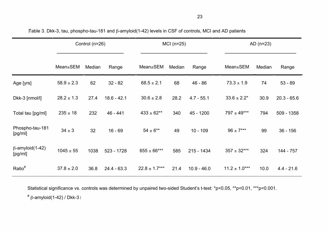
CSF Dkk-3 levels from 25 MCI and 23 AD patients were determined by IEMA and
compared with the control group. Dkk-3 values of MCI patients were slightly but not
significantly increased (30.6 ± 2.8 nmol/l vs. 28.2 ± 1.3 nmol/l). Like in plasma, the
levels of the glycoprotein were significantly elevated in the CSF of patients with AD
(33.6 ± 2.2 nmol/l). Patients age characteristics and CSF Dkk-3 levels are given in
Table 3. The mean age of the control group was significantly lower compared with
the patient cohorts with MCI and AD, however, as Dkk-3 levels in CSF do not alter
with age (Fig. 1C) this difference in chronological age could not be causative for the
specific increase in Dkk-3 of AD patients.
Dkk-3 correlates with tau and phospho-tau-181 levels in CSF
CSF levels of total tau and phospho-tau-181 were significantly increased in patients
with MCI and significantly further increased in patients with AD (Table 3). To analyze
correlations between Dkk-3 and tau levels, the individuals of each cohort (Control,
MCI and AD) were classified into three commensurate groups according to their total
tau levels. In all cohorts with increasing total tau levels also Dkk-3 levels increased
revealing positive correlations of the proteins (Dkk-3 levels [nmol/l]: Control: 21.9 ±
1.7 [total tau<150 pg/ml], 29.1 ± 1.9 [150-250], 30.2 ± 2.0 [>250]; MCI: 18.3 ± 3.8
[<200], 30.9 ± 4.5 [200-500], 38.5 ± 3.6 [>500]; AD: 28.5 ± 2.1 [<650], 31.2 ± 2.9
[650-850], 40.7 ± 4.5 [>850]). The same analysis was performed for phospho-tau-
181 and again revealed positive correlations between Dkk-3 and phospho-tau-181 in
all three cohorts (Dkk-3 levels [nmol/l]: Control: 22.9 ± 1.5 [phospho-tau-181<20

12
pg/ml], 27.7 ± 1.4 [20-40], 32.8 ± 2.4 [>40]; MCI: 17.3 ± 3.7 [<30], 30.6 ± 4.0 [30-60],
38.6 ± 3.8 [>60]; AD: 25.8 ± 2.2 [<70], 33.4 ± 2.2 [70-100], 37.6 ± 4.2 [>100]).
Dkk-3 and the ratio �-amyloid(1-42) / Dkk-3 as classifiers for diagnosis
In contrast to Dkk-3, CSF levels of �-amyloid(1-42) were significantly decreased in
patients with MCI and significantly further reduced in patients with AD (Table 3).
A one-dimensional scatter plot of the �-amyloid(1-42) levels showed a high
heterogeneity of the individual values in the three cohorts (Fig 3B). Given the
negative correlation between Dkk-3 and �-amyloid(1-42) we analyzed the ratio of �-
amyloid(1-42) / Dkk-3 (Table 3). A one-dimensional scatter plot of this ratio showed
reduced heterogeneity compared with �-amyloid(1-42) levels alone (Fig 3C),
suggesting the use of the �-amyloid(1-42) / Dkk-3 ratio instead of �-amyloid(1-42)
levels to differentiate between controls, MCI and AD patients.
To further substantiate these findings the accuracy of the �-amyloid(1-42) / Dkk-3
ratio in comparison to �-amyloid(1-42) to discriminate the single cohorts was
assessed by ROC analysis. The ability to segregate between two cohorts was
increased using the ratio in all cases, control subjects vs. MCI patients (Fig. 3D; �-
amyloid(1-42): AUC = 0.817; ratio: AUC = 0.894), control subjects vs. AD patients
(Fig. 3E; �-amyloid(1-42): AUC = 0.981; ratio: AUC = 1.0) and MCI vs. AD patients
(Fig. 3F; �-amyloid(1-42): AUC = 0.812; ratio: AUC = 0.902).

13
Discussion
Dkk-3 in CSF of controls, MCI and AD patients
To our knowledge this is the first report of Dkk-3 in human CSF. Our data
demonstrate that Dkk-3 is present in CSF at high concentrations (28.2 ± 1.3 nmol/l)
compared with plasma (1.22 ± 0.04 nmol/l). Given the fact that CSF mainly
represents an ultra filtrate of plasma and therefore the total protein concentration is
highly reduced (0.15 – 0.45 mg/ml vs. 60 – 80 mg/ml in plasma) the high content of
Dkk-3 is even more astonishing. This high concentration of Dkk-3 in CSF indicates
an important function of the protein in the brain/CSF compartment. Of all body fluids
we tested, Dkk-3 levels are by far highest in CSF [Dkk-3 levels in seminal fluid are
similar or higher to that in plasma (2.59±0.41 nmol/l; range 1.62-5.25 nmol/l; n=10),
while levels of Dkk-3 were below detection limit in urine (<5 pmol/l; n=3)].
In contrast to plasma Dkk-3 levels, which increase with age (Zenzmaier et al.
2008a), Dkk-3 levels in CSF did not alter significantly with age as shown in the
present study. However, due to the lack of CSF samples from younger patients, we
could not include a cohort of young adults (age 20 – 30 yrs).
Due to the high Dkk-3 content and the proximity to the diseased tissue, we
hypothesized CSF might represent a valuable source to trace changes in Dkk-3
levels associated with neurodegenerative disorders. Thus CSF samples from
patients suffering from MCI and AD were analyzed and compared with healthy
controls. Indeed a significant elevation of Dkk-3 levels in AD patients was observed,
indicating a potential role of the protein in the development of the disease and its use
for diagnostic purposes.

14
Dkk-3 levels in plasma of controls, depressed, MCI and AD patients
Similar to CSF, Dkk-3 levels in plasma of patients suffering from AD, but not MCI or
depression, was significantly elevated compared with healthy controls. However, in
this study we did not differentiate between MCI subtypes. Most of the patients with
amnestic MCI convert to AD (Jicha et al. 2006). Further studies for amnestic MCI
patients compared with patients with other MCI subtypes should reveal whether Dkk-
3 levels differ among MCI subgroups, and these studies will clarify to which extent
amnestic MCI patients are similar to AD patients.
The origin of this Dkk-3 increase in plasma of AD patients is not resolved. One
source could be endothelial cells where Dkk-3 is reported to be expressed (Kupatt et
al. 2005, Goodwin et al. 2006). Furthermore, upregulation of Dkk-3 in endothelial
cells has been demonstrated in various tumors tissues (Zenzmaier et al. 2008b, St
Croix et al. 2000, Untergasser et al. 2008). High expression of the protein has also
been reported in a subset of adult human pancreatic beta cells (Hermann et al.
2007).
Given the high concentration of Dkk-3 in CSF, a major source of Dkk-3 in plasma
might also be resorption of CSF. This hypothesis is further supported by the fact that
the AD-related elevation of Dkk-3 is to a similar extend in both sources.
Potential sources of CSF Dkk-3
There are several potential sources for the high Dkk-3 levels in CSF. CSF is mainly
produced in the choroid plexus and represents an ultra filtrate of plasma. Therefore
the total protein content is very low compared with plasma. However, the
composition of CSF is modified by the choroid plexus, where Dkk-3 could be
transferred from the plasma by an active transport mechanism, or produced locally
by the epithelial lining of the plexus. Our data demonstrate that these epithelial cells

15
of the choroid plexus produce Dkk-3 and therefore it is likely, that at least a major
fraction of Dkk-3 present in CSF is derived from this source.
Furthermore, DKK3 gene expression has been reported in the human cortex
especially in pyramidal cells (Ftouh et al. 2005) and our data demonstrate that these
cells also produce Dkk-3 protein. Diffusion of the protein through the brain tissue
might also contribute to Dkk-3 CSF levels.
Dkk-3 as a diagnostic biomarker for dementia
�-Amyloid(1-42), one of the best established and widely used biomarkers for
diagnosis of AD (Fagan et al. 2009, Tapiola et al. 2009, Shaw et al. 2009),
segregates the studied cohorts with high sensitivity and specificity. Given the
increased Dkk-3 and decreased �-amyloid(1-42) levels in CSF of AD patients, the
ratio of �-amyloid(1-42) / Dkk-3 was analyzed as a classifier for disease by ROC
analysis. While the accuracy to discriminate between AD patients and controls did
not change significantly (due to the already excellent accuracy when using �-
amyloid(1-42) levels alone), the sensitivity and specificity of the ratio as classifier
was clearly superior to �-amyloid(1-42) levels, indicating the value of Dkk-3 as an
additional biomarker.
However, it is well established that the measurement of amyloid(1-42), tau and
phospho-tau-181 in CSF can be used to diagnose AD with high sensitivity and
specificity, and the additional information provided by Dkk-3 levels might not justify
its use for routine diagnosis in CSF. Alternatively, the research for plasma-derived
biomarkers is of high importance, because the invasive lumbar puncture and
collection of CSF limits the diagnosis of dementia. We observed an increase of Dkk-
3 levels associated with AD in plasma similar to that in CSF, indicating that the
increase in plasma levels might be directly associated with disease status and that

16
Dkk-3 levels in CSF and plasma are interrelated either by active or passive transport
over the blood-brain barrier. Thus, the measurement of Dkk-3 in plasma may help to
overcome this problem and may be useful in diagnosing AD. ROC analysis of Dkk-3
plasma levels as a classifier for AD diagnosis revealed a fair accuracy, suggesting
that Dkk-3 plasma levels indeed can be useful for the diagnosis of dementia when
weighed in combination with other molecular markers.
Conclusions
In summary, the present study revealed the presence of high levels of Dkk-3 in CSF
which is at least in part secreted by epithelial cells of the choroid plexus. With a
recently established sensitive and specific IEMA for Dkk-3 significant changes in the
plasma and CSF levels were revealed in patients suffering from AD, while Dkk-3
levels in samples derived from Depression or MCI patients were unchanged
compared with control subjects. Future work will be set up to study the potential role
of Dkk-3 in the development of AD and to further analyze its utility as a diagnostic
marker for neurodegenerative diseases.
Acknowledgements
The authors wish to thank Roswitha Plank for her excellent technical support.
Disclosure Statement
There are no actual or potential conflicts of interest.

17
References
Bian, H., Van Swieten, J. C., Leight, S. et al. (2008) CSF biomarkers in
frontotemporal lobar degeneration with known pathology. Neurology, 70,
1827-1835.
Bibl, M., Mollenhauer, B., Esselmann, H. et al. (2008) Cerebrospinal fluid
neurochemical phenotypes in vascular dementias: original data and mini-
review. Dement Geriatr Cogn Disord, 25, 256-265.
Blasko, I., Lederer, W., Oberbauer, H., Walch, T., Kemmler, G., Hinterhuber, H.,
Marksteiner, J. and Humpel, C. (2006) Measurement of thirteen biological
markers in CSF of patients with Alzheimer's disease and other dementias.
Dement Geriatr Cogn Disord, 21, 9-15.
Blennow, K. (2004) CSF biomarkers for mild cognitive impairment. J Intern Med,
256, 224-234.
Blennow, K. (2005) CSF biomarkers for Alzheimer's disease: use in early diagnosis
and evaluation of drug treatment. Expert Rev Mol Diagn, 5, 661-672.
Desai, A. K. and Grossberg, G. T. (2005) Diagnosis and treatment of Alzheimer's
disease. Neurology, 64, S34-39.
Diep, D. B., Hoen, N., Backman, M., Machon, O. and Krauss, S. (2004)
Characterisation of the Wnt antagonists and their response to conditionally
activated Wnt signalling in the developing mouse forebrain. Brain Res Dev
Brain Res, 153, 261-270.
Fagan, A. M., Head, D., Shah, A. R., Marcus, D., Mintun, M., Morris, J. C. and
Holtzman, D. M. (2009) Decreased cerebrospinal fluid Abeta(42) correlates
with brain atrophy in cognitively normal elderly. Ann Neurol, 65, 176-183.

18
Fradinger, E. A. and Bitan, G. (2005) En route to early diagnosis of Alzheimer's
disease--are we there yet? Trends Biotechnol, 23, 531-533.
Ftouh, S., Akbar, M. T., Hirsch, S. R. and de Belleroche, J. S. (2005) Down-
regulation of Dickkopf 3, a regulator of the Wnt signalling pathway, in elderly
schizophrenic subjects. J Neurochem, 94, 520-530.
Goodwin, A. M., Sullivan, K. M. and D'Amore, P. A. (2006) Cultured endothelial cells
display endogenous activation of the canonical Wnt signaling pathway and
express multiple ligands, receptors, and secreted modulators of Wnt
signaling. Dev Dyn, 235, 3110-3120.
Henley, S. M., Bates, G. P. and Tabrizi, S. J. (2005) Biomarkers for
neurodegenerative diseases. Curr Opin Neurol, 18, 698-705.
Hermann, M., Pirkebner, D., Draxl, A., Berger, P., Untergasser, G., Margreiter, R.
and Hengster, P. (2007) Dickkopf-3 is expressed in a subset of adult human
pancreatic beta cells. Histochem Cell Biol, 127, 513-521.
Humpel, C. and Marksteiner, J. (2009) Peripheral biomarkers in dementia and
Alzheimers disease. In: The Handbook of Neuropsychiatric Biomarkers,
Endophenotypes and Genes, Volume III: Metabolic and Peripheral
Biomarkers, (M. S. Ritsner ed.). Springer.
Jicha, G. A., Parisi, J. E., Dickson, D. W. et al. (2006) Neuropathologic outcome of
mild cognitive impairment following progression to clinical dementia. Arch
Neurol, 63, 674-681.
Krupnik, V. E., Sharp, J. D., Jiang, C. et al. (1999) Functional and structural diversity
of the human Dickkopf gene family. Gene, 238, 301-313.
Kupatt, C., Horstkotte, J., Vlastos, G. A. et al. (2005) Embryonic endothelial
progenitor cells expressing a broad range of proangiogenic and remodeling

19
factors enhance vascularization and tissue recovery in acute and chronic
ischemia. Faseb J, 19, 1576-1578.
Lewczuk, P. and Wiltfang, J. (2008) Neurochemical dementia diagnostics: State of
the art and research perspectives. Proteomics, 8, 1292-1301.
Mao, B., Wu, W., Li, Y., Hoppe, D., Stannek, P., Glinka, A. and Niehrs, C. (2001)
LDL-receptor-related protein 6 is a receptor for Dickkopf proteins. Nature,
411, 321-325.
McKeel, D. W., Jr., Price, J. L., Miller, J. P., Grant, E. A., Xiong, C., Berg, L. and
Morris, J. C. (2004) Neuropathologic criteria for diagnosing Alzheimer disease
in persons with pure dementia of Alzheimer type. J Neuropathol Exp Neurol,
63, 1028-1037.
McKhann, G., Drachman, D., Folstein, M., Katzman, R., Price, D. and Stadlan, E. M.
(1984) Clinical diagnosis of Alzheimer's disease: report of the NINCDS-
ADRDA Work Group under the auspices of Department of Health and Human
Services Task Force on Alzheimer's Disease. Neurology, 34, 939-944.
Mollenhauer, B., Bibl, M., Wiltfang, J., Steinacker, P., Ciesielczyk, B., Neubert, K.,
Trenkwalder, C. and Otto, M. (2006) Total tau protein, phosphorylated tau
(181p) protein, beta-amyloid(1-42), and beta-amyloid(1-40) in cerebrospinal
fluid of patients with dementia with Lewy bodies. Clin Chem Lab Med, 44,
192-195.
Niehrs, C. (2006) Function and biological roles of the Dickkopf family of Wnt
modulators. Oncogene, 25, 7469-7481.
Petersen, R. C., Doody, R., Kurz, A. et al. (2001) Current concepts in mild cognitive
impairment. Arch Neurol, 58, 1985-1992.

20
Ray, S., Britschgi, M., Herbert, C. et al. (2007) Classification and prediction of clinical
Alzheimer's diagnosis based on plasma signaling proteins. Nat Med, 13,
1359-1362.
Shaw, L. M., Vanderstichele, H., Knapik-Czajka, M. et al. (2009) Cerebrospinal fluid
biomarker signature in Alzheimer's disease neuroimaging initiative subjects.
Ann Neurol.
St Croix, B., Rago, C., Velculescu, V. et al. (2000) Genes expressed in human tumor
endothelium. Science, 289, 1197-1202.
Tapiola, T., Alafuzoff, I., Herukka, S. K., Parkkinen, L., Hartikainen, P., Soininen, H.
and Pirttila, T. (2009) Cerebrospinal fluid {beta}-amyloid 42 and tau proteins
as biomarkers of Alzheimer-type pathologic changes in the brain. Arch Neurol,
66, 382-389.
Untergasser, G., Steurer, M., Zimmermann, M., Hermann, M., Kern, J., Amberger,
A., Gastl, G. and Gunsilius, E. (2008) The Dickkopf-homolog 3 is expressed in
tumor endothelial cells and supports capillary formation. Int J Cancer, 122,
1539-1547.
Wu, W., Glinka, A., Delius, H. and Niehrs, C. (2000) Mutual antagonism between
dickkopf1 and dickkopf2 regulates Wnt/beta-catenin signalling. Curr Biol, 10,
1611-1614.
Zenzmaier, C., Sklepos, L. and Berger, P. (2008a) Increase of Dkk-3 blood plasma
levels in the elderly. Exp Gerontol, 43, 867-870.
Zenzmaier, C., Untergasser, G., Hermann, M., Dirnhofer, S., Sampson, N. and
Berger, P. (2008b) Dysregulation of Dkk-3 expression in benign and
malignant prostatic tissue. Prostate, 68, 540-547.

Table 1. MS/MS analysis of Dkk-3 isolated from CSF
Sequence MH+ Mass, % Position AA, %
GLLFPVCTPLPVEGELCHDPASR 2564.35 7.09 204 - 226 6.69
LLDLITWELEPDGALDR 1969.13 5.44 227 - 243 4.86
EVPDEYEVGSFMEEVR 1914.93 5.29 281 - 297 4.86
Total 6448.41 17.82 54 16.41
The identified amino acid (AA) sequences after chymotryptic digestion, the 1+ charge state [(M+H)+] of the Dkk-3 protein fragments,
the percent mass, the positions of amino acid residues, and percentage of the amino acid sequence (AA, %) obtained by database
search using MASCOT are shown. The mature Dkk-3 protein without signal sequence was taken as the basis for analysis.!

22
Table 2. Dkk-3 plasma levels of controls, depression, MCI and AD patients
Age Characteristics [yrs] Dkk-3 plasma levels [nmol/l]
n Mean±SEM Median Range Mean±SEM Median Range
Control 25 70.6 ± 1.2 69 61 - 80 1.21 ± 0.04 1.22 0.89 - 1.87
Depression 15 67.7 ± 1.5 67 60 - 82 1.13 ± 0.06 1.14 0.79 - 1.52
MCI 25 72.9 ± 1.7 70 62 - 90 1.23 ± 0.05 1.24 0.67 - 1.83
AD 25 78.5 ± 1.4 80 64 - 90 1.33 ± 0.04* 1.36 0.96 - 1.88
Statistical significance vs. controls was determined by unpaired two-sided Student’s t-test: *p<0.05
23
!Table 3. Dkk-3, tau, phospho-tau-181 and �-amyloid(1-42) levels in CSF of controls, MCI and AD patients
Control (n=26) MCI (n=25) AD (n=23)
Mean±SEM Median Range Mean±SEM Median Range Mean±SEM Median Range
Age [yrs] 58.9 ± 2.3 62 32 - 82 68.5 ± 2.1 68 46 - 86 73.3 ± 1.9 74 53 - 89
Dkk-3 [nmol/l] 28.2 ± 1.3 27.4 18.6 - 42.1 30.6 ± 2.8 28.2 4.7 - 55.1 33.6 ± 2.2* 30.9 20.3 - 65.6
Total tau [pg/ml] 235 ± 18 232 46 - 441 433 ± 62** 340 45 - 1200 797 ± 49*** 794 509 - 1358
Phospho-tau-181[pg/ml]
34 ± 3 32 16 - 69 54 ± 6** 49 10 - 109 96 ± 7*** 99 36 - 156
�-amyloid(1-42)
[pg/ml]1045 ± 55 1038 523 - 1728 655 ± 66*** 585 215 - 1434 357 ± 32*** 324 144 - 757
Ratio# 37.8 ± 2.0 36.8 24.4 - 63.3 22.8 ± 1.7*** 21.4 10.9 - 46.0 11.2 ± 1.0*** 10.0 4.4 - 21.6
Statistical significance vs. controls was determined by unpaired two-sided Student’s t-test: *p<0.05, **p<0.01, ***p<0.001.
#�-amyloid(1-42) / Dkk-3!

0
10
20
30
Plasma CSF
0
10
20
30
< 55
Age
A
B
70 kDa
CSF
C
CSF
blocked
Zenzmaier et al. Figure 1


0.0
0.2
0.4
0.6
0.8
1.0
0.0 0.2 0.4 0.6 0.8 1.0
1 - Specificity
Se
ns
itiv
ity
Zenzmaier et al. Figure 3
D
0.0
0.2
0.4
0.6
0.8
1.0
0.0 0.2 0.4 0.6 0.8 1.0
1 - Specificity
Se
ns
itiv
ity
0.0
0.2
0.4
0.6
0.8
1.0
0.0 0.2 0.4 0.6 0.8 1.0
1 - Specificity
Se
ns
itiv
ity
0
500
1000
1500
2000
b-a
my
loid
(1- 4
2)
[pg
/ml]
b
Control MCI AD
A B
E F
0
25
50
75
rati
o [
b-a
my
loid
(1-4
2)
/ D
kk
-3]
Control MCI AD
[bb
amyloid
ratio
C vs. MCI C vs. AD MCI vs. AD
0.0
0.2
0.4
0.6
0.8
1.0
0.0 0.2 0.4 0.6 0.8 1.0
1 - Specificity
Se
ns
itiv
ity
C
C vs. AD
Plasma Dkk-3
PlasmaDkk-3


![Detection of genes mutations in cerebrospinal fluid ...plasma or CSF to monitor the tumor progression and/or treatment responses [3–5]. In patients with brain tumor, the plasma ctDNA](https://img.pdfslide.net/doc/110x75/60cb589de02f2457014cfd5e/detection-of-genes-mutations-in-cerebrospinal-fluid-plasma-or-csf-to-monitor.jpg)