Embed Size (px)
Citation preview

Sheffield Molecular Genetics Facility
Preparing an Unenriched Microsatellite LibraryBy Deborah A. Dawson and Jagroop Pandhal
TIMETABLEDay a.m. p.m.
1 DNA digestion Check DNA digest on agarose gel and do size selection
2 Precipitation Set up ligation reaction3 Check ligation by PCR Run PCR products on agarose gel4 Transformations, grow at 37C overnight5 Picking colonies, grow at 37C overnight on a shaker (100rpm)6 Hedge hogging, grow at 37C overnight7 Fixing filters8 Set up probing of filters9 Wash filters, put down autorad film in cassettes containing filters
(with fresh radiation leave overnight but with older readiation leave at -70C for 7 to 10 days)
10 Develop autorad and identify positive colonies
Re- grow selected colonies (250rpm)
11 Plasmid preps Quantify plasmid preps. Set up sequencing PCR
12 Precipitations of sequencing reactions
DNA sequencing on ABI3730
13 DNA analysis using SeqEd BLAST against NCBI database14 Sequence with reverse
primer (as above) Set up sequencing PCR
Precipitations of sequencing reactions
15 DNA sequencing on ABI3730
DNA analysis using SeqEd to compare forward and reverse sequencing and creat a consensus sequence
16 Design primers Test 4 primer sets before continuing large scale
17-100 Repeat stages as above to design 30-50 primer sets
Usually requires 75 positives sequenced – 50 of which are reverse sequenced and consensus sequences designed from.
1

Sheffield Molecular Genetics Facility1. DNA digestion and Size Selection
Add:- 100ng/µl DNA………………..17µl 10 x Reaction buffer………… 2µl Mbo1 (Qbiogene)……………...1µl (1 unit) (Make up to 20µl with autoclaved ddH20 if necessary)
Total volume: 20µlIncubate in a water bath at 37°C for 2 hours.
Run DNA digest with original DNA on 0.8% agarose. Calculate approximate concentration of digested DNA loaded by amount loaded. Include concentration standards (100ng, 200ng and 500ng) and size markers (ø x 174 RF Hae III and λ DNA/Hind III –available from “In Vitrogen”) to enable selection of DNA digest of 200 to 800bp.
Example: Lane 1- λ DNA/Hind II Lane 2- ø x 174 RF Hae III Lane 3- 100ng standard Lane 4- 200ng standard Lane 5- 500ng standard Lane 6- original DNA Lane 7- DNA digest
2

Sheffield Molecular Genetics Facility
2. Gel clean up system
To retrieve the desired DNA, size 200 to 800bp, from agarose gels the “Wizard SV Gel and PCR Clean-Up System” can be used (available from Promega Corporation, Cat. # A9282).Function
The system is designed to extract and purify DNA fragments of 100bp to10kb from standard or low-melt agarose gels in either TAE or TBE.
Process
▪ Cutting out DNA Band.
1) Identify 200-800bp region using size standards.
2) Cut DNA (200 to 800bp) from agarose gel using a new, unused scalpel blade for every different DNA band.
3) Use a hand held UV light or any weak UV light, a facemask and gloves to ensure no skin is exposed to harmful rays.
4) To reduce the amount of agarose accompanying the DNA band turn the gel slice on its side and remove excess agarose.
5) Place DNA slice in a sterile eppendorf.
Dissolving the Gel Slice
1) Weigh an empty eppendorf.
2) Weigh the gel slice in the eppendorf and record this total weight.
3) Subtract the weight of the empty eppendorf from the total weight to obtain the weight of the gel slice.
4) Add Membrane Binding Solution at a ratio of 10µl of solution per 10mg of agarose gel slice.
5) Vortex this mixture.
6) Incubate at 65°C for 10 minutes or so until the gel slice is completely dissolved.
3

Sheffield Molecular Genetics Facility7) Centrifuge the tube briefly at room temperature to ensure the contents are at the bottom of the tube.
DNA Purification
1) Place a SV Minicolumn in a Collection Tube for each dissolved gel slice.
2) Transfer the dissolved gel mixture to the SV Minicolumn assembly and incubate for 1 minute at room temperature.
3) Centrifuge the SV Minicolumn assembly in centrifuge at 14,000rpm for 1 minute.
4) Remove the SV Minicolumn and discard the liquid in the Collection Tube. Return the SV Minicolumn to the Collection Tube.
5) Add 700µl Membrane Wash Solution (previously diluted with 95% ethanol) to the SV Minicolumn. Centrifuge for 1 minute at 14,000rpm.
6) Empty the Collection Tube as before and repeat centrifugation with 500µl Membrane Wash Solution at 14,000rpm for 5 minutes.
7) Empty the Collection Tube and re-centrifuge the assembly at 14,000rpm for 1 minute.
8) Transfer the SV Minicolumn to a clean eppendorf. Add 50µl Nuclease-Free Water directly to the centre of the column. Incubate at room temperature for 1 minute and then centrifuge for 1 minute at 14,000rpm.
9) Discard the SV Minicolumn and store the eppendorf containing eluted DNA at -20°C.
The amount of DNA extracted will be approximately half the amount which was loaded on the gel in the first place i.e. if ~1µg is loaded the digest will contain ~0.5µg of DNA fragments, (in 50µl of nuclease free water the final DNA concentration is 10ng/µl).
4

Sheffield Molecular Genetics Facility
3. Precipitation step
A precipitation step is necessary if the concentration of size selected DNA needs to be increased or if the size selected DNA fragment needs to be cleaned.
1) To 20l digestion mix add (i) 2l 3M sodium acetate (pH 5.5) (ii) 55l clean 100% ethanol
2) Flick to mix and briefly spin.
3) Incubate at -70C for 20mins.
4) Centrifuge at 13,000 rpm for 13mins (whilst still frozen). Orientate eppendorfs with the hinges outermost so the location of the DNA pellet is known.
5) Discard supernatant using a 20l pipette, taking extra care not to the dislodge pellet.
6) Add 1ml of pre-cooled 70% ethanol (stored at -20C) and invert gently to mix three times.
7) Centrifuge at 13,000 rpm for 13mins.
8) Discard supernatant and dry DNA pellet under a lamp (~20mins). The DNA pellet will look like a small translucent flake of skin.
9) Resuspend DNA pellet in 20µl ddH20.
5

Sheffield Molecular Genetics Facility
4. Ligation
For a successful ligation you need to have ten times more vector (PUC 19 Bam H1 cut plasmid) than DNA fragment inserts for successful insertion to occur. This makes the ratio of ends for ligation at 1:1. The concentration of PUC 19 Bam H1 plasmid is usually 50ng/µl.
The table below shows the ligation mixture. The best procedure is to use the ratios of 1:1, 3:1 and 1:3 of DNA to PUC plasmid to achieve a concentration range of transformant colony numbers, as well as checking calculations and progress so far.NOTE: the amount of DNA and PUC Bam plasmid in the table is worked out in relation to the concentrations in the examples used so far. For instance:- 1:1 ratio-1µl of 10ng/µl DNA = 10ng total DNA 2µl of 50ng/µl PUC Bam plasmid = 100ng total (hence 10 x more giving a 1:1 number of ends ratio).
Genomic DNA 1:1 PUC 19 Bam H1 BAP
Genomic DNA 1:3 PUC 19 Bam H1 BAP
Genomic DNA 3:1 PUC 19 Bam H1 BAP
Size selected MboI genomic DNA fragment (~10ng/µl)
1µl (10ng) 0.33µl (3.33ng) 3µl (30ng)
*PUC 19 Bam H1 plasmid (50ng/µl)
2µl (100ng) 2µl (100ng) 2µl (100ng)
**5 x ligation buffer 2µl 2µl 2µlLigase enzyme (1 unit/µl)
1µl 1µl 1µl
Autoclaved ddH20 4µl 4.67µl 2µlTotal volume 10µl 10µl 10µl*PUC 19 Bam H1 plasmid supplied by Qbiogene. **5 x ligation buffer supplied by Invitrogen.
The mixtures are put into small 0.5µl eppendorfs and incubated in a DNA Perkin Elmer thermal cycler at 15°C (program 25- please check) for 1 to 2 days.Check the temperature of the thermo cycler using a thermometer before and during use. Remove and flick tubes several times to mix contents two times per day.
To check ligation reactions- run the following PCR:-
6

Sheffield Molecular Genetics Facility10x PCR buffer 1µl
F sequencing primer (PUC M13F) 1µl
R sequencing primer (PUC M13R) 1µlMgCl2 (50mM) 0.2µlddH20 4.75µldNTP’s 1lTaq 0.05µlLigation DNA 1µlTOTAL 10µl
Program: 94°C- 3 mins 94°C- 1 min; 50°C 1 min; 72°C 30 sec (30 times) 72°C- 2 mins
Run PCR products on 0.8% gel with concentration standards 10ng, 50ng, 100ng, size standards ø x 174 RF Hae III and λ DNA/Hind III. Include uncut PUC plasmid (10 and 100ngng) and PUC 19 plasmid Bam H1 BAP (10ng and 100ng). The uncut PUC plasmid should appear as one band. The PUC 19 plasmid Bam H1 BAP should produce three bands, due to the three forms of DNA- relaxed, coiled and supercoiled DNA.
7

Sheffield Molecular Genetics Facility
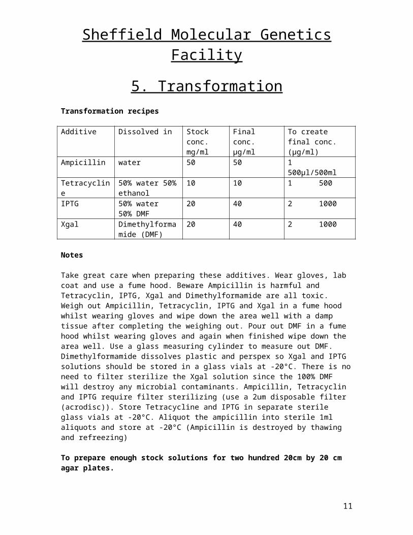
5. TransformationTransformation recipes
Additive Dissolved in Stock conc.mg/ml
Final conc.µg/ml
To create final conc. (µg/ml)
Ampicillin water 50 50 1 500µl/500mlTetracycline 50% water 50%
ethanol10 10 1 500
IPTG 50% water50% DMF
20 40 2 1000
Xgal Dimethylformamide (DMF)
20 40 2 1000
Notes
Take great care when preparing these additives. Wear gloves, lab coat and use a fume hood. Beware Ampicillin is harmful and Tetracyclin, IPTG, Xgal and Dimethylformamide are all toxic. Weigh out Ampicillin, Tetracyclin, IPTG and Xgal in a fume hood whilst wearing gloves and wipe down the area well with a damp tissue after completing the weighing out. Pour out DMF in a fume hood whilst wearing gloves and again when finished wipe down the area well. Use a glass measuring cylinder to measure out DMF. Dimethylformamide dissolves plastic and perspex so Xgal and IPTG solutions should be stored in a glass vials at -20°C. There is no need to filter sterilize the Xgal solution since the 100% DMF will destroy any microbial contaminants. Ampicillin, Tetracyclin and IPTG require filter sterilizing (use a 2um disposable filter (acrodisc)). Store Tetracycline and IPTG in separate sterile glass vials at -20°C. Aliquot the ampicillin into sterile 1ml aliquots and store at -20°C (Ampicillin is destroyed by thawing and refreezing)
To prepare enough stock solutions for two hundred 20cm by 20 cm agar plates.
Additive Solid stored
Amount Dissolved in Filter sterilise
Aliquot into
Ampicillin 4°C 1.0g 20ml water yes 1ml or 1.5mlTetracycline -20°C 0.2g 10ml water 10ml
100%ethanolyes Sterile glass *
vialIPTG -20°C 0.4g 10ml water 10ml DMF yes Sterile glass *
vial (foil covered)
Xgal -20°C 0.4g 20ml DMF no Sterile glass * vial (foil covered)
The DMF in the Xgal and IPTG solutions makes them TOXIC.Xgal and IPTG are also toxic / harmful
8

Sheffield Molecular Genetics FacilityPreparing Agar Plates
A bottle of 500ml of agar will make four or at a stretch five 20cm by 20 cm agar plates.1. Wipe out the required number of clean 20cm by 20cm Perspex plates with 70% ethanol. Ensure they are dry and replace the lids.
2. Remove the top from one bottle of 500ml of agar and melt the agar in the microwave for 5 minutes on high power. If any solid agar is still present after 5 minutes on high power turn down the microwave to medium power and heat for another 5 minutes. Take care that the agar doesn’t boil over as it can cause nasty burns. Once melted the agar can be stored in the 55°C water bath for a few hours but really should be used to make agar plate immediately. It should not be stored at 55°C any longer than a few hours.
3. Place the top back on the bottle of agar and cool under the cold water tap until it is hand hot.
4. Only when agar is hand hot should the ampicillin, tetracyclin, Xgal and IPTG be added. These additives will be destroyed by temperatures higher than hand hot.
Additive Stock Concentration Amount of stock to add to 500ml of agarAmpicillin 50 mg/ml 1.5mlTetracyclin 10mg/ml 0.5 ml
IPTG 20 mg/ml 1.0 ml
Xgal 20mg/ml 1.0 ml
5. Gently swirl the agar several times to mix in the additives. Try to avoid the production of any bubbles.6. Pour approximately 100ml of agar slowly over the whole area of the bottom of the Perspex plate and tip the plate slightly from left to right and top to bottom (if needed) to ensure the agar forms an even layer over the whole of the plate.7. Cover with the lid leaving a small gap for the evaporation to escape. Keep the lids slightly open and allow the agar to solidify for approximately 20 minutes Allow agar to solidify further and dry for 2 hours at room temperature. Plate should be dried well otherwise a lawn of colonies instead of individual colonies will be produced when the transformation mixes are spread out. If available the best place to dry the agar plates is for 15 minutes with the lid completely off in a sterile flowhood. Plates once prepared should be used as soon as possible e.g. within the hour,.
9

Sheffield Molecular Genetics FacilityTransformation Controls
a - Competent cells only (no DNA) electroporated (or not electroporated).To check that no microbial contaminants are present in the cell aliquots. There should be no growth on the agar plates since the untransformed competent cells do not possess ampicillin resistance. This can also be checked without electroporating the cells by simply pipetting 10ul of cells onto an agar plate containing the required antibiotics.
b - Bluescript cut (no CIP treatment) and then religated to itself with no insert To check the ligation (Bluescript religation) is working and that the competent cells are efficiently taking up the DNA. All blue colonies should be produced.
c - Bluescript ligated to genomic DNA fragments (not size selected or enriched for microsatellites). This will tell us if the ligation between genomic DNA and Bluescript is working and can be used to calculate the efficiency of the cells to take up DNA.
d - Bluescript cut, CIP treated and then religated to itself with no insert. The best result would be that no colonies should be produced because the ends of the Bluescript should have been acted on by the CIP and prevented from religating. If very few blue colonies (i.e. less than 700) appear this is good. These colonies are produced when a few molecules of the Bluescript have not been acted on by the CIP so have religated. These best results are obtained when higher amounts of CIP have been used to dephosphorylate the Bluescript vector.In practice colonies are usually produced but the numbers should be 1000 fold less than the numbers of colonies for Bluescript cut, CIP treated and ligated to genomic DNA fragments. The number of colonies produced is dependent on the amount of CIP used to dephosphorylate the Bluescript. Expect 100 - 1000 colonies from the whole 545ul of transformation mix. Expect 60% blues and 40% whites. These whites are produced when the CIP has nibbled back the Bluescript DNA into the lac Z region. Best results = lower numbers of colonies and higher percentages of blues.
e - Bluescript cut, CIP treated and ligated to genomic DNA fragments (not size selected or enriched for microsatellites) – expect many colonies of blues and whites. From 50ul of transformation mix expect 5000. If no colonies are produced there may have been too much CIP used to dephosphorylate the vector. Try reducing the amount of CIP used by 20 fold or 200 fold.
f - Uncut PUC19 or Bluescript To check the electroporation is working and the competent cells are competent (ie can be transformed) Record the amount of bluescript used (10ng is enough) in order to calculate the efficiency of the cells to take up DNA. Ensure the plasmid used (PUC19, Bluescript , etc) carries ampicillin resistance. Use 0.1 - 1.0 ng of DNA.
g - SOC only, not electroporated This is to check for microbial contaminants present in the SOC. Plate out 50ul of SOC onto a 2cm square area of agar plate containing additives. No microbial growth should occur at all. If any does (i.e. colonies or fungi, etc) all the other transformations will also be contaminated and will be no good.
After each transformation record the number of colonies produced per 50ul of transformation mix and the percentages of blues and whites. If there are any ampicillin satellites or contaminant this should be recorded too.
10
Sheffield Molecular Genetics FacilityBLANK FILTER library- expect some colonies - miscellaneous DNA which hybridises to the Hybond filter during the enrichment hybridisation.
11

Sheffield Molecular Genetics FacilityPreparation of High Efficiency E. coli XL1 Competent Cells.1. Autoclave 15 psi 15 minutes two 2 L flasks each containing 500 ml LB (see p24) 1.5 L UHP waterAutoclave 10 psi 15 minutes or Filter Sterilise 20 ml 10% glycerol2. The XL1 culture is in the -80°C. Set up an overnight of XL1 cells in 10 ml LB + 10 ul 10 mg/ml tetracycline only (No ampicillin) in a sterile universal at 37c.(10mg/ml tetracyclin, stored at -20°C) Next Day - start early since the cells need 3 hrs to grow and their preparation takes all afternoon.3. Add 500ul 10mg/ml tetracycline to each of the two flasks of 500 ml LB (No ampicillin).4. If the XL1 overnight culture has grown well inoculate each of the two flasks containing 500 ml of L-broth with 5 ml (1/100 volume) of the fresh overnight culture of XL1.5. Incubate flasks containing cells at 37°C with vigorous shaking (250 rpm) until an OD600nm of 0.5 to 0.7 is reached. This takes approximately 3 hours but check OD600nm after 2 hours.6. Place the autoclaved UHP water and 10 % glycerol in the ice machine.7. After two hours incubation at 37°C measure OD600nm on the spectrophotometer (in lab 331). Zero the spectrophotometer using unused LB media. If the OD600nm is between 0.5 and 0.7 harvest the cells.If the OD600nm is less than 0.5 place the flasks back at 37°C and measure OD600nm in 30minutes.If the OD600 is above 0.7 then start again 8. Switch on the Sorval centrifuge place the GSA rotor inside and allow it to cool down to 0 to 4°C.The cells after they have been centrifuged must be kept on ice at all times and the Sorval centrifuge and solutions used must be kept ice cold.To harvest the cells:(a) Chill the flasks on ice for 15 to 30 minutes (b) Divide the two flasks of culture into the six centrifuge pots, (201D glassware cupboard)(c) Centrifuge in a pre-cooled GSA rotor at 4000 rpm for 10 min with brake on.9. Resuspend pellets in a total of 1 L of ice cold water (approximately 160ml per each of the six pots) using a sterile 10 ml pipette. Centrifuge 4000 rpm for 10 minutes.10. Resuspend in 0.5 L of ice cold water (approximately 80ml per pot) using a sterile 10ml plastic pipette. Centrifuge 4000 rpm for 10 minutes.11. Resuspend in ~20 ml ice cooled 10% glycerol (approximately 3ml per pot). Centrifuge 4000 rpm for 10 minutes.12. Resuspend to a final volume of 2 to 3 ml in ice cooled 10% glycerol (0.5 ml per pot) whilst keeping cells on ice using a 1 ml pipette. Transfer cells to a sterile universal tube for ease of handling.13. This suspension should be frozen in 80µl aliquots on dry ice in flip top eppendorfs (should be about 40 aliquots). 40µl of cells are used per transformation14. The competent cells can be used fresh or should be stored at -70°C. The cells will last at least 6 months under these conditions. Once the cells are thawed they should be discarded if not used. If the cells are accidentally defrosted they should be thrown away.15. Test the cells before use. The cell concentration should be about 3 x 10¹⁰ cells / ml. Calculate their transformation efficiency. Transform them with a RF plasmid such as Bluescript.Use 1µl of 1ng/µl Bluescript and plate out on a 20 by 20 cm agar plate (containing Ampicillin, Tetracyclin, Xgal and IPTG). Calculate the number of transformants per µg of DNA.
12

Sheffield Molecular Genetics FacilityHigh Efficiency Electro-transformation E. coli XL1 Competent Cells.
Have prepared enough LB agar plates upon which the transformation mix will be plated out. 1 – 2 plates are needed per transformation. Use 0.2 cm width electroporation cuvettes (these have yellow tops). Use a separate cuvette per species. The cuvettes are labelled 'single use only' but really are OK to reuse for different satellites in the same species. Eventually they will blow (bang and spark) and should be then thrown away. The cell DNA mix which was in the cuvette when it blew will need re-electroporating.1. Place prelabelled electrotransformation cuvettes on ice and SOC in an eppendorf rack.2. Prepare DNA - To 1ul ligation mix DNA add 4µl water, flick to mix, spindown. Place on ice. (DNA should be in a low ionic strength buffer such asTE, water is OK. DNA containing too much salt will make the sample tooconductive and causes arching (bang + spark) at high voltage.3. Prelabel fresh eppendorfs into which (cell / DNA / SOC) mix will be placed afterelectro transformation. It is not necessary to place these on ice.4. Just before use thaw the competent cells (stored at -80c) and place them onice.Cells should be in 40 or 80µl aliquots. 40µl of cells are used per transformation. Add 40ul of cells to DNA / water mix. Flick to mix well (cell / DNA / water), spin down in picofuge and let sit on ice ~1 min.5. Switch on and set the Gene Pulser apparatus at 2350V wait for the “Ready” light to come on.6. Transfer the mixture (of cells / DNA) to a cooled 0.2 cm electroporation cuvette and shake the suspension to the bottom of the cuvette, wipe off water from outside of cuvette. Place the cuvette in chamber slide, and push the cuvette whilst in the slide into the chamber until the cuvette is seated between the electrical contacts in the base of the chamber. Press the "Pulse" button once. The green light for pulse in progress should come on.7. Allow the machine to beep 4 times then remove the cuvette from the chamber and immediately add 500µl of SOC (at room temp) to the cuvette. Quickly mix the cells with the SOC by inverting the eppendorf or pipetting up and down three times using a 1ml pipette. (This rapid addition of SOC after the pulse is very important in maximising the recovery of transformants).8. Gently transfer the cell suspension to the fresh prelabelled eppendorf using the 1ml pipette to transfer most of the cell mix. Avoid excessive pipetting. Then use a 200µl pipette to retrieve cell mix from the bottom of the cuvette. Wash out the cuvette very well using the UHP water jet 4 -5 times.NB Remember to use a fresh cuvette for each different species.9. Incubate at room temperature for 1 minute. The transformation mix just be plated out immediately after the 1minute incubation time and can not be stored. DO NOT LEAVE LONGER THAN 5 MINUTES!NB Do not incubate any longer than this time or else the bacterial cells carrying themicrosatellite will divide/reproduce and lots of copies of the same microsatellite will result.10. Plate on selective medium which usually is LB agar containing ampicillin, tetracyclin, IPTG and Xgal (See page 23 for recipe) and incubate at 37°C overnight. The cells are very fragile so spread the bacteria very gently on the agar using a light weight glass spreader which can be prepared from a glass Pasteur pipette. Check that the internal temperature of the incubator is 37°C and is stable. Temperatures above 37°C will kill the transformants. The plates should not be incubated at 37c for more than 18 hours. 10 hours is probably the ideal length of time to incubate. Longer incubation periods lead to the production of satellite colonies especially around the “real” transformant colony. This makes it very difficult to pick the real transformant without contaminating it with other colonies. You must be 100% sure that any colonies picked are not mixed with any other colony.
13

Sheffield Molecular Genetics FacilityHow much transformation mix should be plated out?CONTROLS For the uncut Bluescript control. If 10ng of uncut Bluescript DNA was used in the transformation 10ul of transformation mix should be plated out on half or a quarter of a 20 by 20 cm agar plate. This should produce 5000 (- 500 000) colonies (if 5000 colonies are produced the cell efficiency is 2.7 * 107). 100ul of sterile SOC will have to be added to the 10ul of transformation mix since 10ul is too small a volume to be spread out.10ul of transformation mix should also be plated out onto half of a 20cm by 20cm agar plate for the following controls. Just before spreading add 10 ul of the transformation to 100ul of SOC to enable even spreading out of the transformation mix on the agar plate.1 - Digested Bluescript religated to itself (No CIP treatment and no insert ligated)
2 - Digested Bluescript ligated to precipitated digested genomic DNA (not enriched)3 - Digested Bluescript CIP treated religated to itself4 - Digested Bluescript CIP treated and ligated to precipitated digested genomic DNA (unenriched)Expected Results
1. 10 000+ all blue colonies (1-2 whites)2. 10 000+ all blue colonies (10 whites)3. 500 blue colonies (no whites) (1% of Bluesccriptremains either uncut or unCIPed)4. 10 000 colonies, 50-70% whites If the transformation has not worked there will be no colonies for any of the above controls.If the ligations have not worked there will be no colonies for number 3 and 4 of the above controls.If the library has not worked allthe controls will give the expected results but there will be no colonies on the agar plates on which the transformed library ligation was plated.
EXPECTED TRANSFORMANT NUMBERS FOR THE ATTEMPTED LIBRARIES
For the attempted libraries one 20cm by 20cm agar plate is usually divided in half and 50ul of transformation mix plated on one half with 500 ul of transformation mix plated on the other from this initially plate you should estimate the volume of transformation mix to spread over one plate to produce an even spread of colonies which can be easily picked with a toothpick. For the attempted libraries the number of transformants produced depends on the enzyme originally used to digest the genomic DNA, the microsatellite type being enriched for, and other factors.If an enzyme is used which is a frequent cutter (eg Mbo I) then relatively more genomic fragments should reach the enriched fraction so expect higher numbers of transformant colonies.Tetranucleotide repeats are rarer than dinucleotide repeats so expect fewer transformant colonies for libraries enriched for tetranucleotide repeats.
Between 1000 and 100, 000 colonies should be produced from a single transformation using 1ul of the ligation mix. Half up to 70% of this should be whites. If you get these sorts of figures then I would be hopeful that a good library has been produced.
Values of 2000 - 4000 colonies per 20cm by 20cm agar plate is the ideal number from which single colonies can be easily picked.Values of 10 000 colonies per 20cm by20cm agar plate starts to make the picking of single colonies more difficult. When higher densities of colonies are on the plates the colonies also tend to be smaller so making picking more difficult.
14
Sheffield Molecular Genetics FacilityTransformation results recordingRecord the volume of ligation mix used (usually 1ul with 5ul water added to it), the volume of competent cells used (usually 40ul) and the volume of SOC used (usually500ul).Record the volume of transformation mix plated out. The area of plate upon which the transformation mix was platedRecord the number of colonies produced by the volume of transformation mix plated.Record the number of white colonies and blue colonies.Estimate the volume of transformation mix which should have been plated out per 20cm by 20cm agar plate to produced a nice spread of colonies ideal for picking (ie 2000 - 4000 colonies / agar plate).Calculate the Efficiency of the Competent Cells to Take up DNA. To calculate how competent the cells are we need to calculate how many colonies are produced per ug of DNA. For the transformation from which the efficiency is going to be calculated use a standard plasmid for which the concentration has been accurately recorded e.g. Bluescript, PUC, pGEM, etc. equations amount of DNA used in the ligation x fraction used in transformation x fraction of transformation plated out = Z(1ug / Z) x colonies from Z = number of colonies / ug (= efficiency of the competent cells)example calculationamount of DNA used in the ligation-
= 10ng (0.01ug)fraction used in transformation = 1/10 = 1ng (0.001ug)fraction of transformation plated out = 1/200 = 0.005ng (0.000005µg)(5pg)
this 5pg (0.000005ug) of DNA produced 400 colonies the efficiency of the competent cells to take up DNA = 400 / 0.000005 = 80 000 000 (8 to the power of 7), ie 80 000 000 colonies are produced per ug of DNA (which is dodgy / okish)1 to the power of 11 colonies / ug of DNA 100 000 000 000 =best efficiency ever achieved1 to the power of 10 10 000 000 000 = very good1 to the power of 9 1000 000 000 = OK1 to the power of 8 100 000 000 = OK (usually get this)1 to the power of 7 10 000 000 = dodgy1 to the power of 6 or less 1 000 000 = discard and try again !NB The number of colonies produced per ug of DNA also depends on the number of cells present too. I am assuming a 40ul aliquot of cells was used. Competent cells can be kept a few months at
-70 but their efficiency will fall. Every batch of cells prepared should have their efficiency checked before use.
15
Sheffield Molecular Genetics Facility
6. Picking Colonies
After the colonies have grown overnight :-1. Prepare microtitre NUNC plates containing Luria Broth+ glycerol+ ampicillin Add 600ul of 50mg/ml ampicillin to 200ml of LB+20% glycerol (recipe on next page). Mix well, pour into a sterile small tray (eg a 200ul pipette tip box lid) and usingthe multichannel pipette aliquot 100ul into each microtitre well.It is best to prepare the plates and use them Do not let plates sit at room temperature. It is best to prepare the plates and use them fresh. Once made they should be kept in the fridge when not being picked into. Take from the fridge and allow 5 minutes for the plate to warm up before picking colonies into it. Plates can be kept in the fridge one to two days.It is best to prepare and use the plates fresh but the plates containing the LB+ glycerol+ ampillicin can be prepared beforehand and stored at -70c but ensure they have reached room temperature before picking colonies into them! Defrosting and refreezing plates should be avoided since this will destroy the ampicillin.2. Pick the white colonies into microtitre plates containing LB+ 20% glycerol + ampicillin either at room temperature or which have been prewarmed to 37c. CHECK THE PLATEPERFECTLY HORIZONTAL AT ALL TIMES OTHERWISE COLONIES WILL BECOME MIXED AND THE PLATE SHOULD BE THROWN AWAY.3. Mix the tips vigouresly in the wells. It takes approx 15mins to pick one microtitre plates worth of colonies (ie 96 colonies).4. Grow up at 37c for at least 6 hours or preferably overnight with gentle shaking (100 rpm max). Do not grow for more than one day. Store the plates upright at -80c.
16

Sheffield Molecular Genetics FacilityTransformation solution recipes
Luria broth:-1% Bactotryptone 10g 30g 50g0.5% Bacto Yeast Extract
5g 15g 25g
1.0% NaCl 10g 30g 50gwater 950ml 2850ml 4750mlTOTAL 1L 3L 5L
Autoclave 20 mins 15 psi
LB agar:-Prepare 500ml in a 1 Litre bottle to prevent it boiling over when microwaved. Add 7.5g BBL Grade A agar to 500ml LB (before it is autoclaved) then autoclave 20 mins 15psi.
LB glycerol:- 1% Bactotryptone 10g 0.5% Bacto Yeast Extract 5g 1.0% NaCl 10g water 750ml glycerol 200ml
Place 200 ml into 5 bottles and autoclave 20 mins 15psi.
SOC:- Concentration required Amount needed2% bactotryptone 2g0.5% Bacto Yeast Extract 0.5g10mM NaCl 1ml of 1M2.5mM KCl 250µl of 1Mwater Make up to 97mls
Autoclave for 20mins 15psi, allow to cool at room temperature
Then add-
Concentration required Amount needed10mM MgCl2 1ml of 2M10mM MgSO4 1ml of 2M20mM glucose 1ml of 2M
pH to 7.0 with approx 20µl 5M Sodium Hydroxide. Filter sterilise the SOC into sterile 1ml aliquots in eppendorfs and store in the fridge. Store some 5 - 10ml aliquots in sterile universals too. Check that there is no microbial contamination before using any of the SOC aliquots.
1M NaCl 5.844g on 100ml water1M KCl 7.456g in 100ml water1M MgSO4. 24.648g of MgSO4. 7H20 in 100ml water
17

Sheffield Molecular Genetics Facility1M MgCl2 20.03g of MgCl2. 6H20 in 80ml water, make
up to 100ml
Autoclave all these solutions for 20 mins, 15psi.
1M Glucose 18g in 80ml water, allow to dissolve, make up to 100ml(C6H12O6) Sterilise by filtration or autoclave 15 mins at 10psiMW=180.16g
18
Sheffield Molecular Genetics Facility
7. Hedgehogging
Grow the Positives Colonies on to Hybond Filters (aka Hedgehogs)1. Prepare LB Agar+Tetracyclin+Ampicillin plates which are deep in agar. To ensure the agar plates are deep use one 500ml bottle to prepare three 20cm by 20cm plates.2. Lay hybond filter the size of a microtitre plate on the surface of the agar. There is enough space on each large agar plate for three microtitre plate size filters, two filters fit on much more easily. 3. Transfer the colonies using a “hedgehog” from the microtitre plates onto the hybond N filters laid over LB Agar+Tetracycillin+Ampicillin plates. Apply pressure to each corner and the sides of the hedgehog since the teeth of the hedgehog are slightly different lengths. Mark the position of four corner colony spots using a papermate biro (papermate ink doesnt come off during the hybridisation). Ensure the hedgehog is sterilised by flaming with 70% ethanol and cools to room temperature in between each transfer of cells. It can be cooled by dipping it in unused (sterile) LB agar before dipping it into the microtitre plate or placing it in the freezer for a short time. 4. Gently circle the hedgehog teeth around in the wells before transferring them to the hybond nylon filter. This should mix the bacteria in the well.5. Incubate at 37°C overnight. Remember to check the oven temperature before leaving the agar plates overnight.
19
Sheffield Molecular Genetics Facility
8. Fixing colonies- Preparing the Filters for Probing .
Using the microwave method - Ref. L. Bulewela et al (1979), Nar 17(1), 452.
1. Place the 2xSSC / 5xSDS solution in the 55°C water bath until the SDS has redissolved and it has reached approximately 40°C (this takes approximately 15 minutes). NB If it is a warm day the SDS will already be dissolved. Alternatively use the microwave (high power for approximately 5 minutes) to warm the 2xSSC / 5xSDS solution.
2. Pour approximately 100ml 2xSSC / 5xSDS prewarmed to 37°C into a clean tray.
3. Place a glass plate over the tray.
4. Dip a 20cm by 20cm piece of Whatman 3mm paper into the warm 2xSSC / 5xSDS in the tray until it is soaked. Lay it on the glass plate avoiding bubbles.
5. Dip a second 20cm by 20cm piece of Whatman into the warm 2xSSC / 5xSDS in the tray place it over the first, again avoiding air bubbles.
6. Lay Hybond filters on top of the Whatman 3mm paper avoiding air bubbles leave 3 (to 5) minutes. A 20cm by 20cm piece of Whatman should be big enough for three Hybond microtitre filters. Using a 3ml plastic pipette squirt 2xSSC / 5xSDS over the filters ensuring the solution covers each colony.
7. Drain off as much of the solution as possible and place Whatman with filter(s) on top into the microwave. Microwave for approximately 3 minutes on high power checking they do not burn until they are dry and almost crisp (the DNA is fixed bythis method so UV treatment is not needed). It is important to ensure the filters are crisp - the bacteria cells have to be broken and the DNA fixed onto the Hybond filter securely otherwise it will be washed off during the probing. Note: Bacterial culture hedgehogged onto overlapping pieces of Hybond will not grow up to form colonies. The two overlapping pieces of Hybond thick make a barrier which is too thick for the bacterial cells to absorb nutrients through.
2xSSC / 5xSDS Recipe
SDS (Dodecyl Sulphate, Sodium Salt) is harmful (especially be inhalation) and should be weighed out in the upstairs fumehood whilst wearing gloves. Wipe down the weighing out area well with a damp cloth after weighing out.In the fumehood add 50g SDS to 1 Litre 2xSSC (prewarm 2xSSC to 37°C)Heat further whilst stirring if the SDS is not dissolving but be careful the solution doesn’t boil over.
20

Sheffield Molecular Genetics Facility
9. Probing the Filters for Microsatellites . ControlsFor a negative control include a blank 3 cm2 piece of hybond filter. Label using either a pencil or papermate biro since these are the only labels which do not wash off during the hybridisation. For a positive control label (using either a pencil or papermate biro) a 2 cm2 piece of Hybond filter with the probe to be used.Make 0.5ug of the probe DNA upto 7ul using water and spot onto a 2 cm2 piece of Hybond filter. 7ul will spread approximately 1 cm2 so creating a concentration of 50ng/mm2 on the filter. UV fix the DNA by covering the UV transilluminator with a piece of cling film and placing the filters squares on there DNA side down. Expose 2 minutes then turn the filter over and expose 2 minutes on the other side. Alternatively as a positive control, you could use a filter which has been stripped but probed previously and found to contain positives for the particular probe to be used.
1. Rinse filters in 2xSSC ensuring filter are completely wet. If probing “hedgehog” filters pour off the dirty 2xSSC replace with fresh 2xSSC.
2. Prepare the prehybrisation solution. The volume needed depends on the number of filters to be probed and the type of hybridisation vessel to be used. The filters should be free to move without sticking to each other. Bottles are good for small pieces of filter but chambers are better for probing “hedgehog” filters. A maximum of 10 “hedgehog” filters can be probed together in one chamber.If probing in small bottles use 30ml of prehybridisation solution (minimum 20ml).If probing in large bottles use 50ml of prehybridisation solution.If probing in a chamber use 50-100ml of prehybridisation solutionEach chamber will hold a maximum of 100ml prehybridisation solution; do not add more than this.
Add BSA (Albumin, bovine, fraction V, stored in fridge) to the prehybrisation solution. Add 1g BSA per 100ml of prehybridisation solution whilst mixing prehybridisation solution vigorously with a magnetic flea.
3. When the BSA is completely dissolved pour prehybridisation solution containing BSA into chamber. Add filters ensuring they lay flat and don’t clump together, place burnt filters on the top of the rest, tighten screws finger tight only and place in 65°C oven with gentle shaking. Remember to include a blank piece of Hybond.
Approximately 10 “hedgehog” filters is the maximum which will fit in the chamber, ensure there is still being enough space for them to move around separately.
4. In screw topped eppendorfs place50ng probe DNA (eg CA.GT, GA.CT, TTTC.GAAA)Make up to 30.5ul with UHP waterPlace screw top back onBoil on heating block (105c) for 5-10 minutes
(a minimum of 10ng/ul of DNA is needed, using 300ng still works fine)
21

Sheffield Molecular Genetics Facility5. Place on ice / While still hot **add 6.0ul OLB (stored at -20°C)
1.2ul BSA (stored at -20°C)** If the DNA is allowed to cool slowly the strands which have been separated will join back together. We need to keep them separated, this can be done by placing the DNA immediately on ice or if you are quick enough add the next solutions very quickly while the DNA is still hot.
6. Behind Screens Add 0.5ul 32P dCTP (5uCi) (stored at -20 in lead container) 0.4ul Klenow (3.6 Units) (from Pharmacia, stored at -20°C)
7. Flick to mix, spin 10 seconds, and place in 37°C heating block for at least one hour.
8. Add 200ul distilled water then boil in the heating block at 105c, 5-10 minutes and behind screens add to the filters in the prehybridisation solution which are in the hybridisaton chambers.
9. Incubate overnight with shaking at 55°C
10. Wash CA/Gas with 0.1xSSC, 0.1%SDS at 65°C with shaking until the counts per second is 20-50.NB WASH CONDITIONS ARE DIFFERENT FOR TETRA AND WHEN PROBING WITH 32P dATP SEE NEXT PAGE.Approximately three 15 minute washes will be needed. Once the count per second is between 20-50 place the filters in 2xSSC (at room temperature).The filters can be stored in 2xSSC for 1-2 hours but it is best to place them in cassette immediately. Wash filters probed with tetra probes or an AT probe very carefully since the probe is easily washed off. Wash with 3xSSC, 0.1%SDS at 60-55c with shaking until counts per second is between 10-20. Check after 1 minute if the counts are 10 or below stop the washing immediately by placing the filters in 2xSSC (room temperature). If the filters are above 50cps use 2xSSC, 0.1%SDS11. After washing expect there to be 20-30 counts per second (cps) for the CA and GA positives. If the cps is 20-30 it should be possible to develop the autorad after 5 hours.If the counts are less than this leave it down overnight. If 50 cps incubate at -70°C for 5 hours. If more than 50 cps wash more to bring down the cps to 50 and background.12. Carefully record the co-ordinates of positives by placing a thick piece of glass over the filter or behind the screens place another autorad film in the cassette and leave at -70c for 7-10 days. This is to check the co-ordinates of the positives since the filter outline will become visible on the autorad.
Ref. Analytical Biochem 132 6-13 (1983) Andrew P.,Feinberg & Bert VogelsteinAtechnique for radiolabelling DNA restriction endonuclease fragments to ahigh specific activity
22
Sheffield Molecular Genetics FacilityPROBING WITH TETRANUCLEOTIDE PROBES CONTAINING AT LEAST ONE C/G.Use the precipitated PCRed GATC and TTTC. Same protocol as for CA/GA probing with the following exceptions -Use 1ul (min 10ng needed) of the probe which was saved from the PCR amplification in stage C. This DNA has been ligated and extended and then PCR amplified. The hybridisation should be performed at 55°C (60°C works too but not as well, 65°C will not work). Washing should also be performed at 55c and much more carefully - gently quickly rinsed then rinsed again for only 1-2 minutes with 3xSSC, 0.1 %SDS (5xSSC, 0.1 %SDS may be better). Keep checking the filters with the Geiger counter. Usually a couple of very gentle 1 minute washes are enough! Compare the levels of radioactivity with those of the blank. When the blank is only 5 counts or less stop the washing by immediately placing the filters in 2xSSC at room temperature. Expect 10 counts for the TTTC / GAAA and GATC positives. Leave the autorad at -70c overnight. If higher counts (e.g. 10-15) were recorded try developing the autorad after 5 hours. PROBING WITH DINUCLEOTIDES AND TETRANUCLEOTIDE PROBES CONTAINING A / T USING 32P labelled dATP32P labelled dATP is used instead of 32P labelled dCTP.A different oligo labelling mix has to be used.Hybridisation should be done at 55°C.Washing should also be done at 55°C using 3xSSC, 0.1% SDS (5xSSC, 0.1% SDS may be better). Keep checking the filters with the Geiger counter during the washing. Usually a couple of very gentle 1 minute washes are enough.
23

Sheffield Molecular Genetics Facility
Prehybridisation solution. (Church Gilbert Buffer) (Final Concentration Created) 250ml 1M Sodium Phosphate Buffer (pH 7.4) (0.25M)70g SDS (n-dodecyl Sulphate, Sodium Salt) (7%)2ml 0.5M EDTA (1mM)make up to 1 Litre with distilled water
warm solution to help the SDS to dissolve1 M Sodium Phosphate Buffer (pH 7.4). Klenow (supplied by Pharmacia)167.8g NaHPO4.2H2O Klenow Fragment make up to 800ml with distilled water (of DNA Polymerase I)pH to 7.4 with Orthophosphoric Acid (needs approx. 7ml) FPLCpure, clonedmake up to 1043ml with distilled water. 500 Units @ 9,100 U/ml
Cat. No. 27-0928-02
OLIGO-LABELLING OF DNA FRAGMENTSSolutions needed1. Solution O 1.250M Tris-HCl 15.13g Tris 100mls
0.125M MgCl2 2.54g MgCl26H2O
2. dNTPs (store at -20°C)Sigma D-6500 0.1MdATP 187µl/10mgSigma T-8635 0.1M dTTP 207µl/10mg in T.E separately Sigma D-4135 0.1M dGTP 200µl/10mg
T.E 3mM Tris pH 7.0 300µl 1M Tris pH 7.0 80µl 0.25M EDTA make up to 100ml
3. Solution A 1000µl solution O 18µl § mercaptoethanol (14.3M) TOXIC 5µl each dATP,dTTP,dGTP (each 0.1M)
4. Solution B 2M HEPES (titrated to pH 6.6 with 4M NaOH, store at 40C)TOXIC 5.20g /10ml
1.04g / 2ml5.
5. Solution C Hexanucleotides (store at -20°C) suspend 50 OD units in 550µl T.E to give
concentration of 90 OD/ml Pharmacia pD(N)6 Cat No. 27-2166-016. OLB Buffer Mix solution A: B: C ratio 10: 25: 15 360:900:540µl(50µl is sufficient for 15 reactions- freeze at -20c in 50 or 100µl aliquots)
7. Stop Mix 20mM NaCl 1ml 1M 20mM Tris-HCl pH 7.5 1ml 1M50ml 2mM EDTA 50µl 0.2M 0.25% SDS 500µl 25%(store in 1ml aliquots at-20°C)Stop mix can be added to the probe after incubation at 37°C optional
8. BSA 10mg/ml in distilled water (use BRL enzyme grade # 5561)
24

Sheffield Molecular Genetics FacilityAfter developing the Autorad.
Identify which positive clones in the microtitre plate correspond to strong signals on the autorad. Strong signals on the autorad usually correspond to long runs of repeats but weak signals can also
correspond to long runs of repeats. Sometimes a strong signal can be caused by an insert which contains a series of small runs of the repeat motif. Shorter stretches of the repeat motif are
unlikely to be polymorphic. Occasionally there may be an autorad signal but no repeat present - the signal being caused by the presence of a high proportion of the same base type present in the insert as is present in the probe. The occurrence of this is rare. Bear in mind that the G content of the rest of the insert as well as the microsatellite itself affects the strength of the positive signal on the autorad. Klenow builds up one complementary strand per strand of supplied template and we are using 32P labelled dCTP. The amount of bacterial growth through the Hybond filter is also a
major factor affecting the strength of the positive signal on the autorad.
25

Sheffield Molecular Genetics Facility
10. Re-grow colonies 1) Warm up microtitre plate at 37°C for about half an hour.
2) Add 600l of 50ng/l Ampicillin to 500mls of LB broth. Place 3.6ml LB broth in 5ml universal tubes.
3) Transfer 20l of positive colony from microtitre plate to universal tubes. Include a blank with no added colony to ensure no contamination has taken place.
4) Store overnight at 37°C and agitate at 250rpm.
26

Sheffield Molecular Genetics Facility
11. Plasmid prep
To retrieve the plasmid from grown up colonies the “GenElute Plasmid Mini Prep Kit” supplied by Sigma can be used.
1) Harvest cells- Pour LB broth containing bacterial culture into an eppendorf and spin for 1 minute at 13,000rpm. Discard supernatant and repeat with remaining broth (approximately 3 spins).2) Resuspend cells- Completely resuspend bacterial pellet in 200µl Resuspension solution (RNAse solution is added prior to this- product code R6148). Vortex and pipette up and down thoroughly.3) Lyse cells- Lyse the resuspended cells by adding 200µl of Lysis solution. Immediately mix the contents by gentle inversion (6-8 times) until the mixture becomes clear and viscous. Do not vortex and do not let the lysis reaction exceed 5 minutes.4) Neutralize- Precipitate the cell debris by adding 350µl of the Neutralization/Binding Solution. Gently invert the tube (4-6 times). Pellet the cell debris by centrifuging at 13,000rpm for 10 mins.5) Prepare column- Insert a GenElute Miniprep Binding Column into provided microcentrifuge tube and add 500µl of the Column Preparation Solution. Spin at 13,000rpm for 1 minute. Discard the flow-through liquid.6) Load cleared lysate- Transfer the cleared lysate from the neutralisation step to the prepared column and centrifuge at 13,000rpm for 1 minute. Discard the flow-through liquid.7) Wash column- Add 750µl of the diluted wash solution (diluted with ethanol- see preparation instruction manual) to the column. Centrifuge at 13,000rpm for 1 minute. 8) Discard the flow-through liquid and centrifuge again at 13,000rpm for 2 mins.9) Elute DNA- Transfer the column to a fresh collection tube. Add 80µl of ddH20 and centrifuge at 13,000rpm for 1 minute. The DNA is now present in the eluate and is ready for immediate storage at -20°C.
Quantify Plasmid DNA
On 0.8% agarose- run 1µl of plasmid DNA with 14µl of 1x Orange G loading buffer with concentration standards 10ng, 50ng, 100ng, 200ng and size markers ø x 174 RF Hae III and λ DNA/Hind III.Quantifying plasmid DNA is essential to avoid adding too little or too much template to the sequencing reaction, which results in poor quality data. For plasmid DNA a total of approximately 100ng is suitable for most insert sizes.
27

Sheffield Molecular Genetics Facility
28

Sheffield Molecular Genetics Facility
12. DNA Cycle Sequencing using ABI BigDye Terminator Mix v1.1
Primers used for sequencing…
Forward Sequencing primer
M13 Universal Sequencing Primer (Pharmacia) (5’-3’) GTA AAA CGA CGG CCA GT (17 mer)
M13 Reverse Sequence Primer (Pharmacia) (5’-3’) CAG GAA ACA GCT ATG AC (17 mer)
1) Set up reactions in AbGene 96-well plate using the recipe below
Ingredients Volume (µl) BigDye Terminator Mix v1.1 1.0Sequencing dilution buffer (5x) 1.5Autoclaved ddH20 1.5Primer (1µM) (either forward or Reverse) 1.6Plasmid DNA + autoclaved ddH20 4.4 Total volume 10.0µl
2) Cover plate with a rubber seal (avoid using oil). Run reactions on PCR tetrad machines using the hot-lid setting.
Thermal cycling program1. 96°C 1 min2. 94°C 10 sec3. 50°C 5 sec4. 60°C 4 min5. Repeat cycle (step 2 to 4) 29 times6. 10°C Hold
29

Sheffield Molecular Genetics Facility
30
Sheffield Molecular Genetics Facility
13. Precipitation of BigDye Terminator v1.1 cycle sequencing reactions
Ethanol/EDTA/sodium acetate precipitation
1) Label a clean sterile eppendorf for each reaction; don’t attempt more than 12 samples.
2) Add 2µl of 125mM EDTA, 2µl 3M sodium acetate (pH 5.2), 10µl autoclaved ddH20 and 50µl 100% ethanol to each tube. (Ensure EDTA and sodium acetate is at the bottom of the tube).
3) Transfer 10µl of sequencing reactions to the labelled 1.5ml eppendorf and briefly vortex.
4) Incubate at room temperature for 15 mins in the dark* (put in a drawer).
5) Spin at 13,000rpm for 15 mins. Orientate eppendorfs with the hinges outermost so the location of the DNA pellet is known.
6) Set a P200 pipette to 80µl and aspirate off the supernatant, taking care not to dislodge the pellet. If residual ethanol/EDTA/NaOAc drops are left behind, carefully remove with as lint-free kimwipe (i.e. roll a corner to make a tissue wick). Ignore drops adjacent to the DNA pellet.
7) Add 195µl 70% ethanol (pre-cooled at -70C).
8) Spin at 13,000rpm for 5 mins
9) Repeat step 6 using a P200 pipette set to 200µl.
10) Place eppendorfs upright in a rack with lids open, cover with a lint-free tissue and air-dry at room temperature in the dark for 15 to 60 minutes or until dry.
*Dark conditions are recommended by Sigma due to the photosensitive BigDye. However it is known not to be essential.
31
Sheffield Molecular Genetics Facility
14. DNA SEQUENCING ON THE ABI3730 48- CAPILLARY INSTRUMENT
This method assumes that you are using a 36cm array and the standard protocols. AT YOUR BENCH Prepare your plate. Re-suspend your precipitated sequence reactions in 10uL of Hi-Di formamide. Plate out 5uL of this re-suspension and 5uL of Hi-Di formamide (i.e. a 1:2 dilution) onto an ABI 96-well optical reaction plate. Use 10uL ddH20 in empty lanes, rather than formamide. Each plate contains two 48-lanes runs; odd columns = run1 and even columns = run2. See attached guide to row/column loading order. Cover plate with clean rubber septa and ensure there are no bubbles at the bottom of wells by spinning in a post-PCR salad spinner. Denature at 95°C for 3 minutes (in a PCR machine or hot block) and then place on ice until you load it into the machine.
AT THE 3730 Re-start the 3730 computer (password is ‘grouse’).Click on Unified Data Collection; wait for the icons to switch from red to yellow to green. Minimize.Create a new Plate Record → open Plate Manager. If you have an existing Plate Record template then edit this (plate name and sample names) in Microsoft XL, import directly into Plate Manager (click ‘Import’) and proceed to Run Scheduler (below). Otherwise click on ‘New’. New Plate Dialogue box will open. ID (type the name of your plate. This must be unique)Name (type the name of your plate)Application (Sequencing Analysis)Plate type (96-well)Plate sealing (Septa)Owner name/Operator name (your initials)Click OKThe Sequencing Analysis Plate Editor will now open. Enter your Sample Names.Select a Results Group. You have to create your own Results Group initially. Select an Instrument Protocol (3730_POP7_StdSeq_BDTv1). Select an Analysis Protocol (StdSeq_KB_BDTv1.1). Instructions for creating/editing Results Groups, Instrument Protocols and Analysis Protocols appear over page. Fill down (CtrlD) so that all samples contain all necessary information.Export the Plate Record as a template (E:\AppliedBiosystems\UDC\DataCollection \Data\PlateRecordTemplates). In the future this plate record can be edited in XL (plate name and sample names) and directly imported into the Plate Manager. Click OK.Open Run Scheduler. Click Search in the Input Stack pane → the Add Plate to Input Stack window opens. Type of search → Advanced Enter your search criteria, then click Search. Select your plate and click Add → Done. Your plate will appear in the Current Runs pane of the Run Scheduler window.Place your plate into a black tray and cover with a white clip-on cover. Place into the 3730.
32

Sheffield Molecular Genetics FacilityClick the green ► to run your plate.Monitor your run in Instrument Status, Capillary Viewer and Array Viewer.When your run is finished it will be dumped into the E drive (Applied Biosystems/UDC/data collection/data). You must then copy this file to one of the lab PC’s.Use SeqScape v2.0 or Sequence Analysis v5.0 to analyse your data.
CREATING A RESULTS GROUP
Each user has their own Results Group which defines (i) how files & folders are named, (ii) where files are saved and (iii) the type of analysis (i.e. sequencing). To create an Results Group, select New from the drop-down menu in the Sequencing Analysis Plate Editor, or click on the Results Group icon in the explorer pane. The Results Group Editor will open. General – enter the Results Group name and owner (you).Analysis – click ‘Do autoanalysis’.Destination – use the default file destination (E:\AppliedBiosystems\UDC\DataCollection \Data). This folder will be emptied monthly so always copy your folders onto Alfred (G:\). Naming – in the upper pane (Sample File Name Format), under ‘Format’, select ‘Sample name’, then ‘Plate ID’ and then finally ‘Capillary #’. Use the same procedure to label the ‘Run Folder’ as ‘Plate ID_Date of run_Run #’.Click OK to save. Your Results Group will now appear in the Sequencing Analysis Plate Editor.
CREATING AN INSTRUMENT PROTOCOL
The instrument protocol contains everything necessary to run the instrument. It is comprised of a Dye-Set, which specifies the sequencing chemistry (dye-set E for BigDye v1.1 and dye-set Z for BigDye v3.1), and a Run Module, which specifies the capillary array length (36cm or 50cm), type of run (i.e. standard verses rapid sequencing) and the polymer (POP7). To create or edit an Instrument protocol select New from the drop-down menu in the Sequencing Analysis Plate Editor, or from within the Sequence Analysis Protocol Manager. The Protocol Editor will open. Select a Run Module (e.g. StdSeq36_POP7).Select a Dye-set (e.g. E-BigDyeV1). Set Type to ‘Regular’. Click OK to save.
CREATING AN ANALYSIS PROTOCOL
An analysis protocol contains all the settings necessary for analysis and post-processing. It is comprised of a basecaller algorithm (e.g. KB) and a DyeSet/Primer (or Mobility) file (.mob) that is specific to each combination of polymer and sequencing chemistry. To create or edit an Analysis Protocol click on New from the Plate Editor drop-down menu, or do the same from the Protocol Manager. This will open the Sequence Analysis Protocol Editor. Under the General tab give the protocol a name and indicate your preference of sequence file formats. Under the Basecalling tab select the basecaller algorithm and the DyeSet/Primer file. The KB.bcp algorithm is preferred because it provides quality values (QV) for each base and also recognises mixed bases. Use DyeSet/Primer file KB_3730_POP7_BDTv1.mob. In the ‘Ending Base’ pane, you can also specify when you want basecalling to end. This is particularly important if your sequence products are short. Under the mixed bases tab you can choose to identify mixed bases
33

Sheffield Molecular Genetics Facility(i.e. heterozygotes) and specify the threshold detection level. Finally under the clear range tab you can instruct the protocol to trim low quality data from the ends of your sequence automatically. Click OK to save your analysis protocol.
34
Sheffield Molecular Genetics Facility
15. ANALYSING DNA SEQUENCES IN SEQSCAPE V2.0
Two applications are available for analysis of DNA sequence data generated by the 3730 capillary instrument. Sequence Analysis v5.0 is software that will perform base calling of DNA sequences and enables individual DNA sequence chromatograms to be visualized and edited. It is useful if you have small numbers of sequences. SeqScape v2.0 is software that combines the initial base calling of DNA sequence data performed by Sequence Analysis v5.0, with advanced sequence editing and alignment tools (e.g. such as in Sequence Editor and Sequencher). Sequence Analysis v5.0 is therefore redundant if you use SeqScape. This protocol provides a brief introduction to the analysis of DNA sequences in SeqScape v2.0.
Re-start a lab computer using the Windows 2000 operating system.Double click on the SeqScape v2.0 software and login (TABLAB, password = grouse). If you get a warning message “no disk in drive” press continue.
Before you can import and analyse your DNA sequence data you need to create a Project template. This is comprised of a Reference Data Group, Analysis Protocol, Analysis defaults and Display Settings. Select Tools and open the SeqScape Manager First, create a Reference Data Group ( = a known reference sequence to which your sequences will be aligned and compared. Each user needs their own RDG).Select the Reference Data Group tab → Click NewRDG Properties will open. Complete the tabs:General – specify a name that is unique and has meaningROI – add one or more Reference Segments (= known reference sequences): Click on Add Ref. Segment –prompts you to import a GenBank nucleotide (.gb) file (recommended). Alternatively, click on Paste Ref. Segment – this allows you to copy-and-paste a nucleotide sequence straight into the RDG. You may also define ROIs (Regions Of Interest such as exons, introns), arrange them into layers, create links to a library of known alleles and import specific NT (nucleotide) and AA (amino acid) variants.Click OK to save your RDG.
For standard sequencing runs using BigDye Terminator Mix v1.1 the Analysis Protocol (3730_StdSeq_POP7_BDTv1.1) and Analysis defaults (3730_POP7_BDTv1.1) already exist. If you ran a standard sequencing module on the 3730 using BDTv1.1 then proceed to the ‘Create a Project Template’ step below. If you’re using a different version of BigDye Terminator mix (v2.0, v3.0 or v3.1), or doing either Rapid or Long-Read sequencing, you will need to create a new Analysis Protocol and new Analysis defaults. See over the page for instructions.
To create a Project Template, select the Project Template tab and click New.Give the template a name (i.e. the same as your RDG name)Select your Reference Data Group, Analysis Defaults (e.g. 3730_POP7_BDTv1.1) and Display Settings (use ‘DefaultDisplaySettings_v2’) from the drop-down menus (Note that the default display settings should be OK for all types of sequence runs and all chemistries, but they can be edited by selecting Properties in the Display Settings tab.
35
Sheffield Molecular Genetics FacilityClick OK to save Close SeqScape Manager
Now you can create your New Project and import your samplesFile → New ProjectSelect your Project TemplateSpecify a Project NameClick New and your project will open. Click File → Import Samples to Project (CtrlM)The Import Samples window will open.Locate your sample files in the explorer pane. You should have transferred these files from the 3730pc E: drive to your own folder on G: drive (Alfred).At this point you can either: (a) Add Samples (adds all samples you highlight or all samples in the selected folder), OR (b) if you have multiple samples from each of a number of individuals (e.g. forward & reverse sequences), then you can group these samples into Specimens (= individuals). To do this (1) click New Specimen, (2) highlight this specimen in the Samples to Add pane, (3) then select just the samples for that specimen in the explorer pane, and (4) click ‘Add Samples’. (5) Repeat for each specimen. You will need to manually edit specimen names in the Project Navigator after the samples have been imported. Alternatively you can use the Auto Add feature if, within your sample names, text delimiters flank a common specimen name. Click OK when you have finished adding samples.Your specimens & samples will appear in the Project Navigator pane in the main SeqScape window (notice the presence of a red line through each specimen icon– this indicates that they are yet to be analysed).Click the green ► to analyse your samples. This should be fast.SeqScape will perform basecalling, assign quality values to each base, identify mixed bases (if you requested it to) and trim low quality data from the ends of your sequences. It will also automatically assemble and align samples within specimens and construct a consensus sequence for each specimen. Sequences will be automatically reverse complemented, where necessary.
CREATING AN ANALYSIS PROTOCOL Select the Analysis Protocol tab → Click NewThe Analysis Protocol Editor will open. Complete the tabs:General – specify a name that captures the application (e.g. Long-Read), polymer (POP7) and sequencing chemistry (e.g. BDTv3.1).Basecalling – select a basecaller and the appropriate DyeSet/Primer (i.e. mobility) file from the drop-down lists. The recommended basecaller is KB.bcp, because it provides quality values (QV) for each base and can identify mixed bases (heterozygotes). The DyeSet/Primer file is specific for each combination of basecaller and sequencing chemistry. You can also indicate how the software should recognise the end of your sequence fragment.Mixed Bases – select mixed base detection and specify threshold (if required).Clear Range – provide instructions for trimming low quality data. Filter – use the default values Click OK. (Note: you could export your Analysis Protocol from Unified Data Collection [on the 3730 computer] and import it directly into SeqScape v2.0 by clicking Import rather than New above).
CREATING NEW ANALYSIS DEFAULTS Select the Analysis Defaults tab → Click New
36

Sheffield Molecular Genetics FacilityNew Analysis Settings will open. Complete the tabs:General – specify a name that captures the application (e.g. Long-Read) and sequencing chemistry (e.g. BDTv3.1).Sample – Select the appropriate Analysis Protocol from the drop-down menu.Specimen – Click ‘Base call samples’.Otherwise use the default valuesClick OK
Reviewing your analysisBefore editing any of your sequences check to see if your analysis was successful.In the Project Navigator explorer panel click open each specimen to determine if samples have been successfully assembled into segments (local alignments).If any samples could not be assembled they will appear under ‘unassembled’. This could indicate a failed or poor quality sequence, or that the analysis settings are inappropriate.There are two ways to check:First, click on the unassembled sample in the Project Navigator panel.View the Annotation tab. Signal intensity should be 50 → 1000 for each base. Values <50 probably indicate too little template. Values >1000 may be corrected by reducing injection time (Edit the Instrument protocol on the 3730 and re-run).View the Sequence and Electrophenogram tabs. Click the ‘Show/Hide sample QVs’ icon to view quality values. How does the sequence look?Second, click Analysis → Report ManagerThe Report Manager provides links to 9 reports! (quickly explore these)Select Analysis QC ReportThe Specimen Analysis pane indicates if Basecalling, Filtering and Assembly of each specimen has been successful (Green box), partially successful (Yellow triangle) or unsuccessful (Red octagon). Reasons for Yellow or Red outcomes are provided in the Sample Analysis pane. Sometimes poor results can be corrected or improved by editing the analysis parameters and applying them to the affected samples/specimens.If necessary edit your analysis settings for failed samples and re-analyse. Instructions to do this are provided over the page.
Viewing and editing your dataWhen you are satisfied with the analysis you can edit your dataFirst, turn on the Audit Trail (keeps track of all the edits you make and why you made them):Select Tools → Options from the main SeqScape windowSelect Authentication & Audit and click the Audit Trail On check boxIn the Audit Reason pane you can specify reasons for making changesClick OK to exit the Options dialogue box.
Next, click the Project icon in the Project Navigator. Your specimen consensus sequences will appear, aligned to the Reference sequence. This is called Project View. Variable sites in the alignment are highlighted as coloured lines on the reference sequence. Your current position in the alignment is indicated by the rectangle over the reference sequence.Click the triangle (►) adjacent to one (or more, or all) of your specimens so that you can view the assembled & aligned samples. Click on the consensus sequence to view the electrophenogram (EP) snippets in that region of the alignment.To centre the EP snippet use CtrlZ.To edit the EP snippet range open Display Settings ( ds ), click View and edit ‘EP range’.
37

Sheffield Molecular Genetics FacilityClick the ‘Show/Hide sample QVs’ and ‘Show/Hide consensus QVs’ icons to view quality value bars for each base. Blue bars (QV 25-50) indicate that there is low probability (≤0.31%) of an incorrect base call at that position. Yellow (QV 15-24) and red (QV 0-14) bars indicate a progressively higher probability of error. Note that you should be most concerned with the specimen consensus QVs. YOU SHOULD MANUALLY EDIT: (1) all low (red) & medium (yellow) QV bases, (2) all known and unknown variants (i.e. all polymorphic base positions), (3) all mixed base calls and (4) all ambiguous base calls. But you should visually check the whole sequence! Click on the ‘View column selector’ icon (this highlights a single base for easy viewing)Use the TAB key to jump through the sequence to the next base that requires editing. To specify Tab settings, select ‘Multiple…’ from the Tab jump to next drop-down menu (top RHS). The Multiple Tab Jump Settings box opens. Select options from the list and click OK.To actually insert, delete or change a base simply highlight the position on the sample or consensus sequence and change the base. Changes made to the consensus will be reflected in all samples that make up that specimen. Edited base changes appear in lowercase to distinguish them from unedited bases. If Audit Trail is activated you will be prompted for a reason for making each change.
Exporting your dataWhen you have completed editing you can export Sample Sequence files, Consensus sequences and also Project alignments in a number of formats, including FASTA, SEQ and AB1.To export a specimen consensus sequence:Select File → Export → Consensus Sequence, and then export.
HOW TO EDIT YOUR ANALYSIS PROTOCOL FOR RE-ANALYSIS From within the SeqScape main project window, click Analysis → Sample ManagerClick Edit Analysis Protocol.Edit Basecaller, DyeSet/Primer file, Ending Base settings, Mixed Bases settings, the Clear Range and Filter settings. Click OK.Select the samples in the Sample Manager that you wish to apply the new protocol to.Click Apply Analysis Protocol.Select your new Analysis protocol from the drop-down list. Click OK and then Apply.Click the green ► to re-analyse your samples.Check the Project Navigator and the relevant reports in Report Manager.
x
38