Embed Size (px)
Citation preview

HUGGINS-SULLIVAN, SIOBHAN, M.S. Does Internal Mental Imagery Help Maintain
Muscle Strength and Force Steadiness During Immobilization? (2012)
Directed by Dr. Renee Newcomer Appaneal 83 pp.
Research has shown that immobilization such as that which occurs during
treatment to an injury can result in significant muscle strength and force steadiness loss
within the first week (Lundbye-Jensen & Nielsen, 2008; Newsom, Knight, & Balnave,
2003). Research has examined the efficacy of imagery in minimizing strength-loss during
a period of immobilization (Newsom, Knight, & Balnave, 2003; Stenekes, Geertzen,
Nicolai, De Jong, & Mulder, 2009). While promising, limitations remain with regards to
type of imagery used and structures immobilized. This study assessed the effectiveness of
using internal kinesthetic mental imagery to maintain thenar muscle group strength
during immobilization of the thumb. Participants’ thenar muscle group strength was
measured pre- and post-immobilization period in adduction, abduction, opposition and
flexion. Force steadiness was also evaluated pre- and post-immobilization at 5%, 25%
and 50% of maximum thumb flexion force. All participants were immobilized for seven
days on their non-dominant hand in a thumb spica cast. During the immobilization
period, both the control and experimental groups were instructed to limit the use of their
non-dominant hand. The experimental group completed a daily 8-minute imagery script.
Results of separate repeated measure 2 (group) x2 (pre/post) ANOVAs failed to support
the effect of imagery to maintain muscle strength or force steadiness following 7-days of
immobilization. Future research should add a familiarization session to the protocol to
allow participants to become more accustomed to the unique testing procedures.

i
DOES INTERNAL MENTAL IMAGERY HELP MAINTAIN MUSCLE STRENGTH
AND FORCE STEADINESS DURING IMMOBILIZATION?
by
Siobhan Huggins-Sullivan
A Thesis Submitted to
the Faculty of the Graduate School at
The University of North Carolina at Greensboro
in Partial Fulfillment
of the Requirements for the Degree
Master of Science
Greensboro
2012
Approved By
__________________________
Committee Chair

ii
APPROVAL PAGE
This thesis has been approved by the following committee of the Faculty of The
Graduate School at The University of North Carolina at Greensboro.
Committee Chair________________________________
Committee Members________________________________
________________________________
_________________________________
Date of Acceptance by Committee
_________________________________
Date of Final Oral Examination

iii
TABLE OF CONTENTS
Page
LIST OF TABLES………………………………………………………………………...v
LIST OF FIGURES………………………………………………………………………vi
CHAPTER
I. INTRODUCTION…………………………………………………………......1
Purpose and Hypothesis……………………..…………………………….6
II. REVIEW OF THE LITERATURE…………………………………………....8
Perceptions of Psychological Skills in Sport Injury Rehabilitation…….....9
Athletes Use of Psychological Skills in Injury Rehabilitation...................
Mental Imagery Use in Injury Rehabilitation……………………………
Force Steadiness.........................................................................................
Future Research Directions........................................................................
III. METHODS..………………..………………………………………………..32
Participants……………………………………………………….………
Measures……………………………………………………….………...
Procedures………….......………………………………………………...
Data Analysis…………………………….......…………………………..
IV. RESULTS...………………………………………………………………….40
Participants…………………………………………………………….…40
Preliminary Analysis…………………………............……………….….40
Thenar Muscle Group Strength……………………………………….….41
Force Steadiness…………………………………………….……………46
Adherence to Protocol……….…………………….....……………….….49
V. DISCUSSION………………………........………………………………….
Thenar Muscle Group Strength...............................……………………...52
11
12
26
29
32
32
35
38
51

iv
Force Steadiness……………………………..........……………………...
Imagery Ability……………………………………………....…………..
Adherence…………………………………......……………………...….
Limitations………………………..……………………………………....
Strengths…………………………….....................………………….…...
Future Research……………………………..................………………....
Conclusions……..…………………….…….………………………….....
REFERENCES…………………………………………………………………………..
APPENDIX A. EDINBURGH HANDEDNESS SURVEY…………………………......69
APPENDIX B. VIVIDNESS OF MOVEMENT IMAGERY QUESTIONAIRE-2…….
APPENDIX C. LAB SETUP.......................................................................................... ..76
APPENDIX D. PROPER CAST CARE INSTRUCTIONS.....………………………….
APPENDIX E. IMAGERY SCRIPT…………………………………………….............78
APPENDIX F. THUMB DIRECTIONAL MOVEMENTS…………………….………
56
58
59
61
61
62
63
83
71
64
77

v
LIST OF TABLES
Page
Table 1. Means and standard deviation for imagery ability…………………………..….
Table 2. Means and standard deviations for thenar muscle group strength………….......
Table 3. Means and standard deviations of force steadiness.............................................
41
43
47

vi
LIST OF FIGURES
Page
Figure 1. Percent of abduction strength lost post immobilization………………….….....
Figure 2. Percent of adduction strength lost post immobilization.……………..………...
Figure 3. Percent of opposition strength lost post immobilization…………………….....
Figure 4. Percent of flexion strength lost post immobilization……...…………………...
Figure 5. Percent gain of force steadiness at 5% of maximum flexion
strength post immobilization………………………………………………...
Figure 6. Percent gain of force steadiness at 25% of maximum flexion strength post
strength post immobilization………………………………………………...
Figure 7. Percent gain of force steadiness at 50% of maximum flexion strength post
strength post immobilization………………………………………………...
44
44
45
45
48
48
49

1
CHAPTER I
INTRODUCTION
Injured athletes are typically in a rehabilitation protocol under the medical model
that lacks mind-body integration thus typically focusing only on body part and not other
effects of the injury (Green, 1992). Psychological skills can be used to incorporate the
mind-body integration during rehabilitation (Green, 1992; Law, Driediger, Hall, &
Forwell, 2006). Researchers have recommended rehabilitation professionals’ use of
psychological skills in injury rehabilitation (Hamson-Utley, Martin, & Walters, 2008;
Ievleva & Orlick, 1991; Brewer, Jeffers, Petitpas, & Van Raalte, 1994; Weise & Weiss,
1987; Wiese, Weiss, & Yukelson, 1991; Driediger, Hall, & Callow, 2006). As a result,
athletic trainers are required to have psychological skills education in their curriculum
(Board of Certification, 2006).
Researchers have examined athletes’ and rehabilitation professionals’ perceptions
of psychological skill use (Brewer, Jeffers, Petitpas, & Van Raalte, 1994; Hamson-Utley,
Martin, & Walters, 2008; Wiese, Weiss, & Yukelson, 1991). Generally perceptions of
psychological skill use are favorable for both athletes and rehabilitation professionals.
Although perceptions are positive, research has shown that psychological skills are
underused in rehabilitation (Hamson-Utley, Martin, & Walters, 2008). One reason for
this could be the inexperience with psychological skills for most rehabilitation
professionals (Wiese, Weiss, & Yukelson, 1991).

2
Studies have been performed to examine athletes’, who have not received prior
formal training in psychological skills, use of psychological skills during injury
rehabilitation (Ievleva & Orlick, 1991; Scherzer, et al., 2001). Ievleva and Orlick (1991)
found a correlation between the use of psychological skills and improved healing rates.
Scherzer et al. (2001) found a correlation between the use of psychological skills and
improved rehabilitation adherence. These studies interviewed athletes after the injury
rehabilitation was completed and asked what types of psychological skills they
remembered performing. Researchers have advocated for intervention studies focused on
one or more specific psychological skills (Weise & Weiss, 1987; Wiese, Weiss, &
Yukelson, 1991; Scherzer, et al., 2001; Brewer, Jeffers, Petitpas, & Van Raalte, 1994).
Mental imagery is a particular mental skill that has been recommended to be
effective in injury rehabilitation (Christakou & Zervas, 2007; Driediger, Hall, & Callow,
2006; Brewer, Jeffers, Petitpas, & Van Raalte, 1994; Weise & Weiss, 1987). Mental
imagery is the re-creation of experiences using all of one’s senses including kinesthetic
sense, without actually performing any movements (Weinberg, 2008). Studies reviewing
athletes’ use of mental imagery during injury rehabilitation show that many athletes use it
on their own, without prior instruction (Driediger, Hall, & Callow, 2006; Law, Driediger,
Hall, & Forwell, 2006; Monsma, Mensch, & Farroll, 2009; Sordoni, Hall, & Forwell,
2000). Law et al. (2006) recommended that mental imagery intervention studies include
pre- and post-measures in injury rehabilitation, in order to be able to conclude results
were due to the mental imagery intervention. Controlled intervention studies will show
how mental imagery effects injury rehabilitation outcomes. The findings from these types

3
of studies can be used to inform the development of recommendations for the use of
mental imagery in rehabilitation.
Currently, intervention studies have included both inured and healthy populations
and explored the impact of mental imagery upon a range of dependent variables.
Dependent variables including: pain, edema, range of motion, muscular endurance,
dynamic balance, functional stability, re-injury anxiety, and muscle strength (Christakou,
Zervas, & Lavalle, 2007; Christakou & Zervas, 2007; Cupal & Brewer, 2001; Lorenzo,
Ives, & Sforzo, 2003; Sidaway & Trzaska, 2005; Herbert, Dean, & Gandevia, 1998;
Ranganathan, Siemionow, Liu, Sahgal, & Yue, 2004; Yue & Cole, 1992). These studies
have looked at both injured and healthy populations.
One area that shows future promise is the use of mental imagery to maintain
muscle strength. It is largely accepted that muscle strength gains are the result of muscle
hypertrophy and neural adaptations (Ranganathan, Siemionow, Liu, Sahgal, & Yue,
2004). Neural adaptations are thought to be the basis of most early strength gains.
Neurophysiological research has shown that muscle strength gains may be possible
without muscle contractions (Sidaway & Trzaska, 2005). Mental imagery is thought to
create neural adaptations that may lead to muscle strength gains or prevent strength loss.
The perspective taken during mental imagery has been suggested to impact muscle
strength. Internal imagery has been shown to have greater physiological effects than
external imagery (Ranganathan, Siemionow, Liu, Sahgal, & Yue, 2004). Internal imagery
is when a person imagines himself or herself performing the task, as if looking through
his or her own eyes and external imagery is when a person imagines watching himself or

4
herself perform the task, as if watching a video (Weinberg, 2008). Currently commonly
used rehabilitation techniques to gain muscle strength require a muscle contraction,
which may not always be possible for the patient following injury (Sidaway & Trzaska,
2005). Following an injury, athletes may be in too much pain to generate a muscle
contraction or they may be immobilized and unable to contract their muscle.
Some researchers have found support for mental imagery to maintain muscle
strength (Newsom, Knight, & Balnave, 2003; Ranganathan, Siemionow, Liu, Sahgal, &
Yue, 2004; Sidaway & Trzaska, 2005; Cupal & Brewer, 2001; Yue & Cole, 1992), while
others have not (Stenekes, Geertzen, Nicolai, De Jong, & Mulder, 2009; Herbert, Dean,
& Gandevia, 1998; Lorenzo, Ives, & Sforzo, 2003). Currently, there is no definitive
understanding of the effects of mental imagery on muscle strength (Sidaway & Trzaska,
2005). Methodological inconsistencies could account for many of the contradictory
findings within the literature. Studies vary on the type of imagery used, the content of
imagery, as well as intervention length. Internal imagery has been shown to create the
greatest physiological benefits (Ranganathan, Siemionow, Liu, Sahgal, & Yue, 2004) but
only four studies specifically used an internal imagery script (Ranganathan, Siemionow,
Liu, Sahgal, & Yue, 2004; Newsom, Knight, & Balnave, 2003; Sidaway & Trzaska,
2005; Yue & Cole, 1992). Protocols for imagery intervention also varied among
researchers from as little as 4 days to 6 months in duration. Manipulation checks for
imagery adherence varied among researchers and were not well reported across studies.
This makes it hard to understand if the protocol was actually followed by participants or
if adherence issues may account for the results found. Also some immobilization studies

5
have tested muscles that were not fully immobilized during the intervention (Newsom,
Knight, & Balnave, 2003).
Force steadiness is the ability to match a given sub maximal force with limited
fluctuations (Bandholm, Radmussen, Aagaard, Jenson, & Diederichsen, 2006). Force
steadiness has been previously studied during wrist immobilization (Lundbye-Jensen &
Nielsen, 2008) but never during a mental imagery intervention. After just a week of wrist
immobilization significant changes are found in force steadiness. Other researchers have
looked at the effects of strength training during periods of bed rest on force steadiness
(Shinohara, Yoshitake, Kouzaki, Fukuoka, & Fukunaga, 2003; Mulder, et al., 2011). In
both of these studies strength training helped decrease the fluctuation in force steadiness
usually found during bed rest.
If research shows that mental imagery helps to maintain muscle strength, then
protocols can be developed for the use of mental imagery in injury rehabilitation. This
could allow for muscle strength training to begin while a patient is still immobilized or is
too weak to contract his or her muscles. Earlier muscle strength training may lead to
shorter rehabilitation periods and quicker advancement to functional exercises.
Maintaining strength through mental imagery may help maintain force steadiness as well
Evidence supporting the benefits of mental imagery could encourage the use of mental
imagery amongst rehabilitation professionals for their patients. Also, the use of mental
imagery can help connect the mind-body integration that researchers have advocated for
(Green, 1992; Law, Driediger, Hall, & Forwell, 2006). Mind-body integration can

6
improve rehabilitation outcomes for injured patients (Richardson & Latuda, 1995; Green,
1992).
Purpose and Hypothesis
The purpose of this study is to explore if internal mental imagery can prevent the
loss of thenar muscle strength during thumb immobilization. This question is important
because immobilization has been shown to result in significant muscle strength loss
within the first week (Newsom, Knight, & Balnave, 2003). Research has explored the use
of mental imagery to prevent the loss of muscle strength during immobilization but the
present study will improve upon methodological limitations. Studies on the use of mental
imagery to prevent the loss of muscle strength during immobilization have looked at
other hand musculature but none have researched the thenar muscle group. Also studies
have tested hand muscles that were not fully immobilized during the immobilization
period. Thumb immobilization would prevent any use of the thenar muscles, which can
help control for extra movement that may have occurred during daily living or the
imagery task in previous studies. Furthermore, using internal kinesthetic imagery, which
has been shown to be the most effective type of imagery (Ranganathan, Siemionow, Liu,
Sahgal, & Yue, 2004), should improve the effects of the imagery performed.
A secondary purpose of the study is to determine if mental imagery can help
maintain force steadiness during immobilization. Force steadiness can be used to assess
sensory-motor control in a muscle (Bandholm, Radmussen, Aagaard, Jenson, &
Diederichsen, 2006). At the time of this study no research has measured force steadiness
during a mental imagery intervention.

7
The information obtained from this research may foster a better understanding of
imagery as an effective intervention to maintain muscle performance (i.e. strength and
steadiness). Results may foster the use of psychological skills during the rehabilitation of
patients undergoing thumb immobilization. It may also lead to future immobilization
research of other structures and the efficacy of mental imagery to offset muscle strength
loss during musculoskeletal injury. Based on previous research, the hypothesis of the
study is that internal kinesthetic imagery will maintain thenar muscle strength and also
maintain force steadiness during thumb immobilization.

8
CHAPTER II
REVIEW OF THE LITERATURE
Annually it is estimated that 3.7 million emergency room visits are a result of
sport or recreation. These visits are estimated to cost $680 million in health care expenses
(Burt & Overpeck, 2001). With a current emphasis on educating the public to pursue
regular physical activity these costs are likely to increase (U.S. Department of Health and
Human Services, 2000). Athletic injury is becoming increasingly more common in sport
but little documentation exists about the use of psychological skills in rehabilitation
(Wiese, Weiss, & Yukelson, 1991; Weise & Weiss, 1987). Much of the literature
regarding athletic injury focuses only on the physical aspect of treatment (Law,
Driediger, Hall, & Forwell, 2006). Traditional rehabilitation programs are under the
medical model, which focuses only on the injured body part and lacks mind-body
integration (Green, 1992). Mind-body integration has been shown to promote the healing
process (Richardson & Latuda, 1995; Green, 1992; Green, 1992; Richardson & Latuda,
1995). The use of psychological skills can help incorporate the mind-body integration
(Green, 1992; Law, Driediger, Hall, & Forwell, 2006). Psychological skills have been
recommended for use in injury rehabilitation (Hamson-Utley, Martin, & Walters, 2008;
Ievleva & Orlick, 1991; Brewer, Jeffers, Petitpas, & Van Raalte, 1994; Weise & Weiss,
1987; Wiese, Weiss, & Yukelson, 1991; Driediger, Hall, & Callow, 2006). Although
recommendations for psychological skill use in rehabilitation have been made, research

9
has shown that psychological skills are underused in rehabilitation by rehabilitation
professionals (Hamson-Utley, Martin, & Walters, 2008).
Weise and Weiss (1987) developed five strategies they believed were most
important for the sports medicine team to use in injury rehabilitation. These strategies
included: effective communication skills, goal setting, relaxation and imagery, positive
self-talk, and social support. Little empirical research existed for these recommendations,
Weise and Weiss instead grounded their recommendations in psychological principles
and theory. Weise and Weiss felt that the increase in sport participation, which leads to
an increase in sport injury, warranted more empirical research of psychological skills in
injury rehabilitation. Following their recommendations, many researchers started
studying the use of psychological skills in sport injury rehabilitation.
Perceptions of Psychological Skills in Sport Injury Rehabilitation
Brewer et al. (1994) surveyed college students and injured athletes about their
perceptions of psychological interventions during injury rehabilitation. In the first
experiment, 161 college students completed surveys on their feelings about psychological
interventions. Students reported mostly positive feelings toward psychological
interventions. In the second experiment, 20 injured athletes from a local sports medicine
clinic completed the same survey after having a brief (15-20 min) instruction on goal
setting, imagery, and counseling. This group also perceived psychological interventions
positively. One limitation noted by the authors was that there was only a brief
introduction to the psychological skills in experiment two, when ideally each of those
techniques would have been introduced separately and more in depth.

10
Wiese, Weiss and Yukelson (1991) surveyed athletic trainers on their thoughts
about the use of psychological skills during injury rehabilitation. During a national
convention, 115 athletic trainers were surveyed with an instrument developed specifically
for this study. Athletic trainers rated the majority of psychological skills as important or
very important. Some contradictions came out of the athletic trainers’ rankings, such as
ranking interpersonal skills very high but the need for more knowledge on listening skills
very low. Improving listening and communication skills would improve interpersonal
skills. Athletic trainers also did not rank relaxation, imagery, and concentration
development as being as important as most of the other techniques (i.e. goal setting,
interpersonal communication, social support, and reinforcement). Researchers felt one
reason for this could be the relative inexperience with these psychological skills.
Athletic trainers and physical therapists are two groups of professionals that
commonly work with injured athletes during rehabilitation. Hamson-Utley, Martin and
Walters (2008) looked at perceptions of psychological skills in injury rehabilitation by
athletic trainers and physical therapists. Current educational requirements for athletic
trainers state they must learn about psychological aspects of injury rehabilitation.
Physical therapists do not have this same requirement. Athletic trainers are specifically
expected to have education in “knowledge of psychological effects related to
rehabilitation, recovery and performance” and “skills in using appropriate psychosocial
techniques in rehabilitation” (Board of Certification, 2006, p. 21). The Attitudes About
Imagery survey was given to 665 athletic trainers and physical therapists to determine
attitudes about the effectiveness of imagery, self-talk, goal setting, and pain control on

11
rehabilitation adherence and recovery speed. Athletic trainers showed more positive
attitudes towards psychological interventions in rehabilitation than physical therapists.
This may be due to the increased exposure athletic trainers have to psychological skills in
their educational training.
Research has shown that the perception of psychological skills use in injury
rehabilitation is positive for both rehabilitation professionals and athletes (Brewer,
Jeffers, Petitpas, & Van Raalte, 1994; Hamson-Utley, Martin, & Walters, 2008; Hamson-
Utley, Martin, & Walters, 2008; Wiese, Weiss, & Yukelson, 1991). Brewer et al. (1994)
found positive attitudes toward psychological skills for both college students and injured
athletes in a sports medicine clinic, even though both groups only received a short
introduction to psychological skills. Athletic trainers surveyed by Wiese, Weiss and
Yukelson (1991) ranked most psychological skills as important or very important during
injury rehabilitation. Athletic trainers also show more positive attitudes toward
psychological skills in injury rehabilitation than physical therapists (Hamson-Utley,
Martin, & Walters, 2008).
Athletes Use of Psychological Skills in Injury Rehabilitation
Researchers have also investigated the use of mental skills by athletes during
rehabilitation when they have not been formally trained on mental skills. Ievleva and
Orlick (1991) surveyed 39 athletes from a sports medicine clinic who were recovering
from a grade II ankle sprain or grade II medial collateral ligament sprain. Athletes were
ranked based on their recovery time as fast, average, or slow healers. The groups were
then compared by their responses to the Sport Injury Survey on their use of mental skills.

12
Fast healers had higher use of mental skills than slow healers. This supports the
hypothesis that mental skills can improve injury rehabilitation. One weakness of this
study is it grouped together athletes recovering from two different types of injuries to
measure their healing rate even though rehab and recovery differences existed. This could
have impacted the findings if more fast healers had the same type of injury, which may
have accounted for the increased healing rate not the use of mental skills.
Scherzer et al (2001) measured psychological skill use and rehabilitation
adherence in 54 patients undergoing anterior cruciate ligament reconstruction
rehabilitation. Using three subscales (goal setting, healing imagery, and positive self-talk)
of the Sport Injury Survey, researchers examined the relationship between psychological
skill use and adherence to the rehabilitation plan. Goal setting was positively associated
with adherence. Imagery use was not associated with adherence, although it was thought
by the researchers to still contribute to improved recovery. Positive self-talk was
positively related to adherence although most respondents reported not using positive
self-talk. Although these results are promising, the correlational design of the study does
not indicate that the psychological skills use was the reason for improved adherence.
Mental Imagery Use in Injury Rehabilitation
Much of the literature on psychological skills use in injury rehabilitation
advocates for future empirical research isolating one or more psychological skills (Wiese,
Weiss, & Yukelson, 1991; Weise & Weiss, 1987; Brewer, Jeffers, Petitpas, & Van
Raalte, 1994; Scherzer, et al., 2001). Imagery is often recommended as one of the most
effective psychological skills for sport injury rehabilitation (Weise & Weiss, 1987;
13
Brewer, Jeffers, Petitpas, & Van Raalte, 1994). Imagery is the re-creation of experiences
using all of one’s senses including one’s kinesthetic sense, without actually performing
any movements (Weinberg, 2008). Imagery has most often been studied in the context of
sport training (Sordoni, Hall, & Forwell, 2000). It is one of the most commonly used
psychological skills by athletes (Law, Driediger, Hall, & Forwell, 2006). There have been
many claims to the therapeutic benefits of imagery during injury rehabilitation, though
few well-controlled studies exist (Ievleva & Orlick, 1991; Law, Driediger, Hall, &
Forwell, 2006; Driediger, Hall, & Callow, 2006).
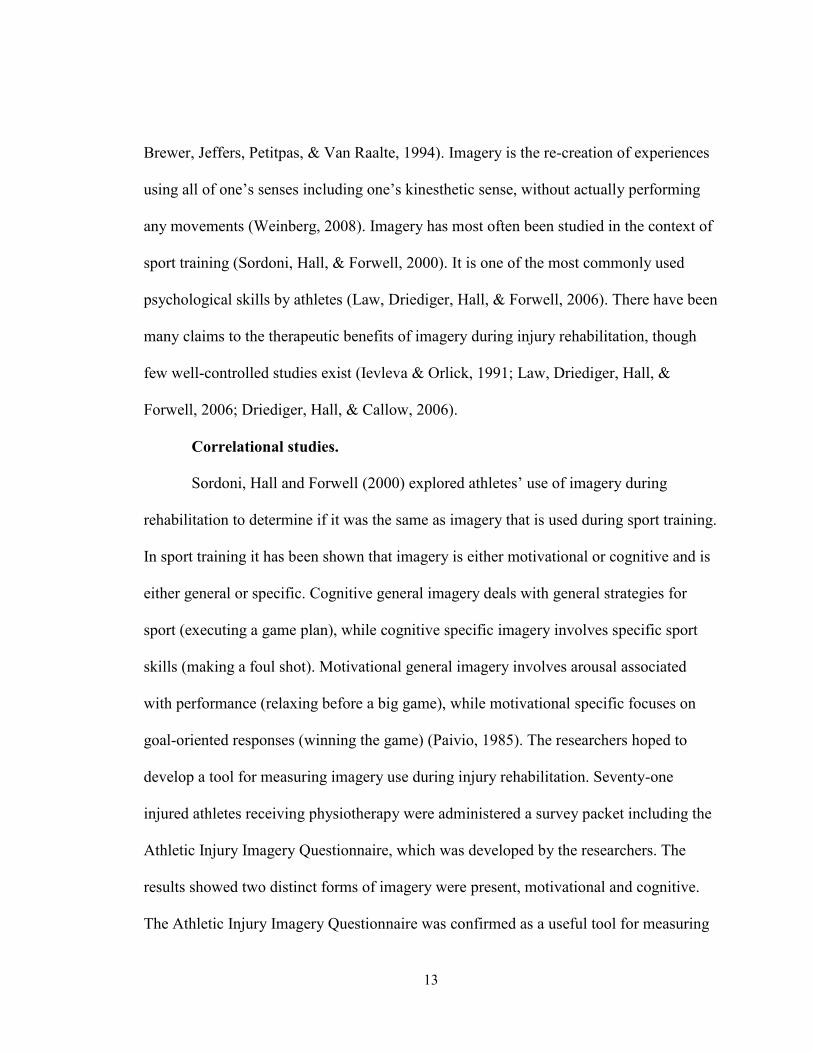
Correlational studies.
Sordoni, Hall and Forwell (2000) explored athletes’ use of imagery during
rehabilitation to determine if it was the same as imagery that is used during sport training.
In sport training it has been shown that imagery is either motivational or cognitive and is
either general or specific. Cognitive general imagery deals with general strategies for
sport (executing a game plan), while cognitive specific imagery involves specific sport
skills (making a foul shot). Motivational general imagery involves arousal associated
with performance (relaxing before a big game), while motivational specific focuses on
goal-oriented responses (winning the game) (Paivio, 1985). The researchers hoped to
develop a tool for measuring imagery use during injury rehabilitation. Seventy-one
injured athletes receiving physiotherapy were administered a survey packet including the
Athletic Injury Imagery Questionnaire, which was developed by the researchers. The
results showed two distinct forms of imagery were present, motivational and cognitive.
The Athletic Injury Imagery Questionnaire was confirmed as a useful tool for measuring

14
athletes’ imagery use during injury recovery. It was also shown that athletes use imagery
less during injury rehabilitation compared with regular sport training. Although the
Athletic Injury Imagery Questionnaire is a useful tool it is not based on empirical
knowledge of imagery actually used by injured athletes, instead it is based in
psychological theory of imagery use during sport competition (Driediger, Hall, & Callow,
2006).
Driediger, Hall, and Callow (2006) gathered empirical knowledge about what types
of imagery athletes were actually using during injury rehabilitation. Using an interview
method, the researchers spoke with ten athletes undergoing physiotherapy for an athletic
injury for at least two weeks. The researchers aimed to answer four questions: When do
injured athletes use imagery? Where do they use imagery? Why do they use imagery? What
are injured athletes imaging? Athletes were more likely to use imagery during the
physiotherapy session than before or after it. The reasons for using imagery were varied but
included pain management, healing, rehearsal of movements, and motivation. Athletes were
imaging a variety of things both positive and negative. Some reported imaging themselves
completing a rehabilitation exercise or returning to practice without restrictions. Athletes
believed that imagery served a valuable role in injury recovery. Athletes reported using less
imagery during injury rehabilitation than during sport training, which was similar in to what
Sordoni, Hall, and Forwell (2000) found.
Monsma, Mensch, and Farroll (2009) investigated the use of imagery during
rehabilitation and its effects on return-to-play anxiety. The Sport Imagery Questionnaire,
the Sport Anxiety Scale, and feelings about return to practice or competition form was

15
given to 36 athletes undergoing injury rehabilitation for at least eight days. Athletic
trainers working with these athletes were given an injury description form and asked to
rate the injury severity. All athletes reported no formal training in imagery. Of the 25
athletes who completed the survey, 68% (n=17) reported using imagery. The longer an
athlete was injured, the less imagery they seemed to use. The length of injury time was
positively related to somatic anxiety. The use of sport specific imagery by an athlete was
positively related to a more efficacious return to previous level of skill after injury.
Considering the researchers were specifically interested in imagery use during injury
rehabilitation, the Athletic Injury Imagery Questionnaire may have been a more
appropriate measure to use instead of the Sport Imagery Questionnaire, since the Athletic
Injury Imagery Questionnaire is specifically designed to assess injury imagery.
Law et al (2006) surveyed 83 athletes with lower leg injuries undergoing
physiotherapy to determine if imagery use helped reduce perceived pain and improve
limb functioning. The survey packet included the Athletic Injury Imagery Questionnaire-
2, Visual Analogue Scale for pain, the lower extremity functional scale and questions
concerning their use of imagery for pain management and satisfaction with rehabilitation.
Of the respondents, 42% (n=35) reported using imagery to manage pain and were
grouped together in the pain imagery group, while the remaining 58% (n=48) were placed
in the no pain imagery group. Athletes in the pain imagery group reported more
satisfaction with rehabilitation but there were no differences amongst the groups on
perceived pain or limb functioning. One reason for this may have been because athletes’
imagery use was only measured once, and differences between the groups may have been

16
more apparent at the beginning of the rehabilitation period. The researchers recommend
more controlled studies using imagery as an intervention with pre- and post-measures.
Psychological skills can incorporate a mind-body approach to improve the
outcomes of sport injury rehabilitation (Green, 1992; Law, Driediger, Hall, & Forwell,
2006). Imagery is one of the most commonly recommended psychological skills in sport
injury rehabilitation (Weise & Weiss, 1987; Brewer, Jeffers, Petitpas, & Van Raalte,
1994). Research has shown that many athletes are already using imagery during injury
rehabilitation (Sordoni, Hall, & Forwell, 2000; Driediger, Hall, & Callow, 2006;
Monsma, Mensch, & Farroll, 2009; Law, Driediger, Hall, & Forwell, 2006). Although
athletes are using imagery in injury rehabilitation it is often to a lesser extent than during
sport training (Sordoni, Hall, & Forwell, 2000; Driediger, Hall, & Callow, 2006). These
studies looked at the imagery athletes were currently using, but controlled intervention
studies can be used to show how the use of mental imagery affects injury rehabilitation
outcomes. Exploring the process of imagery in sport injury is not only of theoretical
importance but also of clinical importance (Christakou & Zervas, 2007). Law et al.
(2006) recommended more intervention studies that included pre- and post-measures with
imagery in injury rehabilitation.
Intervention studies.
Christakou and Zervas (2007) conducted an intervention study using imagery with
athletes undergoing physiotherapy for a grade II ankle sprain. The researchers
investigated the effectiveness of an imagery intervention on pain, edema, and range of
motion. Eighteen male athletes were randomly split into two groups. The intervention

17
group completed 12 sessions during 4 weeks of physiotherapy treatment. The control
group just completed the normal physiotherapy during the 4 weeks. Participants in the
imagery group were asked to imagine themselves performing the physiotherapy exercises
as vividly as possible. No significant results were found, but there was an increased range
of motion, and decreased pain and edema in the intervention group. The effect size was
large for pain (d= .86) and medium for range of motion (d= .52) and edema (d= .71).
Researchers noticed there was a greater difference in pain between the groups during the
second session and recommended starting imagery interventions as soon as possible after
the injury.
Chrisakou, Zervas, and Lavelle (2007) conducted a similar study examining the
effects of imagery on muscular endurance, dynamic balance, and functional stability
during a grade II ankle rehabilitation. Twenty athletes undergoing physiotherapy were
randomly assigned into two groups. The imagery intervention group completed 12
sessions during 4 weeks of physiotherapy treatment. The control group completed the
normal physiotherapy during the 4 weeks. Participants in the imagery group were asked
to imagine themselves performing the physiotherapy exercises as vividly as possible. A
single hop test for distance and a single hop test for time were performed to measure
functional stability. Dynamic balance was measured on a Biodex system and muscle
endurance was measured with a rising on heels test, rising on toes test, and walking down
stairs. Significantly greater muscle endurance was found in the intervention group, but no
other significant results were found. Treatment effects were large for the rising on toe test
(d= .85), the rising on heel test (d= .70), the dynamic balance (d= .90), and the single leg

18
hop for time (d= .91). Treatment effects for single leg hop for distance were small.
Researchers concluded that the significant increase in muscular endurance was most
likely due to central processes adaptations as a result of the imagery.
Cupal and Brewer (2001) explored the use of relaxation and guided imagery in
patients after undergoing anterior cruciate ligament reconstruction. Using thirty
participants, the researchers randomly assigned three groups: treatment, placebo and
control. The inclusion of a placebo group was the first in sport injury psychology with
athletes in rehabilitation. The treatment group received a total of ten sessions,
approximately 2 weeks apart over six months, on relaxation and guided imagery.
Researchers used internal, external, visual, and kinesthetic imagery during the
intervention. The sessions were geared towards the rehabilitation goals during the phase
of recovery the patients were in, and were recorded. The treatment group was also asked
to listen to their recorded sessions daily between sessions. Actual compliance was only
4.4 times a week on average. The placebo group was asked to spend 10-15 minutes a day
visualizing a peaceful scene. No data on compliance rates were given for the placebo
group. The control group received no additional intervention and just progressed through
their injury rehabilitation with their physical therapist. Physical therapists working with
the patients were blind to their study involvement and group membership. Re-injury
anxiety and pain were measured on a 10-point scale at the beginning and conclusion of
the study. Knee strength was measured with a Cybex machine 24 weeks post operatively
and compared to the uninjured knee. Current physical therapy protocols for anterior
cruciate ligament repair rehabilitation call for attaining 80-85% of the strength of the

19
contralateral knee. Re-injury anxiety and pain were significantly lower in the treatment
group compared with the placebo and control group. Knee strength was significantly
greater in the treatment group compared with the placebo and control group. Although
this significant improvement in knee strength was found, researchers did not attribute this
to the imagery specifically. Researchers provided two reasons why they believed the
increased knee strength was seen: 1) the treatment intervention promoted the belief that
the patients recovery was within their own control, and 2) reductions in re-injury anxiety
and pain allowed patients to engage more fully in the physical therapy sessions. Mental
imagery could not be specifically identified as the cause of the muscle strengths gains
because mental imagery was only one of two mental skills used in this study.
One area that shows promise for future research is the use of imagery to maintain
muscle strength. Lorenzo, Ives, and Sforzo (2003) investigated the effect of education
and mental imagery on knee extension strength. Sixteen college volunteers, with no
previous education in neuromuscular physiology, were randomly divided into two
groups, one receiving two one hour sessions about muscle physiology, neural control of
muscle force, and imagery training and the second receiving two one hour sessions about
general health and fitness. Isokinetic knee extensor strength was measured with five
maximal contractions pre and post intervention. There was no effect seen in the treatment
group. The researchers provided three reasons for why this may have occurred: 1) the
quality of imagery was not ascertained, 2) training had no relevance to the task, and 3)
instructions and attentional focus were inappropriate. Since the quality of the imagery
was not known, it is hard to know if the participants actually completed the imagery task
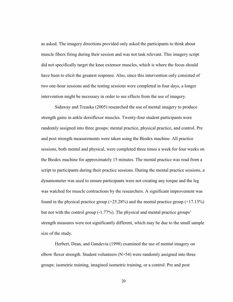
20
as asked. The imagery directions provided only asked the participants to think about
muscle fibers firing during their session and was not task relevant. This imagery script
did not specifically target the knee extensor muscles, which is where the focus should
have been to elicit the greatest response. Also, since this intervention only consisted of
two one-hour sessions and the testing sessions were completed in four days, a longer
intervention might be necessary in order to see effects from the use of imagery.
Sidaway and Trzaska (2005) researched the use of mental imagery to produce
strength gains in ankle dorsiflexor muscles. Twenty-four student participants were
randomly assigned into three groups: mental practice, physical practice, and control. Pre
and post strength measurements were taken using the Biodex machine. All practice
sessions, both mental and physical, were completed three times a week for four weeks on
the Biodex machine for approximately 15 minutes. The mental practice was read from a
script to participants during their practice sessions. During the mental practice sessions, a
dynamometer was used to ensure participants were not creating any torque and the leg
was watched for muscle contractions by the researchers. A significant improvement was
found in the physical practice group (+25.28%) and the mental practice group (+17.13%)
but not with the control group (-1.77%). The physical and mental practice groups’
strength measures were not significantly different, which may be due to the small sample
size of the study.
Herbert, Dean, and Gandevia (1998) examined the use of mental imagery on
elbow flexor strength. Student volunteers (N=54) were randomly assigned into three
groups: isometric training, imagined isometric training, or a control. Pre and post

21
treatment strength measurements were taken using a Biodex machine. Participants were
trained three times a week for eight weeks. Each training session was supervised and
participants were asked to complete six 10-second maximal isometric contractions with a
60-second rest period between contractions. All training sessions occurred while set up
on the Biodex machine. Directions for each group were given via tape-recorded
messages. The researchers did not provide a detailed script, so it is unclear if the
participants used internal or external imagery, which could affect the results. Strength
increased in all groups. The isometric training group had a 17.8% increase, imagined
training had a 6.8% increase, and the control had a 6.5% increase. Methodological
questions are raised since all three groups experienced an increase in strength gains. The
researchers presumed this was a result of the familiarity with the testing procedures at the
posttest. The imagined isometric training group was not statistically different from the
control, which raises questions about the use of mental imagery for strength gains. The
lack of clear directions for the use of imagery may also have accounted for the lack of
gains in the imagined isometric training group.
Herbert, Dean, and Gandevia (1998) and Sidaway and Trzaska (2005) both used
physical practice groups compared with mental practice groups and got different results.
Both muscle groups that were used would be considered highly trained since both are
used in daily activity (ankle dorsiflexors are used for ambulation). It is also interesting to
note that Herbert, Dean and Gandevia (1998) found a 6.5% increase in their control group
and Sidaway and Trzaska (2005) found a 1.77% decrease in their control group. One key
difference between the studies is the description of imagery provided. Sidaway and

22
Trzaska (2005) provided a much more detailed imagery script to participants which may
account for the positive effect that was shown.
Ranganathan et al. (2004) explored the use of mental imagery on fifth finger
abductor and elbow flexor strength. It is generally assumed that muscle strength gains are
from two main factors: neural adaptations and muscle hypertrophy. Cortical
representation differs in more proximal muscles, such as elbow flexors, than distal
muscles, such as fifth finger abductors. Researchers were interested if cortical
representation differences would create differences in strength gains. Thirty right-hand
dominant, previously untrained participants were randomly separated into either a fifth
finger abductor or elbow flexion mental training group. Each group was then compared to
a control group of eight subjects recruited later. The mental training sessions were fifteen
minutes long and performed five times a week for twelve weeks. This was one of the few
studies that mentioned specifically using an internal imagery script in the study. Both
groups showed statistically significant increased muscle strength compared with the
control group. The fifth finger abductors mental imagery group showed a 40% strength
increase and the elbow flexors mental imagery group showed a 13.5% increase. One
reason given for the difference in strength gains was that the elbow flexors are used more
frequently in daily life and are already highly trained, while the fifth finger abductors are
rarely used.
Yue and Cole (1992) compared imagined and maximal voluntary contractions of
the fifth metatarsal abductor. Thirty healthy participants were randomly divided into three
groups: imagery, contraction, and control. The imagery and contraction groups completed

23
20 sessions over a four-week period. All participants completed a pre-test fifth metatarsal
adductor maximum voluntary strength task to get a baseline measure of strength. The
imagery group was asked to imagine completing 15 repetitions of the pre-test adductor
strength task with 20-second rest intervals during each session. EMG activity was
measured during the imagery task to insure the muscle was inactive. The contraction
group completed this same task but actually performed 15 maximal contractions. The
imagery group experienced a 22% increase, while the contraction group had a 29.75%
increase and the control group had a 3.7% gain. The imagery and contractions groups had
a statistically significant increase in strength. There was no statistical significance
between the imagery and contraction group strength gains. This supports the idea that
strength increases can occur from an imagery task. Researchers indicated that this could
have therapeutic implications for use in combating strength loss due to immobilization.
Regaining strength during injury rehabilitation is usually a main therapeutic goal
but can be even more important during immobilization. Stenekes et al. (2009) explored
the use of mental imagery during immobilization after a flexor tendon surgical repair in
the hand. Using 25 participants, the researchers assigned the group into an imagery group
and a control group. Prior to group assignment participants completed the Vividness of
Movement Imagination Questionnaire. Participants with a score >72 were not admitted to
the motor imagery score, because scores greater than 72 reflected poor imagery skill.
Only 1 participant scored greater than 72 and was then assigned to the control group, all
others were assigned randomly. The imagery group was instructed to perform eight
imagery sessions a day. Among other measurements, the researchers measured grip

24
strength and pinchmeter (thumb pinch strength to each finger). There was no statistically
significant difference between the control and treatment group for the strength measures.
At the end of the intervention, the researchers asked about compliance to the imagery
sessions. The researchers reported that the participants were not completely compliant.
Participants averaged 100 sessions, ranging from 2-294 sessions completed. The
researchers did not indicate how many total sessions were possible.
The imagery script that accompanied this intervention was not very descriptive
and did not employ the use of all senses, which is known to improve imagery results
(Weinberg, 2008). The treatment and control groups differed in the number of tendons
ruptured on average for each participant (treatment=2.3, control 1.5). This difference
could have resulted in greater strength losses in the treatment group, which would offset
any gains in comparison with the control group. Analogous groups at the start of the
intervention make it easier to compare differences post treatment amongst the groups.
Newsom, Knight, and Balnave (2003) also looked at mental imagery to maintain
grip strength following immobilization, this time in a healthy population. In this study, 18
healthy participants non-dominant forearm was immobilized for ten days. Strength loss
has been shown to occur rapidly during the first week of immobilization. Participants’
grip strength, isometric wrist flexion, and isometric wrist extension were measured both
pre- and post-immobilization. Group assignment was done randomly. The mental
imagery group was asked to participate in three five-minute imagery sessions a day
guided by an audiotape. Participants were asked to imagine themselves gripping a ball.
Participants responded that they completed between 26-30 sessions with a mean of 28

25
1.7. There was no significant difference between the groups; but the mental imagery
group showed no significant loss in strength (-1.5%), while the control group experienced
a larger loss in strength (-16.3%). The study did have some limitations. The way that the
participants were casted did allow for some gripping, as well as third finger and thumb
opposition. This could have allowed for movement during the imagery sessions although
the treatment group was instructed to not move their hand during the imagery. These
findings suggest that mental imagery can be effective in maintaining muscle strength
during immobilization.
The research in this area is often contradictory with some findings supporting the
use of mental imagery to improve muscle strength (Newsom, Knight, & Balnave, 2003;
Ranganathan, Siemionow, Liu, Sahgal, & Yue, 2004; Sidaway & Trzaska, 2005; Cupal &
Brewer, 2001; Yue & Cole, 1992), and other findings not supporting it (Stenekes,
Geertzen, Nicolai, De Jong, & Mulder, 2009; Herbert, Dean, & Gandevia, 1998; Lorenzo,
Ives, & Sforzo, 2003). Potential explanations of the inconsistencies may be likely due to
methodological inconsistencies. First, it is understood that an internal mental imagery
creates the greatest physiological benefits (Ranganathan, Siemionow, Liu, Sahgal, &
Yue, 2004; Hale, 1982; Harris & Robinson, 1986) but many of these studies did not
employ this type of mental imagery or were unclear in the type of imagery used. Internal
imagery has been shown to create a greater muscular response than external imagery
(Hale, 1982; Harris & Robinson, 1986). Only four studies specifically used an internal
imagery script (Ranganathan, Siemionow, Liu, Sahgal, & Yue, 2004; Newsom, Knight,
& Balnave, 2003; Sidaway & Trzaska, 2005; Yue & Cole, 1992). Cupal and Brewer

26
(2001) used internal imagery mixed with external, visual and kinesthetic. Interestingly,
all four studies that used an internal imagery script found support for the use of mental
imagery to maintain or promote muscle strength. Another potential explanation for
disparate findings is that researchers have used mental imagery that is not relevant to the
task. Harris and Robinson (1986) showed that localized response during an imagery task
is specific to the muscle group being used. Task irrelevant imagery scripts will fail to
produce the muscle response desired. Protocols for the imagery intervention also vary
amongst researchers. Some use the intervention in as few as four days and others up to
six months. Research has shown that most muscle strength loss occurs within the first
week of immobilization after which little additional strength loss occurs (Newsom,
Knight, & Balnave, 2003). Also, the total number of sessions completed varies, as well as
whether the sessions were completed alone or with a clinician. In studies where sessions
were completed without a clinician, manipulation checks for adherence to the protocol
were not always employed. Having better knowledge of actual adherence to the imagery
script could help explain the results.
Force Steadiness
Force steadiness is the ability to match a given sub-maximal force with limited
fluctuations. Force steadiness can be used to assess sensory-motor control in a muscle
(Bandholm, Radmussen, Aagaard, Jenson, & Diederichsen, 2006). Force steadiness is
important in fine motor tasks such as writing.
Lundbye-Jensen and Nielsen (2008) studied strength and force steadiness changes
following one week of wrist and hand immobilization. Ten healthy participants were

27
immobilized in a non-dominant, forearm, wrist and hand cast. Strength and force
steadiness measures were obtained on two different days prior to immobilization and
immediately following immobilization, as well as one week post-immobilization.
Maximal wrist flexion and extension decreased significantly (p < 0.001 and p = 0.004,
respectively) following immobilization, and returned to baseline values one-week post
immobilization. Force steadiness was measured at 10% of maximal muscle strength in
the flexor carpi radialis and abductor pollicis brevis. Force steadiness increased following
immobilization. There was an increased variability in flexor carpi radialis from 2.5 and
2.7% at pretest to 5.2% following immbolization (p = 0.042). In the abductor pollicis
brevis variability increased from 3.0 and 3.2% at pretest to 6.2% following
immobilization (p = 0.048). With just one week of immobilization there was significant
decreases in muscle strength and significant increases in force steadiness. Researchers
believe this change was due to central nervous adaptations since there were no changes
seen in the muscle.
Mulder et al. (2011) examined the effects of bed rest and resistance training on
force steadiness. Participants (N = 22) were randomly assigned to resistance exercise
group (n = 7), resistance exercise plus whole body vibration (n = 7), or inactive control
group (n = 8). All participants completed a 60-day head down tilt bed rest protocol. The
exercise intervention took approximately 23 minutes and was performed 3x a week.
Exercises included: bilateral squats, single leg heel raises, double leg heel raises and back
extension. Prior to the bed rest protocol participants performed four plantar flexion force
steadiness tasks at 20%, 40%, 60%, 80% of maximal plantar flexion strength for 15s
28
trials. Force fluctuations are reported as coefficient of variation (CV=SD/M). During the
data analysis both resistance training groups’ data was pooled together. Although the data
was not reported, researchers said there was no statistical difference between the two
resistance training groups. Across all levels CV increased significantly (p < 0.005) more
for the bed rest control group (from 0.31 0.10% to 0.92 0.63%) than the resistance-
training group (from 0.39 0.09% to 0.54 0.72%). The largest increases in CV
occurred at 20% of maximal plantar flexion strength. Bed rest resulted in the loss of
plantar flexion strength as well an increase in CV. These results show that resistance
training is partially successful in controlling the loss of plantar flexor force steadiness due
to bed rest.
Shinohara et al. (2003) looked at strength and force steadiness in the knee and
ankle extensor muscles of the legs. Twenty healthy participants were recruited for the
study; six were assigned to the bed rest and strength training group and fourteen to the
bed rest control group. The bed rest protocol was for 20 days. Strength training was
completed 16 out of 20 days and included bilateral calf raises and leg press. Force
steadiness was measured at 2.5, 5.0, 7.5 and 10% of maximal strength. Strength
significantly decreased in the bed rest only group (p < 0.05), while the bed rest and
strength training group had no significant difference in strength before and after bed rest.
The CV for each individual at ankle extensors averaged 88% increase in the control
group which was significantly greater (p < 0.05) than the resistance training group (41%).
For the knee extensors the CV increased by average 22% in the control group which was

29
significantly greater (p < 0.05) than the strength training group (4%). Strength training
was able to counteract the effects of bed rest on force fluctuations.
Physical inactivity, such as bed rest, joint immobilization or limb unloading, have
been linked to muscle strength loss, atrophy and neural alterations in the muscle
(Lundbye-Jensen & Nielsen, 2008; Mulder, et al., 2011; Shinohara, Yoshitake, Kouzaki,
Fukuoka, & Fukunaga, 2003). Past research has shown that strength training can improve
maximal force and force steadiness. Research suggests that strength training effects the
neural mechanisms important for maintaining force steadiness (Mulder, et al., 2011;
Shinohara, Yoshitake, Kouzaki, Fukuoka, & Fukunaga, 2003).
Currently there has been no research done on the effects of mental imagery on
force steadiness. Research has shown that mental imagery can be used to offset the loss
of strength during immobilization (Newsom, Knight, & Balnave, 2003). Based on the
ability of strength training to offset the loss of force steadiness during immobilization, it
is hypothesized that mental imagery may help maintain force steadiness as well.
Future Research Directions
In order to advance the knowledge in the area of mental imagery and muscle
strength, better research needs to be done. Specifically controlled studies that address
limitations of previous research need to be performed. Within immobilization studies,
protocols of at least 7 days must be used, with an internal imagery script that is task
relevant to the targeted muscles. Also the targeted muscles should be completely
immobilized to prevent any unwanted movements during the intervention. Once this

30
research is completed we can then begin to have a better understanding of the effects of
mental imagery on muscle strength.
Currently, there is no definitive understanding of the effects of mental imagery on
muscle strength (Sidaway & Trzaska, 2005). As previous researchers have recommended
(Law, Driediger, Hall, & Forwell, 2006), more controlled imagery intervention studies
are needed. If future research can show that mental imagery can help maintain muscle
strength, protocols can be developed for the use of mental imagery in injury
rehabilitation. Currently, rehabilitation techniques to gain muscle strength require a
muscle contraction, which may not always be possible for the patient following injury
(Sidaway & Trzaska, 2005). Thus, mental imagery could allow for muscle strength
training to begin while a patient is still immobilized or is too weak to contract their
muscles. Earlier muscle strength training may lead to shorter rehabilitation periods and
quicker advancement to functional exercises. This could encourage the use of mental
imagery amongst rehabilitation professionals, potentially create better outcomes for their
patients, and support evidence-based practice.
The purpose of this research study is to explore if internal mental imagery can
prevent the loss of thenar muscle group strength during thumb immobilization. This will
address prior limitations by using an internal mental imagery script that is also task
relevant. A thumb spica cast will be used to completely immobilize the thenar muscles of
the non-dominnant hand for 7 days. It is expected that internal mental imagery will
maintain thenar muscle strength during immobilization. A secondary purpose is to

31
explore if internal mental imagery can help maintain force steadiness during thumb
immobilization.
32
CHAPTER III
METHODS
Participants
Participants were recruited from local universities and the community at large to
be invited to participate in the study. Respondents were interviewed prior to participation
to make sure they met all of the inclusion criteria. Inclusion criteria included: 1) no prior
injury, that required splinting or casting, to the non-dominant thumb in the past three
years, 2) ability to access the Internet and listen to an imagery script for five-minutes a
day for seven consecutive days, 3) no skin condition on the non-dominant hand that could
be affected by casting and 4) ability to listen to and understand English. A sample of 20
participants was recruited for this study. Two participants dropped from participation
prior to completing the study, resulting in a total of 18 participants who completed the
study for a retention rate of 90%. Participants were both male (N=5) and female (N=13)
and ranged in age between 19-35 (M= 24.1, SD= 4.93). Based on their id number,
participants were randomized into two groups (i.e., imagery and control) by a research
assistant.
Measures
Hand dominance.
Participant hand dominance was determined based on the Edinburgh Handedness
Inventory (Oldfield, 1971) (Appendix A). Participants marked hand preference on ten

33
common tasks (e.g. writing) to determine hand dominance, and then items were summed
for a total score that may range between -100 and 100. Scores greater than 40 confirm
right hand dominance and scores less than -40 confirm left hand dominance. Scores equal
to and between 40 and -40 confirm ambidextrous hand use. No participants scored in the
ambidextrous scale. The non-dominant hand was used for all tasks during the experiment.
Imagery ability.
The Vividness of Movement Imagery Questionnaire-2 (VIMQ-2) (Roberts,
Callow, Markland, & Bringer, 2008) was used to assess participants’ imagery ability
(Appendix B). The VIMQ-2 rates the vividness of imagery on three types of imagery:
internal, external, and kinesthetic during 12 imagined tasks. Items for each type of
imagery are summed for a score that may range from 12-60, with lower scores indicating
better vividness of imagery for each specific type of imagery. Participants were not
excluded based upon imagery ability as some past studies have done.
Thenar muscle group strength.
The dependent variable of this study is thenar muscle group strength measured pre- and
post-immobilization. Thenar muscle group strength was tested through maximum
voluntary muscle contractions (MVC). The thenar muscle group includes: abductor
pollicis brevis, flexor pollicis brevis, adductor pollicis brevis, and opponens brevis. MVC
was measured using a force transducer (Grass FT03, Grass Technologies, West Warwick,
RI). Specifically thumb flexion, opposition, adduction and abduction were measured, to
target the four muscles of the thenar muscle group. Participants were secured to the
testing table and the force transducer and instructed on how to perform a MVC. Three

34
trials of maximal force were recorded for each thumb direction tested for each
participant. The force transducer recorded the force generated for every trial and was
saved to a lab computer. The greatest MVC trial within 10% of another MVC trial was
recorded as the MVC. If no trials were within 10% of a previous trial, then additional
trials were completed. The analog signal obtained from the force transducer was digitized
(CED 1401, Cambridge UK) and processed (Spike 2, CED, Cambridge UK; Excel,
Microsoft, Redman WA) to acquire force data. Data from Spike 2 was recorded in Volts
(V) and converted to Force (N) using a known calibration factor (mV x 20.98 = g). The
mean force was taken of the largest .5 second, visually identified, of the strongest MVC
trial in each of the four directions (abduction, adduction, flexion, and extension).
Force steadiness.
Force steadiness is a measure of one’s ability to maintain submaximal isometric
contraction over a period of time (Bandholm, Radmussen, Aagaard, Jenson, &
Diederichsen, 2006). This was measured using a force-matching task that had the
participant try to match a constant force displayed on the monitor. Participants were in
the same testing position used for the MVC trials. Force steadiness was only measured in
thumb flexion. Force steadiness was measured at three levels: 5%, 25%, and 50% of
MVC. Each trial the target force was placed in the center of the screen and lasted 20
seconds. For force steadiness, the steadiest 10-second portion of each trial was identified
visually for use. The magnitude of force fluctuations is reported as a coefficient of
variation of the force signal (CV= S.D./Mean) (Danion & Galléa, 2004). CV scores that
are larger indicate a greater fluctuation in force and decreased force steadiness in that

35
muscle (Shinohara, Yoshitake, Kouzaki, Fukuoka, & Fukunaga, 2003). The order of the
force steadiness trials were randomized for participants.
Adherence.
Two Blackboard (Blackboard Inc.) organizations were created to measure
adherence to the protocol. Participants were assigned to an organization based upon
group assignment. Regardless of group assignment, all participants were asked to log on
to Blackboard twice daily to answer two questions: Have you used your casted hand at all
today? And have you driven at all today? The imagery group was also asked to complete
the imagery script accessible through their organization, twice daily. Participants were
instructed to log in once in the morning and once at night. The number of times each
participant answered the questions was recorded to measure adherence. Participants could
have answered the questions a total of 14 times during the 7-day intervention.
Procedures
The University of North Carolina Greensboro’s Institutional Review Board
approved the data collection and recruitment procedures. During the first testing session,
subjects completed informed consent for their participation in the study. After informed
consent, each participant completed the VIMQ-2 and the Edinburgh Handedness
Inventory. Thenar muscle group strength testing and force steadiness measures were
performed prior to immobilization to establish a baseline. The primary researcher
completed all pre- and post- intervention muscle strength testing. This was done to avoid
the threat of inter-tester reliability on the data collected. Participants were seated
comfortably upright in the lab with their elbow flexed to 90 degrees and their non-

36
dominant hand supinated and resting on a wooden board secured to the table. The non-
dominant thumb was secured to a finger splint using medical tape. Participants were
secured to the board using velcro straps across the hand and forearm. The force
transducer base was bolted to the wooden board. The force transducer was attached to the
thumb using an adjustable metal pipe clamp and metal wire (Appendix C). This allowed
for adjustments based on participant hand size. Care was taken to keep thumb placement
uniform across participants. Participants were instructed to increase their strength over a
3 second countdown to reach their maximum force and then maintain the force for
another 3 seconds. Three trials were completed for each thumb direction. The greatest
MVC trial within 10% of another MVC trial was recorded as the MVC. If no trials were
within 10% of the other trials, then additional trials were completed until this was
achieved.
After pre-intervention muscle strength testing, all subjects were immobilized in a
fiberglass thumb spica cast by the primary researcher, who is a licensed and certified
athletic trainer. This type of cast immobilized the thumb, which limited the thenar muscle
group. The thumb was placed into flexion to allow for shortening of the flexor pollicis
brevis. Muscles immobilized in a shortened position show increased strength loss
(Wagatsuma, Yamazaki, Mizuno, & Yamada, 2001). Participants were immobilized for
seven days.
After the cast was set participants were given cast care instructions (Appendix D)
and instructions on how to access the Blackboard organizations established for the study.
These organizations could be accessed from any computer connected to the Internet, and

37
participants accessed the appropriate content based on their group assignment (imagery
or control). Participants were instructed to not tell the primary researcher what group they
were enrolled in and to address any questions regarding access or use of the Blackboard
organization to the research assistant. . A research assistant randomly assigned
participants to the imagery group (n=10) and control group (n=8). The primary researcher
was blinded to participants’ group assignment and served as a control for experimenter
bias during post immobilization strength testing. This helped to ensure that accurate
strength measurements were taken and minimized expectancy threats to internal validity
of the study.
Regardless of group assignment participants were asked to log on to Blackboard
twice daily to answer two questions: Have you used your casted hand at all today? And
have you driven at all today? These questions were intended to make sure both groups
were logging on daily and staying active in the experiment. Also it allowed for there to be
a tally of how many times the organization was accessed by each participant to determine
adherence to the protocol. In order to encourage adherence, participants gained one entry
into a $100 Target gift card raffle for every time they answered the questions. Both
organizations also had an electronic copy of the cast care instructions for participants.
Participants assigned to the imagery group were able to access the mental imagery audio
script (Appendix E) from the organization. The audio file was approximately 8 minutes
long. The mental imagery script guided participants through five imagined contractions in
the four directions of muscle strength testing. Participants were instructed to listen to the
script twice daily in a quiet location. During the muscle strength testing care was taken to

38
clearly explain the four directions of thumb movement being tested to assist the imagery
group while listening to the mental imagery script. The imagery group was also provided
with a document with photos showing them each thumb direction to remind them of the
thumb movement directions while they were completing the script (Appendix F).
After the seven-day immobilization period, subjects met again with the primary
researcher for cast removal and lab testing. The casting tape being used (3M Softcast)
allowed for easy removal with scissors. The primary researcher retested thumb strength
and force steadiness following the same pre-test protocols.
Data Analysis
Group differences in age and VMIQ-2 were analyzed using a one-way ANOVA.
Chi square analyses were run for gender and hand dominance. These analyses were run to
identify potential group differences in age, imagery ability, gender, and hand dominance
at baseline.
The primary research question was: does internal mental imagery prevent the loss
of thumb strength during thumb immobilization? To examine this question, four
repeated-measures 2 (group: imagery, control) x 2 (time: baseline, posttest) ANOVAs
were performed with each of the MVC measures as dependent variables (abduction,
adduction, flexion, and extension). The secondary research question was: does internal
mental imagery help maintain force steadiness during immobilization? To examine this
question, three separate 2 (group) x 2 (pre/post) repeated-measures ANOVAs were
performed with each of the levels of force steadiness (5%, 25%, and 50%) as dependent
variables. Supplemental to these analyses, between group adherence rates were compared

39
using a one-way ANOVA. Analyses were run at alpha=.05 to determine statistical
significance. Data was analyzed using IBM SPSS Statistics, version 19.

40
CHAPTER IV
RESULTS
Participants
Twenty participants were recruited for the study but two participants,
discontinued participation before completion of the study due to an uncomfortable fit of
the cast. A sample of 18 participants (5 men, 13 women) completed this study, and they
ranged in age from 19-35 years (M = 24.1, SD = 4.93). Of the 18 participants, sixteen
were right-hand dominant and two were left-hand dominant. Participants were randomly
assigned to either the imagery group (n=10) or control group (n=8).
Preliminary Analysis
The control group included six females and two males, between 19-32 years of
age (M = 22.63, SD = 4.207), all of whom were right-hand dominant. The imagery group
included seven females and three males, between 21-35 years of age (M = 25.2, SD =
5.371), nine of whom were right-hand dominant and two of whom were left-hand
dominant. Imagery and control groups did not differ by gender, X2 = .471, df = 1, p=
.492, nor did they differ by hand dominance, X2 =1.818, df = 1, p= .178.
Means and standard deviations for groups’ imagery ability scores are presented in
Table 1. As can be seen, the control group scored slightly lower on internal and external
imagery ability compared with the imagery group; however these differences were not
statistically significant, F’s (1,16)= 0.816 and 0.240, p’s= .380 and .631, respectively.
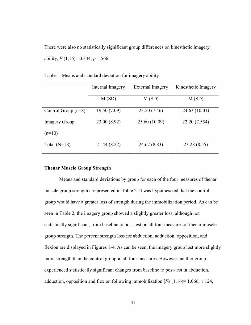
41
There were also no statistically significant group differences on kinesthetic imagery
ability, F (1,16)= 0.344, p= .566.
Table 1. Means and standard deviation for imagery ability
Internal Imagery External Imagery Kinesthetic Imagery
M (SD) M (SD) M (SD)
Control Group (n=8) 19.50 (7.09) 23.50 (7.46) 24.63 (10.01)
Imagery Group
(n=10)
23.00 (8.92) 25.60 (10.09) 22.20 (7.554)
Total (N=18) 21.44 (8.22) 24.67 (8.83) 23.28 (8.55)
Thenar Muscle Group Strength
Means and standard deviations by group for each of the four measures of thenar
muscle group strength are presented in Table 2. It was hypothesized that the control
group would have a greater loss of strength during the immobilization period. As can be
seen in Table 2, the imagery group showed a slightly greater loss, although not
statistically significant, from baseline to post-test on all four measures of thenar muscle
group strength. The percent strength loss for abduction, adduction, opposition, and
flexion are displayed in Figures 1-4. As can be seen, the imagery group lost more slightly
more strength than the control group in all four measures. However, neither group
experienced statistically significant changes from baseline to post-test in abduction,
adduction, opposition and flexion following immobilization [Fs (1,16)= 1.066, 1.124,

42
0.127, and 0.970, ps= .317, .305, .727, and .339, ηp2s= .062, .066, .008, and .057,
respectively].

43
Table 2. Means and standard deviations for thenar muscle group strength
Control Group Imagery Group Total Pre Post M Pre Post M Pre Post M
M (SD) M (SD) M (SD) M (SD) M (SD) M (SD) Abduction 58.14 N
(42.36 N)
49.61 N
(54.87 N)
8.53 N 55.13 N
(28.25 N)
23.53 N
(5.60 N)
31.6 N 56.47 N
(34.11 N)
35.12 N
(37.87 N)
21.35 N
Adduction 163.30 N
(76.02 N)
130.36 N
(53.19 N)
32.94 N 219.45 N
(86.83 N)
142.67 N
(88.16 N)
76.78 N 194.50 N
(84.83 N)
137. 20 N
(72.93 N)
57.30 N
Opposition 158.90 N
(86.62 N)
108.13 N
(34.31 N)
50.77 N 171.39 N
(76.46 N)
106.03 N
(60.74 N)
65.36 N 165.84 N
(78.90 N)
106.93 N
(49.39 N)
58.91 N
Flexion 135.07 N
(67.49 N)
110.20 N
(46.20 N)
24.87 N 184.22 N
(67.00 N)
126.48 N
(66.09 N)
57.74 N 162.38 N
(69.88 N)
119.24 N
(57.11 N)
43.14 N
43
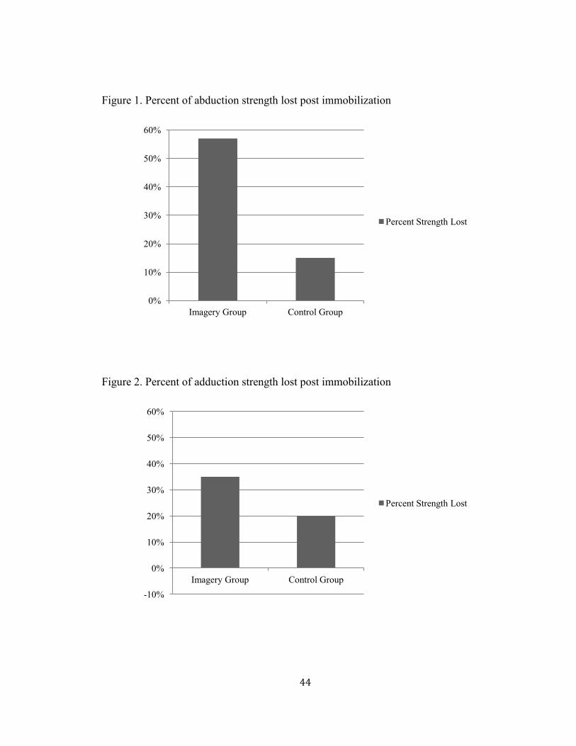
44
Figure 1. Percent of abduction strength lost post immobilization
Figure 2. Percent of adduction strength lost post immobilization
0%
10%
20%
30%
40%
50%
60%
Imagery Group Control Group
Percent Strength Lost
-10%
0%
10%
20%
30%
40%
50%
60%
Imagery Group Control Group
Percent Strength Lost

45
Figure 3. Percent of opposition strength lost post immobilization
Figure 4. Percent of flexion strength lost post immobilization
-10%
0%
10%
20%
30%
40%
50%
60%
Imagery Control
Percent Loss
-10%
0%
10%
20%
30%
40%
50%
60%
Imagery Control
Percent Loss
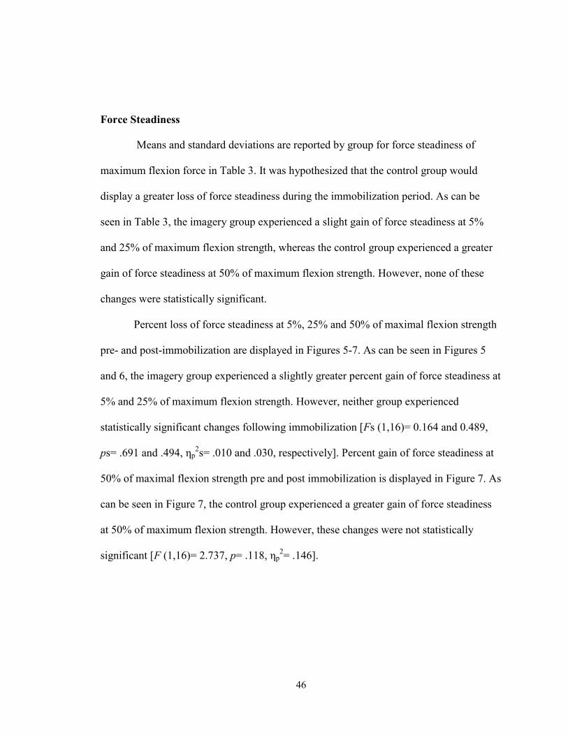
46
Force Steadiness
Means and standard deviations are reported by group for force steadiness of
maximum flexion force in Table 3. It was hypothesized that the control group would
display a greater loss of force steadiness during the immobilization period. As can be
seen in Table 3, the imagery group experienced a slight gain of force steadiness at 5%
and 25% of maximum flexion strength, whereas the control group experienced a greater
gain of force steadiness at 50% of maximum flexion strength. However, none of these
changes were statistically significant.
Percent loss of force steadiness at 5%, 25% and 50% of maximal flexion strength
pre- and post-immobilization are displayed in Figures 5-7. As can be seen in Figures 5
and 6, the imagery group experienced a slightly greater percent gain of force steadiness at
5% and 25% of maximum flexion strength. However, neither group experienced
statistically significant changes following immobilization [Fs (1,16)= 0.164 and 0.489,
ps= .691 and .494, ηp2s= .010 and .030, respectively]. Percent gain of force steadiness at
50% of maximal flexion strength pre and post immobilization is displayed in Figure 7. As
can be seen in Figure 7, the control group experienced a greater gain of force steadiness
at 50% of maximum flexion strength. However, these changes were not statistically
significant [F (1,16)= 2.737, p= .118, ηp2= .146].

47
Table 3. Means and standard deviations of force steadiness at 5, 25, and 50% maximal force strength
Control Group Imagery Group Total
Pre Post M Pre Post M Pre Post M M (SD) M (SD) M (SD) M (SD) M (SD) M (SD)
5% .154
(.198)
.122 (.208) .032 .084 (.144) .021 (.108) .064 .115 (.168) .066
(.163)
.049
25% .057
(.066)
.060 (.109) -.003 .052 (.058) .024 (.009) .028 .054 (.060) .040
(.072)
.014
50% .094
(.137)
.025 (.013) .069 .025 (.017) .024 (.007) .001 .056 (.095) .024
(.010)
.032
46
47
47

48
Figure 5. Percent gain of force steadiness at 5% of maximum flexion strength post
immobilization
Figure 6. Percent gain of force steadiness at 25% of maximum flexion strength post
immobilization
0%
10%
20%
30%
40%
50%
60%
70%
80%
Imagery Control
Percent Loss
-10%
0%
10%
20%
30%
40%
50%
60%
70%
80%
Imagery Control
Percent Loss

49
Figure 7. Percent gain of force steadiness at 50% of maximum flexion strength post
immobilization
Adherence to Protocol
Of the 18 participants to complete the study, the average number of times they
logged onto Blackboard was 11.06 (SD= 2.485) out of 14 possible log-ins. This reflects
an adherence rate of 79%. The control group had a mean adherence of 11.75 (SD=
1.669). The imagery group had a mean adherence of 10.5 (SD= 2.953), but there was no
significant between group difference, F (1,16)= 1.134, p= .303.
Participants reported using their casted hand an average of 5.39 (SD= 1.754) days.
The control group reported using their casted hand an average of 5.00 (SD= 2.000) days.
The imagery group reported using their casted hand an average of 5.70 (SD= 1.567) days.
There was no significant between group difference for reported hand use [F (1,16)= .695,
p= .417].
Participants reported driving an average of 5.28 (SD= 2.052) days. The control
group reported driving an average of 5.50 (SD= 1.512) days. The imagery group reported
0%
10%
20%
30%
40%
50%
60%
70%
80%
Imagery Control
Percent Loss

50
driving an average of 5.10 (SD= 2.470) days. There was no significant between group
difference for reported days driven [F (1,16)= .160, p= .694].

51
CHAPTER V
DISCUSSION
Past research has recommended more controlled intervention studies on the use of
mental imagery to maintain muscle strength (Stenekes, Geertzen, Nicolai, De Jong, &
Mulder, 2009; Newsom, Knight, & Balnave, 2003). Findings have been inconsistent
amongst past researchers on the effects of mental imagery on muscle strength.
Differences in methodology could help explain the inconsistencies. While different types
of imagery have been employed, internal imagery has been shown to create the greatest
muscular response (Hale, 1982; Harris & Robinson, 1986). Task-relevant imagery is also
an important factor in creating muscular response (Harris & Robinson, 1986). In past
research, muscles being tested were not always fully immobilized. To address these
limitations, the purpose of this current study was look at the effects of task-relevant
internal kinesthetic mental imagery on thenar muscle group strength during thumb
immobilization. The primary hypothesis was that mental imagery would maintain thenar
muscle group strength during a 7-day period of thumb immobilization. Additionally, this
study also explored the effects of task-relevant internal kinesthetic mental imagery on
thumb flexion force steadiness. Similar to thenar muscle group strength, it was expected
that mental imagery would also maintain thenar flexion force steadiness during a 7-day
period of thumb immobilization.

52
Thenar Muscle Group Strength
The primary hypothesis, that mental imagery would maintain thenar muscle group
strength during a 7-day thumb immobilization, was not supported. There was no
statistically significant difference found between groups on any of the four measures of
thenar muscle strength. The imagery group actually had a greater average strength loss
than the control group on all four measures. For example, the imagery group lost 57% of
thenar muscle strength compared with the control group who lost 15% of thenar muscle
strength in abduction. In adduction the imagery group loss 35% of strength compared
with the control group loss of 20%. In opposition the imagery group loss 38% of strength
compared with the control group loss of 32%. In flexion the imagery group loss 31% of
strength compared with the control group loss of 18%. It is important to note that both
groups loss muscle strength during the weeklong immobilization period.
This finding is consistent with previous studies that failed to demonstrate mental
imagery would maintain muscle strength (Stenekes, Geertzen, Nicolai, De Jong, &
Mulder, 2009; Lorenzo, Ives, & Sforzo, 2003; Herbert, Dean, & Gandevia, 1998).
However, some studies have demonstrated support for mental imagery to maintain
muscle strength (Newsom, Knight, & Balnave, 2003; Ranganathan, Siemionow, Liu,
Sahgal, & Yue, 2004; Sidaway & Trzaska, 2005; Yue & Cole, 1992). Several potential
explanations for the current findings are provided below, and these include thenar
muscles being highly trained, length of the intervention, validity of baseline measures,
and participant learning effect.

53
The thenar muscle group can be considered highly trained muscles since they are
used frequently in daily living. Therefore, it may be harder to maintain strength through a
mental imagery intervention with highly trained muscles. Three previous studies have
looked at the effect of mental imagery on muscle strength in highly trained muscles
(Sidaway & Trzaska, 2005; Herbert, Dean, & Gandevia, 1998; Ranganathan, Siemionow,
Liu, Sahgal, & Yue, 2004). Rangathan et al. (2004) examined the effects of mental
imagery on both highly trained muscles (elbow flexors) and rarely used muscles (fifth
finger abductors). Ranganathan et al. (2004) found decreased rates of strength gains in
the elbow flexors, which only gained 13.5% compared with the fifth finger abductors,
which gained 40% of strength during the imagery intervention. The fifth finger abductors
had a significantly greater gain in strength compared with the elbow flexor group.
Herbert, Dean, and Gandevia (1998) also examined the effects of mental imagery on
elbow flexors but found no significant differences in strength compared to the control
group. In contrast, Sidaway and Trzaska (2005) did find significant strength gains in
ankle dorsi flexors, a highly trained muscle group, after a mental imagery intervention.
The failure to support mental imagery in maintaining muscle strength may also
have been due to the length of intervention. Sidaway and Trzaska (2005) examined an
intervention that was 4 weeks in duration, Herbert, Dean and Gandevia (1998) tested an
8-week intervention, and Ranganathan et al. (2004) tested a 12-week intervention.
Sidaway and Trzaska (2005) and Herbert and Dean (1998) both found support for the use
of mental imagery to maintain muscle strength. It is possible that 4 weeks may represent
the minimal optimal intervention time frame for strength gains. Since this study was only

54
one week in length, a longer intervention may be necessary in order to find significant
differences in muscle strength. Sidaway and Trzaska (2005) also provided a very detailed
internal imagery script. It is unclear how detailed the other two imagery scripts were but
Ranganathan et al. (2004) did also report using an internal script. Internal mental imagery
has been shown by researchers to create the greatest physiological response in muscles
(Hale, 1982; Harris & Robinson, 1986). An internal and task specific mental imagery
script was developed for this intervention.
Event though an internal and task specific imagery script was used some of the
language in the script may have made it hard for participants to complete it as directed.
Anatomical terms for thumb directions were used (adduction, abduction, opposition and
flexion). Although participants were instructed on these terms during the lab session and
provided pictures depicted these directions, unfamiliarity with the terms may have
confused participants. At the end of the study there was no check to how well participants
understood the imagery script. A script that replicates common tasks such as Newsom,
Knight, and Balnave’s(2003) imagery script that required gripping a ball, may have been
easier to understand for participants.
Another reason the hypothesis was not supported may be due to errors in baseline
measures. Of the eight participants in the control group, five of them gained strength
from pre to post immobilization testing in at least one of the four directions. One
participant gained strength in all four directions following immobilization. In the
immobilization group four of the eleven participants gained strength in at least one
direction. Since strength loss is expected within seven days of immobilization (Newsom,

55
Knight, & Balnave, 2003), the strength gains found may be due to inaccurate baseline
measures. Baseline measures could be inaccurate due to participants not putting forth
100% effort during the pre-immobilization trials. If participants did not produce 100%
effort at the pre-immobilization testing an accurate baseline could not be established.
Another reason for the increase in strength may be due to participants’ familiarity with
the testing protocol. A learning effect may have occurred between the pre-test and post-
test that resulted in increased strength gains after the immobilization period. Since the
strength task was unique, participants may have showed increased score because they
learned how to perform the task more effectively. The same researcher did all of the
testing to try to limit inter tester reliability errors.
Although the changes found in thenar muscle group strength did not support the
hypothesis, they were similar to what has been reported previously (Stenekes, Geertzen,
Nicolai, De Jong, & Mulder, 2009; Lorenzo, Ives, & Sforzo, 2003; Herbert, Dean, &
Gandevia, 1998). Lorenzo, Ives, and Sforzo (2003) examined the effects of mental
imagery and education on knee extensor strength. There was no effect seen in the
treatment group compared with the control group. The researchers did indicate that the
mental imagery was not relevant to the strength task. Task relevant imagery is needed to
create the greatest muscular response (Harris & Robinson, 1986). Stenkes et al. (2009)
explored the use of mental imagery to maintain strength after immobilization for flexor
tendon surgery of the hand. There was no significant difference between the control and
imagery group. The imagery group did have a greater number of average ruptured
tendons (2.3) compared with the control group (1.5). Also actual compliance to the

56
protocol was not well documented. Participants completed their imagery sessions at home
without supervision, which is similar to this study. Supervised imagery sessions with a
practitioner would help make sure participants were completing the imagery sessions as
directed.
Other studies have demonstrated support for mental imagery to maintain muscle
strength (Newsom, Knight, & Balnave, 2003; Ranganathan, Siemionow, Liu, Sahgal, &
Yue, 2004; Sidaway & Trzaska, 2005; Yue & Cole, 1992). Newsom, Knight, and
Balnave (2003) examined the use of mental imagery to maintain muscle strength
following wrist immobilization in healthy participants. The control group lost a
significant amount of strength following immobilization while the imagery group did not.
In this study the tested structures were not completely immobilized during the
immobilization period, which may have allowed for extra contractions especially during
the imagery sessions. Yue and Cole (1992) found a significant increase in fifth metatarsal
abductor strength following an imagery intervention. Using healthy participants over a
four-week period the imagery group experienced a 22% increase in strength compared
with the control group’s 3.7% gain. Fifth finger abductors are not considered highly
trained muscles, so they may respond better to a mental imagery intervention than thenar
muscles.
Force Steadiness
Force steadiness is measured to assess sensory motor control of a muscle
(Bandholm, Radmussen, Aagaard, Jenson, & Diederichsen, 2006), and to date, the impact
of mental imagery on force steadiness has not been tested. In this study, force steadiness

57
was measured at 5, 25 and 50% of maximum flexion force, and it was hypothesized that
the control group would display a greater loss of force steadiness during the
immobilization period. Yet, there were no significant differences between the groups at
any of the three levels of force steadiness, and the results did not support the hypothesis.
Four participants in the control group improved on all three levels of force steadiness
tested, compared to only two participants in the imagery group who improved on all three
levels. One reason that may explain these findings is that participants were more familiar
with the force steadiness task at the post-immobilization testing. Since the force
steadiness task was unique, the learning effect may account for the improvements
between the pre and post immobilization values.
Also, while no prior study has examined the impact of mental imagery to maintain
force steadiness this is an important area to research. Force steadiness has been shown to
increase after periods of bed rest or immobilization (Lundbye-Jensen & Nielsen, 2008;
Shinohara, Yoshitake, Kouzaki, Fukuoka, & Fukunaga, 2003; Mulder, et al., 2011) .
Lundbye-Jensen and Nielsen (2008) found significant increases in force steadiness
variability following one week of wrist immobilization. Participants had an increase of
variability from 3.2% of MVC to 6.2% of MVC in their abductor pollicis brevis.
Researchers also noticed that strength training during periods of bed rest helped maintain
force steadiness (Mulder, et al., 2011; Shinohara, Yoshitake, Kouzaki, Fukuoka, &
Fukunaga, 2003). Shinohara et al. (2003) had participants on bed rest only and bed rest
with strength training and found strength training helped counteract the increase in force
steadiness in ankle extensors. The control group had an 88% increase in force steadiness
58
while the strength-training group experienced a 41% increase. The control group and
strength-training group were significantly different in their post-bed rest force steadiness.
Mulder et al. (2011) compared plantar flexor force steadiness changes for healthy
subjects who completed bed rest or bed rest with resistance training. Across all levels,
force steadiness increased significantly more for the bed rest only group. The largest
increase in force steadiness occurred at 20% of MVC.
Force steadiness has been shown to increase after periods of immobilization
(Lundbye-Jensen & Nielsen, 2008) . Researchers have also shown that strength training
can help maintain force steadiness during bed rest (Mulder, et al., 2011; Shinohara,
Yoshitake, Kouzaki, Fukuoka, & Fukunaga, 2003). Bed rest and immobilization are
similar because both create joint unloading thus resulting in strength loss of the affected
area. Based on the previous research that showed strength training helped maintain force
steadiness (Mulder, et al., 2011; Shinohara, Yoshitake, Kouzaki, Fukuoka, & Fukunaga,
2003), it was hypothesized that mental imagery could also help maintain force steadiness.
Since mental imagery was not found to have a significant effect on maintaining thenar
muscle strength, it makes sense that there were no significant effects found on force
steadiness. Also force steadiness was not specifically targeted during the imagery script.
Imagery Ability
Imagery ability was assessed using the VMIQ-2 to determine if there was a
difference in imagery ability between groups that may account for the results found.
Scores range from 12-60 in each section of the VMIQ-2: internal, external and
kinesthetic. Both groups scored very low on each imagery scale, which means they have

59
good imagery ability. Specifically for the internal imagery scale, the imagery group had a
mean score 4 points higher than the control group. The VMIQ-2 recommends a score
below 36 (moderate imagery ability) for anyone participating in an imagery intervention.
Stenkes et al. (2009) used imagery ability as a means for group exclusion, not letting low
imagery ability participants into the imagery group. In the current study participants were
not grouped based on imagery ability. Imagery ability was assessed prior to the
intervention to determine if imagery skill might account for differences between groups.
Since all of the participants in the imagery group had a score less than 36, poor imagery
ability did not seem to contribute to the lack of differences between groups. Although all
of the participants in this study had at least moderate imagery ability, participants with
poor imagery ability might need further training in imagery prior to taking on an imagery
intervention.
Adherence
The imagery group had an adherence rate of 75%, and the control group had an
adherence rate of 84%, and there was no significant difference between the groups for
adherence. Three participants in the imagery group completed the questions less than 10
times and only one participant answered the questions all 14 times. In the control group
only one participant answered the questions fewer than 10 times and two participants
answered the questions all 14 times. Nonetheless, the adherence rate in the current study
is similar to other studies. Newsom, Knight, and Balnave (2003) reported their imagery
group completed an average of 28 out of 30 sessions for an adherence rate of 93%. Cupal

60
and Brewer (2001) reported their participants averaged listening to the imagery script 4.4
days a week for an adherence rate of 63%.
Participants in the imagery group were assumed to have completed the imagery
script when they answered the control questions. The mental imagery script took an
additional 8 minutes. A tally of the number of times the imagery script was listened to
completely was not obtained. Participants could have answered the questions without
completing the imagery script, which may have altered the adherence rate. Since
participants completed the imagery script online, at their convenience there is no way to
know if the imagery was done under ideal conditions. Participants were instructed to
complete the script in a quiet location, free from distractions. It is unknown if participants
were contracting their muscles or not during the imagery script. If the imagery sessions
had been done with a clinician, the environmental setting could have been controlled and
the hand could have been watched for extra movement and contractions during the
imagery session.
Participants reported using their casted hand an average of 5.39 days even though
they were asked to treat their hand as if they had really injured it. Participants self-
reported driving an average of 5.28 days. Given that the immobilization was only for 7
days, participants using their hand so frequently could have affected the amount of
strength lost. Since there was no significant difference between groups for hand use or
driving, any muscle contractions that may have maintained strength during the
intervention probably occurred equally across both groups.
61
Limitations
One limitation of this study is that participants were only casted for seven days.
Although the most strength loss is expected within the first seven days of immobilization
(Newsom, Knight, & Balnave, 2003), most injuries requiring casting usually take 6-8
weeks of immobilization to heal. Immobilization that closely matches clinical protocols
will give a better assessment of total strength loss during an injury recovery. Another
limitation of the study was participants were only asked to complete the imagery script
twice daily; a more frequent imagery intervention may produce better results. Also an
imagery intervention longer than a week may create a greater loss in muscle strength. The
imagery sessions were completed alone at the participants’ convenience; supervised
imagery sessions may produce better results. The thenar muscles are considered highly
trained muscles and it is harder to maintain strength through a mental imagery
intervention in highly trained muscles. Another limitation is that some participants gained
strength or improved in force steadiness after the immobilization period. This was
unexpected and may be due to inaccurate baseline measures or a learning effect from pre-
test to post-test. Lastly, only 18 participants completed the study, more participants might
provide a better idea of the effects of mental imagery on strength and force steadiness.
Strengths
This study is able to add to the existing knowledge base of the effects of mental
imagery on muscle strength during immobilization. This study used internal mental
imagery, which has been shown by past researchers as the most effective type of imagery
for physiological benefits (Ranganathan, Siemionow, Liu, Sahgal, & Yue, 2004; Harris &
62
Robinson, 1986; Hale, 1982). This was the first study to look at the effects of mental
imagery on the thenar muscle. Also by using a thumb spica cast the thumb was
completely immobilized during the intervention, limiting the thenar muscle group. Past
researchers have indicated that force steadiness can be maintained through strength
training during bed rest (Mulder, et al., 2011; Shinohara, Yoshitake, Kouzaki, Fukuoka,
& Fukunaga, 2003). At the time of this intervention there was no previous research found
on force steadiness during a mental imagery intervention. This study provides force
steadiness as a new measure for future researchers looking at the effects of an imagery
intervention.
Future Research
Future research can improve upon this study by adding a familiarization lab day
the day before the pre-test session. This would help account for day-to-day variability as
well as lower the learning effect that was seen at the post-test. At this session participants
would practice the strength measures and force steadiness tasks and become more
accustomed with the unique testing procedures. Also a third group that completed the
strength testing but was not immobilized could be added to asses changes in the thenar
muscle group strength and force steadiness during the week long intervention. All
participants in this study were healthy, future research should try to use injured
participants. With injured participants there is less risk of unwanted muscle movements
during the immobilization period.
Participants in this study were only immobilized for one week. Future research
should look at longer immobilization periods at a minimum of 4 weeks, or that closely

63
resemble clinical immobilization periods of 6-8 weeks. This study focused on thumb
musculature but other commonly immobilized muscles should be researched to determine
if mental imagery might be more effective in different muscle groups. Some commonly
immobilized muscles that can be researched are wrist flexors and extensors and ankle
dorisflexors and plantar flexors.
Conclusions
The results of this study suggest that mental imagery did not have a significant
effect on muscle strength or force steadiness during a 7-day period of immobilization.
This was the first study to look at the effects of mental imagery on force steadiness.
Future researchers can improve upon this study by adding a familiarization session to the
protocol, as well as a third group that does not undergo the immobilization.

64
REFERENCES
Bandholm, T., Radmussen, L., Aagaard, P., Jenson, B. R., & Diederichsen, L. (2006).
Force steadiness, muscle activity and maximal muscle strength in subjects with
subacromial impingement syndrome. Muscle and Nerve, 34, 631- 639.
Board of Certification. (2006). Role dilineation study, fifth edition.
Brewer, B. W., Jeffers, K. E., Petitpas, A. J., & Van Raalte, J. L. (1994). Perceptions of
psychological interventions in the context of sport injury rehabilitation. The Sport
Psychologist, 8, 176-188.
Burt, C. W., & Overpeck, M. D. (2001). Emergency visits for sports-related injuries.
Annals of Emergency Medicine , 37 (3), 301-308.
Christakou, A., & Zervas, Y. (2007). The effectiveness of imagery on pain, edema, and
range of motion in athletes with a grade II ankle sprain. Physical Therapy in
Sport, 8, 130-140.
Christakou, A., Zervas, Y., & Lavalle, D. (2007). The adjunctive role of imagery of the
functional rehabilitation of a grade II ankle sprain. Human Movement Science ,
141-154.
Cupal, D. D., & Brewer, B. W. (2001). Effects of relaxation and guided imagery on knee
strength, reinjury anxiety, and pain following anterior cruciate ligament
reconstruction. Rehabilitation Psychology, 46 (1), 28-43.
65
Danion, F., & Galléa, C. (2004). The relation between force magnitude, force steadiness
and muscle co-contraction in the thumb during precision grip. Neuroscience
Letters, 368, 176-180.
Driediger, M., Hall, C., & Callow, N. (2006). Imagery use by injured athletes: A
qualitative analysis. Journal of Sports Sciences, 24 (3), 261-271.
Green, L. B. (1992). The use of imagery in the rehabilitation of injured athletes. The
Sport Psychologist, 6, 416-428.
Hale, B. (1982). The effects of internal and external imagery on muscular and ocular
concomitants. Journal of Sport Psychology, 4, 379-387.
Hamson-Utley, J. J., Martin, S., & Walters, J. (2008). Athletic trainers' and physical
therapists' perceptions of the effectiveness of psychological skills within sport
injury rehabilitation programs. Journal of Athletic Training, 43 (3), 258-264.
Harris, D. V., & Robinson, W. J. (1986). The effects of skill level on EMG activity
during internal and external imagery. Journal of Sport Psychology, 8, 105-111.
Herbert, R. D., Dean, C., & Gandevia, S. C. (1998). Effects of real and imagined training
on voluntary muscle activation during maximal isometric contractions. Acta
Physiol Scand, 163, 361-368.
Ievleva, L., & Orlick, T. (1991). Mental links to enhanced healing: An exploratory study.
The Sport Psychologist, 5, 25-40.
Law, B., Driediger, M., Hall, C., & Forwell, L. (2006). Imagery use, percieved pain, limb
functioning and satisfaction in athletic injury rehabilitation. New Zealand Journal
of Physiotherapy, 34 (1), 10-16.

66
Lorenzo, J., Ives, J. C., & Sforzo, G. A. (2003). Knowledge and imagery of contractile
mechanisms do not improve muscle strength. Perceptual and Motor Skills, 97,
141-146.
Lundbye-Jensen, J., & Nielsen, J. B. (2008). Central nervous adaptations following 1 wk
of wrist and hand immoblization. Journal of Applied Physiology, 105, 139-151.
Monsma, E., Mensch, J., & Farroll, J. (2009). Keeping your head in the game: Sport-
specific imagery and anxiety among injured athletes. Journal of Athletic Training,
44 (4), 410-417.
Mulder, E. R., Horstman, A. M., Gerrits, K., Massa, M., Kleine, B. U., de Haan, A., et al.
(2011). Enhanced physiological tremor deteriotates plantar flexor torque
steadiness after bed rest. Journal of Electromyography and Kinesiology, 384-393.
Newsom, J., Knight, P., & Balnave, R. (2003). Use of mental imagery to limit strength
loss after immobilization. Journal of Sport Rehabilitation, 12, 249-258.
Oldfield, R. C. (1971). The assessment and analysis of handedness: The Edinburh
inventory. Neuropschologia, 9, 97-113.
Paivio, A. (1985). Cognitive and motivational functions of imagery in human
performance. Can J Appl Sport Sci, 10, 22S-28S.
Ranganathan, V. K., Siemionow, V., Liu, J. Z., Sahgal, V., & Yue, G. (2004). From
mental power to muscle power- gaining streght by using the mind.
Neuropsychologia, 42, 994-956.
Richardson, P. A., & Latuda, L. M. (1995). Therapuetic imagery and athletic injuries.
Journal of Athletic Training, 30 (1), 10-12.
67
Roberts, R., Callow, N., Markland, D., & Bringer, J. (2008). Movement imagery ability:
Development and assessment of a revised version of the vividness of movement
imagery questionnaire. Journal of Sport and Exercise Psychology, 30, 200-221.
Scherzer, C. B., Brewer, B. W., Cornelius, A. E., Van Raalte, J. L., Petitpas, A. J., Sklar,
J. H., et al. (2001). Psychological skills and adherence to rehabilitation after
reconstruction of the anterior cruciate ligament. Journal of Sport Rehabilitation,
10, 165-172.
Shinohara, M., Yoshitake, Y., Kouzaki, M., Fukuoka, H., & Fukunaga, T. (2003).
Strength training counteracts motor performance losses during bed rest. Journal
of Applied Physiology, 95, 1485-1492.
Sidaway, B., & Trzaska, A. (2005). Can mental practice increase ankle dorsiflexor
torque? Physical Therapy, 85 (10), 1053-1060.
Sordoni, C., Hall, C., & Forwell, L. (2000). The use of imagery by athletes during injury
rehabilitation. Journal of Sport Rehabilitation, 9, 329-338.
Stenekes, M. W., Geertzen, J. H., Nicolai, J.-P. A., De Jong, B. M., & Mulder, T. (2009).
Effects of motor imagery on hand function during immobilization after flexor
tendon repair. Arch Phys Med Rehabil, 9, 553-559.
U.S. Department of Health and Human Services. (2000). Healthy people 2010:
Understanding and improving health (2nd ed.). Washington, DC: Government
Printing Office.
68
Wagatsuma, A., Yamazaki, Y., Mizuno, K., & Yamada, S. (2001). Molecular properties
and gene experssion of albumin in the skeletal muscle followign hindlimb
immoblization in a shortened position. Acta Neuropatho , 101, 540-546.
Weinberg, R. (2008). Does imagery work? Effects on performance and mental skills.
Journal of Imagery Research in Sport and Physical Activity, 3 (1), 1-21.
Weise, D. M., & Weiss, M. R. (1987). Psychological rehabilitation and physical injury:
Implications for the sports medicine team. The Sport Psychologist, 1, 318-330.
Wiese, D. M., Weiss, M. R., & Yukelson, D. P. (1991). Sport psychology in the training
room:A survey of athletic trainers. The Sport Psychologist, 5, 15-24.
Yue, G., & Cole, K. J. (1992). Strength increases from the motor program: Comparison
of training with maximal voluntary and imagines muscle contractions. Journal of
Neurophysiology, 67 (5), 1114-1123.

69
APPENDIX A
EDINBURGH HANDEDNESS INVENTORY
Please indicate with a check () your preference in using your left or right hand in the
following tasks.
Where the preference is so strong you would never use the other hand, unless absolutely
forced to, put two checks ().
If you are indifferent, put one check in each column ( | ).
Some of the activities require both hands. In these cases, the part of the task or object for
which hand preference is wanted is indicated in parentheses.
Task / Object Left Hand Right Hand
1. Writing
2. Drawing
3. Throwing
4. Scissors
5. Toothbrush
6. Knife (without fork)
7. Spoon
8. Broom (upper hand)
9. Striking a Match (match)
10. Opening a Box (lid)
Total checks: LH = RH =
Cumulative Total CT = LH + RH =
Difference D = RH – LH =
Result R = (D / CT) 100 =
Interpretation:
(Left Handed: R < -40)
(Ambidextrous: -40 R +40)

70
(Right Handed: R > +40)

71
APPENDIX B
VIVIDNESS OF MOVEMENT IMAGERY QUESTIONNAIRE-2
Participant: Age: Gender:
Movement imagery refers to the ability to imagine a movement. The aim of this questionnaire is to determine the vividness of your
movement imagery. The items of the questionnaire are designed to bring certain images to your mind. You are asked to rate the vividness
of each item by reference to the 5-point scale. After each item, circle the appropriate number in the boxes provided. The first column is
for an image obtained watching yourself performing the movement from an external point of view (External Visual Imagery), and the
second column is for an image obtained from an internal point of view, as if you were looking out through your own eyes whilst
performing the movement (Internal Visual Imagery). The third column is for an image obtained by feeling yourself do the movement
(Kinaesthetic imagery). Try to do each item separately, independently of how you may have done other items. Complete all items from
an external visual perspective and then return to the beginning of the questionnaire and complete all of the items from an internal visual
perspective, and finally return to the beginning of the questionnaire and complete the items while feeling the movement. The three ratings
for a given item may not in all cases be the same. For all items please have your eyes CLOSED.
71
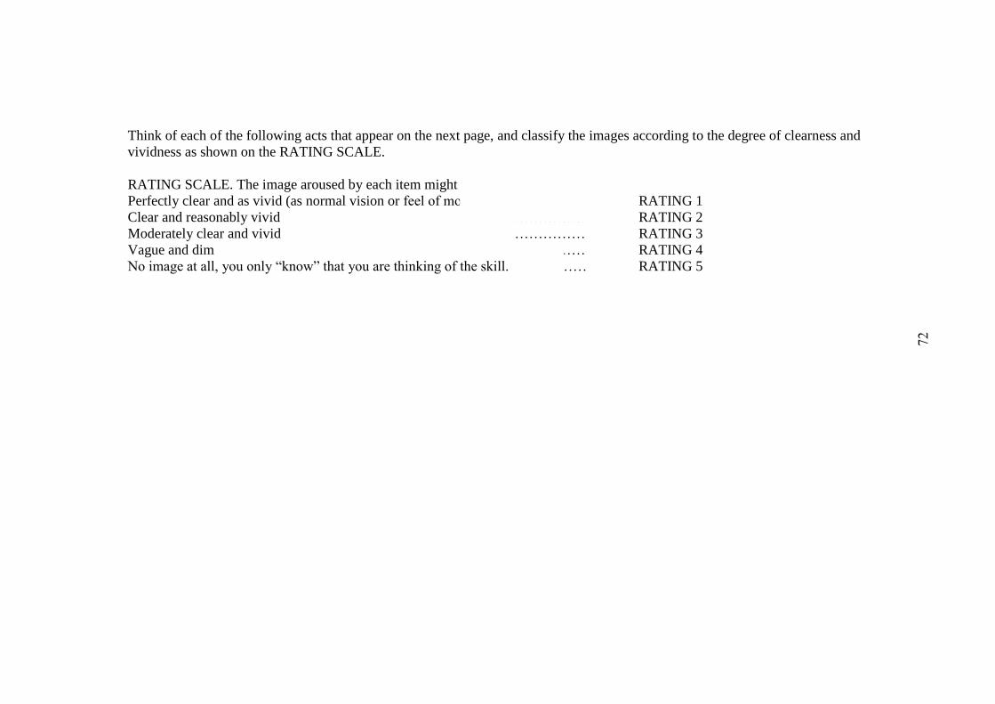
72
Think of each of the following acts that appear on the next page, and classify the images according to the degree of clearness and
vividness as shown on the RATING SCALE.
RATING SCALE. The image aroused by each item might be:
Perfectly clear and as vivid (as normal vision or feel of movement) …………… RATING 1
Clear and reasonably vivid …………… RATING 2
Moderately clear and vivid …………… RATING 3
Vague and dim …………… RATING 4
No image at all, you only “know” that you are thinking of the skill. …………… RATING 5
72
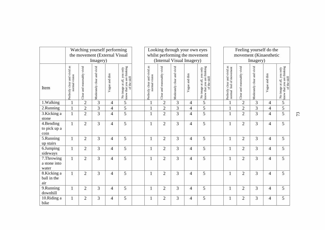
73
Watching yourself performing
the movement (External Visual
Imagery)
Looking through your own eyes
whilst performing the movement
(Internal Visual Imagery)
Feeling yourself do the
movement (Kinaesthetic
Imagery)
Item
Per
fect
ly c
lear
an
d v
ivid
as
no
rmal
vis
ion
Cle
ar a
nd
rea
son
ably
viv
id
Mod
erat
ely
cle
ar a
nd
viv
id
Vag
ue
and
dim
No
im
age
at a
ll, you
only
kno
w t
hat
you
are
th
ink
ing
of
the
skil
l
Per
fect
ly c
lear
an
d v
ivid
as
no
rmal
vis
ion
Cle
ar a
nd
rea
son
ably
viv
id
Mod
erat
ely
cle
ar a
nd
viv
id
Vag
ue
and
dim
No
im
age
at a
ll, you
only
kno
w t
hat
you
are
th
ink
ing
of
the
skil
l
Per
fect
ly c
lear
an
d v
ivid
as
no
rmal
fe
el o
f m
ovem
ent
Cle
ar a
nd
rea
son
ably
viv
id
Mod
erat
ely
cle
ar a
nd
viv
id
Vag
ue
and
dim
No
im
age
at a
ll, you
only
kno
w t
hat
you
are
th
ink
ing
of
the
skil
l
1.Walking 1 2 3 4 5 1 2 3 4 5 1 2 3 4 5 2.Running 1 2 3 4 5 1 2 3 4 5 1 2 3 4 5 3.Kicking a
stone 1 2 3 4 5 1 2 3 4 5 1 2 3 4 5
4.Bending
to pick up a
coin
1 2 3 4 5 1 2 3 4 5 1 2 3 4 5
5.Running
up stairs 1 2 3 4 5 1 2 3 4 5 1 2 3 4 5
6.Jumping
sideways 1 2 3 4 5 1 2 3 4 5 1 2 3 4 5
7.Throwing
a stone into
water
1 2 3 4 5 1 2 3 4 5 1 2 3 4 5
8.Kicking a
ball in the
air
1 2 3 4 5 1 2 3 4 5 1 2 3 4 5
9.Running
downhill 1 2 3 4 5 1 2 3 4 5 1 2 3 4 5
10.Riding a
bike 1 2 3 4 5 1 2 3 4 5 1 2 3 4 5
72
73

74
11.Swinging
on a rope 1 2 3 4 5 1 2 3 4 5 1 2 3 4 5
12.Jumping
off a high wall 1 2 3 4 5 1 2 3 4 5 1 2 3 4 5
73
74

75
Scoring: Add up the responses in each section to get a score for:
External Visual Imagery (EVI): Watching yourself
performing the movement
Internal Visual Imagery (IVI): Looking through your
own eyes whilst performing
the movement
Kinaesthetic Imagery (KIN): Feeling yourself do
the movement
Each score should range between 12 and 60 (as there are 12 items, with a score of 1 to 5 for each.). The lower the score, the
better the athlete is at using that particular imagery type. There is no “gold-standard” with regards to what score would indicate
that an athlete is very skilled at a particular type of imagery. However, as a general rule we would suggest that if you are going
to undertake an imagery intervention using a particular type of imagery (e.g., IVI) then the athlete should have a total IVI score
of no more than 36. This value corresponds to a moderate level of imagery ability. Therefore, your athlete would be able to
image to some degree, which should improve the effectiveness of the imagery intervention. If the athlete had a lower score
than this, you might encourage them to develop their imagery skills by doing some basic imagery exercises before planning a
specific sporting imagery intervention.
74
75

76
APPENDIX C
LAB SETUP

77
APPENDIX D
PROPER CAST CARE INSTRUCTIONS
Keep the cast dry. When showering please wrap your cast in a plastic bag to help
keep it dry.
The fiberglass cast will take approximately 1 hour to completely dry. Take care
not to bump or hit your cast until it is completely dry.
Do not put anything inside the cast to scratch the skin. This may result in an
infection. You can put ice packs over the cast at any itchy area. A hair dryer set to
cool setting may be blown into the cast to relieve itching.
Your fingers should have feeling and be warm and pink. If they become
discolored or numb please call Siobhan Huggins-Sullivan.
Do not try to trim or cut the cast yourself. This can result in injury. If you want
the cast removed prior to the completion of the immobilization period please
contact Siobhan Huggins-Sullivan.

78
APPENDIX E
IMAGERY SCRIPT
Make sure you’re in a quiet location and seated in a comfortable position. Before
we get started I want to remind you that during the imagery script you are supposed to
imagine completing the contractions with your casted thumb.
To begin I want you to take a couple of depth breaths and get into a comfortable
and relaxed position. Try to keep your breathing relaxed throughout the imagery you are
about to complete. As you take your deep breaths make sure you are relaxing your casted
hand and arm.
Imagine you are seated in the same position that you were in the research lab
earlier this week with your thumb secured to the lab table. Today we are going to practice
five imagined maximal contractions in four different directions. The directions of the
contractions will be the same as completed in the research lab.
Flexion
First we are going to practice thumb flexion. Remember that flexion is pulling
towards the base of your palm. I want you to imagine contracting your thumb as hard as
possible towards flexion. Hold this contraction for 3…2...1… and then release the
contraction and relax your muscles. (5 seconds relaxation)
As you begin flexion contraction number two feel the resistance from the force
transducer against your thumb. Hold this contraction for 3…2...1… and then release the
contraction and relax your muscles. (5 seconds relaxation)

79
As you begin flexion contraction number three feel your muscles get stronger and
produce more force. Hold this stronger contraction for 3…2...1… and then release the
contraction and relax your muscles. (5 seconds relaxation)
As you begin flexion contraction number four feel your thumb pulling the force
transducer further and further. Hold this contraction for 3…2...1… and then release the
contraction and relax your muscles. (5 seconds relaxation)
For your fifth and final flexion contraction imagine contracting your muscle as
hard as possible. Pulling harder than you have on your last four attempts. Hold this
contraction for 3…2...1… and then release the contraction and relax your muscles. (5
seconds relaxation)
Abduction
Next we are going to practice thumb abduction. Abduction is pulling away from
your palm towards your forearm. I want you to imagine contracting your thumb as hard
as possible towards abduction. Hold this contraction for 3…2...1… and then release the
contraction and relax your muscles. (5 seconds relaxation)
As you begin abduction contraction number two feel the resistance from the force
transducer against your thumb. Hold this contraction for 3…2...1… and then release the
contraction and relax your muscles. (5 seconds relaxation)
As you begin abduction contraction number three feel your muscles get stronger
and produce more force. Hold this stronger contraction for 3…2...1… and then release
the contraction and relax your muscles. (5 seconds relaxation)

80
As you begin abduction contraction number four feel your thumb pulling the force
transducer further and further. Hold this contraction for 3…2...1… and then release the
contraction and relax your muscles. (5 seconds relaxation)
For your fifth and final abduction contraction imagine contracting your muscle as
hard as possible. Pulling harder than you have on your last four attempts. Hold this
contraction for 3…2...1… and then release the contraction and relax your muscles. (5
seconds relaxation)
Adduction
Third we are going to practice thumb adduction. Adduction is moving your thumb
toward your pointer finger. I want you to imagine contracting your thumb as hard as
possible towards adduction. Hold this contraction for 3…2...1… and then release the
contraction and relax your muscles. (5 seconds relaxation)
As you begin adduction contraction number two feel the resistance from the force
transducer against your thumb. Hold this contraction for 3…2...1… and then release the
contraction and relax your muscles. (5 seconds relaxation)
As you begin adduction contraction number three feel your muscles get stronger
and produce more force. Hold this stronger contraction for 3…2...1… and then release
the contraction and relax your muscles. (5 seconds relaxation)
As you begin adduction contraction number four feel your thumb pulling the force
transducer further and further. Hold this contraction for 3…2...1… and then release the
contraction and relax your muscles. (5 seconds relaxation)

81
For your fifth and final adduction contraction imagine contracting your muscle as
hard as possible. Pulling harder than you have on your last four attempts. Hold this
contraction for 3…2...1… and then release the contraction and relax your muscles. (5
seconds relaxation)
Opposition
Finally we are going to practice thumb opposition. Opposition is moving your
thumb toward the top of your pinky finger. I want you to imagine contracting your thumb
as hard as possible towards opposition. Hold this contraction for 3…2...1… and then
release the contraction and relax your muscles. (5 seconds relaxation)
As you begin opposition contraction number two feel the resistance from the force
transducer against your thumb. Hold this contraction for 3…2...1… and then release the
contraction and relax your muscles. (5 seconds relaxation)
As you begin opposition contraction number three feel your muscles get stronger
and produce more force. Hold this stronger contraction for 3…2...1… and then release
the contraction and relax your muscles. (5 seconds relaxation)
As you begin opposition contraction number four feel your thumb pulling the
force transducer further and further. Hold this contraction for 3…2...1… and then release
the contraction and relax your muscles. (5 seconds relaxation)
For your fifth and final opposition contraction imagine contracting your muscle as
hard as possible. Pulling harder than you have on your last four attempts. Hold this

82
contraction for 3…2...1… and then release the contraction and relax your muscles. (5
seconds relaxation)
This concludes your imagery script for this session

83
APPENDIX F
THUMB DIRECTIONAL MOVEMENTS
As a reminder thumb flexion is moving your
thumb towards the base of your palm.
Thumb opposition is moving toward the tip
of your pinky finger.
Thumb abduction is moving away from
your palm and toward your forearm.
Thumb adduction is moving toward your
palm and pointer finger.