Embed Size (px)
Citation preview

DOI 10.2478/v10181-010-0008-1Original article
Transcriptional pattern of TGF-β1 inhibitoryeffect on mouse C2C12 myoblasts
differentiation
Z. Wicik, T. Sadkowski, M. Jank, T. Motyl
Department of Physiological Sciences, Faculty of Veterinary Medicine, Warsaw University of Life Sciences– SGGW, Nowoursynowska 159, 02-776 Warsaw, Poland
Abstract
The aim of the present study was to define the effect of TGF-β1 on C2C12 myoblasts myogenesis.TGF-β1 together with its receptor is a negative auto-paracrine regulator of myogenesis, which in-fluences the proliferation, differentiation, and functions of muscle cells. TGF-β1 exerts highly signifi-cant inhibitory effect on differentiation of C2C12 mouse myoblasts manifested by the impairment ofcell fusion and very low expression of myosin heavy chain. The study of differentiating C2C12 mousemyoblasts treated with TGF-β1 revealed 502 genes (436 down-regulated and 66 up-regulated) withstatistically different expression. TGF-β1-regulated genes were identified to be involved in 29 biologi-cal processes, 29 molecular functions groups and 59 pathways. The strongest inhibiting effect ofTGF-β1 was observed in the cadherin and Wnt pathways. The key-genes that could play the role ofTGF-β1 targets during myoblasts differentiation was identified such as: Max, Creb1, Ccna2, Bax,MdfI, Tef, Tubg1, Cxcl5, Rho, Calca and Lgals4.
Key words: C2C12 myoblasts, differentiation, DNA microarrays, myogenesis, TGF-β1
Introduction
Transforming growth factor-β1 (TGF-β1) belongsto the transforming growth factor beta super-family,a large group of proteins which play a significant rolein regulation of cell growth and differentiation.TGF-β1 was described in 1983, as a first factor be-longing to this family (Miyazono et al. 1988). TGF-β1is a 25 kDa homodimeric protein consisting of twosubunits, each with a molecular weight 12.5 kDa and112 amino-acids joined by disulphide bounds. TGF-β1is synthesized as a 110 kDa propeptide containingshort hydrophobic signaling peptide, latency asso-ciated peptide (LAP) and an active TGF-β1 domainin C-terminal region (Klass et al. 2009). TGF-β1 is an
Correspondence to: T. Motyl, e-mail: [email protected], tel./fax: (+48) 22 847 24 52
inhibitor of myogenic cell proliferation and differenti-ation. Results presented by Zimowska et al. (2009)indicate that the control of TGF-β1 activity is import-ant to improve regeneration of injured muscle andaccelerate myoblast differentiation, in part throughchanges in glycosaminoglycans composition of musclecell environment. TGF-β1 can also inhibit mitosis inepithelial and epidermal cells, blood precursor cells,liver, ovary, lymphatic and endothelial cells. The roleof TGF-β1 is also the regulation of angiogenesis andsimulation of extracellular matrix components syn-thesis. TGF-β1 is a subject of intense investigationsaiming to evaluate its role in pathogenesis of differentdiseases, especially tumors (Wrighton et al. 2009).The signal transduction of all proteins belonging to
Polish Journal of Veterinary Sciences Vol. 13, No. 4 (2010), 629-638
Brought to you by | Uniwersytet Przyrodniczy w PoznaniuAuthenticated | 150.254.174.70
Download Date | 2/14/14 9:15 AM

TGF-β superfamily, including TGF-β1, occursthrough two types of trans-membrane receptors withserine-theronine kinase activity. TGF-β1 initiatesdownstream signals by binding directly to type II re-ceptor (RII) and activation of type I receptor (RI) (LeGrand et al. 2007). Afterwards the presence ofSmad2/Smad3 dimers (R-Smads) enables phos-phorylation of RII and RI receptor complex and for-mation of a heterodimer with Smad4. TheR-Smad/Smad4 complex is then translocated into thenucleus where it binds with one of DNA-binding pro-teins which serve as activators or repressors of targetgene transcription (Moustakas 2002).
TGF-β1 modulates myoblasts activity by inhibitionof their proliferation and differentiation, which wasshown in vitro in C2C12, BC3H1 and L6 cell lines( Olson 1992, Kollias et al. 2006). Inhibition of differ-entiation occurs by down-regulation of MyoD and my-ogenin expression. Moreover, it was shown thatTGF-β1 could directly influence myostatin (growthand differentiation factor 8 – GDF-8) expression(Budasz-Świderska et al. 2005). The inhibition of my-oblast proliferation by TGF-β1 occurs through inhibi-tion of Cdk4 expression at the translational level,leading to cell cycle arrest in the G1 phase. Inhibitionof myoblast differentiation by TGF-β1 occurs bySmad signaling pathway, which activates or suppressestarget genes. Smad-3 binds to the E-box sequence ofmuscle regulatory genes and inhibits binding of bHLHproteins with this sequence (Klass et al. 2009). Apartof TGF-β1 there are other factors which can suppressmyoblast differentiation e.g. myostatin (GDF-8)(Budasz-Świderska et al. 2005), glycogen synthasekinase 3 beta (GSK3β) (Van der Valden et al. 2008)or inhibitors of differentiation (Id) (Clever et al.2010).
So far the knowledge about genes negatively regu-lated by TGF-β1 during myogenesis is still obscure. Inthe present study, DNA microarray method was ap-plied to investigate a wide-spectrum of gene express-ion profiling to identify the potential genes which areresponsible for inhibition of C2C12 myogenic cellsdifferentiation by TGF-β1.
Materials and Methods
Media and reagents
DMEM with Glutamax, phosphate buffered saline(PBS) [pH 7.4], fetal bovine serum (FBS), horseserum (HS) and antibiotics: penicillin-streptomycin,fungizone and gentamycin sulphate were purchasedfrom Gibco BRL (UK). Primary monoclonal rabbitanti-mouse MyHC (H-300) antibody was delivered bySanta Cruz Biotechnology Inc. (USA). Alexa Fluor488 chicken anti-rabbit IgG secondary antibody and
7-aminoactinomycin D (7-AAD) were purchasedfrom Sigma-Aldrich (Germany). Sterile conical flasks,and Lab-Teks were supplied by Nunc Inc. (USA).Sterile Petri dishes and disposable pipettes were pur-chased from Corning Glass Co. (USA).
Cell culture
The mouse skeletal muscle cell line C2C12 waspurchased from European Collection of Animal CellCulture (UK). Cells were cultured as described before(Budasz-Świderska et al. 2005). Preliminary experi-ments were done to assess the most effective dose ofTGF-β1 and time of the experiment. 2 ng/ml TGF-β1supplementation of experimental medium and 6-daytime duration have been chosen. Control medium didnot contain TGF-β1. After sixth day, cells were har-vested for RNA isolation or fixed for staining for con-focal microscopy.
Experimental procedures
For RNA isolation, C2C12 cells were cultured onPetri dishes in proliferation medium (10%FBS/DMEM/antibiotics) until 80% of confluence.The medium was then replaced with differentiationmedium (2% HS/DMEM/antibiotics) for next 6 days.Pictures of each stage of differentiation were takenusing contrast-phase microscopy. After sixth day, cellswere harvested and stored at -80oC until analysis. Forconfocal microscopy cells were cultured on a 8-cham-ber Lab-Tek slides until they reached 80% confluenceand then were differentiated with 2% HS/DMEM for6 days. Afterwards, cells were fixed by dehydratation.Five separate experiments were performed.
RNA isolation and validation, probe labelling, hy-bridization, signal detection and quantification weredone as described before (Sadkowski et al. 2008,2009). In this study Mouse Genome OpArray v 4.0.1(Operon, Germany) slides were used.
Confocal Microscopy
The cells were fixed by washing in 0.25% parafor-maldehyde and were incubated in ice-cold 70% meth-anol. Samples were stored at -80oC until staining.Then cells were incubated for 1 h with the primaryantibody (anti-MyHC) diluted 1:250 with PBS andafter triple washing were incubated 1 hour with AlexaFluor 488 secondary antibody 1:500 (anti-rabbit) at4oC in the dark. The cells were finally incubated for 30min with 5 μg/ml of 7-aminoactinomycin D (7-AAD)at room temperature in the dark to stain the DNA ofnuclei. Preparations were stored in the dark at 4oC for
630 Z. Wicik et al.
Brought to you by | Uniwersytet Przyrodniczy w PoznaniuAuthenticated | 150.254.174.70
Download Date | 2/14/14 9:15 AM
24h before analysis. Images from confocal microscopeFV-500 (Olympus Opticals, Germany) were analyzedusing MicroImage software (Olympus, Germany). Foreach culture stage 15 pictures were taken.
Results
The influence of TGF-β1 on differentiationof C2C12 myoblasts
TGF-β1 exerted a strong inhibitory effect on dif-ferentiation of mouse C2C12 myoblasts. It was evi-dent in contrast phase images of control cultureswhere myotubes were visible from 48 h of differenti-ation period. (Figure 1a, upper panel). Addition ofTGF-β1 (2 ng/ml) exerted an inhibitory effect onfusion of myoblasts and formation of myotubes duringthe 6-day observation period (Figure 1a, lower panel).Moreover, there was observed an increase in thenumber of necrotic cells in TGF-β1-treated cultures.Inhibition of C2C12 myoblasts differentiation byTGF-β1 was also shown by immunofluorescenceanalysis of myosin heavy chain expression in cultureson the sixth day of myoblasts differentiation.MyHC-related fluorescence was visible only in con-focal images containing myotubes from control cul-tures (Figure 1b, upper panel). There was no MyHCexpression in TGF-β1-treated cultures (Figure 1b,lower panel). Integrated optical density analysis(IOD) was calculated using MicroImage software.There was a significantly (P ≤ 0.0001) lower express-ion of MyHC in TGF-β1-treated myoblasts in com-parison with control cultures (Fig. 1c).
The influence of TGF-β1 on transcriptomic profileof differentiating C2C12 myoblasts
Global potentiometric analysis of gene expressionprofile in TGF-β1-treated C2C12 myoblasts was per-formed using the DNA microarray technique. Com-parison of transcriptomic profiles between cellstreated with TGF-β1 and control cultures revealed502 genes, which exhibited statistically significant dif-ferences in expression, with at least 1.6 fold change.TGF-β1 treatment caused down-regulation of 436genes and an up-regulation of 66 genes. The role ofthese particular genes was evaluated with Panther sof-tware, that allows to classify identified genes in re-spect to their molecular function, biological processand metabolic pathways.
Classification of identified genes according totheir molecular functions revealed 29 groups of genessignificantly affected by TGF-β1 (Fig. 2). The majorityof genes were associated with groups such as: nucleic
acid binding (65), receptors (51), transcription factors(38), transferase (30) and hydrolase (25).
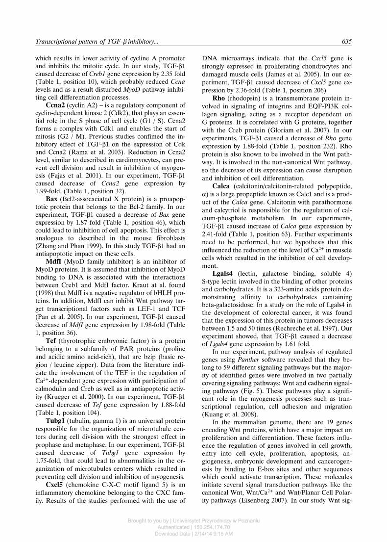
Classification of genes according to biological pro-cesses revealed 29 processes in which genes signifi-cantly affected by TGF-β1 were involved (Fig. 3). Thehighest numbers of genes were involved in: signaltransduction (106), nucleoside, nucleotide and nucleicacid metabolism (81), developmental processes (64)and protein metabolism and modification (57). Analy-sis of the results according to Pathway Architect sof-tware (Stratagene USA) showed connections betweenthe products of genes identified in this study andgenes described in the literature. This allowed to pres-ent the network of reciprocal interactions betweenprotein products of the investigated genes. In the net-work there were also proteins of putative special sig-nificance, which formed junctions converging withother pathways. The genes encoding these proteinsplay or could play an essential role in proliferationand the developmental processes. The key genes, withdecreased expression under the influence of TGF-β1were: Max protein (Max), cAMP responsive elementbinding protein 1 (Creb1), cyclin A2 (Ccna2), Bcl2-as-sociated X protein (Bax), MyoD family inhibitor(MdfI), thyrotroph embryonic factor (Tef), tubulingamma1 (Tubg1), chemokine (C-X-C motif) ligand5 (Cxcl5), rhodopsin (Rho). Whereas key genes, withincreased expression were: calcitonin/calcitonin-re-lated polypeptide alpha (Calca) and lectin, galactosebinding, soluble 4 (Lgals4) (Fig. 4).
Classification of genes according to signalingpathways revealed that 59 signaling pathways were af-fected by TGF-β1. Analysis of the role of the identifi-ed genes in various pathways showed that the largestnumber of gene products were involved in the Wntsignaling pathway represented by 12 genes, cadherinsignaling pathways represented by 10 genes (Pcdhgb8,Wnt16, Erbb4, Pcdhgc4, Pcdhb16, Fat3, Dchs2,Pcdhb20, Pcdh1, Pcdhb17), inflammation mediated bychemokine and cytokine signaling pathway represen-ted by 7 genes (Fprl1, Tyk2, Plce1, Alox12, Xcr1,Inppl1, Rgs4), Huntington disease pathway represen-ted by 7 genes (Synj2, Arfip2, Grin2c, Dnahc8,AI427515, Dnahc17, Bax), Notch signaling pathwayrepresented by 5 genes (Numb, Dll4, Heyl, Fut8,Ncstn) and TGF-beta signaling pathway representedby 3 genes (Dcp1a, Smad5, Foxp2). To localize theproducts of gene expression of the metabolic path-ways in skeletal muscle fibers Pathway Architect sof-tware was used. The result of this analysis was thenetwork describing the relationship between the prod-ucts of the identified genes belonging to the most rep-resented pathways: Wnt and cadherin signaling path-way (Fig. 5). Among the genes of the Wnt signalingpathway 5 groups of genes were distinguished: al-pha-catenin – 1 gene (Ctnnal1); cadherin – 8 genes(Pcdhb17; Pcdhgc4; Pcdhb16; Fat3; Dchs2; Pcdhb20;
Transcriptional pattern of TGF-β inhibitory... 631
Brought to you by | Uniwersytet Przyrodniczy w PoznaniuAuthenticated | 150.254.174.70
Download Date | 2/14/14 9:15 AM

a)
b)
c)
Fig. 1. a) Six days differentiation process of C2C12 myoblasts (100x magnification). Control culture stimulated to differentiationin DMEM/2% HS for 48, 72, 96 and 144 hours (upper panel). Experimental culture maintained in above medium supplementedwith 2 ng/ml of TGF-β1 (lower panel), b) Images from confocal microscopy showing expression of MyHC (Alexa 488 – green)and the DNA of nuclei (7-AAD – red) after six days of differentiation in control (upper panel) and TGF-β1 treated culture(lower panel), c) Influence of TGF-β1 on MyHC levels in myotubes of C2C12 mouse myoblasts. Results are showed as mean± SEM of MyHC integrated optical density (IOD). Means were calculated from 15 digital images for each sample. Values a andb indicate a significant statistical difference (P≤0.0001) between experimental culture treated with 2 ng/ml TGF-β1 and controlculture analyzed with t-Student test (n = 5).
632 Z. Wicik et al.
Brought to you by | Uniwersytet Przyrodniczy w PoznaniuAuthenticated | 150.254.174.70
Download Date | 2/14/14 9:15 AM
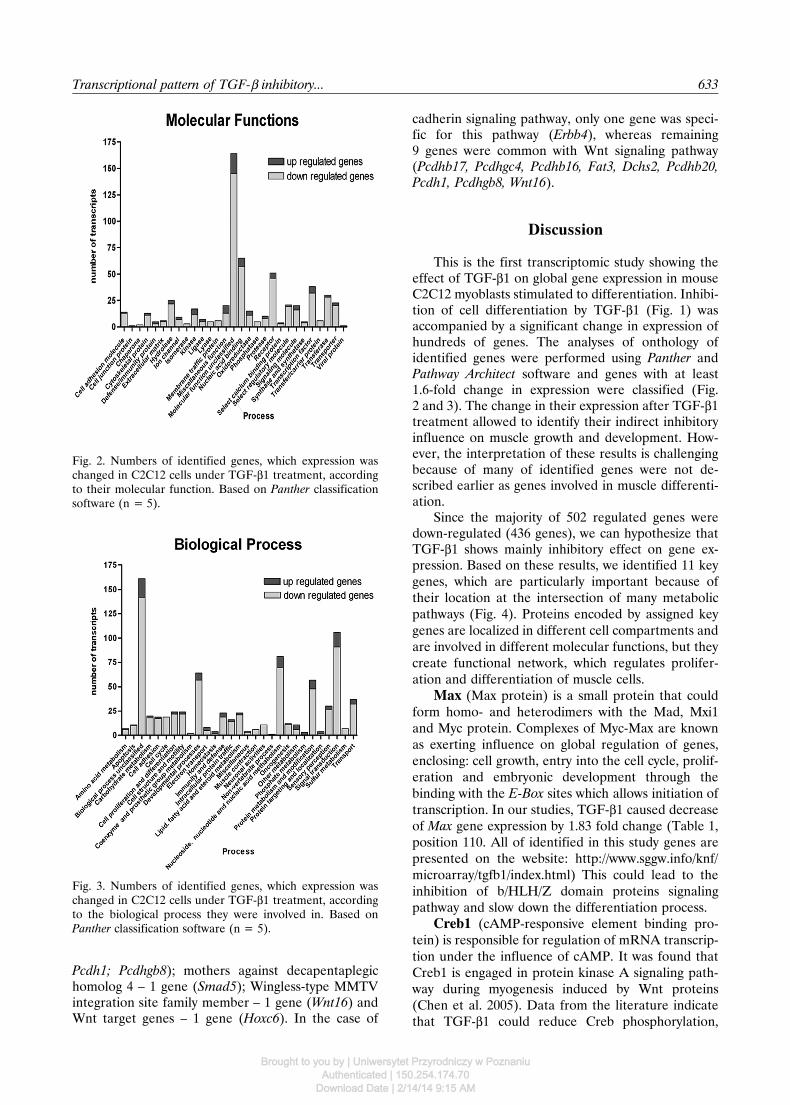
Fig. 2. Numbers of identified genes, which expression waschanged in C2C12 cells under TGF-β1 treatment, accordingto their molecular function. Based on Panther classificationsoftware (n = 5).
Fig. 3. Numbers of identified genes, which expression waschanged in C2C12 cells under TGF-β1 treatment, accordingto the biological process they were involved in. Based onPanther classification software (n = 5).
Pcdh1; Pcdhgb8); mothers against decapentaplegichomolog 4 – 1 gene (Smad5); Wingless-type MMTVintegration site family member – 1 gene (Wnt16) andWnt target genes – 1 gene (Hoxc6). In the case of
cadherin signaling pathway, only one gene was speci-fic for this pathway (Erbb4), whereas remaining9 genes were common with Wnt signaling pathway(Pcdhb17, Pcdhgc4, Pcdhb16, Fat3, Dchs2, Pcdhb20,Pcdh1, Pcdhgb8, Wnt16).
Discussion
This is the first transcriptomic study showing theeffect of TGF-β1 on global gene expression in mouseC2C12 myoblasts stimulated to differentiation. Inhibi-tion of cell differentiation by TGF-β1 (Fig. 1) wasaccompanied by a significant change in expression ofhundreds of genes. The analyses of onthology ofidentified genes were performed using Panther andPathway Architect software and genes with at least1.6-fold change in expression were classified (Fig.2 and 3). The change in their expression after TGF-β1treatment allowed to identify their indirect inhibitoryinfluence on muscle growth and development. How-ever, the interpretation of these results is challengingbecause of many of identified genes were not de-scribed earlier as genes involved in muscle differenti-ation.
Since the majority of 502 regulated genes weredown-regulated (436 genes), we can hypothesize thatTGF-β1 shows mainly inhibitory effect on gene ex-pression. Based on these results, we identified 11 keygenes, which are particularly important because oftheir location at the intersection of many metabolicpathways (Fig. 4). Proteins encoded by assigned keygenes are localized in different cell compartments andare involved in different molecular functions, but theycreate functional network, which regulates prolifer-ation and differentiation of muscle cells.
Max (Max protein) is a small protein that couldform homo- and heterodimers with the Mad, Mxi1and Myc protein. Complexes of Myc-Max are knownas exerting influence on global regulation of genes,enclosing: cell growth, entry into the cell cycle, prolif-eration and embryonic development through thebinding with the E-Box sites which allows initiation oftranscription. In our studies, TGF-β1 caused decreaseof Max gene expression by 1.83 fold change (Table 1,position 110. All of identified in this study genes arepresented on the website: http://www.sggw.info/knf/microarray/tgfb1/index.html) This could lead to theinhibition of b/HLH/Z domain proteins signalingpathway and slow down the differentiation process.
Creb1 (cAMP-responsive element binding pro-tein) is responsible for regulation of mRNA transcrip-tion under the influence of cAMP. It was found thatCreb1 is engaged in protein kinase A signaling path-way during myogenesis induced by Wnt proteins(Chen et al. 2005). Data from the literature indicatethat TGF-β1 could reduce Creb phosphorylation,
Transcriptional pattern of TGF-β inhibitory... 633
Brought to you by | Uniwersytet Przyrodniczy w PoznaniuAuthenticated | 150.254.174.70
Download Date | 2/14/14 9:15 AM

Fig. 4. Network of mutual interactions between TGF-β1 (blue), products of identified key genes (yellow), and products of othergenes described in the literature (red). The analysis was performed by using Pathway Architect software.
Fig. 5. Schematic illustration of supposed influence ofTGF-β1 on Wnt signalling pathway and cadherin signalingpathway through β-catenin and APC complex, leading to de-crease of genes transcription and inhibition of myogenesis.
634 Z. Wicik et al.
Brought to you by | Uniwersytet Przyrodniczy w PoznaniuAuthenticated | 150.254.174.70
Download Date | 2/14/14 9:15 AM
which results in lower activity of cycline A promoterand inhibits the mitotic cycle. In our study, TGF-β1caused decrease of Creb1 gene expression by 2.35 fold(Table 1, position 10), which probably reduced Ccnalevels and as a result disturbed MyoD pathway inhibi-ting cell differentiation processes.
Ccna2 (cyclin A2) – is a regulatory component ofcyclin-dependent kinase 2 (Cdk2), that plays an essen-tial role in the S phase of cell cycle (G1 / S). Ccna2forms a complex with Cdk1 and enables the start ofmitosis (G2 / M). Previous studies confimed the in-hibitory effect of TGF-β1 on the expression of Cdkand Ccna2 (Rama et al. 2003). Reduction in Ccna2level, similar to described in cardiomyocytes, can pre-vent cell division and result in inhibition of myogen-esis (Fajas et al. 2001). In our experiment, TGF-β1caused decrease of Ccna2 gene expression by1.99-fold. (Table 1, position 32).
Bax (Bcl2-assocaciated X protein) is a proapop-totic protein that belongs to the Bcl-2 family. In ourexperiment, TGF-β1 caused a decrease of Bax geneexpression by 1.87 fold (Table 1, position 46), whichcould lead to inhibition of cell apoptosis. This effect isanalogous to described in the mouse fibroblasts(Zhang and Phan 1999). In this study TGF-β1 had anantiapoptotic impact on these cells.
MdfI (MyoD family inhibitor) is an inhibitor ofMyoD proteins. It is assumed that inhibition of MyoDbinding to DNA is associated with the interactionsbetween Creb1 and MdfI factor. Kraut at al. found(1998) that MdfI is a negative regulator of bHLH pro-teins. In addition, MdfI can inhibit Wnt pathway tar-get transcriptional factors such as LEF-1 and TCF(Pan et al. 2005). In our experiment, TGF-β1 causeddecrease of MdfI gene expression by 1.98-fold (Table1, position 36).
Tef (thyrotrophic embryonic factor) is a proteinbelonging to a subfamily of PAR proteins (prolineand acidic amino acid-rich), that are bzip (basic re-gion / leucine zipper). Data from the literature indi-cate the involvement of the TEF in the regulation ofCa2+-dependent gene expression with participation ofcalmodulin and Creb as well as in antiapoptotic activ-ity (Krueger et al. 2000). In our experiment, TGF-β1caused decrease of Tef gene expression by 1.88-fold(Table 1, position 104).
Tubg1 (tubulin, gamma 1) is an universal proteinresponsible for the organization of microtubule cen-ters during cell division with the strongest effect inprophase and metaphase. In our experiment, TGF-β1caused decrease of Tubg1 gene expression by1.75-fold, that could lead to abnormalities in the or-ganization of microtubules centers which resulted inpreventing cell division and inhibition of myogenesis.
Cxcl5 (chemokine C-X-C motif ligand 5) is aninflammatory chemokine belonging to the CXC fam-ily. Results of the studies performed with the use of
DNA microarrays indicate that the Cxcl5 gene isstrongly expressed in proliferating chondrocytes anddamaged muscle cells (James et al. 2005). In our ex-periment, TGF-β1 caused decrease of Cxcl5 gene ex-pression by 2.36-fold (Table 1, position 206).
Rho (rhodopsin) is a transmembrane protein in-volved in signaling of integrins and EQF-PI3K col-lagen signaling, acting as a receptor dependent onG proteins. It is correlated with G proteins, togetherwith the Creb protein (Gloriam et al. 2007). In ourexperiments, TGF-β1 caused a decrease of Rho geneexpression by 1.88-fold (Table 1, position 232). Rhoprotein is also known to be involved in the Wnt path-way. It is involved in the non-canonical Wnt pathway,so the decrease of its expression can cause disruptionand inhibition of cell differentiation.
Calca (calcitonin/calcitonin-related polypeptide,α) is a large propeptide known as Calc1 and is a prod-uct of the Calca gene. Calcitonin with parathormoneand calcytriol is responsible for the regulation of cal-cium-phosphate metabolism. In our experiments,TGF-β1 caused increase of Calca gene expression by2.41-fold (Table 1, position 63). Further experimentsneed to be performed, but we hypothesis that thisinfluenced the reduction of the level of Ca2+ in musclecells which resulted in the inhibition of cell develop-ment.
Lgals4 (lectin, galactose binding, soluble 4)S-type lectin involved in the binding of other proteinsand carbohydrates. It is a 323-amino acids protein de-monstrating affinity to carbohydrates containingbeta-galactosidose. In a study on the role of Lgals4 inthe development of colorectal cancer, it was foundthat the expression of this protein in tumors decreasesbetween 1.5 and 50 times (Rechreche et al. 1997). Ourexperiment showed, that TGF-β1 caused a decreaseof Lgals4 gene expression by 1.61 fold.
In our experiment, pathway analysis of regulatedgenes using Panther software revealed that they be-long to 59 different signaling pathways but the major-ity of identified genes were involved in two partiallycovering signaling pathways: Wnt and cadherin signal-ing pathways (Fig. 5). These pathways play a signifi-cant role in the myogenesis processes such as tran-scriptional regulation, cell adhesion and migration(Kuang et al. 2008).
In the mammalian genome, there are 19 genesencoding Wnt proteins, which have a major impact onproliferation and differentiation. These factors influ-ence the regulation of genes involved in cell growth,entry into cell cycle, proliferation, apoptosis, an-giogenesis, embryonic development and cancerogen-esis by binding to E-box sites and other sequenceswhich could activate transcription. These moleculesinitiate several signal transduction pathways like thecanonical Wnt, Wnt/Ca2+ and Wnt/Planar Cell Polar-ity pathways (Eisenberg 2007). In our study Wnt sig-
Transcriptional pattern of TGF-β inhibitory... 635
Brought to you by | Uniwersytet Przyrodniczy w PoznaniuAuthenticated | 150.254.174.70
Download Date | 2/14/14 9:15 AM
naling pathway was represented by 12 down-regulatedgenes such as: Pcdhgb8 (↓1.89), Wnt16 (↓2.02),Ctnnal1 (↓2.01), Pcdhgc4 (↓1.95), Pcdhb16 (↓1.99),Fat3 (↓1.98), Dchs2 (↓1.89), Pcdhb20 (↓2.18), Pcdh1(↓2.08), Pcdhb17 (↓1.68), Smad5 (↓1.63), Hoxc6(↓2.28). Wnt factors in vivo modulate myogenesis,preventing impaired cell adhesion and increasing con-current protein transcription. The Wnt signaling path-way is also involved in the regulation of Myf5, MyoD,kinase A and Creb level of transcription during my-ogenesis. Wnt factors also influence the expression ofPax3 and Pax7 together with Wnt6, which enablesβ-catenin stabilization during c-myc activation(Buckingham 2006).
Cadherins are glycoproteins involved in cell ad-hesion associated with Ca2+ ions. These proteins haveextracellular regions that mediate cell-cell interactionsand play an important role in cell death, signaling,differentiation and migration. These proteins alsohave a transmembrane domain and a cytoplasmic tailthat binds catenins. Catenins link the cadherin to theactin cytoskeleton and play a significant role in cellu-lar signaling. α-Catenins bind indirectly to cadherinsvia interaction with β-catenin. In our study cadherinsignaling pathway was represented by 10 down-regu-lated genes such as: Pcdhgb8 (↓1.89), Wnt16 (↓2.02),Erbb4 (↓2.06), Pcdhgc4 (↓1.95), Pcdhb16 (↓1.99),Fat3 (↓1.98), Dchs2 (↓1.89), Pcdhb20 (↓2.18), Pcdh1(↓2.08), Pcdhb17 (↓1.68).
The main path of signal transduction through theWnt pathway is the canonical Wnt. To date, majorsignaling pathways downstream of the Frizzled (Fz)receptor have been identified including a canonical orWnt/β-catenin dependent pathway and the non-ca-nonical or β-catenin-independent pathway which canbe further divided into the Planar Cell Polarity andthe Wnt/Ca2+ pathways and these branches are nowbeing actively investigated at the molecular and bio-chemical levels (Komiya 2008). The canonical path-way is activated when Wnt proteins bind to their cellmembrane receptors, which are the 10 known mem-bers of the frizzled family of transmembrane proteins.Signal transduction from this ligand/receptor interac-tion occurs through the cytoplasmic protein Dishev-elled (Dsh) which promotes the inactivation of glyco-gen synthase kinase 3 (GSK3). Absence of Wnt fac-tors leads to degradation of cytoplasmatic β-cateninand results in inhibition of gene transcription. Theinhibition of GSK3 activity by the Wnt signal resultsin the cellular accumulation of β-catenin, which thentranslocates to the nucleus and forms a transcriptionalenhancer complex with LEF/TCF DNA-binding pro-teins. Degradation of β-catenin occurs with contribu-tion of 26S proteasome, serine-treonine kinases, CKI,GSK3, axin and APC protein (Polakis 2002). Ourstudy indicates a significant influence of TGF-β1 ongenes involved in these proteins expression, with the
exception of APC and GSK3. Our results also indicatea strong decrease of α-catenin gene expression.α-Catenin binds to cytoplasmatic domain of cadherintype I and is responsible for organization and functionof cadherins by binding them to actin of the cytos-keleton (Nelson et al. 2004). Integrity of structuralcadherin-catenin complex is regulated byserine-treonine phosphorylation of β-catenin or epi-thelial cadherin (E-cadherin). Our study also showeda down-regulation of genes encoding α-catenin pre-cursor proteins and Erbb4. Erbb4 protein is an im-portant stimulator of cardiac muscle myogenesis andplays significant role in EGFR pathway. These resultsconfirm the influence of TGF-β1 on myogenesisthrough Wnt and cadherin signaling pathways (Gar-cia-Rivello et al. 2005).
Other interesting Wnt pathways involved in em-bryogenesis process are non-canonical: the PlanarCell Polarity pathway (PCP pathway) and theWnt/Ca2+ pathway. Those pathways are often referredas the β-catenin-independent. PCP pathway furthermodulates canonical signaling for dorsal axis forma-tion and PCP signaling for gastrulation cell move-ments. The Wnt/Ca2+ pathway emerged with the find-ing that some Wnts and Fz receptors can stimulateintracellular Ca2+ release from ER and this pathway isdependent on G-proteins (Komiya 2008). It is inter-esting because our studies indicated that TGF-β1could decrease expression of Rho protein engagedinto PCP pathway and the level of Ca2+-related pro-teins engaged into Wnt/Ca2+ pathway such as cal-modulin.
Other studies investigating the role of TGF-β1suggest that this cytokine can act independently ofWnt signaling pathway. It was found that an increaseof expression of zinc finger Slug/Snail proteins caninhibit E-cadherin transcription and activation ofFGFR, Erbb1 i Erbb2 (Peinado et al. 2003). The ef-fect of increased Slug/Snail expression resulting ina loss of cell adhesion and an increase of migration,just in case of β-catenin accumulation, what can be anevidence of some independence from Wnt signalingpathway. In the studies mentioned above it was shownthat Wnt signaling pathway regulates E-cadherin ex-pression and that Slug could be a target gene forβ-catenin/TCF complex. This complex could playa repressor function on E-cadherin promoter. De-crease of E-cadherin expression could impair cell ad-hesion and reduces the cells affinity to Wnt factors,which is a result of changes in β-catenin protein levels.Activation of transcription of target genes byβ-catenin is coordinated with TGF-β1 signaling path-way and connected with Smad proteins. Based on stu-dies of TGF-β1 and myostatin action it can be con-cluded that inhibition of C2C12 cells differentiation ismediated by c-Ski protein, which binds R-Smad pro-tein (Budasz-Świderska et al. 2005). It was also seen in
636 Z. Wicik et al.
Brought to you by | Uniwersytet Przyrodniczy w PoznaniuAuthenticated | 150.254.174.70
Download Date | 2/14/14 9:15 AM

our studies, since we observed significant decrease ofSmad5 expression. Smad5 is a receptor-regulatedSmad (R-Smad), which is a transcriptional modulatoractivated by BMP (bone morphogenetic proteins)type 1 receptor kinase (Miyazono et al. 2003). How-ever, with the level of significance used in our study itwas not possible to show interdependence betweenlevels of c-Ski, TGF-β1 and myostatin. It has also tobe mentioned that Smad proteins, TCF/LEF andcatenin-β are able to exert an effect on transcriptiononly when they are involved in those processes alto-gether (Labbe et al. 2000, Nishita et al. 2000). Varietyof TGF-β1 receptor interactions could intersectthrough Wnt pathway, regulate availability ofβ-catenin, break catenin-cadherin bonds and blockcadherin expression (Nelson and Nusse 2004).
Conclusions
In conclusion, TGF-β1 impairs the processes ofgenes transcription leading to inhibition of myogeniccell differentiation. The conclusions are based on factsthat: 1) the majority of regulated genes weredown-regulated; 2) three of eleven so-called key genesare genes strictly related with regulation of transcrip-tion, 3) TGF-β1-regulated signaling pathways wereWnt and cadherin signaling pathways, which areclosely cross-related and involved in the process ofgenes transcription. Myogenesis disturbances can bea result of decreased expression of factors directly tak-ing part in processes of gene transcription such asMax, Creb1, Creb5, Tef and inhibiting the expressionof genes encoding proteins, that modulate pathwaysinvolved in cells differentiation, such as Wnt16,Ctnnal1, Ccna2, Pdgfc and protocadherins.
All of 502 identified genes are presented in Table1 available at the website http://www.sggw.info/knf/microarray/tgfb1/index.html
Acknowlegements
This work was supported by the grant No.PBZ-KBN-113/P06/2005 from Ministry of Science andHigher Education.
References
Buckingham M (2006) Myogenic progenitor cells and skel-etal myogenesis in vertebrates. Curr Opin Genet Dev16: 525-532.
Budasz-Świderska M, Jank M, Motyl T (2005) Transforminggrowth factor-beta1 upregulates myostatin expression inmouse C2C12 myoblasts. J Physiol Pharmacol56: 195-214.
Chen AE, Ginty DD, Fan CM (2005) Protein kinase A sig-nalling via CREB controls myogenesis induced by Wntproteins. Nature 433: 317-322.
Clever JL, Sakai Y, Wang RA, Schneider DB (2010) Ineffic-ient skeletal muscle repair in inhibitor of differentiationknockout mice suggests a crucial role for BMP signalingduring adult muscle regeneration. Am J Physiol CellPhysiol 298: C1087-C1099.
Eisenberg LM, Eisenberg CA (2007) Evaluating the role ofWnt signal transduction in promoting the development ofthe heart. Scientific World Journal 7: 161-176.
Fajas L, Paul C, Vie A, Estrach S, Medema R, BlanchardJM, Sardet C, Vignais ML (2001) Cyclin A is a mediatorof p120E4F-dependent cell cycle arrest in G1. Mol CellBiol 21: 2956-2966.
Garcia-Rivello H, Taranda J, Said M, Cabeza-Meckert P,Vila-Petroff M, Scaglione J, Ghio S, Chen J, Lai C,Laguens RP, Lloyd KC, Hertig CM (2005) Dilated car-diomyopathy in Erb-b4-deficient ventricular muscle. AmJ Physiol Heart Circ Physiol 289: H1153-H1160.
Gloriam DE, Fredriksson R, Schith HB (2007) The G pro-tein-coupled receptor subset of the rat genome. BMCGenomics 8: 338.
James CG, Appleton CT, Ulici V, Underhill TM, BeierF (2005) Microarray analyses of gene expression duringchondrocyte differentiation identifies novel regulators ofhypertrophy. Mol Biol Cell 16: 5316-5333.
Klass BR, Grobbelaar AO, Rolfe KJ (2009) Transforminggrowth factor beta1 signalling, wound healing andrepair: a multifunctional cytokine with clinical implica-tions for wound repair, a delicate balance. Postgrad MedJ. 85: 9-14.
Kollias HD, Perry RL, Miyake T, Aziz A, McDermott JC(2006) Smad7 promotes and enhances skeletal muscledifferentiation. Mol Cell Biol 26: 6248-6260.
Komiya Y, Habas R (2008) Wnt signal transduction path-ways. Organogenesis 4: 68-75.
Krueger DA, Warner EA, Dowd DR (2000) Involvement ofthyrotroph embryonic factor in calcium-mediated regula-tion of gene expression. J Biol Chem 275: 14524-14531.
Kuang S, Gillespie MA, Rudnicki MA (2008) Niche regula-tion of muscle satellite cell self-renewal and differenti-ation. Cell Stem Cell 2: 22-31.
Labbe E, Letamendia A, Attisano L (2000) Association ofSmads with lymphoid enhancer binding factor 1/Tcell-specific factor mediates cooperative signaling by thetransforming growth factor-beta and wnt pathways. ProcNatl Acad Sci USA 97: 8358-8363.
Le Grand F, Rudnicki MA (2007) Skeletal muscle satellitecells and adult myogenesis. Curr Opin Cell Biol19: 628-633.
Miyazono K, Hellman U, Wernstedt C, Heldin CH (1988)Latent high molecular weight complex of transforminggrowth factor beta 1. Purification from human platelets andstructural characterization. J Biol Chem 263: 6407-6415.
Miyazono K, Suzuki H, Imamura T (2003) Regulation ofTGF-beta signaling and its roles in progression of tumors.Cancer Sci 94: 230-234.
Moustakas A (2002) Smad signalling network. J Cell Sci115: 3355-3356.
Nelson WJ, Nusse R (2004) Convergence of Wnt,beta-catenin, and cadherin pathways. Science 303: 1483--1487.
Transcriptional pattern of TGF-β inhibitory... 637
Brought to you by | Uniwersytet Przyrodniczy w PoznaniuAuthenticated | 150.254.174.70
Download Date | 2/14/14 9:15 AM
Nishita M, Hashimoto MK, Ogata S, Laurent MN, Ueno N,Shibuya H, Cho KW (2000) Interaction between Wntand TGF-beta signalling pathways during formation ofSpemann’s organizer. Nature 403: 781-785.
Olson EN (1992) Proto-oncogenes in the regulatory circuitfor myogenesis. Semin Cell Biol 3: 127-136.
Pan W, Jia Y, Wang J, Tao D, Gan X, Tsiokas L, Jing N, WuD, Li L (2005) Beta-catenin regulates myogenesis by re-lieving I-mfa-mediated suppression of myogenic regula-tory factors in P19 cells. Proc Natl Acad Sci USA.102: 17378-17383.
Peinado H, Quintanilla M, Cano A (2003) Transforminggrowth factor beta-1 induces snail transcription factor inepithelial cell lines: mechanisms for epithelial mesen-chymal transitions. J Biol Chem 278: 21113-21123.
Polakis P (2002) Casein kinase 1: a Wnt’er of disconnect.Curr Biol 12: R499-R501.
Rama S, Suresh Y, Rao AJ (2003) TGF beta1 inducesmultiple independent signals to regulate human trophob-lastic differentiation: mechanistic insights. Mol Cell En-docrinol 206: 123-136.
Rechreche H, Mallo GV, Montalto G, Dagorn JC, IovannaJL (1997) Cloning and expression of the mRNA of hu-man galectin-4, an S-type lectin down-regulated incolorectal cancer. Eur J Biochem 248: 225-230.
Sadkowski T, Jank M, Zwierzchowski L, Oprządek J, MotylT (2009) Comparison of skeletal muscle transcriptionalprofiles in dairy and beef breeds bulls. J Appl Genet50: 109-123.
Sadkowski T, Jank M, Zwierzchowski L, Siadkowska E,Oprządek J, Motyl T (2008) Gene expression profiling inskeletal muscle of Holstein-Friesian bulls with single-nuc-leotide polymorphism in myostatin gene 5’-flanking re-gion. J Appl Genet 49: 237-250.
Van der Velden JL, Schols AM, Willems J, Kelders MC,Langen RC (2008) Glycogen synthase kinase 3β sup-presses myogenic differentiation through negative regula-tion of NFATc3. J Biol Chem 283: 358-366.
Wrighton KH, Lin X, Feng XH (2009) Phospho-control ofTGF-beta superfamily signaling. Cell Res 19: 8-20.
Zhang HY, Phan SH (1999) Inhibition of myofibroblast apo-ptosis by transforming growth factor beta(1) Am J RespirCell Mol Biol 21: 658-665.
Zimowska M, Duchesnay A, Dragun P, Oberbek A, Morac-zewski J, Martelly I (2009) Immunoneutralization ofTGFβ1 Improves Skeletal Muscle Regeneration: Effectson Myoblast Differentiation and GlycosaminoglycanContent. Int J Cell Biol 2009: 659372.
638 Z. Wicik et al.
Brought to you by | Uniwersytet Przyrodniczy w PoznaniuAuthenticated | 150.254.174.70
Download Date | 2/14/14 9:15 AM