Embed Size (px)
Citation preview

18th Biennial Conference
September 25 th – 28th 2014
Leipzig, Germany
Conference Program
2 18th Biennial Conference
The "International Pharmaco-EEG Society, Association for Electrophysiological Brain Research in Preclinical and Clinical Pharmacology and Related Fields" (IPEG) is a non-profit organisation, established in 1980 and composed of scientists and researchers actively involved in electrophysiological brain research in preclinical and clinical pharmacology, neurotoxicology and related areas of interest.
Conference Program
IPEG 2014 in Leipzig 3
Electrophysiological brain research has a long tradition going back as far as 1875 when the first report on animal electroencephalogram (EEG) was published by Caton. Consequentially, the first recordings from the human skull were reported by Berger in the late 20’s of the last century. Not long thereafter, the intriguing world of the effects of drugs on the EEG opened up, a scientific area now known as pharmaco-EEG research. In 1957, Roth and colleagues reported EEG changes associated with favorable treatment outcome to ECT, laying the foundation of what we now consider as EEG Based Personalized Medicine. With the advent of computerized methods for signal processing and quantification of the EEG, the landscape changed further. These developments highlight the value and long history of EEG as a valuable tool to quantify the effects of pharmacological treatments on the brain, and importantly, to predict their clinical outcome from the brain. To date, the impressive progress in knowledge and methodology in pharmaco-EEG research still enjoys the advantageous exchange of empirical findings and insights between animal and human research. This interdependence is reflected in the program of the 18th biennial IPEG Conference. A second, consistent factor in the advancement of pharmaco-EEG research is the development of novel technologies that brought refinement in the quantification and analysis procedures. In the early years up to 1950 pharmaco-EEG studies relied on skilled hand and eye methods. Next the development of passive electronic filters and amplitude integrators changed our vision on the EEG, while from 1965 onwards the application of computers fostered the measurement of frequency, amplitude, variability, evoked and event related potentials and polysomnography. From 1980 to 1995 multi-lead analysis enabled the development of topographic EEG/ERP (“EEG mapping”) that was paralleled by the development of algorithms to locate sources of electrophysiological activity. The last decade has brought many attempts to improve the understanding and the sophisticated analysis of the electrophysiological brain as three-dimensional organ. Combined application with, for example, imaging techniques (e.g. fMRI) or novel (non-linear) analysis methodology shows that the pharmaco-EEG, with its outstanding temporal characteristics and its unique applicability in man and animal alike, has a powerful potential we are still learning to use. It can help us to further understand brain dynamics and its pathology, and to support (e.g. as a biomarker) the discovery, the development and the targeted application of drugs to treat CNS disorders. It is therefore not so surprising that a small but dedicated society as the IPEG manages to successfully organize a series of biennial meetings on electrophysiological brain research in preclinical and clinical pharmacology and related fields. Progress in pharmaco-EEG research relies on the continuing input from a broad range of experts such as preclinical and clinical pharmacologists, psychiatrists, (neuro-)psychologists, neurologists, biologists, bio-statisticians, and computer scientists. The IPEG scientific meetings aim to bring these experts together in a colorful palette of symposia on pharmaco-EEG research to expand and update the knowledge in this increasingly complex field. We welcome you to this, the 18th IPEG Conference, and look forward to an excellent and exciting meeting.
Ulrich Hegerl Sebastian Olbrich
Leslie Prichep Martijn Arns Marc Jobert
(IPEG2014 Conference Organisation Committee)
Conference Program
4 18th Biennial Conference
Thursday, September 25th The special symposium on EEG Based Personalized medicine in psychiatry and the traditional Training Course will be conducted in parallel. Both programs can be found below.
EEG-based Personalized Medicine in Psychiatry Symposium (Venue I)
9.00-9.15 Introduction to the EEG Based Personalized Medicine day Martijn Arns & Sebastian Olbrich
EEG-Vigilance Regulation: A Framework for treatment prediction?
9:15-10:00 EEG-vigilance regulation in neuropsychiatric disorders Ulrich Hegerl
10.00-10:45 EEG-vigilance and the autonomous nervous system in the prediction of antidepressant treatment: Findings from the iSPOT-D study Sebastian Olbrich
10:45-11:00 Coffee break
Recent results and challenges of prediction of psychiatric treatment outcome.
11:00 - 11:45 The utility of electrocortical measures in characterizing depression and treatment response Natalia Jaworska
11:45 – 12:30 Response prediction of psychiatric treatment – how close is practical application? Jürgen Gallinat
12:30-13:30 Lunch
Personalized medicine, the next step? International multicenter studies: EMBARC and iSPOT
13:30-14:15 EMBARC Study on Biological Biomarkers in Psychiatry Gerard Bruder
14:15-15.00 First results of the iSPOT studies in Depression and ADHD: EEG alpha asymmetry as a gender specific predictor of SSRI treatment outcome Martijn Arns
15:00-15:30 Coffee break
15.30-15.45 The IPPEC, a new industry-academia initiative to set up a global, translational and precompetitive pharmaco-EEG database Gé Ruigt
15:45-16:30 Roundtable
EEG based Personalized Medicine: How close are we from practical application and what are the required steps? Ulrich Hegerl, Sebastian Olbrich, Natalia Jaworska, Jürgen Gallinat, Gerard Bruder, Gé Ruigt and Martijn Arns
Conference Program
IPEG 2014 in Leipzig 5
Training Course (Venue II) The traditional one-day Training Course is delivered by a panel of experts. The series of lectures covers a broad spectrum of aspects related to pharmaco-EEG and its applications.
9:00-9:15 Introduction Ulrich Hegerl & Marc Jobert
9:15-10:00 Animal pharmaco-EEG recording Gé Ruigt
10:00-10:45 Pharmaco-EEG Recording and Analysis in Humans Marc Jobert
10:45-11:00 Coffee break
11:00-11:45 Independent Component Analysis: Method and Application in EEG-research Scott Makeig
11:45-12:30 State-of-the-Art Analysis of high-frequency (gamma range) EEG in Humans Judith Nottage
12:30-13:15 Lunch
13:15-14:00 pharmaco-EEG, its past, present and future, background and scope Gé Ruigt
14:00-14:45 Preclinical EEG – a translatable Biomarker for Drug Discovery Research Pim Drinkenburg
14:45-15:00 Coffee break
15:00-15:45 Safety Pharmacology: Use of Pharmaco-EEG in the anesthetized dog model. Henk J. Van der Linde
15:45-16:30 Assessment of EEG-vigilance Christian Sander
16:30-16:45 Coffee break
16:45-17:30 Cordance and Sleep-EEG – Treatment Response in Depression Marcel Pawlowski
17:30-18:15 (Not yet allocated)
Thursday evening September 25th 19:00-20:00 Turan Itil Memorial Lecture
Bernd Saletu
20:00-22:00 Cocktail party and welcome reception

Conference Program
6 18th Biennial Conference
Friday September 26th (Venue I)
8:00-8:30 Coffee light-breakfast
8:30-9:00 Presidential address and welcome Pim Drinkenburg (IPEG President), Ulrich Hegerl (IPEG 2014), Michael Stumvoll (Medical Faculty Director University of Leipzig)
9:00-9:45 Keynote 1 – QEEG Source Localization in the Identification of Theoretical Underlying Pathophysiology in Subtypes of Neuropsychiatric/Neurological Disorders Leslie Prichep
9:45 10:15 Coffee break
10:15-12:00 Symposium 1 – TMS-EEG – a novel technique to study brain excitability and connectivity (Chair: Ulf Ziemann)
Technique and fundamentals of TMS-EEG Risto Ilmoniemi
Characterization of TMS-evoked EEG potentials Ulf Ziemann
TMS-evoked potentials as a marker of brain maturation in childhood and adolescence. Stephan Bender
Insights into consciousness and unconsciousness by TMS-EEG Mario Rosanova
12:00-13:00 Lunch
13:00-14:30 Symposium 2 – Electropsychopharmacology – enriching psychopharmacology with event related potentials (Chair: Leon Kenemans)
Drug effects in visual spatial-cuing paradigns Leon Kenemans
Genetic polymorphisms in the dopamine and serotonin system as factors in neurophysiological indoces of hypervigilance Ivo Heitland
Functional neuromarkers for psychiatry: clinical applications of event related potentials for diagnosis, prognosis and treatment. Juri Kropotov
14:30-15:00 Coffee break
15:00-15:45 Keynote 2 –From maps to mechanisms: Multimodal Imaging with EEG, fMRI and DTI in Psychiatry Christoph Mulert
15:45-17:30 Oral Presentations Session 1
Electrophysiological correlates of response inhibition across menstual cycle: Go-Nogo potential study Inga Griskova-Bulanova

Conference Program
IPEG 2014 in Leipzig 7
Stimulant medication in pediatric ADHD: Predicting the clinical outcome from
single-dose changes in Event Related Potentials (ERPs) and a Go/No-go test Geir Ogrim
Proof-of-Principle to efficacy in psychiatric patients exemplified by a neuronal-selective nicotinic agonist AZD1446 Peter Boeijinga
Individual Alpha Peak Frequency as an Endophenotype Associated with Affective Predisposition L.I. Aftanas
18:00 Bus to Zoo Leipzig
18:30 Guided Tour through the Zoo Leipzig and Dinner Event at the Hacienda http://www.zoo-leipzig.de/en/your-event/venues/hacienda-las-casas/
Conference Program
8 18th Biennial Conference
Saturday September 27th (Venue II)
8:00-8:30 Coffee light-breakfast
8:30-9:15 Keynote 3 – The clinical value of source-resolved EEG analysis: drowsiness, ADHD, epilepsy, and schizophrenia Scott Makeig
9:15-10:45 Symposium 3 – Fast acting agents and underlying neurotransmitter systems (Chair: Georg Winterer)
Ketamine - perspectives from a neurophysiological point of view Georg Winterer
Pharmaco-EEG study of ketamine in patients with major depressive disorder: QEEG and clinical predictors of antidepressive response Martin Brunovsky
EEG power spectra and connectivity changes in animal models of psychosis - comparisons of glutamatergic, serotonergic and cannabinoid models Tomas Palenicek
Functional connectivity analysis of default mode network changes after ketamine application in healthy subjects Felix Müller
10.45-11.00 Coffee break
11:00-12:15 Oral Presentations Session 2
Resting EEG abnormalities in traumatised refugee adults Mirjana Askovic
Objectively Identifying Abnormal EEG Recordings in Clinical Studies Junshui Ma
Registration of the brain activity under the influence of artificial electromagnetic radiation Reznikov Dmitry
Machine Learning for a Parkinson’s prognosis and diagnosis system based on EEG A Soria-Frisch
12:15-14:00 Lunch and General Assembly
14:00-14:45 Keynote 4 – Arousal systems: the origin of the waking EEG Clifford Saper
14:45-16:15 Symposium 4 – EEG-vigilance regulation (Chair: Ulrich Hegerl)
Assessment of EEG-Vigilance using the VIGALL algorithm Christian Sander
CACNA1C gene variation is linked to EEG-vigilance regulation Philippe Jawinski
Assessment of EEG-based vigilance regulation as early predictor for antidepressant pharmacotherapy Frank Schmidt
EEG vigilance in borderline personality disorder Lucas Kramer
Conference Program
IPEG 2014 in Leipzig 9
16:15-16:45 Coffee break and Poster Viewing
16:15-18:00 Oral Presentations Session 3 (Chair: Jürgen Gallinat)
Placebo-controlled clinical and polysomnographic studies on the acute and chronic effects of electroacupuncture in primary insomnia Gerda Saletu-Zyhlarz
Cordance and REM density derived from REM Sleep as Biomarker for Treatment Response in Depression after Antidepressant Medication – a Follow-up study Marcel Pawlowski
Placebo-controlled EEG topography/tomography and psychometric studies on the acute and chronic effects of electroacupuncture on daytime vigilance in primary insomnia Bernd Saletu
Polysomnographic correlates of subjective sleep onset Frank Pillmann
18:00-20:00 Poster session and drinks
(note that the posters will be presented in the conference hall from Friday morning until Sunday noon)
Conference Program
10 18th Biennial Conference
Conference Program
IPEG 2014 in Leipzig 11
Sunday September 28th (Venue II)
8:00-8:30 Coffee light-breakfast
8:30-10:00 Symposium 5 - Novel IPEG Animal Pharmaco-EEG Guidelines Framework and outline of the novel IPEG animal pEEG Guidelines (Chair: Pim Drinkenburg)
Framework and outline of the novel IPEG animal pEEG Guidelines Pim Drinkenburg
Auditory event-related potentials as back-translational tools for studying neuronal processing during pre-attentive and attentive processing Jesper Frank Bastlund
Rat pharmaco-EEG studies: multi-channel methodology Tomas Palenicek
The power of EEG in a non-human primate Ingrid Philippens
Translational aspects of animal EEG studies in the IMI PharmaCog Consortium for studies on Alzheimer’s Disease Claudio Babiloni
10:00-10:45 Keynote 5 – Pharmaco-Sleep: Five Decades of Research Hartmut Schulz
10:45-11:00 Coffee break
11.00-12:15 Symposium 6 – EEG Based Personalized Medicine in Psychiatry: Current status and future prospects (chair: TBC)
The utility of electrocortical measures in characterizing depression and treatment response Natalia Jaworska
The Methylphenidate in mania project (MEMAP) Michael Kluge
Results of the iSPOT studies in ADHD and Depression: Current status and future prospects Martijn Arns
12:15-12:30 Werner Hermann Prize winner announcement
12:30-13:00 Farewell and adjourn Pim Drinkenburg (IPEG President)

Conference Program
12 18th Biennial Conference
IPEG 2014 in Leipzig 13
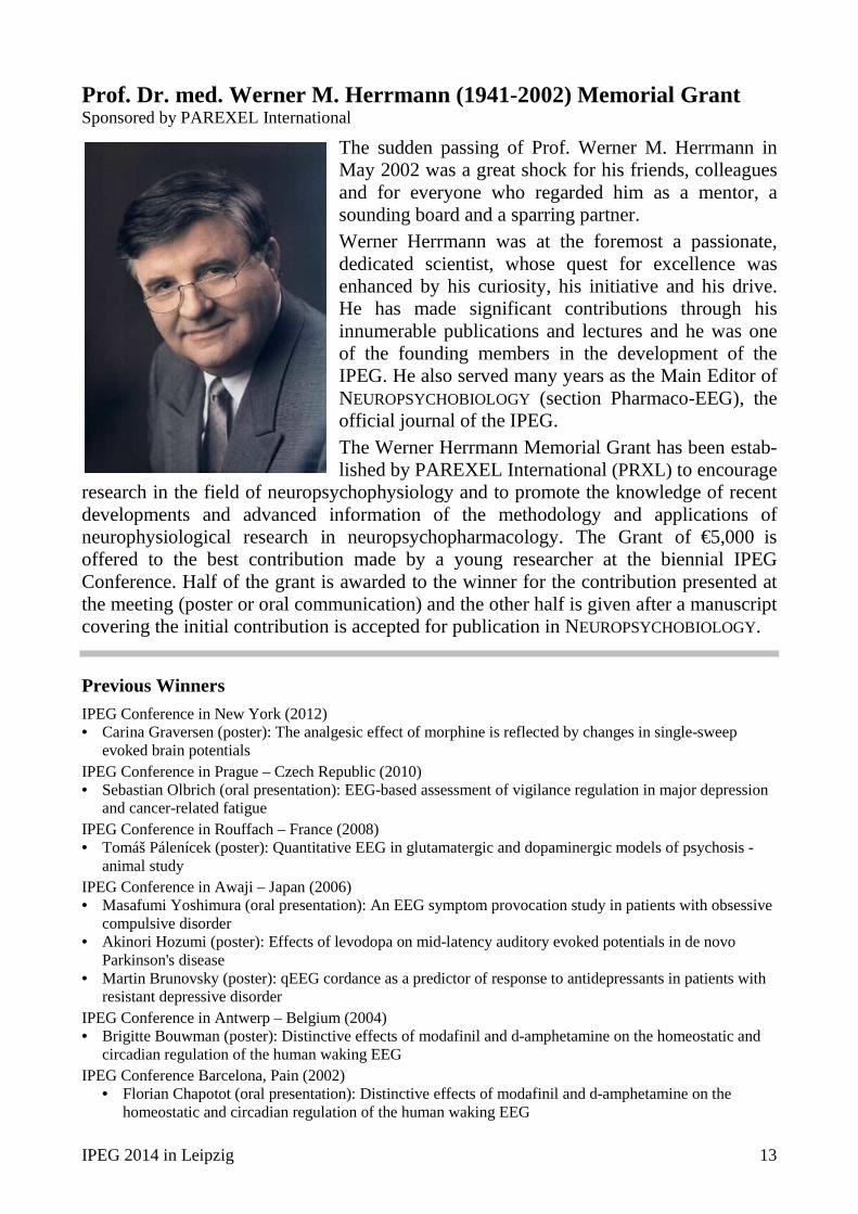
Prof. Dr. med. Werner M. Herrmann (1941-2002) Memorial Grant Sponsored by PAREXEL International
The sudden passing of Prof. Werner M. Herrmann in May 2002 was a great shock for his friends, colleagues and for everyone who regarded him as a mentor, a sounding board and a sparring partner. Werner Herrmann was at the foremost a passionate, dedicated scientist, whose quest for excellence was enhanced by his curiosity, his initiative and his drive. He has made significant contributions through his innumerable publications and lectures and he was one of the founding members in the development of the IPEG. He also served many years as the Main Editor of NEUROPSYCHOBIOLOGY (section Pharmaco-EEG), the official journal of the IPEG. The Werner Herrmann Memorial Grant has been estab-lished by PAREXEL International (PRXL) to encourage
research in the field of neuropsychophysiology and to promote the knowledge of recent developments and advanced information of the methodology and applications of neurophysiological research in neuropsychopharmacology. The Grant of €5,000 is offered to the best contribution made by a young researcher at the biennial IPEG Conference. Half of the grant is awarded to the winner for the contribution presented at the meeting (poster or oral communication) and the other half is given after a manuscript covering the initial contribution is accepted for publication in NEUROPSYCHOBIOLOGY.
Previous Winners
IPEG Conference in New York (2012) • Carina Graversen (poster): The analgesic effect of morphine is reflected by changes in single-sweep
evoked brain potentials IPEG Conference in Prague – Czech Republic (2010) • Sebastian Olbrich (oral presentation): EEG-based assessment of vigilance regulation in major depression
and cancer-related fatigue IPEG Conference in Rouffach – France (2008) • Tomáš Pálenícek (poster): Quantitative EEG in glutamatergic and dopaminergic models of psychosis -
animal study IPEG Conference in Awaji – Japan (2006) • Masafumi Yoshimura (oral presentation): An EEG symptom provocation study in patients with obsessive
compulsive disorder • Akinori Hozumi (poster): Effects of levodopa on mid-latency auditory evoked potentials in de novo
Parkinson's disease • Martin Brunovsky (poster): qEEG cordance as a predictor of response to antidepressants in patients with
resistant depressive disorder IPEG Conference in Antwerp – Belgium (2004) • Brigitte Bouwman (poster): Distinctive effects of modafinil and d-amphetamine on the homeostatic and
circadian regulation of the human waking EEG IPEG Conference Barcelona, Pain (2002)
• Florian Chapotot (oral presentation): Distinctive effects of modafinil and d-amphetamine on the homeostatic and circadian regulation of the human waking EEG
Obituary
14 18th Biennial Conference
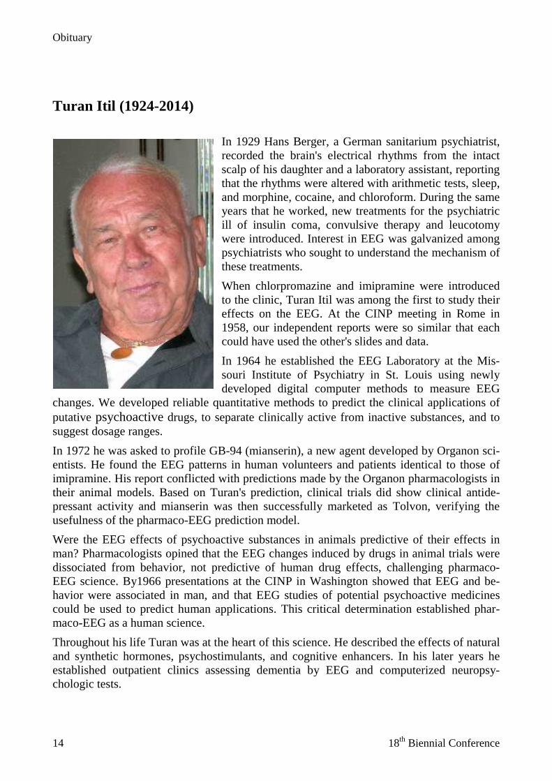
Turan Itil (1924-2014)
In 1929 Hans Berger, a German sanitarium psychiatrist, recorded the brain's electrical rhythms from the intact scalp of his daughter and a laboratory assistant, reporting that the rhythms were altered with arithmetic tests, sleep, and morphine, cocaine, and chloroform. During the same years that he worked, new treatments for the psychiatric ill of insulin coma, convulsive therapy and leucotomy were introduced. Interest in EEG was galvanized among psychiatrists who sought to understand the mechanism of these treatments.
When chlorpromazine and imipramine were introduced to the clinic, Turan Itil was among the first to study their effects on the EEG. At the CINP meeting in Rome in 1958, our independent reports were so similar that each could have used the other's slides and data.
In 1964 he established the EEG Laboratory at the Mis-souri Institute of Psychiatry in St. Louis using newly developed digital computer methods to measure EEG
changes. We developed reliable quantitative methods to predict the clinical applications of putative psychoactive drugs, to separate clinically active from inactive substances, and to suggest dosage ranges.
In 1972 he was asked to profile GB-94 (mianserin), a new agent developed by Organon sci-entists. He found the EEG patterns in human volunteers and patients identical to those of imipramine. His report conflicted with predictions made by the Organon pharmacologists in their animal models. Based on Turan's prediction, clinical trials did show clinical antide-pressant activity and mianserin was then successfully marketed as Tolvon, verifying the usefulness of the pharmaco-EEG prediction model.
Were the EEG effects of psychoactive substances in animals predictive of their effects in man? Pharmacologists opined that the EEG changes induced by drugs in animal trials were dissociated from behavior, not predictive of human drug effects, challenging pharmaco-EEG science. By1966 presentations at the CINP in Washington showed that EEG and be-havior were associated in man, and that EEG studies of potential psychoactive medicines could be used to predict human applications. This critical determination established phar-maco-EEG as a human science.
Throughout his life Turan was at the heart of this science. He described the effects of natural and synthetic hormones, psychostimulants, and cognitive enhancers. In his later years he established outpatient clinics assessing dementia by EEG and computerized neuropsy-chologic tests.
Obituary
IPEG 2014 in Leipzig 15
He was a founder of the International Pharmaco-EEG Group (IPEG), an active scientific member of ACNP and CINP and numerous other EEG and psychopharmacology societies. Werner Herrmann in Berlin, Bernd Saletu in Vienna, Masami Saito in Osaka, and Sevket Akpinar in Ankara, each a leader in pharmaco-EEG science, were his students. Turan was consultant and lead scientist on projects of the World Health Organization.
Turan Itil was born in Bursa, Turkey on August 12, 1924. He received the M.D. degree from Istanbul University in 1948 and moved to the University of Tübingen in Germany for training in neurology. In 1953 he joined the faculty at the University of Erlangen with EEG and psychopharmacology the center of his research. After a decade in St. Louis, he moved to New York Medical College and established the HZI Research Center Laboratory in Tarrytown New York.
He was a vibrant, enthusiastic, and warm-hearted man. He played intensely, enjoyed ping-pong, billiards and roulette. He supported friends and colleagues enthusiastically. He ad-justed to the American culture but on retirement he returned to his family in Turkey. He died at his country home on April 29, 2014 at age 89. He and his wife Ellen had two chil-dren, Kurt and Yasmin. He leaves an extended family in Turkey and New York, friends and students around the world, and a unique body of psychopharmacology science. Max Fink
(May 29, 2014)
Table of Contents
16 18th Biennial Conference
EEG-BASED PERSONALIZED MEDICINE
EEG-vigilance regulation in neuropsychiatric disorders .............................................................. 24 Ulrich Hegerl, Peter Schönknecht, Tilman Hensch, Sebastian Olbrich, Michael Kluge, Hubertus Himmerich, Christian Sander
EEG-vigilance and the autonomous nervous system in the prediction of antidepressant treatment: Findings from the iSPOT-D study ............................................................................... 25
Sebastian Olbrich, Galina Surova, Richard Gevirtz, Evian Gordon, John A Rush, Ulrich Hegerl, Martijn Arns
The utility of electrocortical measures in characterizing depression and treatment response ...... 26 Jaworska Natalia, Blier Pierre, Fusee Wendy, de la Salle Sara, Smith Dylan, Rima Ilhem, Knott Verner
Response prediction of psychiatric treatment – how close is practical application? .................... 27 Jürgen Gallinat
Biosignatures for Personalized Treatment of Depression: Findings for Electrophysiological and Neurocognitive Measures ....................................................................................................... 28
Gerard E. Bruder
First results of the iSPOT studies in Depression and ADHD: EEG alpha asymmetry as a gender specific predictor of SSRI treatment outcome................................................................... 29
Martijn Arns
The IPPEC, a new industry-academia initiative to set up a global, translational and precompetitive pharmaco-EEG database ...................................................................................... 30
Gé SF Ruigt, Pim Drinkenburg
KEYNOTES
Keynote 1 – QEEG Source Localization in the Identification of Theoretical Underlying Pathophysiology in Subtypes of Neuropsychiatric/Neurological Disorders........................................... 32
Leslie S. Prichep
Keynote 2 – From maps to mechanisms: Multimodal Imaging with EEG, fMRI and DTI in Psychiatry ................................................................................................................................................ 33
Christoph Mulert
Keynote 3 – The clinical value of source-resolved EEG analysis: drowsiness, ADHD, epilepsy, and schizophrenia .................................................................................................................... 34
Scott Makeig
Keynote 4 – Arousal systems: The origin of the waking EEG .............................................................. 35 Clifford B. Saper
Keynote 5 – Pharmaco-sleep: Five decades of research ........................................................................ 37 Hartmut Schulz
Table of Contents
IPEG 2014 in Leipzig 17
SYMPOSIA Symposium 1: EEG – Transcranial Magnetic Stimulation (TMS)
Fundamentals of TMS-EEG .......................................................................................................... 40 Risto Ilmoniemi
Pharmaco-TMS-EEG: A novel approach for probing GABAergic neurotransmission in human cortex ................................................................................................................................. 41
Ulf Ziemann, Isabella Premoli, Florian Müller-Dahlhaus
TMS-evoked potentials as a marker of brain maturation in childhood and adolescence .............. 42 Stephan Bender
Insights into consciousness and unconsciousness by TMS-EEG .................................................. 43 Mario Rosanova
Symposium 2: Electropsychopharmacology – Enriching Psychopharmacology with ERPs
Drug effects in visual spatial-cuing paradigms ............................................................................. 44 J.L. Kenemans & H.N.A. Logemann
Genetic polymorphisms in the dopamine and serotonin system as factors in neurophysiological indices of hypervigilance ............................................................................... 45
I. Heitland, K.B.E. Böcker, J.M.P. Baas & J.L. Kenemans
Functional neuromarkers for psychiatry: clinical applications of event related potentials for diagnosis, prognosis and treatment. .............................................................................................. 46
Juri D. Kropotov
Symposium 3: Fast Acting Agents and Underlying Neurotransmitter Systems
Ketamine - perspectives from a neurophysiological point of view ............................................... 47 Georg Winterer
Pharmaco-EEG study of ketamine in patients with major depressive disorder: QEEG and clinical predictors of antidepressive response. .............................................................................. 48
Martin Brunovsky, Jiri Horacek, Peter Sos, Tomas Palenicek, Tomas Novak, Monika Klirova, Cyril Höschl
EEG power spectra and connectivity changes in animal models of psychosis - comparisons of glutamatergic, serotonergic and cannabinoid models ............................................................... 49
Tomas Palenicek, Filip Tyls, Michaela Fujakova, Anna Kubesova, Pavlina Novakova, Lukas Kaderabek, Martin Brunovsky, Jiri Horacek
Functional connectivity analysis of default mode network changes after ketamine application in healthy subjects ...................................................................................................... 50
F. Mueller, G. Winterer
Symposium 4: EEG-Vigilance Regulation
Assessment of EEG-Vigilance using the VIGALL algorithm ...................................................... 51 Christian Sander, Sebastian Olbrich, Janek Spada, Philippe Jawinski, Jue Huang, Daniel Böttger, Tilman Hensch, Ulrich Hegerl
Table of Contents
18 18th Biennial Conference
CACNA1C gene variation is linked to EEG-vigilance regulation ................................................ 52 Philippe Jawinski, Christian Sander, Nicole Mauche, Janek Spada, Madlen Häntzsch, Ralph Burkhardt, Ulrich Hegerl, Tilman Hensch
Assessment of EEG-based vigilance regulation as early predictor for antidepressant pharmacotherapy ........................................................................................................................... 53
Frank M. Schmidt, Marie-Elisa Dietz, Christian Sander, Claudia Nowak, Peter Schönknecht, Hubertus Himmerich, Ulrich Hegerl
EEG vigilance in borderline personality disorder ......................................................................... 54 Lucas Kramer, Christian Sander, Kerstin Herwig, Dorothee Gescher, Ulrich Hegerl, Sabine C. Herpertz, Katja Bertsch
Symposium 5: Novel IPEG Animal Pharmaco-EEG Guidelines
Framework and outline of the novel IPEG animal pEEG Guidelines ........................................... 55 Wilhelmus H.I.M. Drinkenburg
Auditory event-related potentials as back-translational tools for studying neuronal processing during pre-attentive and attentive processing ............................................................. 56
Jesper F. Bastlund
Rat pharmaco-EEG studies: multi-channel methodology ............................................................. 57 Tomas Palenicek, Filip Tyls, Michaela Fujakova, Anna Kubesova, Pavlina Novakova, Lukas Kaderabek, Vaclava Sedlmayerova, Vladimir Krajca, Martin Brunovsky, Jiri Horacek
The power of EEG in a non-human primate ................................................................................. 58 Ingrid H.C.H.M. Philippens
Translational aspects of animal EEG studies in the IMI PharmaCog Consortium for studies on Alzheimer’s Disease ................................................................................................................. 59
Claudio Babiloni, Susanna Lopez, Andrea Soricelli, Giovanni Frisoni, Wilhelmus Drinkenburg, Jesper Bastlund, Gianluigi Forloni, Marina Bentivoglio, Esther Schenker, Sophie Dix, Regis Bordet, Flavio Nobili, Peter Schoenknecht, Jill Richardson on behalf of PharmaCog Consortium
Symposium 6: EEG-based Personalized Medicine in Psychiatry: Current status and future prospects
The utility of electrocortical measures in characterizing depression and treatment response ...... 60 Jaworska Natalia, Blier Pierre, Fusee Wendy, de la Salle Sara, Smith Dylan, Rima Ilhem, Knott Verner
The Methylphenidate in mania project (MEMAP) ....................................................................... 61 Michael Kluge, Ulrich Hegerl, Christian Sander, Jens Dietzel, Roland Mergl, Istvan Bitter, Koen Demyttenaere, Ricardo Gusmão, Ana Gonzalez-Pinto, Victor Perez-Sola, Eduard Vieta, Georg Juckel, Ulrich Zimmermann, Michael Bauer, Pascal Sienaert, Sónia Quintão, Marc-Andreas Edel, Csilla Bolyos, Jose Luis Ayuso-Mateos, Pilar López-García
First results of the iSPOT studies in Depression and ADHD: EEG alpha asymmetry as a gender specific predictor of SSRI treatment outcome................................................................... 62
Martijn Arns
Table of Contents
IPEG 2014 in Leipzig 19
ORAL PRESENTATIONS Session 1
Electrophysiological correlates of response inhibition across menstual cycle: Go-Nogo potential study ............................................................................................................................... 64
Inga Griskova-Bulanova, Ramune Griksiene, Osvaldas Ruksenas
Stimulant medication in pediatric ADHD: Predicting the clinical outcome from single-dose changes in Event Related Potentials (ERPs) and a Go/No-go test ................................................ 65
Geir Ogrim, Jan Brunner, Juri Kropotov
Proof-of-Principle to efficacy in psychiatric patients exemplified by a neuronal-selective nicotinic agonist AZD1446 ........................................................................................................... 66
Peter H Boeijinga, Albena Patroneva
Individual Alpha Peak Frequency as an Endophenotype Associated with Affective Predisposition ................................................................................................................................ 67
Aftanas L.I., Tumyalis A.V.
Session 2
Resting EEG abnormalities in traumatised refugee adults ............................................................ 68 Mirjana Askovic, Louis Mayaud
Objectively Identifying Abnormal EEG Recordings in Clinical Studies ...................................... 69 Junshui Ma, Fei Su, Vladimir Svetnik
Registration of the brain activity under the influence of artificial electromagnetic radiation ...... 70 Reznikov Dmitry
Machine Learning for a Parkinson’s prognosis and diagnosis system based on EEG .................. 71 A. Soria-Frisch, J. Marin, D. Ibañez, S. Dunne, C. Grau, G. Ruffini, J. Rodrigues-Brazète, R. Postuma, J.-F. Gagnon, J. Montplaisir, A. Pascual-Leone
Session 3
Placebo-controlled clinical and polysomnographic studies on the acute and chronic effects of electroacupuncture in primary insomnia ................................................................................... 72
Gerda Maria Saletu-Zyhlarz, Alexander Meng, Peter Anderer, Evelyn Doll, Sergio Rosales-Rodriguez, Michaela Bijak, Daniela Stockenhuber, Helmut Nissel, Bernd Saletu
Cordance and REM density derived from REM Sleep as Biomarker for Treatment Response in Depression after Antidepressant Medication – a Follow-up study ........................... 73
Marcel Pawlowski, Amin Raissi, Martin Dresler, Florian Holsboer, Axel Steiger, Marek Adamczyk
Placebo-controlled EEG topography/tomography and psychometric studies on the acute and chronic effects of electroacupuncture on daytime vigilance in primary insomnia ................. 74
Bernd Saletu, Alexander Meng, Peter Anderer, Evelyn Doll, Sergio Rosales-Rodriguez, Michaela Bijak, Daniela Stockenhuber, Helmut Nissel, Gerda Maria Saletu-Zyhlarz
Polysomnographic correlates of subjective sleep onset ................................................................ 75 Frank Pillmann
Table of Contents
20 18th Biennial Conference
POSTER PRESENTATIONS QEEG and 19 Channel Neurofeedback as Clinical Evaluation Tool for Children with Attention, Learning and Emotional Problems ............................................................................... 78
Theresia Stöckl-Drax
The brain’s instantaneous emotional evaluation : real-time prefrontal gamma using sLORETA in children and young adults ....................................................................................... 79
Theresia Stöckl-Drax, Sabrina Mielke, Michael Keane
A Hierarchical EEG Artifacts Removing Method Based on Threshold Enhanced SWT ............. 80 Jiawei Wang, Fei Su
Machine-Learning-based Quantification of Brain-Age and Diagnosis of Cognitive Impairment .................................................................................................................................... 81
Brian Murphy, Yuqiao Gu, Giulia Cazzoli, Massimo Poesio, Gabriele Miceli
Elevated resting gamma power in people undertaking methadone treatment for opiate dependence .................................................................................................................................... 82
Grace Y Wang, Rob Kydd, Trecia A Wouldes, Maree Jensen, Bruce R Russell
Frankfurt clock paradigm vs. Bern clock paradigm: task difficulty effects on visuospatial processing ...................................................................................................................................... 83
Ingrida Antonova, Anja Baenninger, Axel Kohler, Inga Griskova-Bulanova, Thomas Dierks, Thomas Koenig
Identity authorization based on Electroencephalogram ................................................................ 84 Pengyu Li, Fei Su
Aberrant functional connectivity in AD patients: an EEG-LORETA study ................................. 85 Keiichiro Nishida, Roberto Pascual-Marqui, Masafumi Yoshimura, Yuichi Kitaura, Hiroshi Mii, Toshiaki Isotani, Toshihiko Kinoshita
A case of visual pseudo-hallucinations induced by clozapine and bupropion co-administration associated with epileptiform EEG modifications .................................................. 86
Anna Castelnovo, Simone Cavallotti, Silvio Scarone, Mariapaola Canevini , Armando D’Agostino
VIGALL 2.0: A free software tool for semi-automated EEG vigilance research ......................... 87 Daniel Böttger, Christian Sander, Sebastian Olbrich, Johannes Jödicke, Ulrich Hegerl
Impact of chronic smoking on P3 components in a three-stimulus oddball paradigm ................. 88 Tilman Hensch, Nicole Mauche, Christian Sander, Philippe Jawinski, Cornelia Enzenbach, Sebastian Olbrich, Peter Schönknecht, Ulrich Hegerl
QEEG correlates of sensory craving episodes in a case of girl with autism: the role of female hormonal changes .............................................................................................................. 89
Svetla Velikova
Effect of smoking withdrawal on EEG-vigilance ......................................................................... 90 Janek Spada, Christian Sander, Philippe Jawinski, Sebastian Olbrich, Tilman Hensch, Ivonne Burgos Guerrero, Ulrich Hegerl
Number of cigarettes smoked per day predicts auditory evoked N1/P2 amplitude ...................... 91 Philippe Jawinski, Nicole Mauche, Janek Spada, Cornelia Enzenbach, Christian Sander, Ulrich Hegerl, Tilman Hensch
Table of Contents
IPEG 2014 in Leipzig 21
EEG connectivity analysis as stratification method about responders or non-responders in male non-smokers to nicotine administration ............................................................................... 92
Paolo Ranzi, Jan Freund
Functional and Structural Biomarkers in Major Depression and Antidepressant Treatment: A simultaneous EEG-fMRI study ................................................................................................. 93
Galina Surova, Matthias Gawlitza, Ulrich Hegerl, Karl-Titus Hoffmann, Donald Lobsien, Peter Schönknecht, Sebastian Olbrich
Sleep deficits in mild cognitive impairment are associated with increased plasma amyloid-β levels and cortical thinning ........................................................................................................ 94
Mayely P. Sanchez-Espinosa, Mercedes Atienza, Jose L. Cantero
The use of a data driven LORETA Progress Report (LPR) to determine the most deviant Z score maximal voxel for LORETA neurofeedback in a patient post neurosurgery for the removal of two right frontal lobe cysts ......................................................................................... 95
Deborah R. Simkin, Phil Jones, Joel Lubar
Preclinical evaluation of antiepileptic drugs using qEEG methods in a mouse model of mesial-temporal lobe epilepsy ....................................................................................................... 96
Benoît Pouyatos, Venceslas Duveau, Céline Bouyssières, Carine Dumont, Yann Roche, Corinne Roucard
The role of different serotonergic receptors in 2C-B induced changes in quantitative EEG in rats ............................................................................................................................................. 97
Filip Tylš, Tomáš Páleníček, Pavlína Nováková, Lukáš Kadeřábek, Michaela Fujáková, Anna Kubešová, Jiří Horáček
The pharmaco-EEG during the process of dying; a neglected aspect ........................................... 98 Clementina M van Rijn, Annika Lüttjohann, Hans Krijnen, Saskia Menting-Hermeling, Martin Perescis, Anton ML Coenen, Lyudmila V Vinogradova, Marijtje LA Jongsma
Is EEG biomarker integration the key to personalized medicine? Evidence from zygosity prediction in twins ......................................................................................................................... 99
Sonja Simpraga, Simon-Shlomo Poil, Huibert D. Mansvelder, Dorret I. Boomsma, Eco J.C. de Geus, Dirk Smit, Klaus Linkenkaer-Hansen
The Neurophysiological Biomarker Toolbox (NBT) for clinical M/EEG .................................. 100 Simon-Shlomo Poil, Sonja Simpraga, Richard Hardstone, Huibert D. Mansvelder, Klaus Linkenkaer-Hansen
Anterior Cingulate Cortex Theta Current Density during REM Sleep. A Predictor for Treatment Response in Major Depression? ................................................................................ 101
Thomas von Werder, Lisa Müller, Amin Raissi, Axel Steiger, Michael Czisch, Marcel Pawlowski
The advantage of high-resolution EEG recordings for cognitive evaluation .............................. 102 Jurij Dreo, Andreja Emeršič, Daniel Attia, Grega Repovš, Zvezdan Pirtošek
A normative database of cognition during rest based on the Amsterdam Resting-State Questionnaire for clinical trials and therapeutic interventions .................................................... 103
S. Simpraga, S.-S. Poil, R. Hardstone, B.A. Diaz, J.S. Benjamins, H.D. Mansvelder, E. van Someren, K. Linkenkaer-Hansen
Table of Contents
22 18th Biennial Conference
A two-step machine learning discriminant algorithm to predict the outcome of stimulant medication in children with ADHD using quantitative EEG and event-related potentials from go/no-go tasks. .................................................................................................................... 104
Kompatsiari K, Blunck A, Candrian G, Mueller A, Ogrim G, Kropotov JD
sLORETA and LDAEP in bipolar affective patients and healthy controls during transient induction of emotionally neutral and negative mood state ......................................................... 105
Viktorinová M, Brunovský M, Novák T, Goetz M, Horáček J
Frequency of Occurrence and Description of Abnormalities in Mild or Moderate Traumatic Brain Injury or Concussion, as Identified by Dense Array Electroencephalography (DEEG) ... 106
Michael B Russo, Judi Profant, Ryan Michael Nillo, Nolan Endicott, Turan Itil

IPEG 2014 in Leipzig 23
EEG-BASED PERSONALIZED MEDICINE
EEG-based personalized Medicine
24 18th Biennial Conference
EEG-vigilance regulation in neuropsychiatric disorders Ulrich Hegerl, Peter Schönknecht, Tilman Hensch, Sebastian Olbrich, Michael Kluge, Hubertus Himmerich, Christian Sander (1) Department of Psychiatry and Psychotherapy, University Hospital Leipzig, University of
Leipzig, GERMANY
The human brain can take over different global functional states not only during sleep (sleep stages, e.g. slow wave sleep, REM sleep) but also during wakefulness. These different states of CNS-arousal are called vigilance stages and can best be observed during the transition from active wakefulness to drowsiness and sleep onset using electroencephalography (EEG). A recently developed EEG-based algorithm (Vigilance Algorithm Leipzig, VIGALL) enables fast and standardized classification of EEG-segments into 7 different vigilance stages. This allows an objective assessment of the level as well as the regulation of vigilance (e.g. by analyzing the time course of vigilance fluctuations). A variety of clinical and preclinical arguments indicate that the precise regulation and adaptation of vigilance is not only of fundamental importance for all higher organisms but also plays a pathogenetic role in psychiatric disorders such as depression, mania and ADHD. Within the vigilance model of affective disorders and ADHD [1] the hyperactivity and sensation seeking observed in ADHD and mania is interpreted as an autoregulatory attempt of the organism to stabilize vigilance regulation by increasing external stimulation, comparable to the irritated behavior of overtired children. In line with this concept the possible antimanic effects of methylphenidate are presently studied in an international placebo-controlled RCT [2]. Correspondingly the withdrawal and sensation avoidance in major depression is interpreted as a reaction to a state of tonically high vigilance [1]. In unmedicated patients with major depression a hyperstable regulation of vigilance has been found during EEG recordings under quiet rest [3]. This finding has since then been replicated in independent samples. Based on these findings as well as other arguments it will be discussed whether the vigilance regulation can be considered to be a diagnostic marker and a predictor of treatment response useful for clinical and research purposes.
Keywords: vigilance regulation, affective disorders, ADHD, diagnostic marker, response prediction
Corresponding author: [email protected]
[1] Hegerl U, Hensch T. The vigilance regulation model of affective disorders and ADHD. Neurosci Biobehav Rev. 2012 Oct 22. pii: S0149-7634(12)00175-3. doi: 10.1016/j.neubiorev.2012.10.008. [Epub ahead of print].
[2] Kluge M, Hegerl U, Sander C, et al. Methylphenidate in mania project (MEMAP): study protocol of an international randomised double-blind placebo-controlled study on the initial treatment of acute mania with methylphenidate. BMC Psychiatry. 2013 Feb 27;13:71.
[3] Hegerl U, Wilk K, Olbrich S, Schoenknecht P, Sander C. Hyperstable regulation of vigilance in patients with major depressive disorder. World J Biol Psychiatry. 2012 Sep;13(6):436-46.
EEG-based personalized Medicine
IPEG 2014 in Leipzig 25
EEG-vigilance and the autonomous nervous system in the prediction of antidepressant treatment: Findings from the iSPOT-D study Sebastian Olbrich(1), Galina Surova(1), Richard Gevirtz(1), Evian Gordon(1), John A Rush(1), Ulrich Hegerl(1), Martijn Arns(2,3,4) (1) Dept. of Psychiatry and Psychotherapy, University of Leipzig, Germany (2) Dept. of Experimental Psychology, Utrecht University, Utrecht, The Netherlands (3) Research Institute Brainclinics, Nijmegen, The Netherlands (4) Dept. of Cognitive Neuroscience, Donders Institute for Brain Cognition and Behaviour, Radboud
UMC, Nijmegen, The Netherlands
To overcome the limitations of the syndrome-based diagnostic routine in neuropsy-chiatric disorder and to provide biomarker-informed decisions for treatment, recently the Research Domain Criteria have been initialized [1]. They include separate criteria for autonomous and arousal systems. Based on findings of altered wakefulness-regulation and autonomous function in major depressive disorder (MDD), the goal of this study therefore was to investigate the predictive value for treatment outcome of central nervous system (CNS) and autonomous nervous system (ANS) arousal and their interaction in a large cohort of patients from the iSPOT-D trial that received either a selective-serotonin reuptake-inhibitor (SSRI) or a serotonin-norepinephrine-reuptake-inhibitor (SNRI).
Methods: CNS and ANS-arousal (defined by electroencephalogram vigilance and heart rate) and their change over time were assessed during rest. Differences of treatment outcome as defined by the decline of Hamilton Rating Scale for Depres-sion- (HRSD) from baseline to week 8 after treatment initiation for the whole sample and for SSRI and SNRI groups separately were analysed using a binary logistic re-gression model and repeated measure analysis of variance (ANOVA).
Results: Responders and remitters were characterized by a steeper decline of CNS-arousal. Subgroup analysis showed that this effect was only present for the SSRI arm whereas SNRI responders showed a more pronounced increase of ANS-arousal. Fur-ther, SSRI responders showed a correlation between ANS and CNS measures, SSRI non-responders and the whole SNRI subgroup did not.
Conclusions: CNS and ANS-arousal during rest predict positive treatment outcome to antidepressant medication. The differences of CNS and ANS-profiles for SSRI or SNRI prediction are interpreted as neurophysiological traits that indicate responsive-ness to different drug-classes rather than disorder specific aspects.
Keywords: CNS-arousal, ANS-arousal, antidepressant, personalized medicine, major depressive disorder
Corresponding author: [email protected]
[1] Insel T et al. (2010): Research Domain Criteria (RDoC): Toward a New Classification Framework for Research on Mental Disorders. American Journal of Psychiatry 167: 748–751.
EEG-based personalized Medicine
26 18th Biennial Conference
The utility of electrocortical measures in characterizing depression and treatment response Jaworska Natalia(1), Blier Pierre(2), Fusee Wendy(2), de la Salle Sara(2), Smith Dylan(2), Rima Ilhem(2), Knott Verner(2) (1) Department of Psychiatry, McGill University, Montreal, QC, Canada (2) University of Ottawa, Institute of Mental Health Research, Ottawa, ON, Canada
Background: Assessments of electroencephalographic (EEG) activity and event-related potentials (ERPs) in major depressive disorder (MDD) have provided insight into the electrocortical abnormalities associated with the disorder. Such indices have also emerged as candidates for predicting and optimizing treatment outcomes. Methods: Individuals with MDD (N=53; 25 females) were tested prior to, and after 1 and 12 weeks of antidepressant treatment (escitalopram [ESC] + bupropion [BUP], ESC or BUP). Treatment responders exhibited a >50% decrease in depression scores by week 12. Healthy, non-depressed controls (HCs) were also tested (N=43; 23 females). We assessed resting EEG activity (32 electrodes; mastoid-reference), P3a/b ERPs elicited by an auditory oddball task as well as auditory evoked potentials (AEPs) and associated loudness dependence of the AEP (LDAEP) slopes. In addition to power and amplitude/latency measures of EEG and ERPs, respectively, standardized low-resolution brain electromagnetic tomography (sLORETA) was used. Data mining techniques were employed to determine if EEG power at week 1 predicted treatment response. Results: Depressed individuals (especially males) had greater overall frontal and parietal alpha power and increased subgenual anterior cingulate cortex (sgACC)-localized theta2 activity relative to HCs. Treatment responders exhibited high, and non-responders low, frontal baseline alpha2 power. Posterior alpha2 power and sgACC-localized theta2 activity strongly discriminated ESC responders/non-responders. Non-responders had smaller baseline P3a/b amplitudes than responders and HCs. Regarding the LDAEP, baseline N1 sLORETA-LDAEP discriminated responders/non-responders. Finally, data mining indicated that increased week 1 theta (midfrontal and CP1 electrodes) and decreased delta power (at F4) characterized responders, while decreased theta and increased delta in these regions were characteristic of non-responders (high classification accuracy). Conclusions: Electrocortical features differentiated individuals with MDD and HCs, as well as being associated with treatment response, though gender-specific effects emerged. Establishing standardized recording and analyses guidelines, coupled with normative cut-off values, are critical next steps towards utilizing electrocortical measures in guiding clinical decision-making.
Keywords: depression, EEG, ERPs, treatment, response, prediction
Corresponding author: [email protected]
EEG-based personalized Medicine
IPEG 2014 in Leipzig 27
Response prediction of psychiatric treatment – how close is practical application? Jürgen Gallinat
Klinik und Poliklinik für Psychiatrie und Psychotherapie Universitätsklinikum Hamburg-Eppendorf, Martinistraße 52, 20246 Hamburg
Response prediction in psychiatric treatment processes is a fundamental clinical target because treatment response appears normally after several weeks and the success of disease prophylaxis and maintenance therapy can be assessed only after years. Different biological and non-biological response predictors have been evaluated in psychiatry in recent years. Classification- and machine learning tools have been established to increase the precision of response prediction as well as the identification of diagnostic categories and biological subgroups. Although promising results have been published, the application of response predictors in clinical psychiatry is still rare or unusual. Several reasons are responsible for this situation and will be described in the present talk. Possible solutions and future research strategies to develop the utility of response predictors in clinical practice will be discussed.
Keywords: biological treatment, response prediction, diagnostic classification, brain imaging
Corresponding author: [email protected]
EEG-based personalized Medicine
28 18th Biennial Conference
Biosignatures for Personalized Treatment of Depression: Findings for Electrophysiological and Neurocognitive Measures Gerard E. Bruder
Department of Psychiatry, Columbia University College of Physicians & Surgeons
Although a variety of treatments are available for depression, clinicians have no way of knowing whether or not a patient will benefit from a specific treatment. There is growing evidence that electrophysiological measures (resting EEG and evoked or event-related potentials) have may have value as biological markers for predicting clinical response to antidepressants. This presentation will focus on findings for three measures that have been associated with treatment response: (1) EEG power and asymmetry in the alpha band; (2) theta in the rostral anterior cingulate cortex (rACC); and (3) loudness dependence of auditory evoked potentials (LDAEP). These measures are currently being used in a multi-site project “EMBARC—Establishing Moderators and Mediators of Antidepressant Response for Clinical Care”. Since no one test is likely to prove sufficient for predicting response to different antidepressants, this project aims to develop a multivariate biosignature that integrates across clinical, neuroimaging, electrophysiological and behavioral neurocognitive markers for predicting treatment response. In this project, patients are randomized double-blind to 8 weeks of treatment with the SSRI sertraline or placebo, with nonresponders switched to sertraline or bupropion (SNRI). The following measures are obtained at baseline and one week after treatment: structural MRI, fMRI (during resting state, emotional recognition, and reward task), resting EEG, LDAEP, and neurocognitive tests assessing word fluency, choice RT, working memory, and cognitive control. In addition, a study in 40 healthy adults was first conducted to evaluate the test-retest reliability of these measures. Findings from this reliability study and a prior study using the same resting EEG, LDAEP and neurocognitive tests as the EMBARC study will be presented, which support the potential value of these measures for developing clinical aids for selecting treatments for depression. Corresponding author: [email protected]
EEG-based personalized Medicine
IPEG 2014 in Leipzig 29
First results of the iSPOT studies in Depression and ADHD: EEG alpha asymmetry as a gender specific predictor of SSRI treatment outcome Martijn Arns(1,2,3) (1) Research Institute Brainclinics, Nijmegen, The Netherlands (2) Dept. of Experimental Psychology, Utrecht University, Utrecht, The Netherlands (3) Dept. of Cognitive Neuroscience, Donders Institute for Brain Cognition and Behaviour, Radboud
UMC, Nijmegen
Background: Measures of alpha electroencephalogram (EEG) activity often — but not always — differentiate depressed patients from normal controls. Further, some evidence suggests that overall antidepressant response may be associated with greater baseline alpha EEG activity. This study aimed to determine whether occipital alpha and frontal alpha asymmetry would distinguish outpatients with major depression from controls, whether these measures behave as overall and differential predictors of outcome to a Selective Serotonin Reuptake Inhibitor (SSRI) and a Serotonin Norepinephrine Reuptake Inhibitor (SNRI), and to explore the effects of gender on these patterns.
Methods: In the international Study to Predict Optimized Treatment Response in Depression (iSPOT-D) and ADHD (iSPOT-A), a multi-center, international, randomized, prospective open-label trial. In iSPOT-D, 1008 major depressive disorder participants were randomized to escitalopram, sertraline or venlafaxine-extended release. In iSPOT-A, 332 children with ADHD were recruited and prescribed with methylphenidate. In addition 336 adults and 157 children were recruited as a control group. Treatment response was established after eight weeks using the 17-item Hamilton Rating Scale for Depression or the clinician rated ADHD-RS-IV. The resting electroencephalogram was measured at baseline in the eyes closed and eyes open conditions.
Results: No differences in electroencephalogram alpha for occipital and frontal cortex, or for alpha asymmetry, were found in participants with major depressive disorder compared to controls. Alpha in the occipital and frontal cortex were not associated with treatment outcome. However, a gender and drug-class interaction effect was found for frontal alpha asymmetry (F4-F3). Relatively greater right frontal alpha (less activity) in women only was associated with a favorable response to the SSRI escitalopram and sertraline. No such effect was found for the SNRI venlafaxine-extended release. The results for iSPOT-A will also be presented.
Conclusions: In women only, pretreatment alpha electroencephalogram predicted response to Selective Serotonin Reuptake Inhibitors, but not to venlafaxine-extended release. Future studies should separately analyze effects in alpha for men and women.
Corresponding author: [email protected]
EEG-based personalized Medicine
30 18th Biennial Conference
The IPPEC, a new industry-academia initiative to set up a global, translational and precompetitive pharmaco-EEG database Gé SF Ruigt(1), Pim Drinkenburg(2) (1) IPPEC, The Netherlands (2) IPEG, Belgium
The International Precompetitive Pharmaco-EEG Consortium (IPPEC) was established to address the lack of a database containing high quality pharmaco-EEG data on established reference drugs, which would allow for validation of pEEG-based (translational) biomarkers. The establishment of such a database would not only facilitate academic research into biomarkers but will also funnel the collective interest of a number of pharmaceutical industries to compare the EEG effects of their own proprietary drugs with high quality reference data. The initial database consists of vigilance-controlled clinical pEEG data in healthy volunteers, but it is the intention to extend the database in due course with additional pEEG data under different conditions (evoked potential and pharmaco-sleep data) and in different populations (clinical data from various patient and age groups, as well as reference pEEG data from different pre-clinical species). The database will contain both raw EEG data in EDF+ format as well as derived data and for the latter an extensive signal analysis and statistical toolbox will be developed together with specialized academic groups. The database will consist of different layers corresponding with the quality level of the data, the highest level being quality-controlled data obtained according to the IPEG guidelines[1,2]. Access rights will vary for the different layers with secured parts of the database safeguarded for proprietary data, when required. Access, extension, quality control and implementation of database and toolbox will be governed by the non-profit IPPEC foundation.
At the moment the consortium is setting up a series of clinical studies to validate the consistency of pEEG data across sites and to generate a core clinical dataset.
Keywords: pharmaco/EEG, database, reference drugs, translational, consortium
Corresponding author: [email protected] [1] Jobert, Marc, et al.: Guidelines for the recording and evaluation of pharmaco-EEG data in man: the International
Pharmaco-EEG Society (IPEG). Neuropsychobiology 2012; 66(4):201-220. [2] IPEG guidelines for animal pEEG studies, in preparation

IPEG 2014 in Leipzig 31
KEYNOTES
32 18th Biennial Conference
Keynote 1 – QEEG Source Localization in the Identification of Theoretical Underlying Pathophysiology in Subtypes of Neuropsychiatric/Neurological Disorders Leslie S. Prichep
Director, Quantitative Neurophysiological Brain Research Laboratories, New York University School of Medicine, New York, NY, USA
An extensive scientific literature attests to the clinical utility of quantitative EEG (QEEG) in the identification of brain dysfunction in neuropsychiatric disorders. Using source localization, different underlying pathophysiology has been shown to exist within subtypes of many such disorders. Evidence of thalamocortical dysrhythmia (TCD) as the theoretical underlying mechanism in subtypes of several neuropsychiatric/neurological disorders will be presented. Examples will include studies in obsessive compulsive disorder (OCD), chronic neuropathic pain, and chronic tinnitus: [1] OCD patients (n=27) were found to contain two subtypes with different frequency specific characteristics and different underlying sources. TCD sources were hypothesized for one of the subtypes; [2] A large population of chronic pain patients (n=87) were studied in both high and low pain states. While all showed evidence of activation of the “Pain Matrix”, several different subtypes were found within the population. Subtypes were characterized by different frequency specific abnormalities with different underlying sources, some of which were consistent with low-frequency oscillations present in TCD: [3] A large population of tinnitus patients (n=124) were found to contain different subtypes and while all subtypes shared certain cortical and subcortical sources, others features were distinctive to different subtypes. As with the other disorders, there were subtypes of tinnitus that supported a TCD underlying mechanism. The potential role which such information could play in optimization of treatment will be discussed.
Keywords: Source Localization, QEEG Subtypes, Thalamocortical Dysrhythmia (TCD), Neuropsychiatric Disorders
Corresponding author: [email protected]
IPEG 2014 in Leipzig 33
Keynote 2 – From maps to mechanisms: Multimodal Imaging with EEG, fMRI and DTI in Psychiatry Christoph Mulert
Department for Psychiatry and Psychotherapy, University Hamburg-Eppendorf. Germany
For an advanced understanding of brain function and structure both under normal conditions and in neuropsychiatric disorders it is important to integrate findings related to functional segregation and functional integration of brain networks. While functional Magnetic Resonance Imaging is a perfect tool in order to localize brain function, it can address connectivity aspects only indirectly. On the other hand, synchronized brain activity can be nicely investigated with electrophysiological techniques. Here, recent methodological progress has enabled us to investigate different aspects of coherence between different brain regions in the source space without the problem of volume conduction. In this talk, several examples will be provided about integration of several methods such as EEG, fMRI and DTI in order to get a more comprehensive understanding of brain function. One example will be the neurophysiological mechanisms involved in conscious auditory perception and pathophysiological mechanisms underlying auditory verbal hallucinations in schizophrenia.
34 18th Biennial Conference
Keynote 3 – The clinical value of source-resolved EEG analysis: drowsiness, ADHD, epilepsy, and schizophrenia Scott Makeig
Swartz Center for Computational Neuroscience, Institute for Neural Computation, University of California, San Diego, USA
Although EEG was the first functional ‘brain imaging’ modality (Berger, 1926), it has long suffered from a relative lack of contributions by engineers and biophysicists to make it more than a relatively oscure ‘scalp imaging’ modality. The fundamental problem, of course, is that the point-spread function (from coherent local cortical field activity within a small area of cortex to its projection to scalp recording electrodes) is so broad, whereby nearly every electrode records a weighted mixture of cortical EEG source activities from nearly every cortical area.
Fortunately, the mixture weights are fixed by head geometry and tissue conductivi-ties, which are relatively stable though difficult to measure directly. Unfortunately, the inverse problem of determining the distribution of source potentials that produce a given EEG channel or montage map is underdetermined -- choices between very many possibly contributing source patches cannot be made on the basis of a scalp map specified by many fewer EEG recording channels. To get around this problem requires exploring the spatial information inherent in the co-variations among EEG channel recordings over time.
Nearly twenty years ago I ran across a new mathematical method for doing so effi-ciently, Independent Component Analysis (ICA). This began a long study of source-resolved EEG brain imaging or electrocortical source imaging, since brain sources outside of cortex are rare or difficult to locate and are less well understood and characterized. Our research has now reached a stage at which applications to clinical research involving EEG are proving fruitful.
I will briefly describe four such studies in progress. The first concerns EEG source-level monitoring of alertness versus drowsiness. The second involves a set of ERP data collected during cognitive tasks from a group of ADHD and control children by my collaborator Sandra Loo of UCLA. The third involves modeling of seizure dynamics in ECoG data invasively recorded to plan surgery for epilepsy. The fourth involves data collected by collaborator Gregory Light of UCSD from schizophrenic and control subjects in an auditory deviance response paradigm. Results to date in all four investigations justify confidence that a great deal of clinically relevant and usable information about distributed brain processes and their pathologies, contained in high-density scalp EEG data, is now ready for scientific study and exploration.
Corresponding author: [email protected]
IPEG 2014 in Leipzig 35
Keynote 4 – Arousal systems: The origin of the waking EEG Clifford B. Saper
Department of Neurology, Harvard Medical School, Beth Israel Deaconess Medical Center, Boston, MA, USA 02215
Although Hans Berger discovered the fundamental relationship of the frequency of EEG oscillations to level of wakefulness in 1929, the cellular basis for this phenomenon has remained elusive. Recent advances, however, have brought us closer to understanding the basis for the EEG waves that are now used every day in many branches of medicine.
Work from Mircea Steriade and others showed that the fundamental EEG frequency of a slab of isolated cerebral cortex is an approximately 1 Hz high voltage slow wave pattern. Thus, this is the frequency generated by the cerebral cortex in isolation from ascending inputs.
The midfrequency EEG (3-15 Hz) was long thought to be due primarily to the interactions of the thalamus and the cerebral cortex. Work from Steriade as well as David McCormick and others emphasized the generation of oscillations in the range of sleep spindles (8-13 Hz) from the intrinsic dynamics of the thalamic relay nuclei, the thalamic reticular nucleus, and the cerebral cortex.
However, several different studies over the years, beginning with Jaime Villablanca in cats in the 1970’s and continuing with studies from the Vanderwolf and Buszaki labs in rats the 1980’s, and our own lab in 2010, have looked at the role of the thalamus in generating the midfrequency oscillations in the waking EEG. Each has found that even with extensive thalamic ablations, the power spectrum of the EEG in the midfrequencies is indistinguishable during wake from the baseline EEG. We have seen a case in our own hospital of a woman with bilateral thalamic hemorrhages destroying most of the thalamus bilaterally. Although the patient was in a persistent vegetative state clinically, her eyes-open EEG during the daytime showed only mild slowing, with the dominant frequencies in the mid-theta range.
Meanwhile, modern neuroanatomical studies have uncovered a wide rang of cell groups from the mesopontine tegmentum, through the caudal hypothalamus and basal forebrain, that project directly to the cerebral cortex, and recent physiological studies indicate that these pathways are likely to be underlie most of the oscillations in the midfrequency EEG.
Surprisingly, extensive deletions of the monoaminergic pathways, e.g., from the locus coeruleus or the histaminergic tuberomammillary nucleus, have failed to cause much change in the waking EEG pattern, or in the amount of total wakefulness. The most extensive EEG slowing has occurred with lesions of the glutamatergic neurons in the parabrachial nucleus. These neurons project to both the lateral hypothalamus and the basal forebrain, as well as directly to the prefrontal cortex. Lesions of the orexin
36 18th Biennial Conference
neurons in the lateral hypothalamus do not cause any change in the baseline EEG (although they do cause narcolepsy). But disrupting the activity of the glutamatergic neurons in the supramammillary nucleus cause EEG slowing and excessive sleepiness. The supramammillary neurons also project to the basal forebrain and directly to the cerebral cortex, especially the dentate gyrus and CA2 fields of the hippocampus.
Some supramammillary neurons fire in bursts that are phase-locked to the cortical EEG. This property is shared with some neurons in both the cholinergic and GABAergic populations of the basal forebrain. In addition, the GABAergic parvalbumin-expressing neurons in the basal forebrain inhibit GABAergic parvalbumin-expressing neurons in the cerebral cortex. This disinhibition of the cerebral cortex produces high frequency gamma band oscillations, characteristic of the awake cerebral cortex. Extensive cell-specific lesions of the basal forebrain eliminate all cortical EEG patterns beyond the basic 1 Hz intracortical oscillation.
These studies suggest a new model, in which the waking EEG is largely driven by neurons in the basal forebrain, under the influence of suprammamillary and parabrachial inputs.
Keynotes
IPEG 2014 in Leipzig 37
Keynote 5 – Pharmaco-sleep: Five decades of research Hartmut Schulz
Systematic changes of the electroencephalographic activity (EEG) became the basis for classifying different stages of sleep. Alterations of the electrooculographic (EOG) and electromyographic (EMG) activity, which occur periodically and in synchrony with specific EEG patterns, led to the definition of two different stages of sleep, rapid eye movement (REM) and non-REM sleep, which are subject to different principles of physiological regulation. The new understanding of sleep organization spurred intensive research on the neurophysiological and biochemical processes which are responsible for the complex phenomenology of sleep. The new understanding of sleep, which was later supplemented by chronobiological thinking and research on circadian rhythms, led to our current understanding of sleep and the sleep-wake cycle.
These developments strongly influenced pharmacological thinking and the further development of centrally active drugs with a potential to influence sleep and vigilance. Benzodiazepines became the first class of drugs which were tested systematically by sleep EEG recordings in animals and humans for their sleep-promoting properties. Since different centrally active drugs appeared to have specific effects on the structure of sleep, the sleep EEG became an indispensable instrument to investigate potential effects of a compound in its early development.
The rapid development of computer technology and biosignal analysis over the last decades dramatically enlarged the possibilities of the pharmaco-EEG to recognize and classify drug effects, and to search for indicators of individual response strength. Drug-dependent changes in sleep EEG background activity (spectral analysis), pattern, and changes of the topographical distribution of EEG signals (high density EEG) became instruments of choice for drug development.
Another driving force was the rapid growth of sleep medicine, which followed the progress of basic sleep science. The diversity of sleep disorders (insomnias, hypersomnias, parasomnias, sleep-related movement disorders, sleep rhythm disorders, and respiratory disorders in sleep), stimulated research for new treatment options. The most recent example is the development of compounds that modulate the orexinergic system, a key player in vigilance regulation. Thus, the quantified sleep-EEG has become a powerful instrument for the study of centrally active compounds. Corresponding author: [email protected]

38 18th Biennial Conference

IPEG 2014 in Leipzig 39
SYMPOSIA
Symposium 1
40 18th Biennial Conference
Fundamentals of TMS-EEG Risto Ilmoniemi
Deptartment for Biomedical Engineering and Computational Science (BECS), Aalto University School of Science, Finland
The combination of transcranial magnetic stimulation (TMS) and electroencephalo-graphy (EEG) allows one to measure directly how the brain reacts to magnetic stimulation. In this talk, the basic principles of using EEG and TMS concurrently will be discussed. Key issues are the technical requirements of the instrumentation, physiological artifacts such as those produced by muscle activation or auditory evoked responses, artifact rejection techniques such as signal-space projection (SSP) and independent-component analysis (ICA), and the use of TMS-EEG to obtain information about cortical excitability, time-resolved connectivity and instantaneous state of the brain. The possibility of developing a closed-loop connection from a computer-controlled TMS-EEG system to the neurodynamics of the brain will be briefly discussed.
Symposium 1
IPEG 2014 in Leipzig 41
Pharmaco-TMS-EEG: A novel approach for probing GABAergic neurotransmission in human cortex Ulf Ziemann, Isabella Premoli, Florian Müller-Dahlhaus Dept. Neurology & Stroke, and Hertie Institute for Clinical Brain Research, University of
Tübingen, Germany
Combining transcranial magnetic stimulation (TMS) and electroencephalography (EEG) constitutes a powerful tool to directly assess human cortical excitability and connectivity.[1] TMS of the primary motor cortex elicits a sequence of TMS-evoked EEG potentials (TEPs). It is currently speculated that inhibitory neurotransmission through gamma-amino butyric acid type A receptors (GABAARs) modulates early TEPs (< 50ms after TMS), whereas gamma-amino butyric acid type B receptors (GABABRs) play a role for later TEPs (at around 100ms after TMS).[2] However, the physiological underpinnings of TEPs have not been directly tested yet. In a recent series of experiments,[3] we have studied the role of GABAAR/GABABR activation for TEPs in healthy subjects using a pharmaco-TMS-EEG approach. We tested the effects of a single oral dose of alprazolam or diazepam (classical benzodiazepines acting as allosteric positive modulators at α1-, α2-, α3- and α5-subunit-containing GABAARs), zolpidem (a hypnotic acting as positive modulator with high affinity at the α1-GABAAR), and baclofen (a GABABR agonist) on TEP amplitudes in double-blinded, placebo-controlled, crossover studies. Alprazolam and diazepam increased the amplitude of the negative potential at 45ms after stimulation (N45) and decreased the negative component at 100ms (N100), whereas zolpidem increased the N45 only. In contrast, baclofen specifically increased the N100 amplitude. Findings provided first direct evidence that the N45 represents activity of α1-subunit containing GABAARs, while the N100 represents activity of GABABRs. Pharmaco-TMS-EEG opens a novel window of opportunity to study dysfunctional GABAA-/GABAB-related inhibition in disorders such as epilepsy or schizophrenia. [1] Ziemann U (2011) Transcranial Magnetic Stimulation at the Interface with Other Techniques: A
Powerful Tool for Studying the Human Cortex. The Neuroscientist 17:368 - 381 [2] Rogasch NC, Fitzgerald PB (2013) Assessing cortical network properties using TMS-EEG. Human
brain mapping 34:1652-1669 [3] Premoli I, Castellanos N, Rivolta D, Belardinelli P, Bajo R, Zipser C, Espenhahn S, Heidegger T,
Müller-Dahlhaus F, Ziemann U (2014) TMS-EEG signatures of GABAergic neurotransmission in the human cortex. Journal of Neuroscience 34:5603–5612
Keywords: Pharmaco-TMS-EEG; GABAergic inhibition; benzodiazepines; baclofen
Corresponding author: [email protected]
Symposium 1
42 18th Biennial Conference
TMS-evoked potentials as a marker of brain maturation in childhood and adolescence Stephan Bender
Symposium 1
IPEG 2014 in Leipzig 43
Insights into consciousness and unconsciousness by TMS-EEG Mario Rosanova Department of Biomedical and Clinical Sciences, University of Milan, Milan, Italy
Sensory perception, voluntary motor acts, cognitive functions and conscious experience require the fast and causal interaction between specialized thalamocortical modules (effective connectivity). Combining Transcranial Magnetic Stimulation with electroencephalography (TMS-EEG) allows to directly and non-invasively measure cortical effective connectivity with appropriate temporal resolution. Over the past few years, we performed TMS-EEG measurements when consciousness is lost in physiological, pharmacological and pathological conditions. TMS-EEG measurements revealed that in non-REM (NREM) sleep, deep sedation and Vegetative State (VS), cortical areas lose their ability to interact effectively, despite being still excitable. On the contrary, recovery of consciousness in wakefulness, dreaming, minimally conscious state (MCS) and emergence from MCS (EMCS) are associated with resurgence of cortical effective connectivity.
Indeed, theoretical neuroscience suggests that consciousness requires the coexistence of integration and information in corticothalamic networks, otherwise defined as brain complexity. In a recent study, we developed a synthetic index to measure the complexity of cortical responses to TMS based on the calculation of algorithmic complexity. This index, called Perturbational Complexity Index (PCI), was always high in wakefulness, irrespectively of TMS stimulation site and intensity, but dropped drastically when subjects lost consciousness in NREM sleep, in deep sedation with midazolam, and during general anesthesia with propofol and xenon. In all these conditions, PCI was invariably reduced resulting in a clear-cut distinction between the distributions of conscious and unconscious states. Notably, PCI in patients with a stable clinical diagnosis of VS was as low as in NREM sleep and anesthesia, but was invariably higher in subjects who regained consciousness, including MCS, EMCS and locked-in syndrome patients.
Keywords: TMS, EEG, consciousness
Corresponding author: [email protected]
Symposium 2
44 18th Biennial Conference
Drug effects in visual spatial-cuing paradigms J.L. Kenemans & H.N.A. Logemann
Experimental Psychology, Helmholtz Institute, Utrecht University, The Netherlands
Selective attention involves focusing on specific inputs or output categories. Selective attention to a location in visual space is often probed using a spatial-cuing paradigm (VSC). The cue indicates the most likely location for the subsequent target (for behavioral responding). Focusing of attention (‘bias’) is commonly assessed based on the difference in reaction time (RT) between valid and non-informative, neutral cues (benefit analysis). Shifting of attention after an invalid cue is thought to involve a separate mechanism (‘disengagement’, inferred from cost analysis, invalid versus neutral). Numerous human VSC studies have been conducted using various classes of drugs including depressants (e.g., clonidine, benzodiazepines) as well as stimulants (e.g., methylphenidate, nicotine). These older studies have yielded seemingly paradoxical effects, e.g., depressants producing enhanced disengagement, and stimulants and depressants producing effects in the same direction. Here we present a new, computational model (Bias-Bottom-up or BIBU) for older data, as well as new data enriched with brain-potential recordings (Electropsychopharmacology or EPP). We demonstrate that traditional cost-benefit analysis and BIBU converge with respect to enhanced disengagement under nicotine but diverge with respect to clonidine effects (enhanced disengagement versus reduced bias, respectively). Furthermore, new EPP data suggest parallel effects of clonidine (reduced bias and reduced disengagement), as well as bias reduction rather than disengagement enhancement for nicotine.
Keywords: Drugs, Visual-spatial cuing, Cost-benefit analysis, Computational model, Electropsychopharmacology
Corresponding author: [email protected]
Symposium 2
IPEG 2014 in Leipzig 45
Genetic polymorphisms in the dopamine and serotonin system as factors in neurophysiological indices of hypervigilance I. Heitland, K.B.E. Böcker, J.M.P. Baas & J.L. Kenemans
Experimental Psychology, Helmholtz Institute, Utrecht University, The Netherlands
Genetic differences in the dopamine (DA) and serotonin (5HT) systems are involved in a variety of neurophysiological and behavioral processes ranging from simple cognitive functions to complex emotional behavior. However, relatively little studies have been performed that compare these genetic effects on both simple cognition, as e.g. novelty processing, with more complex behaviors as punishment and reward processing. Here, we investigated the effects of dopaminergic (dopamine transporter [DAT1], catecholamine-O-methyltransferase val158met [COMT]) and serotonergic (serotonin transporter [5HTTLPR]) polymorphisms on two different experimental paradigms while an electroencephalogram was recorded in 60 healthy female participants. These were an auditory oddball paradigm (P3a, P3b) and a monetary gambling task (FRN, feedback P3). Across both task, we observed generally higher event related potentials for genotypes associated with relatively high tonic neurotransmitter levels of the serotonergic (5HTTLPR short-allele carriers) and dopaminergic (DAT1 nine-repeat carriers, COMT met/met homozygotes) polymorphisms under study. Together with recent psychophysiology findings from others and our lab, these results suggest that interindividual variability in 5HT and DA functioning might reflect, at least to some extent, individual differences in hypervigilance rather than very specific, unique contributions to different processes.
Corresponding author: [email protected]
Symposium 2
46 18th Biennial Conference
Functional neuromarkers for psychiatry: clinical applications of event related potentials for diagnosis, prognosis and treatment. Juri D. Kropotov(1,2,3) (1) Institute of the Human Brain of Russian Academy of Sciences, St. Petersburg, Russia, (2) Institute of Psychology, Norwegian University of Science and Technology, Trondheim, Norway (3) Andrzej Frycz Modrzewski Krakow University, Krakow, Poland
In the first part of the lecture an overview of studies on event related potentials (ERPs) in the normal and diseased brain is presented. It has been consistently shown that the ERP waves such as N1, mismatch negativity and P300 fit the criteria for biomarkers: 1) have high test-retest reliability; 2) consistently reflect experimental manipulations in sensory, cognitive and emotional domains and, 4) discriminate psychiatric conditions from healthy population with quite large effect sizes. It’s a common view that ERP waves are the sum of activities from widely distributed cortical areas and must be decomposed into separate latent components with distinct functional meanings. On the other hand, each psychiatric condition is characterized by multiple dysfunctions in complex brain systems, and consequently must be indexed by multiple ERP components obtained in different behavioral paradigms.
The second part of the lecture deals with new methodological approaches emerged recently to facilitate clinical application of ERP. The new methods include: 1) the single-subject and group ICA-based ERP decomposition into separate functionally meaningful latent components, 2) non-parametric methods for mapping generators of ERP components into 3D tomograms; 3) constructing ERP normative databases and methods for comparing individual ERP components with the normative data.
The third part of the lecture presents 10 years of my experience of applying ERPs in clinical practice. The experience includes studies on: 1) test-retest reliability of ERP latent components; 2) ERP neuromarkers of ADHD, autism, OCD, schizophrenia, TBI and depression; 3) ERP indexes of neuropsychological domains such as energization, monitoring, task switching, etc.; 4) predicting effects and side-effects of Ritalin in ADHD population; 5) creating neurofeedback protocols on the basis of ERP assessment; 6) creating tDCS protocols on the basis of ERP assessment (clinical results in autistic and stroke patients are demonstrated); 7) monitoring the effects of treatments by ERPs.
Keywords: Event-related potentials, Independent component analysis, psychiatry, effect of Ritalin
Corresponding author: [email protected]
Symposium 3
IPEG 2014 in Leipzig 47
Ketamine - perspectives from a neurophysiological point of view Georg Winterer(1,2) (1) PharmaImage Biomarker Solutions GmbH Berlin (2) Experimental and Clinical Research Center (ECRC), Charité University Medicine Berlin,
Germany
Objective: We compared clinical, behavioral, electrophysiological and functional brain imaging (fMRI) indices with regard to its measurement stability, sensitivity and specificity to detect subanesthetic ketamine effects in humans.
Methods: Within-subject, randomized, placebo-controlled pharmacoimaging study in twenty-four male healthy volunteers. Subjects were administered low-dose S-ketamine (bolus prior to functional imaging: 0.1mg/kg during 5min, thereafter continuous infusion: 0.015625mg/kg/min) or placebo (ketamine vs. placebo measurements were separated by one week). Subjects performed a visual oddball task during simultaneous functional magnetic resonance imaging (fMRI) [TR = 3.4 sec.]with continuous 32-channel recording of event-related potentials (P300), electrodermal activity (EDA) and reaction time. Before and after drug intervention, psychopathological status was assessed using the Positive and Negative Syndrome Scale (PANSS) and the Altered State of Consciousness (5D-ASC) Rating Scale.
Results: P300 amplitude and corresponding BOLD responses were diminished in the ketamine condition in cortical regions being involved in sensory processing/selective attention. In both measurement modalities separation of drug conditions was achieved with area under the curve (AUC) values of 0.5-0.9. Ketamine effects were also observed in the clinical, behavioral and peripheral physiological domains (Positive and Negative Syndrome Scale, reaction hit and false alarm rate, electrodermal activity (EDA) and heart rate) which were in part related to the P300/fMRI measures. AUC measures were considerably higher (> 0.8) for the (neuro-)physiological measures (EDA, P300-amplitude) compared to BOLD-responses in any region-of-interest (~0.5-0.7). Measurement stability was comparable for both modalities.
Discussion: When using subanesthetic ketamine in drug trials, (neuro-)physiological measures appear to offer some (methodological) advantages over fMRI.
Keywords: Simultaneous fMRI/EEG, electrodermal activity, subanesthetic ketamine, drug trials.
Corresponding author: [email protected] Musso F, Brinkmeyer J, Ecker D, London MK, Thieme G, Warbrick T, Wittsack HJ, Saleh A, Greb W,
de Boer P, Winterer G: Ketamine effects on brain function- simultaneous fMRI/EEG during a visual oddball task. Neuroimage. 2011; 15;58(2):508-25
Symposium 3
48 18th Biennial Conference
Pharmaco-EEG study of ketamine in patients with major depressive disorder: QEEG and clinical predictors of antidepressive response. Martin Brunovsky(1,2), Jiri Horacek(1,2), Peter Sos(1,2), Tomas Palenicek(1,2), Tomas Novak(1,2), Monika Klirova(1,2), Cyril Höschl(1,2) (1) Prague Psychiatric Center, Ustavni 91, 18103, Prague 8, Czech Republic (2) 3rd School of Medicine, Charles University, Ruska 87, 10000 Prague 10, Czech Republic
Objective: Treatment resistance in depression is a common clinical problem that constitutes a major challenge for nowadays psychopharmacology. Ketamine and other NMDA (N-methyl-D-aspartate) antagonists produce fast-acting antidepressant-like effects, although the underlying mechanism is still not fully understood. In our study the time-course of effects of ketamine was assessed in depressive patients by QEEG to elucidate changes associated with treatment response.
Methods: In a double-blind, cross-over, placebo-controlled study we assessed the effect of single infusion of ketamine (0.54 mg/kg within 30min) in 30 inpatients with major depressive disorder. EEG data of 27 subjects were analysed during the infusion (10min and 30min) and 24hours, 3 and 7days after ketamine administration using standardized low-resolution electromagnetic tomography (sLORETA). Response to treatment was defined as a ≥50% reduction of MADRS score.
Results: Ketamine induced acute (10min and 30min) decrease of parietooccipital alpha-1 and alpha-2 and increase of gamma-sources in all subjects. 11 of 27 subjects who responded to medication (41%) were characterized by excess of mediofrontal delta and theta sources in comparison to non-responders. Moreover, only the responders showed significant changes (decrease of fast activities in left temporal lobe) 24 hours, 3 and 7 days after infusion, while no significant changes were observed in non-responders. We have also found a significant correlation between the BPRS score during ketamine infusion and MADRS score at day 7 (i.e. the higher the intensity of psychotomimetic symptoms during infusion the more pronounced alleviation in depressive symptoms 7 days after the infusion).
Conclusion: Our results suggest that an acute increase of mediofrontal cortical sources of slow rhythms could be potential biomarkers to differentiate responders and non-responders to ketamine in major depression. Moreover, the antidepressive effect of ketamine seems to be undoubtedly connected with patient`s psychotomimetic experience.
Keywords: QEEG, ketamine; antidepressant; major depressive disorder
Corresponding author: [email protected]
Supported by grants IGA MZCR NT12024 and NT13403 and by project PRVOUK P34.
Symposium 3
IPEG 2014 in Leipzig 49
EEG power spectra and connectivity changes in animal models of psychosis - comparisons of glutamatergic, serotonergic and cannabinoid models Tomas Palenicek(1), Filip Tyls(1,2), Michaela Fujakova(1,2), Anna Kubesova(1,2), Pavlina Novakova(1,3), Lukas Kaderabek(1,2), Martin Brunovsky(1), Jiri Horacek(1)
(1) Prague Psychiatric Center, Prague; (2) Faculty of Medicine, Charles University in Prague (3) Faculty of Science, Charles University in Prague, Czech Republic.
Pharmacological models of psychosis bring a unique tool for studying brain discon-nection in humans as well as in animals. Even though several electrophysiological biomarkers have already been described in schizophrenia, little is known about EEG biomarkers in pharmacological models of psychosis and about the translational va-lidity of these data. Studies on EEG brain connectivity in rodents under these circum-stances are extremely rare. To elucidate the characteristic patterns of EEG connectiv-ity in freely moving rats we have conducted a series of experiments in serotonergic, glutamatergic and cannabinoid models of psychosis. Using multiple cortical elec-trodes, EEG spectral and connectivity analyses were performed on selected episodes of behavioral inactivity – a model of resting EEG. The analyses showed consistent changes that were specific for each of the models used. Glutamatergic models with ketamine and dizocilpin (MK-801) induced a typical global increase in high fre-quency oscillations. On the contrary, in serotonergic models (psilocin, mescaline, LSD, DOB, 2C-B) a global power decrease was observed. The cannabinoid (THC) model showed a transient decrease in gamma oscillations. A common denominator in glutamatergic and serotonergic models was a global decrease in connectivity expressed as a decreased coherence in most of the frequency bands. On the contrary, in the cannabinoid model an increase in connectivity was observed. A translational validity of these findings is supported by findings in schizophrenia patients and by re-cent human studies with ketamine, psilocybin and THC/cannabis. Recently obtained results with drugs modulating serotonergic, dopaminergic glutamatergic and cannabinoid signaling will also be discussed in the light of potential therapeutic implications.
Keywords: animal models of psychosis, EEG power spectra, EEG coherence, rats, translational approach
Corresponding author: [email protected]
(1) Fujáková M et al.: The effect of ((−)-2-oxa-4-aminobicyclo[3.1.0]hexane-2,6-dicarboxylic acid (LY379268), an mGlu2/3 receptor agonist, on EEG power spectra and coherence in ketamine model of psychosis. Pharmacology Biochemistry and Behavior 2014; 122:212-221.
(2) Páleníček T et al.: Behavioral, neurochemical and pharmaco-EEG profiles of the psychedelic drug 4-bromo-2,5-dimethoxyphenethylamine (2C-B) in rats. Psychopharmacology 2013; 225(1): 75-93.
(3) Páleníček T et al.: Electroencephalographic spectral and coherence analysis of ketamine in rats: correlation with behavioral effects and pharmacokinetics. Neuropsychobiology 2011; 63(4):202-218.
This work is supported by projects VG20122015080, VG20122015075, NT13897, PCP00023752, ECGA278006 and PRVOUKP34.
Symposium 3
50 18th Biennial Conference
Functional connectivity analysis of default mode network changes after ketamine application in healthy subjects F. Mueller, G. Winterer (1) Department of Anesthesiology and Operative Intensive Care Medicine, Charité University
Medicine Berlin, Germany
Objective: Hallucinations are a common side effect of NMDA receptor antagonist administration (e.g. ketamine). Still, the mechanisms that cause hallucinations are not completely understood [1]. It was hypothesized that ketamine-induced visual hallucinations are associated with alterations of functional connectivity within the Default Mode Network (DMN) and the primary visual cortex in resting state fMRI. A within-group comparison between resting state fMRI data of healthy subjects after placebo and ketamine administration will be used to explore changes within these networks. Scores of Positive and Negative Syndrome Scale (PANSS) and the Altered State of Consciousness (5D-ASC) Rating Scale are used as covariates to explore ketamine-related changes. Methods: Resting state fMRI data, collected as part of an earlier study [2] were used to perform a functional connectivity ROI-to-ROI analysis. Out of 24 healthy subjects, data sets of 17 subjects were analysed. For 7 subjects ketamine data couldn’t be acquired, because drug-induced symptoms forced premature termination of scanning. For each subject, 100 volumes of the ketamine and placebo condition (TR=3.4s, total scan length: 5 min 40 sec) were realigned, slice timing corrected and normalised to MNI space using SPM8. ROI-to-ROI was performed using CONN toolbox. Network connectivity is compared between placebo and ketamine condition and scale measurements are regressed out as covariates. Findings: In group level analysis the overall functional connectivity after ketamine application is impaired, especially the parietal inferior lobe shows low connectivity compared to the rest of the brain. These changes are correlated with values of the Positive Syndrome Scale and items of the 5D-ASC.
Discussion: Ketamine-induced impairment of functional connectivity in the default network including the visual association cortex appears to be relevant for the development of visual hallucinations.
Keywords: fMRI, funct. connectivity analysis, NMDA –receptor antagonist, ketamine
Corresponding author: [email protected] [1] Rolland B et al.: Pharmacology of Hallucinations: Several Mechanisms for One Single Symptom?
Biomed Res Int. 2014; 2014:307106. [2] Musso F. et al.: Ketamine effects on brain function- simultaneous fMRI/EEG during a visual oddball
task. Neuroimage.2011; 15;58(2):508-25
Symposium 4
IPEG 2014 in Leipzig 51
Assessment of EEG-Vigilance using the VIGALL algorithm Christian Sander, Sebastian Olbrich, Janek Spada, Philippe Jawinski, Jue Huang, Daniel Böttger, Tilman Hensch, Ulrich Hegerl
Department of Psychiatry and Psychotherapy, University Hospital Leipzig, University of Leipzig, GERMANY
The human brain is a highly integrated network which has different global functional states. Based on the temporal-spatial pattern of scalp recorded EEG activity, these so called vigilance stages (i.e. levels of “brain-arousal”) cannot only be separated during sleep (e.g. slow wave sleep, REM) but also during the transition from active wakefulness to drowsiness and sleep onset. The fundamental importance of the precise adaptation of vigilance to the environment is obvious: Vigilance levels determine the behavioral and neurophysiological response to stimuli and tasks. Vigilance regulation has been found to be intraindividually stable with considerable interindividual differences [1]. Under prolonged resting conditions most subjects show gradual declines to lower vigilance stages, while others exhibit very rapid drops (unstable regulation) or no declines for several minutes (hyperstable regulation). This trait is modulated by factors such as sleep deficits, substance consumption, motivation, and disease related factors. Research on this central neurophysiological mechanism has been hampered by the lack of valid and time economic means of assessment. Gold standard to study wakefulness regulation is the Multiple Sleep Latency Test which provides no information about the transitions between different vigilance stages during wakefulness. However, changes of EEG activity during this transition period are well described [2]) and based on this knowledge a computer-algorithm VIGALL (Vigilance Algorithm Leipzig) has been developed [3]. VIGALL automatically attributes to EEG segments one of seven vigilance stages which can be observed from high alertness (stage 0) to relaxed wakefulness (stages A1, A2, A3) to drowsiness (stages B1, B2/3) up to sleep onset (stage C). An overview on vigilance assessment and an introduction to the VIGALL algorithm will be given and data from validation studies presented, in which the vigilance stages are compared to different behavioral and autonomic parameters.
Keywords: vigilance regulation, VIGALL, wakefulness, resting state
Corresponding author: [email protected] [1] Van Dongen HP, Vitellaro KM, Dinges DF. Individual differences in adult human sleep and wakefulness:
Leitmotif for a research agenda. Sleep. 2005; 28(4): 479-96. [2] Hegerl U, Hensch T. The vigilance regulation model of affective disorders and ADHD. Neurosci Biobehav Rev.
2012 Oct 22. pii: S0149-7634(12)00175-3. doi: 10.1016/j.neubiorev.2012.10.008. [Epub ahead of print]. [3] Hegerl U, Sander C, Böttger D, Mauche N, Olbrich S, Zeller F. Vigilance Algorithm Leipzig VIGALL Version
2.0. Manual. [available for download at: www.uni-leipzig.de/~vigall]
Symposium 4
52 18th Biennial Conference
CACNA1C gene variation is linked to EEG-vigilance regulation Philippe Jawinski(1,2), Christian Sander(1,2), Nicole Mauche(1,2), Janek Spada(1,2), Madlen Häntzsch(1,3), Ralph Burkhardt(1,3), Ulrich Hegerl(1,2), Tilman Hensch(1,2) (1) LIFE – Leipzig Research Center for Civilization Diseases, Universität Leipzig (2) Department of Psychiatry and Psychotherapy, Universität Leipzig (3) Institute of Laboratory Medicine, Clinical Chemistry and Molecular Diagnostics, University
Hospital Leipzig
Objectives: According to the vigilance regulation model[1], manic and depressive episodes may partly be induced by profound alterations in vigilance (i.e., neurophysiological arousal). Aside from that, vigilance regulation is currently investigated as a putative response predictor in treatment of mania and depression. Furthermore, Bipolar Disorder (“manic-depressive illness”, BD) has been shown highly heritable and genome-wide association studies established several risk alleles. The present study assessed whether carriers of the most reliable BD risk alleles differ in vigilance regulation compared to non-risk carriers when faced with a twenty-minute eyes-closed resting EEG paradigm. Methods: We selected participants of the large scale Leipzig Health Care Study (LIFE[2]), who completed a comprehensive medical examination and were free of any current neurological or psychiatric disorder. During the EEG paradigm, participants were allowed to follow their natural decline of vigilance. EEGs were analyzed applying the Vigilance Algorithm Leipzig (VIGALL), a LORETA-based tool attributing one out of seven vigilance stages to short EEG segments. Moreover, participants were genotyped for ten of the most replicable BD risk variants utilizing TaqMan OpenArray Plates. The final sample comprised 587 participants (M=70.9 yrs, SD=3.6 yrs, 332 male). Results: Vigilance regulation was most strongly linked to a variation within CACNA1C (rs1006737, p=.002, η²=.012) with risk-allele carriers showing faster vigilance declines. Conclusion: The reported association is in line with the vigilance regulation model and consistent with the notable role of ion-channels in BD, since CACNA1C encodes an alpha-1 subunit of the L-type calcium channel. Further studies should replicate this finding and elucidate possible causal pathways in vigilance regulation.
Keywords: EEG-vigilance regulation, arousal, genetics, CACNA1C, Bipolar Disorder
Corresponding author: [email protected] [1] Hegerl U & Hensch T (2012). The vigilance regulation model of affective disorders and ADHD.
Neurosci Biobehav Rev. doi: 10.1016/j.neubiorev.2012.10.008. [2] This publication is supported by LIFE – Leipzig Research Center for Civilization Diseases, Universität
Leipzig. This project was funded by means of the European Social Fund and the Free State of Saxony.
Symposium 4
IPEG 2014 in Leipzig 53
Assessment of EEG-based vigilance regulation as early predictor for antidepressant pharmacotherapy Frank M. Schmidt, Marie-Elisa Dietz, Christian Sander, Claudia Nowak, Peter Schönknecht, Hubertus Himmerich, Ulrich Hegerl
Department of Psychiatry and Psychotherapy, University Hospital Leipzig, Leipzig, Germany
Impairments of sleep-wake regulation are core symptoms of major depressive disorders (MDD) and are probably related to alterations in transmitter release such as noradrenaline, serotonin and cortisol. Wakefulness regulation (i.e. vigilance, neurophysiological arousal) can be assessed with resting-state EEG by applying the Vigilance Algorithm Leipzig (VIGALL). VIGALL classifies 1 sec EEG segments into vigilance stages (stage 0: active wakefulness; stages A1, A2, A3: relaxed wakefulness; stages B1, B2/3: drowsiness and stage C: sleep). It is a replicated finding that unmedicated patients with MDD show a hyperstable vigilance regulation with fewer declines to lower vigilance stages and higher proportions of high vigilance stages compared to healthy subjects [1]. The current study addresses the question whether the EEG-based vigilance regulation predicts clinical response to antidepressants in patients suffering from MDD. It is hypothesized that a hyperstable vigilance regulation predicts a favourable response to a 4 weeks treatment with antidepressants (either escitalopram or mirtazapine). It is secondly hypothesized that patients with a shift in EEG-based vigilance regulation from hyperstable to more physiological regulation during the first 2 weeks of treatment show higher response rates than patients with no such changes. 15 min resting-state EEG recordings are conducted before (T1) and 2 weeks after starting antidepressant treatment (T2). On T1, T2 and 4 weeks following start of AT (T3), HAMD-17 (primary outcome criterion), IDS-C and BDI-II are assessed. Response is defined as reduction in HAMD ≥50% from T1- T3, remission as HAMD <7 points at T3. Results from datasets of 80 included participants shall be presented in the talk.
Keywords: EEG-vigilance regulation, major depressive disorder, response predictor
Corresponding author: [email protected] [1] Hegerl U, Wilk K, Olbrich S, Schoenknecht P, Sander C. Hyperstable regulation of vigilance in patients
with major depressive disorder. World J Biol Psychiatry 2012; 13: 436-46.
Symposium 4
54 18th Biennial Conference
EEG vigilance in borderline personality disorder Lucas Kramer(1), Christian Sander(2), Kerstin Herwig(1), Dorothee Gescher(1), Ulrich Hegerl(2), Sabine C. Herpertz(1), Katja Bertsch(1) (1) Department of Psychiatry and Psychotherapy, University of Heidelberg, Germany (2) Department of Psychiatry and Psychotherapy, University of Leipzig, Germany
Borderline personality disorder (BPD) is characterized by emotional instability and impulsivity. Both features could be associated with instable vigilance regulation. Vigilance can be understood as “brain arousal”, whereas the regulation of vigilance refers to the regulation of the transition from wakefulness to sleep. In different psychiatric disorders, such as mania or ADHD evidence for instable vigilance regulation was found. According to the vigilance model of affective disorders [1], these dysfunctions in vigilance regulation could be the cause for autoregulatory creation of either stimulating or restraining environments and may thus be an explanation for impulsive/avoidant patterns of behavior associated with these disorders.
Methods: 20 minutes resting EEG-recordings with closed eyes was assessed in 45 unmedicated patients with BPD and of 45 HC (matched for gender, age, and intelligence). Vigilance regulation was assessed and assigned to the different stages of vigilance using the Vigilance Algorithm Leipzig (VIGALL).
Results: Preliminary results suggest differences in vigilance regulation between the two groups. Due to the presented association between instable regulation of vigilance and emotional instability we expect patients with BPD to show lower stages of vigilance than the healthy individuals.
Discussion/Conclusion: According to recent vigilance models, variations in the regulation of vigilance could add to the explanation of emotional instability and impulsivity. As these two patterns of behavior are the key features of BPD, the current results of the study could not only help understanding the etiology but also improving pharmaceutical and psychotherapeutical interventions.
Keywords: EEG vigilance regulation, emotional instability, BPS
Corresponding author: [email protected] [1] Hegerl, U., Hensch, T., The vigilance regulation model of affective disorders and ADHD. Neurosci.
Biobehav. Rev. (2012)
Symposium 5
IPEG 2014 in Leipzig 55
Framework and outline of the novel IPEG animal pEEG Guidelines Wilhelmus H.I.M. Drinkenburg
Janssen Research & Development, Department of Neuroscience Discovery, Division of Janssen Pharmaceutica NV. Turnhoutseweg 30, B-2340 Beerse, Belgium
One explicit goal of the International Pharmaco-EEG Society (IPEG) is the development of principles of training and guidelines for pharmacological research using electrophysiological and neurophysiological methodologies.
From the 1980’s onward the IPEG has published and revisited such guidelines in white papers and manuscripts for clinical pharmacoEEG and pharmacoSleep studies, with most recent updates published in Neuropsychobiology in 2012 and 2013, respectively [1][2]. Main objectives are, next to raising awareness of state-of-the-art pEEG methodology, to increase pEEG data quality and where possible foster harmonization to allow more meaningful pEEG data comparisons and interpretations.
Over the past few years an effort is undertaken to compose a de novo set of IPEG guidelines that would cover preclinical, animal research methodologies for the first time in (IPEG) history. There are however some clear reasons why such guidelines have not been composed earlier and why composition takes considerable more time and effort: while standardisation has clear advantages within clinical settings, this is not so clear in preclinical pEEG research, in which research hypotheses dictate the use of non-standardized, experiment-tailored methodology. Moreover, while clinical pEEG work is mostly non-invasive for obvious reasons; preclinically, the variety in EEG recording electrodes and recording locations is huge including almost all brain areas. Experimental design is further diversified due to the different purposes of the animal studies: e.g. basic research, biomarker or drug discovery studies. Lastly, species-differences further contribute to specific possibilities and limitations for using pEEG methodology.
Notwithstanding the huge challenge, a working committee is now making progress in preparing a novel animal guidelines manuscript. An outline of the draft manuscript will be presented, also as an introduction to further symposium speakers, who will each cover examples of animal pEEG methodology, including rodent AEP and advanced EEG analysis, non-human primate research, and of some harmonisation initiatives thus far.
Keywords: IPEG guidelines; animal pEEG; translational research; biomarkers
Corresponding author: [email protected] [1] Jobert et al. (2012) Guidelines for the Recording and Evaluation of Pharmaco-EEG Data in Man – International
Pharmaco-EEG Society (IPEG) Neuropsychobiology 66:201-220 [2] Jobert et al. (2013) Guidelines for the Recording and Evaluation of Pharmaco-Sleep Studies in Man – International
Pharmaco-EEG Society (IPEG) Neuropsychobiology 67:127–167
Symposium 5
56 18th Biennial Conference
Auditory event-related potentials as back-translational tools for studying neuronal processing during pre-attentive and attentive processing Jesper F. Bastlund
H Lundbeck A/S, Synaptic transmission, Valby, Denmark
Event-related magnetoencephalography (MEG) and electroencephalography (EEG) studies of pre-attentive and attentive processing of auditory-evoked potentials (AEP) have demonstrated several neuronal processing deficits associated psychiatric and neurological diseases. More specifically, reduced amplitude of sensory AEPs, sensory gating deficits as well as reduced mismatch negativity (MMN) amplitude and P300 ERP amplitude and latency, has been repeatedly reported e.g. in schizophrenia. Moreover, abnormalities in evoked cortical gamma-band (30–100 Hz) synchrony have been observed in schizophrenia, with an increased or unchanged level of gamma during resting state when investigating in assays like auditory steady-state responses (ASSR).
In an attempt to bridge the translational gap between humans and rodents and provide more human confidence in detailed mechanistic investigations at Lundbeck, we have setup at battery of rodent AEP assays. AEPs in rodents produce similar waveforms and evoked oscillations across species. This has led to a series of experiments studying the effect of pharmacological substances, disease relevant pathology, cross species translation and single neuron firing patterns in pre-attentive and attentive AEP paradigms.
The presentation will provide an overview of our current level of understanding of back-translational rodent AEP assays and their value for translational and mechanistic studies.
Corresponding author: [email protected]
Symposium 5
IPEG 2014 in Leipzig 57
Rat pharmaco-EEG studies: multi-channel methodology Tomas Palenicek(1), Filip Tyls(1,2), Michaela Fujakova(1,2), Anna Kubesova(1,2), Pavlina Novakova(1,3), Lukas Kaderabek(1,2), Vaclava Sedlmayerova(4), Vladimir Krajca(4), Martin Brunovsky(1), Jiri Horacek(1)
(1) Prague Psychiatric Center; (2) 3rd Faculty of Medicine, Charles University in Prague (3) Faculty of Science, Charles University in Prague, Czech Republic (4) Faculty of Biomedical Engineering, Czech Technical University in Prague, Czech Republic
The translational validity of findings from animals to humans is always an issue. In electrophysiology studies different recording conditions in animals compared to humans are typically used. One of the main issues is the lack of “resting condition” and “eyes closed condition” in animals. A second important factor is that human EEG is typically recorded from the calve, most typically respecting 10/20 montage, while in animals only a few cortical electrodes are used from isolated regions. Due to this fact advanced methods of EEG analysis typically used in humans are difficult to use in animals. In our laboratory we use a multichannel recording in the Wistar rat from 12 active electrodes above the frontal, temporal and parietal cortex and we recently made a pilot recording in an adaptation of a 10/20 system for rats with 19 cortical electrodes involving the occipital cortex. The EEG was recorded with the same amplifier that is used in our human recordings and the analysis of the signal was performed using the same software used for human EEG data. We check the animals for their behavioral state during the recording and subsequently perform analyses in episodes corresponding to each of these states. To date we have analyzed almost two hundred samples of 10 min EEG signals from rats with 12 active electrodes and we have performed EEG spectral and coherence analysis of the signal from epochs corresponding to behavioral activity vs inactivity. We have found out that behavioral activity compared to inactivity typically induces a positive power peak in 7-8 Hz, increases low gamma power up to 40 Hz and induces a decrease in delta power. An increase of coherence in theta, alpha, beta, high beta and low gamma along with a decrease in delta coherence is also present. Similar observations have been also found under different pharmacological manipulations and also with 19 cortical electrodes. This indicates that behavioral activity is a significant confounding factor if EEG is analyzed in freely moving rats. Subsequent comparison of episodes of behavioral inactivity in animals treated with drugs that share the same mechanisms of action shows a high similarity of findings. Finally, when these data from pharmacological models with ketamine and psilocin are compared to recent human findings with the same compounds, they also show a high level of similarity indicating that that our approach has a significant translational validity. EEG epochs corresponding to behavioral inactivity can therefore be recommended as a model of resting EEG. Future directions with quantitative analysis of the rat EEG in research will also be presented.
Keywords: animal models, EEG power spectra, EEG coherence, rats, translational approach
Corresponding author: [email protected] This work is supported by projects VG20122015080, VG20122015075, NT13897, PCP00023752, ECGA278006 and PRVOUKP34.
Symposium 5
58 18th Biennial Conference
The power of EEG in a non-human primate Ingrid H.C.H.M. Philippens
Biomedical Primate Research Centre (BPRC), GH Rijswijk, the Netherlands
Recording of cortical activity by means of the electroencephalogram (EEG) has contributed greatly to the understanding of normal brain function, including sleep, and for studying neurodegenerative disorders in animals and humans. Therefore, EEG and related evoked-related-potentials (ERPs) are used extensively in pre-clinical neuroscience research.
Neurophysiology in an animal model closely related to human can bridge the gap between standardized rodents studies and the clinic. This makes the non-human primate a relevant model for neurophysiological studies.
In conventional scalp EEG, using wires, there is a need to restrain the monkey to attach the device for recording. Consequently, opportunities to monitor behavioral parameters at the same time are severely restricted. Implantable telemetry for remote monitoring of bio-potentials in unrestrained animals may cover this problem.
Under anesthesia, the transmitter is implanted inside the intra-peritoneal area and the EEG leads are tunneled subcutaneously to the skull. To increase the spatial resolution the electrodes are placed in a small hole in the skull touching the dura mater. The advantage is a better resolution, higher amplitudes, less noise owing to muscle activity and a non-restricted setup without wires allowing the monkey to move freely reducing possible stress effects. After placement, the device can stay for as long as needed in a study. By using magnetic switches to activate the transmitter, the battery durability can be saved.
Recovery of marmoset monkeys from the surgical intervention takes about 4 weeks[1]. This 4-week period is based on the recovery of the body weight to the normal pre-surgery value. On jumping and axial turning behavior the recovery was observed two weeks after the surgery allowing normal behavioral pattern.
In my presentation, I will give an overview of different neurophysiological applications, including ERPs and sleep EEG [2], in the marmoset monkey as a model for brain disorders. I will show that EEG is an elegant tool for neuroscience research in non-human primates.
Corresponding author: [email protected]
(1) Bakker J et al.: Recovery time after intra-abdominal transmitter placement for telemetric (neuro)physiological measurement in freely moving common marmosets (Callitrix jacchus). Animal Biotelemetry 2014; 2:10.
(2) Verhave PS et al.: REM sleep behavior disorder in the marmoset MPTP model of early Parkinson disease. Sleep 2011; 34(8):1119-25.
Symposium 5
IPEG 2014 in Leipzig 59
Translational aspects of animal EEG studies in the IMI PharmaCog Consortium for studies on Alzheimer’s Disease Claudio Babiloni(1), Susanna Lopez(1), Andrea Soricelli(2), Giovanni Frisoni(3), Wilhelmus Drinkenburg(4), Jesper Bastlund(5), Gianluigi Forloni(6), Marina Bentivoglio(7), Esther Schenker(8), Sophie Dix(9), Regis Bordet(10), Flavio Nobili(11), Peter Schoenknecht(12), Jill Richardson (13) on behalf of PharmaCog Consortium
(1) University of Rome “La Sapienza”; University of Foggia; IRCCS San Raffaele Pisana, Roma, Italy;
(2) IRCCS SDN Naples, Italy (3) IRCCS Fatebenefratelli, Brescia, Italy
University of Geneve, Switzerland; (4) Janssen Research & Development, Beerse,
Belgium; (5) H. Lundbeck A/S, Valby, Denmark;
(6) Mario Negri Institute, Milan, Italy; (7) University of Verona, Verona, Italy; (8) Institut de Recherches Servier, Paris, France; (9) Lilly Research Laboratories, Windlesham,
UK; (10) University of Lille 2, Lille, France; (11) IRCCS San Martino Hospital, Genoa, Italy; (12) University of Leipzig, Leipzig, Germany; (13) GlaxoSmithKline R&D, Stevenage, UK
Background: Current pharmacological treatments of Alzheimer’s disease (AD) include the use of acetylcholinesterase inhibitors (AChEIs), based on the cholinergic hypothesis of the disease. However, converging evidence has shown a strong and unpredictable inter-individual variation in response to AChEI treatment. This motivates a better understanding of the neurophysiological aspects of the disease and the quest for true translational biomarkes applicable to both humans and animals for improving early stages of drug discovery. Electroencephalographic (EEG) markers are quite promising to this regard.
Methods: The IMI PharmaCog Consortium (January 2010-June 2015; Grant Agreement n°115009, www.pharmacog.org) has probed neurophysiological mechanisms of cortical neural synchronization in pathological aging as revealed by an advanced spectral analysis of resting state EEG rhythms in groups of normal subjects (N) and amnesic mild cognitive impairment (MCI) subjects. Back translation was tested on on-going EEG rhythms in wild type and transgenic mice (i.e. PDAPP, TASTPM, and triple mutations).
Preliminary Results: delta (<4Hz) EEG power density was abnormal (1) in MCI subjects in relation to A beta amyloid in cerebrospinal fluid and (2) in transgenic mice (i.e. PDAPP, TASTPM, and triple mutations) when compared to wild type ones.
Tentative Conclusions: Delta EEG power density may reflect AD processes in amnesic MCI subjects with signs of brain amyloidosis and may have a back-translation counterpart in transgenic mouse models of AD. Future research will have to evaluate the impact of this methodological approach in drug discovery.
Keywords: qElectroencephalography (qEEG), Translational models, Drug discovery, Alzheimer's disease (AD), PharmaCog
Corresponding author: [email protected]
Symposium 6
60 18th Biennial Conference
The utility of electrocortical measures in characterizing depression and treatment response Jaworska Natalia(1), Blier Pierre(2), Fusee Wendy(2), de la Salle Sara(2), Smith Dylan(2), Rima Ilhem(2), Knott Verner(2) (1) Department of Psychiatry, McGill University, Montreal, QC, Canada (2) University of Ottawa, Institute of Mental Health Research, Ottawa, ON, Canada
Background: Assessments of electroencephalographic (EEG) activity and event-related potentials (ERPs) in major depressive disorder (MDD) have provided insight into the electrocortical abnormalities associated with the disorder. Such indices have also emerged as candidates for predicting and optimizing treatment outcomes. Methods: Individuals with MDD (N=53; 25 females) were tested prior to, and after 1 and 12 weeks of antidepressant treatment (escitalopram [ESC] + bupropion [BUP], ESC or BUP). Treatment responders exhibited a >50% decrease in depression scores by week 12. Healthy, non-depressed controls (HCs) were also tested (N=43; 23 females). We assessed resting EEG activity (32 electrodes; mastoid-reference), P3a/b ERPs elicited by an auditory oddball task as well as auditory evoked potentials (AEPs) and associated loudness dependence of the AEP (LDAEP) slopes. In addition to power and amplitude/latency measures of EEG and ERPs, respectively, standardized low-resolution brain electromagnetic tomography (sLORETA) was used. Data mining techniques were employed to determine if EEG power at week 1 predicted treatment response. Results: Depressed individuals (especially males) had greater overall frontal and parietal alpha power and increased subgenual anterior cingulate cortex (sgACC)-localized theta2 activity relative to HCs. Treatment responders exhibited high, and non-responders low, frontal baseline alpha2 power. Posterior alpha2 power and sgACC-localized theta2 activity strongly discriminated ESC responders/non-responders. Non-responders had smaller baseline P3a/b amplitudes than responders and HCs. Regarding the LDAEP, baseline N1 sLORETA-LDAEP discriminated responders/non-responders. Finally, data mining indicated that increased week 1 theta (midfrontal and CP1 electrodes) and decreased delta power (at F4) characterized responders, while decreased theta and increased delta in these regions were characteristic of non-responders (high classification accuracy). Conclusions: Electrocortical features differentiated individuals with MDD and HCs, as well as being associated with treatment response, though gender-specific effects emerged. Establishing standardized recording and analyses guidelines, coupled with normative cut-off values, are critical next steps towards utilizing electrocortical measures in guiding clinical decision-making.
Keywords: depression, EEG, ERPs, treatment, response, prediction
Corresponding author: [email protected]
Symposium 6
IPEG 2014 in Leipzig 61
The Methylphenidate in mania project (MEMAP) Michael Kluge(1), Ulrich Hegerl(1), Christian Sander(1), Jens Dietzel(1), Roland Mergl(1), Istvan Bitter(2), Koen Demyttenaere(3), Ricardo Gusmão(4), Ana Gonzalez-Pinto(5,6), Victor Perez-Sola(6,7), Eduard Vieta(6,8), Georg Juckel(9), Ulrich Zimmermann(10), Michael Bauer(10), Pascal Sienaert(3), Sónia Quintão(4), Marc-Andreas Edel(9), Csilla Bolyos(2), Jose Luis Ayuso-Mateos(11,12,6), Pilar López-García(12,6)
(1) Department of Psychiatry and Psychotherapy, University of Leipzig, Germany (2) Department of Psychiatry and Psychotherapy, Semmelweis University, Budapest, Hungary (3) Department of Psychiatry, University Psychiatric Center KU Leuven, Leuven, Belgium (4) CEDOC, Clínica Universitária de Psiquiatria e Saúde Mental, Lisbon, Portugal (5) Department of Psychiatry, Hospital Santiago Apostol, Vitoria, Spain (6) Centro de Investigació n Biomé dica en Red de Salud Mental, CIBERSAM, Madrid, Spain (7) Department of Psychiatry, Hospital de la Santa Creu i Sant Pau, Barcelona, Spain (8) Department of Psychiatry, Hospital Clinic, University of Barcelona, Spain (9) Department of Psychiatry and Psychotherapy, University of Bochum, Germany (10) Department of Psychiatry and Psychotherapy, University Hospital Dresden, Germany (11) Department of Psychiatry, Universidad Autonoma de Madrid, Spain (12) Instituto de Investigación Sanitaria Princesa (IP), Madrid, Spain
BACKGROUND: Treatment of patients with acute mania remains a considerable medical challenge since onset of action of antimanic medication is delayed for several days. Psychostimulants could have an earlier onset of action. This assumption is based on the 'vigilance regulation model of mania' which postulates that vigilance is unstable in manic patients. Accordingly, vigilance-stabilising psychostimulants could be more useful than conventional treatment in acute mania. We present here the study protocol of a trial intended to study the efficacy and safety of methylphenidate in the initial treatment of acute mania.
METHODS/DESIGN: A multi-centre, randomised, double-blind, placebo-controlled clinical trial will be conducted in 88 bipolar in patients with acute mania. Male and female patients older than 18 years will be randomised to treatment with either methylphenidate (20 to 40 mg/day) or placebo for 2.5 days, given once or twice daily. The main outcome measure is the reduction in the Young Mania Rating Scale (YMRS) after 2.5 days of treatment. Other outcome measures include the Positive and Negative Syndrome Scale-Excited Component (PANSS-EC) the Clinical Global Impression-Bipolar Scale (CGI-BP), the Screen for Cognitive Impairment in Psychiatry (SCIP), actigraphy and the EEG-'Vigilance Algorithm Leipzig' (VIGALL).
DISCUSSION: A positive study outcome of the proposed study could substantially impact our understanding of the etiopathogenesis of mania and open new treatment perspectives.
Keywords: Mania, Methylphenidate, RCT, EEG, Vigilance
Corresponding author: [email protected]
Symposium 6
62 18th Biennial Conference
First results of the iSPOT studies in Depression and ADHD: EEG alpha asymmetry as a gender specific predictor of SSRI treatment outcome Martijn Arns(1,2,3) (1) Research Institute Brainclinics, Nijmegen, The Netherlands (2) Dept. of Experimental Psychology, Utrecht University, Utrecht, The Netherlands (3) Dept. of Cognitive Neuroscience, Donders Institute for Brain Cognition and Behaviour, Radboud
UMC, Nijmegen
Background: Measures of alpha electroencephalogram (EEG) activity often — but not always — differentiate depressed patients from normal controls. Further, some evidence suggests that overall antidepressant response may be associated with greater baseline alpha EEG activity. This study aimed to determine whether occipital alpha and frontal alpha asymmetry would distinguish outpatients with major depression from controls, whether these measures behave as overall and differential predictors of outcome to a Selective Serotonin Reuptake Inhibitor (SSRI) and a Serotonin Norepinephrine Reuptake Inhibitor (SNRI), and to explore the effects of gender on these patterns. Methods: In the international Study to Predict Optimized Treatment Response in Depression (iSPOT-D) and ADHD (iSPOT-A), a multi-center, international, randomized, prospective open-label trial. In iSPOT-D, 1008 major depressive disorder participants were randomized to escitalopram, sertraline or venlafaxine-extended release. In iSPOT-A, 332 children with ADHD were recruited and prescribed with methylphenidate. In addition 336 adults and 157 children were recruited as a control group. Treatment response was established after eight weeks using the 17-item Hamilton Rating Scale for Depression or the clinician rated ADHD-RS-IV. The resting electroencephalogram was measured at baseline in the eyes closed and eyes open conditions. Results: No differences in electroencephalogram alpha for occipital and frontal cortex, or for alpha asymmetry, were found in participants with major depressive disorder compared to controls. Alpha in the occipital and frontal cortex were not associated with treatment outcome. However, a gender and drug-class interaction effect was found for frontal alpha asymmetry (F4-F3). Relatively greater right frontal alpha (less activity) in women only was associated with a favorable response to the SSRI escitalopram and sertraline. No such effect was found for the SNRI venlafaxine-extended release. The results for iSPOT-A will also be presented.
Conclusions: In women only, pretreatment alpha electroencephalogram predicted response to Selective Serotonin Reuptake Inhibitors, but not to venlafaxine-extended release. Future studies should separately analyze effects in alpha for men and women.
Corresponding author: [email protected]

IPEG 2014 in Leipzig 63
ORAL PRESENTATIONS
Oral Presentations – Session 1
64 18th Biennial Conference
Electrophysiological correlates of response inhibition across menstual cycle: Go-Nogo potential study Inga Griskova-Bulanova, Ramune Griksiene, Osvaldas Ruksenas
Department of Neurobiology and Biophysics, Vilnius University
Background: Go-Nogo task was shown to be promising in studies of inhibitory processes in neuropsychiatric disorders [1]. Inhibitory processes, recorded with EEG, are related to GABAergic transmission and affected by female sex steroid hormones [2]. However, hormonal effect on Go-Nogo task is not known.
Methods: 24 healthy females were recruited during one of the menstrual cycle (MC) phase: 1) early follicular; 2) late follicular; 3) and mid-luteal. The levels of salivary 17β-estradiol and progesterone were measured to validate phases retrospectively. An unwarned equiprobable auditory Go/NoGo task was used [3]. Stimuli were presented in blocks of 150 tones binaurally via headphones; ISI was fixed at 1100 ms. Tones were 1000 Hz and 1500 Hz, presented in random order. Participants were instructed to press a button with their dominant hand in response to one of the tones, designated as the ‘target’. EEG was recorded continuously referenced to the mastoids. Data was segmented into -100 to 500ms epochs and epochs containing artefacts were removed. Fz, Cz, Pz electrodes were analysed.
Results: Reaction times in Go task did not differ between MC phases. During Go task significant differences between MC phases were observed in 130-160 ms time window over Fz and Cz electrodes. During NoGo condition there were two significant time intervals where differences between MC phases were observed: at 130-160 ms over Fz and Cz and 150-185 ms over Pz and 240-250 ms over Fz and 270-300 ms over Cz and Pz. The differences were caused by more positive amplitudes in the late follicular phase, where highest levels of 17β-estradiol and intermediate levels of progesterone were obtained.
Conclusion: The results suggest that inhibitory processes, as measure in Go-NoGo task, may depend on the phase of menstrual cycle.
Keywords: response inhibition, Go-Nogo, menstrual cycle, auditory, ERPs
Corresponding author: [email protected] [1] Chun et al.: Can P300 distinguish among schizophrenia, schizoaffective and bipolar I disorders? An
ERP study of response inhibition. Schizophr Res. 2013; 151(1-3):175-84 [2] Griskova-Bulanova et al. (2014) 40 Hz auditory steady-state response in females: When is it better to
entrain? Acta Neurobiol Exp (Wars). 2014;74(1):91-7. [3] Barry and De Blasio (2013) Sequential processing in the equiprobable auditory Go/NoGo task: a
temporal PCA study. Int J Psychophysiol. 2013; 89(1):123-7
Oral Presentations – Session 1
IPEG 2014 in Leipzig 65
Stimulant medication in pediatric ADHD: Predicting the clinical outcome from single-dose changes in Event Related Potentials (ERPs) and a Go/No-go test Geir Ogrim(1), Jan Brunner(2), Juri Kropotov(3) (1) Institute of Psychology, Norwegian University of Science and Technology, Trondheim Norway (2) Department of Physical Medicine and Rehabilitation, St. Olav’s Hospital, Norway (3) Institute of the Human Brain, Russian Academy of Sciences, St. Petersburg, Russia
Background: The aim of the present study was to find predictors of clinical response to stimulants based on single-dose changes in ERPs and behavior in a cued visual Go/No-go test. Method: Eighty medication naïve children (8-17 years) diagnosed with ADHD underwent an EEG recording including a 20 min. cued visual Go/No-go task from which ERPs were computed. Before the onset of a four weeks systematic medication try-out, the recording was repeated on a single dose of methylphenidate. Two psychologists, ignorant of the ERP results, independently rated each patient as responder (RE) or non-responder (non-RE), based on daily ratings during the try-out period. Result: There were 60 REs and 20 non-REs. At test 1 non-REs performed better than REs on all behavioral measures from the Go/No-go test (omissions, commissions, reaction time (RT) and RT variability). At time 2 the non-RE group showed moderate improvements on these measures. Clear and significant improvements were seen in the RE group. Most of the ERP components were almost identical at time 2 in both groups. The amplitude of the P3 No-go component – overriding of prepared response - was close to normal at time 1 in non-REs, and did not change at time 2. In the RE group a significant increase of amplitude of this component was seen at time 2. The ERP component, Contingent Negative Variation (CNV), reflecting preparatory processes, was close to normal in the non-REs at time 1, while the RE group had a lower amplitude. At time 2 the difference between the groups had disappeared. The ERP component cue P3, with a parietal distribution, reflecting orienting to cue information, was stronger (closer to normal) in the RE group at time 1 compared with non-REs. At time 2 this component was even stronger in REs, but did not change in non-REs. Discussion and Implications: The deviances from normal in REs seem to be related to dopaminergic frontal systems, while the deviances in non-REs were smaller and related to parietal dysfunctions. Significant changes in behavior and some ERPs after a single-dose of stimulants in ADHD patients seem to be useful in predicting the quality of medication response.
Corresponding author: [email protected]
Oral Presentations – Session 1
66 18th Biennial Conference
Proof-of-Principle to efficacy in psychiatric patients exemplified by a neuronal-selective nicotinic agonist AZD1446 Peter H Boeijinga(1), Albena Patroneva(2) (1) Independent Biomedical Consultant Neuroelectric Markers, Roderen, France
(2) Takeda Development Center Americas, Deerfield IL, USA
Disease-related functional symptoms in Alzheimer’s Disease and even pre-dementia states are believed to be the result of synaptic dysfunctioning and objective markers via neuroelectric ERP recordings(1) are advocated here. There are not many study results of nootropics/ antidementia agents for healthy controls in the literature(2). In order to enlarge our knowledge in the field, drug effects for a novel NNR have been investigated in a similar fashion in both a cohort of patients and in cognitivly normal young subjects given scopolamine. The aim has been to open a gateway to research hypotheses on diagnostics, but mostly towards efficacy evaluation in a broad clinical and drug-development perspective.
In 22 medication free and non-nicotine using patients diagnosed for probable AD , doses of AZD1446 10 mg and 60 or placebo were tested in double-blind, randomized, crossover study on Day 1 and repeated administration for 60 mg once daily (od) and 60 mg three times daily given for an additional 6 days. Measurements of auditory oddball P300 and qEEG were performed at 1 h, 3h and 8 h after administration. The conditions were mental counting for P300, and qEEG was obtained during vigilance controlled and wakeful resting state.
A matching study with 19 male healthy volunteers used the same techniques to study the capacity of AZD1446 10 mg and 80 or placebo to reverse scopolamine-induced deteriorated P300 latency and an EEG-signal pattern of slow wave and alpha power changes. Positive control was 5 mg donepezil and has been used in both studies on day 1 only.
The results have shown that in patients there was no consistent pattern in the time-course of the effects in qEEG and ERP on Day 1; P300 latency showed an improvement in latency on for 60 mg od. on day 7, whereas for all-day EEG inspection consensus for decreases in slow wave and increased alpha power (mostly relative magnitude) was achieved. Scopolamine was associated with the expected impairments in healthy males (3). AZD1446 decreased the P300 latency reaching statistical significance for the 80 mg dose. As regards qEEG, AZD1446 attenuated dose-dependently the scopolamine induced alpha changes in a vigilance controlled state and mimicked the effects of positive control, donepezil.
The results underscore the predictivity of surrogate markers for successful modulation of synaptic dysfunctioning in patients. Potential benefits are unveiled in neuroelectric markers more readily when (at least a shallow) activation is maintained or the brain is working in attentional/executive task(s).
Keywords: Neuronal Nicotinic drug, scopolamine model, Alzheimer's Disease, P300
Corresponding author: [email protected] [1] Olichney JM et al.: Biomarkers of Synaptic Dysfunction Across the Stages of Alzheimer’s Disease .
Journal of Alzheimer’s Disease 2011, 26:215–228 [2] Boeijinga PH (2002). Objective markers of drug effects on brain function by way of scalp potential
recordings in healthy volunteers. Dialogues Clin Neurosci 2002, 4 (4):388-394 [3] Frodl-Bauch T, Bottlender R, Hegerl U. (1999) Neurochemical substrates and neuroanatomical
generators of the event-related P300. Neuropsychobiology 1999, 40:86-94.

Oral Presentations – Session 1
IPEG 2014 in Leipzig 67
Individual Alpha Peak Frequency as an Endophenotype Associated with Affective Predisposition Aftanas L.I., Tumyalis A.V.
Federal State Budgetary Institution “Scientific Research Institute of Physiology and Basic Medicine”, Novosibirsk, Russia
Neuroimaging data such as from EEG, fMRI, PET scans, and so forth, can be considered stable endophenotypes or biomarkers incorporating both the effects of nature and nurture. This potentially makes such markers ideal candidate biomarkers, which have the potential to predict vulnerability to affective and psychosomatic disorders as well as treatment outcome for both neuropharmacological (e.g., antidepressants) and neuromodulation (e.g., neurofeedback) treatments of these pathological states. Among them individual alpha peak frequency (iAPF) of electroencephalogram (EEG) is an endophenotype supposedly featuring brain’s ‘‘Intel core’’ processing speed. Several studies have now demonstrated that a fast iAPF is associated with better memory performance and creativity, higher speed of information processing, more efficient biofeedback training, whereas a slow iAPF might be considered a nonspecific predictor for nonresponse to treatments such as stimulant, antidepressant and antipsychotic medication [1]. Up to date little is known about association of iAPF and emotion. In order to address this idea we implemented two models of laboratory induced emotions on healthy right-handed volunteers (n=62): anticipatory anxiety (awaiting of unavoidable aversive punishment) (1) and experience of discrete emotions of anger and joy (evoked by the recall generation method). Multichannel EEG, SCR and cardiovascular reactivity variables (FinapresTM) were simultaneously recorded. The participants were divided into the low and high frequency groups according to median posterior iAPF at Cz location. It was revealed that while experiencing joy vs. anger the high iAPF group exhibited greater positive arousal relative to negative one (“positivity bias”) (as indexed by intensity of experience, accessibility of recent positive memories, SCR amplitude, systolic arterial blood pressure and heart rate reactivity). By contrast, low iAPF subjects were marked by “negativity bias” predominantly due to insufficiency of positive emotional arousal. It is suggested that iAPF is an endophenotype featuring individual affective predisposition and affective coping styles.
Keywords: Individual alpha peak frequency - iAPF, endophenotype, emotion, emotional reactivity.
Corresponding author: [email protected] [1] Arns, M. (2012). EEG Based Personalized Medicine in ADHD: Individual alpha peak frequency as an
endophenotype associated with non-response. Journal of Neurotherapy, 16, 123-141.
Oral Presentations – Session 2
68 18th Biennial Conference
Resting EEG abnormalities in traumatised refugee adults Mirjana Askovic(1), Louis Mayaud(2) (1) The NSW Service for the Treatment and Rehabilitation of Torture and Trauma Survivors (2) Mensia Technologies SA
Background: Several studies have indicated abnormalities in electroencephalogram (EEG) in clients with Post Traumatic Stress Disorder. However, this is the first study of refugees. The Service for the Treatment and Rehabilitation of Torture and Trauma Survivors (STARTTS) in partnership with Mensia Technologies SA has undertaken a retrospective analysis of the EEGs of hundred traumatised refugee clients from a variety of ethnic backgrounds.
Aims: The study aimed to investigate EEG abnormalities in traumatised refugees at rest.
Methods: 19-channel EEG was recorded for 20 minutes with eyes open and closed, and de-artifiacted. Analysis focused on energy in different frequency bands as well as non-linear and complexity metrics as detailed by Arns et al.[1] This analysis was carried out in the sensor space (per electrode), globally (average value over all electrodes), and in the source space (per anatomical area using real-time LORETA reconstruction).
Findings: A preliminary case-by-case analysis revealed two frequent deviations: right temporal-parietal (T6) excess in alpha and frontal lobe hypercoherence in alpha band. These deviations were therefore hypothesized to be significant features among the refugee population. To test this hypothesis, we will present a statistical analysis of this dataset, including an additional analysis using non-linear and complexity metrics.
Discussion: Identification and functional understanding of neuromarkers for PTSD and related disorders in refugee population could have important implications for selection and evaluation of treatment.
Keywords: Refugees, PTSD, EEG, Neuromarkers
Corresponding author: [email protected] [1] Arns, Martijn, Alexander Cerquera, Rafael M. Gutiérrez, Fred Hasselman, and Jan A. Freund. "Non-
linear EEG analyses predict non-response to rTMS treatment in major depressive disorder." Clinical Neurophysiology (2013)
Oral Presentations – Session 2
IPEG 2014 in Leipzig 69
Objectively Identifying Abnormal EEG Recordings in Clinical Studies Junshui Ma(1), Fei Su(2), Vladimir Svetnik(1) (1) Merck Research Laboratories, Merck Sharp & Dohme (MSD), USA (2) Beijing University of Posts and Telecommunications (BUPT), China
No matter how careful an EEG clinical study is implemented, signal quality of some EEG recordings can still be problematic due to various reasons. Although artefact reduction methods [1] can rectify some low-quality recordings, it is almost unavoidable that some other abnormal recordings have to be rejected from subsequent analysis as “outliers”. Identifying abnormal EEG recording plays a critical role in determining how variable EEG data are, and thus how large an underlying EEG effect can be reliably detected by the study; it is also frequently a controversial topic due to its subjective nature. This paper addresses this dilemma by proposing an objective approach for identifying abnormal recordings. The proposed approach is based on an observation that the power spectral density (PSD) profile of each person’s normal EEG recordings is as unique and stabile as this person’s signature. This observation has been independently confirmed by separate research groups in personal identification area, in which a person’s PSD profiles were used as his/her “signature” to identify him/her. It was shown that, over a period of a year, a person’s PSD profiles were not dramatically changed by many factors, including his/her age, diet, circadian effects, etc. [2]. Since several EEG recordings are usually collected from one person in a clinical study, based on the observation, we propose to identify abnormal recordings by analysing a person’s recordings simultaneously. A mega-analysis of a large amount of historical EEG clinical studies help us construct the distribution of persons’ normal PSD profiles, which allows us to recommend the thresholds for objectively separating abnormal recordings from normal ones. The proposed approach can improve the reproducibility of EEG clinical results, which is of critical importance for a wider acceptance of Pharmaco-EEG as a routine drug development modality.
Keywords: EEG signal quality, Outliers, Within-subject variability, Detectable EEG effect, EEG clinical Study
Corresponding author: [email protected] [1] Junshui Ma, Peining Tao, Sevinc Bayram, Vladimir Svetnik, "Muscle artifacts in multichannel EEG:
Characteristics and reduction," Clinical Neurophysiology, January 10, 2012. [2] Fei Su, Liwen Xia, Anni Cai, Junshui Ma, "Evaluation of Recording Factors in EEG-based Personal
Identification: a Vital Step in Real Implementations", Proceedings of IEEE International Conference on Systems, Man, and Cybernetics (SMC), Istanbul, Turkey, October, 2010.
Oral Presentations – Session 2
70 18th Biennial Conference
Registration of the brain activity under the influence of artificial electromagnetic radiation Reznikov Dmitry (1) Moscow State University of Medicine and Dentistry (2) Educational Center of Nanomaterials and Nanotechnologies of the Moscow State University of
Civil Engineering
Introduction: Nowadays medicine starts moving from hospitals to patients as well as functional diagnostic. However, artificial electromagnetic radiation (EMR) is making electroencephalography (EEG) unuseful in not special prepared shielded rooms. Wide range of electromagnetic radiation: from 3 to 3000 MHz creating many artifacts (changes of the EEG that caused by non-brain activity).
Objectives: Identify, describe and make a list of artifacts, which are caused by electromagnetic radiation in the range from 3 to 3000 MHz.
Materials and Methods: An innovation methodic of detecting, and registration of EEG artifacts have been developed especial for this research. Experiment conducted on the group of children, 8-10 years old. Research used different EEGs (Japan and Russian manufactured) and special equipment for EMR-shielding, EMR-analyzing and generating of the EMR.
Results: More than 10 new different artifacts, which are caused by EMR, have been detected, described, and listed.
Conclusions: Created list of the artifacts will make possible to develop filters for any EEG-systems, which will allow making EEG easily avoiding EMR artifacts at field conditions, at the patient home, or at any other not special prepared places.
Keywords: EEG, EMR, artifacts, protection
Corresponding author: [email protected]
Oral Presentations – Session 2
IPEG 2014 in Leipzig 71
Machine Learning for a Parkinson’s prognosis and diagnosis system based on EEG A. Soria-Frisch(1), J. Marin(1), D. Ibañez(1), S. Dunne(1), C. Grau(1), G. Ruffini(1), J. Rodrigues-Brazète(2,3), R. Postuma(2,3), J.-F. Gagnon(2,3), J. Montplaisir(2,3), A. Pascual-Leone(4) (1) Neuroscience Business Unit, Starlab Barcelona SL, Spain (2) Centre d'Études Avancées en Médecine du Sommeil, Hopital du Sacre-Coeur, Montreal, Canada (3) Department of Psychiatry, Université de Montréal, Canada (4) Berenson-Allen Center for Noninvasive Brain Stimulation, Beth Israel Deaconess Medical
Center and Harvard Medical School, Boston, MA, USA
It has been known for some years that REM Behaviour Disorder (RBD) is a risk factor for later development of a neurodegenerative disease [1]. Here we present novel data on the prognostic value of EEG metrics in individually predicting the development of a synucleinopathy years after diagnosis of RBD. We worked with EEG data from a cohort of 118 subjects (half healthy controls, half RBD patients), where 80% of the patients developed a synucleinopathy (either Parkinsons’ Disease or Lewy Body Dementia) at an average follow-up of 8 years. A Machine Learning framework and the associated performance evaluation procedure [2] were developed for classifying each individual patient’s baseline EEG, i.e. when diagnosed with idiopathic RBD. The approach included a detailed analysis of spectral and synchrony EEG features, and its classification through Support Vector Machines. Classification results show an excellent performance in terms of Area Under the Curve (AUC 94-98%) in all problems of operational relevance, i.e. RBD diagnosis, synucleinopathy prognosis, and PD prognosis, when taking homogeneous subject groups into account. In this context the main problem is the group characterization when including subjects with short follow-up (1 year). This increases the uncertainty in the assignment of the subjects to one or other group. Therefore it is important to include time to conversion in order to improve even more the prediction of neurodegeneration resulting from idiopathic RBD through machine learning. Summarizing we can state that project results definitely advance in the classification of EEG as a tool for conversion prognosis of synucleinopathies (PD or LBD) in addition to clinical, neuropsychological, and brain imaging data, which can have a real impact on both early treatment and drug development for neuroprotection.
Keywords: Machine Learning, EEG data analysis, REM Behavior Disorder, Parkinsons’ Disease Corresponding author: [email protected] [1] Livia Fantini, M et al.:Slowing of electroencephalogram in rapid eye movement sleep behavior disorder.
Annals of neurology, 2003, 53(6):774-780. [2] N. Japkowicz and M. Shah (2011). “Evaluating Learning Algorithms: A classification perspective”,
Cambridge University Press
Acknowledgement: The described work has been partially funded by The Michael J. Fox Foundation for Parkinson’s. Research under Rapid Response Innovation Awards 2013.
Oral Presentations – Session 3
72 18th Biennial Conference
Placebo-controlled clinical and polysomnographic studies on the acute and chronic effects of electroacupuncture in primary insomnia Gerda Maria Saletu-Zyhlarz(1), Alexander Meng(2,3), Peter Anderer(1), Evelyn Doll(1), Sergio Rosales-Rodriguez(1), Michaela Bijak(2,3), Daniela Stockenhuber(2,3), Helmut Nissel(2,3), Bernd Saletu(1,4)
(1) Department of Psychiatry and Psychotherapy, Medical University of Vienna, Austria (2) Austrian Society of Acupuncture, Neurological Center Rosenhügel, Vienna (3) Johannes Bischko Institute of Acupuncture, Vienna (4) Sleep Laboratory Rudolfinerhaus, Vienna
Objectives: Exploratory, placebo-controlled, clinical and polysomnographic studies evaluated acute and chronic effects of electroacupuncture on sleep and awakening in primary insomnia patients over 10 weeks.
Methods: Twelve patients (9 f, 38.6+11.1 years) were investigated in 5 sleep laboratory nights (pre-treatment, acute placebo and electroacupuncture nights [randomized], 2 nights after 10-week treatment 1x/week). Observer ratings (CGI/DOTES) and self-ratings (QOL, PSQI, ISI, SAS, SDS, ESS, IRLSSG, AS, SSA) were performed pre and post treatment. A total of 10 needles were applied to specific acupuncture points, which were subjected to randomized real or sham stimulation by an electric generator.
Results: The CGI-S improved from moderate (4+0.6) to borderline disturbance (2.1+1.0) (p<0.05, Wilcoxon). The PSQI, ISI and SAS improved on the 1%, subjective sleep and awakening on the 5% level. PSG showed a reduction of sleep latency to S1, wake-time within TSP, WASO and awakenings and improvements of SE from 81.2+9.4 to 89.7+6.7% (p<0.05) from pre-treatment to placebo. The following treatment nights did not differ from placebo, except for a further latency decrease after chronic acupuncture. S1 decreased from pre-treatment to placebo, S2, S3+S4 and REM increased. Respiratory and PLM parameters did not show any significant changes, except for an improvement of the oxygen desaturation index.
Conclusion: Clinical ratings improved significantly after 10-week electroacupuncture. In the PSG, even sham acupuncture induced significant improvements of sleep initiation, maintenance and efficiency, a decrease in S1, an increase in S2, S3+4 and SREM, and an improvement of the desaturation index, with values remaining on this level thereafter.
Keywords: Electroacupuncture, primary insomnia, clinical measures, polysomnography
Corresponding author: [email protected]
Oral Presentations – Session 3
IPEG 2014 in Leipzig 73
Cordance and REM density derived from REM Sleep as Biomarker for Treatment Response in Depression after Antidepressant Medication – a Follow-up study Marcel Pawlowski, Amin Raissi, Martin Dresler, Florian Holsboer, Axel Steiger, Marek Adamczyk
Max Planck Institute of Psychiatry, Munich, Germany
Introduction: Cordance is known as a predictor of treatment outcome in depression [1], invented for resting-state EEG and based on minimum 19 EEG channels. REM densitiy is based on EOG channels during REM sleep only and showed likewise predictive potential [2]. A combination of cordance and REM density is not known yet.
Hypothesis: To evaluate whether prefrontal cordance in theta frequency band derived from REM sleep EEG and/or REM density derived from REM sleep EOG after the first week of antidepressant medication predicts the treatment response after 4 weeks of drug therapy in depressed patients.
Method: 33 in-patients (22 females, 13 males) with a depressive episode were recruited. Patients were treated with various antidepressants. Response to treatment was defined as a ≥ 50% reduction of HAM-D score at the end of four weeks of active medication. Sleep EEG of patients was recorded after the first and the fourth week of medication. Cordance was computed for prefrontal EEG channels in theta frequency band during tonic REM sleep. REM density was calculated as an index regarding EOG channels during REM sleep reflecting rapid eye movement intensity.
Findings: This follow-up study corroborates our previous findings. The group of 14 responders had significantly higher prefrontal theta cordance in relation to the group of 21 non-responders after the first week of antidepressant medication (P<.001). Furthermore, prefrontal cordance of all patients showed significant positive correlation (r=.55; P<.001) with the improvement of HAM-D score between the inclusion week and fourth week of medication. REM density alone showed no significant difference between responders and non-responders (P=.013), whereas the combination of cordance and REM density showed a better positive predictive value of therapy response (93 %) than cordance alone.
Discussion: Our results suggest that prefrontal cordance alone or combined with REM density provides a predictor for the response to antidepressant treatment in depressed patients.
Keywords: biomarker, sleep, cordance, REM sleep
Corresponding author: [email protected]
[1] Cook IA, Leuchter AF, Morgan M, et al. Early changes in prefrontal activity characterize clinical
responders to antidepressants. Neuropsychopharmacology. 2002;27:120–131. [2] Steiger A, Kimura M. Wake and sleep EEG provide biomarkers in depression. Journal of Psychiatric
Research. 2010: 44:242--252
Oral Presentations – Session 3
74 18th Biennial Conference
Placebo-controlled EEG topography/tomography and psychometric studies on the acute and chronic effects of electroacupuncture on daytime vigilance in primary insomnia Bernd Saletu(1,4), Alexander Meng(2,3), Peter Anderer(1), Evelyn Doll(1), Sergio Rosales-Rodriguez(1), Michaela Bijak(2,3), Daniela Stockenhuber(2,3), Helmut Nissel(2,3), Gerda Maria Saletu-Zyhlarz(1) (1) Department of Psychiatry and Psychotherapy, Medical University of Vienna, Austria (2) Austrian Society of Acupuncture, Neurological Center Rosenhügel, Vienna, Austria (3) Johannes Bischko Institute of Acupuncture, Vienna, Austria (4) Sleep Laboratory Rudolfinerhaus, Vienna, Austria
Objectives: While interest in treating insomnia with acupuncture is high, controlled clinical sleep studies are scarce and objective data on daytime vigilance lacking. Thus, one aim of this sham-controlled polysomnographic, psychometric and electrophysiological neuroimaging trial was to study the immediate and chronic effects of electroacupuncture on daytime vigilance in primary insomnia.
Methods: Twelve patients (9 f, age: 21-58 years, mean: 39.5 years) with primary insomnia (DSM-IV-TR 307.42) were included in the study. QEEGs were evaluated after acute electroacupuncture/placebo and chronic 10-week treatment (one 30-min electroacupuncture session/week) utilizing EEG mapping and EEG tomography (low-resolution brain electromagnetic tomography = LORETA). Psychometry included various noo- and thymopsychic measures.
Results: EEG mapping of the acute effects of electroacupuncture compared with both baseline and sham acupuncture demonstrated a significant decrease in absolute and relative delta and theta power as well as an increase in alpha power, specifically fast alpha-2. Chronic electroacupuncture induced similar EEG changes, with an additional increase in absolute beta power and an acceleration of the delta/theta and total centroid over the right occipitotemporal region. Spatial distribution will be described on the basis of the LORETA data. Psychometrically, some variables improved after sham acupuncture and remained on this level thereafter.
Conclusions: As compared with baseline and sham acupuncture, both acute and chronic electroacupuncture induced QEEG changes indicative of improvements of daytime vigilance. On the behavioural level, this vigilance increase induced partial thymo- and noopsychic improvements, which also became obvious after sham acupuncture and subsequently remained on this level, suggesting a placebo effect.
Keywords: Electroacupuncture, primary insomnia, daytime vigilance, QEEG, psychometry
Corresponding author: [email protected]
Oral Presentations – Session 3
IPEG 2014 in Leipzig 75
Polysomnographic correlates of subjective sleep onset Frank Pillmann
Department of Psychiatry, Psychotherapy and Psychosomatics, Martin Luther University Halle-Wittenberg, Germany
The transition from wake to sleep is a fluctuating phenomenon probably driven by the interaction of several sleep promoting and sleep disturbing processes [1]. Nevertheless, people usually give estimates of the time it takes them to fall asleep with reasonable (subjective) confidence. In polysomnographic sleep reports, sleep latency is reported as a single scalar value. Several algorithms for the determination of sleep onset have been reported, but reliability and validity of these data are still problematic. A low concordance of objective and subjective sleep latency has long been observed, and there are several explanations for this effect [2].
Multivariate analyses were used in a large sample of polysomnographically examined patients with different diagnoses (n=248) to assess the best EEG predictors of subjective sleep onset latency reported the morning after the examination. The result was replicated in a second non-clinical sample (n=98).
Subjective sleep onset latency was best predicted by the amount of wake after sleep onset with latency to sleep stage s1 or s2 had less influence. Likewise, in the second sample wake after sleep onset was by far the best predictor of subjective sleep onset latency.
The subjectively experienced sleep onset latency is strongly influenced by wake episodes following the first major vigilance drop conventionally taken to indicate sleep onset. The currently accepted polysomnographic criterion for sleep onset (time to first epoch scored as sleep) [3] may therefore be inadequate. Alternatives should be discussed in the light of the dynamic nature of sleep onset.
Keywords: sleep onset, polysomnography, vigilance, self-report
Corresponding author: [email protected] [1] Ogilvie, R. D. (2001): The process of falling asleep. Sleep Medicine Reviews 5: 247-270. [2] Harvey, A. G. and N. K. Y. Tang (2012): (Mis)perception of Sleep in Insomnia: A Puzzle and a
Resolution. Psychological Bulletin 138: 77-101. [3] American Academy of Sleep Medicine (2007): AASM manual for the scoring of sleep and associated
events. Westchester, IL: American Academy of Sleep Medicine.

Oral Presentations – Session 3
76 18th Biennial Conference

IPEG 2014 in Leipzig 77
POSTER PRESENTATIONS
Posters
78 18th Biennial Conference
QEEG and 19 Channel Neurofeedback as Clinical Evaluation Tool for Children with Attention, Learning and Emotional Problems Theresia Stöckl-Drax
Pediatric Neurodevelopmental Clinic, Director, Gauting/Munich, Germany
Attention, learning and emotional problems can have different causes which cannot be easily and clearly distinguished by clinical testing methods. But QEEG and even more live19 channel training with Z-Scores and different task conditions give very detailed insights in the specific functioning and dysregulations of an individual’s brain.
The evaluation described below was developed and refined with more than 300 children tested between June 2012 and April 2014. The goal is to get as much information as possible in only one session of 45-60 minutes. The clinical intake evaluation of the child is optimized by including a quantitative neurometric analysis of an eyes open (EO) and eyes closed (EC) EEG acquisition combined with a real time analysis of the child’s (in vivo) brain functioning: display of the actual brain waves, the Z-score values, instant brain maps with different task conditions, games with a challenge condition. In addition current source density (CSD) sLORETA of the different wave frequencies, their distribution and velocity are shown and how the brain evaluates emotions. The session ends with a brief individual 19 channel training with video feedback.
The data was collected with the Brainmaster 24E, a 24 channel EEG and DC amplifier with BrainAvatar software and an EEG cap (Pamel), the real time analysis of the data and the further evaluation is performed through comparing the patient’s obtained scores to an FDA 510K compliant normative database (Neuroguide, Brain DX).
Because of the usefulness of the information obtained from using this QEEG method regarding diagnosis, treatment options and medication, the author recommends that QEEG and an interactive neurofeedback session be included as a standard component in the diagnosis of and treatment planning for children with attention, learning and emotional problems.
Keywords: QEEG, Neurofeedback, ADD, ADHD, learning, emotions
Corresponding author: [email protected] (1) Arns, M. (2012), EEG-Based personalized medicine in ADHD: individual alpha peak frequency as an
endophenotype associated with nonresponse. Journal of Neurotherapy, 16, 123-141. (2) Collura T., Thatcher, R. Smith M. Lambos, W. & Stark C. (2008) EEG biofeedback training using live
Z-Scores and a normative data base. In Budzinsky, H. et. al.: Introduction to QEEG and Neurofeedback (pp. 103-140).
Posters
IPEG 2014 in Leipzig 79
The brain’s instantaneous emotional evaluation : real-time prefrontal gamma using sLORETA in children and young adults Theresia Stöckl-Drax(1), Sabrina Mielke(2), Michael Keane(3) (1) Pediatric Neurodevelopmental Clinic and (2) Family Therapy Center, Gauting/Munich, Germany (3) Actualise Neurofeedback Training Clinic, Dublin, Rep. of Ireland.
Attention and learning problems frequently have a significant emotional impact on children, while emotions can influence attention and learning capabilities. Frontal gamma activity has been associated with mood and emotional state [1], but real time assessment in children has not yet been performed. The clinical assessment presented here was developed and refined with more than 300 children tested between June 2012 and April 2014.[2] The actual study, with three steps studied and documented, contains 30 children and young adults (ages 6-20). Through BrainAvatar software from Brainmaster Technologies, prefrontal gamma activity can be visualized 3-dimensionally and instantaneously as real-time sLORETA image reconstruction and quantification. As a first step, pictures from the Karolinska emotional faces are shown to the patients and the frontal gamma response is assessed, Secondly the children are confronted with persons or activities from their own life, which are considered to be pleasant or unpleasant, and these are talked about. In a third step children are encouraged to move their frontal gamma to the right or to the left side and then are asked about words, thoughts or inner pictures. The most consistent finding is that the brain instantaneously evaluates what is displayed, said or comes to mind in an emotional way. In most patients there is constant shifting of prefrontal gamma from right to left and vice versa. In some persons the gamma is more fixed at one side, but can always be at least briefly moved to the other side through positively or negatively loaded words, pictures or thoughts. A personalized therapeutic approach could go further: to enable patients to change their individual brain from a more negative to a more balanced mode through learning to move their gamma from the left to the right frontal area and keeping it balanced.
Keywords: emotions, sLORETA, neurofeedback, prefrontal gamma, QEEG
Corresponding author: [email protected] [1] Bonnstetter, R.J., Collura, T.F., Bonnstetter, B.J. & Hebets, D. (2012) Trait & State-Based Depression
Indicators Measured using sLORETA Imaging and Quantification. Proceedings of the 2012 International Pharmaco-EEG Society Meeting, New York.
[2] Stöckl-Drax, T. (2014) QEEG and 19 channel neurofeedback as clinical evaluation tool for children with attention, learning and emotional problems, submitted to IPEG 2014
Posters
80 18th Biennial Conference
A Hierarchical EEG Artifacts Removing Method Based on Threshold Enhanced SWT Jiawei Wang, Fei Su
Beijing University of Posts and Telecommunications, Beijing, 100876, China
Abstract - Using EEG signal as the biometric modality for personal identification is highly secure with anti-forgery and intrusion prevention. However, the recorded EEG signals tend to be contaminated by various artifacts, such as power line interference, the Electro-Oculogram (EOG) and Electromyography (EMG), which may cause severe impact on the performance of EEG-based personal identification system.
Principal Component Analysis (PCA) and blind source separation (BSS) techniques [1]
are widely utilized in multichannel measurements. But only a few effective methods have been proposed for single channel EEG. And in some of those techniques, additional reference electrodes are required. For practical purpose and commercialization [2], portable EEG equipment with single electrode is used for signal acquisition in this thesis. This single electrode portable EEG equipment may introduce serious noise. While applying techniques to signal acquired from portable EEG equipment, some of them such as Single Channel ICA and EEMD-ICA [3] perform bad when EEG activity is contaminated seriously.
In this paper, a hierarchical artifacts removing method based on threshold enhanced SWT is proposed. It combines the subject specific threshold to eliminate the EOG spikes and a stationary wavelet transformation (SWT) filter to remove other artifacts. The proposed method preserves the neuronal activity maximally while removing artifacts. The experimental results show the effectiveness of this artifacts removal method. Meanwhile, based on the well-designed experiments, the quantitative comparisons of the artifacts removing effects of the proposed method and EEMD-ICA are also given.
Keywords: single channel EEG, empirical-mode decomposition (EMD), independent component analysis (ICA), stationary wavelet transformation (SWT), personal identification
Corresponding author: [email protected] [1] Jung, T. P., Makeig, S., Humphries, C., Lee, T. W., Mckeown, M. J., Iragui, V., & Sejnowski, T. J.
(2000). Removing electroencephalographic artifacts by blind source separation. Psychophysiology, 37(2), 163-178.
[2] Fei Su, Liwen Xia, et al. "EEG-based personal identification: from proof-of-concept to a practical system." Pattern Recognition (ICPR), 2010 20th International Conference on. IEEE, 2010.
[3] Mijovic, Bogdan, et al.: Source separation from single-channel recordings by combining empirical-mode decomposition and independent component analysis. Biomedical Engineering, IEEE Transactions on 57.9 (2010): 2188-2196.
Posters
IPEG 2014 in Leipzig 81
Machine-Learning-based Quantification of Brain-Age and Diagnosis of Cognitive Impairment Brian Murphy(1), Yuqiao Gu(2), Giulia Cazzoli(2), Massimo Poesio(3), Gabriele Miceli(2) (1) Queen's University Belfast
(2) University of Trento (3) University of Essex
Pharmacological research lacks a cheap, mobile and objective measure of health and impairment in cognitive function. Such a device could be used to initially evaluate neurological conditions for cohort selection, track the progress of conditions in individuals, and measure the effects of medication.
We present a preliminary study that uses EEG and machine-learning computational methods to evaluate cognitive function in healthy and demented individuals. Forty healthy controls (in age cohorts, 25-80 yrs) and 10 patients (65-90 yrs) with prodromal and early-stage forms of dementia participated. Each took part in a short experimental EEG session (~30 mins), in which they viewed and silently named images of animals and tools [1,2].
Conventional ERP analyses over cohorts showed that neural responses were slower and weaker in older healthy groups, relative to younger groups. Time-windowed linear machine-learning methods were used to evaluate individual participants, by quantifying speed and strength of neural signatures that differentiate between living (animals) and non-living (tools) kinds. Healthy participants showed gradual increase in latency and decrease in classification performance with age. Impaired individuals were outliers with respect to this pattern, as though their brains had aged prematurely.
To determine the diagnostic power of these methods, a logistic machine-learning method was used to classify left-out participants as to their healthy/impaired status. Summary statistics of neural signature response were combined with information that was readily available after the session: age, and manual response latencies. This diagnostic method has precision of 83% in differentiating healthy from impaired individuals (sensitivity 78%, specificity 87%).
Keywords: machine learning, cognitive impairment, dementia, diagnosis
Corresponding author: [email protected] [1] Murphy, B., Poesio, M., Bovolo, F., Bruzzone, L., Dalponte, M., & Lakany, H. (2011). EEG decoding
of semantic category reveals distributed representations for single concepts. Brain and language, 117(1), 12-22.
[2] Gu, Y., Cazzolli, G., Murphy, B., Miceli, G., & Poesio, M. (2013). EEG study of the neural representation and classification of semantic categories of animals vs tools in young and elderly participants. BMC Neuroscience, 14(Suppl 1), P318.
Posters
82 18th Biennial Conference
Elevated resting gamma power in people undertaking methadone treatment for opiate dependence Grace Y Wang(1), Rob Kydd(2,3), Trecia A Wouldes(3), Maree Jensen(4), Bruce R Russell(2,4) (1) Department of Psychology, Auckland University of Technology, New Zealand (2) Centre for Brain Research (3) Department of Psychological Medicine (4) School of Pharmacy, University of Auckland, New Zealand
Methadone maintenance treatment (MMT) has been used as a treatment for opiate dependence since the mid 1960’s. Evidence suggests that methadone binds to mu opiate receptors as do other opiates, and induces changes in neurophysiological function. For example, resting oscillatory power within delta and theta bands is increased in people undertaking MMT relative to non-drug users. However, little is known, about how power within the higher frequency gamma band (>30 HZ) while at rest changes in those stabilised on MMT despite its association with the excitation-inhibition balance within pyramidal-interneuron networks.
Our study investigated differences in resting gamma power (37-41 HZ) between patients undertaking MMT for opiate dependence (n=32, mean age 39.4±5.1 years, mean duration of MMT 7.29±6.39 years, current dose 70.9±40.6 mg/day) and non-drug users (n=22, mean age 36.1±6.6 years). A Neuroscan (4.3) 40 sensor shielded Quickcap was used to acquire EEG data from 26 sites according to the international 10/20 system.
Results show that spontaneous gamma power was significantly greater in the MMT group during the eyes open condition and most pronounced in central-partial and occipital areas (p-values from 0.001 to 0.02). The sLORETA between-group comparison revealed that relative to non-drug users, patients undertaking MMT exhibited significantly increased activity (p=0.01) in the occipital lobe, including the cuneus, lingual gyrus and middle occipital gyrus. These findings suggest that the integrative function of visual processing in patients undertaking MMT is possibly impaired, which is likely caused by an imbalance of excitation and inhibition between glutamatergic cells and GABAergic interneurons.
Key words: EEG gamma, methadone treatment, opiate dependence
Corresponding author: [email protected]
Posters
IPEG 2014 in Leipzig 83
Frankfurt clock paradigm vs. Bern clock paradigm: task difficulty effects on visuospatial processing Ingrida Antonova(1,2), Anja Baenninger(2), Axel Kohler(3), Inga Griskova-Bulanova(1), Thomas Dierks(2), Thomas Koenig(2) (1) Vilnius University, Faculty of Natural Sciences, Department of Neurobiology and Biophysics,
Vilnius, Lithuania (2) University Hospital of Psychiatry, Department of Psychiatric Neurophysiology, Bern,
Switzerland (3) University of Osnabrueck, Institute of Cognitive Science, Osnabrueck, Germany
Clocks tasks are commonly used to explore the visuospatial processing. Various versions exist; however, the effect of task difficulty has never been evaluated. This study aimed to compare two established clock task paradigms [1, 2] and evaluate task difficulty effects. Fourteen (14) healthy participants performed each task. Participants were instructed to press one of two buttons whenever a target or non-target stimulus appeared. EEG was recorded from 76 channels. In task 1 [1] stimuli were schematic analogue clocks with five angular disparities: 30º, 60º, 90º, 120º, 150º and yellow or white hands. Difficulty of the task was manipulated by switching a target from an angle (30º and 60º targets) to a colour (yellow target), resulting in 2 difficulty levels. In task 2 [2] stimuli were schematic analogue clocks with five angular disparities: 30º, 45º, 60º, 75º, 90º and three different lengths of the hands: long, medium, and short. A target was a 60º angle. Difficulty of a task was manipulated by length of the hands, resulting in 3 difficulty levels. Spatially defined transiently stable states of the ERP (microstates) associated with brain activation were identified and t-maps were quantified. In task 1, significant (p<0.05) a significant main effect of difficulty was observed between 200-350 ms, and difficulty levels and target vs. non-target interacted in the angle condition between 350-700 ms. Task 2 showed differences between all 3 difficulty levels between 200-500 ms. T-maps comparing difficulty levels in these time windows differed topographically between both tasks. This is evidence that the effect of difficulty level is task dependent and that difficulty related brain activation depends on the type of difficulty. Conclusion: Different t-maps induced by two paradigms showed that difficulty of these paradigms is different and cannot be simply compared.
Keywords: visuospatial processing, clock task, EEG, microstate, task difficulty
Corresponding author: [email protected]
[1] Sack A. T. et al.: Imaging the brain activity changes underlying impaired visuospatial judgments: simultaneous fMRI, TMS, and behavioral studies. Cerebral Cortex 2007, 17(12):2841-2852.
[2] Vannini P. et al.: Task demand modulations of visuospatial processing measured with functional magnetic resonance imaging. NeuroImage 2004, 21(1):58-68.
This work was partialy supported by Sciex-NMS.
Posters
84 18th Biennial Conference
Identity authorization based on Electroencephalogram Pengyu Li(1), Fei Su(1) (1) Beijing University of Posts and Telecommunications, China
Although the concept of using Electroencephalogram (EEG) for personal identification has been validated in several studies, some unanswered practical and theoretical questions, such as need a long time to extract EEG signal and the equipment is too complex to use , prevent this technology from further development for application in real world. Some study have get almost 98% accuracy rate in identification with a closed data set which collect with a single-channel portable equipment [1]. However, the method to reject someone who should not be contained in the data set is poor studied. If EEG want to be further developed for commercialization, rejection should be as important as identification. We call the system which could not only identify but also reject as authorization. This paper describes use of EEG signal as biometric characteristic for person authorization. Based on a well-designed personal authorization experiment using EEG recordings in data set [1] (split 10% of subjects as rejection set, the rest consist of training set and testing set), we force on 2 important questions, which are (1) using metric learning creatively to reduce the length of EEG signal in time-domain. We can just us 30s instead of 180s [1] and can get the same accuracy rate in identification. (2) using ad-boost to construct a Cascade one-leave-out classifier to authorization and can get 70.3% rejection accuracy rate and 73.1% accept rate. Finally, we can get almost 71.6% authorization rate in the whole data set include rejection and testing set.
Keywords: authorization, rejection, metric learning, ad-boost cascade classifier
Corresponding author: [email protected] [1] EEG-based Personal Identification: from Proof-of-Concept to A Practical System [2] Leave-one-out authentication of persons using 40 Hz EEG oscillations
Posters
IPEG 2014 in Leipzig 85
Aberrant functional connectivity in AD patients: an EEG-LORETA study Keiichiro Nishida(1), Roberto Pascual-Marqui(1, 2), Masafumi Yoshimura(1), Yuichi Kitaura(1), Hiroshi Mii(1), Toshiaki Isotani(3), Toshihiko Kinoshita(1) (1) Department of Neuropsychiatry, Kansai Medical University , Japan (2) The KEY Institute for Brain-Mind Research, University of Zurich, Switzerland (3) Laboratory for Brain-Mind Research, Faculty of Nursing Shikoku University, Japan
Alzheimer’s disease (AD) is characterized by cognitive dysfunctionof memory processes concurrent with an abnormal default mode network (DMN). To investigate the electrophysiology of these dysfunctions, we examined lagged physiological connectivity by using standardized low resolution electromagnetic tomography (sLORETA) in resting state EEG [1]. Resting, eyes closed EEG was recorded from 19 patients with mild AD (MMSE: 21.2, SD: 2.4, age: 69.4, SD:9.6 y, right handed) and 22 healthy control subjects (age: 66.1, SD: 6.0 y, right handed). Maps of functional connectivity of oscillatory activity were computed between cortical parahippocampal seed points with the rest of the cortex. Separately, functional connectivity specifically between the constituent regions of the DMN were also studied. These results were compared between these two groups, in seven frequency bands. Middle beta frequency connectivity was significantly lower between the right parahippocampus seed point and right superior frontal gyrus, right precentral gyrus, and right occipital lobe regions in patients with AD. Within the DMN regions, high alpha and middle beta connectivities were lower between the posterior cingulate gyrus and the right inferior parietal lobe. Our results are in agreement with previous fMRI findings [2] that reported decreased connectivity between parahippocampus, where neurological degeneration appears in AD, with other brain regions. Additionally, the dysconnectivity between hubs of the DMN as revealed by LORETA provides insight into the partuclar form of the abnormality of DMN in AD that has been reported in previous resting state networks studies using fMRI [3].
Conclusion: LORETA connectivity reveals not only the neurophysiological dysfunction between parahippocampal cortex and other brain areas, but also the aberrance in the resting state DMN in AD patients. [1] Assessing interactions in the brain with exact low-resolution electromagnetic tomography. Pascual-
Marqui RD, Lehmann D, Koukkou M, Kochi K, Anderer P, Saletu B, Tanaka H, Hirata K, John ER, Prichep L, Biscay-Lirio R, Kinoshita T. Philos Trans Math Phys Eng Sci. 2011, 13;369(1952):3768-84
[2] Changes in hippocampal connectivity in the early stages of Alzheimer's disease: evidence from resting state fMRI.Wang L, Zang Y, He Y, Liang M, Zhang X, Tian L, Wu T, Jiang T, Li K. Neuroimage. 2006 Jun;31(2):496-504. Epub 2006 Feb 9.
[3] The brain's default network: anatomy, function, and relevance to disease. Buckner RL, Andrews-Hanna JR, Schacter DL. Ann N Y Acad Sci. 2008 Mar;1124:1-38.
Posters
86 18th Biennial Conference
A case of visual pseudo-hallucinations induced by clozapine and bupropion co-administration associated with epileptiform EEG modifications Anna Castelnovo, Simone Cavallotti, Silvio Scarone, Mariapaola Canevini , Armando D’Agostino
Department of Health Sciences, Università degli Studi di Milano, Milan, Italy
The addition of antidepressants (ADs) to second-generation antipsychotics (SGAs) is a frequently adopted strategy in clinical practice. However, the relative lack of studies combining newer ADs and SGAs significantly undermines the possibility of evidence-based decisions. Among others, the efficacy of bupropion - a combined dopamine and norepinephrine agonist - in Schizophrenia is biologically plausible, and the risk for bupropion-induced psychosis seems negligible [1]. The risk of seizures is relevant, especially in association with clozapine, given its known epileptogenic potential at all stages of treatment and at common therapeutic dosage. We present a case of a patient treated with clozapine whose antipsychotic therapy was augmented with bupropion. During co-administration, he developed visual pseudo-hallucinations that resolved after bupropion was discontinued. EEG recording revealed slowing of background activity and diffuse slow abnormalities intermingled with spikes over temporo-occipital areas, confirming that the hallucinatory experiences were of epileptic origin. Indeed, a second EEG showed a complete disappearance of interictal epileptiform abnormalities after bupropion was discontinued. Although the tonic-clonic type is the most frequently described drug-induced seizure, other subtypes, such as myoclonic, atonic, or partial seizures, may also occur [2]. We aim to inform clinicians about this particular type of seizures that can be induced by clozapine in association regimen.
Keywords: clozapine, bupropion, temporo-occipital seizures, schizophrenia
Corresponding author: [email protected] [1] Englisch S, et al. Risks and benefits of bupropion treatment in schizophrenia: a systematic review of the
current literature. Clinical Neuropharmacology 2013;36:203-15 [2] Wong J, Delva N. Clozapine-induced seizures: Recognition and treatment. Canadian Journal of
Psychiatry. 2007;52:457-463
Posters
IPEG 2014 in Leipzig 87
VIGALL 2.0: A free software tool for semi-automated EEG vigilance research Daniel Böttger(1), Christian Sander(1,2), Sebastian Olbrich(1,2), Johannes Jödicke(1), Ulrich Hegerl(1) (1) Clinic and Policlinic for Psychiatry and Psychotherapy, University of Leipzig (2) Leipzig Research Center for Civilization Diseases (LIFE)
The Vigilance Algorithm Leipzig (VIGALL) has already proven a useful tool for EEG vigilance research, e.g. studies on EEG vigilance correlates of clinical depression [1] or medication response in attention deficit/hyperactivity disorder [2].
It is being used for several ongoing studies, in particular at the Leipzig Research Center for Civilization Diseases (LIFE). To facilitate improved research, a new version has been implemented as an add-in for the BrainVision Analyzer 2.0. This poster presents its features and uses.
VIGALL 2.0 implements many improvements over the previous version, including high-speed batch processing of large datasets, improved discrimination of vigilance stages using slow eye movements (SEM) detection and support for event-related potentials (ERP) analysis. It may be used to find neurobiological biomarkers for neuropsychiatric diagnosis and intervention prediction, for neurofeedback-based vigilance training and to control for vigilance in functional magnetic resonance imaging (fMRI) and ERP studies. A graphical user interface and full manual make it easy and comfortable to use. It is now available for free from the project web site: http://www.uni-leipzig.de/~vigall .
Keywords: eeg vigilance, software, automation, depression, adhd
Corresponding author: [email protected] [1] Olbrich S, et al (2012). EEG-Vigilance Regulation Patterns and their discriminate Power to separate
patients with major depression from healthy controls. Neuropsychobiology, 65: 188-194. [2] Sander C, et al (2010). EEG-vigilance and response to stimulants in paediatric patients with attention
deficit/hyperactivity disorder. Clin Neurophysiol 121:1511-1518.
Posters
88 18th Biennial Conference
Impact of chronic smoking on P3 components in a three-stimulus oddball paradigm Tilman Hensch(1,2), Nicole Mauche(1,2), Christian Sander(1,2), Philippe Jawinski(1,2), Cornelia Enzenbach(1), Sebastian Olbrich(1,2), Peter Schönknecht(1,2), Ulrich Hegerl(1,2) (1) LIFE – Leipzig Research Center for Civilization Diseases, Universität Leipzig (2) Department of Psychiatry and Psychotherapy, Universität Leipzig
Background: The P3 component is related to cognitive functions and to central information processing associated with attention and memory processes. Additionally, the P3 is associated with psychopathology and effects of cognition-improving and -impairing drugs. A reduction of the P3 amplitude in chronic smokers has been reported in some studies. Furthermore, this reduction has also been reported in former smokers indicating either a pre-existing trait and/or long-lasting effects of smoking. Moreover, it was found that P3 amplitude is negatively correlated with smoking level [1], which may reflect substance dose-effects and/or a pre-existing liability to higher cigarette consumption. Only few studies have examined chronic effects of smoking on P3 components using an auditory oddball paradigm. Furthermore, to the best of our knowledge, only one study examined the P3a subcomponent using, however, a two-tone oddball paradigm[1], which is suboptimal to elicit a P3a component. The current study tries to replicate previous smoking associations applying a three-stimulus oddball paradigm in a well-diagnosed population-based sample of healthy elderly subjects. Methods: From the population-based Leipzig Health Care Study (LIFE[2]) current, former and never smokers without mental or neurological disorders were carefully matched by age, sex and qualification. Subjects were presented with a 15-minute eyes-closed active auditory novelty oddball paradigm. Results & Discussion: P3 components and behavioral measures in current, former and never smokers will be compared, and a dose-response relationship will be analyzed by correlating amount and duration of smoking with P3 parameters. It will be discussed whether smoking status is a relevant confounder in P3a/b research if smoking is kept ad libitum in a naturalistic protocol.
Keywords: smoking, auditory novelty oddball paradigm, P3a, P3b
Corresponding author: [email protected] [1] Mobascher et al. (2010). Int J Psychophysiol. 77(2):166-175. [2] This publication is supported by LIFE – Leipzig Research Center for Civilization Diseases, Universität
Leipzig. LIFE is funded by means of the European Union, by the European Regional Development Fund (ERDF) and by means of the Free State of Saxony within the framework of the excellence initiative.
Posters
IPEG 2014 in Leipzig 89
QEEG correlates of sensory craving episodes in a case of girl with autism: the role of female hormonal changes Svetla Velikova
Smartbrain clinic, Oslo, Norway
Background: There is an extensive literature regarding QEEG features in autistic spectrum, but the way, in which female sex hormones modulate brain rhythms and behavior in these cases is not well explored.
Purpose: This is a retrospective QEEG study in a case of 12-years old girl with autism, who has been reported to develop during the last one year rare episodes of intensive sensorial craving lasting till 3 days. Since the initiation of the episodes approximately coincided with appearance of menarche, the aim of the study was to analyze QEEG changes in link to different phases of menstrual cycle and to compare results with behavioral data.
Method: Nineteen-channels EEGs, collected with frequency 2-3x per week for one year, recorded prior to neurofeedback training were subject of analysis. The records were obtained in resting state with closed eyes for 3 min., and were analyzed using LORETA (Zurich-KEY Institute) software. Analysis was provided with reference to the LORETA current density normative database from Appliedneuroscience (Fl,USA).
Results: The episodes of sensorial craving coincided always with pre-menstrual period and had strongly repeated QEEG correlate: significant increased CSD in Left insular cortex (23-30Hz), which disappeared out of period of sensorial craving.
Conclusions: QEEG analysis reveled correspondence between sensorial craving episodes and oscillatory abnormalities in the left insular cortex during pre-menstrual period in a girl with autism. These data suggest that QEEG studies and Neurofeedback training including girls in puberty and women with autistic spectrum should take into account eventual hormonal induced EEG changes.
Keywords: autism, QEEG, LORETA, sensorial craving
Corresponding author: [email protected]
Posters
90 18th Biennial Conference
Effect of smoking withdrawal on EEG-vigilance Janek Spada(1), Christian Sander(1), Philippe Jawinski(1), Sebastian Olbrich(1), Tilman Hensch(1), Ivonne Burgos Guerrero(2), Ulrich Hegerl(1) (1) Department of Psychiatry and Psychotherapy, University Hospital Leipzig, University of
Leipzig, GERMANY (2)
Introduction : It is well established that smoking has profound effects on multiple brain areas by acting through nicotinic acetylcholine receptors. Direct application of nicotine has been reported to have enhancing effects on cognitive functions in non-smokers, by increasing attention, memory function and reducing reaction time. In accordance with this, smokers under nicotine withdrawal show impaired cognitive functions, which are normalized after resumption of smoking. Findings from EEG and imaging studies suggest that some of these effects might simply be mediated by nicotine-dependent changes in general arousal. However, previous EEG studies were only focused on frequency band power. For a meaningful operationalization of arousal it is necessary to take into account the scalp topography and time-dependence of the frequency bands, as is done in the EEG-vigilance concept [1].
Purpose of the work: The present study aimed to characterize the effect of smoking withdrawal on EEG-vigilance by using the recently developed Vigilance Algorithm Leipzig (VIGALL [2]). VIGALL is able to classify 1s EEG segments as belonging to one of 7 vigilance stages ranging from wakefulness to the beginning of sleep.
Methods and Materials: The experiment was performed in n=10 subjects (40 – 65 years), which had been nicotine dependent for more than 2 years as defined in the ICD-10 and scored > 5 in the Fagerström Test for Nicotine Dependence (FTDN). All subjects participated in a 20 min resting EEG assessment under two conditions: (i) normal cigarette consumption and (ii) 24 h nicotine withdrawal prior to measurement.
Findings: Our preliminary results confirm that under nicotine withdrawal, smokers exhibit a significantly decreased EEG vigilance, with more frequent declines to lower vigilance stages.
Implications for future Research: Studies on cognitive functioning under conditions of smoking withdrawal should take into account changes in basic brain arousal (EEG vigilance). Further experimental investigations are needed to reveal the causal pathways of smoking or withdrawal induced effects.
Keywords: smoking, withdrawal, vigilance, EEG, VIGALL
Corresponding author: [email protected] [1] Hegerl U, Hensch T. The vigilance regulation model of affective disorders and ADHD. Neurosci Biobehav Rev.
2012 Oct 22. pii: S0149-7634(12)00175-3. doi: 10.1016/j.neubiorev.2012.10.008. [Epub ahead of print]. [2] Hegerl U, Sander C, Böttger D, Mauche N, Olbrich S, Zeller F. Vigilance Algorithm Leipzig VIGALL Version
2.0. Manual. [available for download at: www.uni-leipzig.de/~vigall]
Posters
IPEG 2014 in Leipzig 91
Number of cigarettes smoked per day predicts auditory evoked N1/P2 amplitude Philippe Jawinski(1,2), Nicole Mauche(1,2), Janek Spada(1,2), Cornelia Enzenbach(1), Christian Sander(1,2), Ulrich Hegerl(1,2), Tilman Hensch(1,2) (1) LIFE – Leipzig Research Center for Civilization Diseases, Universität Leipzig (2) Department of Psychiatry and Psychotherapy, Universität Leipzig, Germany
Objectives: Intensity dependence of auditory evoked potentials (IAEP) has been suggested as an indicator of serotonergic neurotransmission [1]. In this respect, a steep increase of N1/P2 amplitude with increasing tone intensities reflects low serotonergic activity. Although smoking has been shown to modulate serotonergic activity, there is still a dearth of studies comparing IAEP among current, former and never smokers. The present study assessed whether smoking status is linked to amplitude and slope of the N1/P2 component and whether a dose-response relationship is evident. Methods: We selected participants of the large scale Leipzig Health Care Study (LIFE [2]), who completed a comprehensive medical examination and were free of any current neurological or psychiatric disorder. Never and former smokers were matched to current smokers regarding sex, age and alcohol consumption. The final sample comprised 328 never smokers, 164 former smokers, and 82 current smokers (M=69.6 yrs, SD=3.3 yrs, 349 male). Participants underwent an IAEP paradigm with tones of five intensities (72-96 dBSPL). Results: Smoking status was significantly linked to N1/P2 component with never smokers exhibiting the highest and current smokers exhibiting the lowest amplitudes across intensities (p=.009, η²=.017). Beyond, partial correlation analyses revealed a negative association between current daily cigarette consumption and N1/P2 amplitude at 72, 78, 84 and 90 dB (all p≤.032, all rho≤-.241). Former daily cigarette consumption was marginally linked to N1/P2 amplitude at 90 and 96 dB (all p≤.056, all rho≤-.151). However, neither smoking status (p=.384, η²=.003) nor current daily cigarette consumption (p=.982, rho=-.003) but former daily cigarette consumption (p=.001, rho=-.249) was associated with N1/P2 slope. Conclusion: Although we found only minor indications for a link between smoking and N1/P2 slope, the present study provides first evidence for a dose-response relationship regarding cigarette consumption and N1/P2 amplitude across intensities.
Keywords: smoking, intensity dependence, auditory evoked potentials, N1/P2
Corresponding author: [email protected]
[1] Hegerl, U., & Juckel, G. (1993). Intensity dependence of auditory evoked potentials as an indicator of central serotonergic neurotransmission: a new hypothesis. Biological Psychiatry, 33(3), 173–187.
[2] This publication is supported by LIFE – Leipzig Research Center for Civilization Diseases, Universität Leipzig. LIFE is funded by means of the European Union, by the European Regional Development Fund (ERDF) and by means of the Free State of Saxony within the framework of the excellence initiative.
Posters
92 18th Biennial Conference
EEG connectivity analysis as stratification method about responders or non-responders in male non-smokers to nicotine administration Paolo Ranzi(1), Jan Freund(2) (1) Department of Psychology, University of Oldenburg, Germany (2) Institut für Chemie und Biologie des Meeres, University of Oldenburg, Germany
The topic of predictability of drug action is expanding rapidly. Here it will be shown an explorative analysis regarding the use of EEG coherence measures (connectivity) in order to stratify the sample of subjects (nicotine group) who received nicotine transdermally, by dividing them in two sub-groups of either responders vs non-responders. Non-responders were defined as subjects who have taken nicotine (nicotine group), but who failed in increasing heart rate respect to the placebo group. Increase of heart rate is a physiological effect of nicotine [1]. The subjects were all non-smoker males. They were all young, healthy, right-handed and they refrained from taking any psychoactive drug (caffeine included) 24 hours before the experiment. Fourteen subjects belonged to the nicotine group, whereas sixteen to the placebo group. The experiment was double-blind controlled: either a 7 mg nicotine patch or a placebo patch was randomly administered to the subjects. A within-subject design has been used: all subjects were recorded twice, where the recording sessions were 3 hours apart. Recording was carried out the same time of the day for all subjects. A 64 channels EEG montage has been used. All subjects performed twice both a 7 minutes eyes-closed resting state and a 14 minutes eyes-open task (fixating a cross in the middle of the screen). Blood pressure and heart rate were also collected. Source reconstruction has been computed. Connectivity on pre-selected sources has been carried out. All results were corrected for multiple comparisons.
Keywords: EEG; nicotine; source reconstruction; connectivity;
Corresponding author: [email protected] [1] Ettinger U., Williams S. C., Patel D., Michel T. M., Nwaigwe A., Caceres A., Kumari V.: Effects of
acute nicotine on brain function in healthy smokers and non-mokers: estimation of inter-individual response heterogeneity. Neuroimage 2009, 45(2), 549-561
Posters
IPEG 2014 in Leipzig 93
Functional and Structural Biomarkers in Major Depression and Antidepressant Treatment: A simultaneous EEG-fMRI study Galina Surova(1), Matthias Gawlitza(2), Ulrich Hegerl(1), Karl-Titus Hoffmann(1), Donald Lobsien(2), Peter Schönknecht(1), Sebastian Olbrich(1) (1) Department for Psychiatry University Leipzig (2) Department for Neuroradiology University Leipzig, Germany
Major depressive disorder (MDD) belongs to the leading causes of disability worldwide. Identifying biomarkers facilitating precise diagnosis and predicting response to specific medical treatment is, thus, of critical importance for psychiatric research. At the functional level, several potential electrophysiological biomarkers of MDD like a hyperstable vigilance regulation, alterations of theta power in frontal brain areas and increased synchronisation at alpha and theta frequencies have been reported. In parallel, structural (MRI) and functional (fMRI) magnetic resonance imaging studies suggest altered structures and blood oxygenation level dependent (BOLD) signals in e.g. frontal and prefrontal brain areas. However, the association between the structural and functional alterations in MDD remain vague. Thus, the goal of this study is to merge the benefits of high temporal resolution of electroencephalography (EEG) and the high spatial resolution of the (f)MRI to sharpen the understanding of structural and functional biomarkers in MDD.
Therefore, it is intended to conduct a simultaneous EEG-fMRI resting state measurement of 20 unmedicated MDD-patients and 20 healthy controls. The treatment response to escitalopram will be evaluated by the difference of baseline and 8th week Hamilton Depression Rating Scale. Time courses of EEG-biomarkers including EEG-vigilance regulation and phase synchronization measures will serve as predictors for the fMRI data.
The study is designed to shed more light on the linkage between neurophysiological activity assessed via EEG and structural and functional aspects of brain activity as measured via the (f)MRI in patients suffering from MDD. Besides the discriminative power also the possibility to predict treatment outcome by combining information from both modalities will be evaluated. It is planned to present the study design, the used methods and a preliminary-analysis of 10 depressive patients and 10 healthy controls.
Keywords: Major depressive disorder; Simultaneous EEG–fMRI; Resting-state; Treatment; Biomarker.
Corresponding author: [email protected]
Posters
94 18th Biennial Conference
Sleep deficits in mild cognitive impairment are associated with increased plasma amyloid-β levels and cortical thinning Mayely P. Sanchez-Espinosa, Mercedes Atienza, Jose L. Cantero
Laboratory of Functional Neuroscience, Spanish Network of Excellence for Research on Neurodegenerative Diseases (CIBERNED), Pablo de Olavide University, Seville, Spain
Evidence suggests that amyloid-beta (Aβ) deposition parallels sleep deficits in Alzheimer’s disease (AD). However, it remains unknown whether impaired sleep and changes in plasma Aβ levels are related in amnestic mild cognitive impairment (aMCI) subjects, and whether both markers are further associated with cortical thinning in canonical AD regions. To jointly address this issue, we investigated relationships between changes in physiological sleep and plasma Aβ concentrations in 21 healthy old (HO) adults and 21 aMCI subjects, and further assessed whether these two factors were associated with cortical loss in each group. aMCI, but not HO subjects, showed significant relationships between disrupted slow-wave sleep (SWS) and increased plasma levels of Aβ42. We also found that shortened rapid-eye movement (REM) sleep in aMCI correlated with thinning of the posterior cingulate, precuneus, and postcentral gyrus; whereas higher levels of Aβ40 and Aβ42 accounted for grey matter (GM) loss of posterior cingulate and entorhinal cortex, respectively. These results support preliminary relationships between Aβ burden and altered sleep physiology observed in animal models of AD amyloidosis, and provide precise cortical correlates of these changes in older adults with aMCI. Taken together, these findings open new research avenues on the combined role of sleep, peripheral Aβ levels and cortical integrity in tracking the progression from normal aging to early neurodegeneration.
Keywords: Sleep disturbances, plasma amyloid-beta, cortical thickness, mild cognitive impairment, Alzheimer's disease
Corresponding author: [email protected]
Posters
IPEG 2014 in Leipzig 95
The use of a data driven LORETA Progress Report (LPR) to determine the most deviant Z score maximal voxel for LORETA neurofeedback in a patient post neurosurgery for the removal of two right frontal lobe cysts Deborah R. Simkin(1), Phil Jones(2), Joel Lubar(3) (1) Assistant Clinical Professor, Department of Psychiatry, University of Emory School of Medicine (2) NFTOOLS.org (3) Professor Emeritus, University of Tennessee
Objective: To determine the most deviant maximal voxels targeted during LORETA neurofeedback. Methods: The use of LORETA Z scores have been used for LORETA neurofeedback (NF) protocols. Normally the most deviant center voxels of, for instance, specific Brodmann areas associated with symptoms may be targeted during these NF sessions. However, other voxels within these specific locations may have Z scores which are more deviant than the center voxel. A data driven LORETA Progress Report (LPR) has been developed to determine other maximal voxels that may be associated with the targeted center voxel in specific areas. Using LORETA Z score values, the most deviant voxels associated with the specific Brodmann areas, anatomical locations and 10-20 locations for each 1-30 Hz can be determined and can be found in the LPR under FFT. Likewise, the most deviant Z scores for each Brodmann pair can be displayed for coherence, absolute phase and absolute power under JFTA in the LPR. This program was used in a patient status post neurosurgery for the removal of two right sided, slow growing cysts in the frontal lobes. An MRI was done pre and post neurosurgery. Pre and post assessments were done using the Hamilton Depression Scale (HAM) 17, the Comprehensive Executive Function Inventory (CEFI) and neuropsychological testing. Results: After NF the patient had significant changes in the HAM 17 scale, the CEFI and neuropsychological testing. When this method was utilized, the neurofeedback changes appeared to occur more rapidly. Conclusions: Targeting the most deviant maximal voxels may decrease the number of sessions needed in LORETA neurofeedback in patients’ status post neurosurgery with multiple psychiatric and neuropsychological effects.
Keywords: LORETA neurofeedback, neurosurgery, depression, Z scores, deviant voxels, LORETA progress Report (LPR)
Corresponding author: [email protected]
[1] Pascual-Marqui RD, Michel, CM, Lehmann, D. Low resolution electromagnetic tomography: a new
method for localizing electrical activity in the brain. Int J Psychophysiol 1994; 18: 49-65 [2] Thatcher, RW Introduction In: Handbook on quantitative electroencephalography and EEG
biofeedback. St. Petersberg, Fl: Anipublishing; 2012, p. 10-145 [3] Thatcher, RW, Lubar, JF History of scientific standards of qEEG normative bases. In: Budzinsky T,
Budzinsky, J, Evans, J, et al. editors. Introduction to qEEG and neurofeedback: advanced theory and applications. San Diego, CA: Academic Press; 2008
Posters
96 18th Biennial Conference
Preclinical evaluation of antiepileptic drugs using qEEG methods in a mouse model of mesial-temporal lobe epilepsy Benoît Pouyatos(1), Venceslas Duveau(1), Céline Bouyssières(1), Carine Dumont(1), Yann Roche(1), Corinne Roucard(1) (1) SynapCell SAS, Bâtiment Biopolis, 5 avenue du Grand Sablon, 38700 La Tronche, France
The preclinical development of antiepileptic drugs (AED) has been traditionally done by visually quantifying the seizures in diverse acute animal models. Here, we aim to show that the use of quantitative EEG (qEEG) methods in a chronic model of epilepsy brings precious information for drug development programs. We used the Mesial Temporal Lobe Epilepsy (MTLE) mouse model induced by intrahippocampal injection of kainate, which reproduces most of the features of human MTLE. We present a comprehensive qEEG characterization of the response of the MTLE mouse to the major antiepileptic drugs (AEDs) currently on the market.
Using depth EEG recordings, we tested the dose-response effects of nine AEDs with different mechanisms of action on the occurrence of hippocampal paroxysmal discharges (HPDs). A first level of analysis consisted in analysing the time spent in HPDs (i.e. cumulated duration of HPDs over one hour). In addition, AEDs effects on ictal and interictal signals were studied using qEEG methods (FFT and Morlet Wavelet).
The MTLE mouse displayed a wide range of sensitivities to the AEDs. For some of them, very high doses were necessary to reduce the HPD duration, while others had a positive effect at low “clinical” doses. In parallel, we observed that each drug had a specific spectral signature on the power of HPDs and/or the background EEG. For example, diazepam was effective at low doses on our model but induced an increase of beta/gamma oscillations on the interictal EEG. By contrast, levetiracetam was only effective at high doses, but did not cause any change of EEG power.
These data show that the MTLE mouse displays a specific pharmacology, with a resistance to several AEDs and sensitivity to others. The use of qEEG methods in this model provides a critical and objective tool to assess the effects of new AEDs on the brain activity, by assessing subtle changes in both the ictal and interictal signals.
Keywords: Mesial-Temporal Lobe Epilepsy, animal model, kainate, qEEG, Antiepileptic drugs.
Corresponding author: [email protected]
Posters
IPEG 2014 in Leipzig 97
The role of different serotonergic receptors in 2C-B induced changes in quantitative EEG in rats Filip Tylš(1,2), Tomáš Páleníček(1,2), Pavlína Nováková(1,3), Lukáš Kadeřábek(1,2), Michaela Fujáková(1,2), Anna Kubešová(1,2), Jiří Horáček(1,2) (1) Psychiatric Centre Prague, Czech Republic (2) 3rd Faculty of Medicine, Charles University in Prague, Czech Republic (3) Faculty of Science, Charles University in Prague, Czech Republic
Objectives: The serotonergic agonist, 2C-B, is a phenylethylamine derivative with psychedelic effects in humans. Recently, specific EEG correlates of a “psychedelic state” were found in human MEG studies with another serotonergic drug, psilocybin[1]. In order to elucidate the pharmaco-EEG profile of psychedelics, the effect of 2C-B was studied by means of quantitative EEG (qEEG) in freely moving rats. Since previous binding studies showed a similar action of psychedelics on 5HT2A/C and 5HT1A receptors, we used specific antagonists of these receptors to explore their impact on 2C-B induced changes in qEEG. Methods: Rats were stereotactically implanted under isoflurane anesthesia with 12 active electrodes on the cortical surface. EEG power spectra (local synchronization) and coherence (long projections) were subsequently analyzed comparing the drug effect to the baseline record. In order to avoid movement artifacts, only EEG traces corresponding to behavioral inactivity were included in the analysis. The impact of substances on the EEG signal was evaluated using a pair T-test (p < 0.05). Results: A significant decrement in absolute spectral power of the delta and higher frequency bands was observed as a result of 2C-B administration. The delta band decrement was normalized by all three of the used antagonists. The spectral power changes in higher frequency bands were partially normalized by the 5HT1A antagonist. 2C-B generally decreased EEG intra- and inter-hemispherical coherences in the broad frequency spectrum. The changes in intra-hemispherical coherences were partially reversed by the 5HT1A antagonist. 5HT2A and 5HT2C antagonists normalized both intra- and inter-hemispherical coherences and increases were also observed in the case of the 5HT2A antagonist. Conclusions: QEEG patterns similar to classical hallucinogens were observed after 2C-B administration in our animal models [2]. Serotonin antagonists were able to selectively normalize these changes. However, compared to our results with psilocin[3], 2C-B-induced EEG changes were also mediated via 5-HT2C.
Keywords: psychedelics, EEG spectral power, EEG coherences, 5HT receptors
Corresponding author: [email protected] [1] Muthukumaraswamy SD, Carhart-Harris RL, Moran RJ, et al.: Broadband cortical desynchronization
underlies the human psychedelic state. J.Neurosci. 2013, 33(38):15171-15183. [2] Tylš F, Páleníček T, Fujáková M, Kubešová, Brunovský M, Horáček J, Krajča V: A comparison of
electroencephalographic activity in serotonergic and gultametergic models of psychosis. Abstract book from 28th CINP Congress, 3-7 June 2012; Stockholm, Sweden; p. 211.
[3] Tylš F, Páleníček T, Nováková P, Kadeřábek L, Fujáková M, Kubešová A, Horáček J: The impact of serotonin system modulation on behavior and quantitative EEG in an animal model of psychosis induced by psilocin. European Neuropsychopharmacology 2014, 24(1):S98. ISSN: 0924-977X.
Posters
98 18th Biennial Conference
The pharmaco-EEG during the process of dying; a neglected aspect Clementina M van Rijn(1), Annika Lüttjohann(1), Hans Krijnen(1), Saskia Menting-Hermeling(1), Martin Perescis(1), Anton ML Coenen(1), Lyudmila V Vinogradova(3), Marijtje LA Jongsma(2) (1) Donders Institute for Brain, Cognition and Behaviour, (2) Behavioral Science Institute, Radboud University, Nijmegen, The Netherlands. (3) Institute of Higher Nervous Activity and Neurophysiology, Russian Academy of Sciences,
Moscow, Russia.
Oscillation patterns in the EEG correlate with mental activity, so it is tempting to try to deduce the presence of mental activity from EEG patterns. This issue is especially delicate during the last minutes of life. A number of papers have reported gamma oscillations in the EEG during the dying process and speculated that this might be a manifestation of increased awareness [1-3]. However, both the terminally ill patients reported in [1] and [3], and the rats reported in [2], were not medication-free, whereas it is well known that drugs might cloud the relation between EEG and mental processes.
We have studied EEG patterns from dying rats with and without anaesthetic drugs. We have observed the previously reported increase in gamma activity [2] while the rats were in ketamine anaesthesia. However, in the drug-free condition or in isoflurane anaesthesia, the power of the EEG, including the gamma band, did not increase but faded away [4].
The depth of anaesthesia depends on both the quantity of anaesthetic drugs and the amount of stress present. Even if the depth of anaesthesia would be diminished during the dying process, the drugs are still present and so the relation of the EEG oscillations with mental processes might still be clouded. Therefore, the claim is questionable that gamma activity in a not drug-free EEG is a sign of increased awareness.
We will present terminal EEG oscillations under various other anesthesia regimes. Such knowledge might hold important insight for palliative practices.
Keywords: Process of dying, wave of death, gamma oscillation, pharmaco- EEG
Corresponding author: [email protected] [1] Auyong DB et al.: Processed Electroencephalogram During Donation After Cardiac Death., Anesthesia
and Analgesia 2010; 110(5):1428-1432. [2] Borjigin J et al.: Surge of neurophysiological coherence and connectivity in the dying brain. Proc Natl
Acad Sci U S A. 2013; 110(35):14432-7 [3] Chawla Lakhmir S et L:. Surges of Electroencephalogram Activity at the Time of Death: A Case
Series., JOURNAL OF Palliative Medicine 2009; 12(12):1095-1100. [4] van Rijn CM et al.: Decapitation in rats: latency to unconsciousness and the 'wave of death'. PLoS One.
2011 Jan 27;6(1):e16514. doi: 10.1371/journal.pone.0016514.
Posters
IPEG 2014 in Leipzig 99
Is EEG biomarker integration the key to personalized medicine? Evidence from zygosity prediction in twins Sonja Simpraga(1), Simon-Shlomo Poil(1), Huibert D. Mansvelder(1), Dorret I. Boomsma(2), Eco J.C. de Geus(2), Dirk Smit(2), Klaus Linkenkaer-Hansen(1) (1) Dept. Integrative Neurophysiology, CNCR, Neuroscience Campus Amsterdam, VU University
Amsterdam (2) Dept. Biological Psychology, Neuroscience Campus Amsterdam, VU University Amsterdam
Brain function as reflected in the electroencephalography (EEG) is one of the most heritable human characteristics [1]. Importantly for clinical measurements, the high heritability of EEG biomarkers also implies a high test-retest reliability, which is critical for longitudinal monitoring of treatment effects. Most commonly, however, the functional state of the brain is assessed merely using one type of biomarker even though different biomarker algorithms are available and known to identify complementary information [2]. Recently, we showed that biomarker integration significantly improved the prediction of conversion from mild cognitive impairment to Alzheimer’s disease compared with a single biomarker based prediction [3]. Here, we provide additional proof-of-concept that EEG-based prediction can be improved with the use of the integrative biomarker approach on a twin dataset (n > 630) by classifying whether a twin pair is dizygotic or monozygotic. This is addressed by using novel features of the Neurophysiological Biomarker Toolbox, namely NBTintegration (http://www.nbtwiki.net/), which employs data-mining algorithms to combine the information from multiple biomarkers into a single index.
We demonstrate that using a combination of multiple EEG biomarkers instead of just one we can enhance the classification accuracy dramatically. The integrative biomarker approach provides a comprehensive description of an EEG and better captures the unique phenotype of an individual patient. We conjecture that this knowledge can be used to better estimate changes in longitudinal recordings that aim to quantify treatment effects or identify EEG phenotypes of non-responders, which are critical aims of clinical trials and personalized medicine.
Keywords: electroencephalography, longitudinal monitoring, integrative biomarkers, personalized medicine
Corresponding author: [email protected] [1] Smit et al. Heritability of background EEG across the power spectrum. Psychophysiology 2005;
42(6):691-697. [2] Linkenkaer-Hansen et al. Genetic contributions to long-range temporal correlations in ongoing
oscillations. Journal of Neuroscience 2007; 27(50):13882–13889. [3] Poil et al. Integrative EEG biomarkers predict progression to Alzheimerʼs disease at the MCI stage.
Frontiers in aging neuroscience 2013; 5:58.
Posters
100 18th Biennial Conference
The Neurophysiological Biomarker Toolbox (NBT) for clinical M/EEG Simon-Shlomo Poil, Sonja Simpraga, Richard Hardstone, Huibert D. Mansvelder, Klaus Linkenkaer-Hansen Dept. Integrative Neurophysiology, CNCR, Neuroscience Campus Amsterdam, VU University
Amsterdam, The Netherlands.
Neurophysiological signals are rich in information on both health and disease. Based on non-invasive techniques such as electroencephalography (EEG) or magneto-encephalography (MEG), they can provide important information on the functional state of the brain. Accurate classification of multi-channel EEG is emerging as an important challenge for diagnosis, prognosis, and monitoring of disease progression or treatment response. To capture the information, different biomarkers are needed, e.g., spectral power, peak frequency, cross-channel phase/amplitude correlations, or temporal autocorrelations. However, most studies of resting-state M/EEG only compute a few biomarkers and assess them in isolation even though their combination can have significantly higher sensitivity and specificity [1].
In order to make it easier to calculate, organize, and integrate multiple biomarkers for advanced statistical tests we have developed the Neurophysiological Biomarker Toolbox (NBT, http://www.nbtwiki.net/) [2]. NBT is an open-source Matlab toolbox, which integrates easily with other toolboxes such as EEGlab and Fieldtrip. NBT provides a data processing pipeline, from raw data to final statistical analysis and visualization of M/EEG biomarkers combined with biomarkers from other modalities. The pipeline enables fast processing and the NBT data structure provides an intuitive and predictable interface for programming of advanced statistical analysis. Here, we explain how the NBT processing framework can be applied to a wide range of problems, such as patient group stratification, understanding the effect of different interventions, or for novel prognostics or diagnostic methods based on the integration of multiple biomarkers.
Keywords: electroencephalography, longitudinal monitoring, integrative biomarkers, personalized medicine, diagnostics, prognostics, treatment monitoring.
Corresponding author: [email protected] [1] Poil et al. Integrative EEG biomarkers predict progression to Alzheimerʼs disease at the MCI stage.
Frontiers in aging neuroscience 2013; 5:58. [2] Hardstone et al.: Detrended fluctuation analysis: a scale-free view on neuronal oscillations. Frontiers in
Physiology 2012; 3(November):450.
Posters
IPEG 2014 in Leipzig 101
Anterior Cingulate Cortex Theta Current Density dur ing REM Sleep. A Predictor for Treatment Response in Major Depression? Thomas von Werder, Lisa Müller, Amin Raissi, Axel Steiger, Michael Czisch, Marcel Pawlowski
Max-Planck-Institute of Psychiatry, Munich, Germany.
The management of major depressive disorder (MDD) remains a challenge in clinical care. Patients experience lengthy trial and error periods until a well-tolerated medication leads to recovery. Therefore, the identification of biomarkers for treatment response in MDD would greatly improve the outcome. Increased anterior cingulate cortex (ACC) activity during wakefulness has been shown to be a robust predictor of treatment response in MDD to a variety of medications [1]. The sleep profile of non-medicated depressive patients comprises prolonged sleep latency, periods of intermittent wakefulness, increased light sleep, decreased slow wave sleep, a shortened latency to the first rapid eye movement period (REM) and increased phasic REM sleep [2]. The ACC has a certain key feature in the sleeping state: during REM sleep it reaches its maximum activation compared to light and deep sleep, whereas the surrounding neocortical regions show decreased activation [3]. In the present study we assessed REM sleep of 20 non-medicated depressive patients with a 118 channel high-density electroencephalogram (HD-EEG). ACC theta current density was examined using standardized low-resolution electromagnetic tomography (sLORETA). Preliminary data of region of interest (ROI) ACC analysis show higher activation in the theta band in non-responders (f=7) compared to responders (f=4) at baseline but not after one week of treatment with an antidepressant of doctor’s choice. Furthermore, reduction in Hamilton Depression Rating Scale scores correlates moderately with standardized current density. Hence, our preliminary data suggest that ACC theta activity during REM sleep may serve as a trait marker for treatment response to antidepressive medication.
Keywords: Depression; Biomarker; Sleep; Anterior Cingulate Cortex; sLORETA
Corresponding author: [email protected] [1] Pizzagalli, D.A. (2011). Frontocingulate dysfunction in depression: toward biomarkers of treatment
response. Neuropsychopharmacology, 36, 183-206. [2] Steiger, A. & Kimura, M. (2010). Wake and sleep EEG provide biomarkers in depression. Journal of
psychiatric research, 44, 242-252. [3] Braun A.R., Balkin T.J., Wesensten N.J., Carson, R.E. & Varga M Baldwin P (1997). Regional cerebral
blood flow throughout the sleep-wake cycle - An (H2O)-O-15 PET study. Brain, 120, 1173-1197.
Posters
102 18th Biennial Conference
The advantage of high-resolution EEG recordings for cognitive evaluation Jurij Dreo(1), Andreja Emeršič(1), Daniel Attia(1,2), Grega Repovš(3), Zvezdan Pirtošek(2) (1) Laboratory for Cognitive Neuroscience, Department of Neurology, University Medical Centre
Ljubljana, Slovenia (2) Faculty of Philosophy, University of Vienna, Austria (3) Department of Psychology, Faculty of Arts, University of Ljubljana, Slovenia
A wide range of medications exhibit adverse effects on cognition. This problem becomes most poignant when chronic, polypharmaceutical, CNS-acting drug regimens prescribed in childhood, such as anti-epilepsy treatment are considered[1, 2]. There is a need to develop cost-effective, sensitive and individualized screening techniques that could be used to elucidate possible detrimental iatrogenic influences on either current or future cognitive status. Due to their low cost and non-invasiveness, EEG and event related potential (ERP) measurements are widely used as neurophysiological markers of cognition. Of these, the P3 (or P300) cognitive ERP is probably the most widely used and studied. Most research, however, focuses on just its more prominent features: amplitude and latency measured on as few as 3 electrodes. We tested the sensitivities of different EEG electrode densities to detect slight changes in cognitive processing by comparing their ability to differentiate between a visually and auditory evoked P3. While most older studies using low electrode densities concluded that the visual and auditory P3 do not differ in terms of topography (and consequently in source configuration), newer results using high-resolution EEG have shown differences in P3 topographies between different sensory modalities[3]. We recorded auditory and visual P3s using the oddball task on 17 healthy subjects with a 128 channel EEG. To compare different electrode densities we digitally removed channels to obtain 19, 32, 64 and 122 electrode configurations. We performed a topographical correlational analysis that compared each individual’s auditory and visual P3 data to their respective group-wide averages. Only the 122 electrode configuration revealed statistically significant differences between the auditory and visual P3 topography (p19chan=0.120, p32chan=0.093, p64chan=0.110, p122chan=0.018). We propose that high resolution EEG recordings coupled with topographical analyses might be essential when trying to detect small to moderate cognitive changes due to different physiological perturbations such as pharmacological interventions.
Keywords: cognition, P300, high resolution EEG, surface spline Laplacian
Corresponding author: [email protected]
[1] Eddy, C. M., Rickards, H. E., & Cavanna, A. E. (2012). Behavioral adverse effects of antiepileptic drugs in epilepsy. Journal of clinical psychopharmacology, 32(3), 362-375.
[2] Mula, M. (2012). Recent and future antiepileptic drugs and their impact on cognition: what can we expect?. Expert review of neurotherapeutics, 12(6), 667-671.
[3] Ji, J., Porjesz, B., Begleiter, H., & Chorlian, D. (1999). P300: the similarities and differences in the scalp distribution of visual and auditory modality. Brain topography, 11(4), 315-327.

Posters
IPEG 2014 in Leipzig 103
A normative database of cognition during rest based on the Amsterdam Resting-State Questionnaire for clinical trials and therapeutic interventions S. Simpraga(1), S.-S. Poil(1), R. Hardstone(1), B.A. Diaz(1), J.S. Benjamins(2), H.D. Mansvelder(1), E. van Someren(2), K. Linkenkaer-Hansen(1) (1) Department of Integrative Neurophysiology, Center for Neurogenomics and Cognitive Research
(CNCR), Neuroscience Campus Amsterdam (NCA), VU University Amsterdam, Netherlands (2) Department of Sleep and Cognition, Netherlands Institute for Neuroscience, Amsterdam,
Netherlands
Background: The human brain generates complex patterns of activity and cognition during rest, yet their relationship remains elusive. Despite great advances in characterizing resting-state neurophysiology, linking (electro)physiology to the rich inner experiences during the resting-state has received scant attention. To assess such experiences, we developed the Amsterdam Resting-State Questionnaire (ARSQ) of 54 items for rating feelings and thoughts experienced during wakeful rest [1][2]. Here, we propose that a normative database of resting-state cognition and EEG will prove valuable for clinical trials by giving a more informed assessment of the effects of an intervention.
Methods: We recorded ARSQ in >2000 people and created a normative database of cognition during rest. EEG data was recorded from n > 100 subjects. Data were analyzed using the Neurophysiological Biomarker Toolbox (NBT, www.nbtwiki.net), which is dedicated to the computation of a wide range of classical and novel biomarkers, and correlated these with the ARSQ data.
Results: The ARSQ can be reduced to ten factors of resting-state cognition (Discontinuity of Mind, Theory of Mind, Self, Planning, Sleepiness, Comfort, and Somatic Awareness, Health Concern, Visual Thought, Verbal Thought). We found that ARSQ factors could sub-divide the cohort of subjects into functional phenotypes that further could be related to differences in EEG phenotype.
Conclusions: Our results show that the ARSQ could prove useful for shedding light on the functional implications of disease related variation or treatment effects by successfully combining electrophysiological and cognitive measures
Corresponding author: [email protected] [1] Diaz et al.: The Amsterdam Resting-State Questionnaire Reveals Multiple Phenotypes of Resting-State
Cognition. Frontiers in Human Neuroscience 7 (2013): 446. doi:10.3389/fnhum.2013.00446. [2] Diaz et al.: The ARSQ 2.0 Reveals Age and Personality Effects on Mind-Wandering Experiences.
Frontiers in Psychology 5 (April 3, 2014). doi:10.3389/fpsyg.2014.00271.
Posters
104 18th Biennial Conference
A two-step machine learning discriminant algorithm to predict the outcome of stimulant medication in children with ADHD using quantitative EEG and event-related potentials from go/no-go tasks. Kompatsiari K, Blunck A, Candrian G, Mueller A, Ogrim G(1), Kropotov JD(2)(3)
Brain and Trauma foundation Grison, Switzerland (1) Neuropsychiatric Unit, Østfold Hospital Trust, Fredrikstad, Norway (2) Institute of the Human Brain, Russian Academy of Sciences, Saint Petersburg, Russia (3) Department of Neuropsychology, Andrzej Frycz Modrzewski Krakow University, Poland
Background: Today in clinical practice, the administration of methylphenidate is similar to a trial and error experiment: The success is largely dependent on the clinical experience of the medical doctor, MD, an evidenzbased administration is completely absent. One quantitative method used extensively in clinical and research settings for psychiatric conditions is the EEG (QEEG) and the event related potentials, ERPs. This method has been proven valuable concerning the diagnosis of Attention-Deficit and hyperactivity disorder, ADHD, conveying high discriminate information between patients and healthy controls [1,2].
The present study is looking for a data-driven solution regarding the administration of methylphenidate using the EEG and ERP-technique. Similar to the clinical practice, in a first step the responder-quality is investigated for methylphenidate. If the responder quality is considered good in the individual case, the probability of NON-responder-quality is calculated.
Method: Nineteen-channel electroencephalography (EEG) was recorded in a resting state, eyes-open and eyes-closed conditions, and during the performance of a cued go/no-go task on 98 medication-naïve ADHD patients aged 7–17 years and on 49 controls with the same age distribution as the patients. After the recording, the patients followed a systematic trial on stimulant medication lasting at least 4 weeks. Based on data from rating scales and interviews, two psychologists who were blind to the electrophysiological results independently rated the patients as responders (REs) (N=74) or non-responders (non-REs) (N=24) (Data was collected in Norway[3]). The decomposition of ERP responses into independent components, ICA, was performed to responders and healthy controls. The appropriate filters were included in the analysis together with the raw data of QEEG and ERPs. Using support vector machines, SVM, and a forward feature selection scheme the best discriminant features between the responders and the healthy controls were calculated. In a second step of the analysis, the probability to be a non-responder was calculated.
Discussion: Our results indicate that the EEG/ERP method could give a useful insight into the clinical issue of response to stimulant medication without severe side-effects. To stabilize our results, the database should be enhanced with more non-responder cases which is a rather difficult task due to a highly unbalanced ratio between responders and non-responder cases in real life.
Keywords: ADHD, Methylphenidate, event-related potentials, responder-quality
Corresponding author: [email protected] [1] Mueller A et al.: Discriminating between ADHD adults and controls using independent ERP
components and a support vector machine: a validation study. Nonlinear Biomed Phys 2011; 5:5. [2] Mueller A et al.: Classification of ADHD patients on the basis of independent ERP components using a
machine learning system. Nonlinear Biomed Phys 2010; 4(Suppl 1):S1. [3] Ogrim G et al.: Predicting the clinical outcome of stimulant medication in pediatric attention-
deficit/hyperactivity disorder: data from quantitative electroencephalography, event-related potentials, and a go/no-go test. Neuropsychiatric Disease and Treatment 2014; 10:231–242.
Posters
IPEG 2014 in Leipzig 105
sLORETA and LDAEP in bipolar affective patients and healthy controls during transient induction of emotionally neutral and negative mood state Viktorinová M(1,2,3), Brunovský M(1,2,3), Novák T(1,2,3), Goetz M(4), Horáček J(1,2,3) (1) Prague Psychiatric Center, Czech Republic (2) National Institute of Mental Health, Czech Republic (3) 3rd Faculty of Medicine, Charles University in Prague, Czech Republic (4) University Hospital in Motol, 1st Faculty of Medicine, Charles University in Prague, Czech
Republic
Background: To date, only a few QEEG studies have targeted bipolar disorder patients during euthymia with inconsistent results. The mood induction technique allows comparing different mood conditions in the same subjects during one EEG recording session reducing thus the variability of data.
Objectives: (1) To identify vulnerability markers related to emotional processing in remitted patients with bipolar affective disorder (BD) by means of QEEG and (2) determine its relationship to the level of serotonergic activity measured by the loudness dependence of auditory evoked potential (LDAEP).
Methods: EEG data were obtained during transient sadness and neutral mood state induced by a written, autobiographical script in 13 remitted patients with BD (MADRS < 10, YMRS < 7) and 13 healthy controls (HC). In addition, following the induction of transient sadness, the LDAEP of the N1/P2 component was evaluated by single electrode estimation at Cz. EEG data were analysed using standardized low-resolution electromagnetic tomography (sLORETA).
Results: Compared to healthy controls, the transient induction of emotionally negative mood state (sadness) led to the significant increase of delta and theta current densities in bilateral subgenual and anterior cingulate (BA 25, 32) as well as to the increase of beta-1 sources in right superior frontal gyrus (BA 10, 11). Remitted BD patients showed significantly lower LDAEP than HC suggesting increased serotoninergic activity during euthymia.
Conclusion: The mood challenge paradigm can unmask the trait-marker in remitted BP. Our finding of abnormal neuronal activity in subgenual cingulate (SGC) and superior frontal gyrus (SFG) is in accordance with previous studies of bipolar depression and could be interpreted within the framework of aberrant SGC-SFG connectivity revealed during induced sadness in euthymia patients.
Acknowledgements: This work was supported by grants IGA MZCR NT12024 and NT13337 and by the project PRVOUK P34.
Keywords: QEEG; mood disorders; autobiographic script; LORETA; loudness dependence of the auditory evoked potential
Posters
106 18th Biennial Conference
Frequency of Occurrence and Description of Abnormalities in Mild or Moderate Traumatic Brain Injury or Concussion, as Identified by Dense Array Electroencephalography (DEEG) Michael B Russo(1), Judi Profant(2), Ryan Michael Nillo(3), Nolan Endicott(4), Turan Itil(5) (1) University of Hawaii Burns School of
Medicine (2) University of California, Irvine
(3) Carleton College (4) University of the Pacific (5) New York Medical College (Honorary)
Introduction : Abnormalities in patients with mild traumatic brain injuries[1] have been identified using Dense Array Electroencephalography (DEEG)[1], but frequency of occurrence and description of abnormalities have not been reported. Also, effects of pharmacotherapy as identified by DEEG have not been reported in this patient population. This study reports the frequency of occurrence of abnormalities as well as the effects of pharmacotherapy as identified by DEEG in patients with mild to moderate brain injury or concussion. Methods: DEEG of 60 consecutive patients presenting to a general neurology clinic with mild or moderate brain injury or concussion were evaluated. DEEG was acquired for at least 60 minutes in a wake and sleep state protocol using the EGI, 128 channel system 300[2]. IBM SPSS version 22 was used to analyze the data. Slow waves (> 0.5 seconds), sharp waves (> 0.2 seconds), spikes, or sharp-slow wave bursts (> 0.2 seconds) are reported as occurring in a single focal region or in multiple focal regions, occurring only during wake or during both wake and sleep. Results: 55 of the 60 EEGs were abnormal (91.7%, p<0.001). Of the 55 abnormal EEGs, 17 showed single focus abnormalities (30.9%) and 23 showed multi-focus abnormalities (41.8%). 27 showed spikes (49.1%), 47 showed sharp waves (85.5%), and 23 showed slow waves (41.8%). 49 EEGs were abnormal only during wake (89.1%); 23 during both wake and sleep (41.8%). In patients on stimulants, dominant frequencies increased, usually from the Alpha into the Beta range, during both wake and sleep. Conclusions: DEEG identified abnormalities in 91.7% of 60 patients with mild to moderate traumatic brain injury or concussion. Abnormalities included spikes, sharp waves, and slow waves, seen in single focal regions and multiple focal regions, both during wake and sleep.
Keywords: DEEG, mild to moderate traumatic brain injury, concussion, pharmacotherapy
Corresponding author: [email protected] [1] Slobounov, et al. Residual brain dysfunction observed one year post mild traumatic brain injury:
Combined EEG and balance study. Clinical Neurophysiology. 2012;123(9):1755-1761. [2] EGI system 300 with Hydrocel Geodesic 128 channel Sensor net, and Net Station software EGI.
Eugene, Oregon, USA.

IPEG 2014 in Leipzig 107
108 18th Biennial Conference
The "International Pharmaco-EEG Society, Association for Electrophysiological Brain Research in Preclinical and Clinical Pharmacology and Related Fields" (IPEG) is a non-profit organisation, established in 1980 and composed of scientists and researchers actively involved in electrophysiological brain research in preclinical and clinical pharmacology, neurotoxicology and related areas of interest.