Embed Size (px)
Citation preview
Nucleotide metabolism
Dr. Diala Abu-Hassan, DDS, PhD Medical students-First semester
All images are taken from Lippincott’s Biochemistry textbook except where noted
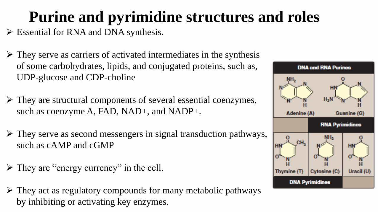
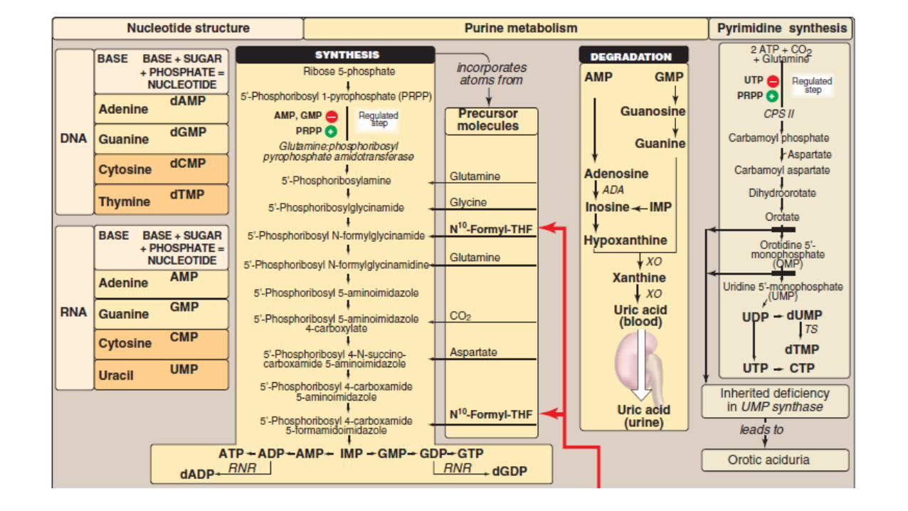
Purine and pyrimidine structures and roles Essential for RNA and DNA synthesis.
They serve as carriers of activated intermediates in the synthesis
of some carbohydrates, lipids, and conjugated proteins, such as,
UDP-glucose and CDP-choline
They are structural components of several essential coenzymes,
such as coenzyme A, FAD, NAD+, and NADP+.
They serve as second messengers in signal transduction pathways,
such as cAMP and cGMP
They are “energy currency” in the cell.
They act as regulatory compounds for many metabolic pathways
by inhibiting or activating key enzymes.
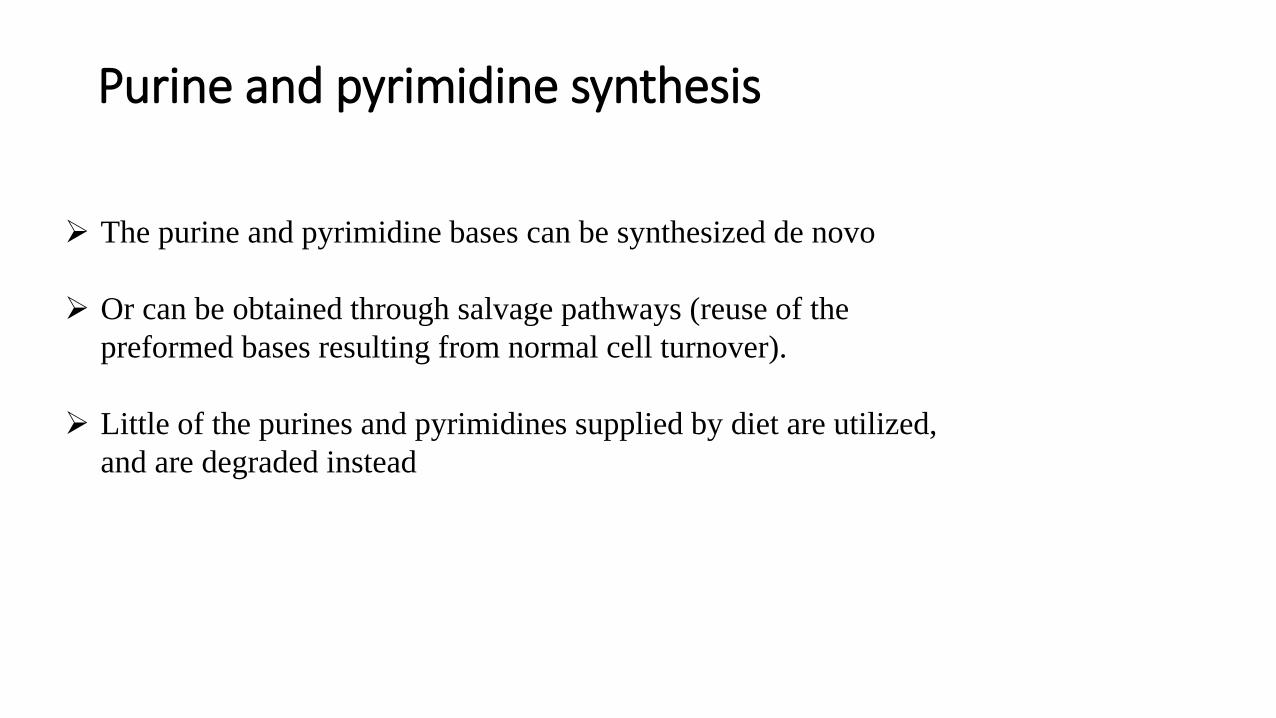
Purine and pyrimidine synthesis
The purine and pyrimidine bases can be synthesized de novo
Or can be obtained through salvage pathways (reuse of the
preformed bases resulting from normal cell turnover).
Little of the purines and pyrimidines supplied by diet are utilized,
and are degraded instead
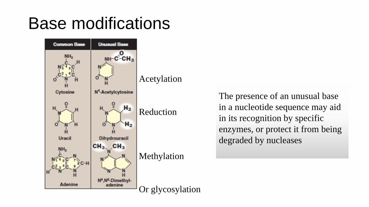
Base modifications
The presence of an unusual base
in a nucleotide sequence may aid
in its recognition by specific
enzymes, or protect it from being
degraded by nucleases
Acetylation
Reduction
Methylation
Or glycosylation
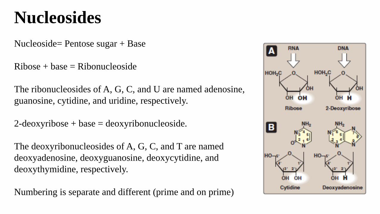
Nucleosides
Nucleoside= Pentose sugar + Base
Ribose + base = Ribonucleoside
The ribonucleosides of A, G, C, and U are named adenosine,
guanosine, cytidine, and uridine, respectively.
2-deoxyribose + base = deoxyribonucleoside.
The deoxyribonucleosides of A, G, C, and T are named
deoxyadenosine, deoxyguanosine, deoxycytidine, and
deoxythymidine, respectively.
Numbering is separate and different (prime and on prime)
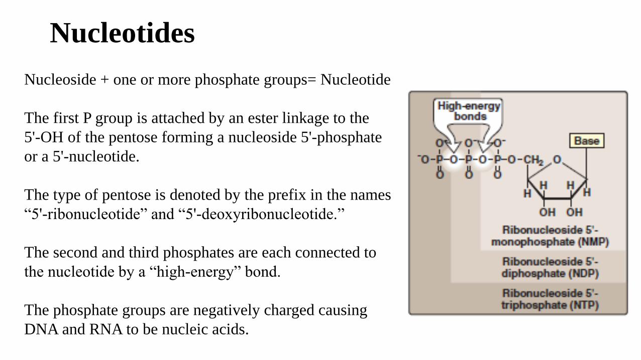
Nucleotides
Nucleoside + one or more phosphate groups= Nucleotide
The first P group is attached by an ester linkage to the
5'-OH of the pentose forming a nucleoside 5'-phosphate
or a 5'-nucleotide.
The type of pentose is denoted by the prefix in the names
“5'-ribonucleotide” and “5'-deoxyribonucleotide.”
The second and third phosphates are each connected to
the nucleotide by a “high-energy” bond.
The phosphate groups are negatively charged causing
DNA and RNA to be nucleic acids.
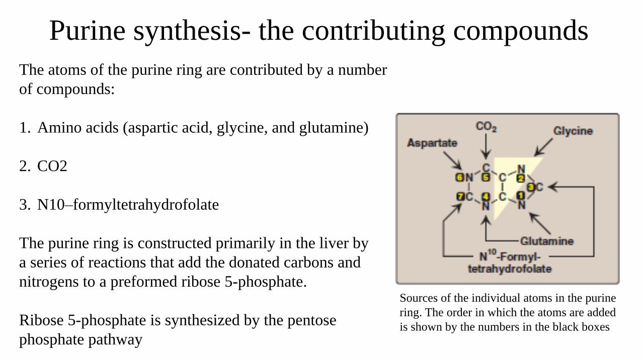
Purine synthesis- the contributing compounds
Sources of the individual atoms in the purine
ring. The order in which the atoms are added
is shown by the numbers in the black boxes
The atoms of the purine ring are contributed by a number
of compounds:
1. Amino acids (aspartic acid, glycine, and glutamine)
2. CO2
3. N10–formyltetrahydrofolate
The purine ring is constructed primarily in the liver by
a series of reactions that add the donated carbons and
nitrogens to a preformed ribose 5-phosphate.
Ribose 5-phosphate is synthesized by the pentose
phosphate pathway
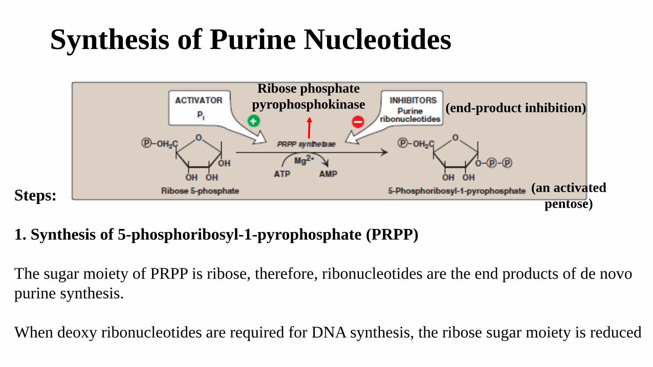
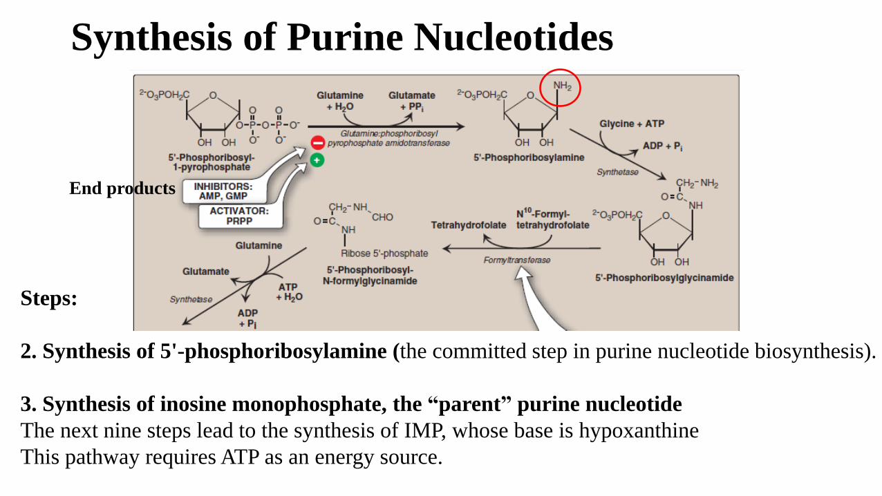
Synthesis of Purine Nucleotides
Steps:
1. Synthesis of 5-phosphoribosyl-1-pyrophosphate (PRPP)
The sugar moiety of PRPP is ribose, therefore, ribonucleotides are the end products of de novo
purine synthesis.
When deoxy ribonucleotides are required for DNA synthesis, the ribose sugar moiety is reduced
(an activated
pentose)
Ribose phosphate
pyrophosphokinase (end-product inhibition)
Synthesis of Purine Nucleotides
Steps:
2. Synthesis of 5'-phosphoribosylamine (the committed step in purine nucleotide biosynthesis).
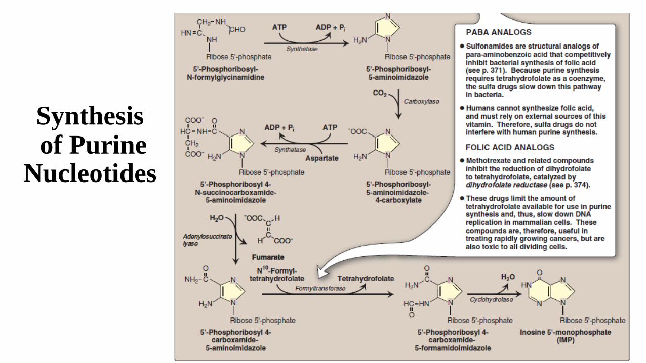
3. Synthesis of inosine monophosphate, the “parent” purine nucleotide
The next nine steps lead to the synthesis of IMP, whose base is hypoxanthine
This pathway requires ATP as an energy source.
End products
Synthesisof Purine
Nucleotides
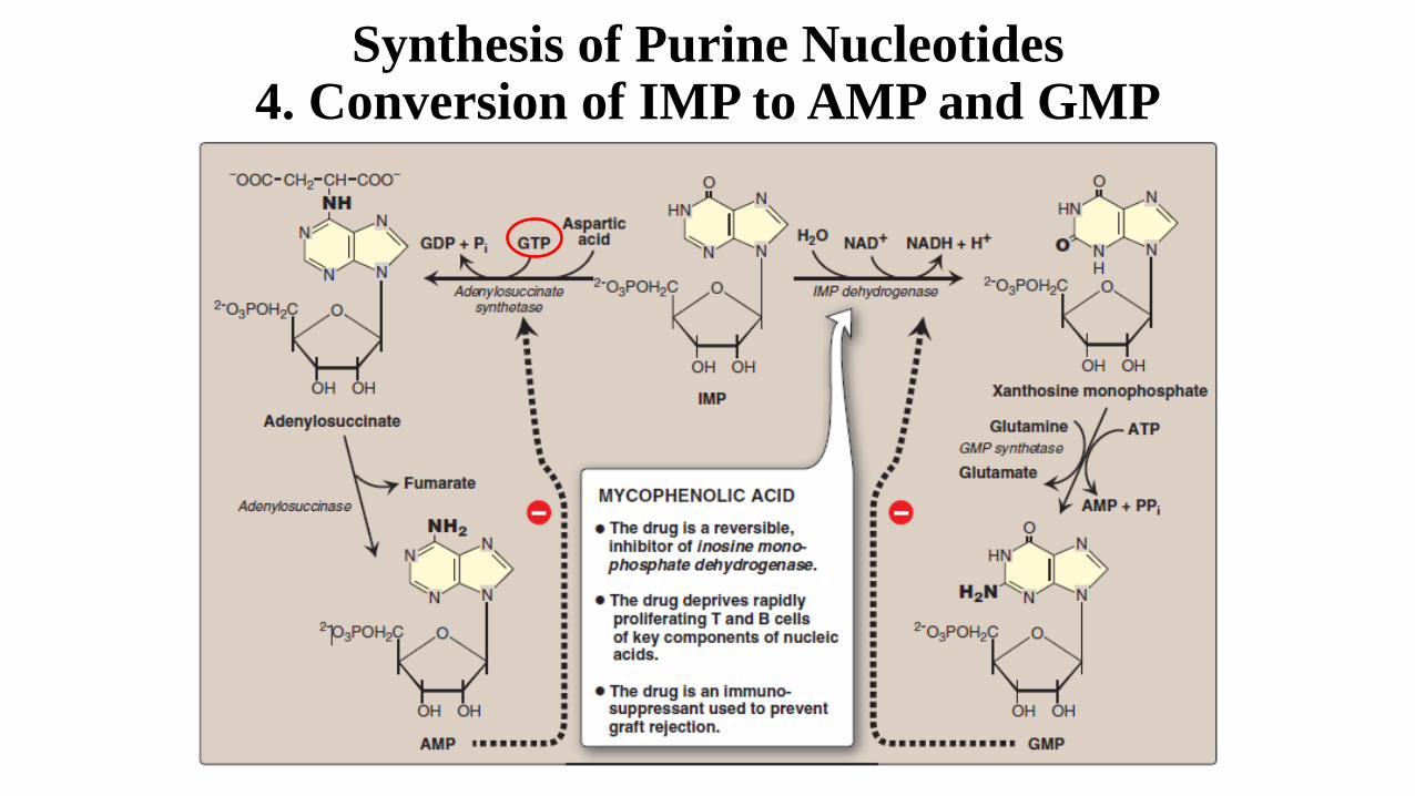
Synthesis of Purine Nucleotides4. Conversion of IMP to AMP and GMP
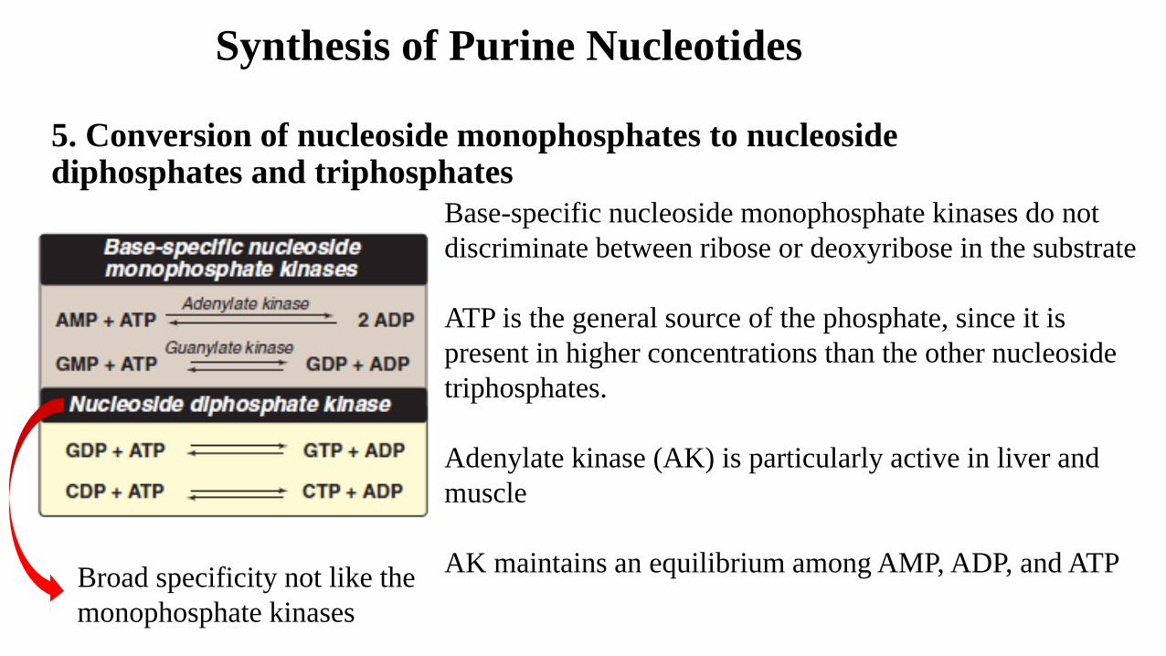
Synthesis of Purine Nucleotides
5. Conversion of nucleoside monophosphates to nucleoside diphosphates and triphosphates
Base-specific nucleoside monophosphate kinases do not
discriminate between ribose or deoxyribose in the substrate
ATP is the general source of the phosphate, since it is
present in higher concentrations than the other nucleoside
triphosphates.
Adenylate kinase (AK) is particularly active in liver and
muscle
AK maintains an equilibrium among AMP, ADP, and ATP Broad specificity not like the
monophosphate kinases
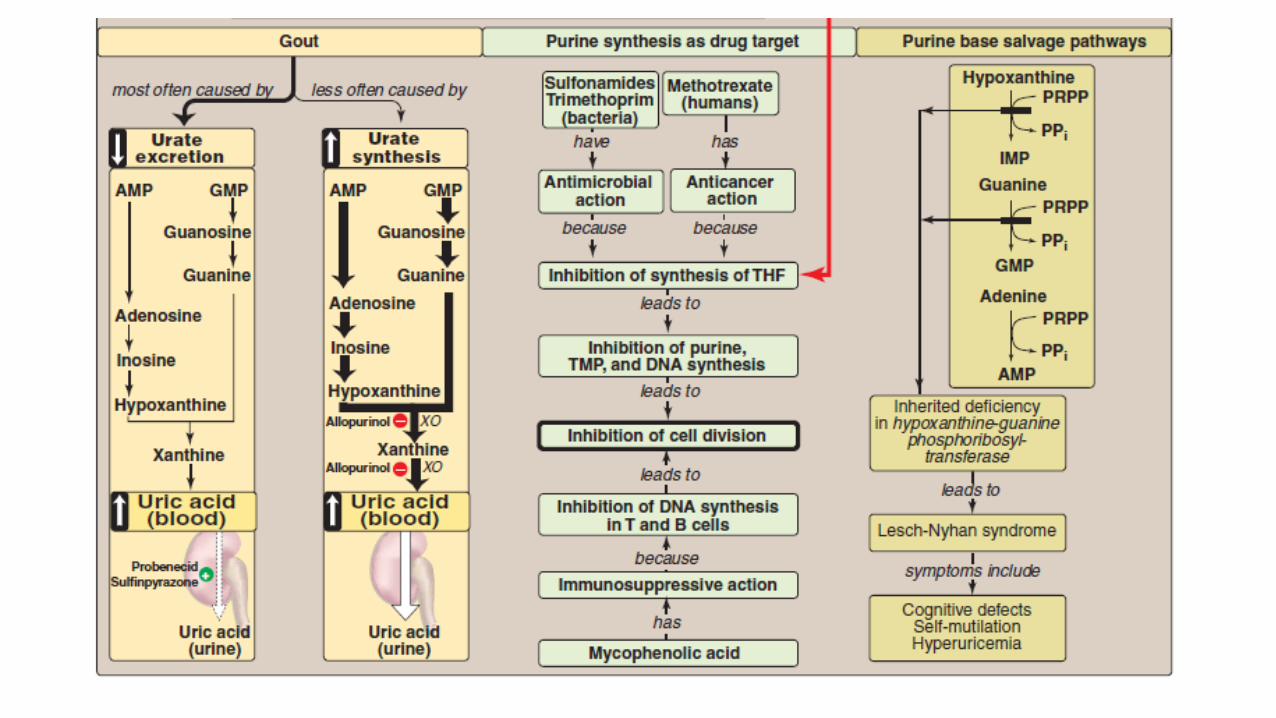
Synthetic inhibitors of purine synthesis
Synthetic inhibitors of purine synthesis (the sulfonamides1), are designed to inhibit the
growth of rapidly dividing microorganisms without interfering with human cell functions
Other purine synthesis inhibitors, such as structural analogs of folic acid (such as,
methotrexate2), are used as drugs that control the spread of cancer by interfering with the
synthesis of nucleotides and, therefore, of DNA and RNA.
Inhibitors of human purine synthesis are extremely toxic to tissues, especially to developing
structures such as in a fetus, or to cell types that normally replicate rapidly, including those
of bone marrow, skin, GI tract, immune system, or hair follicles.
Thus, anticancer drugs result in adverse effects, including anemia, scaly skin, GI tract
disturbance, immunodeficiencies, and hair loss.
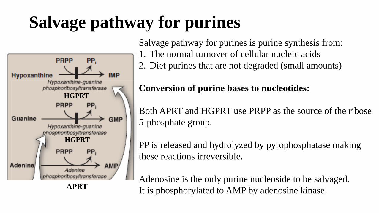
Salvage pathway for purinesSalvage pathway for purines is purine synthesis from:
1. The normal turnover of cellular nucleic acids
2. Diet purines that are not degraded (small amounts)
Conversion of purine bases to nucleotides:
Both APRT and HGPRT use PRPP as the source of the ribose
5-phosphate group.
PP is released and hydrolyzed by pyrophosphatase making
these reactions irreversible.
Adenosine is the only purine nucleoside to be salvaged.
It is phosphorylated to AMP by adenosine kinase.APRT
HGPRT
HGPRT
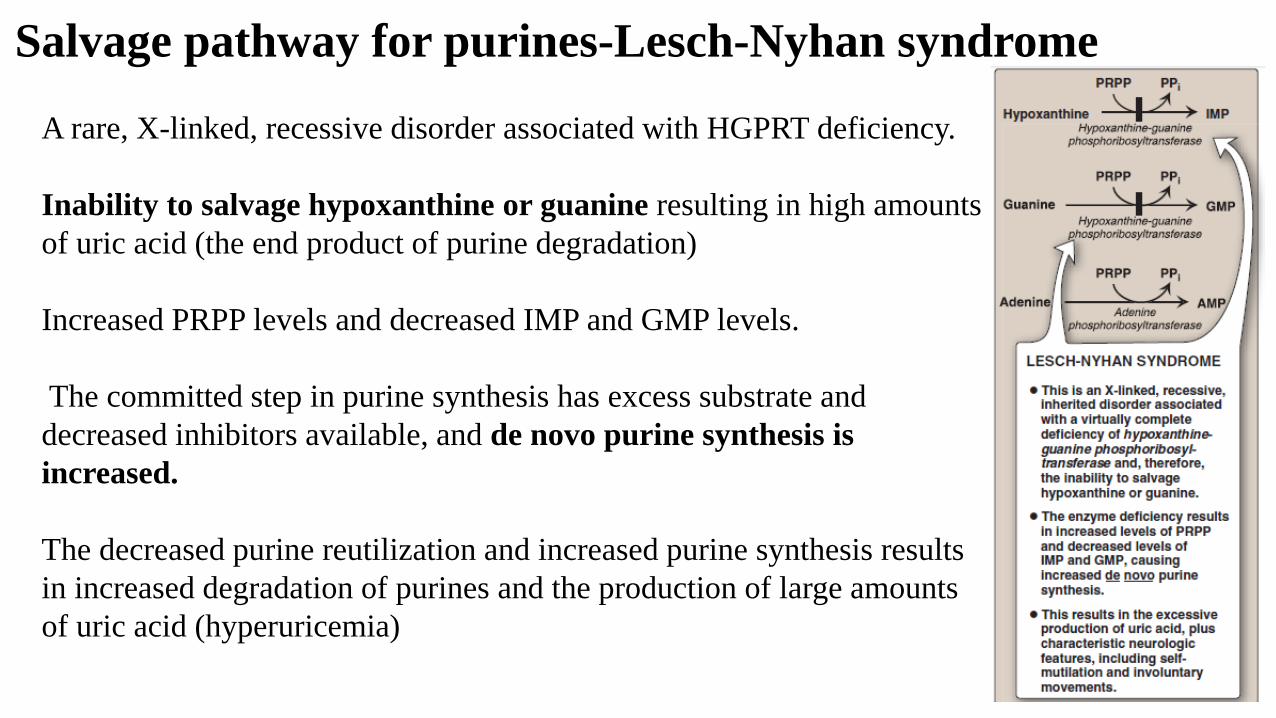
Salvage pathway for purines-Lesch-Nyhan syndrome
A rare, X-linked, recessive disorder associated with HGPRT deficiency.
Inability to salvage hypoxanthine or guanine resulting in high amounts
of uric acid (the end product of purine degradation)
Increased PRPP levels and decreased IMP and GMP levels.
The committed step in purine synthesis has excess substrate and
decreased inhibitors available, and de novo purine synthesis is
increased.
The decreased purine reutilization and increased purine synthesis results
in increased degradation of purines and the production of large amounts
of uric acid (hyperuricemia)
Lesch-Nyhan syndrome
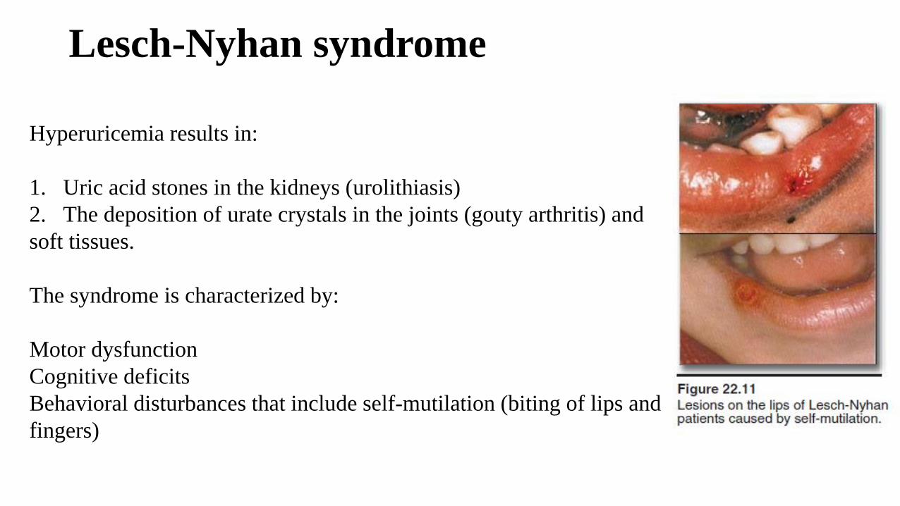
Hyperuricemia results in:
1. Uric acid stones in the kidneys (urolithiasis)
2. The deposition of urate crystals in the joints (gouty arthritis) and
soft tissues.
The syndrome is characterized by:
Motor dysfunction
Cognitive deficits
Behavioral disturbances that include self-mutilation (biting of lips and
fingers)
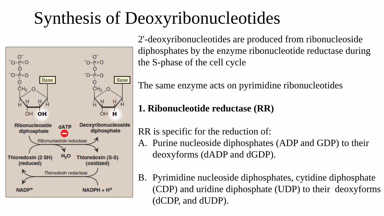
Synthesis of Deoxyribonucleotides2'-deoxyribonucleotides are produced from ribonucleoside
diphosphates by the enzyme ribonucleotide reductase during
the S-phase of the cell cycle
The same enzyme acts on pyrimidine ribonucleotides
1. Ribonucleotide reductase (RR)
RR is specific for the reduction of:
A. Purine nucleoside diphosphates (ADP and GDP) to their
deoxyforms (dADP and dGDP).
B. Pyrimidine nucleoside diphosphates, cytidine diphosphate
(CDP) and uridine diphosphate (UDP) to their deoxyforms
(dCDP, and dUDP).
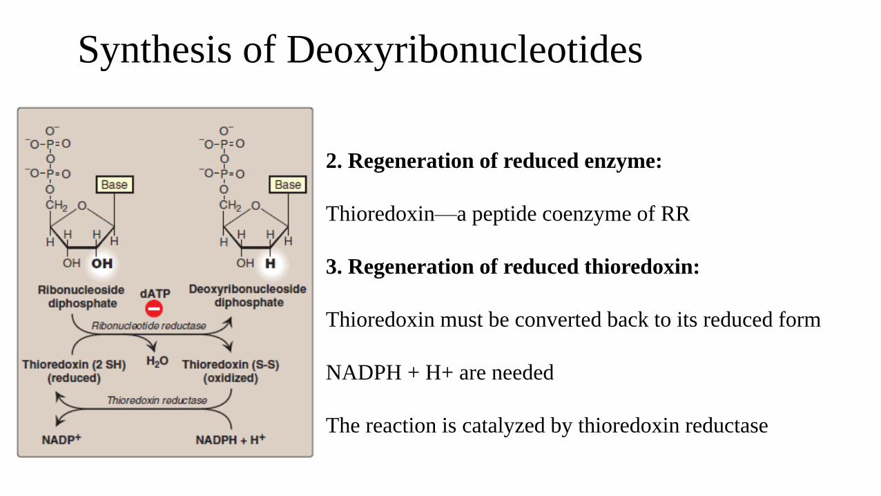
Synthesis of Deoxyribonucleotides
2. Regeneration of reduced enzyme:
Thioredoxin—a peptide coenzyme of RR
3. Regeneration of reduced thioredoxin:
Thioredoxin must be converted back to its reduced form
NADPH + H+ are needed
The reaction is catalyzed by thioredoxin reductase
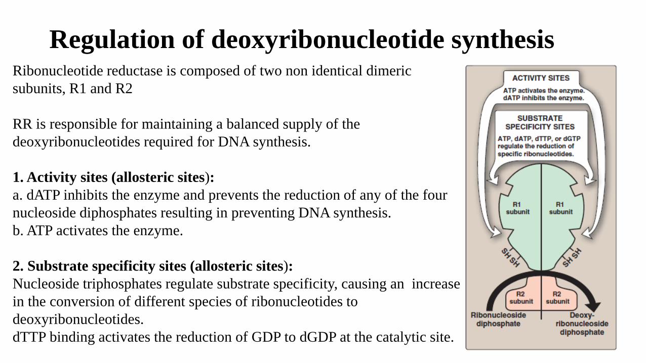
Regulation of deoxyribonucleotide synthesisRibonucleotide reductase is composed of two non identical dimeric
subunits, R1 and R2
RR is responsible for maintaining a balanced supply of the
deoxyribonucleotides required for DNA synthesis.
1. Activity sites (allosteric sites):
a. dATP inhibits the enzyme and prevents the reduction of any of the four
nucleoside diphosphates resulting in preventing DNA synthesis.
b. ATP activates the enzyme.
2. Substrate specificity sites (allosteric sites):
Nucleoside triphosphates regulate substrate specificity, causing an increase
in the conversion of different species of ribonucleotides to
deoxyribonucleotides.
dTTP binding activates the reduction of GDP to dGDP at the catalytic site.
Hydroxyurea and ribonucleotide reductase
The drug hydroxyurea destroys the free radical required for the
activity of ribonucleotide reductase
Hydroxyurea inhibits the generation of substrates for DNA synthesis.
Hydroxyurea has been used in the treatment of cancers such as CML
Degradation of Purine Nucleotides
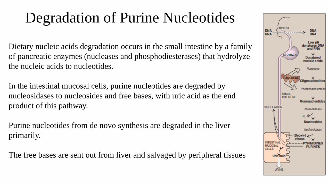
Dietary nucleic acids degradation occurs in the small intestine by a family
of pancreatic enzymes (nucleases and phosphodiesterases) that hydrolyze
the nucleic acids to nucleotides.
In the intestinal mucosal cells, purine nucleotides are degraded by
nucleosidases to nucleosides and free bases, with uric acid as the end
product of this pathway.
Purine nucleotides from de novo synthesis are degraded in the liver
primarily.
The free bases are sent out from liver and salvaged by peripheral tissues
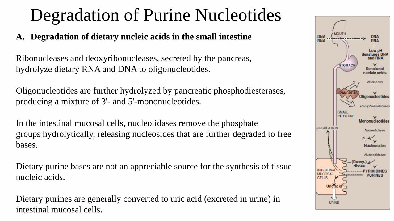
Degradation of Purine NucleotidesA. Degradation of dietary nucleic acids in the small intestine
Ribonucleases and deoxyribonucleases, secreted by the pancreas,
hydrolyze dietary RNA and DNA to oligonucleotides.
Oligonucleotides are further hydrolyzed by pancreatic phosphodiesterases,
producing a mixture of 3'- and 5'-mononucleotides.
In the intestinal mucosal cells, nucleotidases remove the phosphate
groups hydrolytically, releasing nucleosides that are further degraded to free
bases.
Dietary purine bases are not an appreciable source for the synthesis of tissue
nucleic acids.
Dietary purines are generally converted to uric acid (excreted in urine) in
intestinal mucosal cells.
Degradation of Purine Nucleotides
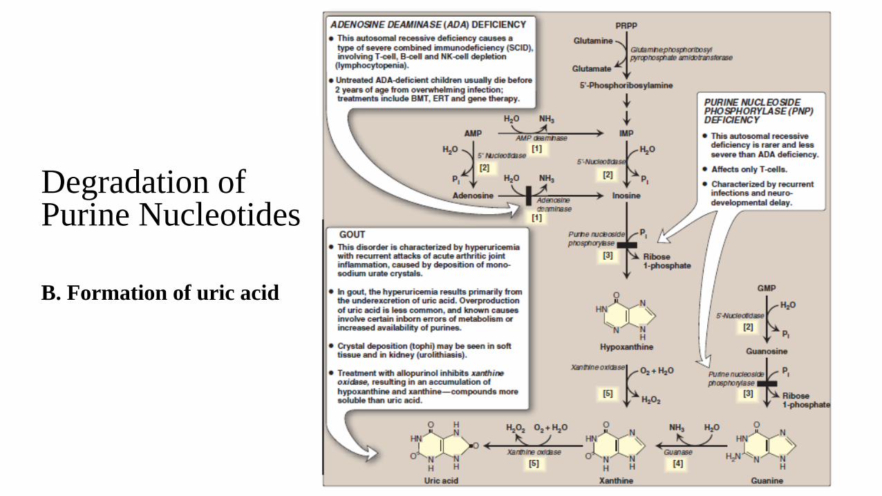
B. Formation of uric acid
Degradation of Purine NucleotidesB. Formation of uric acid
[1] An amino group is removed from AMP to produce IMP by AMP deaminase, or from adenosine
to produce inosine (hypoxanthineribose) by adenosine deaminase.
[2] IMP and GMP are converted into their nucleoside forms––inosine and guanosine––by the action
of 5'-nucleotidase.
[3] Purine nucleoside phosphorylase converts inosine and guanosine into their respective purine
bases, hypoxanthine and guanine.
Note: A mutase interconverts ribose 1- and ribose 5-phosphate.
[4] Guanine is deaminated to form xanthine.
[5] Hypoxanthine is oxidized by xanthine oxidase to xanthine, which is further oxidized by xanthine
oxidase to uric acid, the final product of human purine degradation.
Diseases associated with purine degradation
1. Gout: high levels of uric acid in blood (hyperuricemia)
Hyperuricemia due to either the overproduction or underexcretion of
uric acid.
Hyperuricemia lead to the deposition of monosodium urate crystals
in the joints, and an inflammatory response to the crystals, causing
first acute and then chronic gouty arthritis.
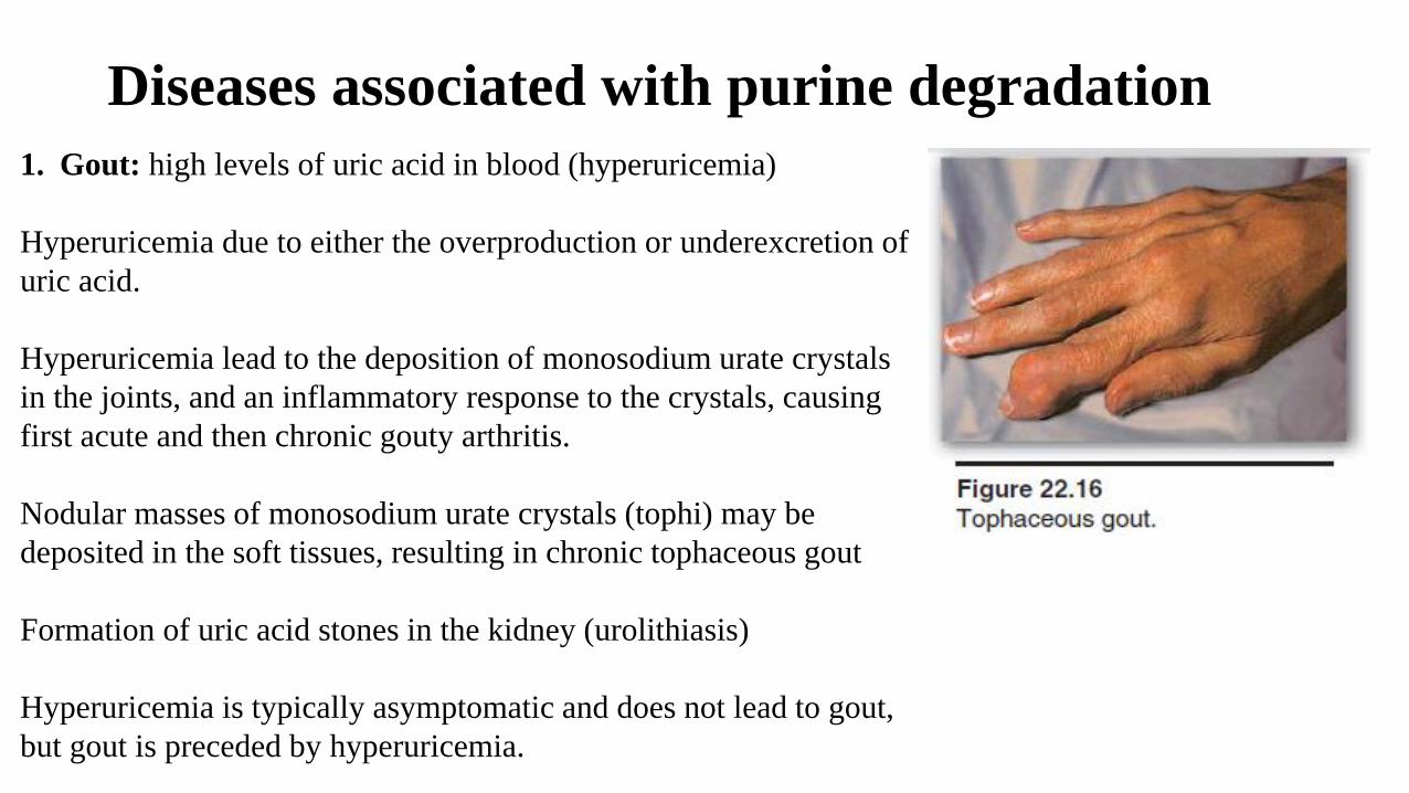
Nodular masses of monosodium urate crystals (tophi) may be
deposited in the soft tissues, resulting in chronic tophaceous gout
Formation of uric acid stones in the kidney (urolithiasis)
Hyperuricemia is typically asymptomatic and does not lead to gout,
but gout is preceded by hyperuricemia.
Diseases associated with purine degradation
1. Gout:
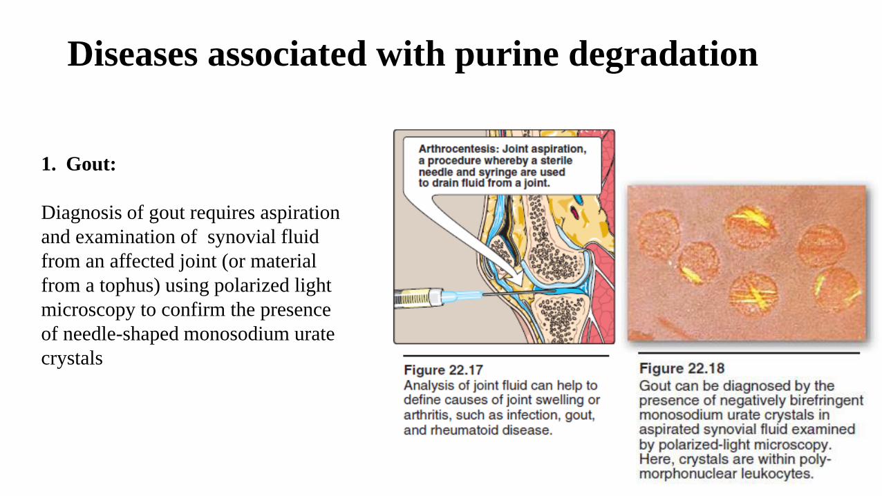
Diagnosis of gout requires aspiration
and examination of synovial fluid
from an affected joint (or material
from a tophus) using polarized light
microscopy to confirm the presence
of needle-shaped monosodium urate
crystals
Causes of hyperuricemia
Underexcretion of uric acid:
Most gout patients In the vast majority of patients,
Underexcretion can be primary (due to unidentified inherent excretory defects)
Or secondary to: 1. A known disease that affects the kidney function in handling urate, such as
lactic acidosis (lactate and urate compete for the same renal transporter)
2. Environmental factors such as drugs (thiazide diuretics)
3. Exposure to lead (saturnine gout)
Overproduction of uric acid: less common.
Several identified mutations in the X-linked PRPP synthetase gene that increase PRPP
production
Treatment of gout
Acute attacks are treated with anti-inflammatory agents.
Anti-inflammatory drugs have no effect on uric acid levels.
Long-term therapy involve lowering the uric acid level to prevent the deposition of urate
crystals.
Uricosuric agents, such as probenecid or sulfinpyrazone increase renal excretion of uric acid
in patients under-excrete uric acid.
Allopurinol (xanthine oxidase inhibitor), a structural analog of hypoxanthine, inhibits uric
acid synthesis in patients with uric acid overproduction
Hypoxanthine and xanthine are more soluble than uric acid and are less likely to initiate an
inflammatory response.
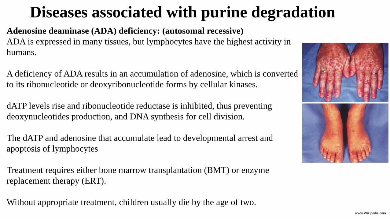
Diseases associated with purine degradationAdenosine deaminase (ADA) deficiency: (autosomal recessive)
ADA is expressed in many tissues, but lymphocytes have the highest activity in
humans.
A deficiency of ADA results in an accumulation of adenosine, which is converted
to its ribonucleotide or deoxyribonucleotide forms by cellular kinases.
dATP levels rise and ribonucleotide reductase is inhibited, thus preventing
deoxynucleotides production, and DNA synthesis for cell division.
The dATP and adenosine that accumulate lead to developmental arrest and
apoptosis of lymphocytes
Treatment requires either bone marrow transplantation (BMT) or enzyme
replacement therapy (ERT).
Without appropriate treatment, children usually die by the age of two.www.Wikipedia.com

Pyrimidine Synthesis
The pyrimidine ring is synthesized before being attached
to ribose 5-phosphate
Ribose 5-phosphate is donated by PRPP.
Pyrimidine Synthesis
A. Synthesis of carbamoyl
phosphate
The regulated step of this
pathway in mammalian cells is
the synthesis of carbamoyl
phosphate from glutamine and
CO2
CPS II is inhibited by UTP (the
end product of this pathway)
CPS II is activated by PRPP
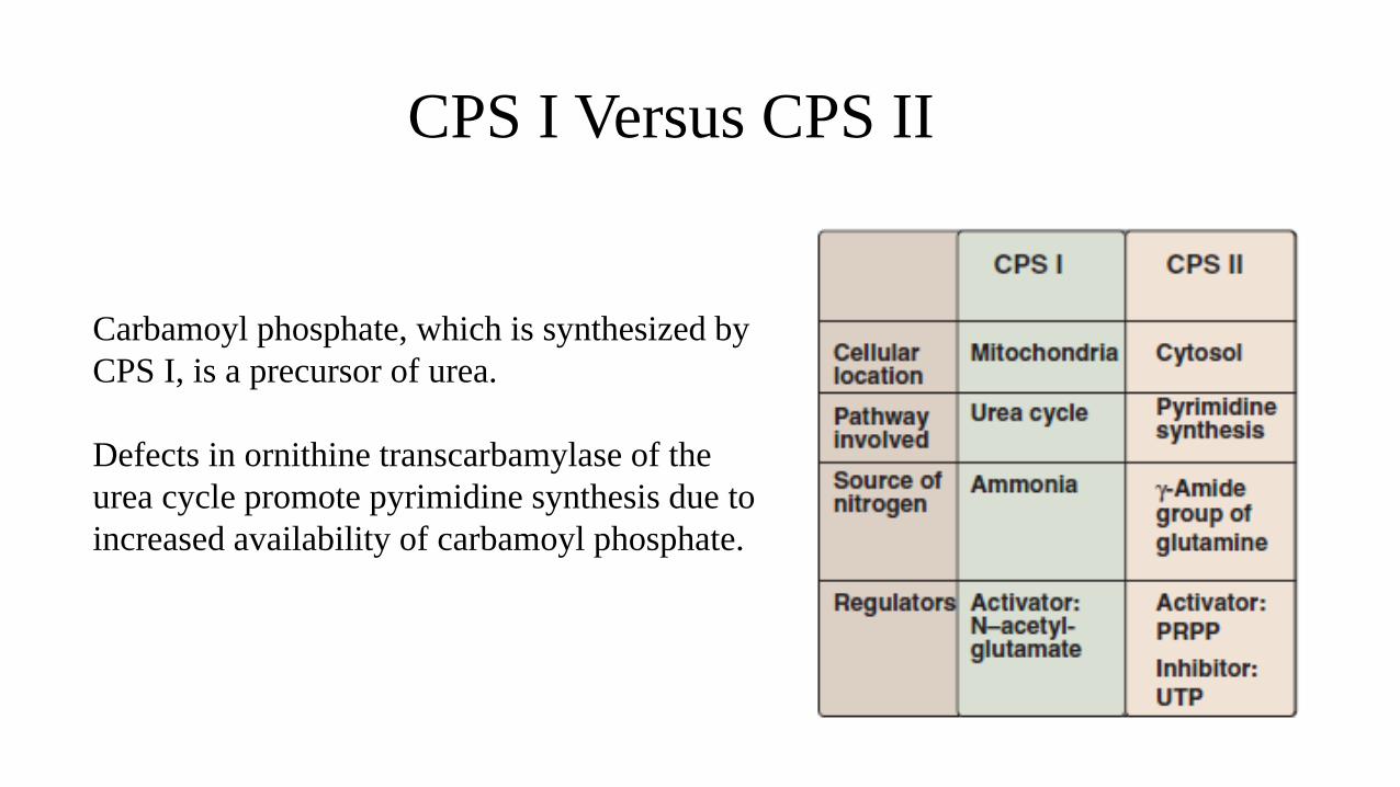
CPS I Versus CPS II
Carbamoyl phosphate, which is synthesized by
CPS I, is a precursor of urea.
Defects in ornithine transcarbamylase of the
urea cycle promote pyrimidine synthesis due to
increased availability of carbamoyl phosphate.
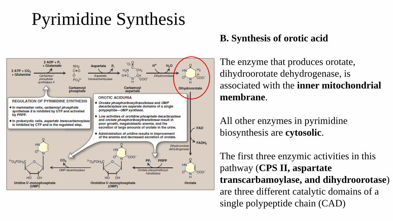
Pyrimidine Synthesis B. Synthesis of orotic acid
The enzyme that produces orotate,
dihydroorotate dehydrogenase, is
associated with the inner mitochondrial
membrane.
All other enzymes in pyrimidine
biosynthesis are cytosolic.
The first three enzymic activities in this
pathway (CPS II, aspartate
transcarbamoylase, and dihydroorotase)
are three different catalytic domains of a
single polypeptide chain (CAD)
Pyrimidine Synthesis
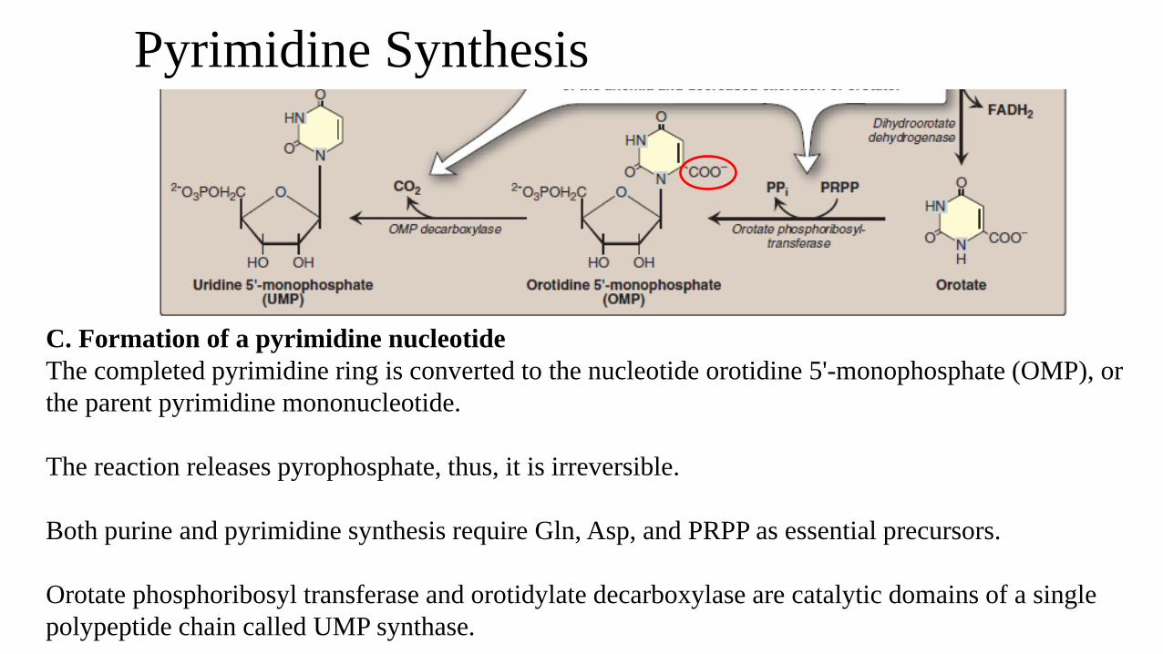
C. Formation of a pyrimidine nucleotide
The completed pyrimidine ring is converted to the nucleotide orotidine 5'-monophosphate (OMP), or
the parent pyrimidine mononucleotide.
The reaction releases pyrophosphate, thus, it is irreversible.
Both purine and pyrimidine synthesis require Gln, Asp, and PRPP as essential precursors.
Orotate phosphoribosyl transferase and orotidylate decarboxylase are catalytic domains of a single
polypeptide chain called UMP synthase.
Pyrimidine Synthesis and Metabolism
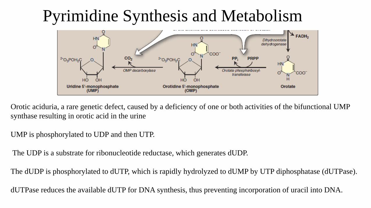
Orotic aciduria, a rare genetic defect, caused by a deficiency of one or both activities of the bifunctional UMP
synthase resulting in orotic acid in the urine
UMP is phosphorylated to UDP and then UTP.
The UDP is a substrate for ribonucleotide reductase, which generates dUDP.
The dUDP is phosphorylated to dUTP, which is rapidly hydrolyzed to dUMP by UTP diphosphatase (dUTPase).
dUTPase reduces the available dUTP for DNA synthesis, thus preventing incorporation of uracil into DNA.
Pyrimidine Synthesis
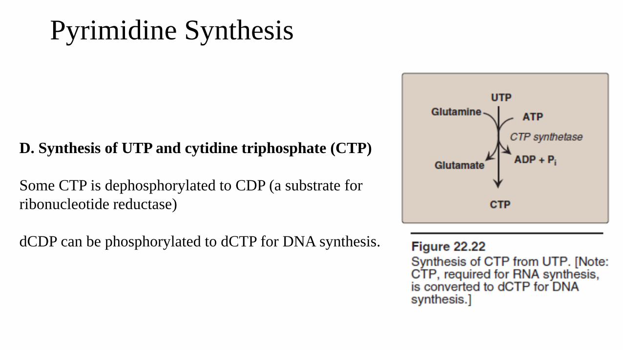
D. Synthesis of UTP and cytidine triphosphate (CTP)
Some CTP is dephosphorylated to CDP (a substrate for
ribonucleotide reductase)
dCDP can be phosphorylated to dCTP for DNA synthesis.
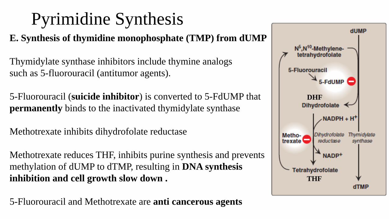
Pyrimidine Synthesis E. Synthesis of thymidine monophosphate (TMP) from dUMP
Thymidylate synthase inhibitors include thymine analogs
such as 5-fluorouracil (antitumor agents).
5-Fluorouracil (suicide inhibitor) is converted to 5-FdUMP that
permanently binds to the inactivated thymidylate synthase
Methotrexate inhibits dihydrofolate reductase
Methotrexate reduces THF, inhibits purine synthesis and prevents
methylation of dUMP to dTMP, resulting in DNA synthesis
inhibition and cell growth slow down .
5-Fluorouracil and Methotrexate are anti cancerous agents
DHF
THF
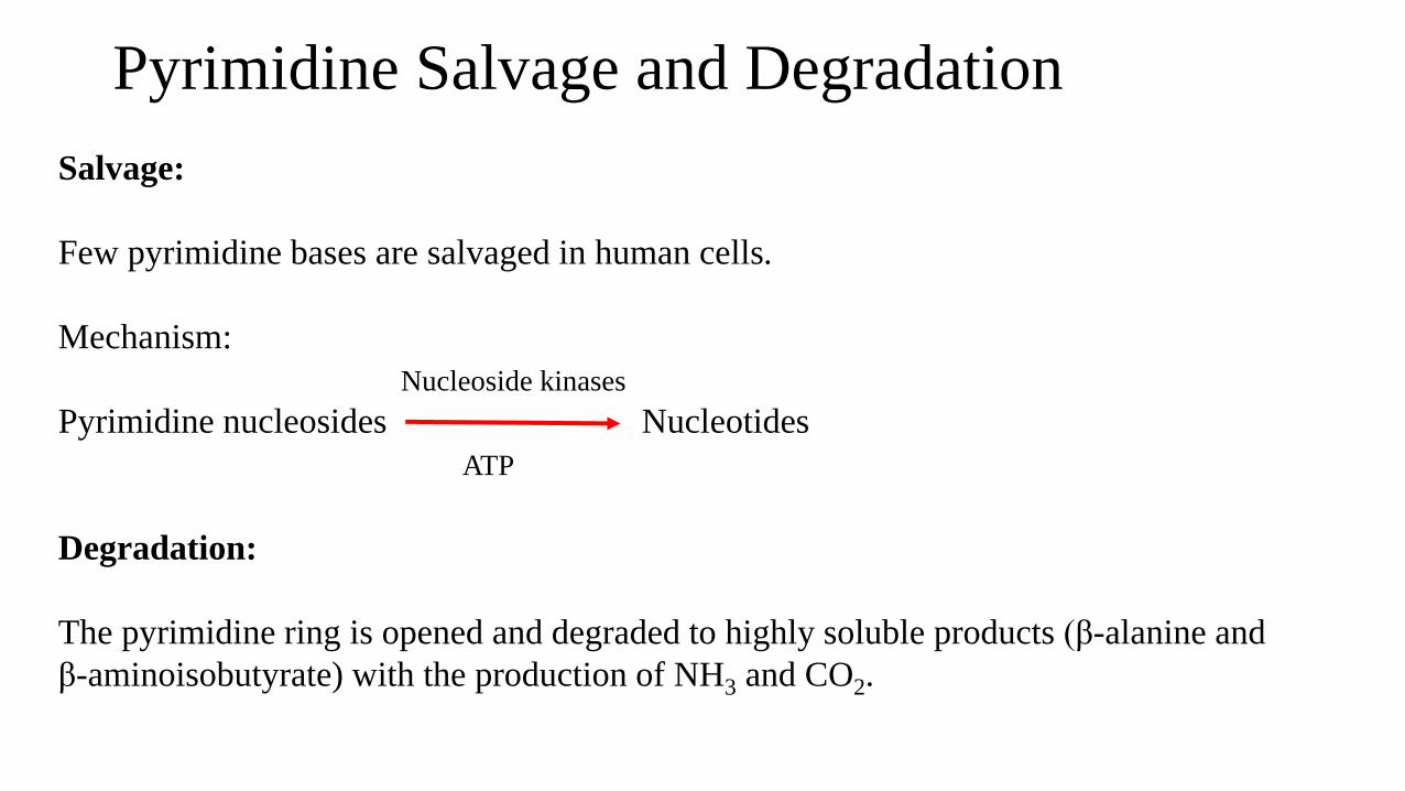
Pyrimidine Salvage and Degradation
Salvage:
Few pyrimidine bases are salvaged in human cells.
Mechanism:
Nucleoside kinases
Pyrimidine nucleosides Nucleotides
ATP
Degradation:
The pyrimidine ring is opened and degraded to highly soluble products (β-alanine and
β-aminoisobutyrate) with the production of NH3 and CO2.


![University of Groningen Antimalarial Drug Discovery ......lack the de novo purine synthesis pathway and take up host cell purines for growth [2, 8]. Inhibition of this pathway was](https://img.pdfslide.net/doc/110x75/5e30b84fcfea694521705426/university-of-groningen-antimalarial-drug-discovery-lack-the-de-novo-purine.jpg)


![Astrocytic ceramide as possible indicator of neuroinflammation...the salvage pathway, the sphingomyelinase pathway, and thedenovopathway[21, 22]. In the salvage pathway, complex sphingolipids](https://img.pdfslide.net/doc/110x75/60dd056ddd29fb7ae4748ab6/astrocytic-ceramide-as-possible-indicator-of-neuroinflammation-the-salvage-pathway.jpg)