Embed Size (px)
Citation preview

Application 1530:
Purified human alpha1-proteinase inhibitor for the treatment of alpha1-proteinase inhibitor deficiency, leading to chronic
obstructive pulmonary disease
PICO Confirmation(To guide a new application to MSAC)
(Version 1.0)
1 | P a g e P I C O C o n fi r m a ti o n – R a ti fi e d 7 J u n e 2 0 1 8A p p l i c a ti o n 1 5 3 0 : P u r i fi e d h u m a n a l p h a 1 - p r o t e i n a s e i n h i b i t o r f o r t h e t r e a t m e n t o f a l p h a 1 - p r o t e i n a s e i n h i b i t o r d e fi c i e n c y

Summary of PICO criteria to define the question to be addressed in an Assessment Report to MSAC
Component DescriptionPatients Ex-smokers or individuals who have never smoked, with severe alpha1-
proteinase inhibitor deficiency (serum levels ≤11 μM) and emphysema with FEV1 <80%.
Note that other populations of interest might include (and evidence could be presented for):
1. Individuals with chronic obstructive pulmonary disease (COPD) and severe alpha1-proteinase inhibitor deficiency (serum levels ≤11 μM)
2. Individuals with emphysema or COPD stratified according to airflow obstruction which is mild, moderate, or severe
3. Individuals with emphysema and genotypes other than ZZ4. Individuals with emphysema and deficiency >11 µM
Prior tests(for investigative medical services )
Alpha1-proteinase inhibitor deficiency: A1-PI serum levels and genotypeLung functions: Spirometry (FEV1/FVC <0.7) and investigations to exclude other conditionsEmphysema: Computed tomography of the lung and chest X-rayOther relevant: Tests for monitoring compliance with smoking cessation, arterial blood gases analysis, sputum examination, other respiratory function investigations
Interventions Augmentation therapy with Prolastin-C, in addition to optimal pharmacological treatment and supportive care; OR
Augmentation therapy with Zemaira, in addition to optimal pharmacological treatment and supportive care.
Comparators Optimal pharmacological treatment and supportive care
Augmentation therapy (in addition to optimal pharmacological treatment and supportive care) with the alternative augmentation agent (i.e. Prolastin-C versus Zemaira)
Outcomes Safety
Incidence and severity of adverse events
Primary effectiveness
Respiratory function measured by spirometry (FEV1) and FEV1/FVC ratio Dyspnoea (measured with a validated tool e.g. Baseline dyspnoea
index, Transition dyspnoea index)
Secondary effectiveness
Mortality, including deaths from respiratory failure Number of exacerbations and hospitalisations associated with COPD Quality of life (measured by validated tool for COPD or respiratory
impairment) Changes in exercise capacity (with tools such as the 6-minute walking
test) The BODE index- body mass index, airflow obstruction, dyspnoea and
exercise index, which is more predictive of mortality then FEV1
Surrogate measures/biomarkers:
2 | P a g e P I C O C o n fi r m a ti o n – R a ti fi e d 7 J u n e 2 0 1 8A p p l i c a ti o n 1 5 3 0 : P u r i fi e d h u m a n a l p h a 1 - p r o t e i n a s e i n h i b i t o r f o r t h e t r e a t m e n t o f a l p h a 1 - p r o t e i n a s e i n h i b i t o r d e fi c i e n c y

Component Descriptiono Lung density measured on computed tomographyo Carbon monoxide (CO) transfer or pulmonary diffusing capacity
for CO Costs
o Costs associated with screening for the conditiono Costs of providing the intervention (including costs associated
with monitoring lung function, compliance with smoking cessation etc.)
o Cost per Quality Adjusted Life Year (QALY)
PICO or PPICO rationale for therapeutic and investigative medical services only
PopulationAlpha1-proteinase inhibitor (A1-PI) or alpha1 antitrypsin (A1-AT) deficiency is a heritable genetic condition that results in decreased circulating (and/or abnormally functioning) A1-PI protein. A1-PI is predominantly synthesized by hepatocytes and released into the bloodstream, where it acts as a serine protease inhibitor, with neutrophil elastase being its primary substrate (de Serres et al. 2003). A1-PI deficiency, defined as ≤30 per cent of normal serum levels, is known to have a role in the development of liver disease and emphysema, and has been hypothesised to be part of pathological processes underlying a range of health conditions.
The application is for patients with A1-PI deficiency, leading to chronic obstructive pulmonary disease (COPD). A1-PI deficiency is associated with a range of problems that fall under the umbrella term of COPD, with panacinar emphysema being the most commonly recognised manifestation. Consultation with clinical experts indicated the main population to be considered during the assessment phase is ex-smokers or patients who have never smoked, with emphysema and severe A1-PI deficiency. However, if available, evidence could also be provided on a broader population (see rationale). The clinical expert advised that cigarette smoking inactivates A1-PI, rendering this expensive product useless in smokers.
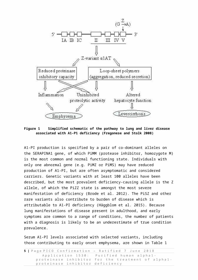
Severe A1-PI deficiency is defined as serum levels below 11 μM (approximately 59 mg/dL) (Hatipoglu and Stoller 2016). Clinically this deficiency manifests as panacinar emphysema or hepatitis, cirrhosis, and/or hepatoma (Pharmacy and Therapeutics 2010). Less commonly, vasculitis and panniculitis are observed (Pharmacy and Therapeutics 2010). The deleterious consequences of A1-PI deficiency for lung and liver function occur via different pathways. In the lungs, neutrophil elastase (which has an important role in fighting infection) is normally bound and inactivated by A1-PI. However, with low levels of A1-PI, enzymatic activity of neutrophil elastase goes unchecked, and ultimately its detrimental impact on elastin compromises the bronchia and alveoli. Conversely, liver damage occurs when the A1-PI protein forms polymers that accumulate within hepatocytes and lead to scarring, inflammation or malignancy. A simplified schematic of the mechanisms underlying disease associated with the most prevalent deficiency-causing allele is shown in Figure 1 (Fregonese and Stolk 2008).
3 | P a g e P I C O C o n fi r m a ti o n – R a ti fi e d 7 J u n e 2 0 1 8A p p l i c a ti o n 1 5 3 0 : P u r i fi e d h u m a n a l p h a 1 - p r o t e i n a s e i n h i b i t o r f o r t h e t r e a t m e n t o f a l p h a 1 - p r o t e i n a s e i n h i b i t o r d e fi c i e n c y
Figure 1 Simplified schematic of the pathway to lung and liver disease associated with A1-P1 deficiency (Fregonese and Stolk 2008)
A1-PI production is specified by a pair of co-dominant alleles on the SERAPINA1 gene, of which PiMM (protease inhibitor, homozygote M) is the most common and normal functioning state. Individuals with only one abnormal gene (e.g. PiMZ or PiMS) may have reduced production of A1-PI, but are often asymptomatic and considered carriers. Genetic variants with at least 100 alleles have been described, but the most prevalent deficiency-causing allele is the Z allele, of which the PiZZ state is amongst the most severe manifestation of deficiency (Brode et al. 2012). The PiSZ and other rare variants also contribute to burden of disease which is attributable to A1-PI deficiency (Häggblom et al. 2015). Because lung manifestations of disease present in adulthood, and early symptoms are common to a range of conditions, the number of patients with a diagnosis is likely to be an underestimate of true condition prevalence.
Serum A1-PI levels associated with selected variants, including those contributing to early onset emphysema, are shown in Table 1 (adapted from Hatipoglu and Stoller 2016)). Prevalence data from Australia is limited, but de Serres et al. (2003) reported gene frequencies per 1000 persons from a range of cohort studies conducted in various Australian populations. De Serres et al. (2003) reported the estimated prevalence of deficient allele carriers in the Australian population is 1 in 8.9 individuals; the majority of whom are carriers. For PiSZ, the prevalence was estimated to be 1 in 841; and for PiZZ, it was estimated at 1 in 5,584. The PiZZ allele contributes to the greatest burden of lung disease in the A1-PI deficient population, and if the population is limited to those with severe deficiency, the treated population would likely be those with the ZZ allele. PASC noted that not all people with ZZ A1-PI deficiency will develop severe emphysema. Based on estimates by commercial sponsors, the incidence of people meeting the criteria for treatment with A1-PI in Australia in 2018
4 | P a g e P I C O C o n fi r m a ti o n – R a ti fi e d 7 J u n e 2 0 1 8A p p l i c a ti o n 1 5 3 0 : P u r i fi e d h u m a n a l p h a 1 - p r o t e i n a s e i n h i b i t o r f o r t h e t r e a t m e n t o f a l p h a 1 - p r o t e i n a s e i n h i b i t o r d e fi c i e n c y
was 252. Considering treatment is expected to be life-long (and not curative), the number of patients being treated will increase cumulatively over time.
PASC recommended the population be restricted to patients with ZZ and null phenotypes, and a FEV1/FVC ratio < 0.7.
Table 1 Serum A1-PI levels associated with normal and SZ or ZZ allele variations known to increase the risk of emphysema (Hatipoglu and Stoller 2016)
Alleles Impact Serum A1-PI levelsMg/dL (Mean [5th–95th Percentile])
Genetic prevalence in the Australian population (de Serres et al. 2003)**Genetic prevalence (95% confidence interval) per 1000 (de Serres et al. 2003)
MM Normal 147 (102–254) Not applicableMS orMZ
Carriers, usually asymptomatic 125 (86–218)90 (62–151)
1 in 121 in 40
SS Slightly increased emphysema risk, mildly symptomatic or asymptomatic
95 (43–154) 1 in 507
SZ Individuals produce less A1-PI than normal and have an increased risk of emphysema
62 (33–108) 1 in 841
ZZ* Most severely affected , individuals have a greatly increased risk of emphysema and liver disease
≤29 (≤29–52) 1 in 5,584
Null Very rare, no A1-PI produced 0 Very rare, cannot be estimated*It has been estimated that the number of individuals with the ZZ form in Australia is 4,126 (between 2,894–5,695)(Blanco et al. 2017).**Genetic prevalence (95% confidence interval) per 1000 for the PiS allele is 44.4 (40.7–48.5); for the PiZ allele it is 13.4 (11.4–15.7)
Estimate of eligible population in Australia
Estimates of A1-PI deficiency prevalence are varied and not necessarily indicative of the patient population eligible for A1-PI augmentation therapy. Patients proposed for treatment are those with airflow restriction consistent with emphysema; therefore, the starting point for a more robust approach to considering eligible population may be use of the burden of COPD in Australia (as these patients may have emphysema and deficiency). It is important to note that while clinical advice indicates the suitable population only includes patients with emphysema (i.e. a subgroup of the COPD population), population studies do not tend to differentiate emphysema and COPD.
The probability (that emphysema will develop) increases across the MZ, SZ and ZZ genotypes, with the most significant contributor being the ZZ genotype (de Serres and Blanco 2014). Good quality data on the burden of emphysema in an Australian population, and specifically the A1-PI deficient population, is difficult to identify because of the propensity for both to be under-recognised (Brode et al. 2012). It is generally accepted that A1-PI deficiency is associated with 1‒3 per cent of all COPD cases, with the ZZ genotype accounting for 0.8 per cent (Janciauskiene et al. 2011; Russo et al. 2016). Table 2 applies this to the prevalence of COPD in the Australian population aged 45 to 64, assuming a constant representation across age-groups. Since patients with A1-PI deficiency might be over-represented in younger age-groups (and younger presentation is an indication for testing), only the prevalence in the 45‒64 year age-group has been presented. These figures are indicative only.1
1 Alternatively data from the ADAPT registry Stockley, RA 2015, 'Antitrypsin Deficiency Assessment and Programme for Treatment (ADAPT): The United Kingdom Registry', Copd, vol.12 Suppl 1pp. 63-68. posits that 4.6% of ZZ individuals are symptomatic, from a total ZZ population of 4,126 estimated ZZ patients in Australia Blanco, I, Bueno, P, Diego, I, Pérez-Holanda, S, Casas-Maldonado, F, Esquinas, C & Miravitlles, M 2017, 'Alpha-1 antitrypsin Pi*Z gene frequency and Pi*ZZ genotype numbers worldwide: an update', International Journal of 5 | P a g e P I C O C o n fi r m a ti o n – R a ti fi e d 7 J u n e 2 0 1 8
A p p l i c a ti o n 1 5 3 0 : P u r i fi e d h u m a n a l p h a 1 - p r o t e i n a s e i n h i b i t o r f o r t h e t r e a t m e n t o f a l p h a 1 - p r o t e i n a s e i n h i b i t o r d e fi c i e n c y

Many individuals may not be aware of their A1-PI status, and for various reasons, may never be tested. It is therefore difficult to predict utilisation. Furthermore, how eligibility is defined (in terms of respiratory features and/or level of circulating A1-PI) will influence the pool of eligible individuals.
Table 2 Prevalence of COPD, ages 45+, 2014–15 (Australian Institute of Health and Welfare 2017) Age group, years Persons, % (95% Confidence interval) Potential number with underlying A1-
P1 deficiency of ZZ origin
45–54 2.5 (1.8–3.2) 621
55–64 4.8 (3.7–6.0) 1,057
Total 1,678Explanatory notes: prevalence of COPD is sourced from the AIHW data. To generate estimates of the potential population with A1-PI ZZ deficiency a prevalence of 0.8% of the COPD population was used. It does not appear that there is a difference in prevalence between males or females in terms of A1-PI deficiency.
A1-PI deficiency-related emphysema
In normal individuals, maximal lung function is attained around 15 to 25 years of age, and remains relatively constant for approximately a decade. After this, it declines by approximately 20 to 25 ml/year, with roughly one litre of loss over the next 50 years (Minai et al. 2008). The Alpha-1 Antitrypsin Deficiency Registry Study Group reports the annual decline in FEV1 in patients with serum A1-PI below 11 µM is between 50‒80ml, compared to 20‒30ml in A1-PI replete individuals (1998).
While some individuals with A1-PI deficiency may have normal lung function and a normal life expectancy, the risk of developing emphysema is high and exacerbated by a history of smoking. Smoking is the most important lifestyle risk factor for development of emphysema in patients with severe A1-PI deficiency, and may both increase the risk of developing emphysema and decrease the age of onset. However, the rate of decline in respiratory function in ex-smokers, smokers and those who have never smoked, once emphysema has developed, is unclear (Evald et al. 1990). Symptoms often present in the third or fourth decade of life, commonly with emphysema that is predominantly in the base of the lungs (Brode et al. 2012). Emphysema is an important cause of mortality for individuals with A1-PI deficiency, and an early study of 124 patients with A1-PI deficiency and symptomatic emphysema found a significantly shortened lifespan, with a mean survival of 16% at 60 years of age, compared with 85% for normal persons (Brantly et al. 1988). The progression of emphysema is characterised by worsening pulmonary function, reduced exercise capacity, increasing symptom burden and negative impact on quality of life.
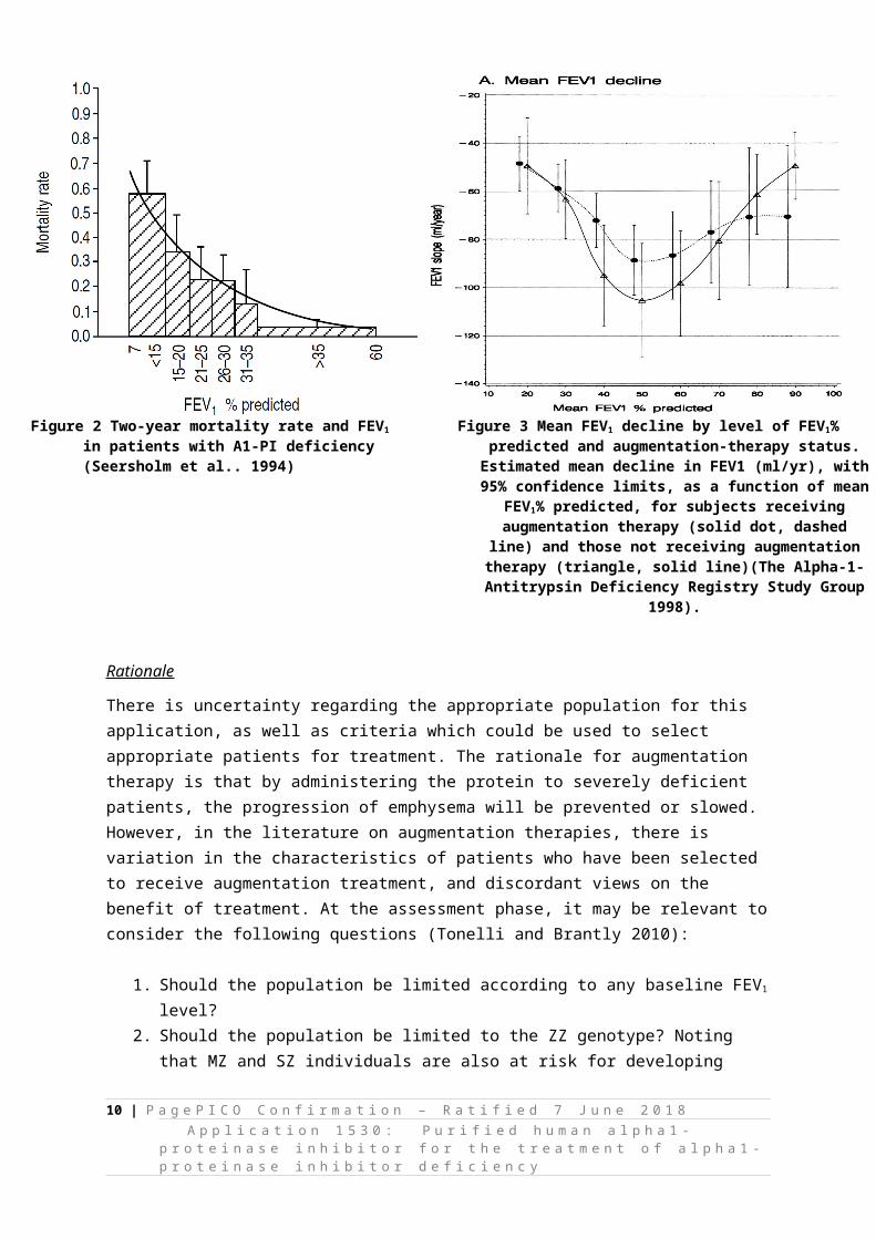
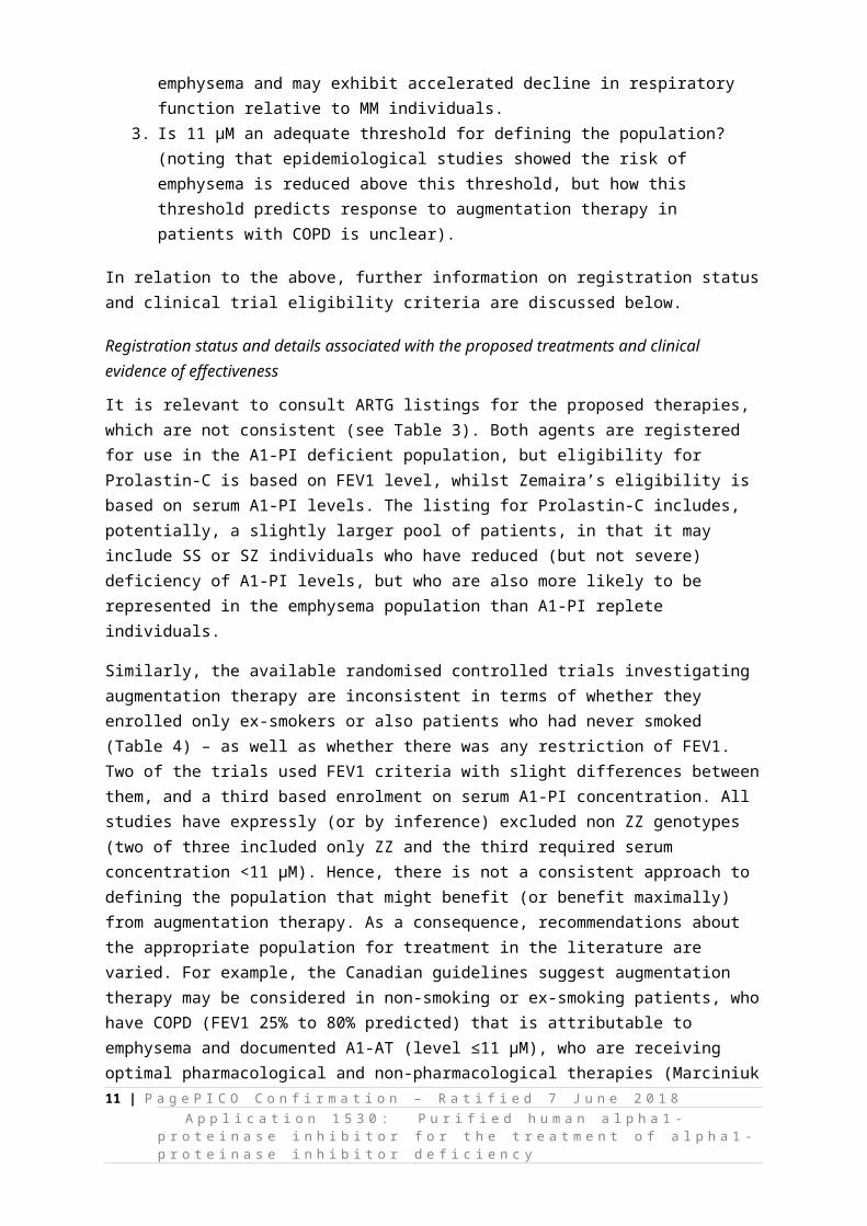
FEV1 has been shown in a number of studies to be an important predictor of survival in patients with emphysema with two-year mortality increasing exponentially once FEV1 falls below one-third of predicted; at this point two-year mortality reaches 50% in patients with FEV1 of 15% of predicted. This is shown in Figure 2 (Seersholm et al. 1994). In considering the overall pattern of FEV1 decline over the course of disease it has been observed that FEV1 declines most rapidly in patients with moderately reduced lung function with a slower decline with mild or severely reduced lung function (U shaped curve). This relationship is shown in Figure 3. When considering augmentation therapy, and the most applicable patient population, it is relevant to consider how the natural history of FEV1
decline might affect the ability to demonstrate efficacy.2
Chronic Obstructive Pulmonary Disease, vol.12pp. 561-569. there would be approximately 190 eligible patients. Since these estimates are disparate it is suggested that there is substantial uncertainty about the best estimate of population size2 When the rate of FEV1 decline (over time) is small, it is likely to be difficult to demonstrate significant differences between groups over a short timeframe. 6 | P a g e P I C O C o n fi r m a ti o n – R a ti fi e d 7 J u n e 2 0 1 8
A p p l i c a ti o n 1 5 3 0 : P u r i fi e d h u m a n a l p h a 1 - p r o t e i n a s e i n h i b i t o r f o r t h e t r e a t m e n t o f a l p h a 1 - p r o t e i n a s e i n h i b i t o r d e fi c i e n c y

Figure 2 Two-year mortality rate and FEV1 in patients with A1-PI deficiency (Seersholm et al.. 1994)
Figure 3 Mean FEV1 decline by level of FEV1% predicted and augmentation-therapy status. Estimated mean decline in FEV1 (ml/yr), with 95%
confidence limits, as a function of mean FEV1% predicted, for subjects receiving augmentation therapy (solid dot, dashed line)
and those not receiving augmentation therapy (triangle, solid line)(The Alpha-1-Antitrypsin Deficiency Registry Study Group 1998).
Rationale
There is uncertainty regarding the appropriate population for this application, as well as criteria which could be used to select appropriate patients for treatment. The rationale for augmentation therapy is that by administering the protein to severely deficient patients, the progression of emphysema will be prevented or slowed. However, in the literature on augmentation therapies, there is variation in the characteristics of patients who have been selected to receive augmentation treatment, and discordant views on the benefit of treatment. At the assessment phase, it may be relevant to consider the following questions (Tonelli and Brantly 2010):
1. Should the population be limited according to any baseline FEV1 level?2. Should the population be limited to the ZZ genotype? Noting that MZ and SZ individuals are
also at risk for developing emphysema and may exhibit accelerated decline in respiratory function relative to MM individuals.
3. Is 11 µM an adequate threshold for defining the population? (noting that epidemiological studies showed the risk of emphysema is reduced above this threshold, but how this threshold predicts response to augmentation therapy in patients with COPD is unclear).
In relation to the above, further information on registration status and clinical trial eligibility criteria are discussed below.
Registration status and details associated with the proposed treatments and clinical evidence of effectiveness
It is relevant to consult ARTG listings for the proposed therapies, which are not consistent (see Table 3). Both agents are registered for use in the A1-PI deficient population, but eligibility for Prolastin-C is based on FEV1 level, whilst Zemaira’s eligibility is based on serum A1-PI levels. The listing for
7 | P a g e P I C O C o n fi r m a ti o n – R a ti fi e d 7 J u n e 2 0 1 8A p p l i c a ti o n 1 5 3 0 : P u r i fi e d h u m a n a l p h a 1 - p r o t e i n a s e i n h i b i t o r f o r t h e t r e a t m e n t o f a l p h a 1 - p r o t e i n a s e i n h i b i t o r d e fi c i e n c y

Prolastin-C includes, potentially, a slightly larger pool of patients, in that it may include SS or SZ individuals who have reduced (but not severe) deficiency of A1-PI levels, but who are also more likely to be represented in the emphysema population than A1-PI replete individuals.
Similarly, the available randomised controlled trials investigating augmentation therapy are inconsistent in terms of whether they enrolled only ex-smokers or also patients who had never smoked (Table 4) – as well as whether there was any restriction of FEV1. Two of the trials used FEV1 criteria with slight differences between them, and a third based enrolment on serum A1-PI concentration. All studies have expressly (or by inference) excluded non ZZ genotypes (two of three included only ZZ and the third required serum concentration <11 μM). Hence, there is not a consistent approach to defining the population that might benefit (or benefit maximally) from augmentation therapy. As a consequence, recommendations about the appropriate population for treatment in the literature are varied. For example, the Canadian guidelines suggest augmentation therapy may be considered in non-smoking or ex-smoking patients, who have COPD (FEV1 25% to 80% predicted) that is attributable to emphysema and documented A1-AT (level ≤11 μM), who are receiving optimal pharmacological and non-pharmacological therapies (Marciniuk et al. 2012). Whilst an American consensus statement recommends augmentation therapy for individuals with established airflow obstruction from A1-PI deficiency (American Thoracic Society/European Respiratory Society 2003) and notes that benefits in individuals with mild or severe airflow obstruction are unclear.
Given the apparent uncertainty about patients most likely to benefit from augmentation therapy, it would be relevant to examine the treatment-modifying effect of emphysema severity and serum A1-PI at the assessment phase.
Table 3 Approved augmentation therapies and their indicationsProduct ARTG ID and detailsPROLASTIN-C ARTG ID 234553: indicated to increase serum Alpha1-PI levels in adults with congenital deficiency of alpha-
1 antitrypsin and with clinically significant emphysema (FEV1 less than 80%). The data for clinical efficacy of PROLASTIN-C is derived from changes in the biomarkers alpha-1 anti-protease level and CT lung density. Efficacy on FEV1 or patient relevant endpoints such as quality of life or pulmonary exacerbations has not been established in randomised clinical trials. Clinical trials have only included patients who were not smoking.
Zemaira ARTG ID 273182: indicated for maintenance treatment, to slow the progression of emphysema in adults with documented severe A1-PI deficiency (A1-PI less than 11 μM) and progressive lung disease. Patients are to be under optimal pharmacologic and non-pharmacologic treatment.
Table 4 Eligibility criteria of the included studies (Gotzsche and Johansen 2016)Study Included patientsDirksen et al. (1999) 58 ex-smokers from Denmark and The Netherlands with alpha-1 antitrypsin deficiency of PI*ZZ phenotype
and moderate emphysema (FEV1 between 30% and 80% of predicted). Patients were required to have ceased smoking at least six months prior to entry in the study.
EXACTLE(Dirksen et al. 2009)
82 ex- or never-smokers from Copenhagen (Denmark), Malmö (Sweden) and Birmingham (UK) with severe alpha-1 antitrypsin deficiency (serum concentration <11 μM)
RAPID(Chapman et al. 2015; McElvaney et al. 2017)
180 ex-smokers from Australia, Canada, Czech Republic, Denmark, Estonia, Finland, Germany, Ireland, Poland, Romania, Russia, Sweden, The Netherlands and the US with alpha-1 antitrypsin deficiency of the ZZ phenotype (168 participants) and moderate emphysema (FEV1 between 35% and 70% of the predicted normal value)
Prior test (investigative services only - if prior tests are to be included)
Diagnosis of COPD
The diagnosis of COPD is generally based on symptoms (breathlessness, cough and sputum), post-bronchodilator FEV1/FCV and patient history (smoking or exposure to noxious gases or particles). According to Campos et al. (2005), A1-PI deficiency is an under recognised condition in which there
8 | P a g e P I C O C o n fi r m a ti o n – R a ti fi e d 7 J u n e 2 0 1 8A p p l i c a ti o n 1 5 3 0 : P u r i fi e d h u m a n a l p h a 1 - p r o t e i n a s e i n h i b i t o r f o r t h e t r e a t m e n t o f a l p h a 1 - p r o t e i n a s e i n h i b i t o r d e fi c i e n c y

is an average interval of seven to eight years between symptom onset and diagnosis. A complicating factor is the prevalence of under-diagnosis and misdiagnosis of emphysema more broadly. The nonspecific and gradual nature of symptoms may prevent patients seeking medical review and physicians may attribute symptoms to acute respiratory infections. Recognising and testing for A1-PI deficiency in the setting of emphysema requires that individuals recognise and act on symptoms and practitioners undertake comprehensive patient workup.
Definitive diagnosis requires spirometry which measures timed expired and inspired volumes. According to the COPD-X guidelines indications for spirometry include (Yang et al. 2017):
breathlessness that seems inappropriate; chronic (daily for two months) or intermittent, unusual cough; frequent or unusual sputum production; relapsing acute infective bronchitis; and risk factors such as exposure to tobacco smoke, occupational dusts and chemicals, and a
strong family history of COPD.
COPD is indicated when the ratio of FEV1 to FVC is <70% and the FEV1 is <80% of the predicted value (normal values are obtained from healthy population studies, and derived from formulas based on height, age, sex and ethnicity). Table 5 provides a guide to severity that was adapted from the 2017 Australian and New Zealand guidelines for diagnosis and management of COPD. Individuals with A1-PI deficiency exposed to environmental air pollutants or who smoke may also experience accelerated lung damage relative to A1-PI replete individuals.
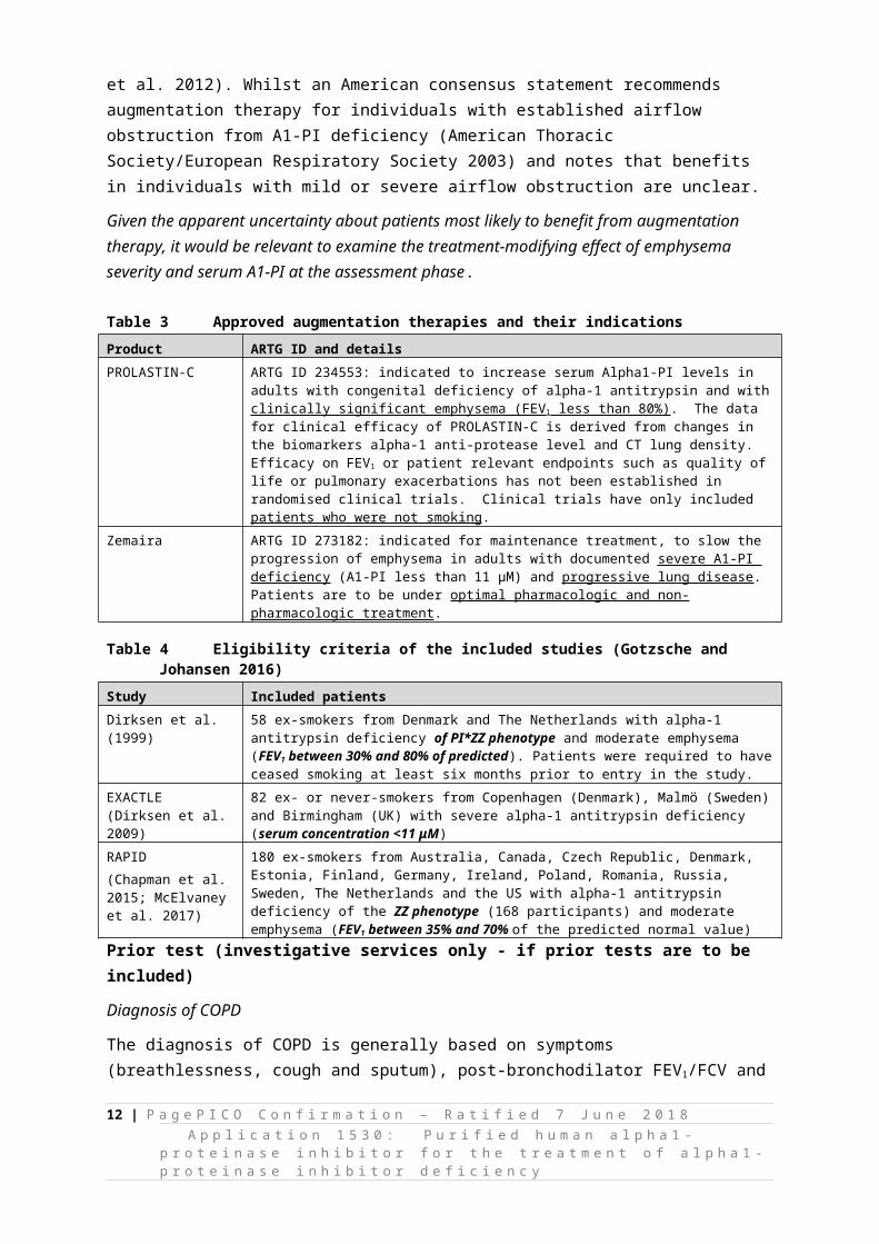
Table 5 Characteristics of COPD in mild, moderate and severe disease (Yang et al. 2017)Mild COPD Moderate COPD Severe COPD
Symptoms Limited symptomsBreathless on moderate exertionRecurrent chest infectionsLittle or no effect on daily activities
Breathless walking on level groundIncreasing limitation of daily activitiesCough and sputum productionExacerbations requiring oral corticosteroids and/or antibiotics
Breathless on minimal exertionsDaily activities severely curtailedExperiencing regular sputum productionChronic coughExacerbations of increasing frequency and severity
Typical FEV1 60 to 80% of predicted 40 to 59% of predicted <40 % of predicted
Diagnosis of emphysema
Emphysema, a form of COPD, is the result of enlargement of air spaces distal to the terminal bronchioles, and destruction of alveolar walls in the absence of obvious fibrosis. This pathological process: reduces alveolar surface area for gas exchange; limits elastic recoil and therefore airflow; and causes airway narrowing, further limiting airflow (Boka 2016). For individuals with A1-PI deficiency, panacinar emphysema predominates, in which the entire alveolus is uniformly destroyed (and this manifests predominantly in the lower lungs). For the diagnosis of emphysema, investigations include chest radiographs and computed tomography. Computed tomography is more sensitive and specific for diagnosing emphysema, however it is predominantly used in patients being considered for surgical interventions and typically is not a routine investigation in COPD (Boka 2016).
A1-PI deficiency testing
When considering the diagnosis of A1-PI deficiency, testing facilitates a more complete diagnosis in symptomatic individuals. In the setting of augmentation therapy, this may alter management, as well 9 | P a g e P I C O C o n fi r m a ti o n – R a ti fi e d 7 J u n e 2 0 1 8
A p p l i c a ti o n 1 5 3 0 : P u r i fi e d h u m a n a l p h a 1 - p r o t e i n a s e i n h i b i t o r f o r t h e t r e a t m e n t o f a l p h a 1 - p r o t e i n a s e i n h i b i t o r d e fi c i e n c y
as provide information about risks for asymptomatic individuals. It also has a role in genetic counselling in relation to reproduction (American Thoracic Society/European Respiratory Society 2003). Currently, there are differences of opinion about which populations should be tested for deficiency, and as yet, there are no Australian guidelines on the issue.
The World Health Organisation recommends all patients with COPD be screened for A1-PI deficiency. The American Thoracic Society/European Respiratory Society statement: ‘Standards for the Diagnosis and Management of Individuals with Alpha1-Antitrypsin Deficiency’ suggests genetic testing is warranted in:
“symptomatic adults with emphysema, chronic obstructive pulmonary disease, or asthma with airflow obstruction that is incompletely reversible after aggressive treatment with bronchodilators”; and
“asymptomatic individuals with persistent obstruction on pulmonary function tests with identifiable risk factors (e.g. cigarette smoking, occupational exposure).”
Furthermore, the statement indicates genetic testing is warranted in siblings of an individual with known deficiency (American Thoracic Society/European Respiratory Society 2003). The Canadian Thoracic Society clinical practice guideline (2012) suggests testing for A1-AT deficiency be considered in “individuals with COPD diagnosed before 65 years of age or with a smoking history of <20 pack years”, and that “targeted testing for A1-AT deficiency not be undertaken in individuals with bronchiectasis or asthma” (Marciniuk et al. 2012).
Diagnosis of A1-PI deficiency involves consideration of:
serum levels of A1-PI (low levels indicated deficiency) A1-PI phenotyping test to determine the type of AAT protein that an individual has A1-PI genotyping to determine the type (i.e. MZ or ZZ etc)
Testing requires a blood sample (in lithium heparin tube if genotyping is required) and consists of an immunoassay and isoelectric focussing for phenotyping (Royal College of Pathologists of Australasia 2014). DNA testing may be done as a follow-up to an alpha-1 antitrypsin level and phenotype. The Royal College of Pathologists manual suggests that if deficiency is documented, genotyping should be performed on the index case and their family. Furthermore, the manual suggests that where there is high clinical suspicion of deficiency, genotyping be performed even if serum deficiency is not identified (Royal College of Pathologists of Australasia 2014).
It is important to note that the clinical relevance of testing for A1-PI deficiency, genotyping and family testing may be altered by availability of a specific treatment option. In the absence of augmentation therapy, patients with emphysema are managed in the same way, irrespective of their A1-PI status; however, if augmentation therapy was broadly available, there would be an additional incentive for testing.
Compliance with smoking cessation
Patients who are current smokers are not eligible for treatment with augmentation therapy, and potential providers may wish to monitor compliance. Urine, blood or saliva testing can all be used to screen for tobacco exposure. Continine is the most frequently-tested metabolite of nicotine and can
10 | P a g e P I C O C o n fi r m a ti o n – R a ti fi e d 7 J u n e 2 0 1 8A p p l i c a ti o n 1 5 3 0 : P u r i fi e d h u m a n a l p h a 1 - p r o t e i n a s e i n h i b i t o r f o r t h e t r e a t m e n t o f a l p h a 1 - p r o t e i n a s e i n h i b i t o r d e fi c i e n c y
be tested in blood or urine (with dipstick testing an option). The type of testing that would be used in practice to monitor this population is uncertain (Lab Tests Online 2015).
InterventionThe proposed intervention is augmentation with alpha-1 antitrypsin concentrate, derived from human plasma and delivered intravenously to the patient. This is an additive intervention, as it will be given in addition to best supportive care for patients with emphysema (Ranes and Stoller 2005). There is uncertainty around level of patient exposure to this therapy that will provide maximal effect, and the randomised controlled trials differ on volume and frequency of product that patients were treated with. The dose in the earlier trial by Dirksen et al. was 250 mg/kg every four weeks, whereas the dose in the later trial by the same lead author (EXACTLE) was 60 mg/kg per week. The more recent RCT (RAPID) reportedly used the same dosing strategy of 60 mg/kg per week. Thus, according to trials reported in the published literature and product manufacturer recommendations, this is the most common dosing strategy. A statistical modelling study of the results of RAPID-RCT and RAPID-OLE stated that the dose of 60 mg/kg per week achieved the desired study outcome (which was serum levels ≤11 µM). However, no evidence in RAPID was found of a plateau in clinical efficacy as A1-PI exposure increases, thus the maximum clinically effective threshold for treatment in these patients has not yet been established (Tortorici et al. 2017). Another option is individualising dosage based on trough levels for each patient, but the benefit of this requires confirmation (Miravitlles et al. 2017).
The clinical expert explained to PASC that treatment needs to be lifelong, or it is a waste of resources.
Two augmentation therapies—PROLASTIN-C and Zemaira (marketed as Respreeza in Europe)—are registered on the ARTG for use in Australia. Although PROLASTIN-C has been in use for longer (with Zemaira joining the market more recently), they are bioequivalent products, with slightly different eligibility criteria (see Table 3).
Therapeutic concentrations of A1-PI are prepared from the blood of plasma donors. The product is presented in a sterile lyophilised powder, in a 1g vial. It needs to be reconstituted in 20mL of water for intravenous administration. Augmentation with alpha1-antitrypsin is administered at an infusion rate of approximately 0.08mL/kg per minute, and infusion takes approximately 15 minutes. Patients may administer at home themselves or with assistance of a carer, when deemed appropriate by the treating specialist and after receiving adequate training (REDACTED)
Both products are provided in a pack containing:
1 vial 1g lyophilised powder; 1 vial 20mL sterile water for injection 1 sterile filter needle; and 1 vented transfer device.
As a note for the future, two additional A1-PI augmentation products are registered with the FDA for use in the United States, being AralastTM (FDA 2012) and GLASSIA (FDA 2010). These products have the same indication, although GLASSIA is provided in a single vial with 1g of A1-PI in 50mL of solution, and AralastTM contains more A1-PI. Shire, the manufacturer of these two products, has stated it has no plans to market them in Australia at this time.
11 | P a g e P I C O C o n fi r m a ti o n – R a ti fi e d 7 J u n e 2 0 1 8A p p l i c a ti o n 1 5 3 0 : P u r i fi e d h u m a n a l p h a 1 - p r o t e i n a s e i n h i b i t o r f o r t h e t r e a t m e n t o f a l p h a 1 - p r o t e i n a s e i n h i b i t o r d e fi c i e n c y
Product information for Prolastin-C reported that the most common adverse events, occurring at a rate of ≥1% were chills, malaise, headache, rash, hot flush, and pruritis. These were reported in two open-label trials, including 62 patients in total, with the most serious adverse event being an abdominal extremity rash.
Researchers are working towards a new product becoming available (aerosolised A1-AP). This would offer an alternative route to the intravenous administration of A1-PI, and is proposed to address the expensive and time consuming nature of A1-PI augmentation. At present, the safety and effectiveness of aerosolised A1-AP have not been confirmed, but a long-term study has not yet been published (Franciosi et al. 2015).
Rationale
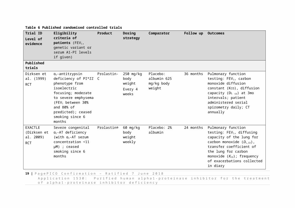
It appears the optimal dose of this therapy has not been confirmed in the literature. The dosing strategy used in published RCTs is provided in Table 6. As stated, a larger volume less frequently administered was used in 1999, and since then, the strategy of 60 mg/kg body weight per week seems to have been used. However, the minimal clinically-effective threshold is yet to be confirmed.
12 | P a g e P I C O C o n fi r m a ti o n – R a ti fi e d 7 J u n e 2 0 1 8A p p l i c a ti o n 1 5 3 0 : P u r i fi e d h u m a n a l p h a 1 - p r o t e i n a s e i n h i b i t o r f o r t h e t r e a t m e n t o f a l p h a 1 - p r o t e i n a s e i n h i b i t o r d e fi c i e n c y

Table 6 Published randomised controlled trialsTrial IDLevel of evidence
Eligibility criteria of patients (FEV1, genetic variant or serum A1-PI levels if given)
Product Dosing strategy Comparator Follow up Outcomes
Published trialsDirksen et al. (1999)RCT
α1-antitrypsin deficiency of PI*ZZ phenotype from isoelectric focusing; moderate to severe emphysema (FEV1 between 30% and 80% of predicted); ceased smoking since 6 months
Prolastin-C 250 mg/kg body weightEvery 4 weeks
Placebo: albumin 625 mg/kg body weight
36 months Pulmonary function testing: FEV1, carbon monoxide diffusion constant (KCO), diffusion capacity (DL CO) at 3mo intervals; patient administered serial spirometry daily; CT annually
EXACTLE(Dirksen et al. 2009)RCT
Severe congenital α1-AT deficiency (with α1-AT serum concentration <11 μM) ; ceased smoking since 6 months
Prolastin® 60 mg/kg body weightweekly
Placebo: 2% albumin 24 months Pulmonary function testing: FEV1, diffusing capacity of the lung for carbon monoxide (DL,CO), transfer coefficient of the lung for carbon monoxide (KCO); frequency of exacerbations collected in diary
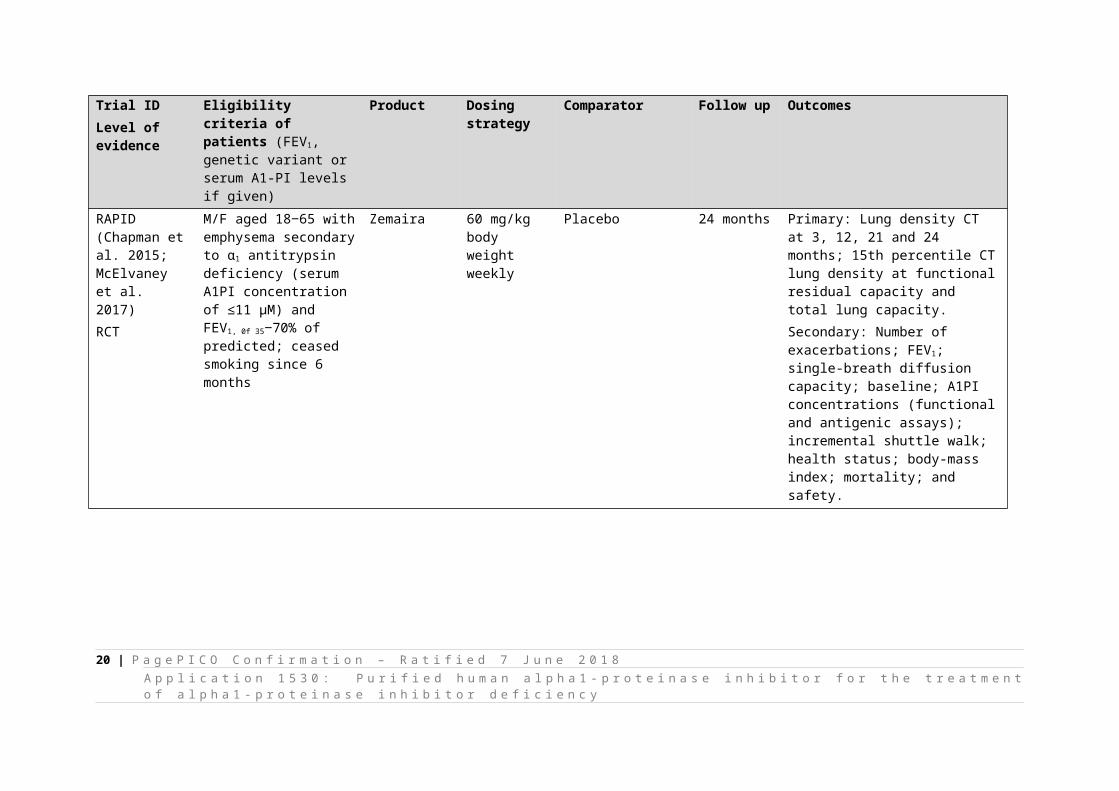
RAPID(Chapman et al. 2015; McElvaney et al. 2017)RCT
M/F aged 18‒65 with emphysema secondary to α1 antitrypsin deficiency (serum A1PI concentration of ≤11 μM) and FEV1, 0f 35‒70% of predicted; ceased smoking since 6 months
Zemaira 60 mg/kg body weightweekly
Placebo 24 months Primary: Lung density CT at 3, 12, 21 and 24 months; 15th percentile CT lung density at functional residual capacity and total lung capacity.Secondary: Number of exacerbations; FEV1; single-breath diffusion capacity; baseline; A1PI concentrations (functional and antigenic assays); incremental shuttle walk; health status; body-mass index; mortality; and safety.
13 | P a g e P I C O C o n fi r m a ti o n – R a ti fi e d 7 J u n e 2 0 1 8A p p l i c a ti o n 1 5 3 0 : P u r i fi e d h u m a n a l p h a 1 - p r o t e i n a s e i n h i b i t o r f o r t h e t r e a t m e n t o f a l p h a 1 -p r o t e i n a s e i n h i b i t o r d e fi c i e n c y
In addition to published trials, a current clinical trial (SPARTA) is due for completion in 2021. SPARTA is a placebo-controlled trial of alpha1-proteinase inhibitor, 60 or 120 mg/kg per week in 339 Australian patients. The primary measured outcome is lung CT scans (Sorrells et al. 2015).
Comparator
Change in practice
As stated, A1-PI deficiency is associated with indications falling under the umbrella term of COPD, in which panacinar emphysema is the most commonly recognised manifestation. Current clinical management treats symptoms of the emphysema, not the underlying causes of the disease (REDACTED). It is expected that augmentation with alpha1-antitrypsin will be provided in addition to best supportive care, which is the care provided to COPD patients regardless of A1-PI status. While best supportive care can be provided via a number of means, the COPD-X guidelines (below) reflect what is currently accepted to be best care.3
Main alternative
The comparator for this intervention is best supportive care for patients with COPD. Strategies for the management of stable COPD are provided in the Australian and New Zealand guidelines for the diagnosis and treatment of COPD (Yang et al. 2017), as follows:
Non-pharmaceutical strategies
Pulmonary rehabilitation and physical activity are strongly evidenced to be effective in optimising function. Pulmonary rehabilitation includes supervised exercise training and can be given in conjunction with any number of the following: behaviour change, nutritional advice, or psychosocial support. Programs can be provided in a hospital outreach department or in the community setting. However, if a patient does not have access to rehabilitation therapy, they should try to be as active as possible, as physical inactivity is linked with increased exacerbations and mortality.
Pharmacological strategies
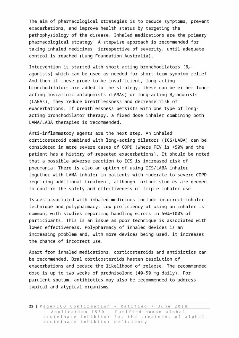
The aim of pharmacological strategies is to reduce symptoms, prevent exacerbations, and improve health status by targeting the pathophysiology of the disease. Inhaled medications are the primary pharmacological strategy. A stepwise approach is recommended for taking inhaled medicines, irrespective of severity, until adequate control is reached (Lung Foundation Australia).
Intervention is started with short-acting bronchodilators (B2-agonists) which can be used as needed for short-term symptom relief. And then if these prove to be insufficient, long-acting bronchodilators are added to the strategy, these can be either long-acting muscarinic antagonists (LAMAs) or long-acting B2-agonists (LABAs), they reduce breathlessness and decrease risk of exacerbations. If breathlessness persists with one type of long-acting bronchodilator therapy, a fixed dose inhaler combining both LAMA/LABA therapies is recommended.
Anti-inflammatory agents are the next step. An inhaled corticosteroid combined with long-acting dilators (ICS/LABA) can be considered in more severe cases of COPD (where FEV is <50% and the patient has a history of repeated exacerbations). It should be noted that a possible adverse reaction to ICS is increased risk of pneumonia. There is also an option of using ICS/LABA inhaler together with
3 REDACTED
14 | P a g e P I C O C o n fi r m a ti o n – R a ti fi e d 7 J u n e 2 0 1 8A p p l i c a ti o n 1 5 3 0 : P u r i fi e d h u m a n a l p h a 1 - p r o t e i n a s e i n h i b i t o r f o r t h e t r e a t m e n t o f a l p h a 1 - p r o t e i n a s e i n h i b i t o r d e fi c i e n c y

LAMA inhaler in patients with moderate to severe COPD requiring additional treatment, although further studies are needed to confirm the safety and effectiveness of triple inhaler use.
Issues associated with inhaled medicines include incorrect inhaler technique and polypharmacy. Low proficiency at using an inhaler is common, with studies reporting handling errors in 50%‒100% of participants. This is an issue as poor technique is associated with lower effectiveness. Polypharmacy of inhaled devices is an increasing problem and, with more devices being used, it increases the chance of incorrect use.
Apart from inhaled medications, corticosteroids and antibiotics can be recommended. Oral corticosteroids hasten resolution of exacerbations and reduce the likelihood of relapse. The recommended dose is up to two weeks of prednisolone (40–50 mg daily). For purulent sputum, antibiotics may also be recommended to address typical and atypical organisms.
Comorbidities that accompany COPD (the main ones being anxiety and depression) increase hospitalisations and these need to be managed. Osteoporotic fractures are also a common problem in patients with COPD, hence bone mineral density testing is important for prevention and monitoring. COPD and its resulting hypoxaemia are known to lead to pulmonary hypertension and right heart failure, especially when occurring with sleep apnoea. When this is suspected clinically, arterial blood gas or a sleep study should be conducted, leading to oxygen therapy or continuous positive airway pressure.
Prevent deterioration
To complement the above-mentioned ‘function-optimising’ steps, behaviour change is recommended. In the hope of preventing deterioration, patients are recommended to cease cigarette smoking (of utmost importance), reduce alcohol consumption, increase physical activity, and avoid environmental irritants.
Another helpful approach is vaccination for influenza and pneumococcal, as it reduces exacerbations due to influenza and pneumococcal at high-risk times. When used together, there is an additional benefit. Adverse effects of vaccines are mild and self-limiting. It is a cost-effective approach to receive the influenza vaccine annually, particularly patients with severe COPD. The five-yearly pneumococcal vaccination will also protect against community-acquired pneumonia, and therefore reduce the probability of COPD exacerbations.
Long term use of supplemental oxygen assists with correction of severe hypoxaemia, and might also improve survival; using supplemental oxygen for longer periods has been reported to have greater benefits. While no benefit of continuous oxygen therapy has been reported for patients with mild or moderate hypoxaemia, 18 hours a day (at least) is recommended for patients with PaO2 of ≤55 mmHg who also have pulmonary hypertension, polycythaemia or right heart failure. All patients with COPD may benefit from ambulatory oxygen when blood is de-saturated due to exertion. For each patient, it is important to review oxygen use, determination of benefit from the oxygen, and need for continued use.
Patients with very severe disease might need lung transplantation or lung volume reduction—by surgery or bronchoscopically. Only certain patients may be considered appropriate for lung volume reduction, including those with severe emphysema, hyperinflation and ongoing symptoms, despite best management and pulmonary rehabilitation. Likewise patients considered for lung transplantation will be those suffering severe functional impairment and airflow obstruction, which
15 | P a g e P I C O C o n fi r m a ti o n – R a ti fi e d 7 J u n e 2 0 1 8A p p l i c a ti o n 1 5 3 0 : P u r i fi e d h u m a n a l p h a 1 - p r o t e i n a s e i n h i b i t o r f o r t h e t r e a t m e n t o f a l p h a 1 - p r o t e i n a s e i n h i b i t o r d e fi c i e n c y
is not appropriately managed by other strategies. Considerable risks are associated with these procedures (Yang et al. 2017). PASC noted that lung transplantation is not curative, because transplant recipients still need A1-PI supplementation to protect their transplanted lungs from the same gradual deterioration.
Rationale
There are no direct comparators for A1-PI augmentation. Best supportive care for patients with COPD, regardless of A1-PI status, is what this patient population receives in clinical practice when A1-PI is not available. Best supportive care can be provided through all major health facilities in Australia.
Best supportive care (as a comparator) aims to only address symptoms of the disease, optimise function and prevent deterioration, whereas A1-PI augmentation claims to slow progression of the disease.
OutcomesAugmentation therapy increases serum A1-PI levels (and therefore A1-PI levels in the lungs), which is purported to prevent or slow progression of emphysema. FDA guidance for industry on COPD trials (2016) recommends that, for trials intended to show effects on disease progression, the primary endpoint should be serial measurement of FEV1 over time, with an expectation that decline will diverge over time (airflow preservation in the treatment arm). However, the primary outcome in some trials of augmentation therapy has been CT measures of lung density and functional residual capacity. The applicant suggests the primary endpoint is: improvement in clinical outcomes (symptoms and breathing) and slowing of progression of lung deterioration. Secondary outcomes suggested include: reduction in exacerbation, occurrence, severity and length, increased quality of life.
Direct measures of impact on disease progression require long term follow-up, and are subject to individual variability attributable to baseline FEV1, concurrent treatments, smoking history and possibly severity of A1-PI deficiency. Other outcomes relevant to this application include surrogate measures of bioavailability and disease-modification.
Safety Incidence and severity of adverse events
Primary effectiveness Respiratory function measured by spirometry (FEV1) and FEV1/FVC ratio Dyspnoea (measured with a validated tool; e.g. baseline dyspnoea index, transition
dyspnoea index)
Secondary Mortality, including deaths from respiratory failure Number of exacerbations and hospitalisations associated with COPD Quality of life (measured by validated tool for COPD or respiratory impairment) Changes in exercise capacity (with tools such as the 6-minute walking test) BODE index- body mass index, airflow obstruction, dyspnoea and exercise index (De Torres,
Thorax 2014; 69:799–804) - this is more predictive of mortality then FEV1 Surrogate measures/biomarkers:
o Lung density measured by computed tomography (CT)
16 | P a g e P I C O C o n fi r m a ti o n – R a ti fi e d 7 J u n e 2 0 1 8A p p l i c a ti o n 1 5 3 0 : P u r i fi e d h u m a n a l p h a 1 - p r o t e i n a s e i n h i b i t o r f o r t h e t r e a t m e n t o f a l p h a 1 - p r o t e i n a s e i n h i b i t o r d e fi c i e n c y

o Carbon monoxide (CO) transfer or pulmonary diffusing capacity for CO (DLCO)
Minimal Clinically Important Differences (MCID) for Commonly Used Outcomes in Chronic Obstructive Pulmonary Disease from Jones et al. (2014)
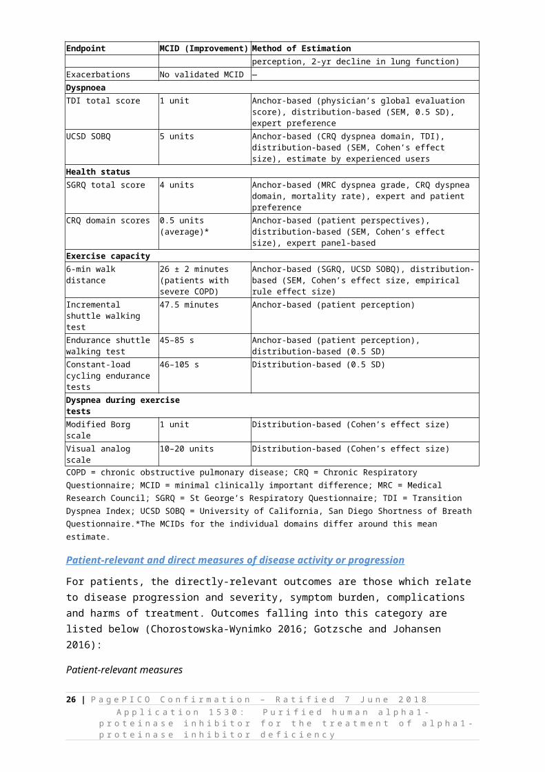
In the context of COPD, validated MCIDs are available for a range of outcomes, including lung function, dyspnoea, health status, and exercise capacity. However, the way in which MCIDs are analysed within COPD trials may vary. Table 7 presents reference values that might be considered during the assessment phase. Published MCIDs for surrogate outcomes (such as lung density measured by CT) were not identified.
Table 7 Minimal Clinically Important Differences for Commonly Used Outcomes in Chronic Obstructive Pulmonary Disease from Jones et al. (2014)
Endpoint MCID (Improvement) Method of EstimationLung function
Trough FEV1 100 ml Anchor-based (exacerbations, patient perception, 2-yr decline in lung function)
Exacerbations No validated MCID —Dyspnoea
TDI total score 1 unit Anchor-based (physician’s global evaluation score), distribution-based (SEM, 0.5 SD), expert preference
UCSD SOBQ 5 units Anchor-based (CRQ dyspnea domain, TDI), distribution-based (SEM, Cohen’s effect size), estimate by experienced users
Health status
SGRQ total score 4 units Anchor-based (MRC dyspnea grade, CRQ dyspnea domain, mortality rate), expert and patient preference
CRQ domain scores 0.5 units (average)* Anchor-based (patient perspectives), distribution-based (SEM, Cohen’s effect size), expert panel-based
Exercise capacity
6-min walk distance 26 ± 2 minutes (patients with severe COPD)
Anchor-based (SGRQ, UCSD SOBQ), distribution-based (SEM, Cohen’s effect size, empirical rule effect size)
Incremental shuttle walking test
47.5 minutes Anchor-based (patient perception)
Endurance shuttle walking test 45–85 s Anchor-based (patient perception), distribution-based (0.5 SD)Constant-load cycling endurance tests
46–105 s Distribution-based (0.5 SD)
Dyspnea during exercise tests
Modified Borg scale 1 unit Distribution-based (Cohen’s effect size)Visual analog scale 10–20 units Distribution-based (Cohen’s effect size)COPD = chronic obstructive pulmonary disease; CRQ = Chronic Respiratory Questionnaire; MCID = minimal clinically important difference; MRC = Medical Research Council; SGRQ = St George’s Respiratory Questionnaire; TDI = Transition Dyspnea Index; UCSD SOBQ = University of California, San Diego Shortness of Breath Questionnaire.*The MCIDs for the individual domains differ around this mean estimate.
Patient-relevant and direct measures of disease activity or progression
For patients, the directly-relevant outcomes are those which relate to disease progression and severity, symptom burden, complications and harms of treatment. Outcomes falling into this category are listed below (Chorostowska-Wynimko 2016; Gotzsche and Johansen 2016):
Patient-relevant measures
Mortality, including death from respiratory failure Dyspnoea (measured with a validated tool e.g. baseline dyspnoea index; transition dyspnoea
index) Number of exacerbations and hospitalisations associated with COPD Changes in exercise capacity (measured with tools such as the 6-minute walking test) Quality of life (preferably measured by validated tool for COPD or respiratory impairment)
17 | P a g e P I C O C o n fi r m a ti o n – R a ti fi e d 7 J u n e 2 0 1 8A p p l i c a ti o n 1 5 3 0 : P u r i fi e d h u m a n a l p h a 1 - p r o t e i n a s e i n h i b i t o r f o r t h e t r e a t m e n t o f a l p h a 1 - p r o t e i n a s e i n h i b i t o r d e fi c i e n c y

Adverse events
Objective physiological measures
Respiratory function measured by spirometry (FEV1) Body mass index, airflow obstruction, dyspnoea and exercise (BODE) index4
Disease-modification and relevant outcomes
Current standard of care for COPD patients consists largely of symptomatic treatments, which improve quality of life, but do not address underlying pathological processes contributing to destruction of lung tissue. Augmentation therapy, by normalising A1-PI levels in the pulmonary tissue, is directed to the underlying imbalance in protease/antiprotease activity in A1-PI deficient patients thereby aiming to provide a sustained alteration in disease progression (Chorostowska-Wynimko 2016). In the context of disease modifying interventions in COPD, it is relevant to consider the definition of disease modification and what outcomes can be used to assess effectiveness of an intervention in terms of disease modification (Halpin and Tashkin 2009).
In 2009, a group of physicians and scientists from the USA, Canada and Europe (in the context of COPD) defined disease modification as “an improvement in, or stabilization of, structural or functional parameters as a result of reduction in the rate of progression of these parameters, which occurs whilst an intervention is applied and may persist even if the intervention is withdrawn” (Halpin and Tashkin 2009). Disease-modification may be assessed using a range of parameters (including those listed above), although respiratory function is the most accepted as a valid surrogate for disease status (U.S. Department of Health and Human Services, Food and Drug Administration Center for Drug Evaluation and Research (CDER) 2016). As such, FEV1 is the most widely used (and commonly accepted) endpoint for assessing efficacy.
However, outcomes such as FEV1 have limitations in disease-modification trials, in that it:
changes slowly over time (therefore requiring long follow-up; generally > 2 years); exhibits individual variability; and (until certain thresholds are reached) has limited correlation with endpoints, such as mortality or
exacerbations (Chorostowska-Wynimko 2016).
Therefore, trials of interventions in COPD have begun to use other supplementary measures that speak to structural changes in the lung, including chest computed tomography (CT), concentration of certain gases in exhaled air or breath condensate, inflammatory mediators or cells in relevant biological fluids, and desmosine and isodesmosine levels in plasma (Ma et al. 2016; U.S. Department of Health and Human Services Food and Drug Administration Center for Drug Evaluation and Research (CDER) 2016).
Of these, lung density measured on CT has emerged as the most robust surrogate measure of disease progression, with studies establishing its correlation with QoL (Miravitlles et al. 2017), FEV1
(Parr et al. 2009), and establishing sensitivity for progression (Stolk et al. 2007) (although there are conflicting views of its utility in the literature). Guidance from the Food and Drug Administration for industry indicates lung density CT can be useful as secondary endpoints to support primary efficacy
4 A recent study by de Torres JP, Casanova C, Marin JM, et al. Prognostic evaluation of COPD patients: GOLD 2011 versus BODE and the COPD comorbidity index COTE. Thorax 2014; 69(9):799–804 found that the BODE index is a useful tool in COPD that is more sensitive at predicting survival than FEV1 alone.
18 | P a g e P I C O C o n fi r m a ti o n – R a ti fi e d 7 J u n e 2 0 1 8A p p l i c a ti o n 1 5 3 0 : P u r i fi e d h u m a n a l p h a 1 - p r o t e i n a s e i n h i b i t o r f o r t h e t r e a t m e n t o f a l p h a 1 - p r o t e i n a s e i n h i b i t o r d e fi c i e n c y
analysis (U.S. Department of Health and Human Services, Food and Drug Administration Center for Drug Evaluation and Research (CDER) 2016). The minimal duration and degree of clinically important improvement qualifying as disease modification, however, are uncertain (Zuwallack and Nici 2012).
During the assessment phase, it may be relevant to provide supporting evidence to establish validity of surrogate measures in demonstrating disease-modification effects. It would also be valid to consider what a minimal clinically-important difference between groups would be.
Criteria for concluding superiority (relative to standard care) should be specified during the assessment phase, in order to support the clinical claim associated with augmentation therapy. In the absence of pre-specified superiority criteria, superiority should be tested with a point estimate and 95% confidence intervals, relative to the null hypothesis that there is no difference between the compared alternatives. To conclude superiority, the 95% confidence interval should exclude the possibility that there is no difference between the compared strategies.
PASC suggested the assessment should consider carer outcome perspectives and additional information about patient experience outcomes.
Healthcare system
Table 8 lists relevant MBS items and known costs associated with providing the intervention and diagnostic services.
Drug acquisition and service provision
Drug acquisition and service provision costs include:
Cost of augmentation product (which will vary, depending on patient weight). Both sponsors recommend a dose of 60 mg/kg/week, but the assessment phase may test different dosing schedules.
Cost of administering the infusion (which may include health service costs and/or cost of patient/carer education on self-administration).
Cost of managing complications (associated with the augmentation therapy).
Cost of screening and testing
Costs associated with testing for emphysema and A1-PI deficiency need to be considered; in particular, cost impacts of testing a population for whom the therapy is not currently indicated.
At present, because knowledge of A1-PI status does not impact the management of patients, screening for deficiency is not a routine component of care for patients with emphysema (unless there is high clinical suspicion, based on age and family history). Availability of this specific therapy is likely to alter this, but the population that could be considered for screening is likely to be smaller than the total COPD population, because:
emphysema is a subgroup of the COPD population; and, individuals with a long history of smoking, who present at older age or with other significant
risk factors for COPD, might not be investigated.
At the broadest level, it is possible that many COPD patients could be screened for A1-PI deficiency, particularly if they develop symptoms at a young age. In the 45 to 54 age group, with a COPD prevalence of 2.5 per cent, there could be as many as 77,625 people eligible for testing. Whilst this is
19 | P a g e P I C O C o n fi r m a ti o n – R a ti fi e d 7 J u n e 2 0 1 8A p p l i c a ti o n 1 5 3 0 : P u r i fi e d h u m a n a l p h a 1 - p r o t e i n a s e i n h i b i t o r f o r t h e t r e a t m e n t o f a l p h a 1 - p r o t e i n a s e i n h i b i t o r d e fi c i e n c y
almost certainly a substantial overestimate, it highlights the magnitude of potential testing or screening. It is suggested this be considered during the assessment phase, in relation to budget impact of the proposed listing. See Table 8 for detail on costs associated with these services.
Resources provided to deliver the comparator
As augmentation therapy will be added to standard care, the main cost will be the additional cost of augmentation agents. Augmentation therapy is not assumed to displace other COPD treatments. Costs will therefore be incurred in both arms of managing COPD that, depending on assumptions about exacerbations and symptoms, may be the same or different between groups.
Potential cost offsets
Potential cost offsets will depend on assumptions about impact of the intervention on morbidity and mortality associated with COPD. If augmentation therapy delays progression, reduces exacerbations or improves symptoms, there will be cost offsets in terms of disease management and hospitalisations.
Table 8 Costs associated with delivering the intervention Item CostMBS items for A1-PI deficiencyCategory 6 Pathology Services: 66635 Alpha-1-antitrypsin - quantitation in serum, urine or other body fluid - 1 or more tests
MBS Fee: $20.10 Benefit: 75% = $15.10 85% = $17.10
Category 6 Pathology Services: 66638 Isoelectric focussing or similar methods for determination of alpha-1-antitrypsin phenotype in serum - 1 or more tests
MBS Fee: $49.05 Benefit: 75% = $36.8085% = $41.70
MBS item for respiratory functionCategory 2 – Diagnostic procedures and investigations: 11503Measurement of the:(a) mechanical or gas exchange function of the respiratory system; or (b) respiratory muscle function; or (c) ventilatory control mechanisms.Various measurement parameters may be used including any of the following: (a) pressures; (b) volumes; (c) flow; (d) gas concentrations in inspired or expired air; (e) alveolar gas or blood; (f) electrical activity of muscles.The tests being performed under the supervision of a specialist or consultant physician or in the respiratory laboratory of a hospital. Each occasion at which 1 or more such tests are performed, not being a service associated with a service to which item 22018 applies.
MBS Fee: $138.65 Benefit: 75% = $104.00 85% = $117.90
MBS items associated with lung imagingCategory 5 Diagnostic Imaging Services: 56301COMPUTED TOMOGRAPHY - scan of chest, including lungs, mediastinum, chest wall and pleura, with or without scans of the upper abdomen, without intravenous contrast medium, not being a service to which item 56801 or 57001 applies and not including a study performed to exclude coronary artery calcification or image the coronary arteries (R) (K) (Anaes.)
MBS Fee: $295.00 Benefit: 75% = $221.25 85% = $250.75
Category 5 Diagnostic Imaging Services: 56301COMPUTED TOMOGRAPHY - scan of chest, including lungs, mediastinum, chest wall and pleura, with or without scans of the upper abdomen, with intravenous contrast medium and with any scans of the chest including lungs, mediastinum, chest wall or pleura and upper abdomen prior to intravenous contrast injection, when undertaken, not being a service to which item 56807 or 57007 applies and not including a study performed to exclude coronary artery calcification or image the coronary arteries (R) (K) (Anaes.)
MBS Fee: $400.00 Benefit: 75% = $300.00 85% = $340.00
Category 5 Diagnostic Imaging Services: 56341COMPUTED TOMOGRAPHY - scan of chest, including lungs, mediastinum, chest wall and pleura, with or without scans of the upper abdomen, without intravenous contrast medium, not being a service to which item 56841 or 57041 applies and not including a study performed to exclude coronary artery calcification or image the coronary arteries (R) (NK) (Anaes.)
MBS Fee: $149.45Benefit: 75% = $112.10 85% = $127.05
20 | P a g e P I C O C o n fi r m a ti o n – R a ti fi e d 7 J u n e 2 0 1 8A p p l i c a ti o n 1 5 3 0 : P u r i fi e d h u m a n a l p h a 1 - p r o t e i n a s e i n h i b i t o r f o r t h e t r e a t m e n t o f a l p h a 1 - p r o t e i n a s e i n h i b i t o r d e fi c i e n c y
Item CostCategory 5 Diagnostic Imaging Services: 56347COMPUTED TOMOGRAPHY - scan of chest, including lungs, mediastinum, chest wall and pleura, with or without scans of the upper abdomen, with intravenous contrast medium and with any scans of the chest including lungs, mediastinum, chest wall or pleura and upper abdomen prior to intravenous contrast injection, when undertaken, not being a service to which item 56847 or 57047 applies and not including a study performed to exclude coronary artery calcification or image the coronary arteries (R) (NK) (Anaes.)
MBS Fee: $202.00 Benefit: 75% = $151.50 85% = $171.70
Category 5 Diagnostic Imaging Services: 58500CHEST (lung fields) by direct radiography (NR)
MBS Fee: $35.35 Benefit: 75% = $26.5585% = $30.05
Category 5 Diagnostic Imaging Services: 58502CHEST (lung fields) by direct radiography (NR)(NK)
MBS Fee: $17.70 Benefit: 75% = $13.30 85% = $15.05
Category 5 Diagnostic Imaging Services: 58503CHEST (lung fields) by direct radiography (R)
MBS Fee: $47.15 Benefit: 75% = $35.40 85% = $40.10
Category 5 Diagnostic Imaging Services: 58505CHEST (lung fields) by direct radiography (R)(NK)
MBS Fee: $23.60 Benefit: 75% = $17.7085% = $20.10
Category 5 Diagnostic Imaging Services: 58506CHEST (lung fields) by direct radiography with fluoroscopic screening (R)
MBS Fee: $60.75 Benefit: 75% = $45.60 85% = $51.65
Augmentation therapyZemaira 1000 mg vial (applicant supplied costs) REDACTED
Prolastin-C 1000 mg vial (applicant supplied costs) REDACTED
MBS item 13915CYTOTOXIC CHEMOTHERAPY, administration of, either by intravenous push technique (directly into a vein, or a butterfly needle, or the side-arm of an infusion) or by intravenous infusion of not more than 1 hours duration - payable once only on the same day, not being a service associated with photodynamic therapy with verteporfin or for the administration of drugs used immediately prior to, or with microwave (UHF radiowave) cancer therapy alone
$51.95 (Annual costs, assuming no self-administration: $2,701)
RationaleNo additional comments.
21 | P a g e P I C O C o n fi r m a ti o n – R a ti fi e d 7 J u n e 2 0 1 8A p p l i c a ti o n 1 5 3 0 : P u r i fi e d h u m a n a l p h a 1 - p r o t e i n a s e i n h i b i t o r f o r t h e t r e a t m e n t o f a l p h a 1 - p r o t e i n a s e i n h i b i t o r d e fi c i e n c y
Current clinical management algorithm for identified population
Figure 4 Current clinical management algorithm for patients with emphysema and FEV1 <80%
22 | P a g e P I C O C o n fi r m a ti o n – R a ti fi e d 7 J u n e 2 0 1 8A p p l i c a ti o n 1 5 3 0 : P u r i fi e d h u m a n a l p h a 1 - p r o t e i n a s e i n h i b i t o r f o r t h e t r e a t m e n t o f a l p h a 1 - p r o t e i n a s e i n h i b i t o r d e fi c i e n c y

Proposed clinical management algorithm for identified population
Figure 5 Proposed clinical management algorithm for patients with emphysema and FEV1 <80%
23 | P a g e P I C O C o n fi r m a ti o n – R a ti fi e d 7 J u n e 2 0 1 8A p p l i c a ti o n 1 5 3 0 : P u r i fi e d h u m a n a l p h a 1 - p r o t e i n a s e i n h i b i t o r f o r t h e t r e a t m e n t o f a l p h a 1 - p r o t e i n a s e i n h i b i t o r d e fi c i e n c y

Proposed economic evaluation
The applicant has specified the comparative claim is one of superiority on the basis that augmentation therapy slows the progression of emphysema in adults with A1-PI deficiency. This is assumed to lead to delayed decline in respiratory function, with consequences for COPD-associated morbidity and mortality. Given the applicant is claiming superiority (relative to currently available treatments), a cost-effectiveness or cost-utility analysis is appropriate to determine if increased health outcomes (and any cost offsets) justify the increased expense. PASC suggested a cost-utility analysis would be most appropriate for this assessment. Based on the plentiful literature on COPD (including utilities for various stages of disease), it should be feasible to present a cost-utility analysis, quantifying the QALY gains associated with the proposal (in order to inform an incremental cost effectiveness ratio).
In considering the proposed economic evaluation, it is relevant to consider that an individual’s baseline level of respiratory function will impact on morbidity and mortality, as well as affecting the capacity for gain from treatment. As COPD is characterised by slow progression, with direct impacts on mortality unlikely to be seen in relatively short trials (unless patients have very severe COPD at baseline), cost-utility analysis will be required to extrapolate outcomes, likely, beyond trial study periods.
PASC may wish to consider what the relevant time-horizon would be for economic analyses in the context of COPD and A1-PI deficiency.
Proposed item descriptor
Augmentation therapy is proposed for reimbursement on the National Products List managed by the National Blood Authority. New blood and blood-related products reviewed by the Jurisdictional Blood Committee may be referred to MSAC for evidence-based evaluation of safety, clinical effectiveness and cost-effectiveness. No MBS item descriptor is required for this application.
24 | P a g e P I C O C o n fi r m a ti o n – R a ti fi e d 7 J u n e 2 0 1 8A p p l i c a ti o n 1 5 3 0 : P u r i fi e d h u m a n a l p h a 1 - p r o t e i n a s e i n h i b i t o r f o r t h e t r e a t m e n t o f a l p h a 1 - p r o t e i n a s e i n h i b i t o r d e fi c i e n c y

ReferencesAlpha-1-Antitrypsin Deficiency Registry Study Group 1998, 'Survival and FEV1 decline in individuals
with severe deficiency of alpha1-antitrypsin. ', Am J Respir Crit Care Med, vol.158(1), pp. 49-59.
American Thoracic Society/European Respiratory Society 2003, 'American Thoracic Society/European Respiratory Society statement: standards for the diagnosis and management of individuals with alpha-1 antitrypsin deficiency', Am J Respir Crit Care Med, vol.168(7), pp. 818-900.
Australian Institute of Health and Welfare 2017, COPD (chronic obstructive pulmonary disease), viewed 15 February 2018, <https://www.aihw.gov.au/reports/asthma-other-chronic-respiratory-conditions/copd-chronic-obstructive-pulmonary-disease/data>.
Blanco, I, Bueno, P, Diego, I, Pérez-Holanda, S, Casas-Maldonado, F, Esquinas, C & Miravitlles, M 2017, 'Alpha-1 antitrypsin Pi*Z gene frequency and Pi*ZZ genotype numbers worldwide: an update', International Journal of Chronic Obstructive Pulmonary Disease, vol.12pp. 561-569.
Boka, K 2016, Emphysema Workup, Medscape, viewed 9 February 2018, <https://emedicine.medscape.com/article/298283-workup#showall>.
Brantly, ML, Paul, LD, Miller, BH, Falk, RT, Wu, M & Crystal, RG 1988, 'Clinical features and history of the destructive lung disease associated with alpha-1-antitrypsin deficiency of adults with pulmonary symptoms', Am Rev Respir Dis, vol.138(2), pp. 327-336.
Brode, SK, Ling, SC & Chapman, KR 2012, 'Alpha-1 antitrypsin deficiency: a commonly overlooked cause of lung disease', CMAJ : Canadian Medical Association Journal, vol.184(12), pp. 1365-1371.
Chapman, KR, Burdon, JG, Piitulainen, E, Sandhaus, RA, Seersholm, N, Stocks, JM, Stoel, BC, Huang, L, Yao, Z, Edelman, JM & McElvaney, NG 2015, 'Intravenous augmentation treatment and lung density in severe alpha1 antitrypsin deficiency (RAPID): a randomised, double-blind, placebo-controlled trial', Lancet, vol.386(9991), pp. 360-368.
Chorostowska-Wynimko, J 2016, 'Disease Modification in Emphysema Related to Alpha-1 Antitrypsin Deficiency', Copd, vol.13(6), pp. 807-815.
de Serres, F & Blanco, I 2014, 'Role of alpha-1 antitrypsin in human health and disease', J Intern Med, vol.276(4), pp. 311-335.
de Serres, FJ, Blanco, I & Fernandez-Bustillo, E 2003, 'Genetic epidemiology of alpha-1 antitrypsin deficiency in North America and Australia/New Zealand: Australia, Canada, New Zealand and the United States of America', Clin Genet, vol.64(5), pp. 382-397.
Dirksen, A, Dijkman, JH, Madsen, F, Stoel, B, Hutchison, DC, Ulrik, CS, Skovgaard, LT, Kok-Jensen, A, Rudolphus, A, Seersholm, N, Vrooman, HA, Reiber, JH, Hansen, NC, Heckscher, T, Viskum, K & Stolk, J 1999, 'A randomized clinical trial of alpha(1)-antitrypsin augmentation therapy', Am J Respir Crit Care Med, vol.160(5 Pt 1), pp. 1468-1472.
Dirksen, A, Piitulainen, E, Parr, DG, Deng, C, Wencker, M, Shaker, SB & Stockley, RA 2009, 'Exploring the role of CT densitometry: a randomised study of augmentation therapy in alpha1-antitrypsin deficiency', Eur Respir J, vol.33(6), pp. 1345-1353.
Evald, T, Dirksen, A, Keittelmann, S, Viskum, K & Kok-Jensen, A 1990, 'Decline in pulmonary function in patients with alpha 1-antitrypsin deficiency', Lung, vol.168 Supplpp. 579-585.
FDA 2010, viewed <https://www.fda.gov/downloads/ApprovedProducts/UCM217890.pdf>.FDA 2012, viewed <https://www.fda.gov/downloads/BloodBloodProducts/ucm092856.pdf>.Franciosi, AN, McCarthy, C & McElvaney, NG 2015, 'The efficacy and safety of inhaled human α-1
antitrypsin in people with α-1 antitrypsin deficiency-related emphysema', Expert Rev Respir Med, vol.9(2), pp. 143-151.
Fregonese, L & Stolk, J 2008, 'Hereditary alpha-1-antitrypsin deficiency and its clinical consequences', Orphanet Journal of Rare Diseases, vol.3pp. 16-16.
Gotzsche, PC & Johansen, HK 2016, 'Intravenous alpha-1 antitrypsin augmentation therapy for treating patients with alpha-1 antitrypsin deficiency and lung disease', Cochrane Database Syst Rev, vol.9pp. Cd007851.
25 | P a g e P I C O C o n fi r m a ti o n – R a ti fi e d 7 J u n e 2 0 1 8A p p l i c a ti o n 1 5 3 0 : P u r i fi e d h u m a n a l p h a 1 - p r o t e i n a s e i n h i b i t o r f o r t h e t r e a t m e n t o f a l p h a 1 - p r o t e i n a s e i n h i b i t o r d e fi c i e n c y
Häggblom, J, Kettunen, K, Karjalainen, J, Heliövaara, M, Jousilahti, P & Saarelainen, S 2015, 'Prevalence of PI*Z and PI*S alleles of alpha-1-antitrypsin deficiency in Finland', European Clinical Respiratory Journal, vol.2pp. 10.3402/ecrj.v3402.28829.
Halpin, DMG & Tashkin, DP 2009, 'Defining Disease Modification in Chronic Obstructive Pulmonary Disease', Copd, vol.6(3), pp. 211-225.
Hatipoglu, U & Stoller, JK 2016, 'alpha1-Antitrypsin Deficiency', Clin Chest Med, vol.37(3), pp. 487-504.
Janciauskiene, SM, Bals, R, Koczulla, R, Vogelmeier, C, Köhnlein, T & Welte, T 2011, 'The discovery of α1-antitrypsin and its role in health and disease', Respir Med, vol.105(8), pp. 1129-1139.
Jones, PW, Beeh, KM, Chapman, KR, Decramer, M, Mahler, DA & Wedzicha, JA 2014, 'Minimal clinically important differences in pharmacological trials', Am J Respir Crit Care Med, vol.189(3), pp. 250-255.
Lab Tests Online 2015, Nicotine / cotinine Lab Tests Online, viewed 22 February 2018, <https://www.labtestsonline.org.au/learning/test-index/nicotine>.
Ma, S, Lin, YY, Cantor, JO, Chapman, KR, Sandhaus, RA, Fries, M, Edelman, JM, McElvaney, G & Turino, GM 2016, 'The Effect of Alpha-1 Proteinase Inhibitor on Biomarkers of Elastin Degradation in Alpha-1 Antitrypsin Deficiency: An Analysis of the RAPID/RAPID Extension Trials', Chronic Obstr Pulm Dis, vol.4(1), pp. 34-44.
Marciniuk, DD, Hernandez, P, Balter, M, Bourbeau, J, Chapman, KR, Ford, GT, Lauzon, JL, Maltais, F, O'Donnell, DE, Goodridge, D, Strange, C, Cave, AJ, Curren, K & Muthuri, S 2012, 'Alpha-1 antitrypsin deficiency targeted testing and augmentation therapy: a Canadian Thoracic Society clinical practice guideline', Can Respir J, vol.19(2), pp. 109-116.
McElvaney, NG, Burdon, J, Holmes, M, Glanville, A, Wark, PA, Thompson, PJ, Hernandez, P, Chlumsky, J, Teschler, H, Ficker, JH, Seersholm, N, Altraja, A, Makitaro, R, Chorostowska-Wynimko, J, Sanak, M, Stoicescu, PI, Piitulainen, E, Vit, O, Wencker, M, Tortorici, MA, Fries, M, Edelman, JM & Chapman, KR 2017, 'Long-term efficacy and safety of alpha1 proteinase inhibitor treatment for emphysema caused by severe alpha1 antitrypsin deficiency: an open-label extension trial (RAPID-OLE)', Lancet Respir Med, vol.5(1), pp. 51-60.
Minai, OA, Benditt, J & Martinez, FJ 2008, 'Natural History of Emphysema', Proceedings of the American Thoracic Society, vol.5(4), pp. 468-474.
Miravitlles, M, Dirksen, A, Ferrarotti, I, Koblizek, V, Lange, P, Mahadeva, R, McElvaney, NG, Parr, D, Piitulainen, E, Roche, N, Stolk, J, Thabut, G, Turner, A, Vogelmeier, C & Stockley, RA 2017, 'European Respiratory Society statement: diagnosis and treatment of pulmonary disease in α<sub>1</sub>-antitrypsin deficiency', European Respiratory Journal, vol.50(5), pp.
Parr, DG, Dirksen, A, Piitulainen, E, Deng, C, Wencker, M & Stockley, RA 2009, 'Exploring the optimum approach to the use of CT densitometry in a randomised placebo-controlled study of augmentation therapy in alpha 1-antitrypsin deficiency', Respir Res, vol.10pp. 75.
Pharmacy and Therapeutics 2010, 'Alpha(1)-Proteinase Inhibitor (Human)', Pharmacy and Therapeutics, vol.35(3 Section 2), pp. 2-6.
Ranes, J & Stoller, JK 2005, 'A review of alpha-1 antitrypsin deficiency', Semin Respir Crit Care Med, vol.26(2), pp. 154-166.
Royal College of Pathologists of Australasia 2014, Alpha-1-antitrypsin, Royal College of Pathologists of Australasia, viewed 8 February 2018 2018, <https://www.rcpa.edu.au/Library/Practising-Pathology/RCPA-Manual/Items/Pathology-Tests/A/Alpha-1-antitrypsin>.
Russo, R, Zillmer, LR, Nascimento, OA, Manzano, B, Ivanaga, IT, Fritscher, L, Lundgren, F, Miravitlles, M, Gondim, HDC, Santos, G, Alves, MA, Oliveira, MV, de Souza, AAL, Sales, MPU & Jardim, JR 2016, 'Prevalence of alpha-1 antitrypsin deficiency and allele frequency in patients with COPD in Brazil', Jornal Brasileiro de Pneumologia, vol.42(5), pp. 311-316.
Seersholm, N, Dirksen, A & Kok-Jensen, A 1994, 'Airways obstruction and two year survival in patients with severe alpha 1-antitrypsin deficiency', Eur Respir J, vol.7(11), pp. 1985-1987.
Sorrells, S, Camprubi, S, Griffin, R, Chen, J & Ayguasanosa, J 2015, 'SPARTA clinical trial design: exploring the efficacy and safety of two dose regimens of alpha1-proteinase inhibitor augmentation therapy in alpha1-antitrypsin deficiency', Respir Med, vol.109(4), pp. 490-499.
26 | P a g e P I C O C o n fi r m a ti o n – R a ti fi e d 7 J u n e 2 0 1 8A p p l i c a ti o n 1 5 3 0 : P u r i fi e d h u m a n a l p h a 1 - p r o t e i n a s e i n h i b i t o r f o r t h e t r e a t m e n t o f a l p h a 1 - p r o t e i n a s e i n h i b i t o r d e fi c i e n c y

Stockley, RA 2015, 'Antitrypsin Deficiency Assessment and Programme for Treatment (ADAPT): The United Kingdom Registry', Copd, vol.12 Suppl 1pp. 63-68.
Stolk, J, Putter, H, Bakker, EM, Shaker, SB, Parr, DG, Piitulainen, E, Russi, EW, Grebski, E, Dirksen, A, Stockley, RA, Reiber, JH & Stoel, BC 2007, 'Progression parameters for emphysema: a clinical investigation', Respir Med, vol.101(9), pp. 1924-1930.
Tonelli, AR & Brantly, ML 2010, 'Augmentation therapy in alpha-1 antitrypsin deficiency: advances and controversies', Ther Adv Respir Dis, vol.4(5), pp. 289-312.
Tortorici, MA, Rogers, JA, Vit, O, Bexon, M, Sandhaus, RA, Burdon, J, Chorostowska-Wynimko, J, Thompson, P, Stocks, J, McElvaney, NG, Chapman, KR & Edelman, JM 2017, 'Quantitative disease progression model of alpha-1 proteinase inhibitor therapy on computed tomography lung density in patients with alpha-1 antitrypsin deficiency', Br J Clin Pharmacol, vol.83(11), pp. 2386-2397.
U.S. Department of Health and Human Services Food and Drug Administration Center for Drug Evaluation and Research (CDER) 2016, Chronic Obstructive Pulmonary Disease: Developing Drugs for Treatment Guidance for Industry, Food and Drug Administration,, viewed 9 February 2018, <https://www.fda.gov/downloads/drugs/guidances/ucm071575.pdf>.
Yang, IA, Brown, JL, George, J, Jenkins, S, McDonald, CF, McDonald, VM, Phillips, K, Smith, BJ, Zwar, NA & Dabscheck, E 2017, 'COPD-X Australian and New Zealand guidelines for the diagnosis and management of chronic obstructive pulmonary disease: 2017 update', Med J Aust, vol.207(10), pp. 436-442.
Zuwallack, RL & Nici, L 2012, 'Modifying the course of chronic obstructive pulmonary disease: looking beyond the FEV1', Copd, vol.9(6), pp. 637-648.
27 | P a g e P I C O C o n fi r m a ti o n – R a ti fi e d 7 J u n e 2 0 1 8A p p l i c a ti o n 1 5 3 0 : P u r i fi e d h u m a n a l p h a 1 - p r o t e i n a s e i n h i b i t o r f o r t h e t r e a t m e n t o f a l p h a 1 - p r o t e i n a s e i n h i b i t o r d e fi c i e n c y