Embed Size (px)
Citation preview
INVERTEBRATE MICROBIOLOGY
Dual BBacterial-Fungal^ Symbiosis in DeltocephalinaeLeafhoppers (Insecta, Hemiptera, Cicadomorpha: Cicadellidae)
Michał Kobiałka1 & Anna Michalik1& Marcin Walczak2
& Teresa Szklarzewicz1
Received: 17 May 2017 /Accepted: 14 September 2017 /Published online: 23 September 2017# The Author(s) 2017. This article is an open access publication
Abstract The symbiotic systems (types of symbionts, theirdistribution in the host insect body, and their transovarialtransmission between generations) of four Deltocephalinaeleafhoppers: Fieberiella septentrionalis, Graphocraerusventralis, Orientus ishidae, and Cicadula quadrinotata havebeen examined by means of histological, ultrastructural, andmolecular techniques. In all four species, two types of symbi-onts are present: bacterium Sulcia (phylum Bacteroidetes) andyeast-like symbionts closely related to the entomopathogenicfungi (phylum Ascomycota, class Sordariomycetes). Sulciabacteria are always harbored in giant bacteriocytes, whichare grouped into large organs termed Bbacteriomes.^ InF. septentrionalis, G. ventralis, and O. ishidae, numerousyeast-like microorganisms are localized in cells of the fatbody, whereas in C. quadrinotata, they occupy the cells ofmidgut epithelium in large number. Additionally, inC. quadrinotata, a small amount of yeast-like microorganismsoccurs intracellularly in the fat body cells and, extracellularly,in the hemolymph. Sulcia bacteria in F. septentrionalis,G. ventralis, O. ishidae, and C. quadrinotata, and the yeast-like symbionts residing in the fat body of F. septentrionalis,G. ventralis, andO. ishidae are transovarially transmitted; i.e.,they infect the ovarioles which constitute the ovaries.
Keywords Leafhoppers . Yeast-like microorganisms .
Symbionts .Ophiocordyceps . Sulcia . Transovarialtransmission
Introduction
The symbiotic microorganisms living in the body of someinsects have a large impact on their development, growth,and survival and, consequently, on their evolution [1–3].Hemip t e r a : Aucheno r r hyncha (Fu lgo romorpha(planthoppers) and Cicadomorpha (leafhoppers, treehoppers,spittlebugs, and cicadas)) are known for their great diversity ofsymbiotic systems (i.e., types of symbionts, their distributionin the body of the host insect, and the mode of their transmis-sion from generation to generation) [4–10]. Since the plant sapconsumed by these hemipterans contains an insufficientamount of amino acids necessary for their proper functioning,the ancestors of extant phloem and xylem feeders acquiredmicroorganisms, which are a source of essential substancesmissing in their diet [11–13]. As a result of an ancient infec-tion, the symbiotic microorganisms are present in all the mem-bers of the particular taxa of insects. The symbionts ofauchenorrhynchans are harbored in the specialized organs ofthe host insect termed bacteriomes or mycetomes. Microbialmutualists are passed from mother to offspring transovarially[14]. Another characteristic feature of bacterial symbionts istheir highly reduced genome, which is the consequence of avery long co-evolution between the microorganism and itshost insect [2, 6, 15].
The histological studies of Müller [16] and Buchner [14] aswell as later ultrastructural and molecular analyses [4–7, 10,17–21] have shown that auchenorrhynchans are, as a rule,colonized by at least two obligate symbiotic microorganisms.As all these symbionts provide essential amino acids to the
* Teresa [email protected]
1 Department of Developmental Biology and Morphology ofInvertebrates, Institute of Zoology and Biomedical Research,Jagiellonian University, Gronostajowa 9, 30-387 Kraków, Poland
2 Department of Zoology, Faculty of Biology and EnvironmentalProtection, University of Silesia, Bankowa 9,40-007 Katowice, Poland
Microb Ecol (2018) 75:771–782DOI 10.1007/s00248-017-1075-y
host, they have been named Bco-primary symbionts^ [4].Molecular analyses of both symbionts and host insects haveshown that the ancient symbiont of Auchenorrhyncha was amember of the Bacteroidetes—bacterium BCandidatus Sulciamuel ler i^ (hereaf ter Sulc ia ) , which infected theauchenorrhynchan’s ancestor over 260 million years ago[22]. In ancestral auchenorrhynchans, Sulcia co-resided withone additional symbiont, which was a member of the classBetaproteobacteria. Most auchenorrhynchans retained the an-cestral betaproteobacterial symbionts; e.g., in Deltocephalinaeleafhoppers, Sulcia co-occurs with BCandidatus Nasuiadeltocephalinicola^ (hereafter Nasuia) [6, 9, 17, 19, 23]; infroghoppers, with BCandidatus Zinderia insecticola^ [7, 24];and in planthoppers, with BCandidatus Vidania fulgoroidea^[5]. During evolution in some lineages, the ancestralbetaproteobacteriumwas replaced by another symbiont—bac-terium or yeast-like microorganism; e.g., most sharpshootersh a v e t w o n u t r i e n t p r o v i d e r s : S u l c i a a n dgammaproteobacter ium BCandidatus Baumanniacicadellinicola^ (hereafter Baumannia) [4, 25] and cicadas—Sulcia and alphaproteobacterium BCandidatus Hodgkiniacicadicola^ [26]. In some auchenorrhynchans, apart from thebacterium Sulcia and its co-symbiont, a third additional asso-ciate occurs, e.g., gammaproteobacterium Arsenophonus inMacrosteles laevis (Cicadellidae, Deltocephalinae) [9] andgammaproteobacterium Sodalis in Aphrophora quadrinotata(Cercopidae) [7]. Moreover, it was observed that in the greenleafhopper Cicadella viridis, the novel bacterium Baumanniahas been more recently replaced by the bacterium Sodalis[18]. In the eared leafhopper Ledropsis discolor(Cicadellidae, Ledrinae), the bacterium Sulcia is accompaniedby yeast-like symbionts, whereas in Ledra auditura andTituria angulata (both Cicadellidae: Ledrinae) [10], in leaf-hopper Scaphoideus titanus (Cicadellidae: Deltocephalinae)[27], and in some Delphacidae planthoppers examined so far(e.g., Nilaparvata lugens, Sogatella furcifera, Laodelphaxstriatellus) [28], ancestral bacterial symbionts have been elim-inated and replaced by yeast-like symbionts. The above datademonstrate continuous and independent symbiont replacingthroughout the evolution of the hemipteran lineagesmentioned.
In this study, we describe the symbiotic system of fourleafhoppers from the subfamily Deltocephalinae: Fieberiellaseptentrionalis (tribe Fieberiellini), Graphocraerus ventralis(tribe Athysanini), Orientus ishidae (tribe Athysanini), andCicadula quadrinotata (tribe Cicadulini). The subfamilyDeltocephalinae with over 6600 species distributed world-wide, classified into 38 tribes, is the biggest one within theCicadellidae family [29]. The phylogeny and classification ofDeltocephalinae leafhoppers are still a subject under discus-sion [29]. As results of earlier studies have indicated thatmembers of the subfamily Deltocephalinae are characterizedby very diverse symbiotic systems [6, 9, 17, 19, 21, 27, 30],
we expect that our study will provide further details on theultrastructure, distribution, systematic affiliation, and mode oftransmission between generations of their symbiotic associ-ates. While F. septentrionalis , G. ventral is , andC. quadrinotata are common in Poland,O. ishidae is a speciesnative to Southeast Asia and adventive in Europe [31].
Material and Methods
Insects
Adult individuals (females) of Fieberiella septentrionalis(Wagner), Graphocraerus ventralis (Fallén), Orientus ishidae(Matsumura), and Cicadula quadrinotata (Fabricius) were col-lected during the late spring and summer, from April toSeptember in the years 2014, 2015, and 2016 in the Polishcities of Kraków, Częstochowa, Katowice, and Bielsko-Biała.F. septentrionalis was collected from white swallow-wortVincetoxicum hirundinaria (Apocynaceae). F. septentrionalis,as a pest of fruit trees and ornamental plants of Rosaceae fam-ily, is a species of economic significance [32]. G. ventralis wascollected from Poa pratensis and Anthoxanthum odoratum(Poaceae) grasses. So far, there is no data on the economic/phytosanitary significance of G. ventralis. O. ishidae was col-lected from the midland hawthorn, Crataegus oxyacantha(Rosaceae). O. ishidae is a species of Asian origin which wasintroduced into Europe and is known as a vector of phytoplas-ma pathogens, which cause the flavescence dorée (FD) diseasein grapevines [33] and peach X disease [34]. C. quadrinotatawas collected from sedges, Carex spp. (Cyperaceae). To date,C. quadrinotata was not examined for the presence of plantpathogens.
Light and Electron Microscopy
The abdomens of about 25 females of each examined specieswere fixed in 2.5% glutaraldehyde solution in 0.1 M phos-phate buffer (pH 7.4) at 4 °C for 3 months. The samples werethen rinsed using 0.1 M phosphate buffer with the addition of5.8% sucrose and, after that, postfixed in 1% solution of os-mium tetroxide in the same phosphate buffer. The materialwas dehydrated in a series of solutions of ethanol with anincreased concentration and acetone and, finally, embeddedin epoxy resin Epon 812 (SERVA, Heidelberg, Germany).The Epon blocks were cut into serial, semithin (1-μm-thick),and ultrathin (90-nm-thick) sections. The sections, stained in1% methylene blue in 1% borax (for histological studies) orcontrasted with lead citrate and uranyl acetate (for ultrastruc-tural studies), were observed and photographed under a suit-able microscope: the Nikon Eclipse 80i light microscope(LM) and JEOL JEM-2100 electron transmission microscope(TEM).
772 Kobiałka M. et al.
DNA Analyses
The total genomic DNAwas isolated from ten adult females ofO. ishidae , F. septentrionalis , G. ventral is , andC. quadrinotata, previously fixed in 100% ethanol. TheDNA was extracted using the Sherlock AX DNA andGenomic Mini AX Yeas t ex t rac t ion k i t s (A&ABiotechnology) following the manufacturer’s protocol andthen stored at − 20 °C for further analyses.
The fungal 18S ribosomal DNA (rDNA) was amplified bya PCR performed with primers NS1 (5′-GTA GTC ATATGCTTG TCT C-3′) [35] and FS2 (5′-TAG GNATTC CTC GTTGAA GA-3′) [36] under the following conditions: an initialdenaturation step at 94 °C for 3 min, followed by 33 cycles at94 °C for 30 s, 54 °C for 40 s, and 70 °C for 1 min and 40 s anda final extension step of 5 min at 72 °C. The PCR product wasmade visible by the use of electrophoresis in 1.5% agarose gelstained with Midori Green (Nippon Genetics Europe), andnext, the appropriate bands were cut and purified using theGel-out purification kit (A&A Biotechnology). The purifiedPCR product was cloned to the pJET1.2/blunt plasmid vectorusing the CloneJET PCR Cloning Kit (Thermo Scientific).The ligated mixtures were then transformed into competentEscherichia coli TOP10F cells which were prepared usingthe E. coli Transformer Kit (A&A Biotechnology). After16 h, the occurrence of the fungal 18S rDNAwas confirmedby diagnostic PCRs from colonies with the following primers:pJET For. (5′-GCCTGAACACCATATCCATCC-3′) andpJET Rev. (5′-GCAGCTGAGAATATTGTAGGAGAT-3′).Thirty positive colonies of each analyzed species were sub-jected to restrictive analysis using anMspI restriction enzyme.The plasmids from the selected colonies were isolated using aPlasmid Mini AX kit (A&A Biotechnology) and then se-quenced. The Sanger sequencing reactions were performedusing the BigDye® Terminator v3.1 kit (Life Technologies).For each sequencing reaction, 3 μl BigDye™ Terminator v3.1Ready Reaction Mix, 1 μl BigDye™ Terminator v1.1 andv3.1 5× sequencing buffer, 5 pmol of the appropriate primer,and 50–250 ng of DNA template were finally mixed in a 10 μlvolume. Cycle sequencing was performed in 100-μl PCRtubes. Incubation took place at 96 °C for 1 min as initialdenaturation step, followed by 25 cycles of 96 °C for 10 s,54 °C for 5 s, and 60 °C for 4-min incubation. In the priorpurification, reaction mixture was then incubated at 4 °C. Thepurified reaction products were separated by electrophoresison the 3730xl DNA Analyzer, following the manufacturer’sreferences (Thermo Fisher).Molecular cloningwas performedfor two individuals of each of the species examined.
The 18S rDNA sequence of yeast-like symbiont ofG. ventralis was not obtained in the PCR using primer NS1/FS2 despite the fact that the presence of these symbionts wasconfirmed by histological and ultrastructural analyses. A sim-ilar situation was described by Nishino and co-workers [10],
who examined yeast-like symbionts of the other leafhopper—Ledropsis discolor. For this reason, in order to establish thesystematic affinity of the yeast-like symbionts of G. ventralis,the 28S rDNA sequence was amplified using primersSymbioT.FWD (5′-AGG GAT TGC CTC AGTAAC GG-3′)and SymbioT.REV (5′-GAC ACC CAAACACTCGCATA-3′) designed using Primer3 software based on available se-quences deposited in the GenBank database (Vanderpool,unpublished).
The 16S rDNA genes of Sulcia symbionts of the examinedspecies of Deltocephalinae were amplified in PCR usingSulcia-specific primers 10CFBF (5′-AGAGTTTGAATCATGGCTCAGGATG - 3 ′ ) a n d 1 5 1 5 R ( 5 ′ - G TACGGCTACCTTGTTACGACTTAG-3′) [22] under the aboveconditions. The product of the PCRs was checked for speci-ficity in 1.5% agarose electrophoresis gel stained with MidoriGreen (Nippon Genetics Europe), and after that, the sampleswere subjected to sequencing. The nucleotide sequences ob-tained were deposited in the GenBank database under theaccession numbers MF536295 and KY923021–KY923029.
Phylogenetic Analysis
The phylogenetic analysis of the Sulcia symbionts was per-formed on the basis of the sequences of their 16S rDNA,whereas for phylogenetic analysis of yeast-like symbionts,their 18S rDNA sequences were used. First, the sequenceswere edited using BioEdit Sequence Alignment Editor 5.0.9[37], and the alignments were generated using Clustal X 1.8[38]. The phylogenetic analyses were conducted usingMrBayes 3.2.2 (Bayesian analysis) andMEGA7.0 (maximumlikelihood analysis) software [39, 40]. In the Bayesian analy-ses, four incrementally Metropolis-coupled MCMC chains(three heated and one cold) were run for ten million genera-tions. The results of the Bayesian analyses were put into visualform using FigTree 1.4.0 software [41].
Results
Ultrastructure and Distribution of SymbioticMicroorganisms
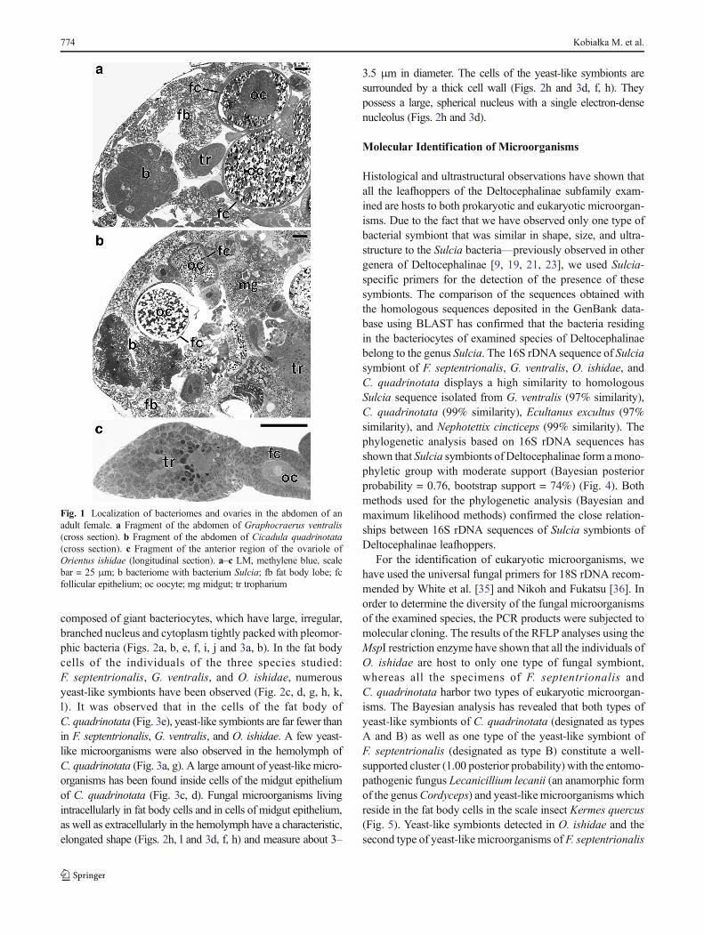
The ultrastructural and histological analyses revealed the pres-ence of two large bacteriomes localized ventro-laterally, onboth sides of the abdomen of each studied species:Fieberiella septentrionalis, Graphocraerus ventralis,Orientus ishidae, and Cicadula quadrinotata. These organsare located between the body wall and the gonads (Fig. 1a,b) and are surrounded by a thin monolayered epitheliumcalled the bacteriome sheath (Figs. 2a, e, i and 3a).Ultrastructural observations did not reveal symbiotic microor-ganisms in the cells of the bacteriome sheath. Bacteriomes are
Dual “Bacterial-Fungal” Symbiosis in Deltocephalinae Leafhoppers (Insecta, Hemiptera, Cicadomorpha:... 773
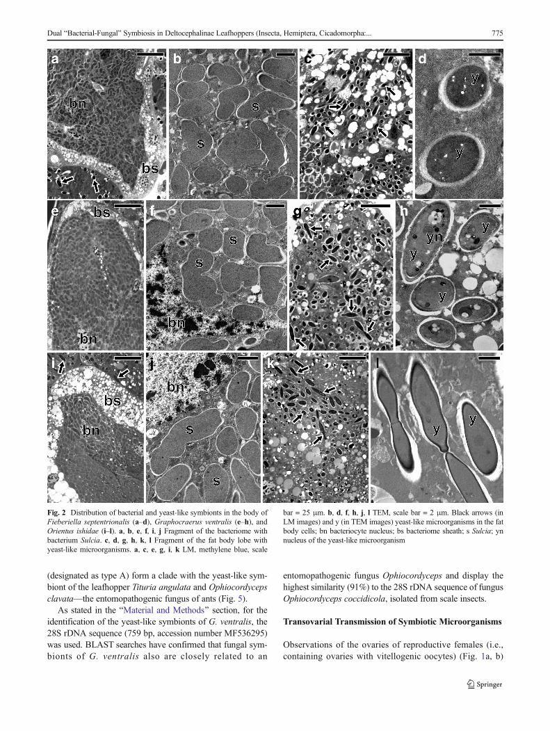
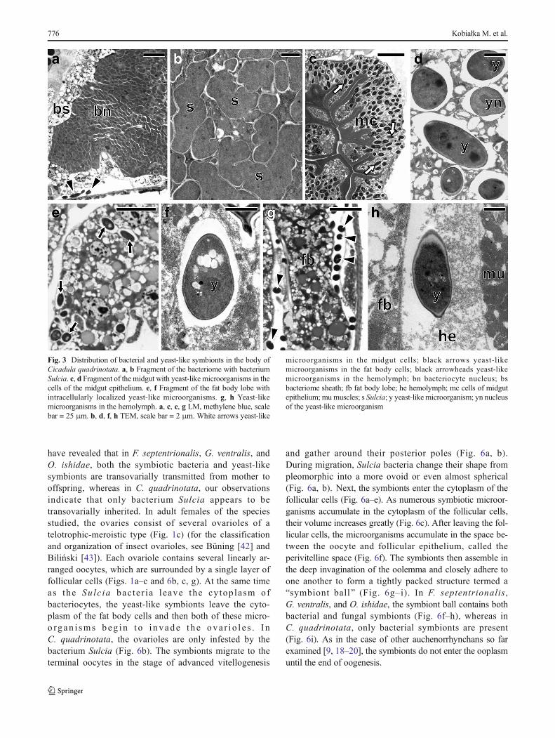
composed of giant bacteriocytes, which have large, irregular,branched nucleus and cytoplasm tightly packed with pleomor-phic bacteria (Figs. 2a, b, e, f, i, j and 3a, b). In the fat bodycells of the individuals of the three species studied:F. septentrionalis, G. ventralis, and O. ishidae, numerousyeast-like symbionts have been observed (Fig. 2c, d, g, h, k,l). It was observed that in the cells of the fat body ofC. quadrinotata (Fig. 3e), yeast-like symbionts are far fewer thanin F. septentrionalis, G. ventralis, and O. ishidae. A few yeast-like microorganisms were also observed in the hemolymph ofC. quadrinotata (Fig. 3a, g). A large amount of yeast-like micro-organisms has been found inside cells of the midgut epitheliumof C. quadrinotata (Fig. 3c, d). Fungal microorganisms livingintracellularly in fat body cells and in cells of midgut epithelium,as well as extracellularly in the hemolymph have a characteristic,elongated shape (Figs. 2h, l and 3d, f, h) and measure about 3–
3.5 μm in diameter. The cells of the yeast-like symbionts aresurrounded by a thick cell wall (Figs. 2h and 3d, f, h). Theypossess a large, spherical nucleus with a single electron-densenucleolus (Figs. 2h and 3d).
Molecular Identification of Microorganisms
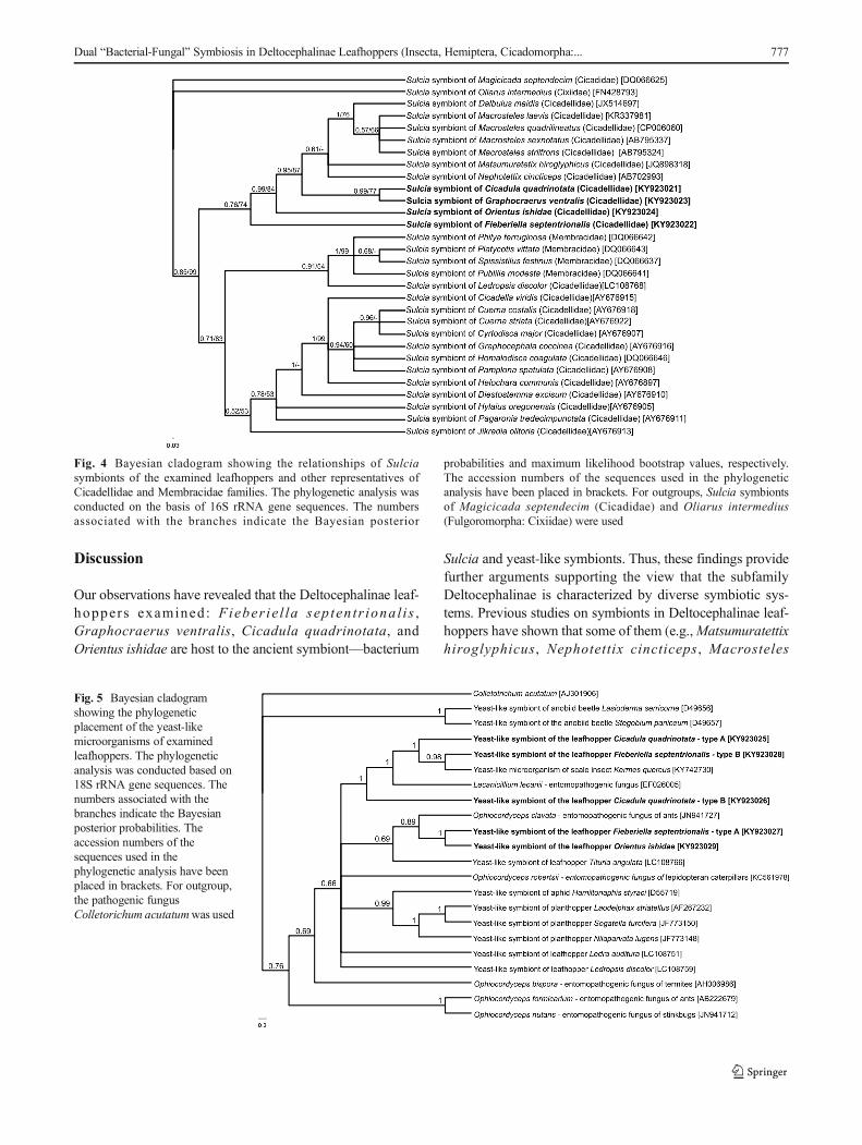
Histological and ultrastructural observations have shown thatall the leafhoppers of the Deltocephalinae subfamily exam-ined are hosts to both prokaryotic and eukaryotic microorgan-isms. Due to the fact that we have observed only one type ofbacterial symbiont that was similar in shape, size, and ultra-structure to the Sulcia bacteria—previously observed in othergenera of Deltocephalinae [9, 19, 21, 23], we used Sulcia-specific primers for the detection of the presence of thesesymbionts. The comparison of the sequences obtained withthe homologous sequences deposited in the GenBank data-base using BLAST has confirmed that the bacteria residingin the bacteriocytes of examined species of Deltocephalinaebelong to the genus Sulcia. The 16S rDNA sequence of Sulciasymbiont of F. septentrionalis, G. ventralis, O. ishidae, andC. quadrinotata displays a high similarity to homologousSulcia sequence isolated from G. ventralis (97% similarity),C. quadrinotata (99% similarity), Ecultanus excultus (97%similarity), and Nephotettix cincticeps (99% similarity). Thephylogenetic analysis based on 16S rDNA sequences hasshown that Sulcia symbionts of Deltocephalinae form amono-phyletic group with moderate support (Bayesian posteriorprobability = 0.76, bootstrap support = 74%) (Fig. 4). Bothmethods used for the phylogenetic analysis (Bayesian andmaximum likelihood methods) confirmed the close relation-ships between 16S rDNA sequences of Sulcia symbionts ofDeltocephalinae leafhoppers.
For the identification of eukaryotic microorganisms, wehave used the universal fungal primers for 18S rDNA recom-mended by White et al. [35] and Nikoh and Fukatsu [36]. Inorder to determine the diversity of the fungal microorganismsof the examined species, the PCR products were subjected tomolecular cloning. The results of the RFLP analyses using theMspI restriction enzyme have shown that all the individuals ofO. ishidae are host to only one type of fungal symbiont,whereas all the specimens of F. septentrionalis andC. quadrinotata harbor two types of eukaryotic microorgan-isms. The Bayesian analysis has revealed that both types ofyeast-like symbionts of C. quadrinotata (designated as typesA and B) as well as one type of the yeast-like symbiont ofF. septentrionalis (designated as type B) constitute a well-supported cluster (1.00 posterior probability) with the entomo-pathogenic fungus Lecanicillium lecanii (an anamorphic formof the genusCordyceps) and yeast-like microorganisms whichreside in the fat body cells in the scale insect Kermes quercus(Fig. 5). Yeast-like symbionts detected in O. ishidae and thesecond type of yeast-like microorganisms of F. septentrionalis
Fig. 1 Localization of bacteriomes and ovaries in the abdomen of anadult female. a Fragment of the abdomen of Graphocraerus ventralis(cross section). b Fragment of the abdomen of Cicadula quadrinotata(cross section). c Fragment of the anterior region of the ovariole ofOrientus ishidae (longitudinal section). a–c LM, methylene blue, scalebar = 25 μm; b bacteriome with bacterium Sulcia; fb fat body lobe; fcfollicular epithelium; oc oocyte; mg midgut; tr tropharium
774 Kobiałka M. et al.
(designated as type A) form a clade with the yeast-like sym-biont of the leafhopper Tituria angulata and Ophiocordycepsclavata—the entomopathogenic fungus of ants (Fig. 5).
As stated in the BMaterial and Methods^ section, for theidentification of the yeast-like symbionts of G. ventralis, the28S rDNA sequence (759 bp, accession number MF536295)was used. BLAST searches have confirmed that fungal sym-bionts of G. ventralis also are closely related to an
entomopathogenic fungus Ophiocordyceps and display thehighest similarity (91%) to the 28S rDNA sequence of fungusOphiocordyceps coccidicola, isolated from scale insects.
Transovarial Transmission of Symbiotic Microorganisms
Observations of the ovaries of reproductive females (i.e.,containing ovaries with vitellogenic oocytes) (Fig. 1a, b)
Fig. 2 Distribution of bacterial and yeast-like symbionts in the body ofFieberiella septentrionalis (a–d), Graphocraerus ventralis (e–h), andOrientus ishidae (i–l). a, b, e, f, i, j Fragment of the bacteriome withbacterium Sulcia. c, d, g, h, k, l Fragment of the fat body lobe withyeast-like microorganisms. a, c, e, g, i, k LM, methylene blue, scale
bar = 25 μm. b, d, f, h, j, l TEM, scale bar = 2 μm. Black arrows (inLM images) and y (in TEM images) yeast-like microorganisms in the fatbody cells; bn bacteriocyte nucleus; bs bacteriome sheath; s Sulcia; ynnucleus of the yeast-like microorganism
Dual “Bacterial-Fungal” Symbiosis in Deltocephalinae Leafhoppers (Insecta, Hemiptera, Cicadomorpha:... 775
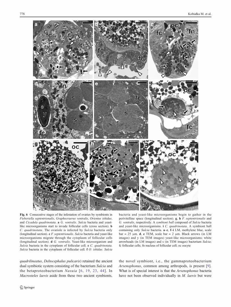
have revealed that in F. septentrionalis, G. ventralis, andO. ishidae, both the symbiotic bacteria and yeast-likesymbionts are transovarially transmitted from mother tooffspring, whereas in C. quadrinotata, our observationsindicate that only bacterium Sulcia appears to betransovarially inherited. In adult females of the speciesstudied, the ovaries consist of several ovarioles of atelotrophic-meroistic type (Fig. 1c) (for the classificationand organization of insect ovarioles, see Büning [42] andBiliński [43]). Each ovariole contains several linearly ar-ranged oocytes, which are surrounded by a single layer offollicular cells (Figs. 1a–c and 6b, c, g). At the same timeas the Sulc ia bac te r i a l eave the cy top lasm ofbacteriocytes, the yeast-like symbionts leave the cyto-plasm of the fat body cells and then both of these micro-o rg an i sms beg i n t o i nvade t h e ova r i o l e s . I nC. quadrinotata, the ovarioles are only infested by thebacterium Sulcia (Fig. 6b). The symbionts migrate to theterminal oocytes in the stage of advanced vitellogenesis
and gather around their posterior poles (Fig. 6a, b).During migration, Sulcia bacteria change their shape frompleomorphic into a more ovoid or even almost spherical(Fig. 6a, b). Next, the symbionts enter the cytoplasm of thefollicular cells (Fig. 6a–e). As numerous symbiotic microor-ganisms accumulate in the cytoplasm of the follicular cells,their volume increases greatly (Fig. 6c). After leaving the fol-licular cells, the microorganisms accumulate in the space be-tween the oocyte and follicular epithelium, called theperivitelline space (Fig. 6f). The symbionts then assemble inthe deep invagination of the oolemma and closely adhere toone another to form a tightly packed structure termed aBsymbiont ball^ (Fig. 6g–i). In F. septentrionalis,G. ventralis, and O. ishidae, the symbiont ball contains bothbacterial and fungal symbionts (Fig. 6f–h), whereas inC. quadrinotata, only bacterial symbionts are present(Fig. 6i). As in the case of other auchenorrhynchans so farexamined [9, 18–20], the symbionts do not enter the ooplasmuntil the end of oogenesis.
Fig. 3 Distribution of bacterial and yeast-like symbionts in the body ofCicadula quadrinotata. a, b Fragment of the bacteriome with bacteriumSulcia. c, d Fragment of the midgut with yeast-like microorganisms in thecells of the midgut epithelium. e, f Fragment of the fat body lobe withintracellularly localized yeast-like microorganisms. g, h Yeast-likemicroorganisms in the hemolymph. a, c, e, g LM, methylene blue, scalebar = 25 μm. b, d, f, h TEM, scale bar = 2 μm. White arrows yeast-like
microorganisms in the midgut cells; black arrows yeast-likemicroorganisms in the fat body cells; black arrowheads yeast-likemicroorganisms in the hemolymph; bn bacteriocyte nucleus; bsbacteriome sheath; fb fat body lobe; he hemolymph; mc cells of midgutepithelium; mumuscles; s Sulcia; y yeast-like microorganism; yn nucleusof the yeast-like microorganism
776 Kobiałka M. et al.
Discussion
Our observations have revealed that the Deltocephalinae leaf-hoppers examined: Fieber ie l la sep ten tr ional i s ,Graphocraerus ventralis, Cicadula quadrinotata, andOrientus ishidae are host to the ancient symbiont—bacterium
Sulcia and yeast-like symbionts. Thus, these findings providefurther arguments supporting the view that the subfamilyDeltocephalinae is characterized by diverse symbiotic sys-tems. Previous studies on symbionts in Deltocephalinae leaf-hoppers have shown that some of them (e.g.,Matsumuratettixhiroglyphicus, Nephotettix cincticeps, Macrosteles
Fig. 5 Bayesian cladogramshowing the phylogeneticplacement of the yeast-likemicroorganisms of examinedleafhoppers. The phylogeneticanalysis was conducted based on18S rRNA gene sequences. Thenumbers associated with thebranches indicate the Bayesianposterior probabilities. Theaccession numbers of thesequences used in thephylogenetic analysis have beenplaced in brackets. For outgroup,the pathogenic fungusColletorichum acutatumwas used
Fig. 4 Bayesian cladogram showing the relationships of Sulciasymbionts of the examined leafhoppers and other representatives ofCicadellidae and Membracidae families. The phylogenetic analysis wasconducted on the basis of 16S rRNA gene sequences. The numbersassociated with the branches indicate the Bayesian posterior
probabilities and maximum likelihood bootstrap values, respectively.The accession numbers of the sequences used in the phylogeneticanalysis have been placed in brackets. For outgroups, Sulcia symbiontsof Magicicada septendecim (Cicadidae) and Oliarus intermedius(Fulgoromorpha: Cixiidae) were used
Dual “Bacterial-Fungal” Symbiosis in Deltocephalinae Leafhoppers (Insecta, Hemiptera, Cicadomorpha:... 777
quadrilineatus, Deltocephalus pulicaris) retained the ancientdual symbiotic system consisting of the bacterium Sulcia andthe betaproteobacterium Nasuia [6, 19, 23, 44]. InMacrosteles laevis aside from these two ancient symbionts,
the novel symbiont, i.e., the gammaproteobacteriumArsenophonus, common among arthropods, is present [9].What is of special interest is that the Arsenophonus bacteriahave not been observed individually in M. laevis but were
Fig. 6 Consecutive stages of the infestation of ovaries by symbionts inFieberiella septentrionalis, Graphocraerus ventralis, Orientus ishidae,and Cicadula quadrinotata. a G. ventralis. Sulcia bacteria and yeast-like microorganisms start to invade follicular cells (cross section). bC. quadrinotata. The ovariole is infected by Sulcia bacteria only(longitudinal section). c F. septentrionalis. Sulcia bacteria and yeast-likemicroorganisms migrate through the cytoplasm of follicular cells(longitudinal section). d G. ventralis. Yeast-like microorganism andSulcia bacteria in the cytoplasm of follicular cell. e C. quadrinotata.Sulcia bacteria in the cytoplasm of follicular cell. f O. ishidae. Sulcia
bacteria and yeast-like microorganisms begin to gather in theperivitelline space (longitudinal section). g, h F. septentrionalis andG. ventralis, respectively. A symbiont ball composed of Sulcia bacteriaand yeast-like microorganisms. i C. quadrinotata. A symbiont ballcontaining only Sulcia bacteria. a–c, f–i LM, methylene blue, scalebar = 25 μm. d, e TEM, scale bar = 2 μm. Black arrows (in LMimages) and y (in TEM images) yeast-like microorganisms; whitearrowheads (in LM images) and s (in TEM images) bacterium Sulcia;fc follicular cells; fn nucleus of follicular cell; oc oocyte
778 Kobiałka M. et al.
always found internalized in the cells of Sulcia bacteria. Itshould furthermore be stressed that a similar phenomenon ofnested symbiosis has been reported in Pseudococcidae mealy-bugs [45–51], as well as in the leafhopper Cicadella viridis[18]. Kobiałka and co-workers [9] suggest that the internal-ized Arsenophonus bacterium, similar to Sodalis-like bacteri-um in Pseudococcidae mealybugs [45] and in the leafhopperC. viridis [18], represents a newly acquired symbiont inM. laevis, which cannot yet be transmitted to offspring on itsown. The co-occurrence inM. laevis of two ancient symbiontsand the symbiont of a more recent origin indicates that thebetaproteobacterial symbiont has not yet been eliminated,whereas the new symbiont has already been acquired. Onthe other hand, a lack of the ancient betaproteobacteriumNasuia in S. titanus, F. septentrionalis, G. ventralis,C. quadrinotata, and O. ishidae [27, this study] suggests thatin some species of Deltocephalinae leafhoppers, this bacteri-um has been already eliminated and replaced by yeast-likesymbionts. It should be stressed that a similar evolutionaryscenario has been presented by Nishino and co-workers[10], who studied the symbiotic systems of three membersof the Ledrinae leafhoppers. Similarly, a lack of Nasuia insidethe bacteriomes of the Deltocephalinae Dalbulus maidis wasreported byBrentassi and co-workers [21], who suggested thatthe leafhopper may have lost some of their symbiotic speciesduring phylogeny.
The results of earlier studies using a paraffin technique[14, 16], and more recent ultrastructural and molecular anal-yses [this study], indicate that the yeast-like symbionts arerather uncommon in members of Deltocephalinae leafhop-pers. The use of molecular phylogenetic analyses revealedthat yeast-like symbionts residing in F. septentrionalis,G. ventralis, C. quadrinotata, and O. ishidae (see Fig. 5)are closely rela ted to the fungi from the generaOphiocordyceps and Cordyceps, which include a widelydistributed fungal entomopathogens [52]. These findingsthus indicate that the fungal entomopathogens, in contrastwith the ancient symbiont, i.e., bacterium Sulcia, infectedthe ancestors of studied species independently from eachother. Then, the acquired fungi evolved into mutualisticsymbionts. Suh and co-workers [53], who studied theyeast-like symbionts of planthoppers, postulated that duringco-evolution, the insect-fungus interaction changed,resulting in modifications in the morphology, life cycle,and physiology of fungal entomoparasites. These fungi losttheir previous filamentous ascomycete form and remainedonly in a yeast-like form. It is worth mentioning that besidesthe alterations in the morphology of yeast-like symbiontsresiding in insects, changes within their genome also oc-curred. Recently, Fan and co-workers [54] have shown thatduring the co-evolution of yeast-like symbionts and its hostinsect, the brown planthopper Nilaparvata lugens, the lossof some genes of yeast-like microorganisms took place.
So far, the fungal symbionts related to entomopathogenshave been found in aphids [55], in planthoppers from theDelphacidae family [28, 53], in anobiid beetles [56], in scaleinsects from the Kerriidae, Dactylopiidae, and Kermesidaefamilies [57–59], and in leafhoppers from the Ledrinae sub-family [10]. In most of the above cases, the yeast-like micro-organisms are the predominant symbionts which reside intheir host insects.
In L. discolor, S. titanus, F. septentrionalis, G. ventralis,and O. ishidae, numerous yeast-like microorganisms are lo-calized in cells of the fat body [10, 27, this study], whereas inC. quadrinotata [this study], they occur both in the cells of themidgut epithelium (in a large amount) and in the fat body cellsand hemolymph (in a small amount). It should be stressed thatBuchner [14] found yeast-like microorganisms in the cells ofmidgut epithelium of C. quadrinotata but did not observethese microorganisms in the fat body. There are two possibleexplanations for this discrepancy: (1) the yeast-like symbiontsmay be present in the fat body cells and hemolymph of onlycertain populations of C. quadrinotata and (2) on account ofthe paraffin technique used, Buchner might have overlookedthese microorganisms. The function of yeast-like microorgan-isms residing in the fat body and hemolymph ofC. quadrinotata remains unknown; however, as these micro-organisms are present in all individuals ofC. quadrinotata anddo not have a negative effect on the growth and developmentof the host insects, it may be possible that they represent anadditional, newly acquired symbiont. It can also not be ruledout that the latter microorganisms may be facultative symbi-onts residing in the examined population of C. quadrinotata.Thus, to elucidate the biological role of these microorganisms,further experiments are needed.
Yeast-like microorganisms and bacterial symbionts inDeltocephalinae leafhoppers: F. septentrionalis, G. ventralis,and O. ishidae [this study], as well as in S. titanus [27], aretransmitted transovarially. It was observed that in all the aboveinsects, the symbionts migrate from the fat body towards theovaries; then, via the cytoplasm of the follicular cells sur-rounding the posterior pole of the terminal oocytes, they enterthe space between the oocyte and follicular epithelium. Itshould be stressed that the same manner of transmission ofboth the bacterial and fungal symbionts has been observed inother auchenorrhynchans [9, 14, 19, 27, 60], which confirmsthe earlier observations that these hemipterans, in spite of alarge diversity of symbionts, developed a uniform mode ofsymbiont transmission [9, 18–21]. In contrast to the mode ofsymbiont transmission mentioned above, the style of inheri-tance of yeast-like microorganisms in C. quadrinotata re-mains unknown. Our observations clearly indicate (seeFig. 6i) that in this species, only bacterial symbionts enterthe ovaries. Thus, the yeast-like microorganismsmust be trans-mitted via a different route. Buchner [14] hypothesized thatthe yeast-like microorganisms of C. quadrinotata, similarly
Dual “Bacterial-Fungal” Symbiosis in Deltocephalinae Leafhoppers (Insecta, Hemiptera, Cicadomorpha:... 779
to the gut bacteria in heteropterans and yeast-like symbionts inbeetles, may contaminate the egg surface. Newly hatched lar-vae consuming symbionts become infected with them. Thelack of a mechanism ensuring the transovarial transmissionof yeast-like symbionts in C. quadrinotata indicates that thesesymbionts were more recently acquired than the bacterial sym-bionts and yeast-like symbionts of other Deltocephalinaeleafhoppers.
The presence of numerous yeast-like symbionts in the fatbody or midgut epithelium of the species studied suggests thatthese microorganisms have an important metabolic function totheir hosts. Data in the literature indicate that yeast-like symbi-onts may play varying roles; e.g., they may be engaged in thedetoxification of food compounds in various beetles, termites,and wood wasps [61] and in amino acid metabolism, sterol bio-synthesis, and nitrogen recycling in planthoppers [54, 62–64].Nishino and co-workers [10] hypothesized that the large size ofthe genome of yeast-like symbionts, in comparison with thesmall genome size of bacterial symbionts, indicates that thesefungal symbionts may play a broader biological function to thehost insect. Therefore, in order to examine the role of yeast-likemicroorganisms, further genomic studies in combination withinsect rearing and symbiont manipulation are required.
2. Moran NA, McCutcheon JP, Nakabachi A (2008) Genomics andevolution of heritable bacterial symbionts. Annu Rev Genet 42:165–190
3. Douglas AE (2011) Lessons from studying insect symbioses. CellHost Microbe 10:359–367
4. Takiya DM, Tran P, Dietrich CH, Moran NA (2006) Co-cladogenesis spanning three phyla: leafhoppers (Insecta:Hemiptera: Cicadellidae) and their dual bacterial symbionts. MolEcol 15:4175–4191
5. Urban J, Cryan J (2012) Two ancient bacterial endosymbionts havecoevolved with the planthoppers (Insecta: Hemiptera: Fulgoroidea).BMC Evol Biol 12:87
6. Bennett GM, Moran NA (2013) Small, smaller, smallest: the originand evolution of ancient dual symbioses in a phloem-feeding insect.Genome Biol Evol 5:1675–1688
7. Koga R, Bennett GM, Cryan JR, Moran NA (2013) Evolutionaryreplacement of symbionts in an ancient and diverse insect lineage.Environ Microbiol 15:2073–2081
8. Koga R, Moran NA (2014) Swapping symbionts in spittlebugs:evolutionary replacement of a reduced genome symbiont. ISME J8:1237–1246
9. Kobiałka M, Michalik A, Walczak M, Junkiert Ł, Szklarzewicz T(2016) Sulcia symbiont of the leafhopper Macrosteles laevis(R ibau t , 1927) ( Insec t a , Hemip te r a , C i cade l l i dae :Deltocephalinae) harbors Arsenophonus bacteria. Protoplasma253:903–912
10. Nishino T, Tanahashi M, Lin CP, Koga R, Fukatsu T (2016) Fungaland bacterial endosymbionts of eared leafhoppers of the subfamilyLedrinae (Hemiptera: Cicadellidae). Appl Entomol Zool 51:465–477
11. Douglas AE (1998) Nutritional interactions in insect-microbialsymbioses: aphids and their symbiotic bacteria Buchnera. AnnuRev Entomol 43:17–37
12. Baumann P (2005) Biology of bacteriocyte-associated endosymbi-onts of plant sup-sucking insects. Annu Rev Microbiol 59:155–189
13. Duncan RP, Husnik F, Van Leuven JT, Gilbert DG, Davalos LM,McCutcheon JP, Wilson ACC (2014) Dynamic recruitment of ami-no acid transporters to the insect-symbiont interface. Mol Ecol 23:1608–1623
14. Buchner P (1965) Endosymbiosis of animals with plant microor-ganisms. Interscience, New York
15. Vogel KJ, Moran NA (2013) Functional and evolutionary analysisof the genome of an obligate fungal symbiont. GenomeBiol Evol 5:891–904
16. Müller HJ (1962) Neuere Vorstellungen über Verbreitung undPhylogenie der Endosymbiosen der Zikaden. Z Morphol ÖkolTiere 51:190–210
17. Ishii Y, Matsuura Y, Kakizawa S, Nikoh N, Fukatsu T (2013)Diversity of bacterial endosymbionts associated with Macrostelesleafhoppers vectoring phytopathogenic phytoplasmas. ApplEnviron Microbiol 79:5013–5022
18. Michalik A, JankowskaW, KotM, Gołas A, Szklarzewicz T (2014)Symbiosis in the green leafhopper, Cicadella viridis (Hemiptera,Cicadellidae). Association in statu nascendi? Arthropod StructDev 43:579–587
19. Kobiałka M, Michalik A, Walczak M, Junkiert Ł, Szklarzewicz T(2015) Symbiotic microorganisms of the leafhopper Deltocephaluspulicaris (FALLÉN, 1806) (Insecta, Hemiptera, Cicadellidae:Deltocephalinae): molecular characterization, ultrastructure andtransovarial transmission. Pol J Entomol 84:155–162
20. Szklarzewicz T, Grzywacz B, Szwedo J, Michalik A (2016)Bacterial symbionts of the leafhopper Evacanthus interruptus(Linnaeus, 1758) (Insecta, Hemiptera , Cicadell idae:Evacanthinae). Protoplasma 253:379–391
21. Brentassi ME, Franco E, Balatti P, Medina R, Bernabei F, DeRemes Lenicov AMM (2017) Bacteriomes of the corn leafhopper,
780 Kobiałka M. et al.
Acknowledgements We are greatly indebted to M.Sc. Ada Jankowskafor her skilled technical assistance. Ultrastructural observations were car-ried out using the JEOL 2100 transmission electron microscope in theLaboratory of Microscopy, Department of Cell Biology and Imaging,Institute of Zoology and Biomedical Research, Jagiellonian University.We would like to thank the anonymous reviewers for their constructivesuggestions and comments which helped us to improve the manuscript.
Funding This study was funded by the research grant 2015/17/N/NZ8/01573 from the National Science Centre, Poland, to Michał Kobiałka.
Compliance with Ethical Standards
Ethical Approval All applicable international, national, and institu-tional guidelines for the animal use were followed.
Conflict of Interest The authors declare that they have no conflicts ofinterest.
Open Access This article is distributed under the terms of the CreativeCommons At t r ibut ion 4 .0 In te rna t ional License (h t tp : / /creativecommons.org/licenses/by/4.0/), which permits unrestricted use,distribution, and reproduction in any medium, provided you give appro-priate credit to the original author(s) and the source, provide a link to theCreative Commons license, and indicate if changes were made.
References
1. Dale C,Moran NA (2006)Molecular interactions between bacterialsymbionts and their hosts. Cell 126:453–465
Dalbulus maidis (DeLong & Wolcott, 1923) (Insecta, Hemiptera,Cicadellidae: Deltocephalinae) harbor Sulcia symbiont: molecularcharacterization, ultrastructure and transovarial transmission.Protoplasma 254:1421–1429
22. Moran NA, Tran P, Gerardo NM (2005) Symbiosis and insect di-versification: an ancient symbiont of sap-feeding insects from thebacterial phylum Bacteroidetes. Appl Environ Microbiol 71:8802–8810
23. Noda H, Watanabe K, Kawai S, Yukuhiro F, Miyoshi T, TomizawaM, Koizumi Y, Nikoh N, Fukatsu T (2012) Bacteriome-associatedendosymbionts of the green rice leafhopper Nephotettix cincticeps(Hemiptera: Cicadellidae). Appl Entomol Zool 47:217–225
24. McCutcheon JP, Moran NA (2010) Functional convergence in re-duced genomes of bacterial symbionts spanning 200 million yearsof evolution. Genome Biol Evol 2:708–718
25. Wu D, Daugherty SC, Van Aken SE, Pai GH, Watkins KL, KhouriH (2006) Metabolic complementarity and genomics of the dualsymbiosis of sharpshooters. PLoS Biol 4:e188
26. McCutcheon JP, McDonald BR, Moran NA (2009) Convergentevolution of metabolic roles in bacterial co-symbionts of insects.Proc Natl Acad Sci U S A 106:15394–15399
27. Sacchi L, GenchiM, Clementi E, Bigliardi E, Avanzatti AM, PajoroM, Negri I, Marzorati M, Gonella E, Alma A, Daffonchio D, BandiC (2008) Multiple symbiosis in the leafhopper Scaphoideus titanus(Hemiptera: Cicadellidae): details of transovarial transmission ofCardinium sp. and yeast-like endosymbionts. Tissue Cell 40:231–242
28. NodaH, Nakashima N, KoizumiM (1995) Phylogenetic position ofyeast-like symbiotes of rice planthoppers based on partial 18SrDNA sequences. Insect Biochem Mol Biol 25:639–646
29. Zahniser JN, Dietrich CH (2013) A review of the tribes ofDeltocephalinae (Hemiptera: Auchenorrhyncha: Cicadellidae).Eur J Taxon 45:1–211
30. Cheung WW-K, Purcell AH (1999) Invasion of bacteroids andBEV bacterium into oocytes of the leafhopper EuscelidiusvariegatusKirschbaum (Homoptera: Cicadellidae): an electron mi-croscopic study. Zool Stud 38:69–75
31. Klejdysz T, Zwolińska A, Walczak M, Kobiałka M (2017) Firstrecord of a potential pest Orientus ishidae (Matsumura 1902)(Hemiptera: Cicadellidae) in Poland. J Plant Prot Res. https://doi.org/10.1515/jppr-2017-0014
32. Gębicki C, Świerczewski D, Szwedo J (2013) Planthoppers andleafhoppers of Poland (Hemiptera: Fulgoromorpha etCicadomorpha). Systematics. Check-list. Bionomy. Ann UppSiles Mus Bytom Entomol 21–22:5–259
33. Lessio F, Picciau L, Gonella E,Mandrioli M, Tota F, AlmaA (2016)The mosaic leafhopper Orientus ishidae: host plants, spatial distri-bution, infectivity, and transmission of 16SrV phytoplasma tovines. Bull Insectol 69:277–289
34. Rosenberger DA, Jones AL (1978) Leafhopper vectors of the peachX disease pathogen and its seasonal transmission from chokecherry.Phytopathology 68:782–790
35. White TJ, Bruns T, Lee S, Taylor J (1990) Amplification and directsequencing of fungal ribosomal RNA genes for phylogenetics. In:Innis MA, Gelfand DH, Sninsky JJ, White TJ (eds) PCR protocols:a guide to methods and applications. Academic Press, San Diego,pp. 315–322
36. Nikoh N, Fukatsu T (2000) Interkingdom host jumping under-ground: phylogenetic analysis of entomoparasitic fungi of the genusCordyceps. Mol Biol Evol 17:629–638
37. Hall TA (1999) BIOEDIT: an user-friendly biological sequencealignment editor and analysis program for Windows 95/98/NT.Nucleic Acids Symp Ser 41:95–98
38. Thompson JD, Gibson TJ, Plewniak F, Jeanmougin F, Higgins DG(1997) The ClustalX windows interface: flexible strategies for
multiple sequence alignment aided by quality analysis tools.Nucleic Acids Res 25:4876–4882
39. Ronquist F, Teslenko M, Van der Mark P, Ayres D, Darling A,Höhna S, Larget B, Liu L, Suchard MA, Huelsenbeck JP (2012)MrBayes 3.2: efficient Bayesian phylogenetic inference and modelselection across a large model space. Syst Biol 61:539–542
40. Tamura K, Stecher G, Peterson D, Filipski A, Kumar S (2013)MEGA6: Molecular Evolutionary Genetics Analysis version 6.0.Mol Biol Evol 30:2725–2729
41. Rambaut A (2009) FigTree v1. 4.0: tree figure drawing tool.Available: http://tree.bio.ed.ac.uk/software/figtree/. Accessed 2July 2014
42. Büning J (1994) The ovary of Ectognatha, the insects s. str. In:Büning J (ed) The insect ovary: ultrastructure, previtellogenicgrowth and evolution. Chapman and Hall, London, pp. 31–305
43. Biliński S (1998) Introductory remarks. Folia Histochem Cytobiol3:143–145
44. Wangkeeree J, Miller TA, Hanboonsong Y (2011) Predominantbacteria symbionts in the leafhopper Matsumuratettixhiroglyphicus—the vector of sugarcane white leaf phytoplasma.Bull Insectol 64:215–216
45. von Dohlen CD, Kohler S, Alsop ST, McManus WR (2001)Mealybug β-proteobacterial endosymbionts contain γ-proteobacterial symbionts. Nature 412:433–435
46. Thao ML, Gullan PJ, Baumann P (2002) Secondary (γ-proteobacteria) endosymbionts infect the primary (β-proteobacteria) endosymbionts of mealybugs multiple times andcoevolve with their host. Appl Environ Microbiol 68:3190–3197
47. Kono M, Koga R, Shimada M, Fukatsu T (2008) Infection dynam-ics of coexisting beta- and gammaproteobacteria in the nested en-dosymbiotic system of mealybugs. Appl Environ Microbiol 74:4175–4184
48. McCutcheon JP, von Dohlen CD (2011) An interdependent meta-bolic patchwork in the nested symbiosis of mealybugs. Curr Biol21:1366–1372
49. Gatehouse LN, Sutherland P, Forgie SA, Kaji R, Christeller JT(2011) Molecular and histological characterization of primary(Betaproteobacteria) and secondary (Gammaproteobacteria) endo-symbionts of three mealybug species. Appl Environ Microbiol 78:1187–1197
50. Husnik F, Nikoh N, Koga R, Ross L, Duncan RP, Fujie M, TanakaM, Satoh N, Bachtrog D, Wilson ACC, von Dohlen CD, Fukatsu T,McCutcheon JP (2013) Horizontal gene transfer from diverse bac-teria to an insect genome enables a tripartite nested mealybug sym-biosis. Cell 153:1567–1578
51. Szabo G, Schulz F, Toenshoff ER, Volland J-M, Finkel OM, BelkinS, HornM (2016) Convergent patterns in the evolution ofmealybugsymbioses involving different intrabacterial symbionts. ISME J 11:715–726
52. SungGH, Hywel-Jones NL, Sung JM, Luangsa-Ard JJ, Shrestha B,Spatafora JW (2007) Phylogenetic classification of Cordyceps andthe Clavicipitaceous fungi. Stud Mycol 57:5–59
53. Suh SO, Noda H, Blackwell M (2001) Insect symbiosis: derivationof yeast-like endosymbionts within an entomopathogenic filamen-tous lineage. Mol Biol Evol 18:995–1000
54. Fan HW, Noda H, Xie HQ, Suetsugu Y, Zhu QH, Zhang CX (2015)Genomic analysis of an ascomycete fungus from the riceplanthopper reveals how it adapts to an endosymbiotic lifestyle.Genome Biol Evol 7:2623–2634
55. Fukatsu T, Ishikawa H (1996) Phylogenetic position of yeast-likesymbiont of Hamiltonaphis styraci (Homoptera, Aphididae) basedon 18S rDNA sequence. Insect Biochem Mol Biol 26:383–388
56. Noda H, Kodama K (1996) Phylogenetic position of yeast-likeendosymbionts of anobiid beetles. Appl Environ Microbiol 62:162–167
Dual “Bacterial-Fungal” Symbiosis in Deltocephalinae Leafhoppers (Insecta, Hemiptera, Cicadomorpha:... 781
57. Vashishtha A, Sharma KK, Lakhanpaul S (2011) Co-existence,phylogeny and putative role ofWolbachia and yeast-like symbiont(YLS) in Kerria lacca (Kerr). Curr Microbiol 63:206–211
58. Vera-Ponce de León A, Sanchez-Flores A, Rosenblueth M,Martínez-Romero E (2016) Fungal community associated withDactylopius (Hemiptera: Coccoidea: Dactylopiidae) and its rolein uric acid metabolism. Front Microbiol 7:954
59. Podsiadło E, Michalik A, Szklarzewicz T (2016) Observations onmicroorganisms infecting Kermes quercus (Linnaeus). XIVInternational Symposium on Scale Insect Studies ISSIS, 13-16.06.2013, Catania, Italy, Book of Abstracts, pp 88
60. Michalik A, Jankowska W, Szklarzewicz T (2009) Ultrastructureand transovarial transmission of endosymbiotic microorganisms inConomelus anceps and Metcalfa pruinosa (Insecta, Hemiptera,Fulgoromorpha). Folia Biol (Kraków) 57:131–137
61. Dowd PF (1992) Insect fungal symbionts: a promising source ofdetoxifying enzymes. J Ind Microbiol 9:149–161
62. Wan PJ, Yang L, Wang WX, Fan JM, Fu Q, Li GQ (2014)Constructing the major biosynthesis pathways for amino acids inthe brown planthopper, Nilaparvata lugens Stål (Hemiptera:Delphacidae), based on the transcriptome data. Insect Mol Biol23:152–164
63. Sasaki T, Kawamura M, Ishikawa H (1996) Nitrogen recycling inthe brown planthopper, Nilaparvata lugens: involvement of yeast-like endosymbionts in uric acid metabolism. J Insect Physiol 42:125–129
64. Noda H, Koizumi Y (2003) Sterol biosynthesis by symbiotes: cy-tochrome P450 sterol C-22 desaturase genes from yeast-like sym-biotes of rice planthoppers and anobiid beetles. Insect BiochemMolBiol 33:649–658
782 Kobiałka M. et al.