Embed Size (px)
Citation preview
Clinical Focuson primary immunodeficiencies
ChronicGranulomatousDisease
IDF MEDICAL ADVISORY COMMITTEE
Rebecca Buckley, MD - ChairDuke University School of Medicine, Durham, NC
Zuhair Ballas, MDUniversity of Iowa, Iowa City, IA
Mark Ballow, MDState University of New York, Buffalo, NY
R. Michael Blaese, MDConsulting Medical Director IDF, Towson, MD
Francisco Bonilla, MD, PhDBoston Children’s Hospital, Boston, MA
Mary Ellen Conley, MDUniversity of Tennessee, Memphis, TN
Charlotte Cunningham-Rundles, MD, PhDMt. Sinai Medical Center, New York, NY
Alexandra Filipovich, MDCincinnati Children’s Hospital, Cincinnati, OH
Thomas Fleisher, MDNational Institutes of Health, Bethesda, MD
Ramsay Fuleihan, MDChildren’s Memorial Hospital, Chicago, IL
Erwin Gelfand, MDNational Jewish Medical and Research Center,Denver, CO
Vivian Hernandez-Trujillo, MDMiami Children’s Hospital, Miami, FL
Steven Holland, MDNational Institutes of Health, Bethesda, MD
Richard Hong, MDBiomosaics, Burlington, VT
Howard Lederman, MD, PhDJohns Hopkins Hospital, Baltimore, MD
Harry Malech, MDNational Institutes of Health, Bethesda, MD
Stephen Miles, MDAll Seasons Allergy, Asthma & Immunology,The Woodlands, TX
Luigi Notarangelo, MDBoston Children’s Hospital, Boston, MA
Hans Ochs, MDSeattle Children’s Hospital, Seattle, WA
Jordan Orange, MD, PhDTexas Children’s Hospital, Houston, TX
Jennifer Puck, MDUniversity of California, San Francisco,San Francisco, CA
John Routes, MDChildren’s Hospital of Wisconsin, Milwaukee, WI
William Shearer, MD, PhDTexas Children’s Hospital, Houston, TX
E. Richard Stiehm, MDUCLA School of Medicine, Los Angeles, CA
Kathleen Sullivan, MD, PhDChildren’s Hospital of Philadelphia, Philadelphia, PA
Troy Torgerson, MD, PhDSeattle Children’s Hospital, Seattle, WA
Jerry Winkelstein, MDBaltimore, MD
ISSUE 15 | JUNE 2013
AUTHORS
Jennifer W. Leiding, MD
Harry L. Malech, MD
Steven M. Holland, MD
This publication was made possible by an educational grant from
This book contains general medical information which cannot be applied safely to any individual case.
Medical knowledge and practice can change rapidly. Therefore, this book should not be used as a substitute
for professional medical advice.
Copyright 2013 by Immune Deficiency Foundation, USA.
Readers may redistribute this article to other individuals for non-commercial use, provided that the text,
html codes, and this notice remain intact and unaltered in any way. Clinical Focus on Primary
Immunodeficiencies: Chronic Granulomatous Disease may not be resold, reprinted or redistributed for
compensation of any kind without prior written permission from Immune Deficiency Foundation. If you have
any questions about permission, please contact: Immune Deficiency Foundation, 40 West Chesapeake
Avenue, Suite 308, Towson, MD 21204, USA; or by telephone at 1-800-296-4433.
www.primaryimmune.org
www.vidararx.com
Immune Deficiency Foundation: Clinical Focus / 1
Authors1 Jennifer W. Leiding, MD2 Harry L. Malech, MD 3 Steven M. Holland, MD
1University of South Florida, Department of Pediatrics,
Division of Allergy, Immunology, and Rheumatology;
Laboratories of 2Host Defenses and 3Clinical Infectious
Diseases, NIAID, NIH
The authors have no relationships to disclose that could
represent or be perceived to represent a conflict of
interest.
Correspondence to:
Jennifer W. Leiding, MD
Children’s Research Institute
140 – 7th Avenue South, Box 9680
St. Petersburg, Florida 33701
History First described in 19571,2 and further characterized in
19593, chronic granulomatous disease (CGD), termed fatal
granulomatous disease of childhood initially, was
characterized by recurrent infections associated with
hypergamma-globulinemia. Over the last six decades, CGD
has evolved from an immunodeficiency associated with
severe infections with poor prognosis to a disease with
effective management and high survival.
Molecular MechanismsCGD is primarily a defect in innate immunity caused by
defective phagocyte NADPH oxidase enzyme, resulting in
the failure of neutrophils and monocytes to produce
superoxide (O2-.) when stimulated upon encountering
bacterial or fungal pathogens, or a variety of soluble
inflammatory stimuli. Within the neutrophil phagosome,
superoxide combines with water to produce hydrogen
peroxide (H2O2) which provides most of the microbicidal
activity from this oxidative burst. The functional NADPH
oxidase is comprised of six proteins, mutations in five of
which lead to CGD4. The sixth protein, Rac2, is involved in
control of the neutrophil cytoskeleton and cell migration,
as well as activation of the NADPH oxidase5. Patients have
some similarities to CGD including recurrent abscesses,
poor wound healing, and decreased neutrophil superoxide
production, but unlike CGD, neutrophilia and severe T-cell
lymphopenia may be present5,6.
Prior to exposure of infections, neutrophils and monocytes
are at rest; the NADPH oxidase is inactive with its subunits
residing in different cell compartments. Some are
membrane bound (gp91phox and p22phox) and others are
in the cytoplasm (p47phox, p67phox, and p40phox). The
gp91phox and p22phox form a single unit called cytochrome
b558 and require each other for expression within
phagocytes, meaning that mutational loss of one results in
the other component also being absent from the cell. After
cellular ingestion of bacteria and fungi, the components of
the NADPH oxidase come together on the surface of the
phagolysosome and catalyze the transfer of an electron
from cytoplasmic NADPH to molecular oxygen inside the
phagolysosome thus creating superoxide radicals. The
metabolites of superoxide, particularly hydrogen peroxide,
contribute directly to bacterial killing but also act as
intracellular signals for non-oxidant dependent pathways4.
Mutations in all five structural genes that comprise the
NADPH oxidase have been found to cause CGD.
Mutations in gp91phox are inherited in an X-linked pattern
and account for ~70% of cases. Autosomal recessive
disease is most commonly caused by mutations in p47phox
occurring in ~25% of cases. The remaining 5% occur due
to defects in p6phox or p22phox7,8. One case of p40phox
deficiency has been reported9 and a second case is
known but has not been reported yet in the literature. The
incidence of CGD is ~1:200,000 based on 2 large
retrospective studies in the United States10 and Europe11.
Other countries have rates that are dependent on the
degree of intermarriage and ethnic practices: 1 in 450,000
in Sweden12; 1 in 300,000 in Japan13; 1 in 111,000 in
Israeli Arabs14.
Chronic Granulomatous Disease
2 / Immune Deficiency Foundation: Clinical Focus
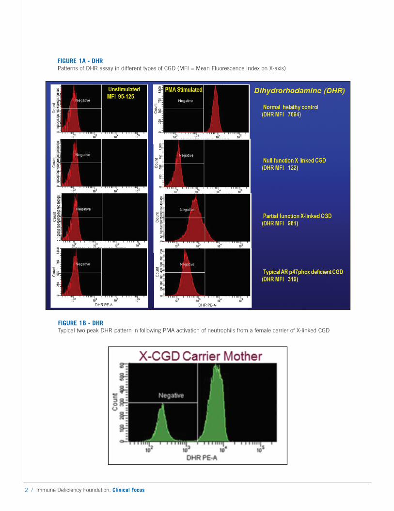
FIGURE 1A - DHRPatterns of DHR assay in different types of CGD (MFI = Mean Fluorescence Index on X-axis)
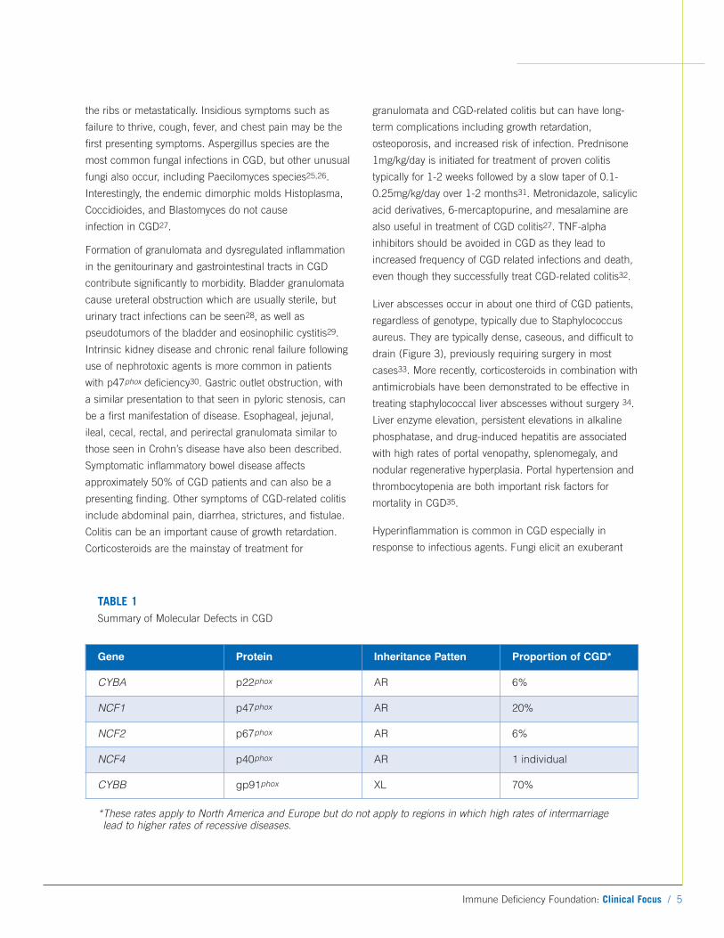
FIGURE 1B - DHRTypical two peak DHR pattern in following PMA activation of neutrophils from a female carrier of X-linked CGD
Immune Deficiency Foundation: Clinical Focus / 3
DiagnosisThe diagnosis of CGD relies on direct measurement of
superoxide production. The nitroblue tetrazolium (NBT)
dye test is the oldest and most recognized diagnostic test
for CGD, but it relies on light microscopy to provide a
mostly qualitative determination of NADPH oxidase
activity. The NBT test is typically performed on a
microscope slide which is read manually to distinguish
reducing (blue-black insoluble formazan precipitate) from
non-reducing (unstained) cells15. Because the NBT is
semi-quantitative, it can miss mild genetic forms of CGD
associated with residual oxidase activity, and has been
largely abandoned and replaced by the dihydrorho-
damine (DHR) assay, which is available from most major
commercial laboratories. The DHR test uses flow
cytometry to measure the production of hydrogen peroxide
in the presence of peroxidase. Oxidation of non-
fluorescent dihydrorhodamine 123 results in production of
highly fluorescent rhodamine 123 in stimulated
neutrophils16. The presence of myeloperoxidase is also
necessary for neutrophils to generate superoxide;
myeloperoxidase deficiency can therefore lead to
abnormal DHR assay results17. The DHR is preferable
because of its ability to distinguish many X-linked patients
from the p47phox deficient autosomal recessive form of
CGD, its sensitivity to very low numbers of functional
neutrophils, its ability to measure residual superoxide
production, its capacity to accurately identify X-linked
carriers, and its ease of use. Figure 1A shows the typical
DHR assay patterns (top to bottom) with neutrophils from
a healthy subject, a typical null oxidase function X-linked
CGD patient, a partial oxidase activity function X-linked
CGD patient, and a typical p47phox deficient AR CGD
patient. The p40phox deficient form of CGD has a DHR
pattern similar to p47phox, while the p22phox and p67phox
CGD patterns can demonstrate null or partial function.
The DHR assay makes it possible to assess the amount of
residual oxidase activity in a patient’s neutrophils by
observing the mean fluorescence index (MFI) of the
peak18. When obtaining a DHR test from a commercial
laboratory on a patient who has or is suspected of having
CGD, it is essential to request that the commercial lab
send the histograms and peak MFI information in addition
to their interpretation of the result. As shown in Figure 1B,
two populations of phagocytes are seen with the DHR in
X-linked female carriers; one normal superoxide producing
population and one abnormal non-superoxide producing
population19. A DHR assay should be requested for the
mother of a male patient with CGD as this can provide
confirmation that the patient has X-linked CGD. If the
mother’s neutrophil DHR pattern shows only a single
normal oxidase peak, this may mean the patient has AR
CGD, but this is only presumptive, because approximately
10% of X-linked CGD patients have new mutations in
which case the mother will not have an X-linked CGD
carrier genotype or phenotype. Infections are uncommon
in female carriers unless there is significant skewing of X-
inactivation such that normal neutrophils are <10%.
However, discoid lupus, photosensitivity rashes, and
aphthous ulcers are common in female carriers20.
Clinical ManifestationsThe majority of patients with CGD present before age 5,
but later presentations, including well into adulthood, have
become more common with the routine use of potent oral
antibiotics and reduction in environmental exposures
(e.g., Salmonella, BCG) in the industrial world. Infections
of the skin, lungs, lymph nodes, and liver represent the
most commonly involved sites. The overwhelming majority
of infections in CGD in North America result from five
organisms: Staphylococcus aureus, Burkholderia cepacia
complex, Serratia marcescens, Nocardia species and
Aspergillus species10. Salmonella, bacille-Calmette Guerin
(BCG), and tuberculosis are important causes of infection
in other parts of the world11. In the setting of antibiotic
prophylaxis, staphylococci are primarily confined to the
liver and lymph nodes. Infection with Burkholderia species
causes pneumonia and rarely sepsis21. Nocardia species
are a common cause of pneumonia but can also cause
osteomyelitis and brain abscesses22. Outside of CGD,
Nocardia infections only occur in the setting of high dose
corticosteroids. Chromobacterium violaceum23 and
Francisella philomiragia24 are uncommon causes of
infection and are virtually pathognomonic of CGD; both
are found in brackish water and can cause sepsis in CGD.
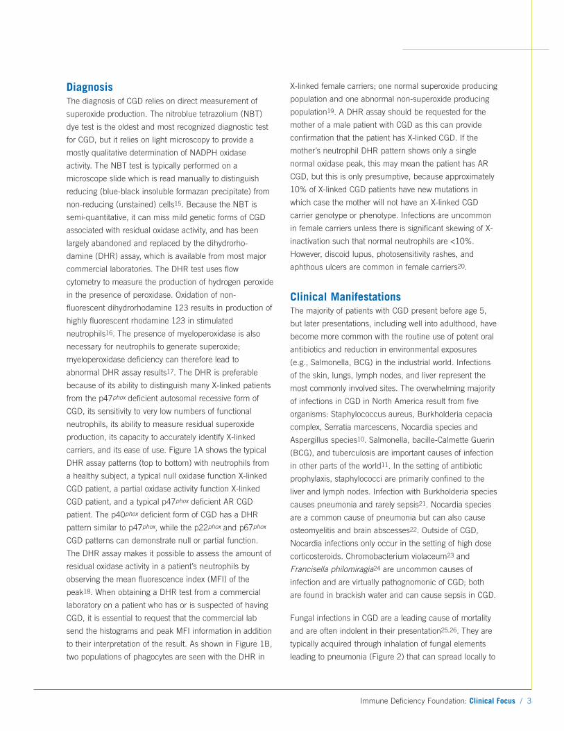
Fungal infections in CGD are a leading cause of mortality
and are often indolent in their presentation25,26. They are
typically acquired through inhalation of fungal elements
leading to pneumonia (Figure 2) that can spread locally to
4 / Immune Deficiency Foundation: Clinical Focus
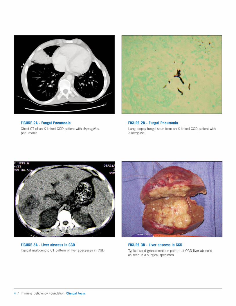
FIGURE 3A - Liver abscess in CGDTypical multicentric CT pattern of liver abscesses in CGD
FIGURE 3B - Liver abscess in CGDTypical solid granulomatous pattern of CGD liver abscessas seen in a surgical specimen
FIGURE 2A - Fungal PneumoniaChest CT of an X-linked CGD patient with Aspergilluspneumonia
FIGURE 2B - Fungal PneumoniaLung biopsy fungal stain from an X-linked CGD patient withAspergillus
Immune Deficiency Foundation: Clinical Focus / 5
the ribs or metastatically. Insidious symptoms such as
failure to thrive, cough, fever, and chest pain may be the
first presenting symptoms. Aspergillus species are the
most common fungal infections in CGD, but other unusual
fungi also occur, including Paecilomyces species25,26.
Interestingly, the endemic dimorphic molds Histoplasma,
Coccidioides, and Blastomyces do not cause
infection in CGD27.
Formation of granulomata and dysregulated inflammation
in the genitourinary and gastrointestinal tracts in CGD
contribute significantly to morbidity. Bladder granulomata
cause ureteral obstruction which are usually sterile, but
urinary tract infections can be seen28, as well as
pseudotumors of the bladder and eosinophilic cystitis29.
Intrinsic kidney disease and chronic renal failure following
use of nephrotoxic agents is more common in patients
with p47phox deficiency30. Gastric outlet obstruction, with
a similar presentation to that seen in pyloric stenosis, can
be a first manifestation of disease. Esophageal, jejunal,
ileal, cecal, rectal, and perirectal granulomata similar to
those seen in Crohn’s disease have also been described.
Symptomatic inflammatory bowel disease affects
approximately 50% of CGD patients and can also be a
presenting finding. Other symptoms of CGD-related colitis
include abdominal pain, diarrhea, strictures, and fistulae.
Colitis can be an important cause of growth retardation.
Corticosteroids are the mainstay of treatment for
granulomata and CGD-related colitis but can have long-
term complications including growth retardation,
osteoporosis, and increased risk of infection. Prednisone
1mg/kg/day is initiated for treatment of proven colitis
typically for 1-2 weeks followed by a slow taper of 0.1-
0.25mg/kg/day over 1-2 months31. Metronidazole, salicylic
acid derivatives, 6-mercaptopurine, and mesalamine are
also useful in treatment of CGD colitis27. TNF-alpha
inhibitors should be avoided in CGD as they lead to
increased frequency of CGD related infections and death,
even though they successfully treat CGD-related colitis32.
Liver abscesses occur in about one third of CGD patients,
regardless of genotype, typically due to Staphylococcus
aureus. They are typically dense, caseous, and difficult to
drain (Figure 3), previously requiring surgery in most
cases33. More recently, corticosteroids in combination with
antimicrobials have been demonstrated to be effective in
treating staphylococcal liver abscesses without surgery 34.
Liver enzyme elevation, persistent elevations in alkaline
phosphatase, and drug-induced hepatitis are associated
with high rates of portal venopathy, splenomegaly, and
nodular regenerative hyperplasia. Portal hypertension and
thrombocytopenia are both important risk factors for
mortality in CGD35.
Hyperinflammation is common in CGD especially in
response to infectious agents. Fungi elicit an exuberant
Gene Protein Inheritance Patten Proportion of CGD*
CYBA p22phox AR 6%
NCF1 p47phox AR 20%
NCF2 p67phox AR 6%
NCF4 p40phox AR 1 individual
CYBB gp91phox XL 70%
*These rates apply to North America and Europe but do not apply to regions in which high rates of intermarriagelead to higher rates of recessive diseases.
TABLE 1Summary of Molecular Defects in CGD
6 / Immune Deficiency Foundation: Clinical Focus
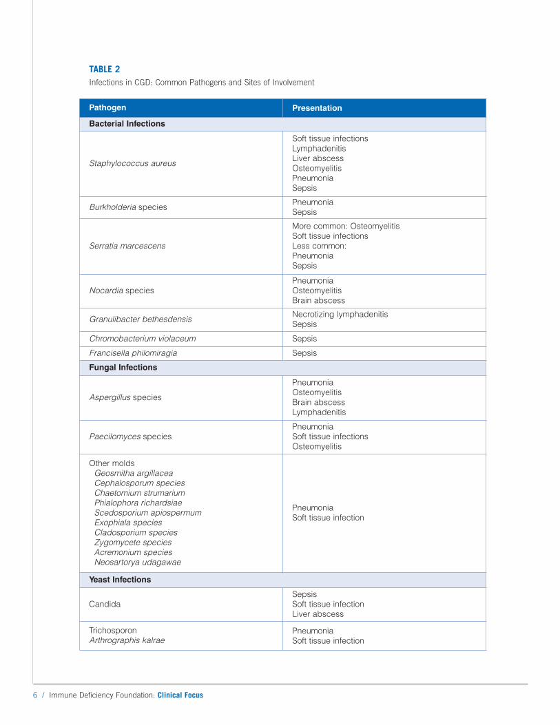
TABLE 2Infections in CGD: Common Pathogens and Sites of Involvement
Pathogen PresentationBacterial Infections
Staphylococcus aureus
Soft tissue infectionsLymphadenitisLiver abscessOsteomyelitisPneumoniaSepsis
Burkholderia speciesPneumoniaSepsis
Serratia marcescens
More common: OsteomyelitisSoft tissue infectionsLess common:PneumoniaSepsis
Nocardia speciesPneumoniaOsteomyelitisBrain abscess
Granulibacter bethesdensisNecrotizing lymphadenitisSepsis
Chromobacterium violaceum Sepsis
Francisella philomiragia Sepsis
Fungal Infections
Aspergillus species
PneumoniaOsteomyelitisBrain abscessLymphadenitis
Paecilomyces speciesPneumoniaSoft tissue infectionsOsteomyelitis
Other moldsGeosmitha argillacea Cephalosporum speciesChaetomium strumariumPhialophora richardsiaeScedosporium apiospermumExophiala speciesCladosporium speciesZygomycete speciesAcremonium speciesNeosartorya udagawae
PneumoniaSoft tissue infection
Yeast Infections
CandidaSepsisSoft tissue infectionLiver abscess
Trichosporon Arthrographis kalrae
Pneumonia Soft tissue infection
Immune Deficiency Foundation: Clinical Focus / 7
inflammatory response regardless of whether the organism
is alive or dead36. “Mulch pneumonitis” is a syndrome
caused by inhalation of aerosolized decayed organic
matter, such as hay or dead leaves, leading to acute
fulminant pneumonitis similar to that seen in
hypersensitivity pneumonitis37. Because of the risk of
pneumonitis, activities that expose CGD patients to
decayed organic matter such as mulching, gardening, leaf
raking, and house demolition should be avoided.
Heightened inflammation has also been described in
chronic colitis31, granulomatous cystitis29, pulmonary
infections with Nocardia38, and staphylococcal liver
abscesses34,39. In each of these cases, directed treatment
of the heightened inflammatory response in addition to
proper anti-microbials is extremely helpful for
clinical resolution.
Many other non-infectious manifestations affect patients
with CGD. Growth delay is common and failure to thrive
can be a presenting symptom as well as compounded by
colitis. Growth may improve in late adolescence and many
affected adults attain normal predicted weight and
height31. Oral manifestations include gingivitis, aphthous
ulcers, and gingival hypertrophy. Characteristically poor
wound healing at sites of surgical incisions leads to wound
dehiscence; other cutaneous manifestations include
photosensitivity, granulomatous lesions, and vasculitis.
Diabetes and renal and cardiovascular disease occur more
commonly in patients with p47phox deficient CGD30.
Discoid lupus erythematosus occurs in CGD but is more
common in X-linked female carriers10,20,40. Other
autoimmune diseases reported in CGD include idiopathic
thrombocytopenic purpura, juvenile idiopathic arthritis,
autoimmune pulmonary disease, celiac disease with co-
existing pulmonary hemosiderosis, myasthenia gravis, IgA
nephropathy, antiphospholipid syndrome, and recurrent
pericardial effusion10,40. Autoimmune disease is likely a
co-morbid condition and not entirely a result of
hyperinflammation associated with CGD. Diagnosis relies
on specific symptoms without an identifiable infectious
cause, presence of serologic markers, and response to
immunosuppression. Autoimmune disease associated with
CGD responds surprisingly well to immunosuppression
such as corticosteroids40.
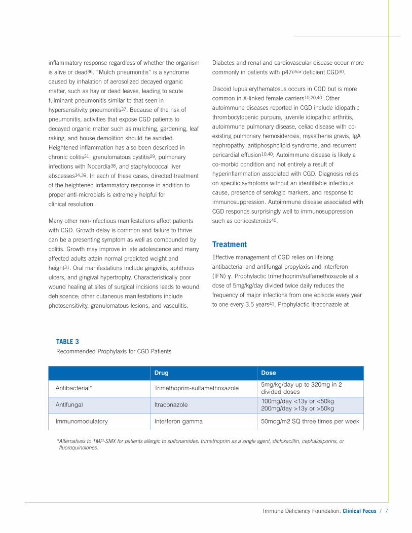
Treatment
Effective management of CGD relies on lifelong
antibacterial and antifungal propylaxis and interferon
(IFN) γ. Prophylactic trimethoprim/sulfamethoxazole at a
dose of 5mg/kg/day divided twice daily reduces the
frequency of major infections from one episode every year
to one every 3.5 years41. Prophylactic itraconazole at
TABLE 3Recommended Prophylaxis for CGD Patients
Drug Dose
Antibacterial* Trimethoprim-sulfamethoxazole5mg/kg/day up to 320mg in 2divided doses
Antifungal Itraconazole100mg/day <13y or <50kg200mg/day >13y or >50kg
Immunomodulatory Interferon gamma 50mcg/m2 SQ three times per week
*Alternatives to TMP-SMX for patients allergic to sulfonamides: trimethoprim as a single agent, dicloxacillin, cephalosporins, orfluoroquinolones.
8 / Immune Deficiency Foundation: Clinical Focus
doses of 100mg daily for patients <13 years or weighing
<50kg and 200mg daily for patients >13 years or weighing
>50kg is effective at reducing the frequency of fungal
infections42. In a large international multicenter
randomized placebo-controlled trial, IFNγ was effective at
reducing the number and severity of infections by 70%
regardless of inheritance pattern, sex, or use of
prophylactic antibiotics43. Dosing of IFNγ is 50mcg/m2
subcutaneously three times per week. Close follow-up of
patients with CGD is recommended. Practitioners should
have a high suspicion and always be in search of new
infections. Evaluation should include tests looking for
evidence of infection such as C-reactive protein or
erythrocyte sedimentation rate, sensitive but non-specific
markers of inflammation27. Anemia may be due to chronic
disease or iron deficiency. Iron deficiency anemia and
hypoalbuminemia often accompany CGD-related colitis31.
Imaging is exquisitely helpful for diagnosis and following
the treatment of infections. Children with CGD should
receive routine vaccinations as recommended by the
American Academy of Pediatrics including live virus
vaccines. Patients with CGD do not have any defect in
immunity to viruses, so they are able to receive live virus
vaccines without adverse effect. Many countries in Europe
and Asia vaccinate children with the tuberculosis live
bacterial vaccine, BCG, but this is not recommended
practice in the U.S. Children with CGD should never
receive the BCG live bacterial vaccine as it can result in a
severe life-threatening systemic BCG infection.
Allogeneic hematopoietic stem cell transplant (HSCT) is
the only known cure for CGD; both myeloablative and non-
myeloablative regimens have been successful44-47. Non-
myeloablative transplants have reduced the risk of
regimen-related toxicity and allow for transplantation in the
setting of active infection44,45,47. CGD is an attractive
target for gene therapy since it results from a single gene
defect and complete protection does not require full
correction of superoxide production, as shown by many
healthy X-linked carriers. Gene therapy protocols using
lentivectors will start soon in the U.S. and Europe.
Although HSCT is an attractive option for cure of patients
with CGD, the overwhelming majority of CGD patients
survive without HSCT, albeit with co-morbidities. Survival
in CGD has improved greatly over the last several decades
and is approximately 90% at 10 years18,48,49. Residual
NADPH oxidase activity correlates well with survival in
CGD. This can be determined directly by DHR or a
specific cytochrome reduction assay, and both tests
correlate quite well with specific mutations. Patient’s with
autosomal recessive CGD typically have higher levels of
residual oxidase activity than patients with X-linked CGD
and therefore, have higher overall survival rates. In general
for X-linked CGD, mutations that abolish protein
expression or that occur in the intracellular FAD or
NADPH binding domains are severe and are associated
with worse overall outcomes, whereas patients with protein
positive mutations in the extracellular domains tend to
have a better outcome. Therefore, the specific X-linked
mutation can be used to predict superoxide production
and overall risk of mortality, providing information that can
help decide about HSCT18.
Over the last 60 years, CGD has taught many lessons in
basic science, infection susceptibility, and the role of
inflammation in immune responses. It has evolved from a
disease characterized as a deficiency of the immune
system with no cure to one of dysregulation with multiple
therapeutic avenues available and a far better outlook for
the CGD patient.
References
1. Berendes H, Bridges RA, Good RA. A fatalgranulomatosus of childhood: the clinical study of anew syndrome. Minnesota medicine 1957;40:309-12.
2. Landing BH, Shirkey HS. A syndrome of recurrentinfection and infiltration of viscera by pigmented lipidhistiocytes. Pediatrics 1957;20:431-8.
3. Bridges RA, Berendes H, Good RA. A fatalgranulomatous disease of childhood; the clinical,pathological, and laboratory features of a newsyndrome. AMA J Dis Child 1959;97:387-408.
4. Segal BH, Leto TL, Gallin JI, Malech HL, Holland SM.Genetic, biochemical, and clinical features of chronicgranulomatous disease. Medicine (Baltimore)2000;79:170-200.
5. Ambruso DR, Knall C, Abell AN, et al. Humanneutrophil immunodeficiency syndrome is associatedwith an inhibitory Rac2 mutation. Proceedings of theNational Academy of Sciences of the United States ofAmerica 2000;97:4654-9.
6. Verbsky J, Thakar M, Routes J. The Wisconsinapproach to newborn screening for severe combinedimmunodeficiency. The Journal of allergy and clinicalimmunology 2012;129:622-7.
7. Roos D, Kuhns DB, Maddalena A, et al. Hematologicallyimportant mutations: the autosomal recessive forms ofchronic granulomatous disease (second update). BloodCells Mol Dis 2010;44:291-9.
8. Roos D, Kuhns DB, Maddalena A, et al. Hematologicallyimportant mutations: X-linked chronic granulomatousdisease (third update). Blood Cells Mol Dis2010;45:246-65.
9. Matute JD, Arias AA, Wright NA, et al. A new geneticsubgroup of chronic granulomatous disease withautosomal recessive mutations in p40 phox andselective defects in neutrophil NADPH oxidase activity.Blood 2009;114:3309-15.
10. Winkelstein JA, Marino MC, Johnston RB, Jr., et al.Chronic granulomatous disease. Report on a nationalregistry of 368 patients. Medicine (Baltimore)2000;79:155-69.
11. van den Berg JM, van Koppen E, Ahlin A, et al. Chronicgranulomatous disease: the European experience. PLoSOne 2009;4:e5234.
12. Ahlin A, De Boer M, Roos D, et al. Prevalence, geneticsand clinical presentation of chronic granulomatousdisease in Sweden. Acta Paediatr 1995;84:1386-94.
13. Hasui M, Japa SGPD. Chronic granulomatous diseasein Japan: Incidence and natural history. PediatricsInternational 1999;41:589-93.
14. Wolach B, Gavrieli R, de Boer M, et al. Chronicgranulomatous disease in Israel: clinical, functional andmolecular studies of 38 patients. Clin Immunol2008;129:103-14.
15. Baehner RL, Nathan DG. Leukocyte oxidase: defectiveactivity in chronic granulomatous disease. Science1967;155:835-6.
16. Vowells SJ, Fleisher TA, Malech HL. Testing for chronicgranulomatous disease. Lancet 1996;347:1048-9.
17. Mauch L, Lun A, O'Gorman MR, et al. Chronicgranulomatous disease (CGD) and completemyeloperoxidase deficiency both yield strongly reduceddihydrorhodamine 123 test signals but can be easilydiscerned in routine testing for CGD. Clin Chem2007;53:890-6.
18. Kuhns DB, Alvord WG, Heller T, et al. Residual NADPHoxidase and survival in chronic granulomatous disease.N Engl J Med 2010;363:2600-10.
19. Elloumi HZ, Holland SM. Diagnostic assays for chronicgranulomatous disease and other neutrophil disorders.Methods Mol Biol 2007;412:505-23.
20. Hafner J, Enderlin A, Seger RA, et al. Discoid lupuserythematosus-like lesions in carriers of X-linkedchronic granulomatous disease. Br J Dermatol1992;127:446-7.
21. Greenberg DE, Goldberg JB, Stock F, Murray PR,Holland SM, Lipuma JJ. Recurrent Burkholderiainfection in patients with chronic granulomatousdisease: 11-year experience at a large referral center.Clinical infectious diseases: an official publication ofthe Infectious Diseases Society of America2009;48:1577-9.
22. Dorman SE, Guide SV, Conville PS, et al. Nocardiainfection in chronic granulomatous disease. Clinicalinfectious diseases: an official publication of theInfectious Diseases Society of America 2002;35:390-4.
23. Sirinavin S, Techasaensiri C, Benjaponpitak S, PornkulR, Vorachit M. Invasive Chromobacterium violaceuminfection in children: case report and review. PediatricInfectious Disease Journal 2005;24:559-61.
24. Mailman TL, Schmidt MH. Francisella philomiragiaadenitis and pulmonary nodules in a child with chronicgranulomatous disease. Can J Infect Dis Med Microbiol2005;16:245-8.
25. Beaute J, Obenga G, Le Mignot L, et al. Epidemiologyand outcome of invasive fungal diseases in patientswith chronic granulomatous disease: a multicenterstudy in France. Pediatr Infect Dis J 2011;30:57-62.
26. Blumental S, Mouy R, Mahlaoui N, et al. Invasive moldinfections in chronic granulomatous disease: a 25-yearretrospective survey. Clinical infectious diseases : anofficial publication of the Infectious Diseases Society ofAmerica 2011;53:e159-69.
27. Holland SM. Chronic Granulomatous Disease. ClinicalReviews in Allergy & Immunology 2010;38:3-10.
28. Walther MM, Malech H, Berman A, et al. The urologicalmanifestations of chronic granulomatous disease. JUrol 1992;147:1314-8.
29. Kontras SB, Bodenbender JG, McClave CR, Smith JP.Interstitial cystitis in chronic granulomatous disease. JUrol 1971;105:575-8.
30. Leiding JW, Marciano BE, Zerbe CS, Deravin SS,Malech HL, Holland SM. Diabetes, Renal andCardiovascular Disease in p47 (phox-/-) ChronicGranulomatous Disease. Journal of clinical immunology2013;33:725-30.
31. Marciano BE, Rosenzweig SD, Kleiner DE, et al.Gastrointestinal involvement in chronic granulomatousdisease. Pediatrics 2004;114:462-8.
32. Uzel G, Orange JS, Poliak N, Marciano BE, Heller T,Holland SM. Complications of Tumor Necrosis Factor-alpha Blockade in Chronic Granulomatous Disease-Related Colitis. Clinical Infectious Diseases2010;51:1429-34.
33. Lublin M, Bartlett DL, Danforth DN, et al. Hepaticabscess in patients with chronic granulomatousdisease. Ann Surg 2002;235:383-91.
34. Leiding JW, Freeman AF, Marciano BE, et al.Corticosteroid Therapy for Liver Abscess in ChronicGranulomatous Disease. Clinical infectious diseases: anofficial publication of the Infectious Diseases Society ofAmerica 2011.
35. Feld JJ, Hussain N, Wright EC, et al. Hepaticinvolvement and portal hypertension predict mortality inchronic granulomatous disease. Gastroenterology2008;134:1917-26.
36. Morgenstern DE, Gifford MA, Li LL, Doerschuk CM,Dinauer MC. Absence of respiratory burst in X-linkedchronic granulomatous disease mice leads toabnormalities in both host defense and inflammatoryresponse to Aspergillus fumigatus. J Exp Med1997;185:207-18.
37. Siddiqui S, Anderson VL, Hilligoss DM, et al. Fulminantmulch pneumonitis: an emergency presentation ofchronic granulomatous disease. Clinical infectiousdiseases: an official publication of the InfectiousDiseases Society of America 2007;45:673-81.
38. Freeman AF, Marciano BE, Anderson VL, Uzel G, CostasC, Holland SM. Corticosteroids in the Treatment ofSevere Nocardia Pneumonia in Chronic GranulomatousDisease. Pediatr Infect Dis J 2011.
39. Yamazaki-Nakashimada MA, Stiehm ER, Pietropaolo-Cienfuegos D, Hernandez-Bautista V, Espinosa-RosalesF. Corticosteroid therapy for refractory infections inchronic granulomatous disease: case reports and reviewof the literature. Ann Allergy Asthma Immunol2006;97:257-61.
40. De Ravin SS, Naumann N, Cowen EW, et al. Chronicgranulomatous disease as a risk factor for autoimmunedisease. Journal of Allergy and Clinical Immunology2008;122:1097-103.
41. Margolis DM, Melnick DA, Alling DW, Gallin JI.Trimethoprim-sulfamethoxazole prophylaxis in themanagement of chronic granulomatous disease. TheJournal of infectious diseases 1990;162:723-6.
42. Gallin JI, Alling DW, Malech HL, et al. Itraconazole toprevent fungal infections in chronic granulomatousdisease. N Engl J Med 2003;348:2416-22.
43. A controlled trial of interferon gamma to preventinfection in chronic granulomatous disease. TheInternational Chronic Granulomatous DiseaseCooperative Study Group. N Engl J Med 1991;324:509-16.
44. Gungor T. Successful low-dose busulfan / full-dosefludarabine based reduced intesntity conditioning inhigh risk pediatric and adult chronic granulomatousdisease patients. In: XIVth Meeting of the EuropeanSociety for Immunodeficiencies; 2010; Istanbul, Turkey; 2010.
45. Kang EM, Marciano BE, DeRavin S, Zarember KA,Holland SM, Malech HL. Chronic granulomatousdisease: Overview and hematopoietic stem celltransplantation. Journal of Allergy and ClinicalImmunology 2011;127:1319-26.
46. Seger RA, Gungor T, Belohradsky BH, et al. Treatment of chronic granulomatous disease with myeloablativeconditioning and an unmodified hemopoietic allograft: a survey of the European experience, 1985-2000. Blood2002;100:4344-50.
47. Soncini E, Slatter MA, Jones LB, et al. Unrelated donorand HLA-identical sibling haematopoietic stem celltransplantation cure chronic granulomatous diseasewith good long-term outcome and growth. Br JHaematol 2009;145:73-83.
48. Jones LB, McGrogan P, Flood TJ, et al. Special article:chronic granulomatous disease in the United Kingdomand Ireland: a comprehensive national patient-basedregistry. Clin Exp Immunol 2008;152:211-8.
49. Martire B, Rondelli R, Soresina A, et al. Clinicalfeatures, long-term follow-up and outcome of a largecohort of patients with Chronic Granulomatous Disease:an Italian multicenter study. Clin Immunol2008;126:155-64.
Immune Deficiency Foundation: Clinical Focus / 9
40 West Chesapeake Avenue, Suite 308Towson, MD 21204800-296-4433410-321-6647410-321-9165 (Fax)[email protected]
40 West Chesapeake Avenue, Suite 308Towson, MD 21204
The Immune Deficiency Foundation, founded in 1980, is the national patient organization
dedicated to improving the diagnosis, treatment and quality of life of persons with primary
immunodeficiency diseases through advocacy, education and research.