Embed Size (px)
Citation preview
Dynamic and Static Magnetic Resonance Angiographyof the Supra-aortic Vessels at 3.0 T
Intraindividual Comparison of Gadobutrol, Gadobenate Dimeglumine,and Gadoterate Meglumine at Equimolar Dose
Jens Harald Kramer, MD,* Elisabeth Arnoldi, MD,* Christopher J. Francois, MD,Þ Andrew L.Wentland, MS,ÞKonstantin Nikolaou, MD,* Bernd J. Wintersperger, MD,*þ and Thomas M. Grist, MDÞ
Purpose: The purpose of this study was the intraindividual comparison of a1.0 M and two 0.5 M gadolinium-based contrast agents (GBCA) using equi-molar dosing in dynamic and static magnetic resonance angiography (MRA)of the supra-aortic vessels.Materials and Methods: In this institutional review boardYapproved study, atotal of 20 healthy volunteers (mean T SD age, 29 T 6 years) underwent 3consecutive supra-aortic MRA examinations on a 3.0 T magnetic resonancesystem. The order of GBCA (Gadobutrol, Gadobenate dimeglumine, andGadoterate meglumine) was randomized with a minimum interval of 48 hoursbetween the examinations. Before each examination and 45 minutes after eachexamination, circulatory parameters were recorded. Total GBCA dose perMRA examination was 0.1 mmol/kg with a 0.03 mmol/kg and 0.07 mmol/kgsplit for dynamic and static MRA, respectively, injected at a rate of 2 mL/s.Two blinded readers qualitatively assessed static MRA data sets independentlyusing pairwise rankings (superior, inferior, and equal). In addition, quantitativeanalysis was performed with signal-to-noise ratio (SNR) and contrast-to-noiseratio (CNR) evaluation as well as vessel sharpness analysis of static MRAusing an in-houseYdeveloped semiautomated tool. Dynamic MRA was eval-uated for maximal SNR. Statistical analysis was performed using the Cohen J,the Wilcoxon rank sum tests, and mixed effects models.Results: No significant differences of hemodynamic parameters were ob-served. In static MRA, Gadobutrol was rated superior to Gadoterate meglu-mine (P G 0.05) and equal to Gadobenate dimeglumine (P = 0.06) with goodto excellent reader agreement (J, 0.66Y0.83). In static MRA, SNR was sig-nificantly higher using 1.0 M Gadobutrol as compared with either 0.5 M agent(P G 0.05 and P G 0.05) and CNR was significantly higher as compared withGadoterate meglumine (P G 0.05), whereas CNR values of Gadobutrol datasets were not significantly different as compared with Gadobenate dimeglu-mine (P = 0.13). Differences in CNR between Gadobenate dimeglumine andGadoterate meglumine were not significant (P = 0.78). Differences in vesselsharpness between the different GBCAs were also not significant (P 9 0.05).Maximal SNR in dynamic MRA using Gadobutrol was significantly higherthan both comparators at the level of the proximal and distal internal carotid ar-tery (P G 0.05 and P G 0.05; P G 0.05 and P G 0.05).Conclusions: At equimolar doses, 1.0 M Gadobutrol demonstrates higherSNR/CNR than do Gadobenate dimeglumine and Gadoterate meglumine, withsuperior image quality as compared with Gadoterate meglumine for dynamic and
static carotid MRA. Despite the shortened bolus with Gadobutrol, no blurring ofvessel edges was observed.
Key Words: magnetic resonance angiography, contrast media, carotidarteries, vessel blurring, image quality, Gadobutrol, Gadobenatedimeglumine, Gadoterate meglumine
(Invest Radiol 2013;48: 121Y128)
A cute arterial events are among the most frequent causes of pre-mature death in the developed world, and cerebrovascular events
have replaced cardiac events as the leading cause of death. Therefore,interest in imaging of the supra-aortic vessels has significantlyincreased.1Y3 In most vascular territories, digital subtraction angiog-raphy still remains the standard of reference.4,5 However, the knowndrawbacks of digital subtraction angiography have spurred the de-velopment of noninvasive techniques.6,7 Because of recent develop-ments, magnetic resonance angiography (MRA) is now routinely used,along with computer tomographic angiography or ultrasound.7Y10 Theimage quality of MRA is enhanced with the use of dedicated gado-linium-based contrast agents (GBCA),11Y14 which improve signal-to-noise ratio (SNR) and contrast-to-noise ratio (CNR).14Y16
However, because of the description of a potential link be-tween the applications of GBCAs and the development of nephro-genic systemic fibrosis (NSF),17,18 efforts are made to reduce theamount of GBCA administered for comprehensive diagnostic imag-ing of the arterial vasculature.8,19 Although earlier studies proposedhigher doses of standard GBCA,20Y22 currently, MRA examinationsare typically performed at single doses (0.1 mmol/kg). Furthermore,the current state-of-the-art MRA of the cervical arteries compriseshigh-resolution static and dynamic imaging. Previously publishedstudies have shown that, using standard GBCA, excellent imagequality is possible using a single dose, and good image quality ispossible even with lower than single doses.9,23Y25 However, reducingthe amount of injected contrast agent results in a shortening of the bolusbecause a certain injection rate is needed to keep the bolus tight.Sampling of the peripheral parts of k-space without the presence ofcontrast agent bolus theoretically leads to blurring of the vessel edges.26
We hypothesized that, in supra-aortic MRA, 1.0 M Gadobutrolis noninferior to 0.5 M Gadobenate dimeglumine and superior toGadoterate meglumine when applied at single equimolar dose. Thepurpose of this study therefore was to intraindividually compare all 3GBCAs in dynamic and static supra-aortic MRAwith application ofidentical imaging and dosage parameters in respect to qualitativeimage quality and quantitative signal parameters.
MATERIALS AND METHODS
Study PopulationThis single-center, blinded, prospective, intraindividual com-
parison study was approved by the institutional review board, andwritten informed consent was obtained from all study volunteers.
ORIGINAL ARTICLE
Investigative Radiology & Volume 48, Number 3, March 2013 www.investigativeradiology.com 121
Received for publication August 16, 2012; and accepted for publication, after re-vision October 2, 2012.
From the *Institute for Clinical Radiology, LudwigMaximilians University HospitalMunich, Munich, Germany; †Department of Radiology, The University ofWisconsin-Madison, Madison, WI; and ‡Department of Medical Imaging,University of Toronto, Toronto, Canada.
Conflicts of interest and sources of funding: Supported by Bayer Healthcare.The authors report no conflicts of interest.Reprints: J. Harald Kramer, MD, Institute for Clinical Radiology, Ludwig Max-
imilians University Hospital Munich, Marchioninistr. 15, 81377 Munich,Germany. E-mail: [email protected].
Copyright * 2013 by Lippincott Williams & WilkinsISSN: 0020-9996/13/4803Y0121
Copyright © 2013 Lippincott Williams & Wilkins. Unauthorized reproduction of this article is prohibited.
Twenty-two healthy men (mean T SD age, 28.7 T 5.9 years;range, 21Y41 years) without history of vascular or renal disease,without contraindication against any of the investigational GBCA, orwithout general magnetic resonance (MR) contraindications wereincluded between June 2011 and August 2011.
Before inclusion in the study, a blood sample was drawn fromevery volunteer and the glomerular filtration rate was calculated us-ing the Cockroft-Gault formula (eGFR) and confirmed to be greaterthan 60 mL/min per 1.73 m2.
Magnetic Resonance Imaging and Study ProtocolAll MRA examinations were performed on a 3.0 T 32-channel
whole-body open-bore MR system (Magnetom Verio; SiemensHealthcare, Erlangen, Germany). For signal reception, a 12-elementhead array coil in combination with a 4-element neck surface coilwas used.
For static MRA, a standard 3-dimensional fast low-angle shotsequence with parameters optimized for 3.0 T was used. Becauseimage quality in terms of SNR is regarded as sufficient at 1.5 T, thepotential increase in signal at 3.0 Twas invested in increasing spatialresolution and shortening the acquisition time. Readout was done in acentric fashion with a time to center of 1 second. Because optimizingsequence parameters for 1 agent would penalize both remainingagents, the sequence settings were chosen to reflect the state-of-the-art 3.0 T MRA parameters. Detailed sequence parameters can befound in Table 1. For dynamic MRA, a time-resolved angiographywith interleaved stochastic trajectories sequence was used.27 Thissequence is based on a view-sharing technique and facilitates insetting the size of the central region of k-space, which is sampled atevery time point, and the sampling density of the remaining periph-eral part of k-space. Because, in this study, a high spatial resolutionstatic data set was acquired as well, dynamic MRAwas optimized fortemporal information. Hence, spatial resolution and potentiallyavailable signal were sacrificed to increase temporal resolution. De-tailed sequence information can be found in Table 1.
The volunteers were imaged at 3 different time points with the3 different contrast agents administered in a randomized fashion anda delay of 48 hours to 30 days between individual GBCA applica-tions. Hemodynamic parameters (eg, blood pressure, heart rate) wererecorded before each scan, and an 18-Gauge venous access was placedin the right antecubital vein. Total GBCA dose per examination was0.1 mmol/kg body weight (BW) with a 70%Y30% split for static anddynamic MRA, respectively. Static MRA was always performed as thefirst of both examinations to exclude any effect of the previously ad-ministered contrast agent on SNR and CNR measurements on static
MRA data sets. All GBCA injections were performed using a powerinjector (Spectris Solaris; Medrad). For the evaluation of bolus arrivaltime, a test bolus technique with a fixed dose of 1 mL of contrast agentinjected at a flow rate of 2 mL/s, followed by a 20 m/L saline chaseinjected at the same rate, was used. Both static and dynamic MRAwereperformed with the same injection parameters.9,27 After each examina-tion, hemodynamic parameters were reassessed and the volunteers weremonitored for the occurrence of adverse events for 60 minutes.
Contrast AgentsThe following Gd-based contrast agents (GBCAs) were used
for this study: Gadobutrol (Gadovist/Gadavist; Bayer Healthcare,Berlin, Germany), Gadobenate dimeglumine (Multihance; Bracco Di-agnostics, Milan, Italy), and Gadoterate meglumine (Dotarem; Guerbet,Roissy CdG Cedex, France).
Gadobutrol is a 1.0 M macrocyclic GBCA with a relaxivity(L mmolj1 sj1) of 5.0 (4.7Y5.3) at 3.0 T. Gadobenate meglumine is a0.5 M linear GBCA with a weak transient protein binding and arelaxivity of 5.5 (5.2Y5.8) at 3.0 T. Gadoterate meglumine is a 0.5 Mmacrocyclic GBCA and a relaxivity of 3.5 (3.3Y3.7) at 3.0 T.14
Image Quality EvaluationQualitative image quality of all static MRA data sets was
assessed independently by 2 radiologists with more than 10 years ofexperience in MRA using pairwise comparisons. The readers wereasked to give their overall impression on whether a data set wasbetter, equal, or worse than the given comparator. The readers in-cluded measures such as vessel conspicuity and vessel homogeneityin their judgment but excluded factors that are not related to a certaincontrast agent, for example, venous enhancement. Both observerswere blinded to any information about the used contrast agent, andafter the independent evaluation, assessments of both readers werecombined, as shown in Table 2.
Quantitative analysis of both static and dynamic MRA datasets was performed on the basis of the assessment of SNR and CNRat the level of the proximal internal carotid artery (ICA) next to thecarotid bifurcation and at the distal ICA just proximal to the carotid Tat the level of the skull base. Because the static and dynamic MRAacquisitions used parallel imaging techniques, SNR and CNR eval-uations were performed by applying the difference method, aspreviously described by Dietrich et al.28 Hence, 2 consecutive un-enhanced data sets were acquired before the contrast agent applica-tion and subtracted from each other. The SD within a region ofinterest (ROI) positioned in the subtracted data set at identical posi-tion as the signal measurement in the enhanced data set was de-fined as image noise for that specific location. For calculation of
TABLE 1. Sequence Details of Static MRA and Dynamic MRA
Static MRA Dynamic MRA
Acquisition time, seconds 21.6 98
Temporal resolution, seconds V 1.85; interp, 0.925
Parallel imaging factor 4 3
Spatial resolution, mm3 0.8 � 0.8 � 0.8 1.4 � 1.4 � 1.4
TR (repetition time), milliseconds] 3.25 2.46
TE (echo time), milliseconds 1.26 0.92
Flip angle, degrees 21 18
Matrix 576 � 342 256 � 176
FOV, mm2 450 � 267 350 � 240
Bandwidth, Hz 620 810
Note the excellent spatial resolution of both techniques and the temporalresolution of less than 2 seconds per frame of dynamic MRA interpolated to0.925 seconds per frame.
TABLE 2. Calculation of Combined Results From Blinded ImageQuality Reading
Reader 1 Reader 2 Combined Result
CA A is superior CA A is superior CA A is superior
CA A is superior both equal CA A is superior
CA A is superior CA B is superior both equal
both equal CA A is superior CA A is superior
both equal both equal both equal
both equal CA B is superior CA B is superior
CA B is superior CA A is superior both equal
CA B is superior both equal CA B is superior
CA B is superior CA B is superior CA B is superior
CA A indicates contrast agent A; CA B, contrast agent B.
Kramer et al Investigative Radiology & Volume 48, Number 3, March 2013
122 www.investigativeradiology.com * 2013 Lippincott Williams & Wilkins
Copyright © 2013 Lippincott Williams & Wilkins. Unauthorized reproduction of this article is prohibited.
contrast between the vessel signal and the surrounding tissues, anROI was positioned within the masseter muscle.
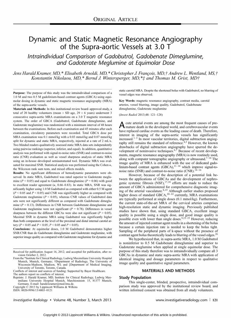
In addition to SNR and CNR evaluation, a semiautomatedquantitative evaluation of the vessel edge sharpness was performedusing an in-houseYdeveloped MATLAB-based tool (MathWorks,Natick, MA). After a user-based identification of the center of thevessel of interest on an axial reformatted slice, the tool automaticallygenerated 6 equally spaced radial spokes in 30-degree intervals. Eachof the 6 spokes provided a line profile and 2 vessel edges, for a totalof 12 vessel edges. Vessel edge sharpness (mm) was defined as thedistance between the 20% and 80% of the maximum signal intensityon each side of the line profile (Fig. 1). Vessel edge sharpness wasaveraged over all 12 vessel edges.
Statistical AnalysisFor the assessment of contrast agent superiority via qualitative
pairwise preference comparisons, a Wilcoxon signed rank test andthe Cohen J for the assessment of interreader agreement wereused. For the assessment of quantitative parameters such as SNR,CNR, and vessel edge sharpness, linear mixed effects models,Kruskal-Wallis tests, and the Wilcoxon rank sum test were used. P G0.05 (2-sided) was considered as statistically significant. Computa-tions were done using measurements of all the volunteers; givenvalues are mean values. Given SDs are mean values of measuredSDs. All computations were done in R for Windows, version 2.12.1(R Development Core Team, 2009).
RESULTSAll contrast agents were administered at 0.1 mmol/kg with a
70%Y30% split for static and dynamic MRA. This resulted in a meanT SD volume of 11.2 (1.1) mL for the 0.5 M agents, 5.6 (0.6) mL forgadobutrol for static MRA, and 4.8 (0.5) mL and 2.4 (0.2) mL for the0.5 M and 1.0 M agents for dynamic MRA, respectively.
Qualitative AnalysisIn none of the volunteers circulatory parameters in terms of blood
pressure and heart rate differed significantly from each other between thethree different exams. No significant differences were observed betweenthe pre-examination and postexamination measurements. In addition,because the investigated cohort consists of healthy, relatively young men,no significantly different circulatory parameters occurred interindividually.
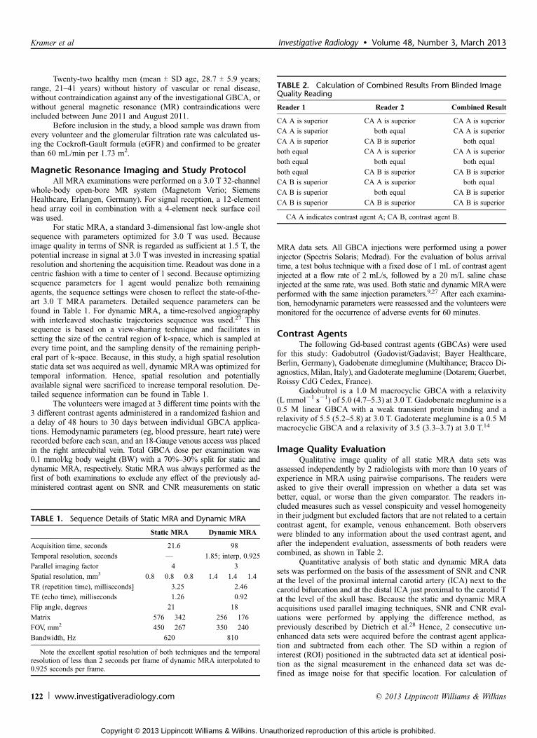
Combined results of the pairwise comparison of static MRAdata sets acquired with Gadobutrol, Gadobenate dimeglumine, andGadoterate meglumine showed Gadobutrol superior to Gadobenatedimeglumine in 10 (50%) cases and to Gadoterate meglumine in 17(85%) cases. It was rated as equal to the comparator in 7 and 2 cases(35% and 10%) when compared with Gadobenate dimeglumine andGadoterate meglumine, respectively. Gadobutrol was judged as in-ferior compared with Gadobenate dimeglumine and Gadoteratemeglumine in 3 and 1 case (15% and 5%), respectively. Gadobenatedimeglumine was judged as superior, equal, or inferior as comparedwith Gadoterate meglumine in 10, 5, and 5 cases (50%, 25%, and25%; Fig. 2; Table 3). The Wilcoxon rank test revealed Gadobutrol tobe insignificantly different compared with Gadobenate dimeglumine(P = 0.057) but significantly superior as compared with Gadoteratemeglumine (P G 0.005). Gadobenate dimeglumine was rated as notsignificantly different from Gadoterate meglumine (P = 0.21). Theinterreader agreement for this qualitative pairwise evaluation wasgood to excellent with Cohen J values of 0.66 to 0.83 for the com-parison of Gadobutrol with Gadobenate dimeglumine (k = 0.66) andGadoterate meglumine (k = 0.83) as well as Gadobenate dimeglu-mine and Gadoterate meglumine (k = 0.66) (Table 4).
Quantitative Analysis
Static MRAGadobutrol featured significantly higher SNR at the level
of the proximal ICA as compared with Gadobenate dimeglumine(P G 0.05) and Gadoterate meglumine (P G 0.05), with values of
FIGURE 1. Example of static MRA of the carotid arteries showing the level of the vessel sharpness evaluation and the reconstructedaxial slice including vessel profile lines. The graph shows an exemplary vessel signal profile with d1 and d1’ representing 20%of the maximal signal, d2 and d2’ representing 80% of the maximal signal and full width half max level.
Investigative Radiology & Volume 48, Number 3, March 2013 Dynamic and Static MRA of the Supra-aortic Vessels
* 2013 Lippincott Williams & Wilkins www.investigativeradiology.com 123
Copyright © 2013 Lippincott Williams & Wilkins. Unauthorized reproduction of this article is prohibited.
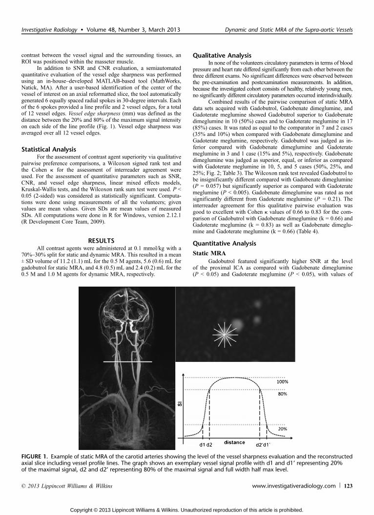
87.4 T 4.2, 47.8 T 4.0, and 44.9 T 3.5 (55% and 51% of SNRachieved with gadobutrol), respectively. In the distal ICA at the levelof the skull base, SNR was not significantly different between allagents with values of 81.7 T 5.6, 58.4 T 7.1 (71%), and 52.0 T 5.8(64%) for Gadobutrol, Gadobenate dimeglumine, and Gadoteratemeglumine, respectively. Contrast-to-noise ratio in MRA data setsacquired with Gadobutrol again was superior as compared withGadobenate dimeglumine (P = 0.13) and significantly better thanGadoterate meglumine (P = 0.03), with values of 71.1 T 3.8, 36.9 T 3.9(52%), and 30.5 T 3.5 (43%), respectively (Fig. 3). Calculation ofvessel sharpness revealed no significant different results for all 3agents with values of 1.17 T 0.24 mm, 1.05 T 0.18 mm, and 1.11 T0.27 mm for Gadobutrol, Gadobenate dimeglumine, and Gadoteratemeglumine, respectively.
Dynamic MRAThe absence of significant interindividual and intraindividual
differences in terms of hemodynamic parameters is reflected by the
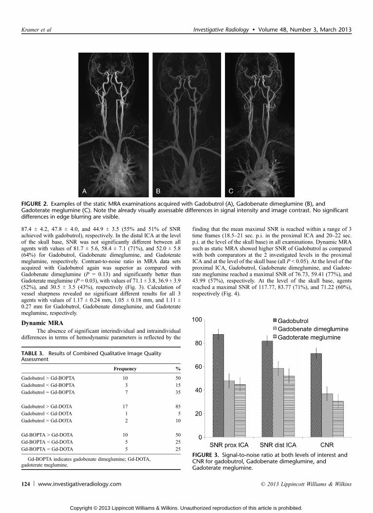
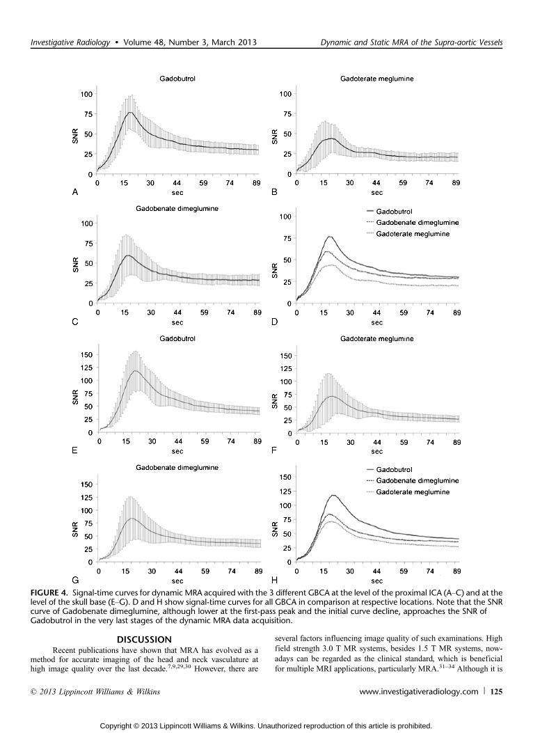
finding that the mean maximal SNR is reached within a range of 3time frames (18.5Y21 sec. p.i. in the proximal ICA and 20Y22 sec.p.i. at the level of the skull base) in all examinations. Dynamic MRAsuch as static MRA showed higher SNR of Gadobutrol as comparedwith both comparators at the 2 investigated levels in the proximalICA and at the level of the skull base (all P G 0.05). At the level of theproximal ICA, Gadobutrol, Gadobenate dimeglumine, and Gadote-rate meglumine reached a maximal SNR of 76.73, 59.41 (77%), and43.99 (57%), respectively. At the level of the skull base, agentsreached a maximal SNR of 117.77, 83.77 (71%), and 71.22 (60%),respectively (Fig. 4).
TABLE 3. Results of Combined Qualitative Image QualityAssessment
Frequency %
Gadobutrol 9 Gd-BOPTA 10 50
Gadobutrol G Gd-BOPTA 3 15
Gadobutrol = Gd-BOPTA 7 35
Gadobutrol 9 Gd-DOTA 17 85
Gadobutrol G Gd-DOTA 1 5
Gadobutrol = Gd-DOTA 2 10
Gd-BOPTA 9 Gd-DOTA 10 50
Gd-BOPTA G Gd-DOTA 5 25
Gd-BOPTA = Gd-DOTA 5 25
Gd-BOPTA indicates gadobenate dimeglumine; Gd-DOTA,gadoterate meglumine.
FIGURE 2. Examples of the static MRA examinations acquired with Gadobutrol (A), Gadobenate dimeglumine (B), andGadoterate meglumine (C). Note the already visually assessable differences in signal intensity and image contrast. No significantdifferences in edge blurring are visible.
FIGURE 3. Signal-to-noise ratio at both levels of interest andCNR for gadobutrol, Gadobenate dimeglumine, andGadoterate meglumine.
Kramer et al Investigative Radiology & Volume 48, Number 3, March 2013
124 www.investigativeradiology.com * 2013 Lippincott Williams & Wilkins
Copyright © 2013 Lippincott Williams & Wilkins. Unauthorized reproduction of this article is prohibited.
DISCUSSIONRecent publications have shown that MRA has evolved as a
method for accurate imaging of the head and neck vasculature athigh image quality over the last decade.7,9,29,30 However, there are
several factors influencing image quality of such examinations. Highfield strength 3.0 T MR systems, besides 1.5 T MR systems, now-adays can be regarded as the clinical standard, which is beneficialfor multiple MRI applications, particularly MRA.31Y34 Although it is
FIGURE 4. Signal-time curves for dynamic MRA acquired with the 3 different GBCA at the level of the proximal ICA (AYC) and at thelevel of the skull base (EYG). D and H show signal-time curves for all GBCA in comparison at respective locations. Note that the SNRcurve of Gadobenate dimeglumine, although lower at the first-pass peak and the initial curve decline, approaches the SNR ofGadobutrol in the very last stages of the dynamic MRA data acquisition.
Investigative Radiology & Volume 48, Number 3, March 2013 Dynamic and Static MRA of the Supra-aortic Vessels
* 2013 Lippincott Williams & Wilkins www.investigativeradiology.com 125
Copyright © 2013 Lippincott Williams & Wilkins. Unauthorized reproduction of this article is prohibited.
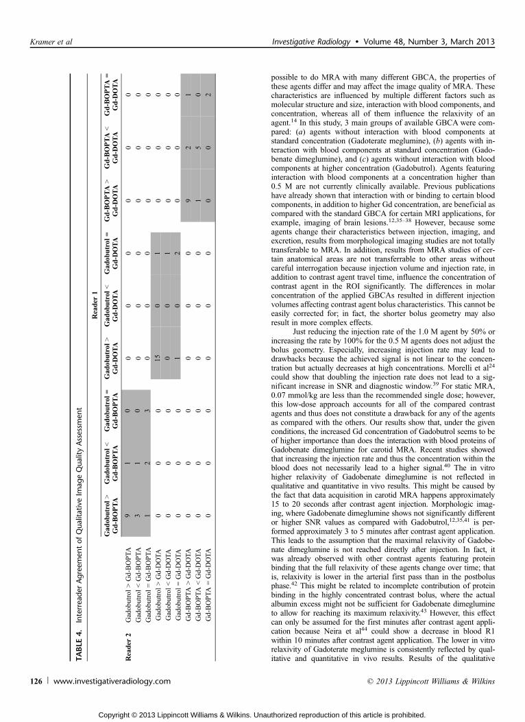
possible to do MRA with many different GBCA, the properties ofthese agents differ and may affect the image quality of MRA. Thesecharacteristics are influenced by multiple different factors such asmolecular structure and size, interaction with blood components, andconcentration, whereas all of them influence the relaxivity of anagent.14 In this study, 3 main groups of available GBCA were com-pared: (a) agents without interaction with blood components atstandard concentration (Gadoterate meglumine), (b) agents with in-teraction with blood components at standard concentration (Gado-benate dimeglumine), and (c) agents without interaction with bloodcomponents at higher concentration (Gadobutrol). Agents featuringinteraction with blood components at a concentration higher than0.5 M are not currently clinically available. Previous publicationshave already shown that interaction with or binding to certain bloodcomponents, in addition to higher Gd concentration, are beneficial ascompared with the standard GBCA for certain MRI applications, forexample, imaging of brain lesions.12,35Y38 However, because someagents change their characteristics between injection, imaging, andexcretion, results from morphological imaging studies are not totallytransferable to MRA. In addition, results from MRA studies of cer-tain anatomical areas are not transferrable to other areas withoutcareful interrogation because injection volume and injection rate, inaddition to contrast agent travel time, influence the concentration ofcontrast agent in the ROI significantly. The differences in molarconcentration of the applied GBCAs resulted in different injectionvolumes affecting contrast agent bolus characteristics. This cannot beeasily corrected for; in fact, the shorter bolus geometry may alsoresult in more complex effects.
Just reducing the injection rate of the 1.0 M agent by 50% orincreasing the rate by 100% for the 0.5 M agents does not adjust thebolus geometry. Especially, increasing injection rate may lead todrawbacks because the achieved signal is not linear to the concen-tration but actually decreases at high concentrations. Morelli et al24
could show that doubling the injection rate does not lead to a sig-nificant increase in SNR and diagnostic window.39 For static MRA,0.07 mmol/kg are less than the recommended single dose; however,this low-dose approach accounts for all of the compared contrastagents and thus does not constitute a drawback for any of the agentsas compared with the others. Our results show that, under the givenconditions, the increased Gd concentration of Gadobutrol seems to beof higher importance than does the interaction with blood proteins ofGadobenate dimeglumine for carotid MRA. Recent studies showedthat increasing the injection rate and thus the concentration within theblood does not necessarily lead to a higher signal.40 The in vitrohigher relaxivity of Gadobenate dimeglumine is not reflected inqualitative and quantitative in vivo results. This might be caused bythe fact that data acquisition in carotid MRA happens approximately15 to 20 seconds after contrast agent injection. Morphologic imag-ing, where Gadobenate dimeglumine shows not significantly differentor higher SNR values as compared with Gadobutrol,12,35,41 is per-formed approximately 3 to 5 minutes after contrast agent application.This leads to the assumption that the maximal relaxivity of Gadobe-nate dimeglumine is not reached directly after injection. In fact, itwas already observed with other contrast agents featuring proteinbinding that the full relaxivity of these agents change over time; thatis, relaxivity is lower in the arterial first pass than in the postbolusphase.42 This might be related to incomplete contribution of proteinbinding in the highly concentrated contrast bolus, where the actualalbumin excess might not be sufficient for Gadobenate dimeglumineto allow for reaching its maximum relaxivity.43 However, this effectcan only be assumed for the first minutes after contrast agent appli-cation because Neira et al44 could show a decrease in blood R1within 10 minutes after contrast agent application. The lower in vitrorelaxivity of Gadoterate meglumine is consistently reflected by qual-itative and quantitative in vivo results. Results of the qualitativeTA
BLE
4.Interrea
derAgree
men
tof
Qua
litativeIm
ageQua
lityAssessm
ent
Reader
1
Gad
obutrol
9Gd-BOPTA
Gad
obutrol
GGd-BOPTA
Gad
obutrol
=Gd-BOPTA
Gad
obutrol
9Gd-D
OTA
Gad
obutrol
GGd-D
OTA
Gad
obutrol
=Gd-D
OTA
Gd-BOPTA
9Gd-D
OTA
Gd-BOPTA
GGd-D
OTA
Gd-BOPTA
=Gd-D
OTA
Reader
2Gadobutrol9Gd-BOPTA
91
00
00
00
0
GadobutrolGGd-BOPTA
31
00
00
00
0
Gadobutrol=Gd-BOPTA
12
30
00
00
0
Gadobutrol9Gd-DOTA
00
015
01
00
0
GadobutrolGGd-DOTA
00
00
01
00
0
Gadobutrol=Gd-DOTA
00
01
02
00
0
Gd-BOPTA
9Gd-DOTA
00
00
00
92
1
Gd-BOPTA
GGd-DOTA
00
00
00
15
0
Gd-BOPTA
=Gd-DOTA
00
00
00
00
2
Kramer et al Investigative Radiology & Volume 48, Number 3, March 2013
126 www.investigativeradiology.com * 2013 Lippincott Williams & Wilkins
Copyright © 2013 Lippincott Williams & Wilkins. Unauthorized reproduction of this article is prohibited.
image quality reading comparing the agents side by side are perfectlysupported by the quantitative measurements. The significantly higherSNR and CNR of MRA data sets acquired with Gadobutrol lead to asignificantly higher qualitative rating of these data sets. However, CNRvalues in MRA are of lesser importance because reading these datasets is mostly done after background subtraction. The different ratio inbetween SNR values for the different agents at different locations canbe explained in several ways, for example, the nonlinearity of signal ascompared with contrast agent concentration and the variation of signalat different flow velocities. Both are true when comparing measure-ments at the proximal and distal ICA. Partial volume effects in smallervessels and the coverage with different coils can explain the differencesin SNR at the evaluated levels achieved with all GBCAs.33,45
Edge blurring, as it theoretically occurs if only the center parts ofk-space are sampled during the presence of contrast agent in the ROI,could be expected because of the small injected volume when usingGadobutrol. Indeed, it was neither quantitatively measurable nor reflec-ted in an impaired qualitative image quality, and no significant differ-ences occurred comparing the 3 evaluated agents. This leads to theconclusion that 0.07 mmol/kg BWof a 1.0 M GBCA injected at 2.0mL/sserves with a sufficiently long contrast agent bolus to avoid disturbingedge blurring; that is, contrast agent is present in the vascular territoryof interest during the entire k-space sampling in an adequate amount.
Since the introduction of dynamic MRA, this technique hasundergone great improvement in spatial and temporal resolution and,nowadays, can add valuable information to an MRA examination.However, there is still a trade-off between dynamic information andspatial resolution, meaning, that dynamic MRA cannot completelyreplace static MRA presently. To further push the image quality ofdynamic MRA, this technique benefits from dedicated contrastagents as well.46 Signal-to-noise ratio evaluation of dynamic MRAdata sets again showed higher values achieved with a highly con-centrated GBCA as compared with the standard 0.5 M GBCA. Thefact that SNR values achieved with dynamic MRA differ from thosemeasured in static MRA and that the ratio between all agents is dif-ferent from this measured in static MRA is sequence inherent. Thetechnique we used for dynamic MRA relies on view sharing whereonly the center part of k-space, which encodes for the contrast in theimage, is sampled for every point in time, and the peripheral parts ofk-space are shared between different frames. In dynamic MRA, theSNR differences between Gadobenate dimeglumine and Gadoteratemeglumine as compared with Gadobutrol are less pronounced than instatic MRA. This can be rationalized by the used sequence settingsand the injection parameters. The sequence used for dynamic MRAfacilitates in setting the size of the central region A of k-space, whichis sampled in every time frame, and the sampling density of the pe-ripheral part B of k-space. Because we selected a size of 17% of theentire k-space for region A, which is relatively small, some parts ofthe central k-space that contribute to the contrasts in the image aresampled as part of the periphery. Sampling density of 20% for regionB in combination with a temporal resolution of 1.86 seconds perframe ends up in a temporal footprint for sampling the entire k-spacedata for 1 time frame of approximately 9 seconds. An injection rate of2 mL/s of 0.03 mmol/kg BW (È4.5 mL for Gadobenate dimeglumineand Gadoterate meglumine/È2.3 mL for Gadobutrol in a 75 kg par-ticipant) results in a bolus shorter than 1 temporal footprint. For avery short bolus, as it is a fact when using Gadobutrol, it means thatnot all central k-space sampling is done during the presence of acontrast agent. However, the resulting signal is averaged from theentire temporal footprint, resulting in a lower signal in the final imagethan the actual reached maximum during acquisition. On the otherhand, for a bolus long enough for a footprint, it results in a highermean signal over the sampling time.
Findings at the later time frames of signal curves beyond the ar-terial peak enhancement show the effect of interaction of Gadobenate
dimeglumine with blood proteins. Signal curves of Gadobutrol andGadoterate meglumine both flatten with time. The curve of Gadobenatedimeglumine also flattens but much less than that of both comparators,most likely because of the prolonged presence of Gadobenate dime-glumine in the blood pool. At the last sampled time point (98 sec. p.i.),the signal curves of Gadobutrol and Gadobenate dimeglumine nearlyintersect; this might explain the finding of other publications that showno significant differences between Gadobutrol and Gadobenate dime-glumine in peripheral MRA.
A limitation of this study is the exclusive evaluation of vol-unteer data sets without any pathologic findings. Thus, evaluation ofthe influence of different SNR on diagnostic accuracy was not pos-sible. However, this was not an aim of the current study and will beevaluated in the future. Three different examinations with requiredintervals in between in a patient with potentially severe carotid ar-tery disease are delaying necessary treatment and are thus ethicallyunjustifiable.
CONCLUSIONThis study indicates that, in static and dynamic MRA of the
carotid arteries, a contrast agent that features a higher Gd concen-tration shows higher qualitative image quality and higher SNR andCNR as compared with 0.5 M agents, regardless of whether theyfeature protein interaction/binding or not. Because we could notconfirm the finding that contrast agents with at least temporarybinding to blood components are beneficial for morphologic imagingfor MRA applications, it seems that the high relaxivity of theseagents due to concentration differences of contrast agent moleculesand human albumin in the first-pass bolus is reached after some timeof interaction with blood components but not directly after injection.However, these agents might be superior because of the higherrelaxivity in later phase imaging. Our results reflect findings in ca-rotid MRA, and the evaluated characteristics might be different inother vascular territories that are imaged with an extended delay afterinjection. Using a highly concentrated GBCA enables comprehensivestatic and dynamicMRA imaging of the head and neck arteries withoutthe need to exceed the recommended dosage of 0.1 mmol/kg BW.
ACKNOWLEDGMENTSThe authors thank Denise Steffinger, Markus Bregler, and
Michele Picciolo for their continuous tremendous help during theexecution of all MRI examinations, Martin Rohrer for his input onmolecular principles of contrast agents, and Gerhard Laub for hisinput on MR sequence basics.
REFERENCES1. Prabhakaran P, Ajay VS, Prabhakaran D, et al. Global cardiovascular disease
research survey. J Am Coll Cardiol. 2007;50:2322Y2328.2. Reddy KS. Cardiovascular disease in non-Western countries. N Engl J Med.
2004;350:2438Y2440.3. Reddy KS, Yusuf S. Emerging epidemic of cardiovascular disease in devel-
oping countries. Circulation. 1998;97:596Y601.4. Remonda L, Senn P, Barth A, et al. Contrast-enhanced 3D MR angiography of
the carotid artery: comparison with conventional digital subtraction angiogra-phy. AJNR Am J Neuroradiol. 2002;23:213Y219.
5. Sundgren PC, Sunden P, Lindgren A, et al. Carotid artery stenosis: contrast-enhanced MR angiography with two different scan times compared with digitalsubtraction angiography. Neuroradiology. 2002;44:592Y599.
6. Anzalone N, Scomazzoni F, Castellano R, et al. Carotid artery stenosis:intraindividual correlations of 3D time-of-flight MR angiography, contrast-enhanced MR angiography, conventional DSA, and rotational angiography fordetection and grading. Radiology. 2005;236:204Y213.
7. Nael K, Ruehm SG, Michaely HJ, et al. High spatial-resolution CE-MRA of thecarotid circulation with parallel imaging: comparison of image quality between2 different acceleration factors at 3.0 tesla. Invest Radiol. 2006;41:391Y399.
8. Kramer H, Runge VM, Morelli JN, et al. Magnetic resonance angiography ofthe carotid arteries: comparison of unenhanced and contrast enhanced tech-niques. Eur Radiol. 2011;21:1667Y1676.
Investigative Radiology & Volume 48, Number 3, March 2013 Dynamic and Static MRA of the Supra-aortic Vessels
* 2013 Lippincott Williams & Wilkins www.investigativeradiology.com 127
Copyright © 2013 Lippincott Williams & Wilkins. Unauthorized reproduction of this article is prohibited.
9. Lohan DG, Tomasian A, Saleh RS, et al. Ultra-low-dose, time-resolved contrast-enhanced magnetic resonance angiography of the carotid arteries at 3.0 tesla.Invest Radiol. 2009;44:207Y217.
10. McCarthy RM, Deshpande VS, Beohar N, et al. Three-dimensional breathholdmagnetization-prepared TrueFISP: a pilot study for magnetic resonance im-aging of the coronary artery disease. Invest Radiol. 2007;42:665Y670.
11. Achenbach M, Figiel JH, Burbelko M, et al. Prospective comparison of imagequality and diagnostic accuracy of 0.5 molar gadobenate dimeglumine and 1.0molar gadobutrol in contrast-enhanced run-off magnetic resonance angiogra-phy of the lower extremities. J Magn Reson Imaging. 2010;32:1166Y1171.
12. Essig M, Lodemann KP, Le-Huu M, et al. Intraindividual comparison ofgadobenate dimeglumine and gadobutrol for cerebral magnetic resonanceperfusion imaging at 1.5 T. Invest Radiol. 2006;41:256Y263.
13. Nikolaou K, Kramer H, Grosse C, et al. High-spatial-resolution multistationMR angiography with parallel imaging and blood pool contrast agent: initialexperience. Radiology. 2006;241:861Y872.
14. Rohrer M, Bauer H, Mintorovitch J, et al. Comparison of magnetic properties ofMRI contrast media solutions at different magnetic field strengths. InvestRadiol. 2005;40:715Y724.
15. Pintaske J, Martirosian P, Graf H, et al. Relaxivity of gadopentetate dimeglu-mine (Magnevist), gadobutrol (Gadovist), and gadobenate dimeglumine (MultiHance)in human blood plasma at 0.2, 1.5, and 3 tesla. Invest Radiol. 2006;41:213Y221.
16. KramerH, RungeVM, Naul LG, et al. BrainMRI with single-dose (0.1mmol/kg)gadobutrol at 1.5 T and 3 T: comparison with 0.15 mmol/kg gadoterate meglu-mine. AJR Am J Roentgenol. 2010;194:1337Y1342.
17. Cowper S. Nephrogenic Fibrosing Dermopathy [NFD/NSFWeb site]. 2001Y2009:Available at http://www.icnfdr.org. Accessed August 10, 2012.
18. Thomsen HS. Nephrogenic systemic fibrosis: a serious late adverse reaction togadodiamide. Eur Radiol. 2006;16:2619Y2621.
19. Katoh M, Spuentrup E, Stuber M, et al. Flow targeted 3D steady-state free-precession coronary MR angiography: comparison of three different imagingapproaches. Invest Radiol. 2009;44:757Y762.
20. Herborn CU, Runge VM, Watkins DM, et al. MR angiography of the renalarteries: intraindividual comparison of double-dose contrast enhancement at1.5 T with standard dose at 3 T. AJR Am J Roentgenol. 2008;190:173Y177.
21. Jourdan C, Heverhagen JT, Knopp MV. Dose comparison of single- vs. double-dose in contrast-enhanced magnetic resonance angiography of the carotid ar-teries: intraindividual cross-over blinded trial using Gd-DTPA. J Magn ResonImaging. 2007;25:557Y563.
22. Thurnher SA, Capelastegui A, Del Olmo FH, et al. Safety and effectiveness ofsingle- versus triple-dose gadodiamide injection- enhanced MR angiographyof the abdomen: a phase III double-blind multicenter study. Radiology. 2001;219:137Y146.
23. Finn JP, Baskaran V, Carr JC, et al. Thorax: low-dose contrast-enhanced three-dimensional MR angiography with subsecond temporal resolutionVinitial re-sults. Radiology. 2002;224:896Y904.
24. Morelli JN, Runge VM, Ai F, et al. Magnetic resonance evaluation of renalartery stenosis in a swine model: performance of low-dose gadobutrol versusgadoterate meglumine in comparison with digital subtraction intra-arterialcatheter angiography. Invest Radiol. 2012;47:376Y382.
25. Wuesten O, Morelli JN, Miller MW, et al. MR angiography of carotid arteryaneurysms in a porcine model at 3 tesla: comparison of two different macro-cyclic gadolinium chelates and of dynamic and conventional techniques.J Magn Reson Imaging. 2012;36:1203Y1212.
26. Hadizadeh DR, Kukuk GM, Fahlenkamp UL, et al. Simultaneous MR arteri-ography and venography with blood pool contrast agent detects deep venousthrombosis in suspected arterial disease. AJR Am J Roentgenol. 2012;198:1188Y1195.
27. Kramer U, Fenchel M, Laub G, et al. Low-dose, time-resolved, contrast-enhanced 3D MR angiography in the assessment of the abdominal aorta and itsmajor branches at 3 tesla. Acad Radiol. 2010;17:564Y576.
28. Dietrich O, Raya JG, Reeder SB, et al. Measurement of signal-to-noise ratios inMR images: influence of multichannel coils, parallel imaging, and recon-struction filters. J Magn Reson Imaging. 2007;26:375Y385.
29. Anzidei M, Napoli A, Marincola BC, et al. High-resolution steady state mag-netic resonance angiography of the carotid arteries: are intravascular agentsnecessary?: feasibility and preliminary experience with gadobenate dimeglu-mine. Invest Radiol. 2009;44:784Y792.
30. Bueltmann E, Erb G, Kirchin MA, et al. Intra-individual crossover comparisonof gadobenate dimeglumine and gadopentetate dimeglumine for contrast-enhanced magnetic resonance angiography of the supraaortic vessels at 3 tesla.Invest Radiol. 2008;43:695Y702.
31. Nael K, Fenchel M, Krishnam M, et al. 3.0 tesla high spatial resolutioncontrast-enhanced magnetic resonance angiography (CE-MRA) of the pul-monary circulation: initial experience with a 32-channel phased array coilusing a high relaxivity contrast agent. Invest Radiol. 2007;42:392Y398.
32. Berg F, Bangard C, Bovenschulte H, et al. Feasibility of peripheral contrast-enhanced magnetic resonance angiography at 3.0 tesla with a hybrid tech-nique: comparison with digital subtraction angiography. Invest Radiol. 2008;43:642Y649.
33. Kramer H, Michaely HJ, Matschl V, et al High-resolution magnetic resonanceangiography of the lower extremities with a dedicated 36-element matrix coil at3 tesla. Invest Radiol. 2007;42:477Y483.
34. Kramer H, Zenge M, Schmitt P, et al. Peripheral magnetic resonance angiog-raphy (MRA) with continuous table movement at 3.0 T: initial experiencecompared with step-by-step MRA. Invest Radiol. 2008;43:627Y634.
35. Anzalone N. Comparative studies of different gadolinium agents in braintumors: differences between gadolinium chelates and their possible influenceon imaging features. AJNR Am J Neuroradiol. 2010;31:981Y982.
36. Anzalone N, Gerevini S, Scotti R, et al. Detection of cerebral metastases onmagnetic resonance imaging: intraindividual comparison of gadobutrol withgadopentetate dimeglumine. Acta Radiol. 2009;50:933Y940.
37. Le Duc G, Corde S, Charvet AM, et al. In vivo measurement of gadoliniumconcentration in a rat glioma model by monochromatic quantitative computedtomography: comparison between gadopentetate dimeglumine and gadobutrol.Invest Radiol. 2004;39:385Y393.
38. Huppertz A, Rohrer M. Gadobutrol, a highly concentrated MR-imaging con-trast agent: its physicochemical characteristics and the basis for its use incontrast-enhanced MR angiography and perfusion imaging. Eur Radiol. 2004;14(suppl 5):M12Y18.
39. Husarik DB, Bashir MR, Weber PW, et al. Contrast-enhanced magnetic reso-nance angiography: first-pass arterial enhancement as a function of gadolinium-chelate concentration, and the saline chaser volume and injection rate. InvestRadiol. 2012;47:121Y127.
40. Wilson GJ, Bastawrous S, Bhargava P, et al. Relaxivities of Protein and non-Protein Binding Gd Agents at High Concentrations in Blood: RelaxationMeasurements. Utrecht, the Netherlands: MRA Club; 2012. Abstract.
41. Seidl Z, Vymazal J, Mechl M, et al. Does higher gadolinium concentration playa role in the morphologic assessment of brain tumors? Results of a multicenterintraindividual crossover comparison of gadobutrol versus gadobenate dime-glumine (the MERIT Study). AJNR Am J Neuroradiol. 2012;33:1050Y1058.
42. Port M, Corot C, Violas X, et al. How to compare the efficiency of albumin-bound and nonalbumin-bound contrast agents in vivo: the concept of dynamicrelaxivity. Invest Radiol. 2005;40:565Y573.
43. Rohrer M. MRI contrast mediaVintroduction and basic properties of the bloodpool agent vasovist. In: Leiner TG, Rohrer M, Schienberg SO, eds. ClinicalBlood Pool MR Imaging. Heidelberg, Germany: Springer Medizinverlag;2008:3Y15.
44. Neira C, Anzidei M, Napoli A, et al. Investigation of the longitudinal relaxationrate of blood after gadobenate dimeglumine administration: sequence optimi-zation, dynamic acquisition, and clinical impact for contrast-enhanced MRangiography of the carotid arteries. Invest Radiol. 2011;46:774Y782.
45. Schultz E, Felix R. Phantom measurements of spatial resolution and the partial-volume-effect in computer tomography (author’s transl) [in German]. Rofo.1978;129:673Y678.
46. Kramer H, Michaely HJ, Requardt M, et al. Effects of injection rate and dose onimage quality in time-resolved magnetic resonance angiography (MRA) byusing 1.0 M contrast agents. Eur Radiol. 2007;17:1394Y1402.
Kramer et al Investigative Radiology & Volume 48, Number 3, March 2013
128 www.investigativeradiology.com * 2013 Lippincott Williams & Wilkins
Copyright © 2013 Lippincott Williams & Wilkins. Unauthorized reproduction of this article is prohibited.