Embed Size (px)
Citation preview

November 2009 1193
Major histocompatibility complexes (MHCs) are cell-sur-face glycoproteins that play an important role in immune re-sponse against infection. MHCs bind to a small peptide de-rived from either host or pathogen proteins, and present themto a T cell as a part of the immune system’s mechanism foridentifying and responding to foreign antigens. The engage-ment of an MHC molecule with a peptide by an antigen re-ceptor on a cell causes the stimulation of the T cell and theactivation of the immune response. The striking characteris-tic of MHCs is their ability to bind to various peptides inorder to ensure an immune response against many possiblepathogens.
MHCs mainly fall into class I and class II. Class I MHC iscomposed of a heavy chain and a b2-immunoglobulin (b2m).The heavy chain is divided into three domains, a1, a2 anda3. The a1 and a2 domains are also functionally expressedas a peptide-binding platform domain, as the two domainsare complicatedly intertwined. The platform domain formsan eight-stranded b-sheet and two a-helical regions, and abound peptide is accommodated between the two a-helicalregions on the b-sheet (Fig. 1A).1) The a3 and b2m domainsare known as the membrane-proximal immunoglobulin-likedomains because they are located between the platform do-main and the cell surface.
Conversely, class II MHC is composed of two asymmetricchains, the a- and b-chains, divided into the a1 and a2 do-mains, and the b1 and b2 domains, respectively. Thus the a1and b1 domains come from different chains, but the two do-mains are also functionally expressed as a peptide-bindingplatform domain, because they are complicatedly inter-twined. Surprisingly, the platform, b2 and a2 domains ofclass II MHC are very similar in structure to the platform,
a3 and b2m domains of class I MHC, respectively (Figs.1A, B).2,3) However, there is a difference in peptide-bindingbetween both classes: Class I MHC binds to the peptide lim-ited in length, usually 8—10 residues,4—6) while class IIMHC binds to the peptide without apparent restriction inlength (ca. 8—23 amino acids in length).7,8)
Previous studies concerning MHCs were interested in thecrystal structure of the platform domain required for peptide-binding and presenting to T cells.1—9) Moreover, some exper-imental and theoretical studies of the peptide-free platformdomain have received attention because its structure has notyet been crystallized in both classes.10—20) On the other hand,the two membrane-proximal domains have attracted less in-terest than the platform domain. Some biochemical studiesinvestigated the two membrane-proximal domains of class IMHC, suggesting the importance of b2m in peptide-bind-ing.21,22) However, there are no experimental or theoreticalstudies concerning the two membrane-proximal domains ofclass II MHC.
In this study, we simulated the dynamics of a whole andpartial model deficient in either of the two membrane-proxi-mal domains for class I and class II using normal modeanalysis, and compared the influence of the two membrane-proximal domains upon the dynamics of the platform domainbetween both classes. Our study suggests that the two mem-brane-proximal domains of class II MHC have a greater in-fluence upon the most important peptide-binding pocket thanthose of class I MHC.
ExperimentalModels Used for Normal Mode Analysis Initial atomic coordinates
were taken from X-ray crystallographic structures stored in the BrookhavenProtein Data Bank (PDB). A structure of human leukocyte antigen (HLA)-
Dynamic Influence of the Two Membrane-Proximal Immunoglobulin-LikeDomains upon the Peptide-Binding Platform Domain in Class I and ClassII Major Histocompatibility Complexes: Normal Mode Analysis
Hiroyuki NOJIMA,a,# Kazuhiko KANOU,a,# Kenshu KAMIYA,b Koichiro ATSUDA,a
Hideaki UMEYAMA,a and Mayuko TAKEDA-SHITAKA*,a
a School of Pharmacy, Kitasato University; 5–9–1 Shirokane, Minato-ku, Tokyo 108–8641, Japan: and b School of Science,Kitasato University; 1–15–1 Sagamihara, Kanagawa 228–8555, Japan.Received March 12, 2009; accepted August 9, 2009; published online August 25, 2009
Major histocompatibility complexes (MHCs) mainly fall into class I and class II. The two classes have simi-lar structures, with two membrane-proximal immunoglobulin-like domains and a peptide-binding platform do-main, though their organizations are different. We simulated the dynamics of a whole and partial model deficientin either of the two membrane-proximal domains for class I and class II using normal mode analysis. Our studyshowed that the influence of the two membrane-proximal domains upon the dynamics of the platform domainwere decisively different between class II and class I. Both membrane-proximal domains (the aa 2 and bb 2 do-mains) of class II MHC, especially the aa 2 domain, influenced the most important pocket that accommodates alarge hydrophobic anchor side chain of the N-terminal side of the bound peptide, though the pocket was not inthe aa 2 domain neighborhood. By contrast, the two membrane-proximal domains (the aa 3 and bb2m domains) ofclass I MHC had little influence upon the most important pocket that accommodates the N-terminal residue ofthe bound peptide. These results suggest that the two membrane-proximal domains of class II MHC have agreater influence upon peptide-binding than those of class I MHC.
Key words major histocompatibility complex; class I; class II; membrane-proximal immunoglobulin-like domain; peptide-binding platform domain; normal mode analysis
Chem. Pharm. Bull. 57(11) 1193—1199 (2009)
© 2009 Pharmaceutical Society of Japan∗ To whom correspondence should be addressed. e-mail: [email protected]# These authors contributed equally to this work.

A2 with a peptide derived from human immunodeficiency virus (HIV) re-verse transcriptase [9 residues: RT309—317 (ILKEPVHGV)] was adoptedas the “class I” model, (PDB code: 1HHJ).6) In the PDB, an A-chain (theheavy chain) of 275 residues (the a1, a2 and a3 domains, G h1—E h275),a B-chain (b2m) of 100 residues (M b20—M b299) and a C-chain (RT pep-tide) of 9 residues were used as the “class I whole” model. A part of the A-chain (the a1 and a2 domains, G h1—R h181), the B- and C-chains wereused as the “class I a3-removed” model, while the A- and C-chains wereused as the “class I b2m-removed” model.
A structure of HLA-DR1 with a peptide derived from influenza virushemagglutinin [13 residues: HA306—318 (PKYVKQNTLKLAT)] wasadopted as the “class II” model (PDB code: 1DLH).3) In the PDB, a D-chain(a-chain) of 180 residues (the a1 and a2 domains, E a3—A a182), an E-chain (b-chain) of 188 residues (the b1 and b2 domains, T b3—A b190)and an F-chain (HA peptide) of 13 residues were used as the “class IIwhole” model. The D-chain, a part of the E-chain (the b1 domain, T b3—Qb92) and the F-chain were used as the “class II b2-removed” model, while apart of the D-chain (the a1 domain, E a3—Y a79), and the E- and F-chainswere used as the “class II a2-removed” model.
The details of domain structure for each model are shown in Table 1.Energy Optimization Each model was energetically optimized to ob-
tain structures for normal mode analysis. In the energy optimization, bondlength, bond angle and dihedral angle were treated as parameters.23) To lowercomputational cost in the energy optimization process, we adopted a slightlymodified Assisted Model Building with Energy Refinement (AMBER)united atom-force field. In this field, aliphatic and aromatic carbon-bondedhydrogen atoms were neglected, and, instead, the three suspected carbonatoms whose atomic weights were 13.02, 14.03 and 15.03, respectively, wereintroduced.24,25) “Slight modification” means that bond-angle and dihedral-angle were neglected in the parameters of a disulfide bond, and the “sus-pected potential” in which the distance between two S atoms was treated asonly a parameter was introduced.23) The validity of the simplified force fieldhas been illustrated in our previous study.26)
A distance-dependent dielectric constant (r/Å) for electrostatic energywas used to consider short-distance electrostatic interactions.27) Position-re-striction energy, E, expressed in Eq. 1 was imposed in the energy optimiza-tion process.
(1)
In Eq. 1, K (kcal/mol Å) is a force constant, and ri and ri0 are displaced and
initial coordinates of the ith atom, respectively. The above restriction wasgradually relaxed by decreasing the K value so that normal mode analysiscould be executed near the initial coordinates. Some energy-optimized struc-tures were prepared from the initial coordinates of a model using differentrelaxation patterns.15,16,26,28—30) All the obtained energy-optimized structures,however, do not have an energy minimum accurate enough to approximate toa harmonic potential. Therefore, we examined the normal modes calculatedusing the obtained energy-optimized structures, and omitted the energy-opti-mized structures improper to normal mode analysis. After all, six proper en-ergy-optimized structures were sampled for a model. Root mean square de-viation (RMSD) between the six energy-optimized structures and the initialcoordinates (for all atoms except hydrogen atoms) ranged from 1.82 to2.46 Å in all models, indicating that the energy-optimized structures are nearthe initial coordinates (Table 1).
Normal Mode Analysis Normal mode analysis was performed usingprograms developed by our laboratory.23) In the normal mode calculation,only dihedral angle was treated as a parameter. A RMS fluctuation at the ithCa atom, Fi, was calculated by Eq. 231):
(2)
in which aki is a projection vector of the kth normal mode with frequency w k
on the Cartesian components of displacement vector Dri, kB is the Bolzmannconstant, and T is the absolute temperature. The fluctuation was calculat-ed by our program, assuming a temperature of 300 K, and was then deter-mined by averaging the data of its six sampled energy-optimized struc-tures.15,16,26,28—30)
When attention is focused on the motion of a component, it can be brokendown into internal and external motions.32) Eckart’s condition was applied toextract the internal motion of a component (for example, a domain) from thewhole motion.33) A “significant fluctuation difference” was calculated be-
� � � �F r k Ti i kl k
k
2 2 2 2� �Δ B (| | / )a ω∑
E K r ri i
i
N
� �( )0 2∑
1194 Vol. 57, No. 11
Table 1. Domain Structures of Each Model and Summaries of Energy-Optimization and Normal Mode Calculations
“Class I” models “Class II” models
“a3-removed” “b2m-removed” “Whole” “b2-removed” “a2-removed” “Whole”
Platform domain a1 (G ha)1—N h86)�a2 (Q h87—R h181) a1 (E a3—Y a79)�b1 (T b3—Q b92)
—a3 a3 —
b2 b2(T h182—E h275) (R b93—A b190)
Membrane-proximal domainsb)
b2m — b2ma2
— a2(M b2
c)0—M b299) (T a80—A a182)
Bound peptideRT
RT RTHA
HA HA(I309—V317) (P306—T318)
Total number of residues 290 284 384 283 278 381
vs. the initial coordinatese) 1.90—2.03 2.01—2.28 2.07—2.25 2.21—2.45 1.97—2.46 1.82—1.96
RMSDd )
between the six energy-optimized 0.53—1.11 0.72—1.18 0.47—1.18 0.67—1.59 0.71—1.44 0.65—1.05
structures f )
Total 1289 1224 1674 1227 1224 1632
Number of �50 cm�1 g) 219—227 211—225 293—303 230—241 213—234 301—317
modes
(�50 cm�1/total) (%) 17.0—17.6 17.2—18.4 17.5—18.1 18.7—19.6 17.4—19.1 18.4—19.4
a) “h” means the heavy chain. b) The b2 and a2 domains of class II MHC structurally correspond to the a3 and b2m domains of class I MHC, respectively. c) “b2”means b2m. d ) RMSD was calculated for all atoms except for hydrogen. e) RMSD was calculated between the initial coordinates and the six energy-optimized structures. f )RMSD was calculated between the six energy-optimized structures. g) Number of modes �50 cm�1 have a range because distributions of normal modes are slightly different be-tween the six energy-optimized structures.

tween two models by Wilcoxon’s rank sum test, a nonparametric test (signifi-cance level 2.5%), using data of the six energy-optimized structures for eachmodel.15,16,26,28,29) If a fluctuation difference at a Ca atom was significant be-tween two models, it was expressed as a “significant fluctuation difference”by subtracting the averages of the two models. If not, it was expressed aszero.
Generally, the dynamic characteristic of a large molecule is heavily influ-enced by low frequency modes.27,31,34—36) In this study, the fluctuation wasexpressed in the low frequency modes (�50 cm�1), as there were little dif-ferences from expression in all-frequency modes (data not shown). A num-ber of the low frequency modes shared 17.0—19.6% of all frequency modesin all models (Table 1).
ResultsThe Fluctuation Differences between the “Class I
Whole” and “Class I aa 3-Removed” Models, and betweenthe “Class II Whole” and “Class II bb2-Removed” ModelsThe a3 domain of class I MHC structurally corresponds tothe b2 domain of class II MHC, as shown in Fig. 1. Thus, thefluctuation difference between “class I whole” and “class Ia3-removed” models was compared with that between the“class II whole” and “class II b2-removed” models. The fluc-tuations of the “class I a3-removed” (Fig. 2A) and “class IIb2-removed” (Fig. 2B) models were superimposed on thoseof the “class I whole” and “class II whole” models, respec-tively. The fluctuation of each model was expressed by theinternal motion of either the platform or another proximaldomain. At a Ca atom, a “significant fluctuation difference”was calculated between the “whole” and “removed” models(yellow lines in Fig. 2), while a “remarkable fluctuation dif-ference” (less than �0.03) was noted in the platform domain(the red lines and red vertical shadows under these lines inFig. 2).
Comparing between the “class I whole” and “class I a3-
removed” models, ten residues showed a “remarkable fluctu-ation difference” (residues h29—30 and h49 in the a1 do-main, and h105—108, h176—177 and h180 in the a2 do-main; Fig. 2A). The “class I” models have two importantpockets to accommodate the N-terminal (I RT309, the “N-terminal side” pocket) and C-terminal (V RT317, the “C-ter-minal side” pocket) residues of the bound peptide.6) The “N-terminal side” pocket is composed of residues h7, h59, h63and h66 in the a1 domain and h159, h167 and h171 in thea2 domain (the red circles in Fig. 2A), while the “C-terminal
November 2009 1195
Fig. 2. Ca Atom RMS Fluctuations of (A) the “Class I Whole” and “Class I a3-Removed” Models and of (B) the “Class II Whole” and “Class II b2-Re-moved” Models
In each figure, the fluctuation of the “removed” model (gray line) is superimposed on that of the “whole” model (black line). The “significant fluctuation difference” between the“removed” and “whole” models is indicated in a yellow line. The “significant fluctuation difference” less than �0.03 is colored red as a “remarkable fluctuation difference” andshadowed by light red vertical line under the red line. The notations of secondary structure, a-helical region, b-strand and loop are shown at the bottom by light green, light blueand gray bars, respectively. In (A), the residues forming two pockets to accommodate the N-terminal (I 309) and C-terminal (V317) residues of RT peptide are marked by red cir-cles and blue triangles, respectively. In (B), the residues forming two pockets to accommodate two hydrophobic anchor side chains in the N-terminal (Y308) and C-terminal (L316)sides of HA peptide are marked by a red circle and a blue triangle, respectively.
Fig. 1. Schematic Structures of (A) Class I (PDB Code, 1HHJ) and (B)Class II (PDB Code, 1DLH) MHCs Separated by Color for Each Domain
Class I MHC is divided into the a1 (green), a2 (yellow) and a3 (red) domains andb2-immunoglobulin (b2m, blue). Class II MHC is divided into the a1 (green), a2(blue), b1 (yellow) and b2 (red) domains. The bound peptides are indicated by graysticks. The a1, a2 and a3 domains and b2m in class I MHC are similar in secondarystructure to the a1, b1, b2 and a2 domains in class II MHC, respectively.
side” pocket is composed of residues h77, h80—81 and h84in the a1 domain and h116, h143 and h146 in the a2 domain(the blue triangles in Fig. 2A). These residues, however, didnot show a “remarkable fluctuation difference.”
By contrast, comparing between the “class II whole” and“class II b2-removed” models, fifteen residues showed a “re-markable fluctuation difference” (residues a6, a26—29,a45 and a47 in the a1 domain, and b21, b86—92 in the b1domain, Fig. 2B). The “class II” models have no pockets toaccommodate both terminal residues of the bound peptidebecause they stick out from the platform domain. To com-pensate for this, the “class II” models have two importantdeep pockets to accommodate two large hydrophobic anchorside chains of the N-terminal side (Y HA308, the “N-termi-nal side” pocket) and the C-terminal side (L HA316, the “C-terminal side” pocket) of the bound peptide.3) The “N-termi-nal side” pocket is composed of residues a31, a32, a43 anda52—54 in the a1 domain and b86 and b89 in the b1 do-main (the red circles in Fig. 2B), while the “C-terminal side”pocket is composed of residues a69, a72, a73 and a76 inthe a1 domain and b9, b57 and b60 in the b1 domain (theblue triangles in Fig. 2B). Among these residues, G b86 andF b89, which participate in the formation of the “N-terminalside” pocket, showed a “remarkable fluctuation difference.”
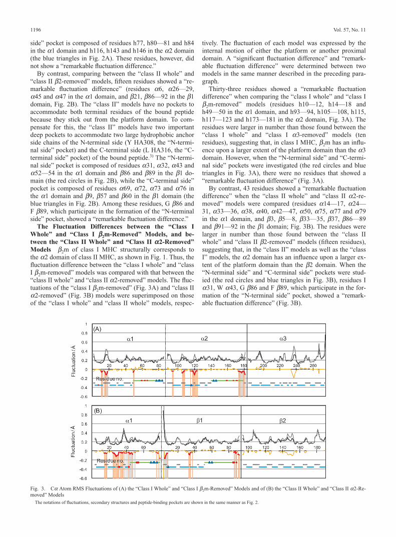
The Fluctuation Differences between the “Class IWhole” and “Class I bb2m-Removed” Models, and be-tween the “Class II Whole” and “Class II aa 2-Removed”Models b2m of class I MHC structurally corresponds tothe a2 domain of class II MHC, as shown in Fig. 1. Thus, thefluctuation difference between the “class I whole” and “classI b2m-removed” models was compared with that between the“class II whole” and “class II a2-removed” models. The fluc-tuations of the “class I b2m-removed” (Fig. 3A) and “class IIa2-removed” (Fig. 3B) models were superimposed on thoseof the “class I whole” and “class II whole” models, respec-
tively. The fluctuation of each model was expressed by theinternal motion of either the platform or another proximaldomain. A “significant fluctuation difference” and “remark-able fluctuation difference” were determined between twomodels in the same manner described in the preceding para-graph.
Thirty-three residues showed a “remarkable fluctuationdifference” when comparing the “class I whole” and “class Ib2m-removed” models (residues h10—12, h14—18 andh49—50 in the a1 domain, and h93—94, h105—108, h115,h117—123 and h173—181 in the a2 domain, Fig. 3A). Theresidues were larger in number than those found between the“class I whole” and “class I a3-removed” models (tenresidues), suggesting that, in class I MHC, b2m has an influ-ence upon a larger extent of the platform domain than the a3domain. However, when the “N-terminal side” and “C-termi-nal side” pockets were investigated (the red circles and bluetriangles in Fig. 3A), there were no residues that showed a“remarkable fluctuation difference” (Fig. 3A).
By contrast, 43 residues showed a “remarkable fluctuationdifference” when the “class II whole” and “class II a2-re-moved” models were compared (residues a14—17, a24—31, a33—36, a38, a40, a42—47, a50, a75, a77 and a79in the a1 domain, and b3, b5—8, b33—35, b37, b86—89and b91—92 in the b1 domain; Fig. 3B). The residues werelarger in number than those found between the “class IIwhole” and “class II b2-removed” models (fifteen residues),suggesting that, in the “class II” models as well as the “classI” models, the a2 domain has an influence upon a larger ex-tent of the platform domain than the b2 domain. When the“N-terminal side” and “C-terminal side” pockets were stud-ied (the red circles and blue triangles in Fig. 3B), residues Ia31, W a43, G b86 and F b89, which participate in the for-mation of the “N-terminal side” pocket, showed a “remark-able fluctuation difference” (Fig. 3B).
1196 Vol. 57, No. 11
Fig. 3. Ca Atom RMS Fluctuations of (A) the “Class I Whole” and “Class I b2m-Removed” Models and of (B) the “Class II Whole” and “Class II a2-Re-moved” Models
The notations of fluctuations, secondary structures and peptide-binding pockets are shown in the same manner as Fig. 2.

The Short Contact of the Two Membrane-ProximalDomains with the Platform Domain If a domain is re-moved from a molecule, the residue which is in the removeddomain neighborhood generally undergoes a fluctuationchange. In both classes, however, the residues of the twomembrane-proximal domain neighborhoods (less than 4 Å,the light and dark gray marks in Fig. 4) were not always inagreement with the residues which showed a “remarkablefluctuation difference.” The “N-terminal side” and “C-termi-nal side” pockets were then studied in both classes (magentaand cyan marks in Fig. 4). In the “class I” models, only oneresidue, Y h116 was in the b2m neighborhood but did notshow a “remarkable fluctuation difference” between the“class I whole” and “class I b2m-removed” models. In the“class II” models, only one residue, F b89 which showed a“remarkable fluctuation difference” between the “class IIwhole” and “class II b2-removed” models, was in the b2 do-main neighborhood (the purple frame in Fig. 4B). I a31 wasin the b2 domain neighborhood, but did not show a “remark-able fluctuation difference” between the “class II whole” and“class II b2-removed” models. I a31, W a43, G b86 and Fb89 showed a “remarkable fluctuation difference” betweenthe “class II whole” and “class II a2-removed” models, butnone of the four residues were in the a2 domain neighbor-hood (the purple and yellow frames in Fig. 4B). Thus, the“N-terminal side” pocket of class II MHC is influenced bythe a2 domain, though it is not in the a2 domain neighbor-hood.
DiscussionAs a complementary theoretical tool, molecular dynamics
has been shown to be a powerful approach for the study offluctuations on the nanosecond time scale.31) Recently, themolecular dynamics simulation on the peptide-class I MHCcomplex vs. the isolated platform domain was performed and
November 2009 1197
Fig. 4. Relationship between “Remarkable Fluctuation Difference” and the Short Contact in the (A) “Class I” and (B) “Class II” Models
In each figure, the “remarkable fluctuation difference” found only between the “class I whole” (or “class II whole”) and “class I a3-removed” (or “class II b2-removed”) modelsis marked on the amino acid sequence in orange. The “remarkable fluctuation difference” found only between the “class I whole” (or “class II whole”) and “class I b2m-removed”(or “class II a2-removed”) models is marked on the amino acid sequence in blue. The “remarkable fluctuation difference” found between the “class I whole” (or “class II whole”)model and both the “class I removed” (or “class II removed”) models is marked on the amino acid sequence in red. The important peptide-binding pockets that accommodate the“N-terminal” (magenta) and “C-terminal” (cyan) sides of the bound peptide, as shown in Fig. 2, are marked on the second top line. The short contact of the platform domain withthe a3 domain (or the b2 domain) and b2m (or the a2 domain) is marked on the third top line in light gray and the bottom line in dark gray, respectively. The short contact was de-fined as occurring where the distance between residues is less than 4 Å.
Fig. 5. Schematic Structures of the Peptide-Binding Platform Domains ofthe (A) “Class I” and (B) “Class II” Models
The domains and peptides are colored as for Fig. 1. In (A), the N-terminal (I309) andC-terminal (V317) residues of RT peptide are colored purple and cyan, respectively. In(B), two residues with a hydrophobic anchor side chain in the N-terminal (Y308) andC-terminal (L316) sides of HA peptide are colored purple and cyan, respectively. Ineach figure, the “remarkable fluctuation difference” found only between the “class Iwhole” (or class II whole”) and “class I a3-removed” (or “class II b2-removed”) mod-els is marked on the structure in orange. The “remarkable fluctuation difference” foundonly between the “class I whole” (“class II whole”) and “class I b2m-removed” (or“class II a2-removed”) models is marked on the structure in blue. The “remarkablefluctuation difference” found between the “class I whole” (“class II whole”) model andboth the “class I removed” (or “class II removed”) models is marked on the structure inred. The residues which participate in the formation of the important peptide-bindingpockets, and showed a “remarkable fluctuation difference,” are marked with the sidechain.

found that the two membrane-proximal domains (the a3 andb2m domain) cannot be neglected in peptide-binding stabilityof the platform domain.37) This study, however, could notclassify the contribution difference between the a3 and b2mdomain influence upon the platform domain. A limited timescale with available computational capability makes the mo-lecular dynamics simulation on the interdomain interactionin a large molecule difficult. To avoid this problem, we se-lected normal mode analysis that has less computational costthan molecular dynamics. However, if two models are com-pared by normal mode analysis based on only one energy-op-timized structure for each model, it is incontrovertible thatthe result contains a trivial computational artifact. To omitsuch a problem, we adopted six energy-optimized structuresfor a model. As shown in Table 1, RMSD between the sam-pled energy-optimized structures ranged from 0.47 to 1.59 Åin all models. This range did not deviate greatly from therange of RMSD between the X-ray crystal structures of classI MHC homologues whose sequence is identical (from 0.09to 1.74 Å) and the range of RMSD between the X-ray crystalstructures of class II MHC homologues whose sequence isidentical (from 0.09 to 1.40 Å), indicating that the energy-optimized structures varied moderately. Our previous studies have inferred the statistical comparison between twomodels using six energy-optimized structures for eachmodel.15,16,26,28,29) For example, the “significant fluctuationdifference” between two models was calculated by the non-parametric Wilcoxon’s rank sum test. Statistical analysis con-vinces us that our studies are not guided based on trivialcomputational artifact.
We attempted to detect the difference between the influ-ence that class I and class II two membrane-proximal do-mains have upon the dynamics of the platform domain. The“remarkable fluctuation differences” found between the“whole” model and the “removed” models are conclusivelyillustrated in Fig. 5. The membrane-proximal domains of the“class I” models little influenced the important peptide-bind-ing pockets (Fig. 5A). On the other hand, both the mem-brane-proximal domains of the “class II” models, especiallythe a2 domain influenced major parts (I a31, W a43, G b86and F b89) of the “N-terminal side” pocket (Fig. 5B), but, asshown in Fig. 4, these residues were not in the a2 domainneighborhood. In both the classes, the “N-terminal side”pocket is more important in peptide-binding than the “C-ter-minal side” pocket. For example, in class I MHC, the N-ter-minus of a bound peptide is accommodated in the “N-termi-nal side” pocket without exception, but the C-terminus of abound peptide is not always accommodated in the “C-termi-nal side” pocket.38) In class II MHC, some experimental andcomputational studies indicated that the introduction of ashort peptide composed of two residues or G b86Y substitu-tion to occupy only the “N-terminal side” pocket induced asignificant stabilization of not only the pocket but also of thewhole peptide-binding groove.14,18,20) The other computa-tional studies discussed the peptide-free form of class IIMHC and demonstrated the flexibility of the “N-terminalside” pocket and its importance in peptide-binding over theother pockets.16,19) Considering these circumstances, our results suggest that the membrane-proximal domains of class II MHC have a greater influence upon peptide-bindingthan those of class I MHC. Such results are probably attrib-
uted to the peptide-binding manner of each class. The “N-terminal side” pocket of class I MHC is located on the insideof the platform domain, and composed of structurally stablea-helices and b-strands (Fig. 5A). On the other hand, com-pared with the former, the “N-terminal side” pocket of classII MHC is located on an edge of the platform domain, andcomposed of a flexible loop (a31 and a32) and a connectingregion (from b86 to b92) between the second a-helices andthe b2 domain (Fig. 5B). Such a flexibility of the “N-termi-nal side” pocket might make the pocket sensitive to the twomembrane-proximal domains, though it is not in the a2 do-main neighborhood. In addition, the delicate cooperation ofthe two membrane-proximal domains with the “N-terminalside” pocket might permit class II MHC to bind to boundpeptides without apparent restriction on length (ca. 8—23amino acids in length).
In this study, we adopted HLA-DR1 as a simulation targetof class II MHC. To examine whether our results using HLA-DR1 showed a major characteristic of class II MHC homo-logues registered up to this point, the conservation of theresidues in the platform domain was investigated by Position-Specific Iterated Basic Local Alignment Search Tool (PSI-BLAST).39) In the a1 domain, 70.0% of the residues whichshowed a “remarkable fluctuation difference” between the“class II whole” model and either of the two “class II re-moved” models (20 residues in 29 residues), were the mostconserved amino acids among class II MHC homologues.This percentages was higher than those of the other residuesin the a1 domain (60.4%, 29 residues in 48 residues). In ad-dition, all the residues which showed a “remarkable fluctua-tion difference” between the “class II whole” model and boththe “class II removed” models (6 residues) were the mostconserved amino acids among class II MHC homologues. Onthe other hand, in the b1 domain, 88.2% of the residueswhich showed a “remarkable fluctuation difference” betweenthe “class II whole” model and either of the two “class II re-moved” models (15 residues in 17 residues), were the mostconserved amino acids among class II MHC homologues.This percentage was a little higher than those of the otherresidues in the b1 domain (84.9%, 62 residues in 73residues). In addition, all the residues which showed a “remarkable fluctuation difference” between the “class IIwhole” model and both the “class II removed” models (6residues), were the most conserved amino acids among classII MHC homologues. We investigated the conservation of themost important pocket of class II MHC, the “N-terminalside” pocket, in detail. In HLA-DR1, I a31, W a43, G b86and F b89 showed a “remarkable fluctuation difference” andwere major in forming the “N-terminal side” pocket. In theseresidues, Wa43, G b86 and F b89 were conserved by 50%,23% and 73% of class II homologues, respectively. The con-servation percentage of G b86 (23%) was not high, but all ofthe three residues were the most conserved amino acidsamong class II MHC homologues. I a31 (with a conserva-tion percentage of 12%) was not highly conserved amongclass II MHC homologues. However, the total conservationpercentage of similar character amino acids (V, L, I and M)was 73% in class II MHC homologues. These results, there-fore, demonstrate that our findings are not limited to HLA-DR1 only but are a major characteristic of class II MHC ho-mologues.
1198 Vol. 57, No. 11
In conclusion, our study showed that the influence of thetwo membrane-proximal domains upon the dynamics of theplatform domain were decisively different between class IIand class I. Both membrane-proximal domains (the a2 andb2 domains) of class II MHC, especially the a2 domain, in-fluenced the most important peptide-binding pocket, the “N-terminal side” pocket, though the pocket was not in the a2domain neighborhood. By contrast, the two membrane-proxi-mal domains (the a3 and b2m domains) of class I MHC hadlittle influence on the “N-terminal side” pocket. These resultssuggest that the two membrane-proximal domains of class IIMHC have a greater influence upon peptide-binding thanthose of class I MHC.
References1) Bjorkman P. J., Saper M. A., Samraoui B., Bennett W. S., Strominger J.
L., Wiley D. C., Nature (London), 329, 506—512 (1987).2) Brown J. H., Jardetzky T. S., Gorga J. C., Stern L. J., Urban R. G.,
Strominger J. L., Wiley D. C., Nature (London), 364, 33—39 (1993).3) Stern L. J., Brown J. H., Jardetzky T. S., Gorga J. C., Urban R. G.,
Strominger J. L., Wiley D. C., Nature (London), 368, 215—221(1994).
4) Zhang W., Young A. C., Imarai M., Nathenson S. G., Sacchettini J. C.,Proc. Natl. Acad. Sci. U.S.A., 89, 8403—8407 (1992).
5) Fremont D. H., Stura E. A., Matsumura M., Peterson P. A., Wilson I.A., Science, 257, 919—927 (1992).
6) Madden D. R., Garboczi D. N., Wiley D. C., Cell, 75, 693—708(1993).
7) Chicz R. M., Urban R. G., Lane W. S., Gorga J. C., Stern L. J., VignaliD. A., Strominger J. L., Nature (London), 358, 764—768 (1992).
8) Rudensky A., Perston-Hurlburt P., Hong S. C., Barlow A., Janeway C.A., Jr, Nature (London), 353, 622—627 (1991).
9) Utz U., Koenig S., Coligan J. E., Biddison W. E., J. Immunol., 149,214—221 (1992).
10) Meng W. S., von Grafenstein H., Haworth I. S., Int. Immunol., 9,1339—1349 (1997).
11) Bouvier M., Wiley D. C., Nat. Struct. Biol., 5, 377—384 (1998).12) Hansen T., Nat. Struct. Biol., 5, 340—341 (1998).13) Meng W. S., von Grafenstein H., Haworth I. S., Int. Immunol., 12,
949—957 (2000).14) Sato A. K., Zarutskie J. A., Rushe M. M., Lomakin A., Natarajan S.
K., Sadegh-Nasseri S., Benedek G. B., Stern L. J., J. Biol. Chem., 275,2165—21732 (2000).
15) Nojima H., Takeda-Shitaka M., Kurihara Y., Adachi M., Yoneda S.,
Kamiya K., Umeyama H., Chem. Pharm. Bull., 50, 1209—1214(2002).
16) Nojima H., Takeda-Shitaka M., Kurihara Y., Kamiya K., Umeyama H.,Chem. Pharm. Bull., 51, 923—928 (2003).
17) Zacharias M., Springer S., Biophys. J., 87, 2203—2214 (2004).18) Gupta S., Höpner S., Rupp B., Günther S., Dickhaut K., Agarwal N.,
Cardoso M. C., Kühne R., Wiesmüller K. H., Jung G., Falk K.,Rötzschke O., PLoS ONE 2008;3:e1814.
19) Painter C. A., Cruz A., López G. E., Stern L. J., Zavala-Ruiz Z., PLoSONE. 2008;3:e2403.
20) Yaneva R., Springer S., Zacharias. M., Biopolymers, 91, 14—27(2009).
21) Vitiello A., Potter T. A., Sherman L. A., Science, 250, 1423—1426(1990).
22) Cook J. R., Myers N. B., Hansen T. H., J. Immunol., 157, 2256—2261(1996).
23) Kamiya K., Sugawara Y., Umeyama H., J. Comput. Chem., 24, 826—841 (2003).
24) Weiner S. J., Kollman P. A., Case D. A., Singh U. C., Ghio C., AlagonaG., Profeta S. J., Weiner P., J. Am. Chem. Soc., 106, 765—784 (1984).
25) Weiner S. J., Kollman P. A., Nguyen D. T., Case D. A., J. Comput.Chem., 107, 230—252 (1986).
26) Nojima H., Takeda-Shitaka M., Kanou K., Kamiya K., Umeyama H.,Chem. Pharm. Bull., 56, 635—641 (2008).
27) Jääskeläinen S., Verma C. S., Hubbard R. E., Linko P., Caves L. S.,Protein Sci., 7, 1359—1367 (1998).
28) Adachi M., Kurihara Y., Nojima H., Takeda-Shitaka M., Kamiya K.,Umeyama H., Protein Sci., 12, 2125—2131 (2003).
29) Kurihara Y., Watanabe T., Nojima H., Takeda-Shitaka M., Kamiya K.,Umeyama H., Chem. Pharm. Bull., 51, 754—758 (2003).
30) Takeda-Shitaka M., Nojima H., Takaya D., Kanou K., Iwadate M.,Umeyama H., Chem. Pharm. Bull., 52, 643—645 (2004).
31) Brooks B., Karplus M., Proc. Natl. Acad. Sci. U.S.A., 80, 6571—6575(1983).
32) Ishida H., Jochi Y., Kidera A., Proteins, 32, 324—333 (1998).33) Eckart C., Phys. Rev., 47, 552—558 (1935).34) Go N., Noguti T., Nishikawa T., Proc. Natl. Acad. Sci. U.S.A., 80,
3696—3670 (1983).35) Levitt M., Sander C., Stern P. S., J. Mol. Biol., 181, 423—447 (1985).36) Nishikawa T., Go N., Proteins, 2, 308—329 (1987).37) Wan S., Coveney P., Flower D. R., J. Comput. Chem., 25, 1803—1813
(2004).38) Collins E. J., Garboczi D. N., Wiley D. C., Nature (London), 371,
626—629 (1994).39) Altschul S. F., Madden T. L., Schäffer A. A., Zhang J., Zhang Z.,
Miller W., Lipman D. J., Nucleic Acids Res., 25, 3389—3402 (1997).
November 2009 1199


![Biochemicallocalization hepatic Na',K+-ATPase on · Biochemicallocalization ofhepatic surface-membrane ... , EC3.6.1.37] between apical and ... proximal renal tubules, and hepatocytes](https://img.pdfslide.net/doc/110x75/5b2f24037f8b9a91438c8c51/biochemicallocalization-hepatic-nak-atpase-on-biochemicallocalization-ofhepatic.jpg)