Embed Size (px)
Citation preview
This journal is©The Royal Society of Chemistry 2016 Chem. Commun., 2016, 52, 9387--9390 | 9387
Cite this:Chem. Commun., 2016,
52, 9387
Dynamic nanoproteins: self-assembledpeptide surfaces on monolayer protectedgold nanoparticles†
Sergio Garcia Martin and Leonard J. Prins*
Here, we demonstrate the formation of dynamic peptide surfaces
through the self-assembly of small peptides on the surface of
monolayer protected gold nanoparticles. The complexity of the
peptide surface can be simply tuned by changing the chemical
nature of the added peptides and the ratio in which these are added.
The dynamic nature of the surface permits adaptation to changes in
the environment.
The exquisite properties of proteins in terms of molecularrecognition and catalysis derive in large part from their sizeand structural complexity.1 Their size permits the presence ofinternal cavities, which are well-shielded from the bulk solvent2
and permit additional advantages like allosteric control bysecondary binding sites3 and multivalent interactions to driveprotein–protein interactions.4 The fact that all proteins arecomposed of a very limited number of amino acids rendersthese structures even more fascinating. Currently, a strongimpetus exists to develop synthetic structures able to match thesize and complexity of proteins for applications in diagnostics,medicine and materials science.5,6 The advantage of syntheticstructures is the ability to design the desired structure andthe possibility of including also non-natural building blocks.Generally, the followed approach relies on the decoration ofmultivalent scaffolds, such as polymers7 or nanoparticles,8 withamino acids or small peptides, although the self-assembly ofsmall (peptidic) molecules has also been extensively used.9
Here, we exploit a combination of both approaches for theformation of multivalent peptide surfaces on monolayer protectedgold nanoparticles. In the last few years we have extensively usedAu NP 1, which are gold nanoparticles (d = 1.8 � 0.4 nm) coveredwith a monolayer of C9-thiols terminating with a 1,4,7-triaza-cyclonane (TACN)�Zn2+ head group, for applications in sensing,catalysis, and systems chemistry.10 A majority of these applications
rely on the high affinity of small oligoanions, such as peptides andnucleotides, for the highly positively charged surface of Au NP 1.Binding constants were high enough to ensure binding of multiplemolecules under saturation conditions even at low micromolarconcentrations under physiologically relevant conditions. Yet, thenoncovalent interactions that drive the self-assembly processesensure that spontaneous exchange processes occur on the mono-layer surface. Here, we show that these two features create thepossibility of forming multivalent peptide surfaces simply byassembling multiple small peptide sequences on Au NP 1. It willbe shown that such an approach offers several advantages. Theself-assembly process facilitates the formation of nanoproteins,since just mixing the components under ambient conditions issufficient. The complexity of the multivalent surface can not onlybe simply tuned by changing the peptide sequence, but also bychanging the ratio of the added peptides. Finally, the dynamicnature of the peptide surface implies that spontaneous adaptationof the surface is possible, enabling the possibility of exploitingdynamic combinatorial chemistry on nanoparticle surfaces.11–13
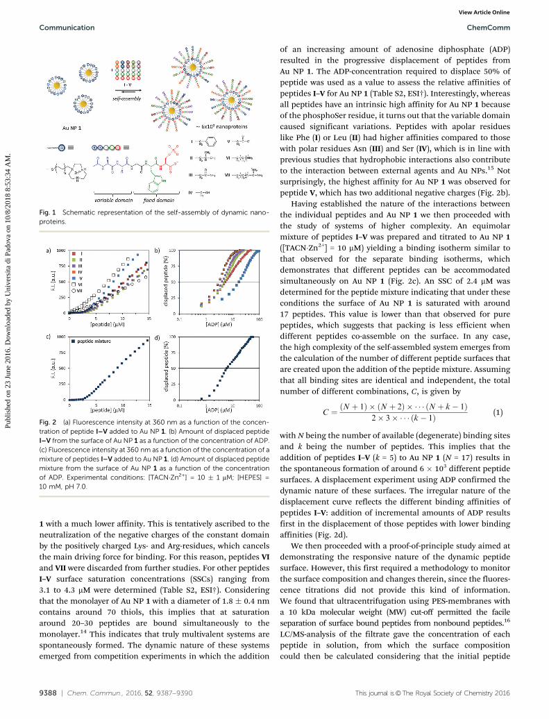
A series of seven different pentapeptides (I–VII) with thegeneral sequence Ac-XXGWS(OPO3
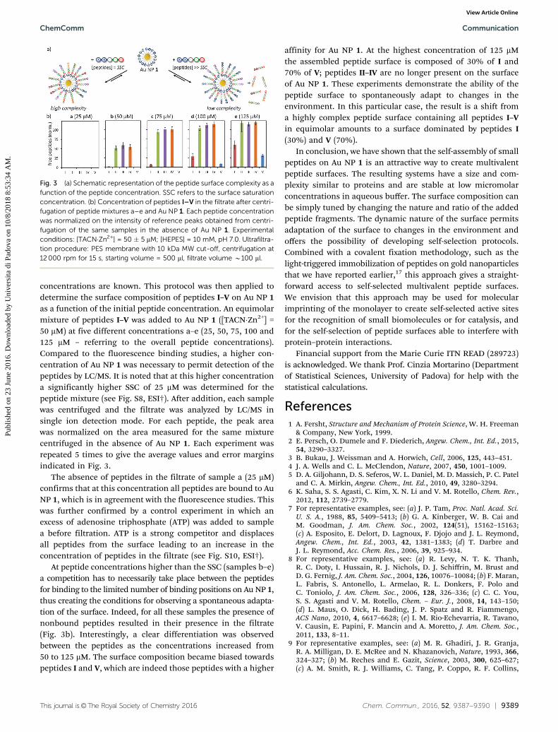
2�)-OH was prepared (Fig. 1).The constant domain was composed of a phosphorylatedSer-residue for binding to Au NP 1, a fluorescent Trp-residuefor monitoring the binding interaction and a Gly-residue as aflexible spacer to outdistance the two remaining residues of thevariable domain. The residues of the variable domain X werechosen from the various subgroups of amino acids rangingfrom apolar (Phe (I), Leu (II)), polar neutral (Asn (III), Ser (IV)),anionic (Asp (V)), to cationic (Lys (VI), Arg (VII)) in order toexplore the compatibility with the self-assembly process (Fig. 1).Two residues of each amino acid were added to enhance theircontribution to the overall properties of the correspondingpeptide. Fluorescence titration experiments of the peptides ata fixed concentration of Au NP 1 ([TACN�Zn2+] = 10 mM) inaqueous buffer at pH = 7.0 revealed a high affinity of allpeptides, except for VI and VII (Fig. 2a). The shallow curvatureof the binding isotherms of the latter peptides, as compared tothose of peptides I–V, indicates that these peptides bind Au NP
Department of Chemical Sciences, University of Padova, Via Marzolo 1,
35131 Padova, Italy. E-mail: [email protected]; Fax: +39 049 8275051
† Electronic supplementary information (ESI) available: Experimental details andcontrol experiments. See DOI: 10.1039/c6cc04786f
Received 8th June 2016,Accepted 23rd June 2016
DOI: 10.1039/c6cc04786f
www.rsc.org/chemcomm
ChemComm
COMMUNICATION
Publ
ishe
d on
23
June
201
6. D
ownl
oade
d by
Uni
vers
ita d
i Pad
ova
on 1
0/8/
2018
8:5
3:34
AM
.
View Article OnlineView Journal | View Issue
9388 | Chem. Commun., 2016, 52, 9387--9390 This journal is©The Royal Society of Chemistry 2016
1 with a much lower affinity. This is tentatively ascribed to theneutralization of the negative charges of the constant domainby the positively charged Lys- and Arg-residues, which cancelsthe main driving force for binding. For this reason, peptides VIand VII were discarded from further studies. For other peptidesI–V surface saturation concentrations (SSCs) ranging from3.1 to 4.3 mM were determined (Table S2, ESI†). Consideringthat the monolayer of Au NP 1 with a diameter of 1.8 � 0.4 nmcontains around 70 thiols, this implies that at saturationaround 20–30 peptides are bound simultaneously to themonolayer.14 This indicates that truly multivalent systems arespontaneously formed. The dynamic nature of these systemsemerged from competition experiments in which the addition
of an increasing amount of adenosine diphosphate (ADP)resulted in the progressive displacement of peptides fromAu NP 1. The ADP-concentration required to displace 50% ofpeptide was used as a value to assess the relative affinities ofpeptides I–V for Au NP 1 (Table S2, ESI†). Interestingly, whereasall peptides have an intrinsic high affinity for Au NP 1 becauseof the phosphoSer residue, it turns out that the variable domaincaused significant variations. Peptides with apolar residueslike Phe (I) or Leu (II) had higher affinities compared to thosewith polar residues Asn (III) and Ser (IV), which is in line withprevious studies that hydrophobic interactions also contributeto the interaction between external agents and Au NPs.15 Notsurprisingly, the highest affinity for Au NP 1 was observed forpeptide V, which has two additional negative charges (Fig. 2b).
Having established the nature of the interactions betweenthe individual peptides and Au NP 1 we then proceeded withthe study of systems of higher complexity. An equimolarmixture of peptides I–V was prepared and titrated to Au NP 1([TACN�Zn2+] = 10 mM) yielding a binding isotherm similar tothat observed for the separate binding isotherms, whichdemonstrates that different peptides can be accommodatedsimultaneously on Au NP 1 (Fig. 2c). An SSC of 2.4 mM wasdetermined for the peptide mixture indicating that under theseconditions the surface of Au NP 1 is saturated with around17 peptides. This value is lower than that observed for purepeptides, which suggests that packing is less efficient whendifferent peptides co-assemble on the surface. In any case,the high complexity of the self-assembled system emerges fromthe calculation of the number of different peptide surfaces thatare created upon the addition of the peptide mixture. Assumingthat all binding sites are identical and independent, the totalnumber of different combinations, C, is given by
C ¼ ðN þ 1Þ � ðN þ 2Þ � � � � ðN þ k� 1Þ2� 3� � � � ðk� 1Þ (1)
with N being the number of available (degenerate) binding sitesand k being the number of peptides. This implies that theaddition of peptides I–V (k = 5) to Au NP 1 (N = 17) results inthe spontaneous formation of around 6 � 103 different peptidesurfaces. A displacement experiment using ADP confirmed thedynamic nature of these surfaces. The irregular nature of thedisplacement curve reflects the different binding affinities ofpeptides I–V: addition of incremental amounts of ADP resultsfirst in the displacement of those peptides with lower bindingaffinities (Fig. 2d).
We then proceeded with a proof-of-principle study aimed atdemonstrating the responsive nature of the dynamic peptidesurface. However, this first required a methodology to monitorthe surface composition and changes therein, since the fluores-cence titrations did not provide this kind of information.We found that ultracentrifugation using PES-membranes witha 10 kDa molecular weight (MW) cut-off permitted the facileseparation of surface bound peptides from nonbound peptides.16
LC/MS-analysis of the filtrate gave the concentration of eachpeptide in solution, from which the surface compositioncould then be calculated considering that the initial peptide
Fig. 1 Schematic representation of the self-assembly of dynamic nano-proteins.
Fig. 2 (a) Fluorescence intensity at 360 nm as a function of the concen-tration of peptide I–V added to Au NP 1. (b) Amount of displaced peptideI–V from the surface of Au NP 1 as a function of the concentration of ADP.(c) Fluorescence intensity at 360 nm as a function of the concentration of amixture of peptides I–V added to Au NP 1. (d) Amount of displaced peptidemixture from the surface of Au NP 1 as a function of the concentrationof ADP. Experimental conditions: [TACN�Zn2+] = 10 � 1 mM; [HEPES] =10 mM, pH 7.0.
Communication ChemComm
Publ
ishe
d on
23
June
201
6. D
ownl
oade
d by
Uni
vers
ita d
i Pad
ova
on 1
0/8/
2018
8:5
3:34
AM
. View Article Online
This journal is©The Royal Society of Chemistry 2016 Chem. Commun., 2016, 52, 9387--9390 | 9389
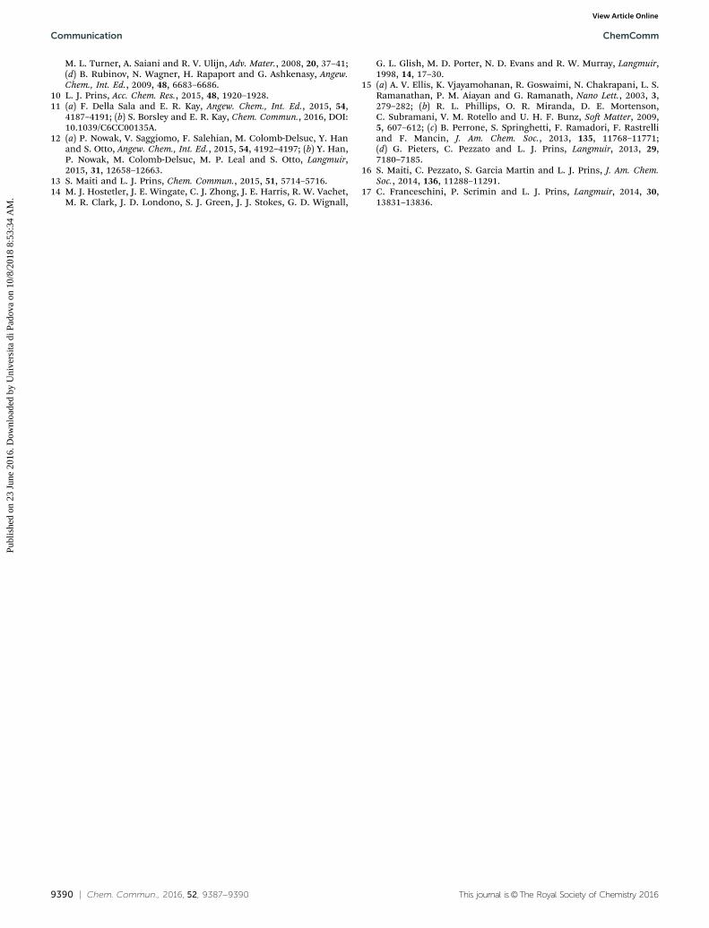
concentrations are known. This protocol was then applied todetermine the surface composition of peptides I–V on Au NP 1as a function of the initial peptide concentration. An equimolarmixture of peptides I–V was added to Au NP 1 ([TACN�Zn2+] =50 mM) at five different concentrations a–e (25, 50, 75, 100 and125 mM – referring to the overall peptide concentrations).Compared to the fluorescence binding studies, a higher con-centration of Au NP 1 was necessary to permit detection of thepeptides by LC/MS. It is noted that at this higher concentrationa significantly higher SSC of 25 mM was determined for thepeptide mixture (see Fig. S8, ESI†). After addition, each samplewas centrifuged and the filtrate was analyzed by LC/MS insingle ion detection mode. For each peptide, the peak areawas normalized on the area measured for the same mixturecentrifuged in the absence of Au NP 1. Each experiment wasrepeated 5 times to give the average values and error marginsindicated in Fig. 3.
The absence of peptides in the filtrate of sample a (25 mM)confirms that at this concentration all peptides are bound to AuNP 1, which is in agreement with the fluorescence studies. Thiswas further confirmed by a control experiment in which anexcess of adenosine triphosphate (ATP) was added to samplea before filtration. ATP is a strong competitor and displacesall peptides from the surface leading to an increase in theconcentration of peptides in the filtrate (see Fig. S10, ESI†).
At peptide concentrations higher than the SSC (samples b–e)a competition has to necessarily take place between the peptidesfor binding to the limited number of binding positions on Au NP 1,thus creating the conditions for observing a spontaneous adapta-tion of the surface. Indeed, for all these samples the presence ofnonbound peptides resulted in their presence in the filtrate(Fig. 3b). Interestingly, a clear differentiation was observedbetween the peptides as the concentrations increased from50 to 125 mM. The surface composition became biased towardspeptides I and V, which are indeed those peptides with a higher
affinity for Au NP 1. At the highest concentration of 125 mMthe assembled peptide surface is composed of 30% of I and70% of V; peptides II–IV are no longer present on the surfaceof Au NP 1. These experiments demonstrate the ability of thepeptide surface to spontaneously adapt to changes in theenvironment. In this particular case, the result is a shift froma highly complex peptide surface containing all peptides I–Vin equimolar amounts to a surface dominated by peptides I(30%) and V (70%).
In conclusion, we have shown that the self-assembly of smallpeptides on Au NP 1 is an attractive way to create multivalentpeptide surfaces. The resulting systems have a size and com-plexity similar to proteins and are stable at low micromolarconcentrations in aqueous buffer. The surface composition canbe simply tuned by changing the nature and ratio of the addedpeptide fragments. The dynamic nature of the surface permitsadaptation of the surface to changes in the environment andoffers the possibility of developing self-selection protocols.Combined with a covalent fixation methodology, such as thelight-triggered immobilization of peptides on gold nanoparticlesthat we have reported earlier,17 this approach gives a straight-forward access to self-selected multivalent peptide surfaces.We envision that this approach may be used for molecularimprinting of the monolayer to create self-selected active sitesfor the recognition of small biomolecules or for catalysis, andfor the self-selection of peptide surfaces able to interfere withprotein–protein interactions.
Financial support from the Marie Curie ITN READ (289723)is acknowledged. We thank Prof. Cinzia Mortarino (Departmentof Statistical Sciences, University of Padova) for help with thestatistical calculations.
References1 A. Fersht, Structure and Mechanism of Protein Science, W. H. Freeman
& Company, New York, 1999.2 E. Persch, O. Dumele and F. Diederich, Angew. Chem., Int. Ed., 2015,
54, 3290–3327.3 B. Bukau, J. Weissman and A. Horwich, Cell, 2006, 125, 443–451.4 J. A. Wells and C. L. McClendon, Nature, 2007, 450, 1001–1009.5 D. A. Giljohann, D. S. Seferos, W. L. Daniel, M. D. Massich, P. C. Patel
and C. A. Mirkin, Angew. Chem., Int. Ed., 2010, 49, 3280–3294.6 K. Saha, S. S. Agasti, C. Kim, X. N. Li and V. M. Rotello, Chem. Rev.,
2012, 112, 2739–2779.7 For representative examples, see: (a) J. P. Tam, Proc. Natl. Acad. Sci.
U. S. A., 1988, 85, 5409–5413; (b) G. A. Kinberger, W. B. Cai andM. Goodman, J. Am. Chem. Soc., 2002, 124(51), 15162–15163;(c) A. Esposito, E. Delort, D. Lagnoux, F. Djojo and J. L. Reymond,Angew. Chem., Int. Ed., 2003, 42, 1381–1383; (d) T. Darbre andJ. L. Reymond, Acc. Chem. Res., 2006, 39, 925–934.
8 For representative examples, see: (a) R. Levy, N. T. K. Thanh,R. C. Doty, I. Hussain, R. J. Nichols, D. J. Schiffrin, M. Brust andD. G. Fernig, J. Am. Chem. Soc., 2004, 126, 10076–10084; (b) F. Maran,L. Fabris, S. Antonello, L. Armelao, R. L. Donkers, F. Polo andC. Toniolo, J. Am. Chem. Soc., 2006, 128, 326–336; (c) C. C. You,S. S. Agasti and V. M. Rotello, Chem. – Eur. J., 2008, 14, 143–150;(d) L. Maus, O. Dick, H. Bading, J. P. Spatz and R. Fiammengo,ACS Nano, 2010, 4, 6617–6628; (e) I. M. Rio-Echevarria, R. Tavano,V. Causin, E. Papini, F. Mancin and A. Moretto, J. Am. Chem. Soc.,2011, 133, 8–11.
9 For representative examples, see: (a) M. R. Ghadiri, J. R. Granja,R. A. Milligan, D. E. McRee and N. Khazanovich, Nature, 1993, 366,324–327; (b) M. Reches and E. Gazit, Science, 2003, 300, 625–627;(c) A. M. Smith, R. J. Williams, C. Tang, P. Coppo, R. F. Collins,
Fig. 3 (a) Schematic representation of the peptide surface complexity as afunction of the peptide concentration. SSC refers to the surface saturationconcentration. (b) Concentration of peptides I–V in the filtrate after centri-fugation of peptide mixtures a–e and Au NP 1. Each peptide concentrationwas normalized on the intensity of reference peaks obtained from centri-fugation of the same samples in the absence of Au NP 1. Experimentalconditions: [TACN�Zn2+] = 50 � 5 mM; [HEPES] = 10 mM, pH 7.0. Ultrafiltra-tion procedure: PES membrane with 10 kDa MW cut-off, centrifugation at12 000 rpm for 15 s, starting volume = 500 ml, filtrate volume B100 ml.
ChemComm Communication
Publ
ishe
d on
23
June
201
6. D
ownl
oade
d by
Uni
vers
ita d
i Pad
ova
on 1
0/8/
2018
8:5
3:34
AM
. View Article Online
9390 | Chem. Commun., 2016, 52, 9387--9390 This journal is©The Royal Society of Chemistry 2016
M. L. Turner, A. Saiani and R. V. Ulijn, Adv. Mater., 2008, 20, 37–41;(d) B. Rubinov, N. Wagner, H. Rapaport and G. Ashkenasy, Angew.Chem., Int. Ed., 2009, 48, 6683–6686.
10 L. J. Prins, Acc. Chem. Res., 2015, 48, 1920–1928.11 (a) F. Della Sala and E. R. Kay, Angew. Chem., Int. Ed., 2015, 54,
4187–4191; (b) S. Borsley and E. R. Kay, Chem. Commun., 2016, DOI:10.1039/C6CC00135A.
12 (a) P. Nowak, V. Saggiomo, F. Salehian, M. Colomb-Delsuc, Y. Hanand S. Otto, Angew. Chem., Int. Ed., 2015, 54, 4192–4197; (b) Y. Han,P. Nowak, M. Colomb-Delsuc, M. P. Leal and S. Otto, Langmuir,2015, 31, 12658–12663.
13 S. Maiti and L. J. Prins, Chem. Commun., 2015, 51, 5714–5716.14 M. J. Hostetler, J. E. Wingate, C. J. Zhong, J. E. Harris, R. W. Vachet,
M. R. Clark, J. D. Londono, S. J. Green, J. J. Stokes, G. D. Wignall,
G. L. Glish, M. D. Porter, N. D. Evans and R. W. Murray, Langmuir,1998, 14, 17–30.
15 (a) A. V. Ellis, K. Vjayamohanan, R. Goswaimi, N. Chakrapani, L. S.Ramanathan, P. M. Aiayan and G. Ramanath, Nano Lett., 2003, 3,279–282; (b) R. L. Phillips, O. R. Miranda, D. E. Mortenson,C. Subramani, V. M. Rotello and U. H. F. Bunz, Soft Matter, 2009,5, 607–612; (c) B. Perrone, S. Springhetti, F. Ramadori, F. Rastrelliand F. Mancin, J. Am. Chem. Soc., 2013, 135, 11768–11771;(d) G. Pieters, C. Pezzato and L. J. Prins, Langmuir, 2013, 29,7180–7185.
16 S. Maiti, C. Pezzato, S. Garcia Martin and L. J. Prins, J. Am. Chem.Soc., 2014, 136, 11288–11291.
17 C. Franceschini, P. Scrimin and L. J. Prins, Langmuir, 2014, 30,13831–13836.
Communication ChemComm
Publ
ishe
d on
23
June
201
6. D
ownl
oade
d by
Uni
vers
ita d
i Pad
ova
on 1
0/8/
2018
8:5
3:34
AM
. View Article Online