Embed Size (px)
Citation preview
r e v b r a s o r t o p . 2 0 1 7;5 2(5):621–624
S
C
Dp
E
U
a
A
R
A
A
K
W
T
F
U
P
T
T
F
I
h2u
OCIEDADE BRASILEIRA DEORTOPEDIA E TRAUMATOLOGIA
www.rbo.org .br
ase Report
ynamic ulnar impaction syndrome in tennislayers: report of two cases�
dgard de Novaes Franca Bisneto
niversidade de São Paulo, Hospital das Clínicas, Instituto de Ortopedia e Traumatologia, São Paulo, SP, Brazil
r t i c l e i n f o
rticle history:
eceived 5 August 2016
ccepted 22 August 2016
vailable online 16 August 2017
eywords:
rist injuries
ennis/injuries
ibrocartilage/injuries
lnocarpal impaction
a b s t r a c t
In this report, two tennis players with symptoms of ulnar impaction syndrome are reviewed.
Both players have neutral ulnar variance. These cases represent dynamic ulnar impaction
syndrome, when the impact between ulna and carpus occurs during conditions of pronated
grip. The literature and the treatment of these two cases are discussed.
© 2017 Sociedade Brasileira de Ortopedia e Traumatologia. Published by Elsevier Editora
Ltda. This is an open access article under the CC BY-NC-ND license (http://
creativecommons.org/licenses/by-nc-nd/4.0/).
Síndrome do impacto ulnocarpal dinâmico em tenistas: relato de doiscasos
alavras-chave:
raumatismos do punho
ênis/lesões
ibrocartilagem/lesões
r e s u m o
O relato apresenta os casos de dois tenistas portadores de dor no bordo ulnar do carpo
com sinais de impacto no semilunar. Ambos são portadores de ulna neutra. Esses casos
representam uma entidade denominada síndrome do impacto ulnocarpal dinâmico na qual
ocorre o impacto entre a cabeca da ulna e o carpo em situacão de pronacão com punho
mpacto ulnocarpal fechado durante a atividade física. A literatura e o tratamento dos dois casos são discutidosno artigo.
© 2017 Sociedade Brasileira de Ortopedia e Traumatologia. Publicado por Elsevier Editora
Ltda. Este e um artigo Open Access sob uma licenca CC BY-NC-ND (http://
� Paper developed at Universidade de São Paulo, Hospital das ClínicasE-mail: [email protected]
ttp://dx.doi.org/10.1016/j.rboe.2017.08.003255-4971/© 2017 Sociedade Brasileira de Ortopedia e Traumatologia.
nder the CC BY-NC-ND license (http://creativecommons.org/licenses/
creativecommons.org/licenses/by-nc-nd/4.0/).
, Instituto de Ortopedia e Traumatologia, São Paulo, SP, Brazil.
Published by Elsevier Editora Ltda. This is an open access articleby-nc-nd/4.0/).
p . 2 0
622 r e v b r a s o r t oIntroduction
Ulnocarpal impaction syndrome (UCIS) is a degenerativelesion characterized by compression or impact of the ulnahead against the semilunar bone and/or the pyramidalbone, with or without lesion of the triangular fibrocartilagecomplex.1–3
Generally associated with the presence of ulna plus, UCISmay also occur in the presence of neutral or minus ulna.4
During pronation there is a relative shortening between theradius and the ulna, and in situations of neutral or minus ulnashorter than 2 mm an impact can occur between the carpusand the ulna head, this condition is called dynamic ulnocarpalimpaction syndrome (UCIS).4
UCIS in the presence of neutral or minus ulna is describedin pronation situations associated with grip strength, commonin sports activities such as tennis or baseball.5
In this report, two cases of tennis players with UCIS willbe presented, and treated arthroscopically. After the presen-tation of the cases, a discussion of the literature will beperformed.
Case report 1
A 20-year-old female patient, who is a tennis player at anAmerican university, with a right wrist triangular fibrocarti-lage lesion, diagnosed at the university.
When she underwent arthroscopy at that medical servicewith fibrocartilage debridement and wrist synovectomy, asso-ciated semilunar chondromalacia was diagnosed.
After seven months of surgery, and intense rehabilitation,the pain persisted preventing her from playing.
On physical examination, she showed pain at maximumsupination; positive fovea sign (pain at ulnar volar palpationof the ulna head); absence of instability of the distal radioulnarjoint (DRUJ); normal wrist ROM and low pain to ulnar deviationagainst resistance.
Imaging demonstrates triangular fibrocartilage lesion,intense synovitis and edema of the semilunar and pyramidalbones. Presence of neutral ulna (Fig. 1) was also noted.
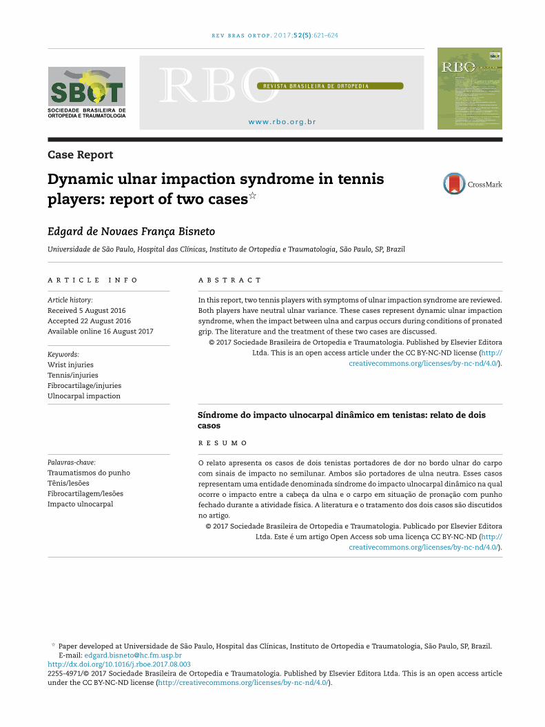
Patient underwent new arthroscopy of the wrist with evi-dent situation of impaction seen in the intraoperative periodduring pronosupination of the wrist. Arthroscopic wafer pro-cedure, as well as debridement of triangular fibrocartilage andloose cartilage of the semilunar (Fig. 2), was performed.
The patient remained immobilized with an antebra-chiopalmar orthesis with epicondylar block during threeweeks. She was referred to rehabilitation, and returnedto her sports activities with no pain four months aftersurgery.
Case report 2
A 49-year-old male patient, amateur tennis player, who playedevery weekend, presented with chronic, progressive pain,mainly when performing forehand technique while playingtennis.
1 7;5 2(5):621–624
On physical examination, he presented with pain at max-imum supination; positive fovea sign, absence of instabilityof the distal radioulnar joint (DRUJ); normal wrist ROM andno pain at ulnar deviation against resistance. This patientalso presented pain on palpation of the projection of theinterosseous scaphosemilunar ligament (SLL), but the Watsontest was negative.
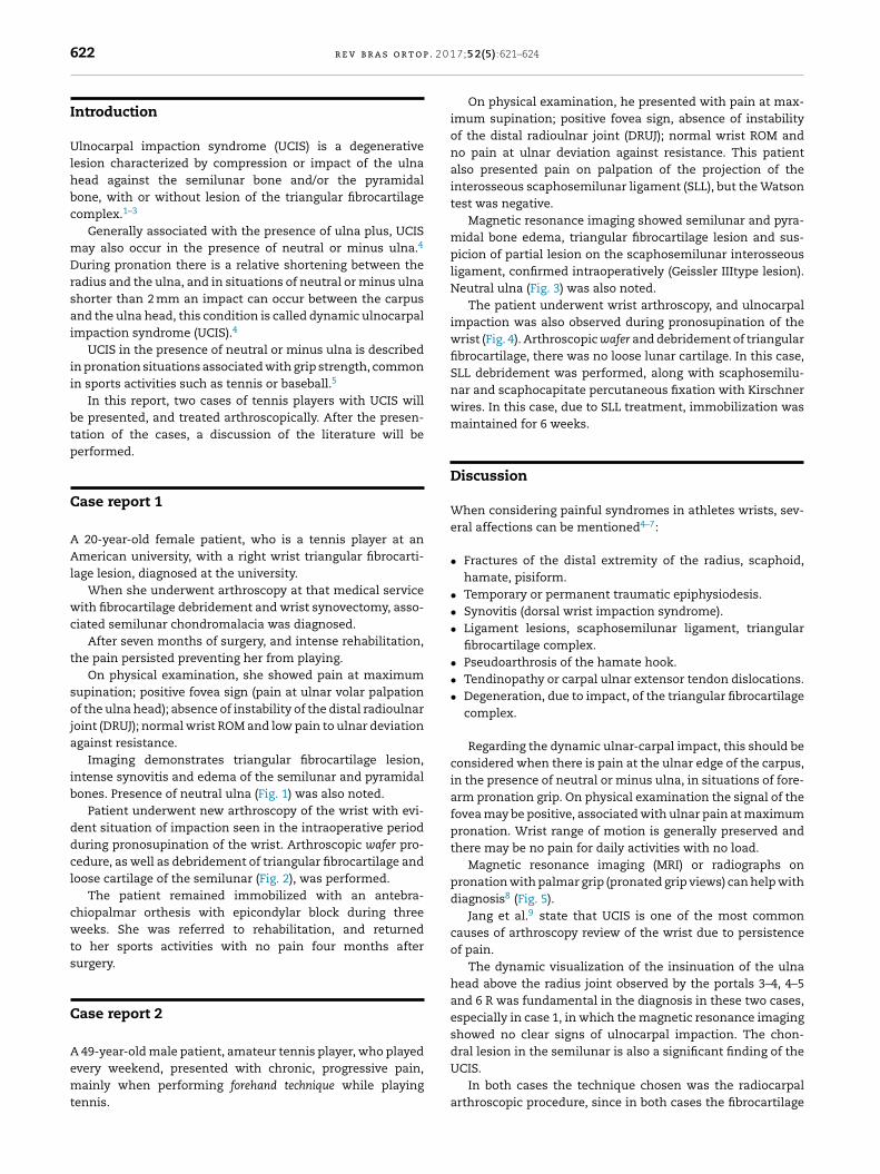
Magnetic resonance imaging showed semilunar and pyra-midal bone edema, triangular fibrocartilage lesion and sus-picion of partial lesion on the scaphosemilunar interosseousligament, confirmed intraoperatively (Geissler IIItype lesion).Neutral ulna (Fig. 3) was also noted.
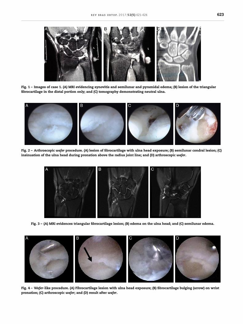
The patient underwent wrist arthroscopy, and ulnocarpalimpaction was also observed during pronosupination of thewrist (Fig. 4). Arthroscopic wafer and debridement of triangularfibrocartilage, there was no loose lunar cartilage. In this case,SLL debridement was performed, along with scaphosemilu-nar and scaphocapitate percutaneous fixation with Kirschnerwires. In this case, due to SLL treatment, immobilization wasmaintained for 6 weeks.
Discussion
When considering painful syndromes in athletes wrists, sev-eral affections can be mentioned4–7:
• Fractures of the distal extremity of the radius, scaphoid,hamate, pisiform.
• Temporary or permanent traumatic epiphysiodesis.• Synovitis (dorsal wrist impaction syndrome).• Ligament lesions, scaphosemilunar ligament, triangular
fibrocartilage complex.• Pseudoarthrosis of the hamate hook.• Tendinopathy or carpal ulnar extensor tendon dislocations.• Degeneration, due to impact, of the triangular fibrocartilage
complex.
Regarding the dynamic ulnar-carpal impact, this should beconsidered when there is pain at the ulnar edge of the carpus,in the presence of neutral or minus ulna, in situations of fore-arm pronation grip. On physical examination the signal of thefovea may be positive, associated with ulnar pain at maximumpronation. Wrist range of motion is generally preserved andthere may be no pain for daily activities with no load.
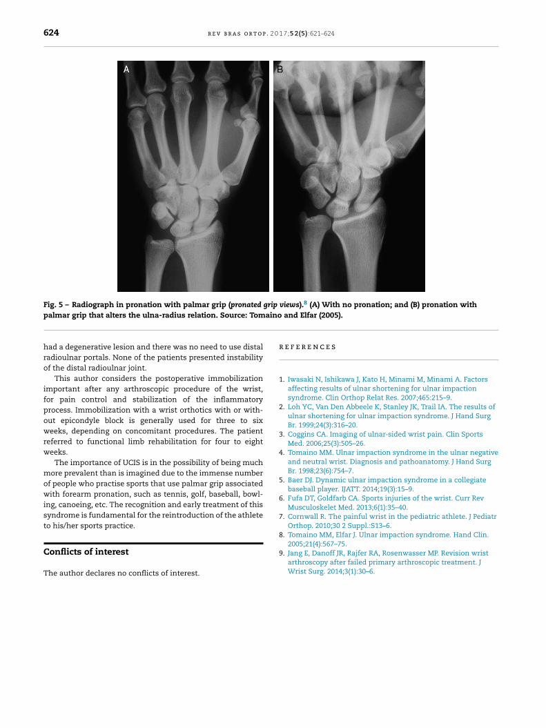
Magnetic resonance imaging (MRI) or radiographs onpronation with palmar grip (pronated grip views) can help withdiagnosis8 (Fig. 5).
Jang et al.9 state that UCIS is one of the most commoncauses of arthroscopy review of the wrist due to persistenceof pain.
The dynamic visualization of the insinuation of the ulnahead above the radius joint observed by the portals 3–4, 4–5and 6 R was fundamental in the diagnosis in these two cases,especially in case 1, in which the magnetic resonance imagingshowed no clear signs of ulnocarpal impaction. The chon-
dral lesion in the semilunar is also a significant finding of theUCIS.In both cases the technique chosen was the radiocarpalarthroscopic procedure, since in both cases the fibrocartilage
r e v b r a s o r t o p . 2 0 1 7;5 2(5):621–624 623
Fig. 1 – Images of case 1. (A) MRI evidencing synovitis and semilunar and pyramidal edema; (B) lesion of the triangularfibrocartilage in the distal portion only; and (C) tomography demonstrating neutral ulna.
Fig. 2 – Arthroscopic wafer procedure. (A) lesion of fibrocartilage with ulna head exposure; (B) semilunar condral lesion; (C)insinuation of the ulna head during pronation above the radius joint line; and (D) arthroscopic wafer.
Fig. 3 – (A) MRI evidences triangular fibrocartilage lesion; (B) edema on the ulna head; and (C) semilunar edema.
Fig. 4 – Wafer-like procedure. (A) Fibrocartilage lesion with ulna head exposure; (B) fibrocartilage bulging (arrow) on wristpronation; (C) arthroscopic wafer; and (D) result after wafer.
624 r e v b r a s o r t o p . 2 0 1 7;5 2(5):621–624
Fig. 5 – Radiograph in pronation with palmar grip (pronated grip views).8 (A) With no pronation; and (B) pronation withaino
r
1
2
3
4
5
6
7
8
palmar grip that alters the ulna-radius relation. Source: Tom
had a degenerative lesion and there was no need to use distalradioulnar portals. None of the patients presented instabilityof the distal radioulnar joint.
This author considers the postoperative immobilizationimportant after any arthroscopic procedure of the wrist,for pain control and stabilization of the inflammatoryprocess. Immobilization with a wrist orthotics with or with-out epicondyle block is generally used for three to sixweeks, depending on concomitant procedures. The patientreferred to functional limb rehabilitation for four to eightweeks.
The importance of UCIS is in the possibility of being muchmore prevalent than is imagined due to the immense numberof people who practise sports that use palmar grip associatedwith forearm pronation, such as tennis, golf, baseball, bowl-ing, canoeing, etc. The recognition and early treatment of thissyndrome is fundamental for the reintroduction of the athleteto his/her sports practice.
Conflicts of interest
The author declares no conflicts of interest.
9
and Elfar (2005).
e f e r e n c e s
. Iwasaki N, Ishikawa J, Kato H, Minami M, Minami A. Factorsaffecting results of ulnar shortening for ulnar impactionsyndrome. Clin Orthop Relat Res. 2007;465:215–9.
. Loh YC, Van Den Abbeele K, Stanley JK, Trail IA. The results ofulnar shortening for ulnar impaction syndrome. J Hand SurgBr. 1999;24(3):316–20.
. Coggins CA. Imaging of ulnar-sided wrist pain. Clin SportsMed. 2006;25(3):505–26.
. Tomaino MM. Ulnar impaction syndrome in the ulnar negativeand neutral wrist. Diagnosis and pathoanatomy. J Hand SurgBr. 1998;23(6):754–7.
. Baer DJ. Dynamic ulnar impaction syndrome in a collegiatebaseball player. IJATT. 2014;19(3):15–9.
. Fufa DT, Goldfarb CA. Sports injuries of the wrist. Curr RevMusculoskelet Med. 2013;6(1):35–40.
. Cornwall R. The painful wrist in the pediatric athlete. J PediatrOrthop. 2010;30 2 Suppl.:S13–6.
. Tomaino MM, Elfar J. Ulnar impaction syndrome. Hand Clin.
2005;21(4):567–75.. Jang E, Danoff JR, Rajfer RA, Rosenwasser MP. Revision wristarthroscopy after failed primary arthroscopic treatment. JWrist Surg. 2014;3(1):30–6.

![Well-founded practice or personal preference: a comparison ...sis of distal forearm fractures [5] and in diagnosis of condi-tions like ulnar impaction syndrome and triangular fibrocartilage](https://img.pdfslide.net/doc/110x75/5f1c1c8c4bf76178453659fa/well-founded-practice-or-personal-preference-a-comparison-sis-of-distal-forearm.jpg)