Embed Size (px)
Citation preview
CASE REPORT Open Access
Early detection of brainstem herniationusing electroencephalography monitoring– case reportNaresh Mullaguri1* , Jonathan M. Beary2 and Christopher R. Newey3,4
Abstract
Background: Continuous electroencephalography (cEEG) is an important neuromonitoring tool in brain injuredpatients. It is commonly used for detection of seizure but can also be used to monitor changes in cerebral bloodflow. One such event that can cause a change in cerebral blood flow is imminent, cerebral herniation. cEEGmonitoring and quantitative electroencephalography (QEEG) can be used as neurotelemetry to detect cerebralherniation prior to onset of clinical signs.
Case presentation: We discuss two cases highlighting the use of cEEG in cerebral herniation accompanied byclinical examination changes. The first case is a patient with multiorgan failure and intracerebral hemorrhage (ICH).Given his coagulopathy status, his ICH expanded. The second case is a patient with intraventricular hemorrhageand worsening obstructive hydrocephalus. In both cases, the cEEG showed increasing regional/lateralized slowing.The Quantitative electroencephalography (QEEG) showed a decrease in frequencies, worsening asymmetry,decreasing amplitude and increasing burst suppression ratio corresponding with the ongoing herniation. Clinically,these changes on cEEG preceded the bedside neurological changes by up to 1 h.
Conclusions: The use of cEEG to monitor patients at high risk for herniation syndromes may identify changesearlier than bedside clinical exam. This earlier identification may allow for an earlier opportunity to intervene.
Keywords: Electroencephalography, Brain injury, Cerebral blood flow, Cerebral herniation
BackgroundElectroencephalography (EEG) is a vital and versatile com-ponent of modern neurotelemetry. Modern computertechnology advances permit complex quantitative EEGspectral analysis. Beyond its more common application inthe detection of seizure activity, EEG also has practicalutility in detecting cerebral ischemia in vasospasm as wellas providing a non-invasive means of intracranial pressuremonitoring and functional stroke prognostication. Wepresent novel case evidence for the utilization of EEG in
the detection of cerebral herniation prior to clinical exam-ination changes with review of recent literature.
Case presentationCase 1A 46-year-old African American male presented to an out-side hospital with 72 h of altered mental status. Past medicalhistory was significant for chronic myelocytic leukemia in ac-celerated phase on dasatinib, ulcerative colitis, polysubstanceabuse (cocaine, cannabinoids, and heroin), and splenic lacer-ation status post splenectomy. On initial examination patientwas combative and disoriented but was otherwise nonfocal.Initial blood work revealed leukocytosis (43,400 cells/mm3),INR> 5, creatinine 1.74mg/dL, and lactic acidosis (pH 7.13,anion gap 30). CT brain showed multifocal intracerebral
© The Author(s). 2020 Open Access This article is licensed under a Creative Commons Attribution 4.0 International License,which permits use, sharing, adaptation, distribution and reproduction in any medium or format, as long as you giveappropriate credit to the original author(s) and the source, provide a link to the Creative Commons licence, and indicate ifchanges were made. The images or other third party material in this article are included in the article's Creative Commonslicence, unless indicated otherwise in a credit line to the material. If material is not included in the article's Creative Commonslicence and your intended use is not permitted by statutory regulation or exceeds the permitted use, you will need to obtainpermission directly from the copyright holder. To view a copy of this licence, visit http://creativecommons.org/licenses/by/4.0/.The Creative Commons Public Domain Dedication waiver (http://creativecommons.org/publicdomain/zero/1.0/) applies to thedata made available in this article, unless otherwise stated in a credit line to the data.
* Correspondence: [email protected] Care, Division of Neurology, Department of Medicine, PrismaHealth Greenville Memorial Hospital, University of South Carolina School ofMedicine, Greenville, SC, USAFull list of author information is available at the end of the article
Mullaguri et al. BMC Neurology (2020) 20:406 https://doi.org/10.1186/s12883-020-01988-7
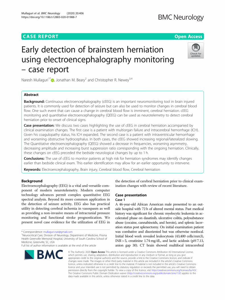
hemorrhages (ICH) in the right frontotemporal region (Fig. 1(A1 and B1)). He was subsequently transferred to the neuro-critical care unit with a coagulation profile suggestive of dis-seminated intravascular coagulation (fibrinogen –undetectable, d-dimer > 35,200 ng/mL, haptoglobulin < 10mg/dL, and activated plasma thromboplastin time of 54.5 s.He was treated with cryoglobulin, fresh frozen plasma andplatelet transfusions but developed tumor lysis syndrome(TLS) with elevated uric acid (12.2mg/L, phosphorous 6.6mg/dL. Repeat neuroimaging 6 h from initial scan showedhematoma expansion. The patient was started on intraven-ous hydration, allopurinol, hydroxyurea, rasburicase, andnilotinib. He developed acute respiratory failure and wasintubated. Peripheral smear confirmed myelocytic leukemiawith monocytic differentiation. Given the acute ICH, he wasnot a candidate for intensive chemotherapy regimen but phe-resis for leukoreduction was initiated. He became
hypotensive requiring multiple vasopressor medications andwas started on broad spectrum antibiotics. Initial EEGshowed continuous generalized slowing maximal in the righthemisphere suggestive of severe encephalopathy withoutseizure activity. Fibrinogen improved to 125mg/dL. RepeatCT brain scan was stable.He was transferred to medical intensive care unit for
management of multiorgan failure and TLS. On hospitalday three at 8:00 am his right pupil became dilated andnon-reactive. Repeat CT brain again was immediately ob-tained and showed stable right frontal hemorrhage al-though with multiple new bilateral supratentorialhemorrhages as well as uncal herniation and midbraincompression (Fig. 2 (A2 and B2)). Although at 09:30 amthe left pupil also became dilated and non-reactive, neuro-surgical intervention was deferred due to coagulopathyand overall poor prognosis. Approximately 1 h prior to left
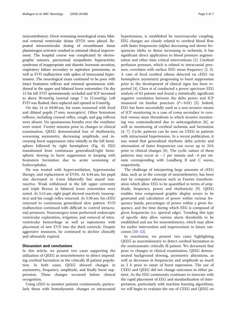
Fig. 1 Computerized tomography of the brain – axial sections. A1, B1 – initial scan showing intracerebral hemorrhage in right frontal andtemporal areas. The midbrain slice shows effacement of quadrigeminal cistern. A2, B2 – Day 3 scan showing large hemorrhage withintraventricular extension, severe cerebral edema with loss of grey-white differentiation, midbrain compression, and bilateral uncal herniation
Mullaguri et al. BMC Neurology (2020) 20:406 Page 2 of 7
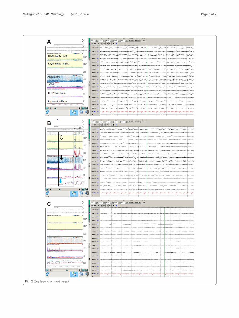
Fig. 2 (See legend on next page.)
Mullaguri et al. BMC Neurology (2020) 20:406 Page 3 of 7
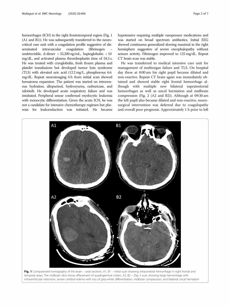
pupillary dilatation, His continuous electroencephalog-raphy (cEEG) showed worsening bilateral cortical dysfunc-tion between 8:25–8:35 am (Fig. 2a). QuantitativeElectroencephalography (QEEG) showed a transition fromdecrease in frequencies, changes in asymmetry, decreasein amplitude, and an increase in burst suppression ratio2 h prior to onset of burst supression (Fig. 2b-c). No EEGreactivity was noted at this time. Despite hyperventilationand hyperosmolar therapy, cerebral herniation was not re-versed. Due to poor prognosis, family requested comfortmeasures and the patient subsequently expired.
Case 2A 76-year-old Caucasian male presented from nursinghome to outside hospital with chief complaint of confusion,loose stools, and chills. He had no focal neurological defi-cits. Past medical history was significant for spinal meta-static cancer of unknown primary origin, chroniccommunicating hydrocephalus with dementia, and baselinegait instability. CT brain showed isolated intraventricularhemorrhage (IVH) and hydrocephalus (Fig. 3a-d). He wastransferred to the neurocritical care unit for further man-agement. MRI and cerebral angiography imaging were
(See figure on previous page.)Fig. 2 Continuous and quantitative electroencephalography changes of case 1. a Baseline EEG shows generalized slowing with a lateralizedslowing in the left hemisphere generalized. b black box indicates the change in QEEG where rhythmicity in the right then left hemisphere dropsout (open arrows) also noted, decrease in asymmetry of the left hemisphere (black arrow) and increasing in burst suppression ratio (blue arrow). cdiffuse suppression
Fig. 3 Computerized tomography of the brain axial section. a showing intraventricular hemorrhage (IVH) with hydrocephalus. b showing rightfrontal external ventricular drain (EVD) placement with no resolution of hydrocephalus. c showing post tPA scan with no resolution of IVH,interval placemnent of left frontal EVD. d showing post surgical changes of IVH evacuation, septostomy and new additional left parietal EVDplacement with no radiological improvement of hydrocephalus
Mullaguri et al. BMC Neurology (2020) 20:406 Page 4 of 7
noncontributory. Given worsening neurological exam, bilat-eral external ventricular drains (EVD) were placed. Re-peated intraventricular dosing of recombinant tissueplasminogen activator resulted in minimal clinical improve-ment. The hospital course was complicated by electro-graphic seizures, paroxysmal sympathetic hyperactivity,syndrome of inappropriate anti diuretic hormone secretion,respiratory failure secondary to aspiration pneumonia, aswell as EVD malfunction with spikes of intracranial hyper-tension. The neurological exam continued to be poor withintact brainstem reflexes and minimal spontaneous with-drawal in the upper and bilateral lower extremities. On day13 his left EVD spontaneously occluded and ICP increasedto above 30mmHg (normal range 7 to 15mmHg). LeftEVD was flushed, then replaced and opened at 0mmHg.On day 14 at 04:00 am, his exam worsened with fixed
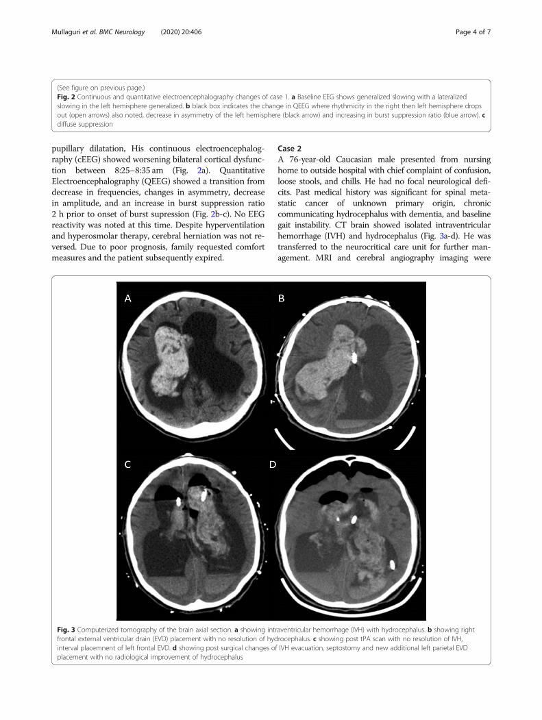
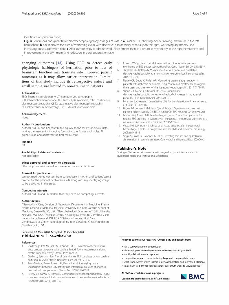
and dilated pupils (7 mm; neuroptics). Other brainstemreflexes, including corneal reflex, cough, and gag reflexeswere absent. No spontaneous breaths over the ventilatorwere noted. Twenty minutes prior to changes in clinicalexamination, QEEG demonstrated loss of rhythmicity,worsening asymmetry, decreasing amplitude, and in-creasing burst suppression ratio initially in the left hemi-sphere followed by right hemisphere (Fig. 4); EEGtransitioned from continuous generalized/right hemi-spheric slowing to burst suppression in keeping withbrainstem herniation due to acute worsening ofhydrocephalus.He was treated with hyperventilation, hyperosmolar
therapy, and replacement of EVDs. At 4:44 am, his pupilsize decreased to 4 mm bilaterally but stayed non-reactive. Weak withdrawal in the left upper extremityand triple flexion in bilateral lower extremities werenoted. At 5:15 am, right pupil showed reactivity (neurop-tics) and his cough reflex returned. At 5:30 am, his cEEGreturned to continuous generalized slow pattern. EVDmalfunction continued with difficult to control intracra-nial pressures. Neurosurgery team performed endoscopicventricular exploration, irrigation, and removal of intra-ventricular hemorrhage as well as septostomy withplacement of new EVD into the third ventricle. Despiteaggressive measures, he continued to decline clinicallyand ultimately expired.
Discussion and conclusionsIn this article, we present two cases supporting theutilization of QEEG as neurotelemetry to detect impend-ing cerebral herniation in the critically ill patient popula-tion. In both cases, QEEG showed changes inasymmetry, frequency, amplitude, and finally burst sup-pression. These changes occurred before clinicalrecognition.Using cEEG to monitor patients continuously, particu-
larly those with hemodynamic changes or intracranial
hypertension, is established by neurovascular coupling.EEG changes are closely related to cerebral blood flowwith faster frequencies (alpha) decreasing and slower fre-quencies (delta or theta) increasing in ischemia; it hassignificant direct application to blood pressure augmen-tation and other time critical interventions [1]. Cerebralperfusion pressure, which is related to intracranial pres-sure, correlates with surface EEG mean frequency [2, 3].A case of focal cerebral edema detected on cEEG viahemisphere asymmetry progressing to burst suppressionprior to the development of clinical signs has been re-ported [4]. Chen et al conducted a power spectrum EEGanalysis of 62 patents and found a statistically significantnegative correlation between the delta power and ICPmeasured via lumbar puncture (P < 0.01) [5]. Indeed,EEG has been successfully used as a non-invasive meansof ICP monitoring in a case of coma secondary to cere-bral venous sinus thrombosis in which invasive monitor-ing was contraindicated due to anticoagulation [6], aswell as monitoring of cerebral ischemia and herniation[4, 7]. Cyclic patterns can be seen on CEEG in patientswith intracranial hypertension. In a recent publication, itwas noted that generalized rhythmic delta activity andattenuation of faster frequencies can occur up to 24 hprior to clinical changes [8]. The cyclic nature of thesepatterns may occur at ~ 1 per minute and ~ 6 per mi-nute corresponding with Lundberg B and C waves,respectively.The challenge of interpreting large amounts of cEEG
data, such as in the concept of neurotelemetry, has beenmet by computer advances such as Fourier transform-ation which allow EEG to be quantified in terms of amp-litude, frequency, power and rhythmicity [9]. QEEGenables time compressed graphic display arrays to begenerated and calculation of power within various fre-quency bands, percentages of power within a given fre-quency, and the time during which EEG is composed ofgiven frequencies (i.e. spectral edge). Trending this typeof specific data allow various alarm thresholds to beestablished and use for neurotelemetry, which may allowfor earlier intervention and improvement in future out-comes [10–12].In conclusion, we present two cases highlighting
QEEG as neurotelemetry to detect cerebral herniation inthe nontraumatic critically ill patient. We document thatprior to changes in clinical examination, QEEG demon-strated background slowing, asymmetry alterations, aswell as decreases in frequencies and amplitude as muchas 2 h prior to onset of burst supression. The use ofCEEG and QEEG did not change outcomes in either pa-tient. As the EEG community continues to innovate withthe rapid placement of EEG and standardization of inter-pretation, particularly with machine learning algorithms,we will begin to evaluate the use of CEEG and QEEG on
Mullaguri et al. BMC Neurology (2020) 20:406 Page 5 of 7
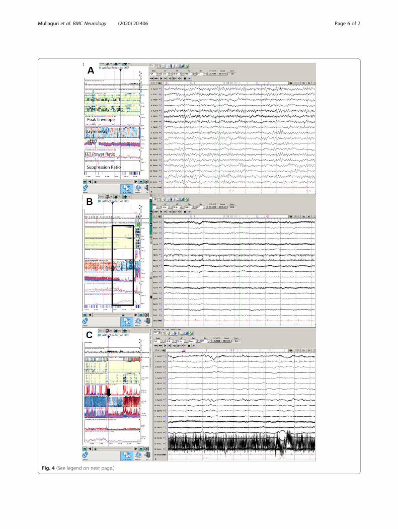
Fig. 4 (See legend on next page.)
Mullaguri et al. BMC Neurology (2020) 20:406 Page 6 of 7
changing outcomes [13]. Using EEG to detect earlyphysiologic harbingers of herniation prior to loss ofbrainstem function may translate into improved patientoutcomes as it may allow earlier intervention. Limita-tions of this study include its retrospective nature andsmall sample size limited to non-traumatic patients.
AbbreviationsEEG: Electroencephalography; CT: computerized tomography;ICH: intracerebral hemorrhage; TLS: tumor lysis syndrome; cEEG: continuouselectroencephalography; QEEG: Quantitative electroencephalography;IVH: intraventricular hemorrhage; EVD: External ventricular drain
AcknowledgementsNone.
Authors’ contributionsAuthors NM, JB, and CN contributed equally to the review of clinical data,writing the manuscript including formatting the figures and tables. Allauthors read and approved the final manuscript.
FundingNA
Availability of data and materialsNot applicable.
Ethics approval and consent to participateEthics approval was waived for case reports at our institutions.
Consent for publicationWe obtained signed consent from patient/case 1 mother and patient/case 2brother for the personal or clinical details along with any identifying imagesto be published in this study.
Competing interestsAuthors NM, JB and CN declare that they have no competing interests.
Author details1Neurocritical Care, Division of Neurology, Department of Medicine, PrismaHealth Greenville Memorial Hospital, University of South Carolina School ofMedicine, Greenville, SC, USA. 2Neurobehavioral Sciences, A.T. Still University,Kirksville, MO, USA. 3Epilepsy Center, Neurological Institute, Cleveland ClinicFoundation, Cleveland, OH, USA. 4Division of Neurocritical Care,Cerebrovascular Center, Neurological Institute, Cleveland Clinic Foundation,Cleveland, OH, USA.
Received: 20 May 2020 Accepted: 30 October 2020
References1. Sharbrough FW, Messick JM Jr, Sundt TM Jr. Correlation of continuous
electroencephalograms with cerebral blood flow measurements duringcarotid endarterectomy. Stroke. 1973;4:674–83.
2. Diedler J, Sykora M. Bast T et al quantitative EEG correlates of low cerebralperfusion in severe stroke. Neurocrit Care. 2009;11:210–6.
3. Sanz-García A, Pérez-Romero M, Pastor J, et al. Identifying causalrelationships between EEG activity and intracranial pressure changes inneurocritical care patients. J Neural Eng. 2018;15:066029.
4. Newey CR, Sarwal A, Hantus S. Continuous electroencephalography (cEEG)changes precede clinical changes in a case of progressive cerebral edema.Neurocrit Care. 2013;18:261–5.
5. Chen H, Wang J, Mao S, et al. A new method of intracranial pressuremonitoring by EEG power spectrum analysis. Can J Neurol Sci. 2012;39:483–7.
6. Threlkeld ZD, Kottapally M, Aysenne A, et al. Continuous qualitativeelectroencephalography as a noninvasive Neuromonitor. Neurohospitalist.2016;6:157–60.
7. Newey CR, Gupta V, Ardelt AA. Monitoring pressure augmentation inpatients with ischemic penumbra using continuous electroencephalogram:three cases and a review of the literature. Neurohospitalist. 2017;7:179–87.
8. Sheikh ZB, Maciel CB, Dhakar MB, et al. Nonepilepticelectroencephalographic correlates of episodic increases in intracranialpressure. J Clin Neurophysiol. 2020;00:1–10.
9. Foreman B, Claassen J. Quantitative EEG for the detection of brain ischemia.Crit Care. 2012;16:216.
10. Rogers JM, Bechara J, Middleton S, et al. Acute EEG patterns associated withtransient ischemic attack. Clin EEG Neurosci Clin EEG Neurosci. 2019;50:196–204.
11. Ghasemi M, Azeem MU, Muehlschlegel S, et al. Prescription patterns forroutine EEG ordering in patients with intracranial hemorrhage admitted to aneurointensive care unit. J Crit Care. 2019;50:262–8.
12. Vespa PM, O'Phelan K, Shah M, et al. Acute seizures after intracerebralhemorrhage: a factor in progressive midline shift and outcome. Neurology.2003;60:1441–6.
13. Singla S, Garcia GE, Rovenolt GE, et al. Detecting seizures and epileptiformabnormalities in acute brain injury. Curr Neurol and Neurosci Rep. 2020;20:42.
Publisher’s NoteSpringer Nature remains neutral with regard to jurisdictional claims inpublished maps and institutional affiliations.
(See figure on previous page.)Fig. 4 Continuous and quantitative electroencephalography changes of case 2. a Baseline EEG showing diffuse slowing, maximum in the lefthemisphere. b box indicates the area of worsening exam with decrease in rhythmicity especially on the right, worsening asymmetry, andincreasing burst suppression ratio. c After osmotherapy is administered (black arrow), there is a return in rhythmicity in the right hemisphere andimprovement in the asymmetry and reduction in burst suppression ratio
Mullaguri et al. BMC Neurology (2020) 20:406 Page 7 of 7