Embed Size (px)
Citation preview
Brainstem Reflexes Herniation Syndromes (CN IX-XII) Lab 7March 24, 2021 - Dr. Krebs ([email protected])
Design & Artwork: The HIVE (hive.med.ubc.ca) 1
Objectives:1. Describe the relationship of the functional anatomy of CN IX - XII and the location of their respective
nuclei to a neurological exam which examines the brainstem.
2. Explain the neuroanatomical pathways associated with brainstem reflexes tested in the conscious and unconscious patient.
3. Describe the relationship between the sympathetic and parasympathetic innervation of the eye to the clinical assessment of eye reflexes.
4. Describe the relationship of changes in upper limb posture of unconscious patient to underlying damage to the brainstem.
5. Describe the consequences of herniation syndromes associated with increases in intracranial pressure.
** NOTE: Interactive PDFs are best viewed on desktop/laptop computers - functionality is not reliable on mobile devices **
Notes: • For identification of the cranial nerves, use online modules and videos,
your atlas and micrographs to locate the nuclei listed.
• On the brain and brainstem specimens, locate cranial nerves IX, X, XI and XII. Note the level at which they are attached to the brainstem.
Videos for Review:
Brainstem Reflexes Herniation Syndromes (CN IX-XII) Lab 7March 24, 2021 - Dr. Krebs ([email protected])
Design & Artwork: The HIVE (hive.med.ubc.ca) 2
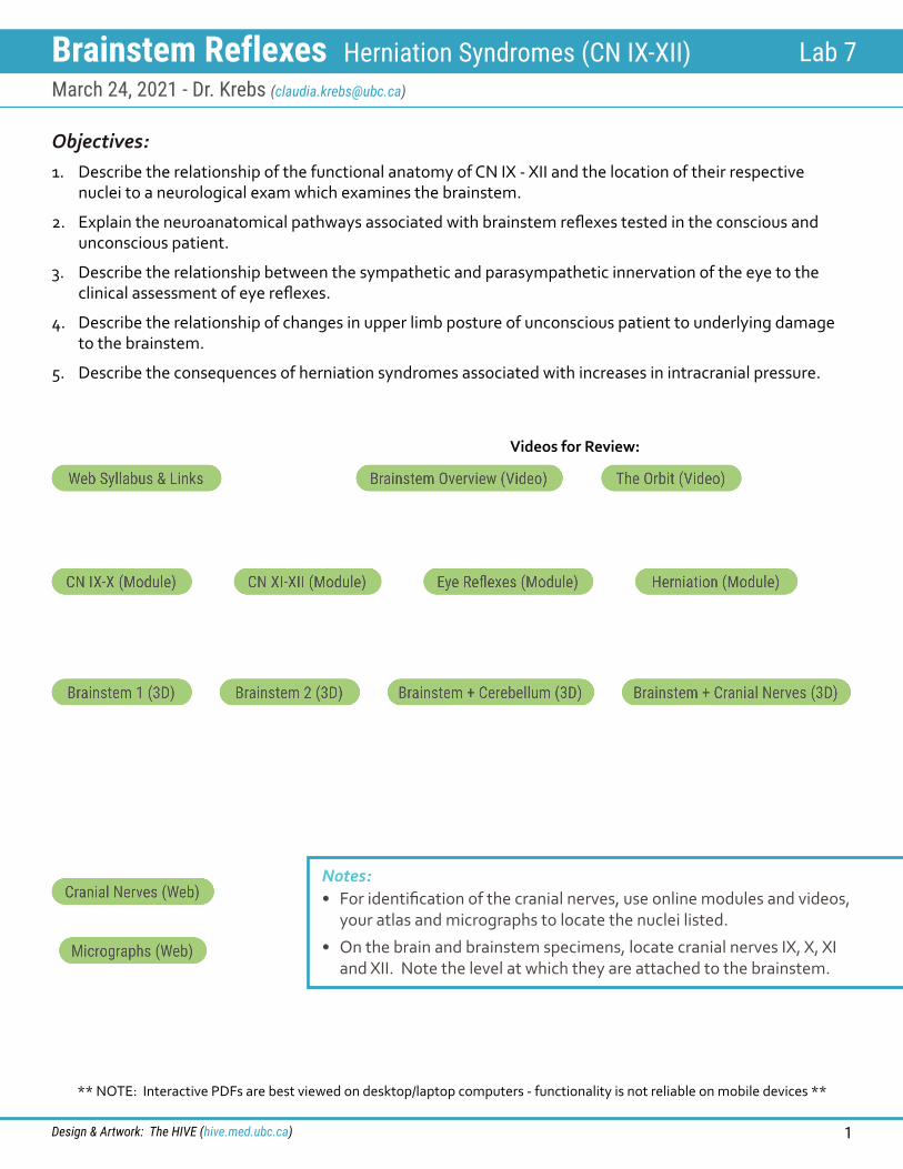
Glossopharyngeal Nerve (CN IX)
Modality Associated Nucleus FunctionMotor(SVE) Nucleus ambiguus Motor to stylopharyngeus muscle
Parasympathetic (GVE) Inferior salivatory nucleus Stimulation of parotid gland
Taste(SVA) Solitary nucleus and tract Taste from posterior 1/3 of tongue
Somatic Sensory(GSA)
Spinal trigeminal nucleus and tract
General sensation from posterior 1/3 of tongue, pharynx, external ear/tympanic membrane
Visceral Sensory(GVA) Solitary nucleus and tract Carotid body, gag sensation from oropharynx
Which foramen does CN IX exit through?
Highlight and label the nuclei associated with CN IX in this diagram and show the types of fibres that comprise this peripheral nerve.
posterior
anterior
Brainstem Reflexes Herniation Syndromes (CN IX-XII) Lab 7March 24, 2021 - Dr. Krebs ([email protected])
Design & Artwork: The HIVE (hive.med.ubc.ca) 3
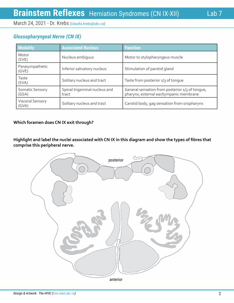
Glossopharyngeal Nerve (CN IX)
Gag Reflex
Modified from Lippincott’s Illustrated Reviews: Neuroscience by C. Krebs, J. Weinberg, E.J. Akesson, and E. Dilli. For educational use only. Copyright © 2017 by Lippincott Williams & Wilkins. All rights reserved.
Brainstem Reflexes Herniation Syndromes (CN IX-XII) Lab 7March 24, 2021 - Dr. Krebs ([email protected])
Design & Artwork: The HIVE (hive.med.ubc.ca) 4
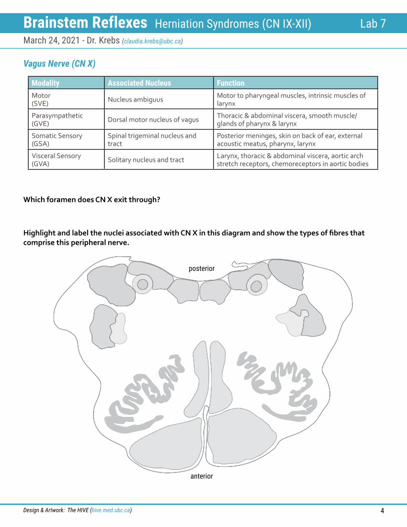
Vagus Nerve (CN X)
Modality Associated Nucleus FunctionMotor(SVE) Nucleus ambiguus Motor to pharyngeal muscles, intrinsic muscles of
larynx
Parasympathetic (GVE) Dorsal motor nucleus of vagus Thoracic & abdominal viscera, smooth muscle/
glands of pharynx & larynx
Somatic Sensory(GSA)
Spinal trigeminal nucleus and tract
Posterior meninges, skin on back of ear, external acoustic meatus, pharynx, larynx
Visceral Sensory(GVA) Solitary nucleus and tract Larynx, thoracic & abdominal viscera, aortic arch
stretch receptors, chemoreceptors in aortic bodies
Which foramen does CN X exit through?
Highlight and label the nuclei associated with CN X in this diagram and show the types of fibres that comprise this peripheral nerve.
posterior
anterior
Brainstem Reflexes Herniation Syndromes (CN IX-XII) Lab 7March 24, 2021 - Dr. Krebs ([email protected])
Design & Artwork: The HIVE (hive.med.ubc.ca) 5
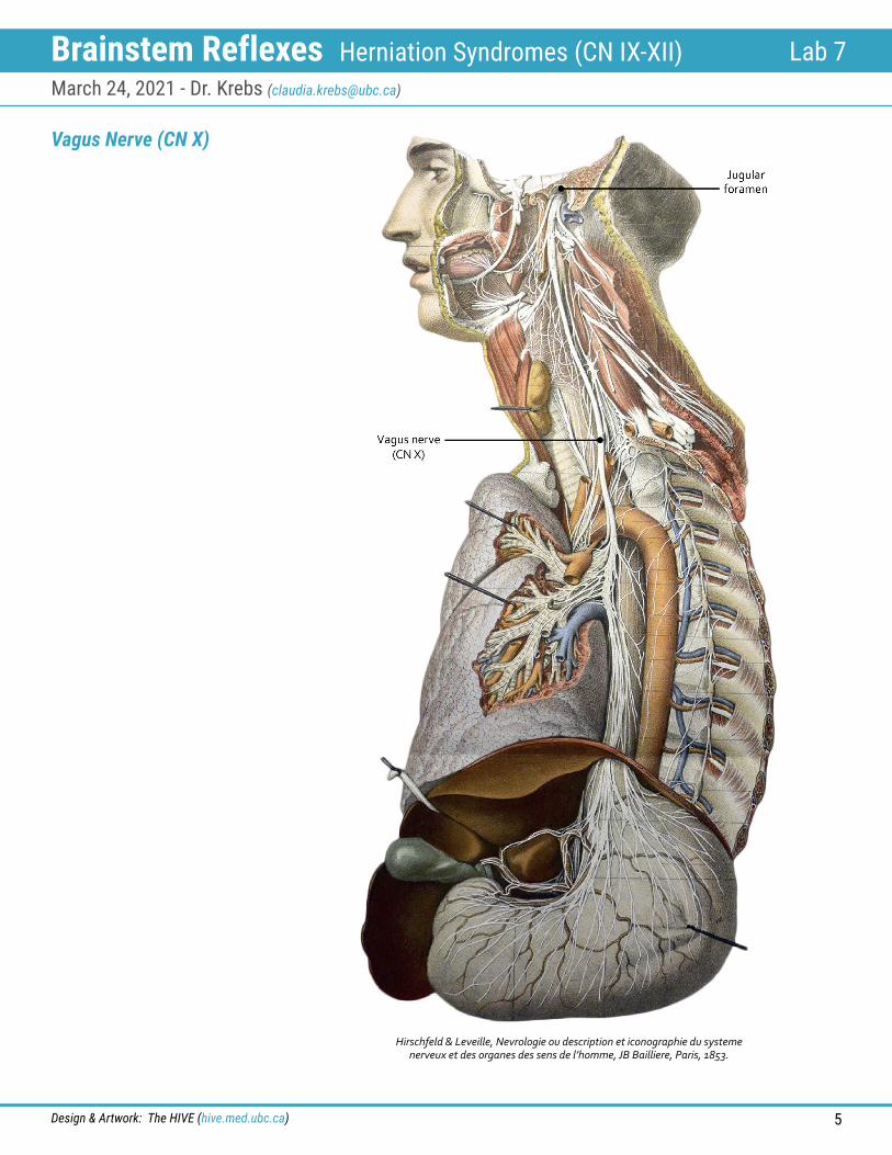
Hirschfeld & Leveille, Nevrologie ou description et iconographie du systeme nerveux et des organes des sens de l’homme, JB Bailliere, Paris, 1853.
Vagus Nerve (CN X)

Brainstem Reflexes Herniation Syndromes (CN IX-XII) Lab 7March 24, 2021 - Dr. Krebs ([email protected])
Design & Artwork: The HIVE (hive.med.ubc.ca) 6
Accessory Nerve (CN XI)
• Its cell bodies are located in the cervical spinal cord.
• CN XI is a motor nerve that innervates sternocleidomastoid and trapezius muscles.
Which foramen does CN XI exit through?
Hirschfeld & Leveille, Nevrologie ou description et iconographie du systeme nerveux et des organes
des sens de l’homme, JB Bailliere, Paris, 1853.
Brainstem Reflexes Herniation Syndromes (CN IX-XII) Lab 7March 24, 2021 - Dr. Krebs ([email protected])
Design & Artwork: The HIVE (hive.med.ubc.ca) 7
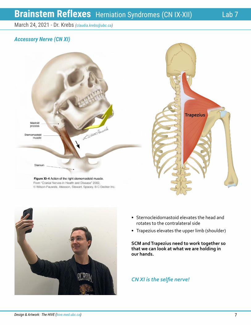
Accessory Nerve (CN XI)
• Sternocleidomastoid elevates the head and rotates to the contralateral side
• Trapezius elevates the upper limb (shoulder)
SCM and Trapezius need to work together so that we can look at what we are holding in our hands.
CN XI is the selfie nerve!
Trapezius
Brainstem Reflexes Herniation Syndromes (CN IX-XII) Lab 7March 24, 2021 - Dr. Krebs ([email protected])
Design & Artwork: The HIVE (hive.med.ubc.ca) 8
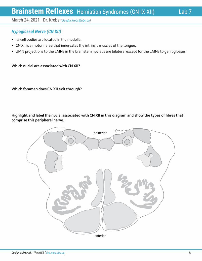
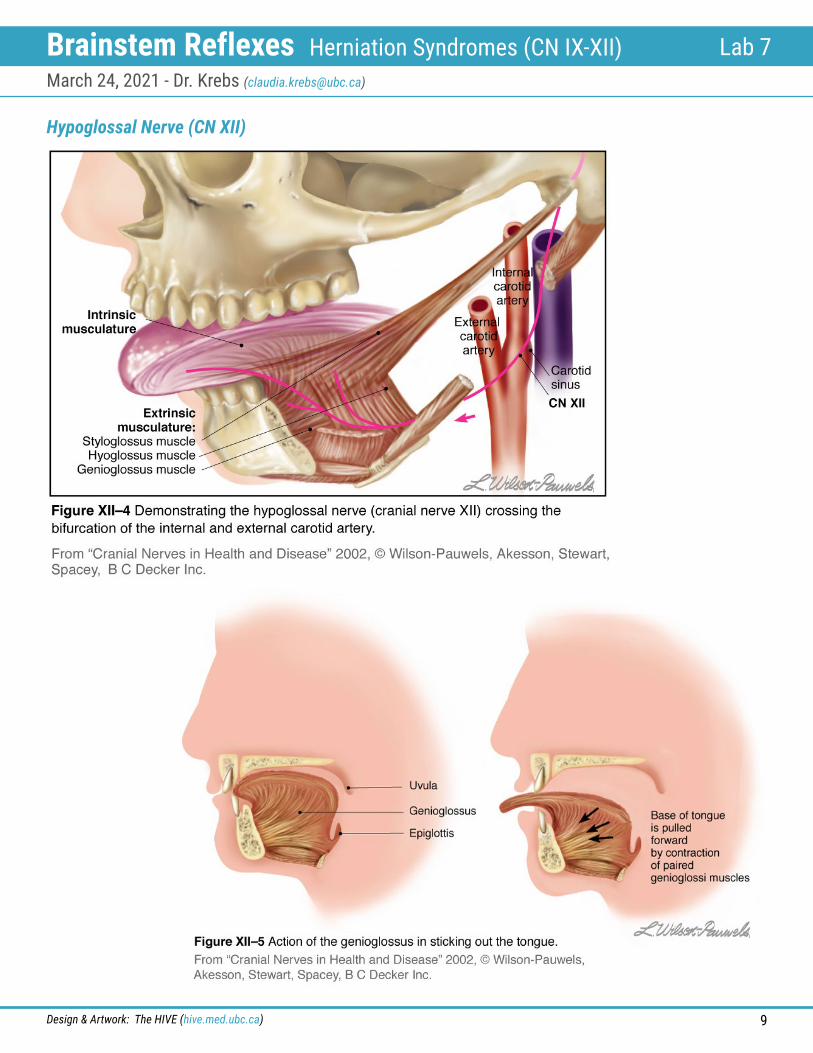
Hypoglossal Nerve (CN XII)
• Its cell bodies are located in the medulla.
• CN XII is a motor nerve that innervates the intrinsic muscles of the tongue.
• UMN projections to the LMNs in the brainstem nucleus are bilateral except for the LMNs to genioglossus.
Which foramen does CN XII exit through?
Which nuclei are associated with CN XII?
Highlight and label the nuclei associated with CN XII in this diagram and show the types of fibres that comprise this peripheral nerve.
posterior
anterior
Brainstem Reflexes Herniation Syndromes (CN IX-XII) Lab 7March 24, 2021 - Dr. Krebs ([email protected])
Design & Artwork: The HIVE (hive.med.ubc.ca) 9
Hypoglossal Nerve (CN XII)
Brainstem Reflexes Herniation Syndromes (CN IX-XII) Lab 7March 24, 2021 - Dr. Krebs ([email protected])
Design & Artwork: The HIVE (hive.med.ubc.ca) 10
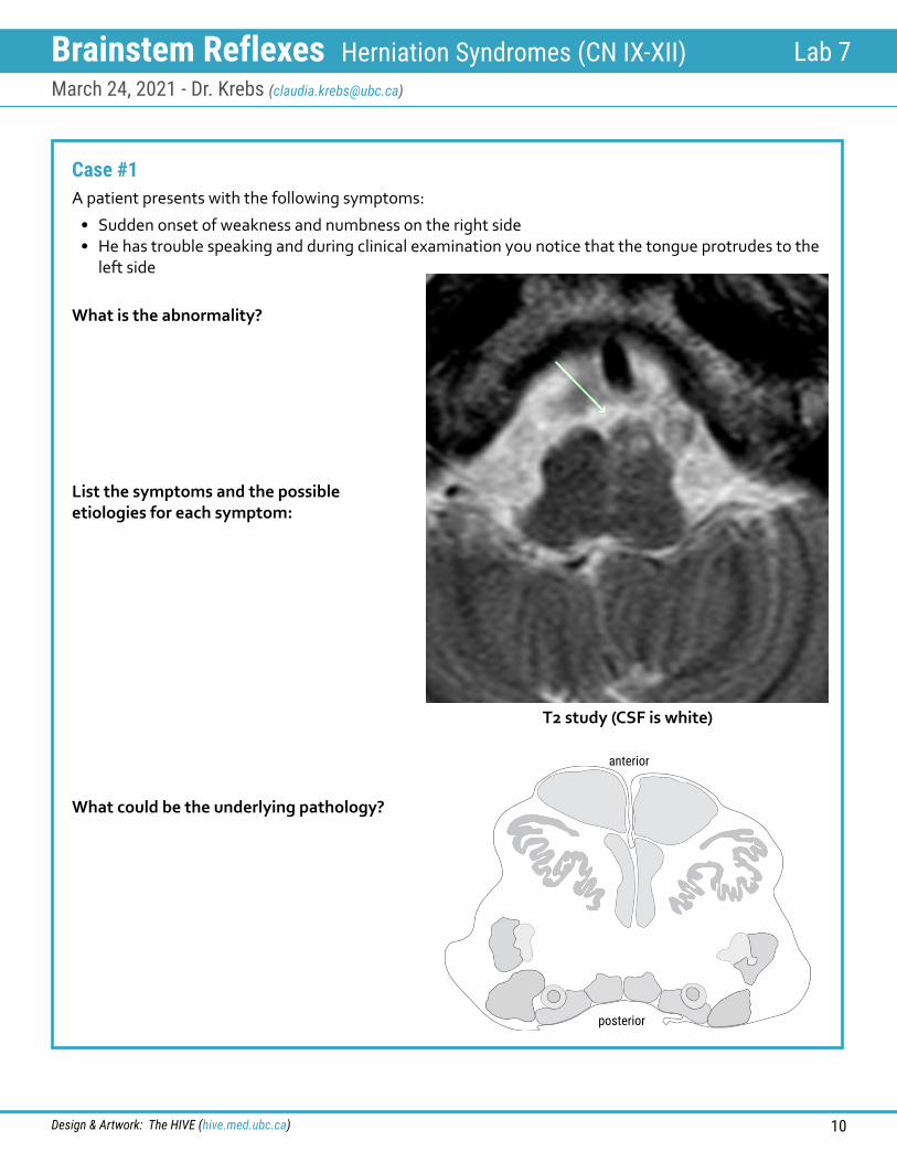
Case #1A patient presents with the following symptoms:
• Sudden onset of weakness and numbness on the right side• He has trouble speaking and during clinical examination you notice that the tongue protrudes to the
left side
What is the abnormality?
List the symptoms and the possible etiologies for each symptom:
What could be the underlying pathology?
T2 study (CSF is white)
posterior
anterior
Brainstem Reflexes Herniation Syndromes (CN IX-XII) Lab 7March 24, 2021 - Dr. Krebs ([email protected])
Design & Artwork: The HIVE (hive.med.ubc.ca) 11
Case #2A patient presents with the following symptoms:
• A progressively hoarse voice• Chronic coughing• Trouble swallowing• Loss of taste in the posterior 1/3 of the tongue on the left side• Weakness of the trapezius on the left
List the symptoms and the possible etiologies for each symptom:
Where would you look for the lesion?
What could be the underlying pathology?
Brainstem Reflexes Herniation Syndromes (CN IX-XII) Lab 7March 24, 2021 - Dr. Krebs ([email protected])
Design & Artwork: The HIVE (hive.med.ubc.ca) 12
Taste
Which cranial nerves carry taste?
Identify on micrographs:
trace structures involved in taste on both gross specimens and micrographs
Which cranial nerve carries taste from the anterior 2/3 of the tongue?
Which cranial nerve carries taste from the posterior 1/3 of the tongue?
Which brainstem nucleus do all taste fibres project to?
nucleus and tractus solitarius
nucleus ambiguus - general location, it is difficult (ambiguous) to locate precisely
dorsal motor nucleus of vagus
spinal trigeminal tract and nucleus
hypoglossal nucleus
Brainstem Reflexes Herniation Syndromes (CN IX-XII) Lab 7March 24, 2021 - Dr. Krebs ([email protected])
Design & Artwork: The HIVE (hive.med.ubc.ca) 13
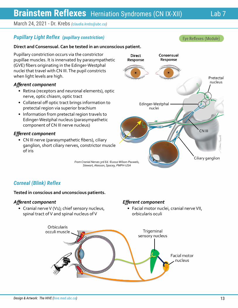
Pupillary Light Reflex (pupillary constriction)
Corneal (Blink) Reflex
Pupillary constriction occurs via the constrictor pupillae muscles. It is innervated by parasympathetic (GVE) fibers originating in the Edinger-Westphal nuclei that travel with CN III. The pupil constricts when light levels are high.
Direct and Consensual. Can be tested in an unconscious patient.
Tested in conscious and unconscious patients.
Afferent component• Retina (receptors and neuronal elements), optic
nerve, optic chiasm, optic tract• Collateral off optic tract brings information to
pretectal region via superior brachium• Information from pretectal region travels to
Edinger-Westphal nucleus (parasympathetic component of CN III nerve nucleus)
Afferent component• Cranial nerve V (V1); chief sensory nucleus,
spinal tract of V and spinal nucleus of V
Efferent component• CN III nerve (parasympathetic fibers), ciliary
ganglion, short ciliary nerves, constrictor muscle of iris
Efferent component• Facial motor nuclei, cranial nerve VII,
orbicularis oculi
From Cranial Nerves 3rd Ed. ©2010 Wilson-Pauwels, Stewart, Akesson, Spacey, PMPH-USA
Brainstem Reflexes Herniation Syndromes (CN IX-XII) Lab 7March 24, 2021 - Dr. Krebs ([email protected])
Design & Artwork: The HIVE (hive.med.ubc.ca) 14
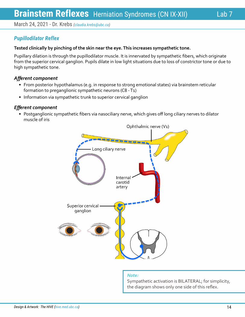
Pupillodilator Reflex
Pupillary dilation is through the pupillodilator muscle. It is innervated by sympathetic fibers, which originate from the superior cervical ganglion. Pupils dilate in low light situations due to loss of constrictor tone or due to high sympathetic tone.
Tested clinically by pinching of the skin near the eye. This increases sympathetic tone.
Afferent component• From posterior hypothalamus (e.g. in response to strong emotional states) via brainstem reticular
formation to preganglionic sympathetic neurons (C8 - T1)• Information via sympathetic trunk to superior cervical ganglion
Efferent component• Postganglionic sympathetic fibers via nasociliary nerve, which gives off long ciliary nerves to dilator
muscle of iris
Note: Sympathetic activation is BILATERAL; for simplicity, the diagram shows only one side of this reflex.
Brainstem Reflexes Herniation Syndromes (CN IX-XII) Lab 7March 24, 2021 - Dr. Krebs ([email protected])
Design & Artwork: The HIVE (hive.med.ubc.ca) 15
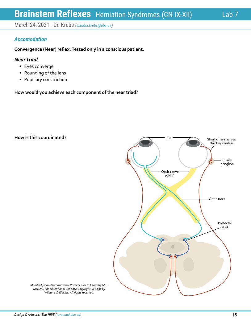
Accomodation
Convergence (Near) reflex. Tested only in a conscious patient.
Near Triad• Eyes converge• Rounding of the lens• Pupillary constriction
How would you achieve each component of the near triad?
How is this coordinated?
Modified from Neuroanatomy Primer Color to Learn by M.E. McNeill. For educational use only. Copyright © 1997 by
Williams & Wilkins. All rights reserved.
Brainstem Reflexes Herniation Syndromes (CN IX-XII) Lab 7March 24, 2021 - Dr. Krebs ([email protected])
Design & Artwork: The HIVE (hive.med.ubc.ca) 16
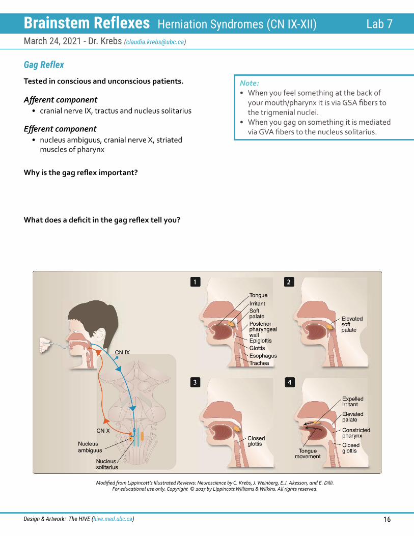
Gag Reflex
Tested in conscious and unconscious patients.
Afferent component• cranial nerve IX, tractus and nucleus solitarius
Efferent component• nucleus ambiguus, cranial nerve X, striated
muscles of pharynx
Why is the gag reflex important?
What does a deficit in the gag reflex tell you?
Note: • When you feel something at the back of
your mouth/pharynx it is via GSA fibers to the trigmenial nuclei.
• When you gag on something it is mediated via GVA fibers to the nucleus solitarius.
Modified from Lippincott’s Illustrated Reviews: Neuroscience by C. Krebs, J. Weinberg, E.J. Akesson, and E. Dilli. For educational use only. Copyright © 2017 by Lippincott Williams & Wilkins. All rights reserved.
Brainstem Reflexes Herniation Syndromes (CN IX-XII) Lab 7March 24, 2021 - Dr. Krebs ([email protected])
Design & Artwork: The HIVE (hive.med.ubc.ca) 17
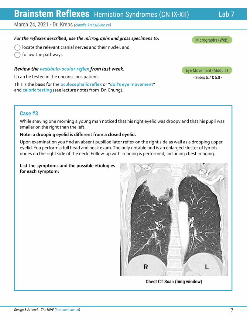
For the reflexes described, use the micrographs and gross specimens to:
locate the relevant cranial nerves and their nuclei, and
follow the pathways
Review the vestibulo-ocular reflex from last week.It can be tested in the unconscious patient.
This is the basis for the oculocephalic reflex or “doll’s eye movement” and caloric testing (see lecture notes from Dr. Chung).
- Slides 5.7 & 5.8 -
Case #3While shaving one morning a young man noticed that his right eyelid was droopy and that his pupil was smaller on the right than the left.
Note: a drooping eyelid is different from a closed eyelid.
Upon examination you find an absent pupillodilator reflex on the right side as well as a drooping upper eyelid. You perform a full head and neck exam. The only notable find is an enlarged cluster of lymph nodes on the right side of the neck. Follow-up with imaging is performed, including chest imaging.
List the symptoms and the possible etiologies for each symptom:
R L
Chest CT Scan (lung window)
Brainstem Reflexes Herniation Syndromes (CN IX-XII) Lab 7March 24, 2021 - Dr. Krebs ([email protected])
Design & Artwork: The HIVE (hive.med.ubc.ca) 18
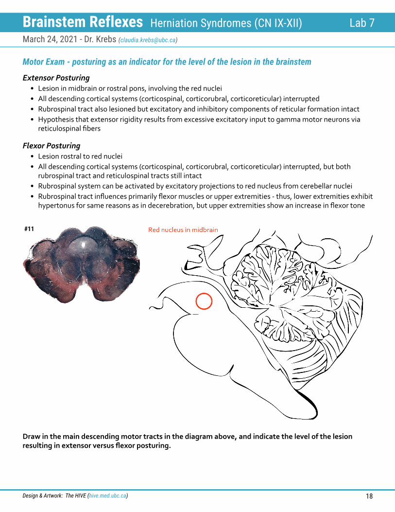
Motor Exam - posturing as an indicator for the level of the lesion in the brainstem
Extensor Posturing• Lesion in midbrain or rostral pons, involving the red nuclei• All descending cortical systems (corticospinal, corticorubral, corticoreticular) interrupted • Rubrospinal tract also lesioned but excitatory and inhibitory components of reticular formation intact• Hypothesis that extensor rigidity results from excessive excitatory input to gamma motor neurons via
reticulospinal fibers
Flexor Posturing• Lesion rostral to red nuclei• All descending cortical systems (corticospinal, corticorubral, corticoreticular) interrupted, but both
rubrospinal tract and reticulospinal tracts still intact• Rubrospinal system can be activated by excitatory projections to red nucleus from cerebellar nuclei • Rubrospinal tract influences primarily flexor muscles or upper extremities - thus, lower extremities exhibit
hypertonus for same reasons as in decerebration, but upper extremities show an increase in flexor tone
#11
Draw in the main descending motor tracts in the diagram above, and indicate the level of the lesion resulting in extensor versus flexor posturing.
Brainstem Reflexes Herniation Syndromes (CN IX-XII) Lab 7March 24, 2021 - Dr. Krebs ([email protected])
Design & Artwork: The HIVE (hive.med.ubc.ca) 19
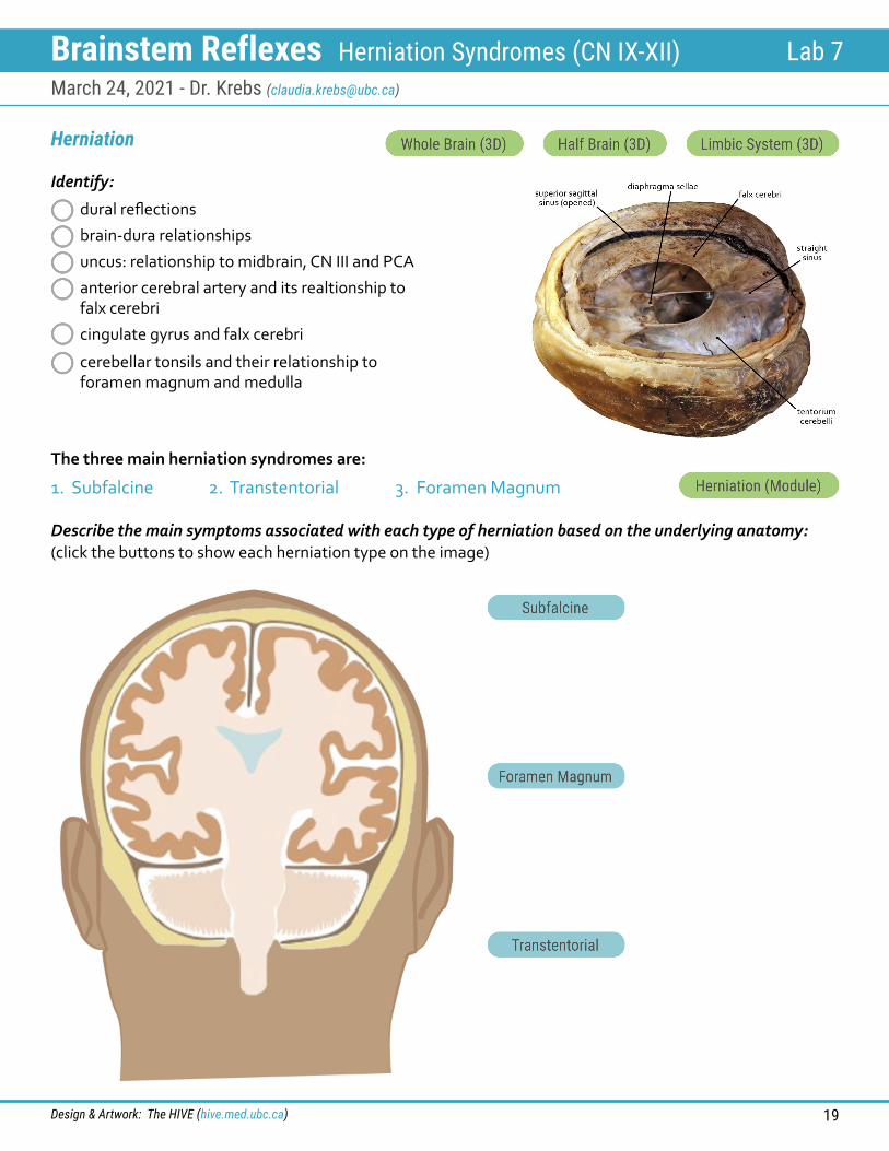
The three main herniation syndromes are:
1. Subfalcine 2. Transtentorial 3. Foramen Magnum
Herniation
Identify:
dural reflections
brain-dura relationships
uncus: relationship to midbrain, CN III and PCA
anterior cerebral artery and its realtionship to falx cerebri
cingulate gyrus and falx cerebri
cerebellar tonsils and their relationship to foramen magnum and medulla
Describe the main symptoms associated with each type of herniation based on the underlying anatomy: (click the buttons to show each herniation type on the image)
Brainstem Reflexes Herniation Syndromes (CN IX-XII) Lab 7March 24, 2021 - Dr. Krebs ([email protected])
Design & Artwork: The HIVE (hive.med.ubc.ca) 20
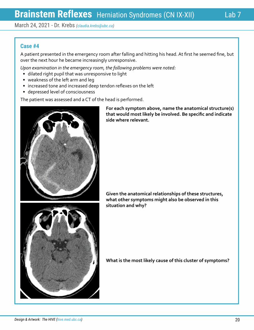
Case #4A patient presented in the emergency room after falling and hitting his head. At first he seemed fine, but over the next hour he became increasingly unresponsive.
Upon examination in the emergency room, the following problems were noted:• dilated right pupil that was unresponsive to light• weakness of the left arm and leg• increased tone and increased deep tendon reflexes on the left• depressed level of consciousness
The patient was assessed and a CT of the head is performed.
For each symptom above, name the anatomical structure(s) that would most likely be involved. Be specific and indicate side where relevant.
Given the anatomical relationships of these structures, what other symptoms might also be observed in this situation and why?
What is the most likely cause of this cluster of symptoms?
Brainstem Reflexes Herniation Syndromes (CN IX-XII) Lab 7March 24, 2021 - Dr. Krebs ([email protected])
Design & Artwork: The HIVE (hive.med.ubc.ca) 21
Recommended Textbooks:Lippincott Illustrated Reviews: NeuroscienceBy: Claudia Krebs, Joanne Weinberg, Elizabeth J. Akesson, Esma DilliLippincott Williams & WilkinsISBN 978-1-4963-6789-1
Neuroanatomy Through Clinical CasesBy: Hal BlumenfeldSinauerISBN 978-0-8789-3613-7
Neuroanatomy in Clinical Context: An Atlas of Structures, Sections, Systems, and SyndromesBy: Duane E. HainesWolters kluwer HealthISBN 978-1-4511-8625-3
Websites:Neuroanatomy | Entrada
RESOURCES
ACKNOWLEDGEMENTS
Artwork & Design:The HIVE, UBC Faculty of Medicine
Instructional Design: Monika FejtekMedical Illustration Lead: Paige BlumerAcademic Lead: Claudia Krebs
Prosector: Lien Vo
THE HIVEUBC