Embed Size (px)
Citation preview

[Type text] Page 0
This course is designed to develop ECG recognition skills and drug treatment knowledge. An American Heart Association course completion certificate in ECG & Pharmacology will be awarded at the end of the class
ECG & Pharmacology
Study Guide
Critical Care Training Center ACLS123.com 818.766.1111
6426 Bellingham Ave. North Hollywood, CA 91606

Critical Care Training Center ACLS123.com 818.766.1111 Page 1
Cardiac Arrhythmias
Pulseless Rhythms
Ventricular Fibrillation
Ventricular Fibrillation (V-Fib or VF) is the most common rhythm that occurs immediately after cardiac arrest. In this rhythm, the ventricles quiver and are unable to uniformly contract to pump blood. It is for this reason that early defibrillation is so imperative. A victim’s chance of survival diminishes rapidly over time once the heart goes into V-Fib; therefore, each minute counts when initiating defibrillation.
There are two types of VF, fine and course. Course VF usually occurs immediately after a cardiac arrest and has a better prognosis with defibrillation. Fine VF has waves that are nearly flat and look similar to asystole. Fine VF often develops after more prolonged cardiac arrest and is much more difficult to correct.
Course VF Fine VF
Ventricular Tachycardia
Stable vs Unstable
Pulse vs No Pulse
Ventricular Tachycardia (VT) can present itself with or without a pulse. When a VT is present and the victim has no pulse, the treatment is the same as the VF. High dose shocks for defibrillation will give the best chance for converting the patient out of pulseless VT.
When defibrillating, heart stops hoping that the heart is viable for normal pace
maker to initiate a rhythm.

Critical Care Training Center ACLS123.com 818.766.1111 Page 2
Pulseless Electrical Activity
Pulseless Electrical Activity (PEA) occurs when the heart is beating and has a rhythm, it can be any rhythm, but the patient does not have a pulse. Always treat the patient, not the rhythm.
1. Problem or Possible correctable causes (H’s & T’s)
2. Epinephrine 1mg 1:10,000 – give to anyone WITHOUT a pulse
In order to treat pulseless rhythm, bradycardia, or tachycardia, identification of the possible underlying causes is essential.
Asystole
Asystole is when there is no detectable cardiac activity on EKG. It may occur immediately after cardiac arrest or may follow VF or PEA. Asystole may also follow a third degree heart block. Treatment of asystole is the same as PEA. The American Heart Association recommends that if a patient is in sustained Asystole for 15 minutes, it is reasonable to call the code, but involve the family in the decision if they are available.
Hypovolemia Hypoxia Hydrogen ion (acidosis) Hypo-/hyperkalemia Hypothermia Tension pneumothorax Tamponade, cardiac Toxins Thrombosis, pulmonary Thrombosis, coronary

Critical Care Training Center ACLS123.com 818.766.1111 Page 3
Rhythms with Pulse – Bradycardia & Blocks
Bradycardia
Bradycardia occurs when the heart is beating too slow (<50 beats per minute). If symptomatic, provide oxygen, given Atropine 0.5mg up to 3mg and call for the transcutaneous pacemaker.
In Sinus Bradycadia, the SA node fires at a rate slower than normal for a person’s age. Athletes may have heart rates less than 50 due to their physical conditioning. Obviously, they would not need treatment. It is possible for patient to have a heart rate of 50 and be asymptomatic; however, if a patient with a heart rate of less than 50 has signs of poor perfusion, begin treatment with oxygen and Atropine 0.5mg
First-Degree AV Block – ALL P WAVES CONDUCTED BUT DELAYED
First-Degree AV Block = prolonged PR interval (>0.20 seconds or 5 small boxes on the ECG strip)
In the first-degree AV block, all of the components of the ECG strip are normal except the PR interval. What happens in this situation is that the impulse from the SA node is delayed at the AV node. All impulses are, however, conducted following the delay.
Critical Care Training Center ACLS123.com 818.766.1111 Page 4
Second-Degree AV Block Type I, (Mobitz I, Wenckebach) – Some P waves conducted, others blocked
Second-degree AV block type I (Mobitz I, or Wenckeback) = progressive lengthening of the PR interval with dropped QRS complexes.
The delay in Second-degree AV block type I also occurs at the AV node. The delay, resulting in progressively lengthening PR interval and then there will be a P wave that is not followed by a QRS complex. Following this event, the cycle starts over again with progressively lengthening PR interval followed by a dropped QRS.
Each repeating Wenckebach series has a consistent P:QRS ratio with one less QRS than Ps in the series.
Block location differentiates second degree AV Type I block from Type II blocks.
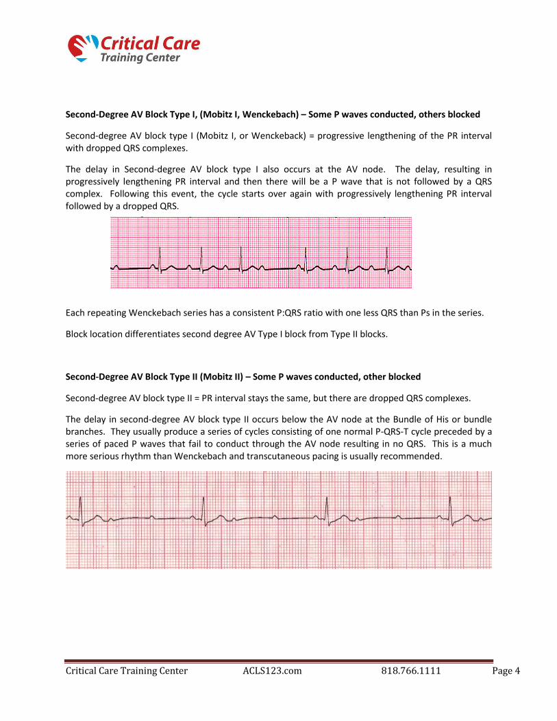
Second-Degree AV Block Type II (Mobitz II) – Some P waves conducted, other blocked
Second-degree AV block type II = PR interval stays the same, but there are dropped QRS complexes.
The delay in second-degree AV block type II occurs below the AV node at the Bundle of His or bundle branches. They usually produce a series of cycles consisting of one normal P-QRS-T cycle preceded by a series of paced P waves that fail to conduct through the AV node resulting in no QRS. This is a much more serious rhythm than Wenckebach and transcutaneous pacing is usually recommended.

Critical Care Training Center ACLS123.com 818.766.1111 Page 5
Third Degree or Complete AV Block
Third Degree or Complete AV Block = no communication in the heart between SA and AV node.
In third-degree or complete AV block, the impulse originating in the SA node is completely blocked. This block may occur at the AV node, bundle of His or bundle branches. In response to this situation, the heart may develop a second pacemaker (either junctional or ventricular) in order to stimulate the ventricles to contract. The location of this “escape pacemaker” will determine if the QRS complexes are wide or narrow. A junctional (narrow QRS complex) escape pacemaker rhythm may possible be stable with a ventricular rate of more than 40 bpm. However, a ventricular (wide QRS complex) escape pacemaker rhythm is usually unstable with a heart rate of less than 40 bpm.
Often heart blocks occur due to cardiac damage following an MI. In high degree blocks (type II and third-degree AV block), characterized by poor perfusion, Atropine 0.5 mg may be given, but prepare for transcutaneous pacing that may be required.
Rhythms with Pulse - Tachycardia
There are three basic groups of tachycardias: sinus tachycardia, supraventricular tachycardia (including atrial tachycardia) and ventricular tachycardia. Fortunately, there is only one algorithm to treat all of them. They key factors are: STABLE vs. UNSTABLE and PULSE vs. NO PULSE. Additional factors are NARROW QRS vs. WIDE QRS and REGULAR vs. IRREGUAR.

Critical Care Training Center ACLS123.com 818.766.1111 Page 6
Sinus Tachycardia
Sinus tachycardia occurs when the SA node is firing at a rate that is faster than normal for a person’s age. The rate is generally 101 to 150 bmp. The key to sinus tachycardia is that all components of a normal ECG are present, P wave, QRS complexes, and T wave. Sinus tachycardia generally starts and stops gradually. There is often a cause such as pain, fever, or agitation that can be identified and treated.
Supraventricular Tachycardia
Supraventricular Tachycardia (SVT) includes any rhythm that begins above the bundle braches. This includes rhythm that begins in the SA node, atrial tissue, or the AV junction. Since the rhythms arise from above the bundle branches, they are characterized by narrow QRS complexes. A supraventricular tachycardia is not the name of a specific arrhythmia. It is a term used to describe a category of regular arrhythmias that cannot be identified more accurately because they have indistinguishable P waves due to their fast rate – usually greater than 150 bpm. The P waves are often indistinguishable because they run into the preceding T waves. The most common SVT rhythms are atrial tachycardia and junctional tachycardia, although Sinus Tachycardia and Atrial Flutter can sometimes also fit into their category with indistinguishable P waves.

Critical Care Training Center ACLS123.com 818.766.1111 Page 7
SVT Treatment
Treatment Question #1 – Stable vs. Unstable
If Unstable – Cardiovert
If Stable, answer question #2
Treatment Questions #2 – Regular vs. Irregular
Rhythm
Regular (SVT or Junctional) = Vagal maneuvers
Adenosine (1st dose = 6mg)
Adenosine (2nd dose = 12mg)
Irregular (A-Fib, A-Flutter, Multi-focal A-Tach) =
Calcium Channel Blockers or Beta Blockers
A = A-fib, A-Flutter
B = Beta Blockers
C = Calcium Channel Blockers (usually used 1st to
slow the rate)