Embed Size (px)
Citation preview
Thorax (1962), 17, 159.
ECTOPIA CORDIS IN MANBY
R. KANAGASUNTHERAM AND J. A. VERZINFrom the Departments of Anatomy and Obstetrics and Gynaecology, University of Khartoum, Sudan
(RECEIVED FOR PUBLICATION AUGUST 18, 1961)
Ectopia cordis in man is a relatively rareanomaly and has been known for many years,being first reported in 1671 by Neil Stensen(Willius, 1948). A translation of the relevantexcerpt from Stensen's article reads, ". . . thesternum was split and the heart, liver and spleen,most of the intestine and right kidney have passedout through the split being thus uncovered." Thisdescription is undoubtedly that of a case of ectopiacordis and antedates the case of Haller (1706,quoted by Byron, 1948) and Martinez (1706),usually regarded as the first to have been reported.Since then over 180 cases have been described.A survey of the available literature indicates
confusion as to the exact meaning of ectopiacordis. Ectopia is derived from the Greek ektopos,meaning away from a place, and implies anabnormality in the position of an organ or a partof the body, often congenital in origin. Accordingto this definition ectopia cordis should includeconditions such as dextrocardia, in which the heartremains within the thorax. Barlow (1938) iscorrect in suggesting that if ectopia cordis is toinclude this type of defect it should be qualifiedas intrathoracic. It seems, however, that ectopiacordis has been mainly limited to those congenitalanomalies in which the heart is not entirely withinthe chest cavity, and Abbott (1927) qualifiesectopia cordis as a " displacement so that the heartpasses out of the thorax and comes to lie eitheron the outer surface of the body or in theabdominal cavity." Even this definition overlooksmalpositions of the heart in relation to the cervicalregion. Similarly, Kalter and Warkany (1959)disregard the cervical and abdominal types of thedefect when they define ectopia cordis orectocardia as a " congenital exposure of the heartdue to insufficient development of the chest."Moreover, it is evident that the term " displace-ment" is inappropriate when referring to thecervical type of ectopia cordis, since in these casesthe heart is merely retained in its embryonicposition in the neck. In view of these difficulties
we suggest that ectopia cordis be defined as acongenital malposition of the heart, partially orcompletely outside the thorax.
CLASSIFICATIONEctopia cordis has been classified by Weese
(1818), by Townsend (1833), and by Rauchbussz(1878), but these authors have not taken intoconsideration all the possible types of the defectand therefore the following classification issuggested.CERVICAL.-The heart is entirely in the cervical
region but the sternum is usually intact.THORACO-CERVICAL.-The heart is partially in
the cervical region but the cranial end of thesternum is defective.THORACIC.-The sternum is defective and the
heart lies outside the thorax, partially orcompletely.THORACO-ABDOMINAL.-This subdivision was
first suggested by Byron (1948), and, accordingto Major (1953), the condition should beaccompanied by a partially absent or cleftsternum, by a diaphragmatic defect, and by amidline abdominal defect of diastasis recti oromphalocoele type.ABDOMINAL.-This presupposes a diaphragmatic
defect allowing the heart to enter the abdominalcavity.Although some 180 cases have been reported,
the condition is comparatively rare, the nomen-clature confusing, and the pathology so obscurethat it seems justifiable to report a further case.In this paper an attempt will be made to discussthe pathogenesis of this interesting phenomenon.
NECROPSY REPORTThe body was that of a partially macerated still-
born full-time female. Pregnancy was reported tohave been uneventful and labour normal. The infantwas well developed, measuring 30 cm. from crown
on 11 July 2018 by guest. Protected by copyright.
http://thorax.bmj.com
/T
horax: first published as 10.1136/thx.17.2.159 on 1 June 1962. Dow
nloaded from
R. KANAGASUNTHERAM and J. A. VERZIN
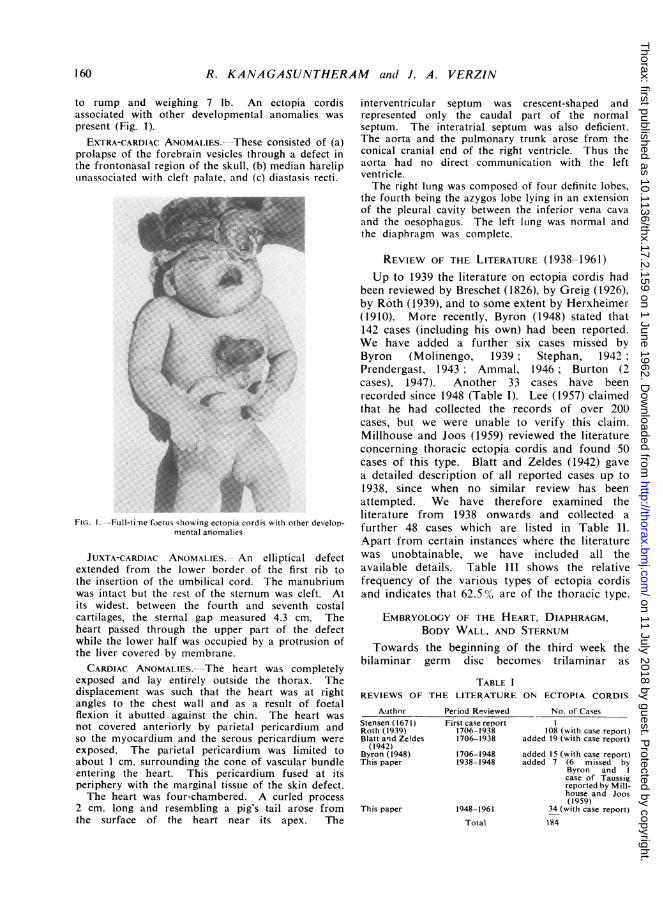
to rump and weighing 7 lb. An ectopia cordisassociated with other developmental anomalies waspresent (Fig. 1).
EXTRA-CARDIAC ANOMALIES.--These consisted of (a)prolapse of the forebrain vesicles through a defect inthe frontonasal region of the skull, (b) median hareliptinassociated with cleft palate, and (c) diastasis recti.
FIG. I.-Full-time foetus showing ectopia cordis with other develop-mental anomalies.
JUXTA-CARDIAC ANOMALIES. -An elliptical defectextended from the lower border of the first rib tothe insertion of the umbilical cord. The manubriumwas intact but the rest of the sternum was cleft. Atits widest. between the fourth and seventh costalcartilages, the sternal gap measured 4.3 cm. Theheart passed through the upper part of the defectwhile the lower half was occupied by a protrusion ofthe liver covered by membrane.CARDIAC ANOMALIES. The heart was completely
exposed and lay entirely outside the thorax. Thedisplacement was such that the heart was at rightangles to the chest wall and as a result of foetalflexion it abutted against the chin. The heart wasnot covered anteriorly by parietal pericardium andso the myocardium and the serous pericardium wereexposed. The parietal pericardium was limited toabout 1 cm. surrounding the cone of vascular bundleentering the heart. This pericardium fused at itsperiphery with the marginal tissue of the skin defect.The heart was four-chambered. A curled process
2 cm. long and resembling a pig's tail arose fromthe surface of the heart near its apex. The
interventricular septum was crescent-shaped andrepresented only the caudal part of the normalseptum. The interatrial septum was also deficient.The aorta and the pulmonary trunk arose from theconical cranial end of the right ventricle. Thus theaorta had no direct communication with the leftventricle.The right lung was composed of four definite lobes,
the fourth being the azygos lobe lying in an extensionof the pleural cavity between the inferior vena cavaand the oesophagus. The left lung was normal andthe diaphragm was complete.
REVIEW OF THE LITERATURE (1938-1961)Up to 1939 the literature on ectopia cordis had
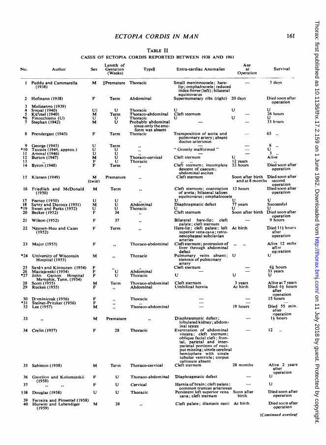
been reviewed by Breschet (1826), by Greig (1926),by Roth (1939), and to some extent by Herxheimer(1910). More recently, Byron (1948) stated that142 cases (including his own) had been reported.We have added a further six cases missed byByron (Molinengo, 1939; Stephan, 1942;Prendergast, 1943; Ammal, 1946; Burton (2cases), 1947). Another 33 cases have beenrecorded since 1948 (Table I). Lee (1957) claimedthat he had collected the records of over 200cases, but we were unable to verify this claim.Millhouse and Joos (1959) reviewed the literatureconcerning thoracic ectopia cordis and found 50cases of this type. Blatt and Zeldes (1942) gavea detailed description of all reported cases up to1938, since when no similar review has beenattempted. We have therefore examined theliterature from 1938 onwards and collected afurther 48 cases which are listed in Table 11.Apart from certain instances where the literaturewas unobtainable, we have included all theavailable details. Table lII shows the relativefrequency of the various types of ectopia cordisand indicates that 62.5% are of the thoracic type.
EMBRYOLOGY OF THE HEART, DIAPHRAGM,BODY WALL, AND STERNUM
Towards the beginning of the third week thebilaminar germ disc becomes trilaminar as
TABLE IREVIEWS OF THE LITERATURE ON ECTOPIA CORDIS
AuthorStensen (1671)Roth (1939)Blatt and Ze!des
(1942)Byron (1948)This paper
This paper
Period Reviewed No. of CasesFirst case report 1
1706-1938 108 (with case report)1706-1938 added 19 (with case report)
1706-1948 added 15 (with case report)1938-1948 added 7 (6 missed by
Byron andcase of Taussigreported by Mill-house and Joos(1959)
1948-1961 34 (with case report)
Total 184
160
on 11 July 2018 by guest. Protected by copyright.
http://thorax.bmj.com
/T
horax: first published as 10.1136/thx.17.2.159 on 1 June 1962. Dow
nloaded from
161ECTOPIA CORDIS IN MAN
TABLE IICASES OF ECTOPTA CORDIS REPORTED BETWEEN 1938 AND 1961
No. AuthorLength of
Sex Gestation(Weeks)
Type§ Extra-cardiac Anomalies
I Puddu and Cammarella(1938)
2 Hofmann (1938)
3 Molinenio (1939)4 Sesyal(1940)5 Ku'inel (1940)*6 Finocchietto (U)7 Stephan (1942)
8 Prendergast (1943)
9*1011121314
George (1945)Taussis (1946, approx.)Ammal (1946)Burton (1947)
oByron (1948)
15 Klassen (1949)
16 Friedlieb and McDonald(1950)
17 Feeney (1950)18 Santy and Duroux (1951)19 Sweet and Parks (1952)20 Becker(1952)
21 Wilson (1952)
22 Nguyen-Huu and Cazes(1952)
23 Major (1953)
*24 University of WisconsinHospital (1953)
25 Sax6i and Kinnunen (1954)26 Maciejewski (1954)*27 John Gaston Hospital
Mernphis, Tenn. (1954)28 Scott (1955)29 Ruckes (1955)
30 D-miniczak (1956)*31 Steiner-Pritzker (1956)32 Lee (1957)
33
M tPremature Thoracic
F Term Abdominal
Small meningocoele; hare-lip; omphalocoele; reducedindex fineer(left); bilateralequinovarus
Supernumerary ribs (right) 20 days
Ull U Thoracic U
M Term Thoraco-abdominal Cleft sternumU U Thoracic U
M U Probably abdominalsince only the ensi-form was absent
F Term Thoracic Transposition c
U
U
U
M
FF
M
(twin)
M
U
M
U
F
F
F
F
M
FF
F
M
F
F
F
M
TermU
U
U
U
Term
Thoraco-cervicalThoracic
,.
Premature
Term
U
U
U
34
37
Term
AbdominalThoracic
,.
..
..
,,9 Thoraco-abdominal
,, Thoracic
U AbdominalU Thoracic
Term Thoraco-abdominal,, Abdominal
U
U
of aorta andpulmonary artery; absentductus arteriosus
- 5 days
s Died soon afteroperation
U
- 26 hoursU
33 hours
65 -
_ _ 6 ,,
"Grossly malformed" - U_ --33
Cleft sternum U Alive12 years
Cleft sternum; incomplete 22 hours Died soon afterdescent of caecum; operationabdominal ascites
Cleft sternum Soon after birth Died soon afterand at 8 months second
operationCleft sternum; coarctation 12 hours Died soon afterof aorta; bilateral talipes operationequinovarus; omphalocoele
T T T~U
Diaphragmatic defectU
Cleft sternum
Bilateral hare-lip; cleftpalate; cleft sternum
Hare-lip; cleft palate; leftsuperior vena cava; retro-oesophageal subclavianarteries
Cleft sternum; protrusion ofliver through abdominaldefect
Pulmonary veins absent;stenosis of pulmonaryartery
Cleft sternum
U
Cleft sternumUmbilical hernia
,, Thoracic
,, Thoraco-abdominal
M Premature
34 Crelin (1957)
35 Sabiston (1958)
36
37
t383940
Gavrilov and Kolomatskii(1958)
Douglas (1958)
Ferreira and Pimentel (1958)Hurwitt and Lebendiger
(1959)
F 28 Thoracic
M Term Thoraco-cervical
F
F
u
u
u
u
Thoraco-abdominal
Cervical
Thoracic
M 28
Diaphragmatic defect;lobulated kidney; abdom-inal testes
Eventration of abdominalviscera; cleft sternum;oblique facial cleft; fron-tal, parietal and inter-parietal portions of occi-put missing; single cerebralhemisphere with singletubular ventricle; corpuscallosum absent
Cleft sternum
Diaphragmatic defect
Hernia of brain; cleft palate;common truncus arteriosus
Persistent left superior venacava; cleft sternum
Cleft palate; diastasis recti
U
77 yearsU
Soon af
At birth
U
ter
USuccessfulU
birth Died soon afteroperation
9 hours
Died 114 hoursafteroperation
Alive 12 mthsafteropzration
U
- 6j hours- 33 years
U U
3 years Alive at 7 yearsAt birth Died 64 hours
afteroperation
15 hours
19 hours Died 55 min.afteroperation
- 14 hours
- 12 ,.
28 months Alive 2 yearsafteroperation
_ U
_ U
Soon after Died soon afterbirth operation
At birth Died soon afteroperation
[Continued overleaf
A°eat
OperationSurvival
on 11 July 2018 by guest. Protected by copyright.
http://thorax.bmj.com
/T
horax: first published as 10.1136/thx.17.2.159 on 1 June 1962. Dow
nloaded from
R. KANAGASUNTHERAM and J. A. VERZIN
TABLE II-co Itinued
No. AuthorLength of
Sex Gestation(Weeks)
Type Extra-cardiac Anomalies
41 Meitner (1959)
42 Millhouse and Joos (1959)43 191.. .
44 Lederman (1959)
45 Brown (1960)
46
4748
Lumsden (1960)
Lambert (1960)This paper
M Term Thoracic
MM
38Term
M Premature
F U
Cleft sternum; harelip; cleftpalate; hydrocephaly;bilateral anophthalmia
Transposition of great ves-sels; hypoplasia of pul-monary artery
Cleft sternum
- 3 days
8,,At birth Died 52 hours
afteroperation
5 days
At birt
F 37 Thoracic or thoraso- Eventration of liver, spleen,abdominal and intestine
F Term Thoracic Prolapse offorebrain vesicles;median harelip; diastasisrecti
th Alive I yearafteroperation
- 12 hours
- Stillborn
* Cases quoted directlY from Millhouse and Joos (1959), who obtained them from personal communications.t Douglas calls his case " ectopia cordis cervicalis." The photograph and anatomical description (" through a deficiency in the centre
part of the chest ") suggest, however, that the ectopia was rather of the thoracic type.: When the length of gestation was not given by the author we relied on the weight of the infant. If this was 5* lb. (2,500 g.) or below,
we resarded the infant as premature.p § When the type of ectopia cordis was not specifically stated in the relevant articles, we based our diagnosis on the anatomical description
orion the photograph.11 U=unknown.
mesoderm differentiates from the primitive streakwhile Hensen's node contributes to the formationof the notochordal process. The mesodermspreads peripherally and by about the 18th daythe intraembryonic coelom makes its firstappearance in the region of the future pericardialcavity. Coelom formation is generally describedas a splitting of the lateral sheet of the intra-embryonic mesoderm into somatic and splanchniclayers. It is perhaps more accurate to regardthe coelomic cavity as resulting from the rapidcoalescence of discrete coelomic spaces (Wyburn,1938; Patten, 1946; and Hamilton, Boyd, andMossman, 1952). Moreover, the coelom has a
significant role to play in the nutrition of the earlyembryo (Streeter, 1942).The heart develops as paired primordia in the
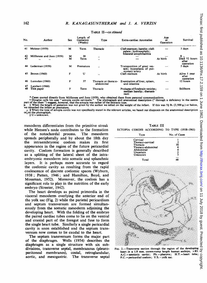
visceral mesoderm overlying the anterior end ofthe yolk sac (Fig. 2) while the parietal pericardiumand septum transversum are formed simultan-eously from the somatic mesoderm adjoining thedeveloping heart. With the folding of the embryothe paired cardiac tubes come to lie on the ventraland cranial part of the foregut and fuse to formthe single heart tube. Similarly a single pericardialcavity is soon established and the septum trans-versum now comes to lie caudal to the heart.The septum transversum forms the major part
of the diaphragm. Wells (1954) describes thediaphragm as a single structure with six sub-divisions, transverse septal, membranous (pleuro-peritoneal membranes), costal, retroglandular,aortic, and mesogastric. The transverse septal
TABLE IIIECTOPIA CORDIS ACCORDING
Type
CervicalThoraco-cervicalThoracicThoraco-abdominalAbdominalUncertainUnknown
Total
TO TYPE (1938-1961)
No. of Cases
2306423
48
A.£C
YY. S.
FIo. 2.-Transverse section through the region of the developingheart in a 1.8 mm. crown-rump length human embryo. x83.A.C.=amniotic cavity; Ph.=pharynx; H.T.=heart tube;P.C. =pericardial coelom; Y.S. =yolk sac.
Ageat
OperationSurvival
162
on 11 July 2018 by guest. Protected by copyright.
http://thorax.bmj.com
/T
horax: first published as 10.1136/thx.17.2.159 on 1 June 1962. Dow
nloaded from
ECTOPIA CORDIS IN MAN
portion forms not only the central tendon butalso the sternal as well as parts of the costal andlumbar portions of the diaphragm. The muscularelements of the diaphragm are said to be derivedfrom the cervical myotomes, but Wells (1954)believes that at least a part of the diaphragmaticmusculature is formed in situ.The somatic mesoderm contributes to the
connective tissue of the anterior body wall whilethe muscular elements are derived from themesoderm of the somites. The epidermis of thebody wall is formed from the embryonic ectoderm.In the thoracic wall and the supra-umbilicalportion of the abdomen there is a stage when theembryonic ectoderm appears to be further awayfrom the midline with the result that confluentsomatic and visceral mesoderms form the liningof the ventral surface of the heart and thesupra-umbilical body wall (Fig. 2).The sternum begins to develop about the fifth
week of intra-uterine life as paired lateralcondensations of mesenchymal cells in the thoracicregion. They are converted into precartilage andadvance towards the midline where they fuse intothe single sternal anlage. The process of fusionproceeds craniocaudally and is complete by theninth week. Simultaneously, the rib cartilagesgrow ventrally from the vertebrae and becomeattached to the sternum. In an experimental studyon the morphogenesis of the mouse sternum,Chen (1953) found that fusion of the bilateralsternal bands could be prevented by delaying themovement of the sternal halves towards oneanother.
DISCUSSIONIt is probable that ectopia cordis originates
during the very early stages of development,perhaps as early as the third week of embryoniclife. The end-results of the anomaly are wellknown, but almost nothing is known of the criticaldevelopmental stages. Although several factsregarding the stages leading to such malformationcan be ascertained from the descriptions of thedifferent grades of the defect, much remains aspeculation. Ectopia cordis in other mammalianembryos, whether experimentally induced orresulting from genetic incompetence, may affordcertain clues, but due care should be taken intransferring such findings to the study of man.Any explanation as to the mode of formation
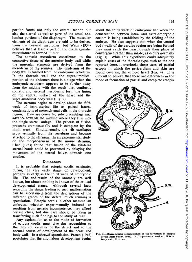
of ectopia cordis must give due recognition tothe different varieties of the defect and to thenormal course of development of the heart andbody wall. In a shrewd speculation, Patten (1946)postulates that the anomalous development begins
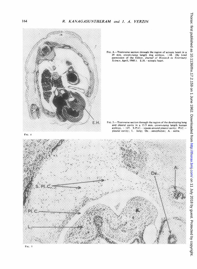
about the third week of embryonic life when thedemarcation between intra- and extra-embryoniccoelom is being established by the folding of theembryo. He also suggests that when the ventralbody walls of the cardiac region are being formedthey must catch the heart outside their place ofconvergence rather than inside, as occurs normally(Fig. 3). While this hypothesis could adequatelyexplain cases of the thoracic type, such as the onereported here, it overlooks those cases of partialectopia in which the pericardium and skin arefound covering the ectopic heart (Fig. 4). It isdifficult to believe that there are differences in themode of formation of partial and complete ectopia
.B.W.
FIG. 3.-Diagrammatic representation of the formation of ectopiacordis (after Patten, 1946). P.C.= pericardial coelom; B.W.=body wall; H.= heart.
163
on 11 July 2018 by guest. Protected by copyright.
http://thorax.bmj.com
/T
horax: first published as 10.1136/thx.17.2.159 on 1 June 1962. Dow
nloaded from
164 R. KANAGASUNTHERAM and J. A. VERZIN
FIG. 4.-Transverse section through the region of ectopic heart in a29 mm. crown-rump length dog embryo, x 44. (By kind
- ~~~~~~~~~~~~~~permissionof the Editor, Journal of Research in Veterinary
~~~~~~.!. =e...pic heart.
Science, April, 1960.). E.H.
E.H. FIG. 5.-Transverse section through the region of the developing lungand pleural cavity in a 13.5 mm. crown-rump length humanembryo, x 127. S.Pi.C. spacesaroundp eural cavity; PliC.pleural cavity;L.S lung; e--oesophaius; A.-aorta.
FIG. 4
. I~~~~~e*#4., .4
S. PI .3C
t-~ ~ ~ ~ ~~~~~~~~~.\ ';,
LI. C. V
.'t'~~'-\.J;, FI.5-rnvrescintruhLh eino h eeoiglnadplerlcvt.na-35m.conruplnt ua
emro 2.SP..-pcsarudperlcvt;P..-
pleural~~~~caiyV,In;O.-ospau;A-ara
FiG. 5
on 11 July 2018 by guest. Protected by copyright.
http://thorax.bmj.com
/T
horax: first published as 10.1136/thx.17.2.159 on 1 June 1962. Dow
nloaded from
ECTOPIA CORDIS INMAN1
74:-.d It.
* 9.-
Aft* 'I X
S524It.4
:t @ ::.s .....0 e _
aF ** * .. A +; tt A ; < ** e 4 >twAt w .* w:t _W3F -
..j
.. 4.I-m...
A... -W
0
e. k
:*.. iA
orr
*.
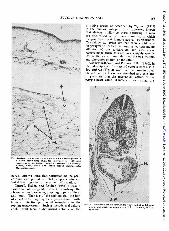
primitive streak, as described by Wyburn (1937)in the human embryo. It is, however, knownthat defects similar to those occurring in manare also found in the lower mammals in whichthe primitive streak is more active. Furthermore,Cantrell et al. (1958) say that there could be adiaphragmatic defect without a correspondingaffliction of the p:ricardium and vice versa.According to them, this requires a highly specificloss of the somatic mesoderm of the one withoutany alteration of that of the other.Kanagasuntheram and Perumal Pillai (1960), in
their description of a case of ectopia cordis in adog embryo (Fig. 4), note that the covering overthe ectopic heart was overstretched and thin andso postulate that the mechanical action of theectopic heart could ultimately break through this
0 -. x
.7'
* : . *.: . : 1
FIG. 6.-Transverse section through the region of a meningocoele ina 29 mm. crown-rump length dog embryo, x 135. (By kindpermission of the Editor, Journal of Research in VeterinaryScience, April, 1960.) S.M. =spaces around meningocoele;M.= meningocoele.
cordis, and we think that herniation of the peri-cardium and partial or total ectopia cordis arebut different grades of the same malformation.
Cantrell, Haller, and Ravitch (1958) discuss asyndrome of congenital defects involving theabdominal wall, sternum, diaphragm, pericardium,and heart. They are of the opinion that the lossof a part of the diaphragm and pericardium resultsfrom a defective portion of mesoderm in theseptum transversum. Such a mesodermal failurecould result from a diminished activity of the
a.
H.
FIG. 7.-Transverse section through the body wall of a 4.0 mm.crown-rump length human embryo, x 135. H. = heart; B.W.=body wall.
165
*4ip
i
on 11 July 2018 by guest. Protected by copyright.
http://thorax.bmj.com
/T
horax: first published as 10.1136/thx.17.2.159 on 1 June 1962. Dow
nloaded from
R. KANAGASUNTHERAM and J. A. VERZIN
thin membrane. Although this may be a plausibleexplanation for the thoracic type of ectopia cordis,it does not throw any light on the mode offormation of the abdominal type of defect.A fourth possibility invokes an abnormal mode
of formation of the coelomic cavity during theearly embryonic stages when the coelom beginsto develop by the coalescence of isolated mesen-chymal clefts. A defective coalescence of theseclefts leading to abnormal adhesions of thestomach and lung to the body wall was observedby Kanagasuntheram (1957). The possibility ofexcessive formation of coelomic spaces, resultingin a reduction of the mesodermal elements, hasnot been sufficiently explored. Fig. 5 showsnormal cavitation occurring in the body walladjoining the pleural cavity in a human embryo,
F. G. P S.T.
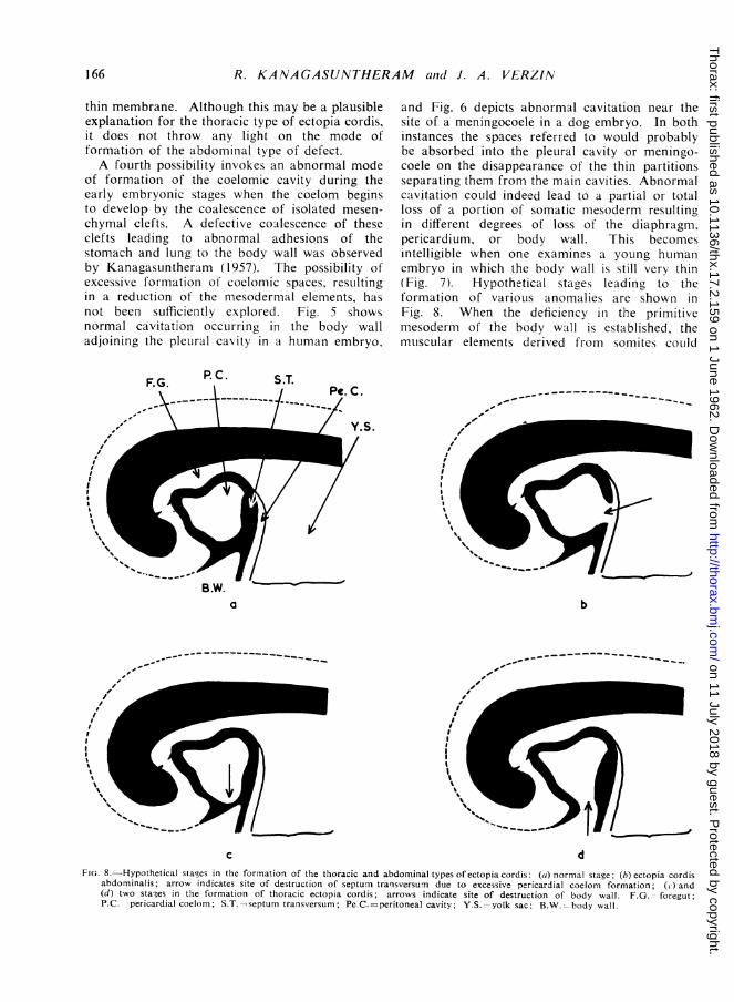
and Fig. 6 depicts abnormal cavitation near thesite of a meningocoele in a dog embryo. In bothinstances the spaces referred to would probablybe absorbed into the pleural cavity or meningo-coele on the disappearance of the thin partitionsseparating them from the main cavities. Abnormalcavitation could indeed lead to a partial or totalloss of a portion of somatic mesoderm resultingin different degrees of loss of the diaphragm,pericardium, or body wall. This becomesintelligible when one examines a young huimanembryo in which the body wall is still very thin(Fig. 7). Hypothetical stage.s leading to theformation of various anomalies are shown inFig. 8. When the deficiency in the primitivemesoderm of the body wall is established, themuiscular elements derived from somites could
PC. C.
Y.S.
B.W.a
-to
b
I
I -q.4
c dFIG. 8.-Hypothetical stages in the formation of the thoracic and abdominal types of ectopia cordis: (a) normal stage; (b) ectopia cordis
abdominalis; arrow indicates site of destruction of septum transversum due to excessive pericardial coelom formation; (c) and(d) two stages in the formation of thoracic ectopia cordis; arrows indicate site of destruction of body wall. F.G. -foregut;P.C. -pericardial coelom; S.T.-septum transversum; Pe C.=peritoneal cavity; Y.S.=yolk sac; B.W.=body wall.
166
on 11 July 2018 by guest. Protected by copyright.
http://thorax.bmj.com
/T
horax: first published as 10.1136/thx.17.2.159 on 1 June 1962. Dow
nloaded from
ECTOPIA CORDIS IN MAN
only migrate ventrally and medially to the edgeof the defect. They would undergo normaldifferentiation and so most of the muscles of theventral body wall would appear to be normalexcept for divarication of the rectus abdominismuscles. Similarly, such a defect in the primitivemesenchyme of the body wall could effectivelyprevent medial migration and fusion of the sternalbands (Chen, 1953). A similar mechanism couldbe envisaged to operate in the diaphragm.The abnormal position of the heart in the
cervical type of ectopia cordis is explicable onthe basis of its situation near the mandibular archduring early development and its subsequentdescent into the thorax. Thus all degrees ofcervical ectopia cordis, ranging from those inwhich the heart projects from the region of themouth cavity to those in which it is partly insidethe thorax, represent various grades of arresteddescent. The abdominal type of ectopia cordiswould entail further abnormal descent of the heartinto the abdomen associated with a deficiency ofthe septum transversum.The hypothesis advanced here not only offers
an explanation of the mode of formation of thedifferent grades of thoracic and abdominal typesof ectopia cordis but also could account forinstances where the pericardium is represented byonly a few fibrous strands. Further support forthis hypothesis is suggested by excessive coelomiccavity formation elsewhere, such as the expansionof the pleural cavity to accommodate an azygoslobe of the lung, a feature of the present case.
SUMMARYA case of thoracic ectopia cordis with extra-
cardiac anomalies is described. The literaturerelating to all varieties of ectopia cordis isreviewed from 1938 onwards. A possibleexplanation of the mode of formation of thisdefect is offered.
We wish to thank Professor H. Butler for hisinterest during the preparation of this paper, andProfessor J. D. Boyd for the loan of the 1.8 mm.and 4 mm. human embryos. Our thanks are alsodue to Mr. J. F. Crane, Department of Anatomy,University of Cambridge, for the photomicrographs,Mr. J. Dew for Fig. 1, and Mrs. R. Kanagasuntheramfor the line drawings.
REFERENCESAbbott, M. E. (1927). In Osler, W., and McCrae, T., Modern
Medicine, 3rd ed., Vol. 4, p. 660.Ammal, P. G. (1946). Antiseptic, 43, 420.Barlow, R. N. (1938). J. Pediat., 12, 58.Becker, T. (1952). Zbl. Chir., 77, 1446.Blatt, M. L., and Zeldes, M. (1942). Amer. J. Dis. Child., 63, 515.Breschet, G. (1826). Rep. gen. Anat. phvsiol. path., 2, pt i. p. 1.Brown, J. J. Mason (1960). J. roy. Coll. Surg. Edinb., 5, 231.Burton, J. F. (1947). Arch. Surg. (Chicago), 54, 79.Byron, F. (1948). J. thorac. Surg., 17, 717.Cantrell, J. R., Haller, J. A., and Ravitch, M. M. (1958). Surg.
Gynec. Obstet., 107, 602.Chen, J. M. (1953). J. Anat. (Lond.), 87, 130.Crelin, E. S. (1957). Yale J. Biol. Med., 30, 38.Dominic7ak, K. (1956). Pediat. pol., 31, 67.Douglas, D. M. (1958). Scot. med. J., 3, 43.Feeney, N. (1950). Trans. Amer. clin. climat. Ass., 62, 259.Ferreira,D., and Pimentel, C. (1958). J. .Ied. P6-to, 36, 781.Friedlieb, O., and McDonald, J. J. (1950). Surgery, 28, 864.Gavrilov, L. F., and Kolomatskii, I. A. (1958). Arkh. anat., Moskova,
35, No. 1, p. 113.George, J. P. (1945). Canad. med. Ass. J., 53, 167.Crreig, D. M. (1926). Edinb. med. J., 33, 480.Hamilton, W. J., Boyd, J. D., and Mossman, H. W. (1952). Human
Embryology, 2nd ed., p. 53. Heffer, Cambridge.Herxheimer, G. (1910). In Schwalbe, E., Die Morphologie der
Missbildungen des Menschen und der Tiere, Teil 3, Lieferung 2,Abt. 2, p. 375. Fischer, Jena.
Hofmann, E. (1938). Mschr. Kinderheilk., 76, 40.Hurwitt, E. S., and Lebendiger, A. (1959). A.M.A. Arch. Surg., 78,
197.Kalter, H., and Warkany. J. (1959). Physiol. Rev., 39, 69.Kanagasuntheram, R. (1957). J. Anat. (Lond.), 91, 188.- and Perumal Pillai, C. (1960). Res. vet. Sci., 1, 172.Klassen, K. (1949). Discussion on Maier, H. C., and Bortone, F.,in
J. thorac. Surg., 18, 851 (1949).Kiihnel, P. (1940). Acta obstet. gynec. scand., 20, 128.Lambert, M. H. (1960). Ned. T. Verlosk., 60, 297.Lederman, D. (1959). Rev. Fac. Med. (Bogotd), 27, 111.Lee, S. T. (1957). Thoravchirurgie, 5, 197.Lumsden, J. W. F. (1960). J. Obstet. Gynaec. Brit. Emp., 67, 299.Maciejewski, J. (1954). Arch. Mal. Cezur, 47, 274.Major, J. W. (1953). J. thorac. Surg., 26, 309.Martinez (1706). Quoted by Barnardo, G. F., in J. Anat. and
Physiol. (Lond.), 32, 325 (1898).Meitner, E. R. (1959). Anat. Anz., 107, 222.Millhouse, R. F., and Joos, H. A. (1959). Amer. Heart J., 57,
470.Molinengo, L. (1939). Atti Soc. Ostet. Ginec., 35,474.Nguyen-Huu and Cazes, G. (1952). Arch. franC, Peliat., 9, 915.Patten, B. M. (1946). Human Embryology, p. 509. Blakiston,
Philadelphia.Prendergast, H. J. (1943). Minn. Med., 26, 182.Puddu, V., and Cammarella, C. (1938). Arch. Mal Caeur, 31, 861.Rauchbussz, C. (1878). Cited by Holmes, J. B., in Johns Hopk.
Hosp. Rep., 18, 287 (1919).Roth, F. (1939). Frankfurt. Path., 53, 60.Ruckes, J. (1955). Zbl. allg. Path. path. Anat., 94, 84.Sabiston, D. C.. Jr. (1958). J. thorac. Surg., 35, 118.Santy, P., and Duroux, P. E. (1951). Lyon Chir., 46, 356.Saxsh. L., and Kinnunen, 0. (1954). Ann. Chir. Gynaec. Fenn., 43,
60.Scott, G. W. (1955). Guv's Hosp. Rep., 104, 55.Sosyal, S. S. (1940). Arch. Med. Enf., 43, 222.Stensen, N. (1671). Cited by Willius, F. A., in Proc. Mayo Clin.,
23, 316 (1948).Stephan (1942). Arch. Gynak., 173, 168.Streeter, G. L. (1942). Contr. Embryol. Carneg. Instn, 30, 211.
(No. 197.)Townsend, R. (1833). Quoted by Byron, F., in J. thorac. Surg., 17,
717 (1948).Weese, C. (1818). Inaug. Dissert., Berlin. Quoted by Blatt, M. L.,
and Zeldes, M., in Amer. J. Dis. Child., 63, 515 (1942).Wells, L. J. (1954). Contr. Embrvol. Carneg. Instn, 35, 107 (No. 236).Willius, F. A. (1948). Proc. Mayo Clin., 23, 316.Wilson, E. J. (1952). Brit. med. J.. 1, 1175.Wyburn, G. M. (1937). J. Anat. (Lond.), 71, 201.- (1938). Ibid., 72, 365.
167
on 11 July 2018 by guest. Protected by copyright.
http://thorax.bmj.com
/T
horax: first published as 10.1136/thx.17.2.159 on 1 June 1962. Dow
nloaded from