Embed Size (px)
Citation preview

2013.23.4 (80) Fiziologia - Physiology 1Fiziologia ‑ Physiology 2010 supplement 1
Official Journal of the Romanian Society of Physiological Sciences
ARDELEAN AUREL (Arad)BADIU GHEORGHE (Constanţa)BĂDĂRĂU ANCA (Bucureşti)BENEDEK GYÖRGY (Szeged)BENGA GHEORGHE (Cluj)BUNU CARMEN (Timişoara)COJOCARU MANOLE (Bucureşti)CUPARENCU BARBU (Oradea)CONSTANTIN NICOLAE (Bucureşti)HAULICĂ ION (Iaşi)IANCĂU MARIA (Craiova)MIHALAŞ GEORGETA (Timişoara)MUNTEAN DANINA (Timişoara)MUREŞAN ADRIANA (Cluj)NESTIANU VALERIU (Craiova) OPREA TUDOR (New Mexico)
CHIEF EDITOR FRANCISC SCHNEIDERCO-CHIEF EDITORS IOANA SISKA CARMEN TATUASSOCIATE EDITORS MIHAI NECHIFOR SORIN RIGAEXECUTIVE EDITORS FLORINA bOjIN GAbRIELA TANASIE DACIANA NISTOR CALIN MUNTEAN
E D I T O R I A L B O A R DPĂUNESCU VIRGIL (Timişoara)PETROIU ANA (Timişoara)POPESCU LAURENŢIU (Bucureşti)RÁCZ OLIVER (Košice)RIGA DAN (Bucureşti)SABĂU MARIUS (Tg. Mureş)SIMIONESCU MAIA (Bucureşti)SIMON ZENO (Timişoara)SAULEA I. AUREL (Chişinău)SWYNGHEDAUW BERNARD (Paris)TANGUAY M. ROBERT (Canada)TATU FABIAN ROMULUS (Timişoara)VLAD AURELIAN (Timişoara)VOICU VICTOR (Bucureşti)ZĂGREAN LEON (Bucureşti)
Publication data: Fiziologia (Physiology) is issued quarterly
Subscription rates: Subscriptions run a full calendar year. Prices
are give per volume, surface postage included.
Personal subscription: Romania - 100 RON, Outside
Romania - 35$ (must be in the name of, billed to, and paid by an
individual. Order must be marked “personal subscription”)
Institutional subscription: 50$ (regular rate)
Single issues and back volumes: Information on availability
and prices can be obtained through the Publisher.
Change of address: Both old and new address should be stated
and send to the subscription source.
Bibliographic indices: We hope this journal will be regularly listed
in bibliographic services, including “Current Contents”.
Book Reviews: Books are accepted for review by special
agreement.
Advertising: Correspondence and rate requests should be
addressed to the Publisher.
1. FOR SUBSCRIPTION ADDRESS
HVB Bank TIMISOARARO 21 BACX 0000000218508250
TIMISOARA – ROMANIAPENTRU REVISTA
„FIZIOLOGIA – PHYSIOLOGY”
2. CORRESPONDENCE SHOULD BE ADDRESSED TO THE CHIEF EDITOR
PROF. DR. FRANCISC SCHNEIDER PO BOX 135
300024 – TIMISOARA – ROMANIAe-mail: [email protected]
Editura EUROSTAMPATel./fax: 0256-204816
ISSN 1223 – 2076
AccREDITED By cNcSIS - B cATEgORy - cODE 240http://journals.indexcopernicus.com/karta.php?action=masterlist&id=4929
http://www.ebscohost.com/titleLists/a9h-journals.pdf
+

Fiziologia - Physiology 2013.23.4 (80)2 Fiziologia ‑ Physiology 2010 supplement2
Official Journal of the Romanian Society of Physiological Sciences
Submission: Only original papers in English are considered and should be sent to:
Prof. dr. Francisc SchneiderChief Editor of “Fiziologia”PO Box 135300024, TIMISOARA, ROMANIATel./Fax: 40-256/490507
Manuscripts should be submitted in triplicate sets of illustrations (of which one is an original), typewritten doublespaced on one side of the paper, with a wide margin.
Conditions: All manuscripts are subject to editorial review. Manuscripts are received with the explicit understanding that they are not under simultaneous consideration by any other publication. Submission of an article for publication implies the transfer of the copyright from the author to the publisher upon acceptance. Accepted papers become the permanent property of “Fiziologia” (Physiology) and may not be reproduced by any means, in whole or in part, without the written consent of the publisher. It is the author’s responsibility to obtain permission to reproduce illustrations, tables, etc. from other publications.
Arrangement:Title page: The first of each paper should indicate the title
(main title underlined), the authors’ names, and the institute where the work was conducted. A short title for use as running head is also required.
Keywords: for indexing purposes, a list of 3-10 keywords in English and Romanian is essential.
Abstract: Each paper needs abstract and title in Romanian and English language, fonts size 9, Arial Narrow.
Bady text: fonts size 10, Arial Narrow.Small type: Paragraphs which can or must be set in smaller
type (case histories, test methods, etc.) should be indicated with a „p” (petit) in the margin on the left-hand side.
Footnotes: Avoid footnotes. When essential, they are numbered consecutively and typed at the foot of the appropriate page, fonts size 8, Arial Narrow.
Tables and illustrations: Tables (numbered in Roman numerals) and illustrations (numbered in Arabic numerals) should be prepared on separate sheets, fonts size 9, Arial Narrow. Tables require a heading, and figures a legend, also prepared on a separate sheet. For the reproduction of illustrations, only good drawings and original photographs can be accepted; negatives or photocopies cannot be used. When possible, group several illustrations on one block for reproduction (max. size 140x188 mm) or provide crop marks. On the back of each illustration indicate its number, the author’s name, and article title. Colour
illustration are reproduced at the author’s expense.References: In the text identify references by Arabic
figures, (in brackets), fonts size 9, Arial Narrow. Material submitted for publication but not yet accepted should be noted as “unpublished data” and not be included in the reference list. The list of references should include only those publications which are cited in the text. The references should be numbered and arranged alphabetically by the authors’ names. The surnames of the authors followed by initials should be given. There should be no punctuation signs other than a comma to separate the authors. When there are more than 3 authors, the names of the 3 only are used, followed by “et al”. abbreviate journal names according to the Index Medicus system. (also see International Committee of Medical Journal Editors: Uniform Requirements for manuscripts submitted to biomedical journals. Ann Intern Med 1982; 96: 766 – 771).
Examples:(a) Papers published in periodicals: Kauffman HF, van der
Heide S, Beaumont F, et al: Class-apecific antibody determination against Aspergillus fumigatus by mean of the enzyme-linked immunosorbent assay. III. Comparative study: IgG, IgA, IgM, ELISA titers, precipitating antibodies and IGE biding after fractionation of the antigen. Int Arch Allergy Appl Immunol 1986; 80: 300 – 306.
(b) Monographs; Matthews DE, Farewell VT: Using and Understanding Medical Statistics. Basel, Karger, 1985.
(c) Edited books: Hardy WD Jr, Essex M: FeLV-inducted feline acquired immune deficiency syndrome: A model for human AIDS; in Klein E(ed): Acquired Immunodeficiency Syndrome. Prog Allergy, Busel, Karger, 1986, vol 37, 353 – 376.
Full address: The exact postal address complete with postal code of the senior author must be given; if correspondence is handled by someone else, indicate this accordingly. Add the E-mail address if possible.
Page charges: There is no page charge for papers of 4 or fewer printed pages (including tables, illustrations and references).
Galley proofs: unless indicated otherwise, galley proofs are sent to the first-named author and should be returned with the least possible delay. Alternations made in galley proofs, other than the corrections of printer’s errors, are charged to the author. No page proofs are supplied.
Reprints: Order forms and a price list are sent with the galley proofs. Orders submitted after the issue is printed are subject to considerably higher prices. Allow five weeks from date of publication for delivery of reprints.
Instructions to Authors

2013.23.4 (80) Fiziologia - Physiology 3
CONTENTS
1. Review: Metformin-From Diabetes to Cancer Therapy .............................................................................................................................................................. 4Deica M, Sima R, Cernat L, Bojin F, Tatu C, Tanasie G,Panaitescu C, Paunescu V
2. Adipose-Tissue Derived Stem Cells: State of the Art Tissue Engineering ................................................................................................................................. 11Hurmuz M, Bojin F, Miu C, Ungurean C, Tatu C, Ionac M, Tatu F
3. Human Recombinant Inteferon-Alpha Effects on Active Behaviors in the Rat Forced Swimming Test ..................................................................................... 17Zahiu D, Goganau I, Braga RI, Baca L, Zagrean L
4. Cortical Activity and Heart Rate Changes as Markers of Noxious Stimulation Response during Chloral Hydrate Anesthesia ................................................... 22Pavel B, Dăneasa A, Călin A, Rădulescu Am, RoscaAe, Braga Ri, Zahiu D, Zăgrean L
5. Arguments for Using Thrombelastography in the Study of Hemostasis Compared to Other Laboratory Tests ......................................................................... 26Oniceanu FM,Ion I, Adumitresi C,Radulescu N, Farcas C, Stefanescu AC, Chirica R
6. Patient Related Prognostic Factors and Therapeutic Strategy in Larynx Squamous Cell Carcinoma ......................................................................................... 31Prodea M, Horhat D,Balica N, Boruga O, Poenaru M
7. In VitroCharacterisation of Tumor-AssociatedFibroblastsfromDifferent Solid Tumors .............................................................................................................. 35Cernat L, Bojin F, Deica M, Anastasiu D, Tanasie G, Ilina R, Paunescu V, Panaitescu V
8. Comorbidities and Risk Factors in Heart Failure with Preserved Ejection Fraction ................................................................................................................... 39Mavrea A, Bucur A, Tomescu M
CUPRINS
1. Metformin: de la diabet la terapiaantitumorală ....................................................................................................................................................................... 4Deica M, Sima R, Cernat L, Bojin F, Tatu C, Tanasie G,Panaitescu C, Paunescu V
2. Celulele stem derivate din ţesutuladipos: actualităţiîningineriatisulară ................................................................................................................................. 11Hurmuz M, Bojin F, Miu C, Ungurean C, Tatu C, Ionac M, Tatu F
3. Efecteleinterferonului-alfa recombinant umanasupracomportamentelor active la testulînotuluiforţat, la şobolan ............................................................... 17Zahiu D, Goganau I, Braga RI, Baca L, Zagrean L
4. Modificărileactivităţii corticale şialeritmuluicardiac ca markeri ai răspunsului la stimulareanociceptivăîntimpulanestezieicucloralhidrat ............................. 22Pavel B, Dăneasa A, Călin A, Rădulescu Am, RoscaAe, Braga Ri, Zahiu D, Zăgrean L
5. Argumentepentruutilizareatrombelastografieiînstudiulhemostazeicomparativ cu alteteste de laborator ............................................................................. 26Oniceanu FM,Ion I, Adumitresi C,Radulescu N, Farcas C, Stefanescu AC, Chirica R
6. Factori de prognostic legaţi de pacientşistrategiiterapeutice în carcinomullaringian cu celulescuamoase ............................................................................. 31Prodea M, Horhat D,Balica N, Boruga O, Poenaru M
7. Caracterizare in vitro a fibroblastelor peri-tumorale izolate din diferite tumori solide ............................................................................................................ 35Cernat L, Bojin F, Deica M, Anastasiu D, Tanasie G, Ilina R, Paunescu V, Panaitescu V
8. Comorbidităţişifactorii de riscîninsuficienţacardiacă cu fracţie de ejecţieprezervată .............................................................................................................. 39Mavrea A, Bucur A, Tomescu M
2013.23.1 (77) Fiziologia - Physiology 3
CONTENTS
1. Review: Cancer-Associated Fibroblasts – Inhabitants of Tumor, However Obscure Underlying the Hallmarks of Cancer ...................................................................................4Cernat L, Bojin F, Deica M, Tanasie G, Paunescu V, Panaitescu C
2. The Eff ects of Rutin and Omega-3 Polyunsaturated Fatty Acids on the Oxidant-Antioxidant Balance in a Rat Model of Adjuvant-Induced Arthritis ....................................13Baltaru D, Chiş IC, Marton A, Socaciu C, Apostu D, Moldovan R, Vlad D, Mureşan A
3. Quercetin, Lycium Barbarum and Chitosan Reverse the Eff ects of Hypobaric Hypoxia and Exert Cardioprotective Eff ects in Rats ..................................................................18Dumitrovici A, Chiş IC, Mureşan A, Marton A, Moldovan R, Vlad D, Borza G, Bolfa P
4. The Importance of Myo-Inositol in the Physiopathology of Hepatic Encephalopathy ........................................................................................................................................23Scheau C, Lazăr M, Bunea M, Ciornei C, Papacocea R, Ion DA, Bădărău AI
5. Pulse Pressure: Prognostic Signifi cance in the Stratifi cation of Cardiovascular Risk ............................................................................................................................................26Bâibâţa D, Ionescu G, Dragomir A, Mancaș S
6. Juvenile Nasopharyngeal Angiofi broma - Histological And Immunohistochemical Landmarks .......................................................................................................................31Gidea DF, Iovanescu G, Ghiran RM, Domuta M, Cotulbea S, Horhat D
7. New Insight into the Pulmonary Vasculitis ...........................................................................................................................................................................................................35Cojocaru M, Rusu E, Vrabie CD, Cojocaru IM
8. Amiodarone Associated Optic Neuropathy, Is It a Real Thing? .............................................................................................................................................................................38Niculescu N, Muntean M, Zolog I
CUPRINS
1. Fibroblastele peritumorale – Componente tumorale cu rol defi nitoriu in cancer .................................................................................................................................................4Cernat L, Bojin F, Deica M, Tanasie G, Paunescu V, Panaitescu C
2. Efectul Rutinului şi al acizilor graşi polinesaturaţi Omega-3 asupra balanţei oxidanţi-antioxidanţi la şobolani cu artrită indusă de adjuvant ...............................................13Baltaru D, Chiş IC, Marton A, Socaciu C, Apostu D, Moldovan R, Vlad D, Mureşan A
3. Quercetinul, Lycium Barbarum şi Chitosanul contracarează efectele hipoxiei hipobare şi exercită un răspuns cardioprotector într-un model de şobolan ............................18Dumitrovici A, Chiş IC, Mureşan A, Marton A, Moldovan R, Vlad D, Borza G, Bolfa P 4. Importanţa mio-inozitolului în fi ziopatologia encefalopatiei hepatice ...............................................................................................................................................................23Scheau C, Lazăr M, Bunea M, Ciornei C, Papacocea R, Ion DA, Bădărău AI
5. Presiunea pulsului: semnifi catia prognostica pentru stratifi carea riscului cardiovascular ...................................................................................................................................26Bâibâţa D, Ionescu G, Dragomir A, Mancaș S
6. Angiofi bromul nasofaringian juvenil – caraceristici histologice şi imunohistochimice ......................................................................................................................................31Gidea DF, Iovanescu G, Ghiran RM, Domuta M, Cotulbea S, Horhat D
7. Actualităţi în vasculita pulmonară ........................................................................................................................................................................................................................35Cojocaru M, Rusu E, Vrabie CD, Cojocaru IM
8. Neuropatia asociată tratamentului cu amiodarona – este o realitate? ................................................................................................................................................................38Niculescu N, Muntean M, Zolog I
Fiziologia ‑ Physiology 2010 supplement2
Official Journal of the Romanian Society of Physiological Sciences
Submission: Only original papers in English are considered and should be sent to:
Prof. dr. Francisc SchneiderChief Editor of “Fiziologia”PO Box 135300024, TIMISOARA, ROMANIATel./Fax: 40-256/490507
Manuscripts should be submitted in triplicate sets of illustrations (of which one is an original), typewritten doublespaced on one side of the paper, with a wide margin.
Conditions: All manuscripts are subject to editorial review. Manuscripts are received with the explicit understanding that they are not under simultaneous consideration by any other publication. Submission of an article for publication implies the transfer of the copyright from the author to the publisher upon acceptance. Accepted papers become the permanent property of “Fiziologia” (Physiology) and may not be reproduced by any means, in whole or in part, without the written consent of the publisher. It is the author’s responsibility to obtain permission to reproduce illustrations, tables, etc. from other publications.
Arrangement:Title page: The first of each paper should indicate the title
(main title underlined), the authors’ names, and the institute where the work was conducted. A short title for use as running head is also required.
Keywords: for indexing purposes, a list of 3-10 keywords in English and Romanian is essential.
Abstract: Each paper needs abstract and title in Romanian and English language, fonts size 9, Arial Narrow.
Bady text: fonts size 10, Arial Narrow.Small type: Paragraphs which can or must be set in smaller
type (case histories, test methods, etc.) should be indicated with a „p” (petit) in the margin on the left-hand side.
Footnotes: Avoid footnotes. When essential, they are numbered consecutively and typed at the foot of the appropriate page, fonts size 8, Arial Narrow.
Tables and illustrations: Tables (numbered in Roman numerals) and illustrations (numbered in Arabic numerals) should be prepared on separate sheets, fonts size 9, Arial Narrow. Tables require a heading, and figures a legend, also prepared on a separate sheet. For the reproduction of illustrations, only good drawings and original photographs can be accepted; negatives or photocopies cannot be used. When possible, group several illustrations on one block for reproduction (max. size 140x188 mm) or provide crop marks. On the back of each illustration indicate its number, the author’s name, and article title. Colour
illustration are reproduced at the author’s expense.References: In the text identify references by Arabic
figures, (in brackets), fonts size 9, Arial Narrow. Material submitted for publication but not yet accepted should be noted as “unpublished data” and not be included in the reference list. The list of references should include only those publications which are cited in the text. The references should be numbered and arranged alphabetically by the authors’ names. The surnames of the authors followed by initials should be given. There should be no punctuation signs other than a comma to separate the authors. When there are more than 3 authors, the names of the 3 only are used, followed by “et al”. abbreviate journal names according to the Index Medicus system. (also see International Committee of Medical Journal Editors: Uniform Requirements for manuscripts submitted to biomedical journals. Ann Intern Med 1982; 96: 766 – 771).
Examples:(a) Papers published in periodicals: Kauffman HF, van der
Heide S, Beaumont F, et al: Class-apecific antibody determination against Aspergillus fumigatus by mean of the enzyme-linked immunosorbent assay. III. Comparative study: IgG, IgA, IgM, ELISA titers, precipitating antibodies and IGE biding after fractionation of the antigen. Int Arch Allergy Appl Immunol 1986; 80: 300 – 306.
(b) Monographs; Matthews DE, Farewell VT: Using and Understanding Medical Statistics. Basel, Karger, 1985.
(c) Edited books: Hardy WD Jr, Essex M: FeLV-inducted feline acquired immune deficiency syndrome: A model for human AIDS; in Klein E(ed): Acquired Immunodeficiency Syndrome. Prog Allergy, Busel, Karger, 1986, vol 37, 353 – 376.
Full address: The exact postal address complete with postal code of the senior author must be given; if correspondence is handled by someone else, indicate this accordingly. Add the E-mail address if possible.
Page charges: There is no page charge for papers of 4 or fewer printed pages (including tables, illustrations and references).
Galley proofs: unless indicated otherwise, galley proofs are sent to the first-named author and should be returned with the least possible delay. Alternations made in galley proofs, other than the corrections of printer’s errors, are charged to the author. No page proofs are supplied.
Reprints: Order forms and a price list are sent with the galley proofs. Orders submitted after the issue is printed are subject to considerably higher prices. Allow five weeks from date of publication for delivery of reprints.
Instructions to Authors
2013.23.1 (77) Fiziologia - Physiology 3
CONTENTS
1. Review: Cancer-Associated Fibroblasts – Inhabitants of Tumor, However Obscure Underlying the Hallmarks of Cancer ...................................................................................4Cernat L, Bojin F, Deica M, Tanasie G, Paunescu V, Panaitescu C
2. The Eff ects of Rutin and Omega-3 Polyunsaturated Fatty Acids on the Oxidant-Antioxidant Balance in a Rat Model of Adjuvant-Induced Arthritis ....................................13Baltaru D, Chiş IC, Marton A, Socaciu C, Apostu D, Moldovan R, Vlad D, Mureşan A
3. Quercetin, Lycium Barbarum and Chitosan Reverse the Eff ects of Hypobaric Hypoxia and Exert Cardioprotective Eff ects in Rats ..................................................................18Dumitrovici A, Chiş IC, Mureşan A, Marton A, Moldovan R, Vlad D, Borza G, Bolfa P
4. The Importance of Myo-Inositol in the Physiopathology of Hepatic Encephalopathy ........................................................................................................................................23Scheau C, Lazăr M, Bunea M, Ciornei C, Papacocea R, Ion DA, Bădărău AI
5. Pulse Pressure: Prognostic Signifi cance in the Stratifi cation of Cardiovascular Risk ............................................................................................................................................26Bâibâţa D, Ionescu G, Dragomir A, Mancaș S
6. Juvenile Nasopharyngeal Angiofi broma - Histological And Immunohistochemical Landmarks .......................................................................................................................31Gidea DF, Iovanescu G, Ghiran RM, Domuta M, Cotulbea S, Horhat D
7. New Insight into the Pulmonary Vasculitis ...........................................................................................................................................................................................................35Cojocaru M, Rusu E, Vrabie CD, Cojocaru IM
8. Amiodarone Associated Optic Neuropathy, Is It a Real Thing? .............................................................................................................................................................................38Niculescu N, Muntean M, Zolog I
CUPRINS
1. Fibroblastele peritumorale – Componente tumorale cu rol defi nitoriu in cancer .................................................................................................................................................4Cernat L, Bojin F, Deica M, Tanasie G, Paunescu V, Panaitescu C
2. Efectul Rutinului şi al acizilor graşi polinesaturaţi Omega-3 asupra balanţei oxidanţi-antioxidanţi la şobolani cu artrită indusă de adjuvant ...............................................13Baltaru D, Chiş IC, Marton A, Socaciu C, Apostu D, Moldovan R, Vlad D, Mureşan A
3. Quercetinul, Lycium Barbarum şi Chitosanul contracarează efectele hipoxiei hipobare şi exercită un răspuns cardioprotector într-un model de şobolan ............................18Dumitrovici A, Chiş IC, Mureşan A, Marton A, Moldovan R, Vlad D, Borza G, Bolfa P 4. Importanţa mio-inozitolului în fi ziopatologia encefalopatiei hepatice ...............................................................................................................................................................23Scheau C, Lazăr M, Bunea M, Ciornei C, Papacocea R, Ion DA, Bădărău AI
5. Presiunea pulsului: semnifi catia prognostica pentru stratifi carea riscului cardiovascular ...................................................................................................................................26Bâibâţa D, Ionescu G, Dragomir A, Mancaș S
6. Angiofi bromul nasofaringian juvenil – caraceristici histologice şi imunohistochimice ......................................................................................................................................31Gidea DF, Iovanescu G, Ghiran RM, Domuta M, Cotulbea S, Horhat D
7. Actualităţi în vasculita pulmonară ........................................................................................................................................................................................................................35Cojocaru M, Rusu E, Vrabie CD, Cojocaru IM
8. Neuropatia asociată tratamentului cu amiodarona – este o realitate? ................................................................................................................................................................38Niculescu N, Muntean M, Zolog I

Fiziologia - Physiology 2013.23.4 (80)4
REVIEWMETFORMIN: FROM DIABETES TO CANCER THERAPYMEDA DEICA, RALUCA SIMA, LAURA CERNAT, FLORINA BOJIN, CARMEN TATU, GABRIELA TANASIE, CARMEN PANAITESCU, VIRGIL PAUNESCUDepartment of Functional Sciences, ”Victor Babes” University of Medicine and Pharmacy Timisoara
ABSTRACTMetformin was approved for clinical use in treatment of many diseases, including hyperglycemia, polychistic ovary syndrome, and metabolic syndrome. The most important use of Metformin is as oral antidiabetic in treatment of diabetes mellitus type 2. First evidence that Metformin could have a role in oncology emerged with the study of possible associations between diabetes and cancer treat-ment. Several studies showed that diabetic patients have increased mortality due to cancer, comparatively to non-diabetic patients, while the patients under Metformin treatment have a better prognostic of cancer evolution compared to patients treated with other antidiabetic drugs. The purpose of this review was to present state-of-the-art regarding multiple roles of Metformin in different types of cancer, and to suggest the possibility of re-purposing this drug in anti-tumor therapy.Key words:Metformin, cancer, signaling pathways, anti-tumor therapy
Received 10th of April 2013.Accepted 25th of September 2013. Address for correspondence: MedaDeica, MD, PhD student, Department of Functional Sciences, Physi-ology, “Victor Babes” University of Medicine and Pharmacy Timisoara, EftimieMurgu Square No. 2A, RO-300041, Timisoara, Romania, Phone/fax: + 40 256490507; email: [email protected]
1. Metformin – oral antidiabeticMetformin history (Figure 1) begins in medieval Europe, when
the botanists noticed that polyuria can be treated using parts of Galegaofficinalis (French liliac) plant. The active substance was not known, nor the fact that type 2 diabetes mellitus is responsible for polyuria. In modern times, the active principle of G. Officinalis – galegine was extracted and new drugs were synthesized – met-formin, phenformin, and buformin (biguanides). Clinical studies demonstrated the efficacy of Metformin in treatment of type 2 diabetes mellitus, and early in the 70’s Metformin was introduced on European market, and in 1995 it was approved for clinical use in United States (1).Nowadays, Metformin is the most prescribed drug in treatment of diabetes mellitus type 2, having more than 40 million prescriptions in United States in 2008.
Metformin was approved for clinical use in treatment of hyper-glycemia, polycystic ovary syndrome, and metabolic syndrome. After oral administration, 50-60% is absorbed at intestinal level in 1-3 hours, and 90% is eliminated at renal level in 12 hours (2). Metformin decreases glucose reabsorption at intestinal level and hepatic gluconeogenesis, but does not stimulate insulin secretion. As a result, increases glucose absorption and use by muscle and adipose tissue. Plasma glucose level decreases only in diabetic patients, not in non-diabetic patients. Moreover, Met-formin increases affinity for insulin of insulin receptors, decreases hyperinsulinemia, and prevents insulin resistance. After few days of administration, insulin level decreases with 25-33% in diabetic and non-diabetic patients. Metformin decreases absorption and oxidation of fatty acids, decreases total cholesterol level, LDL and triglycerides. So that, Metformin is very well tolerated by patients and induces a weight loss of approximately 2 kg. Few side effects may be mentioned: gastro-intestinal, metal taste,
decrease of vitamin B12 concentration, and lactic acidosis (3).
Fig. 1. Chemical structure of Metformin (4)
2. Antitumoral effects of Metformin
Epidemiological studiesFirst evidence that Metformin could have a role in oncology
emerged with the study of possible associations between diabe-tes and cancer treatment. Several studies showed that diabetic patients have increased mortality due to cancer, comparatively to non-diabetic patients, while the patients under Metformin treat-ment have a better prognostic of cancer evolution compared to patients treated with other antidiabetic drugs (5,6).
Evans et al. performed the first epidemiological study, which demonstrates that Metformin reduces cancer risk in diabetic patients (7). Bowker et al., in a study performed on 10,309 diabetic patients, compared cancer incidence during 5 year treatment with insulin, metformin, and sulphonylurea. They observed that patients treated with Metformin presented a lower mortality rate correlated with cancer, compared with insulin and sulphonylurea-treated patients (8). Curie et al. investigated the risk of solid tumors development in patients with type 2 diabetes mellitus, correlated with oral antidiabetic therapy, human insulin,

2013.23.4 (80) Fiziologia - Physiology 5
and insulin analogues. Patients under Metformin treatment presented a lower risk for development of colon and pancreas cancer, compared to patients under other therapeutic regimens (9). In 2010, a meta-analysis concluded that there is a reduction of 31% in cancer occurrence in patients with Metformin treatment, and this is a dose-dependent relationship (10).
In 2009, a retrospective study published by Jiralerspong et al. (11)demonstrated increased efficiency of neoadjuvant chemo-therapy in patients with breast cancer undergoing chronic Metfor-min treatment, both diabetic and non-diabetic. Metformin-treated diabetic patients presented 3 times increased complete response (24%) compared to other diabetic patients (8%). It is important to mention that complete response in non-diabetic patients was of 16%. Although Metformin administration was associated with decreased risck of breast cancer, survival rate was similar for both diabetic and non-diabetic patients, regardless of increased complete response in Metformin-treated group.
It was proven that Metformin use decreases the incidence of breast cancer. Bayraktar et al. (12) investigated association between Metformin administration and survival rate in patients with triple negative breast cancer, characterized by increased aggressiveness, reduced treatment response, and poor prognos-tic. Comparing Metformin-treated group with other antidiabetic-treated patients group, and non-diabetic patients group, the last two groups presented increased risk for metastasis development. Study results show that Metformin administration together with chemotherapy do not impact on survival rate in diabetic patients with triple negative breast cancer.
AMPK/mTOR axisBeneficial effects of Metformin in diabetic patients are due
to gluconeogenesis regression, which induces a decrease in glucose plasma level. Moreover, it increases insulin sensitivity and tissue absorption of glucose. The mechanism underlying these actions is considered to be the inhibition of oxidative phosphorylation, which induces a misbalance of AMP/ATP, with consecutive activation of LKB1-AMPK signaling pathway (13). AMPK is the enzyme regulating metabolic pathways (14), and its activation was correlated with Metformin effect of decreasing glucose concentration (Figure 3).
In most cases, in tumors with rapid growth, tumor cells should survive in an environment deprived of nutrients, so that good functioning of AMPK, regulating energetic homeostasis, becomes vital. Activation is triggered when there is a misbalance between ATP production and consumption, and intracellular AMP/ATPA ratio increases. As a result of AMPK pathway activation, the cell passes from anabolism to catabolism, decreases ATP con-sumption and restores energetic homeostasis. Cellular growth and carbohydrates, lipids, and proteins synthesis are inhibited, while fatty acids oxidation and glucose absorption are stimulated. Thus, tumor cells succeed in survival under metabolic stress conditions (15).
AMPK pathway is under LKB1 control, which is a serine-treo-nine kinase with tumor suppressor role (16).Mutations of LKB1 were associated with Peutz-Jeghers syndromeand increased
risck for tumor development (17). Once activated, LKB1 phos-phorylates AMPK. AMPK can also be activated by other kinases as a response to cellular stress, due to energetic level decrease and reduced AMP/ATP ratio (14). AMPK activation by LKB1 is one of possible mechanisms for Metformin action, while other theories are suggesting that AMPK activation is a side effect of its action on mitochondria. It was demonstrated that Metfor-min induces moderate and specific inhibition of mitochondrial complex I in respiratory chain, while the mechanism underlying this inhibition is unknown. LKB1 and mitochondrial complex I are not the only targets of Metformin. Recently, a new region in ATM gene was discovered (ataxia telangiectasia, mutated), which controls Metformin response in patients with diabetes mellitus type 2 (18). ATM phospholylatesLKB1, which activates AMPK, but can act on AMPK independently of LKB1 (19).ATM tumor suppressor gene is involved in DNA repair mechanism and cell cycle control, thus suggesting a possible explanation of Metfromin anti-tumoral effect.
A direct consequence of AMPK activation is inhibition of mTOR signaling pathway through TCS-2 protein (tuberous sclerosis 2) (20). AMPK can act directly, through phosphorylation of a co-signaling molecule binding to mTOR and inhibiting its action (21). mTOR activation occurs frequently in breast cancer and is associated with poor prognostic. Metformin, through AMPK activation, inhibits mTOR, this being a possible explanation of anti-tumoraleffect observed in breast cancer patients (22). mTOR inhibition can be a result of IGF1 inhibition, or insulin receptor, or AKT (23). Thus, Metformin can inhibit AMPK-dependent mTOR, through decreased insulin or IGF1 level. mTOR pathway is involved in regulation of cellular energetic balance, through modulation of cellular processes of protein synthesis, or au-tophagy (18).
Induction of autophagyAutophagy is a catabolic process in which cell is degrading
its own components with the help of lysosoms. This is a major mechanism of survival, by which cells will relocate the nutrients from less important processes to the essential ones. The mecha-nism by which AMPK can induce autophagy was not completely revealed, but there are speculations about Metformin inducing autophagy by activating AMPK, further activation of p53, which is involved in cellular metabolism.
Inhibition of endogenous lipogenesisAnother consequence of AMPK activation is inhibition of
lipogenesis in tumor cells, which are relying on increased de novo synthesis of fatty acids. The most aggressive breast can-cer phenotypes are endowed with increased lipid metabolism, involved in cellular proliferation and survival. Moreover, when normal cells transform into tumor cells, under the action of spe-cific oncogenes, the enzymes required for de novo synthesis of fatty acids (acetyl-CoA carboxylase and fatty acid synthase) are expressed and activated. Starting from this hypothesis, it was observed that by inhibiting lipid metabolism in tumor cells, there is a blockage in expression and activity of certain oncoproteins.

Fiziologia - Physiology 2013.23.4 (80)6
Metformin effects in normal and tumor cells was characterized by blockage of activation and expression of key enzymes involved in fatty acids biosynthesis and an increase in expression of mitochondrial biogenesis regulators (PCG-1α). It is speculated that these effects have a contribution to the anti-proliferative effects of Metformin, by inhibiting fatty acids biosynthesis and activation of catabolic mechanisms (3).
Decrease of estrogen productionAMPK activation is responsible of aromatase inhibition. Met-
formin inhibits aromatase expression in adipose stromal cells in vitro, thus inducing local decrease of estrogen production (23). Thus, Metformin can be investigated as potential drug in breast cancer prevention, or as adjuvant treatment in cancer types under hormonal influence.
Blockage of mitotic cell cycleAMPK is also involved in cellular division. Metformin decreases
expression of some genes involved in mitosis, such as kinesins, tubulins, histones, polo-like kinases (24). Moreover, by AMPK activation, p53 phosphorylation is induced, which is required for initiation of AMPK-dependant cell cycle blockage (25). Met-formin reduces D1 cyclin level in tumor cells of prostate cancer in vitroandin vivo and blocks cell cycle in G(0)/G(1) phase (26). Similar results were obtained in breast cancer cells, in which case cell cycle blockage required cyclin D, p27Kip1 or p21Cip (27). Cell cycle blockage was observed in pancreatic cells (28) and triple negative breast cancer cell lines (29). It is important to mention that these were the only two studies approaching the apoptotic-inducer effect of Metformin.
Effects on hyperglycemiaDecrease of glucose plasma level can represent an important
anti-tumoral effect. Several studies stressed that type 2 diabetes mellitus and obesity are associated with increased risk of breast, colon, pancreas and endometrial cancer.
A characteristic of tumor cells is the switch from oxidative phosphorylation to glycolysis, as main energy source, This metabolic reprograming is known as Warburg effect, while pro-curing of energy through glycolysis allows tumor cells to adapt in hypoxic environment (30).
Tumor cells express insulin and insulin-like growth factor receptors. Except for metabolic effects, IGF-R stimulates cellular proliferation and metastasis (31). Tumor cells present increased glucose absorption, independent of IGF-R activation, due to Warburg effect. It is considered that IGF-R activation leads to an increased survival rate and proliferation, independent of glucose absorption, while hyperglycemia contributes to IGF-R activa-tion and cellular growth, indirectly, due to stimulation of insulin release. Insulin reduces synthesis of insulin-like growth factor binding protein (IGFBP)-I, IGFBP-II, and IGFBP-III. IGFBP-I binds IGF and inhibits its action, so that lack of this protein leads to increased IGF levels (32).
Hyperinsulinemia can induce an increase of tumor by indirect mechanisms, such as proliferation of epithelial cells, increased
synthesis of sexual hormones and IGF, but also inducing disor-ders in adipokines homeostasis (cytokines involved in cancer pathology). Moreover, IGF activation promotes angiogenesis, which can contribute ot tumor growth (32). It was demonstrated that Metformin inhibits insulin effects on cellular growth by a AMPK/mTOR-dependent mechanism (33).
Impact on chemotherapeutics effectMetformin increases toxicity of some anti-tumoral drugs, such
as paclitaxel, doxorubicin, and cisplatin. In breast and lung can-cer, Metformin acts synergic with paclitaxel and induce blockage of cell cycle, with increased number of cells in G2/M phase and LKB1-dependent inhibition of tumor cells proliferation. Lliopoulos et al. demonstrated the existence of a synergic anti-tumoral effect between Metformin and cisplatin, doxorubicin, paclitaxel in an animal experimental model.
Impact on targeted therapyIn vivo studies showed that Metformin inhibits initiation and
progression of tumors in Her-2 transgenic mice (34). In vitro stud-ies showed that Metformin induces a dose-dependent decrease of Her-2 expression. Thus, low doses block thyrosin-kinase activity of her2, without affecting its expression, while large doses can induce inhibition of her-2 expression (35). Suppres-sion of Her-2 expression is performed by inhibition of mTOR, on an AMPK-partially dependent pathway, by co-incubation with agents blocking oxygen reactive species.
Depending on Her-2 presence or absence on tumor cells surface, Metformin presents two types of actions (24). In case of Her-2 negative breast cancer cells, Metformin suppresses expression of genes involved in mitosis. On the contrary, on Her-2 positive cancer cells, Metformin induces over-expression of genes involved in apoptosis.
In vitro studies regarding interactions between Metformin and drugs used in targeted anti-tumoral therapy, demonstrated that Metformin could be a good adjuvant treatment. A study performed on Her-2 positive tumor cells showed that Metformin acts synergic with anti-Her-2 monoclonal antibody Trastuzumab (Herceptin), in order to eliminate tumor stem cells (36). Also, by blocking mTOR action, Metformin prevents occurrence of Trastuzumab resistance (37). Metformin decreases resistance of Her-2 positive tumor cells to Her-1/Her-2 tyrosine kinase inhibitor (Lapatinib) (38). By its anti-hyperglycemiant action, Metformin decreases IGF level, and consequently inhibits activation of IGF-R, involved in mechanism of resistance to anti-Her-2 therapy (39). We may conclude that Metformin can have a possible therapeutic role in breast cancer resistant to anti-Her-2 therapy, due to prevention of IGF-1R activation.
Effects on tumor stem cellsThe latest discoveries in the field are supporting the idea that
tumors are formed of two subpopulations: tumor cells and tumor stem cells. The latest subtype is characterized by their ability to renewal, differentiation into diverse phenotypes, initiation of tumors, and increased resistance to chemotherapy. This is why

2013.23.4 (80) Fiziologia - Physiology 7
an effective anti-tumor therapy involves tumor destruction and tumor stem cells elimination (40).
In a study performed by Hirsch et al. tumor stem cells sensi-tivity to Metformin was demonstrated using mouse model with human breast cancer xenograft. Interestingly, small doses of Metformin (0.1 or 0.3 mmol/L), which did not have any effect on tumor cells viability, selectively destroyed tumor stem cells, characterized by the CD44+/CD24- phenotype. Researchers demonstrated both in vitro and in vivo that Metformin in combina-tion with doxorubicin can destroy the tumor and prevents cancer rebound for a longer period of time than any other anti-tumor agent administered as monotherapy (41).
Considering the Metformin action on tumor stem cells sub-population, it was supposed that more aggresive cancer types (triple negative and Her2 positive) should present an increased sensitivity to Metformin due to their similar characteristics to stem cells. Recently, Vasquez et al. demonstrated that tumor stem cells (CD44+/CD24-) from Her2 positive tumor cell lines resistant to Trastuzumab have an increased sensitivity to low doses of Metformin.Trastuzumab and Metformin act synergic for elimination of tumor stem cell population (42).
Effects on epithelial-mesenchymal transitionTrying to elucidate the mechanism by which Metformin se-
lectively act on tumor stem cells, the same researcher group observed that Metformin suppresses the epithelial-mesenchymal transition (43). During the carcinogenesis process, epithelial cells lose their epithelial characteristics and acquire mesenchymal cell characteristics (positive for vimentin, myosin, invasive motility), which offers the advantage for survival and invasiveness. Epithe-lial-mesenchymal transition is a process characterized by loss of cellular adhesion, E-cadherin expression and increase cellular motility. Epithelial-mesenchymal transition is often activated dur-ing tumor invasion and metastasis, being essential for acquiring new stem cells and for cellular motility (44). By suppressing some transcription factors (e.g.: cytokines, TGFbeta), Metfor-min eliminates CD44+/ CD-(low) cancer cell subpopulations in case of breast cancer. Metformin exposure impaired loss of E-cadherin in epithelial cells, induced by TGFbeta activation dur-ing epithelial-mesenchymal transition process. Metformin helped keeping E-cadherin location at the contact zone between cells and impaired changes associated with their transition towards mesenchymal cells (size and morphology. A study performed on Madin-Darby canine renal cells (MDCK) showed that Metformin reduces TGFbeta-mediated conversion of MDCK cells towards a spindle-shape aspect. These data suggest that Metformin, by inhibiting TGFbeta, can stop epithelial-mesenchymal transition, including metastasis (45).
Vitamin B12 deficitIt is known that vitamin B12 deficit is one of the adverse ef-
fects of Metformin. In vitro inactivation of B12 vitamin and in vivo deficiency of this vitamin are associated with increased mortality of tumor cells and an improved response to chemotherapy. Con-sidering these characteristics, some scientists proposed vitamin
B12 deficiency as potential anti-tumoral effect of Metfomin (46).
Blockage of HIF-1AMPK is essential for transcription of hypoxia-inducible fac-
tor (HIF-1). HIF-1 represents a key factor in induction of genes facilitating adjustment and survival under hypoxic conditions (47).mTOR signaling pathway activates HIF-1, so that Metfor-min can block the signals transmitted by HIF-1 both through direct inhibition of mTOR, or indirect activation of AMPK. Thus, Metformin has the anti-tumoral effects by stopping the mecha-nism helping the cells to adjust to hypoxia (48).
Fig. 3. Metformin – anti-tumoral effects. Metformin acts on AMPK pathway and activates autophagy through p53, oxidation of fatty ac-ids in mitochondrial complex I and inhibits mTOR signaling pathway, thus impairing cellular growth and protein synthesis, blockage of cell cycle and hypoxia-inducible transcription factor (HIF-1) and inhibi-tion of Her2 expression. Metformin blocks epithelial-mesenchymal
transition. Moreover, Metformin decreases glucose, insulin, and IGF plasma levels.
3. Antitumoral effect of Metformin in different cancer types
Metformin in breast cancerSeveral in vitro and in vivo preclinical trials were performed
related to anti-tumoral action of Metformin in breast cancer (Figure 4). Metformin exhibits its anti-tumoral action in two ways: direct and indirect. Indirect mechanism consists of activa-tion of hepatic AMPK pathway, inducing a decrease of hepatic gluconeogenesis and blood insulin level (49). Metformin can act directly on tumor cells through activation of LKB1/STK11-mediated AMPK pathway and concomitant inhibition of mTOR, thus blocking cellular proliferation and protein synthesis (50). Other potential anti-tumor mechanisms are: blockage of cell cycle and colony formation (35), decreased activity of aroma-tase (applicable in ER positive cancers) (24),and reduced gene expression of HER2 and HER3 (51). Additionally, Metformin delays the occurrence and development of breast cancer in

Fiziologia - Physiology 2013.23.4 (80)8
transgenic mice with Her2 positive cancer (34).Different in vitro studies revealed that Metformin acts synergic with several che-motherapeutical agents. Synergic with monoclonal anti-Her2 antibody (Trastuzumab, HerceptinÔ), Metformin eliminates stem cell populations of Her2 upregulated gene expression carcinoma (42). In animal models of mice with breast cancer xenograft, association with doxorubicin reduces tumor mass and increases duration of tumor remission (41). In case of cells resistant to lapatinib (MCF-7/HER2-LapR), Metformin sup-presses pro-survival signaling pathways involved in acquiring auto-resistance (38). A possible involvement in blockage of epithelial-mesenchymal transition was also noticed. Metformin suppresses CD44+/CD24low immunophenotype characteristic to stem cells and decreases expression of factors involved in epithelial-mesenchymal transition (ZEB1, TWIST1, SNAI2 (Slug) and TGFβ) (43). Anti-metastasis action of Metformin is under investigation. In triple negative cancer cell lines, Metformin suppresses protein CD24, protein associated with metastasis (52).
Fig. 4. Metformin – antitumoral effects in breast cancer. Main action pathway is AMPK, activated by LKB1/STK11. Metformin
has inhibitory action on Her2, epithelial-mesenchymal transition, cell cycle, aromatase, cell proliferation, protein synthesis and
tumor stem cells phenotype. Anti-tumoral effect of some chemo-therapeutic drugs (Trastuzumab, Doxorubicin) is augmented by
Metformin
Metformin in colorectal cancerThere are few data regarding Metformin action on colorec-
tal cancer. Epidemiological studies showed a decrease in occurrence of colorectal cancer in patients with diabetes mellitus type 2 taking Metformin, compared with patients not using this treatment (9,53). In vitro, Metformin activates AMPK pathway and inhibits growth of colon cancer cells (54). Antitumoral effect was also observed in in vivo studies performed on rodent animal models. Systemic treatment with Metformin suppresses development of intestinal polyps in Apc(Min/+) mice with familial adenomatose polyposis (55)and suppresses formation of pathological colorectal cryps induced by deazoximetan (56). Metformin reduces xenografts
development in mice injected with p53-deficient colon cancer cells (26). Clinical study performed on non-diabetic patients demonstrated that Metformin administration in low doses (250mg/day) reduces the number of pathological colorectal crypts(marker of colorectal cancer) and decreases the prolif-erative activity of colon epithelium (57). These studies suggest the need of more detailed investigation of anti-tumoral action of Metformin in colorectal cancer.
Metformin in prostate cancerSeveral epidemiological studies revealed an inverse re-
lationship between diabetes and prostate cancer (58). This is related to several factors, such as metabolism alteration induced by diabetes and anti-tumoralaction of anti-diabetic drugs. In vitro studies demonstrated that Metformin inhibits prostate cancer cells growth, without inducing apoptosis and inhibits expression of cyclin D1 and AMPK-dependent pRb phosphorylation. Moreover, Metformin treatment exhibits and anti-neoplasic action in animal models of LNCap prostate cancer cells xenografts (26). Prostate cancer incidence was 44% reduced in a study on Caucasians males taking Metfor-min (59). In 2010, Patel et al. raised few questions regarding benefic effects of Metformin in prostate cancer. In a study performed on 616 patients, Patel demonstrated that diabetes is associated with cancer rebound after prostatectomy, and Metformin treatment had no beneficial effect in this case (60). These results discouraged the Metformin use in preclinical and clinical trials of prostate cancer.
Metformin in ovarian cancerRecent studies demonstrated that Metformin inhibits prolifera-
tion of two cell lines of endometrial cancer, by blocking cell cycle in phase G1. Moreover, Metformin suppresses mRNA expression for hTERT in the two endometrial cell lines. The effect is mediated by AMPK activation, with consecutive inhibition of mTOR (61). Considering that AMPK inhibits mTOR through Akt, AMPK activa-tion by Metformin can be considered an anti-tumoral strategy in ovarian cancer, characterized by activation of signaling through Akt. Increased Akt expression is given by high prevalence of gene mutations for PTEN protein (phosphatase and tensin homolog) in endometrial cancer.
Polycystic ovary syndrome (PCOS) is one of the most common endocrine disorders, which affect 5-10% of women at fertile age. Treatment used for this syndrome consists of administration of drugs which are increasing insulin sensitivity and mainly, Met-formin prescription. This improves reproductive abnormalities of PCOS and regulates ovulatory processes and menstrual cycle (62). Recently, Metformin was proposed as adjuvant drug in treatment of patients with endometrial cancer (63).
AcknowledgementsThis work was supported by the Sectorial Operational Pro-
gramme for Human Resources Development, financed from the European Social Fund, FSE POSDRU 107/1.5/S/78702 and by UEFISCDI, PNII-Idei, Project No. 318/2011.

2013.23.4 (80) Fiziologia - Physiology 9
REFERENCES1. Bailey CJ, Turner RC. Metformin. NEJM, 1996; 334(9): 574-579.2. Shoelson SE. Inflammation and insulin resistance. J Clin Invest, 2006; 116(7): 1793-1801.3. Del Barco S, Vazquez-Martin A, Cufí S, Oliveras-Ferraros C, et al. Metformin: multi-faceted protection against cancer. Oncotarget, 2011; 2(12): 896-917.4. “Google Image Result for http://upload.wikimedia.org/wikipedia/commons/7/7b/Metformin.jpg.” [Online].5. Giovannucci E, Harlan DM, Archer MC, Bergenstal RM, et al. Diabetes and Cancer: a consensus report. Diabetes Care, 2010; 33(7): 1674-85.6. Johnson JA, Pollak M. Insulin, glucose and the increased risk of cancer in patients with type 2 diabetes. Diabetologia, 2010; 53(10): 2086-88.7. Evans JMM. Metformin and reduced risk of cancer in diabetic patients. BMJ, 2005; 330(7503): 1304-05.8. Bowker SL, Majumdar SR, Veugelers P, Johnson JA. Increased cancer-related mortality for patients with type 2 diabetes who use sulfonylureas or insulin. Diabetes Care, 2006; 29(2): 254-258.9. Currie CJ, Poole CD, Gale EAM. The influence of glucose-lowering therapies on cancer risk in type 2 diabetes. Diabetologia, 2009; 52(9): 1766-77.10. DeCensi A, Puntoni M, Goodwin P, Cazzaniga M, et al. Metformin and Cancer Risk in Diabetic Patients: A Systematic Review and Meta-analysis. Cancer Prev Res, 2010; 3(11): 1451-61.11. Jiralerspong S, Palla SL, Giordano SH, Meric-Bernstam F, et al. Metformin and Pathologic Complete Responses to Neoadjuvant Chemotherapy in Diabetic Patients With Breast Cancer. JCO, 2009; 27(20): 3297-3302.12. Bayraktar S, Hernadez-Aya LF, Lei X, Meric-Bernstam F, et al. Effect of metformin on survival outcomes in diabetic patients with triple receptor-negative breast cancer. Cancer, 2012; 118(5): 1202-11.13. Shaw RJ,Lamia KA, Vasquez D, Koo SH, et al. The Kinase LKB1 Mediates Glucose Homeostasis in Liver and Therapeutic Effects of Metformin. Science, 2005; 310(5754): 1642-46.14. Hardie DG. AMP-activated protein kinase-an energy sensor that regulates all aspects of cell function. Genes Dev, 2011; 25(18): 1895-1908.15. Steinberg GR, KempBE. AMPK in Health and Disease. Physiol Rev, 2009; 89(3): 1025-78.16. Long YC. AMP-activated protein kinase signaling in metabolic regulation. J Clin Invest, 2006; 116(7): 1776-83.17. Forcet C, Billaud M. Dialogue Between LKB1 and AMPK: A Hot Topic at the Cellular Pole. Sci. STKE, 2007; 2007(404): 51.18. Zhou K, Bellenguez C, Spencer CC, Bennett AJ, et al. Common variants near ATM are associated with glycemic response to metformin in type 2 diabetes. Nat Genet, 2011; 43(2): 117-120.19. Alexander A, Walker CL. Differential localization of ATM is correlated with activation of distinct downstream signaling pathways. Cell Cycle, 2010; 9(18): 3685-86. 20. Inoki K, Zhu T, Guan KL. TSC2 Mediates Cellular Energy Response to Control Cell Growth and Survival. Cell, 2003; 115(5): 577-590.21. Gwinn DM, Shackelford DB, Egan DF, Mihaylova MM, et al. AMPK Phosphorylation of Raptor Mediates a Metabolic Checkpoint. Molecular Cell, 2008; 30(2): 214-26.22. Zakikhani M, Dowling R, Fantus IG, Sonenberg N, Pollak M. Metformin Is an AMP Kinase–Dependent Growth Inhibitor for Breast Cancer Cells. Cancer Res, 2006; 66(21): 10269-73.23. Memmott RM, Dennis PA. LKB1 and Mammalian Target of Ra-pamycinAs Predictive Factors for the Anticancer Efficacy of Metformin.
JCO, 2009; 27(34): e226.24. Brown KA, Hunger NI, Docanto M, Simpson ER. Metformin inhib-its aromatase expression in human breast adipose stromal cells via stimulation of AMP-activated protein kinase. Breast Cancer Res Treat, 2010; 123(2): 591-96.25. Jones RG, Plas DR, Kubek S, Buzzai M, et al. AMP-Activated Pro-tein Kinase Induces a p53-Dependent Metabolic Checkpoint. Molecular Cell, 2005; 18(3): 283-93.26. Sahra IB, Laurent K, Loubat A, Giorgetti-Peraldi S, et al. The anti-diabetic drug metformin exerts an antitumoral effect in vitro and in vivo through a decrease of cyclin D1 level. Oncogene, 2008; 27(25): 3576-86.27. Zhuang Y, Miskimins WK. Cell cycle arrest in Metformin treated breast cancer cells involves activation of AMPK, downregulation of cyclin D1, and requires p27Kip1 or p21Cip1. J Molec Signal, 2008; 3(1): 18.28. Wang LW, Li LS, Zou DW, Jin ZD, et al. Metformin induces apop-tosis of pancreatic cancer cells. World J. Gastroenterol, 2008; 14(47): 7192-98.29. Liu B, Fan Z, Edgerton SM, Deng XS, et al. Metformin induces unique biological and molecular responses in triple negative breast cancer cells. Cell Cycle, 2009; 8(13): 2031-40.30. Kroemer G, Pouyssegur J. Tumor Cell Metabolism: Cancer’s Achil-les’ Heel. Cancer Cell, 2008; 13(6): 472-82.31. LeRoith D, Baserga R, Helman L, Roberts Jr. CT. Insulin-like Growth Factors and Cancer. Ann Intern Med, 1995; 122(1): 54-59.32. Kourelis TV, Siegel RD. Metformin and cancer: new applications for an old drug. Med Oncol, 2012; 29(2): 1314-27.33. Kisfalvi K, Eibl G, Sinnett-Smith J, Rozengurt E. Metformin Disrupts Crosstalk between G Protein–Coupled Receptor and Insulin Receptor Signaling Systems and Inhibits Pancreatic Cancer Growth. Cancer Res, 2009; 69(16): 6539-45.34. Anisimov VN, Berstein LM, Egormin PA, Piskunova TS, et al. Effect of metformin on life span and on the development of spontaneous mam-mary tumors in HER-2/neu transgenic mice. Experimental Gerontology, 2005; 40: 685-93.35. Alimova IN, Liu B, Fan Z, Edgerton SM, et al. Metformin inhibits breast cancer cell growth, colony formation and induces cell cycle arrest in vitro. Cell Cycle, 2009; 8(6): 909-915.36. Martin-Castillo B, Dorca J, Vazquez-Martin A, Oliveras-Ferraros C, et al. Incorporating the antidiabetic drug metformin in HER2-positive breast cancer treated with neo-adjuvant chemotherapy and trastu-zumab: an ongoing clinical-translational research experience at the Catalan Institute of Oncology. Ann. Oncol, 2010; 21: 187-89.37. Nahta R, Esteva FJ. HER2 therapy: molecular mechanisms of trastuzumab resistance. Breast Cancer Res, 2006; 8(6): 215.38. Vázquez-Martín A, Oliveras-Ferraros C, del Barco S, Martín-Castillo B, Menéndez JA. mTOR inhibitors and the anti-diabetic biguanide met-formin: new insights into the molecular management of breast cancer resistance to the HER2 tyrosine kinase inhibitor lapatinib (Tykerb®). ClinTranslOncol, 2009; 11(7): 455-59.39. Oliveras-Ferraros C, Vazquez-Martin A, Martin-Castilló B, Pérez-Martínez MC, et al. Pathway-focused proteomic signatures in HER2-overexpressing breast cancer with a basal-like phenotype: new insights into de novo resistance to trastuzumab (Herceptin). Int. J. Oncol, 2010; 37(3): 669-78.40. Schulenburg A, Brämswig K, Herrmann H, Karlic H, et al. Neoplastic stem cells: current concepts and clinical perspectives. Crit. Rev. Oncol. Hematol.,2010; 76(2): 79-98.41. Hirsch HA, Iliopoulos D, Tsichlis PN, Struhl K. Metformin Selectively Targets Cancer Stem Cells, and Acts Together with Chemotherapy to Block Tumor Growth and Prolong Remission. Cancer Res, 2009; 69: 7507-11.

Fiziologia - Physiology 2013.23.4 (80)10
42. Vazquez-Martin A, Oliveras-Ferraros C, Barco SD, Martin-Castillo B, Menendez JA. The anti-diabetic drug metformin suppresses self-renewal and proliferation of trastuzumab-resistant tumor-initiating breast cancer stem cells. Breast Cancer Res Treat, 2011; 126: 355-64.43. Vazquez-Martin A, Oliveras-Ferraros C, Cufí S, Del Barco S, et al. Metformin regulates breast cancer stem cell Ontogeny by transcriptional regulation of the Epithelial-Mesenchymal Transition (EMT) status. Cell Cycle, 2010; 9(18): 3807-14. 44. Mani SA, Guo W, Liao MJ, Eaton EJ, et al. The epithelial-mesen-chymal transition generates cells with properties of stem cells. Cell, 2008; 133(4): 704-15.45. Cufí S, Vazquez-Martin A, Oliveras-Ferraros C, Martin-Castillo B, et al. Metformin against TGFβ-induced epithelial-to-mesenchymal transition (EMT): from cancer stem cells to aging-associated fibrosis. Cell Cycle, 2010; 9(22): 4461-68.46. Garcia A, Tisman G. Metformin, B(12), and enhanced breast cancer response to chemotherapy. J. Clin. Oncol., 2010; 28(2): 19. 47. Lee M, Hwang JT, Lee HJ, Jung SN, et al. AMP-activated protein kinase activity is critical for hypoxia-inducible factor-1 transcriptional activity and its target gene expression under hypoxic conditions in DU145 cells. J. Biol. Chem., 2003; 278(41): 39653-61.48. Hudson CC, Liu M, Chiang GG, Otterness DM, et al. Regulation of hypoxia-inducible factor 1alpha expression and function by the mam-malian target of rapamycin. Mol. Cell. Biol., 2002; 22(20): 7004-14.49. Zhou G, Myers R, Li Y, Chen Y, et al. Role of AMP-activated protein kinase in mechanism of metformin action. JClin Invest, 2001; 108(8): 1167-74.50. Zakikhani M, Dowling R, Fantus IG, Sonenberg N, Pollak M. Metformin Is an AMP Kinase–Dependent Growth Inhibitor for Breast Cancer Cells. Cancer Res, 2006; 66(21): 10269-73.51. Vazquez-Martin A, Oliveras-Ferraros C, del Barco S, Martin-Castillo B, et al. The antidiabetic drug metformin: a pharmaceutical AMPK ac-tivator to overcome breast cancer resistance to HER2 inhibitors while decreasing risk of cardiomyopathy. Ann Oncol, 2009; 20(3): 592-95.52. Vazquez-Martin A, Oliveras-Ferraros C, Cufí S, Del Barco S, et al. The anti-diabetic drug metformin suppresses the metastasis-associated protein CD24 in MDA-MB-468 triple-negative breast cancer cells. Oncol.
Rep., 2011; 25(1): 135-140.53. Libby G, Donnelly LA, Donnan PT, Alessi DR. New users of met-formin are at low risk of incident cancer: a cohort study among people with type 2 diabetes. Diabetes Care, 2009; 32(9): 1620-25.54. Zakikhani M, Dowling RJO, Sonenberg N, Pollak MN. The Effects of Adiponectin and Metformin on Prostate and Colon Neoplasia Involve Activation of AMP-Activated Protein Kinase. Cancer Prev Res, 2008; 1(5): 369-75.55. Tomimoto A, Endo H, Sugiyama M, Fujisawa T, et al. Metformin suppresses intestinal polyp growth in ApcMin/+ mice. Cancer Science, 2008; 99(11): 2136-41. 56. Hosono K, Endo H, Takahashi H, Sugiyama M, et al. Metformin suppresses azoxymethane-induced colorectal aberrant crypt foci by activating AMP-activated protein kinase. Molecular Carcinogenesis, 2010; 49: 662-71.57. Hosono K, Endo H, Takahashi H, Sugiyama M, et al. Metformin Suppresses Colorectal Aberrant Crypt Foci in a Short-term Clinical Trial. Cancer Prev Res, 2010; 3(9): 1077-83.58. Bonovas S, Filioussi K, Tsantes A. Diabetes mellitus and risk of prostate cancer: a meta-analysis. Diabetologia, 2004; 47(6): 1071-78.59. Wright JL, Stanford JL. Metformin use and prostate cancer in Caucasian men: results from a population-based case-control study. Cancer Causes Control, 2009; 20(9): 1617-22.60. Patel T, Hruby G, Badani K, Abate-Shen C, McKiernan JM. Clinical outcomes after radical prostatectomy in diabetic patients treated with metformin. Urology, 2010; 76(5): 1240-44.61. Cantrell LA, Zhou C, Mendivil A,Malloy KM, et al. Metformin is a potent inhibitor of endometrial cancer cell proliferation--implications for a novel treatment strategy. Gynecol. Oncol., 2010; 116(1): 92-98.62. Diamanti-Kandarakis E, Christakou CD, Kandaraki E, Economou FN. Metformin: an old medication of new fashion: evolving new molecu-lar mechanisms and clinical implications in polycystic ovary syndrome. Eur. J. Endocrinol., 2010; 162(2): 193-212.63. Tan BK, Adya R, Chen J, Lehnert H, et al. Metformin treatment exerts antiinvasive and antimetastaticeffects in human endometrial carcinoma cells. J. Clin. Endocrinol. Metab.,2011; 96(3): 808-816.
METFORMIN: DE LA DIABET LA TERAPIA ANTITUMORALA
REZUMATMetformin a fost aprobat pentru uz clinic in tratamentul a numeroase afectiuni, incluzand hiperglicemia, sindromul ovarelor polichistice si sindromul metabolic. Cea mai importanta utilizare a Metforminului este ca antidiabetic oral in tratamentul diabetului zaharat tip 2. Primele dovezi ca Metforminul ar putea avea un rol in oncologie au fost obtinute in studii referitoare la asocierea dintre tratamentul anti-diabetic si anti-tumoral. Numeroase studii au aratat ca pacientii diabetic prezinta o moratalitate crescuta datorata cancerului, comparative cu pacientii non-diabetici, in timp ce pacientii aflati sub tratament cu Metformin au prezentat un prognostic mai bun in cursul evolutiei cancerului, comparativ cu pacientii diabetic aflati sub tratament cu alte medicamente antidiabetice. Scopul acestui review a fost acela de a prezenta mecanismele si modalitatile de actiune actuale ale Metforminului si de a sugera posibilitatea de re-orientare a acestui medicament pentru terapia anti-tumorala. Cuvinte cheie: Metformin, cancer, cai de semnalizare, terapie antitumorala.

2013.23.4 (80) Fiziologia - Physiology 11
ADIPOSE-TISSUE DERIVED STEM CELLS: STATE OF THE ART TISSUE ENGINEERING
HURMUZ M1, BOJIN F1,2, MIU C1, UNGUREAN C3, TATU C1,2, IONAC M1,2, TATU F1,3
1“Victor Babes” University of Medicine and Pharmacy Timisoara2Emergency Clinical County Hospital Timisoara3Emergency Clinical Military Hospital Timisoara
ABSTRACTStem cells represent cells that are capable of differentiating into multiple cell types and to self-renew. Adipose tissue derived stem cells proved to be a true alternative to bone marrow stem cells, due to its accessibility and abundance. Their capacity to differentiate into mesodermal, but also ectodermal and endodermal cells, has made them a source for regenerative medicine. The studies in this field are focusing on trauma, degenerative diseases and even aesthetic surgery. The purpose of this article was to review the latest published scientific papers regarding the use of ASCs therapies. There are still many problems to be solved, but the increasing number of studies conducted worldwide will provide many answers in the years to come.Key words: adipose-derived stem cells, regenerative medicine, differentiation, isolation, tissue engineering
Received 15th of September 2013. Accepted 25th of October 2013. Address for correspondence: Hurmuz Mihai, MD, PhD student, Department of Orthopedics, “Victor Babes” University of Medicine and Pharmacy Timisoara, Eftimie Murgu Square No. 2A, RO-300041, Timisoara, Romania, Phone/fax: + 40 256490507; email: [email protected]
INTRODUCTION
Multipotent mesenchymal stromal cells or mesenchymal stem cells (MSC) are adult stem cells capable of providing functional properties through their capacity to differentiate into multiple cell types (1). The discovery and understand-ing of the capacities of these cells has led to a new domain: the regenerative medicine. The MSC have the capability of multilineage differentiation, such as adipogenic, osteogenic, chondrogenic, but recently they have been reported to exhibit immunosuppressive capacities as well (2).
The traditional source of MSC in the adult was the bone marrow. Although this proved to be a reliable source, the low number of cells isolated and the high morbidity, led to the necessity of exploring other sources. These sources include: muscle (3), adipose tissue (4), connective tissue (5), trabecular bone (6), synovial fluid (7), umbilical cord (8), amniotic fluid and placenta (9, 10).
Although there have been many attempts to generally accept the specific markers for identifying the MSC, some minimal requirements have been proposed by the Interna-tional Society for Cryotherapy (11,12): • Adherence to plastic within standard culture conditions;• CD105+; CD73+; CD90+ ;• CD45-; CD34-; CD14-; CD31- ; • HLA-DR- ;• The ability to differentiate into osteoblasts, chondrocytes and adipocytes.
CHARACTERIZATION OF ADIPOSE TISSUE-DERIVED STEM CELLS (ASCs)
Rodbel, in 1964, was the first scientist to mention the tech-nique of isolating cell populations from adipose blocks harvested from nude mice. The adipose tissue contains a mixture of adult adipocytes and a stromal vascular fraction (SVF). The SVF con-tains, among other cell types (circulating blood cells, fibroblasts, smooth muscle cells, pericytes, endothelial cells), the ASCs. The ASCs represent a small group of these populations (13). The ASCs, being in fact mesenchymal cells, have the capacity to adhere to plastic and consequently they self-select from the rest of the cell populations. Since then, isolating ASCs from adipose tissue has become a standard protocol.
The adipose tissue proved to be an attractive alternative source to that of bone marrow stem cells. It has the advantage of being very accessible, minimally invasive, in large quantities and with a higher proliferation rate (14). At the beginning, the adipose tissue harvested was manually minced, but currently, due to the development of technology, the tissue is finely minced directly during the harvesting procedure (14,15). Usually, the harvest of the adipose tissue samples is performed during abdominal bariatric surgery, lipoaspiration (15) or more recently during knee replacement surgery (16,17).
The International Fat Applied Technology Society stated that “adipose tissue-derived stem cells” (ASCs) should be the term used in relation to the population of cells obtained from adipose tissue. This comes after a long debate, since the early discovery

Fiziologia - Physiology 2013.23.4 (80)12
of the cells, regarding the terms used for describing the popula-tion: “adipose-derived adult stem (ADAS) cells,” “adipose-derived adult stromal cells,” “adipose-derived stromal cells (ADSCs),” “adipose stromal cells (ASCs),” “adipose mesenchymal stem cells (AdMSCs),” “preadipocytes,” “processed lipoaspirate (PLA) cells,” and “adipose-derived stromal/stem cells (ASCs)” (18).
Fig.1. The Immunophenotype of ASCs
Isolation and differentiation
Mesenchymal lineages Osteogenic lineage
Under the right conditions, ASCs, like MSCs, can express genes and proteins according to the osteogenic phenotype. The protocol used for the ASCs differentiation to osteogenic lineage includes the isolation protocol (lipoaspirate is washed with PBS, digested with collagenase, K-NAC medium, then the suspension is remixed, filtered, plated and incubated) and the differentiation protocol (ascorbate-2-phosphate, β-glycerolphosphate and dexamethasone) (19). Another protocol used to differentiate ASCs to ostogenic linieage uses DMEM, FBS, dexamethasone, ascorbate-2-phosphate and β-glycerophosphate (20). In a study from Gimble et al. the osteogenic lineage was obtained with Ascorbic acid, bone morphogenetic protein 2, dexamethasone and 1α,25-dihydroxyvitamin D3 (15). Locke et al. used dexa-methasone, ascorbic acid, β-glycerophosphate and growth factor bone morphogenetic protein 2 (21).
The osteogenic phenotype has a positive expression for CD106, CD13, CD49, CD44, CD90, CD105, CD29, and CD166 (22).
Chondrogenic lineageThe isolation of ASCs requires the lipoaspirate to be washed
with PBS, then the cells to be cultured and then to be suspended in a very low temperature environment. The condrogenic differ-entiation include: DMEM/F-12, ascorbate-2-phosphate, l-proline, dexamethasone, sodium pyruvate, ITS, single or combinations of TGF-β1, TGF-β2 and IGF-I (23). Another protocol for dif-ferentiation uses ascorbic acid, bone morphogenetic protein 6, dexamethasone, insulin, transforming growth factor-β (15). Locke et al. used for chondregenic differentiation insulin, TGF-β1 and ascorbate (21), while Yoshimura et al. used DMEM, FBS, insulin, TGFβ1and ascorbate-2-phosphate (20).
The ASCs differentiated to chondrogenic lineage have a posi-tive cell marker for: CD90, CD73, and CD166 (22).
Adipogenic lineageThe isolation of ASCs from lipoaspirate requires washing with
PBS, collagenase digestion, filtering and then centrifuged. The differentiation protocol for adipogenic lineage includes: Dexa-methasone, isobutyl methylxanthine, indomethacin, insulin and thiazolidinedione (15). Another protocol uses insulin-like growth factor-1 (IGF-1), growth hormone, glucocorticoids, insulin, fatty acids and cyclic adenosine monophosphate (21). Yoshimura et al. obtained adipogenic lineage with the help of DMEM, isobutyl-methylxanthine, dexamethasone, insulin and indomethacin (20).The ASCs differentiated to chondrogenic lineage have a positive cell marker for: CD34 and CD90 (22).
Non-mesenchymal lineage
For many years, it was thought that ASCs can differentiate into mesenchymal lineage, but in the last 5 years studies have proved the ability of ASCs to differentiate into non-mesenchymal lineage (24). Thus, a number of studies have been published, presenting the capacity of ASCs to differentiate into pancreatic, neurogenic, cardiac, hepatic, muscular cell types.
Pancreatic lineageThe ASCs have been suspended with Dulbecco’s modified
Eagle’s medium, then collected and washed with Dulbecco’s modified eagle medium/nutrient mixture. The conditions used to obtain pancreatic lineage were DMEM/F12 medium with glucose, penicillin/streptomycin, nicotinamide, hepatocyte growth factor, pentagastrin, B-27activin-A, exendin-4, serum-free supplement and N-2 Supplement (25).
The pancreatic lineage obtained through differentiation of ACSs has positive cellular markers for c-kit and stem cell factor (SCF) (22).
Neurogenic lineageThe protocol to differentiate ASCs to neurogenic lineage tested
by Gimble et al. used butylated hydroxyanisole, valproic acid and insulin (15). An alternative inductive protocol uses serum-free medium, hEGF, basic FGF, poly-l-lysinated coverslips, BDNF, FBS and all-trans RA (26).
The neurogenic lineage obtained through differentiation of ACSs has positive cellular markers for MHC I, CD90, CD73, CD44, CD29 and CD105 (22). Cardiac lineage
In order to obtain cardiac lineage from lipoaspirate, it needs to be washed with PBS, suspended in DMEM and then centrifuged. The next step is to suspend the pellet in NH4Cl. To induce the differentiation to cardiac lineage the protocol requires DMEM-LG, insulin, transferrin, sodium selenite, FCS, antibiotics bovine serum albumin, ascorbate, dexamethasone and linoleic acid (27). Gimble et al. used transferrin, IL-3, IL-6 and VEGF to induce differentiation (15).

2013.23.4 (80) Fiziologia - Physiology 13
The cardiac lineage obtained through differentiation of ACSs has positive cellular markers for CD105, CD90 and CD73 (22).
Hepatic lineageSeo et al. used adipose tissue harvested after liposuction,
washed the aspirate with PBS, centrifuged it and then suspended in medium α-MEM. The differentiation protocol then used expansion medium without serum, rhOSM dimethyl sulfoxide and rhHGF (28).
The hepatic lineage obtained through differentiation of ACSs has positive cellular markers for CD29, CD90, CD105 and CD44 (22).
Muscular lineageGimble et al. used to differentiate ASCs to muscular lineage
dexamethasone and horse serum (15), while Locke et al. used complete media, horse serum and glucocorticoid such as hydro-cortisone and/or dexamethasone (21).
CLINICAL AND THERAPEUTICAL IMPLICATION OF ASCs
OsteoarticularThe potential of osteogenic lineage of ASCs has been proved
by the first studies describing ASCs (2,4,6,7). Thus, many studies have started analyzing the potential of bone formation in vivo and in vitro in order to obtain bone reconstruction. In the beginning, ASCs were implanted directly in the bony defect, but recently scaffolds have been used to assure a support for the ASCs and a proper environment for cell proliferation (29).
Lendeckel et al. reported in 2004 the usage of a scaffold with ASCs and bone graft in order to obtain the healing of a calvarial defect in a 7 year old girl. Computer tomography at 3 months revealed a great reossification of the defect area (30). In 2009, Mesimäki et al. described a new method to reconstruct a major maxillary defect, resulted after a hemimaxillectomy, in an adult patient using autologous human ASCs. Sándor presented a 85% success in treating 23 patients with craniofacial bony defects using ASCs and resorbable scaffolds, thus proving the utility of biomateri-als and ASCs (31). A more recent report by Pak showed reduction of pain in patients suffering from avascular hip necrosis treated with ASCs, percutaneously injected, in association with platelet rich plasma. At 3 months the patient’s symptoms were improved and the MRI showed a significant bone filling of the defect (32).
CartilageCartilage defects are becoming a real therapeutic problem,
due to an increase in incidence and also to the difficulties in obtaining a viable solution. The problem with cartilage is the low self-renewal capacity. The mesenchymal stem cell’s capacity to differentiate into chondrogenic lineage has led to many studies exploring this solution as a therapy (2,4,7,11). In 2009 Zhang et al. used cultured ASCs mixed with calcium alginate to fill a patel-lofemoral defect created in twenty seven rabbits. The result at 12 weeks revealed full thickness cartilage repair (33). Bahrabi et al. isolated ASCs from rabbit fat and used it to fill a defect created in the auricle cartilage. At six months full thickness coverage of
the defect was observed (34). Furthermore, a recent study investigated the feasibility of
tissue-engineered cartilage in vivo. They concluded that, with the help of bioreactor culturing of ASCs, it is possible to obtain cartilage engineered in vivo (35).
Cardiac and vascular Cardiovascular pathologies are very common and they are
characterized by a high mortality rate. ASCs are capable differen-tiating into cardiomyocytes in vitro, but in vivo their differentiation is debatable. Bai et al. concluded that, by injecting ASCs into infarct areas in mice, the myocardic function would significantly improve (36). However, Cai et al. observed that intramyocardially-injected ACSs differentiated into smooth muscle cells (37).
On the other hand, in a recent study, Wang et al. presented brown ASCs as new source of cardiomyocytes. By using chitosan hydrogel as a support for ASCs in infracted rat hearts, he was able to prove an increased angiogenesis and to maintain the heart function (38).
The loss of contractile function of the myocardium, found in congestive heart failure or other cardiomyopathy, could also benefit from the development of ASCs therapies. When exposed to ASCs and sildenafil combination therapy, mice suffering from dilated cardiomyopathy revealed a preserved left ventricle function (39). In a 2014 study on rats suffering from dilated cardiomyopathy, Liang and Yungfeng managed to prove that by implanting ASCs to myocardial suffering tissue, the functions of the heart could improve significantly (40).
Some studies have focused on limb ischemia. So it was de-termined that ASCs cultured in spheroids improved cell survival, angiogenic factor secretion, neovascularization and limb survival in athymic mice (41). In a recent study, Bura et al. enrolled seven patients with critical limb ischemia and treated them with intra-muscular ASCs. The result was an increase in cutaneous oxygen pressure and an improvement in ulcer wound healing (42).
Neurological and nervous system The capacity of bone marrow mesenchymal stem cells to
express neuronal markers has been taken into account when proposing differentiation toward neuronal lineage. Similarly to other stem cell types, ADSCs have been known to have a differentiation potential into neuronal and glial cells and to be capable of promot-ing neuronal healing by secretion of some nerve growth factors. Following this principles, ACSs have been studied in this manner. It has been proved that ASCs express neuronal markers SSEA-4 and neuronal precursors’ markers nestin and beta-III-tubulin (43). Zhang et al. were among the first to prove the capacity of ASCs to differentiate in vivo and also in vitro intro neuronal-like cells. Yet they were unable to prove a better outcome in spinal cord injuries (44). On the other hand, Moon et al. in a 2014 study on rabbits with an induced spinal ischemic/reperfusion episode revealed that by administrating intrathecal ASCs, the neurological outcome was much improved when compared to the control group (45). Another 2014 study on focal demyelination of rat spinal cord from Ghasemi et al. concluded that ASCs present the capacity to
Fiziologia - Physiology 2013.23.4 (80)14
differentiate into oligodendrocyte phenotype cells and improved remyelination process (46).
Regarding central nervous system injuries, there are studies investigating the cellular therapeutic potential of ASCs. In a rat cerebral ischemic-reperfusion injury study, middle cerebral artery occlusion, ASCs were injected into the cerebral cortex twenty-four hours after the injury, showed that ASCs promote nerve repair and inhibit the neuronal cells death (47). Some central nervous diseases have not been fully understood, so the need to have a decent treatment requires new therapeutically options to be searched. So is the case of Parkinson’s disease. In a recent study of a rat model, the administration of ASCs showed a significant increase in serum TGF-β and MCP-1 levels associated with sig-nificant decrease in serum BDNF, brain dopamine, and brain TH gene expression levels (48). Another neurodegenerative disorder, Alzheimer, has been studied by Ha et al. regarding the effects of injecting intravenously ASCs labelled with multimodal nanoparticle. The study concluded that nanoparticles could become an important tool in observing and treating neurodegenerative diseases (49). Regenerative surgery
The regenerative surgery deals with various types of patholo-gies, from trauma to congenital and even aesthetic problems. In regenerative medicine, in order to provide cell infiltration and angiogenesis, it is necessary to combine cell stems, growth fac-tors and even scaffolds. Consequently, the need for cell therapy was tested by Rigotti et al. who used ASCs in treating severe radiation-induced lesions. He injected ASCs into the injured areas and the result was neovessel formation and a clinical improvement (50). In a study by Yun et al. ASCs subcutaneously injected in pigs skin scar defects revealed an improved scar, in relation to its size, colour, texture (51).
An important side-effect of reconstruction operations is the flap necrosis. Regarding this drawback, Gong et al. tested random pattern flaps in a rabbit model transplanted with ASCs. The results were that flaps injected with ASCs presented a longer survival rate and an increase in the number of capillaries (52).
Another interesting idea tested by Reckhenrich et al., in relation to wound healing and scaring, was to impregnate bioabsorbable suture material with ASCs. The results mark a point in the develop-ment of surgical suture material (53). Furthermore, in a forty patients study, Yoshimura et al. performed breast augmentation surgery using ASCs. The result was an increase in breast circumference and no major complications, no evidence of fibrosis or adhesions, after twelve months. Another study on eighteen patients sustains the idea that ASCs are an effective therapy in breast reconstruction surgery. After 6 months postoperatively the reconstructed breast had a significantly increased volume and a favourable contour (54). Respiratory
A new emerging field of research in ASCs cell therapy focuses on the respiratory diseases. In a study conducted on mice suffering from experimentally induced asthma, ASCs were intravenously injected. Although tracking fluorescent ASCs did not show cell integration or differentiation into airway cells, the ASCs managed
to improve airway hyper-responsiveness and contractile tissue remodelling (55). However, in a recent study on animals suffering from acute respiratory distress syndrome that received ASCs, the condition did not improve and the study concluded that, although not harmful, the cell therapy for this pathology needs further investigation in order to provide good results (56).
Renal and reproductionDue to the paracrine capacity of the ASCs and their stimulating
effects on other cells, ASCs have been used in kidney ischemia. In a murine study, mice suffering from an ischemic-reperfusion injury, ASCs were injected intra-renal immediate after the injury and intravenously at six and twenty-four hour after the injury. The study concluded that ASCs improve the reaction of the suffering tissues, by diminishing the inflammatory response and they suppress the oxidative stress (57). In addition, Wang et al. sustain the idea that ASCs, in the case of ischemic-reperfusion injury, improve the renal function, by limiting the inflammatory reactions and the apoptosis (58).
Even the erectile dysfunction can be addressed to ASCs based therapy. After cavernous nerve crush injury, intracavernous ASCs injection was performed. The result revieled that ASCs can stimulate erectile function recovery, presumably through the paracrine secretion (59). Hyperlipidemia can lead to erectile dysfunction causing nerves and endothelium abnormalities. In a recent study, Huang et al. showed that injecting ASCs into cavernous corpus of hyperlipidemic rats can improve the erectile dysfunction (60). Another cause of erectile dysfunction is repre-sented by Peyronie’s disease. It is a connective tissue disorder causing scar tissue formation. In the active phase of the disease Castiglione et al. injected ASCs directly in the tunica albuginea. After the ASCs injection, the erectile function has increased and the fibrous formation has been prevented (61). FUTURE STUDIES
More than 120 studies are being conducted worldwide regard-ing ASCs (with) the main poles (areas) being East Asia, Europe and USA (http://www.clinicaltrials.gov/). These studies are in different phases of development and they address all kind of pathologies.
Fig.2. Worldwide distribution of clinical studies

2013.23.4 (80) Fiziologia - Physiology 15
CONCLUSIONThe cell therapies are probably the most effervescent line of
research these days. Potentially giving an answer to chronically, degenerative and even congenital pathologies a great effort is placed in financing, conducting and publishing the results regarding ASCs.
There are still many problems to be solved. It is yet to be determined the ideal place and age to harvest the ASCs. There are still unclear mechanisms of action, giving room to lots of speculations regarding the action of these cell therapies. Still, the continuous development of the research in this field will hopefully/probably provide the answers to these questions in the near future.
REFERENCES1. Till JE, McCullough CE. A direct measurement of the radiation sensi-tivity of normal mouse bone marrow cells. Radiat Res 1961;14:213-222.2. Pittenger MF, Mackay AM, Beck SC, Jaiswal RK, Douglas R, Mosca JD, Moorman MA, Simonetti DW, Craig S, Marshak DR. Multilineage potential of adult human mesenchymal stem cells. Scien-ce 1999;284:143-147.3. Dodson MV, Hausman GJ, Guan L et al. Skeletal muscle stem cells from animals I. Basic cell biology. Int J Biol Sci 2010; 6: 465-474.4. Zuk PA, Zhu M, Mizuno H, Huang J, Futrell JW, Katz AJ, Benhaim P, Lorenz HP, Hedrick MH. Multilineage cells from human adipose tis-sue: implications for cell-based therapies. Tissue Eng 2001;7:211-2285. Crigler L, Kazhanie A, Yoon TJ, ZakharI J, Anders J,Taylor B, Virador VM. Isolation of a mesenchymal cell population from murine dermis that contains progenitors of multiple cell lineages. FASEB J 2007; 21: 2053-2061.6. Tuli R, Tuli S, Nandi S, et al. Characterization of multipotential mesenchymal progenitor cells derived from human trabecular bone. Stem Cells 2003; 21: 681-693.7. De Bari C, Dell’accio F, Tylzanowski P, Luyten FP.Multipotent me-senchymal stem cells from adult human synovial membrane. Arthritis Rheum 2001; 44: 1928-1942.8. Bieback K, Kern S, Klüter H, Eichler H. Critical parameters for the isolation of mesenchymal stem cells from umbilical cord blood. Stem Cells 2004; 22: 625-634.9. Anker PS, Scherjon SA, Kleijburg-Van der Keur C, et al. Amniotic fluid as a novel source of mesenchymal stem cells for therapeutic transplantation. Blood 2003; 102: 1548-1549.10. Anker PS, ScherjoN SA, Kleijburg-Van der Keur C, et al. Isolation of mesenchymal stem cells of fetalor maternal origin from human placenta. Stem Cells 2004; 22: 1338-1345.11. Dominici M, Le Blanc K, Mueller I, et al. Minimal criteria for defining multipotent mesenchymal stromal cells. The International Society for Cellular Therapy position statement. Cytotherapy 2006;8:315-317.12. Jorgensen C, Noël D. Regenerative Medicine. Future medicine 2011; 6(6s): 44-51.13. Rodbell M. Metabolism of isolated fat cells. i. effects of hormones on glucose metabolism and lipolysis. J Biol Chem 1964; 239: 375-380.14. Estes BT, Diekman BO, Guilak F. Monolayer cell expansion conditions affect the chondrogenic potential of adipose-derived stem cells. Biotechnol Bioeng 2008; 99: 986-995.15. Gimble JM, Katz AJ, Bunnell BA. Adipose-derived stem cells for regenerative medicine. Circ Res 2007; 100: 1249-1260.16. English A, Jones EA, Corscadden D, Henshaw K, Chapman T, Emery P, et al. A comparative assessment of cartilage and joint fat
pad as a potential source of cells for autologous therapy development in knee osteoarthritis. Rheumatology (Oxford) 2007; 46: 1676-1683.17. Buckley CT, Vinardell T, Thorpe SD, Haugh MG, Jones E, McG-onagle D, et al. Functional properties of cartilaginous tissues engineered from infrapatellar fat pad-derived mesenchymal stem cells. J Bio-mech 2010 43: 920-926.18. Zuk PA. Consensus statement. Paper presented at: International Fat Applied Technology Society 2nd International Meeting; 2004; Pitts-burg, PA19. Kuang-Hung Cheng T-LK, Kung-Kai Kuo, Chang-Chun Hsiao. Hu-man adipose-derived stem cells: Isolation, characterization and current application in regeneration medicine. Genomic Med Biomarkers Health Sci 2011; 3: 53-62.20. Yoshimura K, Suga H, Eto H. Adipose-derived stem/progenitor cells: roles in adipose tissue remodeling and potential use for soft tissue augmentation. Regen Med 2009; 4: 265-273.21. Locke M, Feisst V, Dunbar R. Concise review: Human adipose-derived stem cells: Separating promise from clinical need. Stem Cells 2011; 29: 404-411.22. Sousa BR, Parreira RC, Fonseca EA, et al. Human adult stem cells from diverse origins: An overview from multiparametric immunopheno-typing to clinical applications. Cytometry Part A 2014; 85(1): 43-77.23. Kim JH, Jee MK, Lee SY, Han TH, Kim BS, Kang KS, Kang SK. Regulation of adipose tissue stromal cells behaviors by endoge-nic Oct4 expression control. PLoS One 2009; 4: e7166.24. Zuk PA. The adipose-derived stem cell: Looking back and looking ahead. Mol Biol Cell 2010; 21: 1783-1787.25. Timper K, Seboek D, Eberhardt M, et al. Human adipose tissue-derived mesenchymal stem cells differentiate into insulin, somatostatin, and glucagon expressing cells. Biochem Biophys Res Commun 2006; 341: 1135-40.26. Anghileri E, Marconi S, Pignatelli A, et al. Neuronal differentiation potential of human adipose-derived mesenchymal stem cells. Stem Cells Dev 2008;17: 909-916.27. Choi YS, Dusting GJ, Stubbs S, et al. Differentiation of human adipose-derived stem cells into beating cardiomyocytes. J Cell Mol Med 2010; 14: 878-889.28. Seo MJ, Suh SY, Bae YC, Jung JS. Differentiation of human adi-pose stromal cells into hepatic lineage in vitro and in vivo. Biochem Biophys Res Commun 2005; 328: 258-264.29. Hattori H, Sato M, Masuoka K, et al. Osteogenic potential of hu-man adipose tissue-derived stromal cells as an alternative stem cell source. Cells Tissues Organs 2004; 178: 2-12.30. Lendeckel S, Jodicke A, Christophis P, et al. Autologous stem cells (adipose) and fibrin glue used to treat widespread traumatic calvarial defects: case report. J Maxillofac Surg 2004; 32: 370-373.31. Mesimaki K, Lindroos B, Tornwall J, et al. Novel maxillary recon-struction with ectopic bone formation by GMP adipose stem cells. Int J Oral Maxillofac Surg 2009; 38: 201-209.32. Pak J. Autologous adipose tissue-derived stem cells induce per-sistent bone-like tissue in osteonecrotic femoral heads: A molecular mechanism response. Pain Physician 2012; 15: E345-E345.33. Zhang HN, Li L, Leng P, Wang YZ, Lv CY. Uninduced adipose-derived stem cells repair the defect of full-thickness hyaline cartilage. Chin J Traumatol. 2009; 12(2): 92-7.34. Bahrani H, Razmkhah M, Ashraf MJ, et al. Differentiation of adipose-derived stem cells into ear auricle cartilage in rabbits. J Laryngol Otol 2012; 126(8): 770-4.35. Kang H, Lu S, Peng J, Yang Q, Liu S, et al. In vivo construction of tissue-engineered cartilage using adipose-derived stem cells and bioreactor technology. Cell Tissue Bank, 2014.

Fiziologia - Physiology 2013.23.4 (80)16
36. Bai X, Pinkernell K, Song YH, Nabzdyk C, Reiser J, Alt E. Gene-tically selected stem cells from human adipose tissue express cardiac markers. Biochem Biophys Res Commun 2007; 353: 665-671.37. Cai L, Johnstone BH, Cook TG, Tan J, Fishbein MC, Chen PS, Mar-ch KL. IFATS collection: Human adipose tissue-derived stem cells induce angiogenesis and nerve sprouting following myocardial infarc-tion, in conjunction with potent preservation of cardiac function. Stem Cells 2009; 27: 230-237.38. Wang H, Shi J, Wang Y, Yin Y, et al. Promotion of cardiac differ-entiation of brown adipose derived stem cells by chitosan hydrogel for repair after myocardial infarction. Biomaterials 2014; 35(13): 3986-98.39. Lin YC, Leu S, Sun CK, Yen CH. Early combined treatment with sildenafil and adipose-derived mesenchymal stem cells preserves heart function in rat dilated cardiomyopathy. J Transl Med. 2010; 26: 8-88. 40. Liang Li, Yunfeng Xia. Study of Adipose Tissue-Derived Mesen-chymal Stem Cells Transplantation for Rats with Dilated Cardiomyo-pathy. Ann Thorac Cardiovasc Surg Advance 2014.41. Bhang SH, Cho SW, La WG, et al. Angiogenesis in ischemic tissue produced by spheroid grafting of human adipose-derived stromal cells. Biomaterials 2011; 32(11): 2734-47.42. Bura A, Planat-Benard V, Bourin P, et al. Phase I trial: the use of autologous cultured adipose-derived stroma/stem cells to treat patients with non-revascularizable critical limb ischemia. Cytotherapy 2014; 16(2): 245-57. 43. Zemel’ko VI, Kozhukharova IB, Alekseenko LL, et al. Neurogenic potential of human mesenchymal stem cells isolated from bone mar-row, adipose tissue and endometrium: a comparative study. Tsitolo-giia 2013; 55(2): 101-10.44. Zhang HT, Cheng HY, Cai YQ, Ma X, Liu WP, Yan ZJ, Jiang XD, Xu RX. Comparison of adult neurospheres derived from different origins for treatment of rat spinal cord injury. Neurosci Lett 2009; 458:116-121.45. Moon SM, Kim W, Chung JY, Im W, et al. Neuroprotective effects of adipose-derived stem cells are maintained for 3 weeks against ischemic damage in the rabbit spinal cord. Biomed Res Int 2014; 2014: 539051.46. Ghasemi N, Razavi S, Mardani M, et al. Transplantation of hu-man adipose-derived stem cells enhances remyelination in lysolecithin-induced focal demyelination of rat spinal cord. Mol Biotechnol 2014; 56(5): 470-8.47. Liu XL, Zhang W, Tang SJ. Intracranial transplantation of human ad-ipose-derived stem cells promotes the expression of neurotrophic factors and nerve repair in rats of cerebral ischemia-reperfusion injury. Int J Clin Exp Pathol 2013; 7(1): 174-83.
48. Ahmed H, Salem A, Atta H, Ghazy M, Aglan H. Do adipose tissue-derived mesenchymal stem cells ameliorate Parkinson’s disease in rat model? Hum Exp Toxicol, 2014. 49. Ha S, Ahn S, Kim S, Joo Y, Chong YH, Suh YH, Chang KA. In vivo imaging of human adipose-derived stem cells in Alzheimer’s disease animal model. J Biomed Opt 2014; 19(5): 051206. 50. Rigotti G, Marchi A, Galie M et al. Clinical treatment of radiotherapy tissue damage by lipoaspirate transplant: A healing process mediated by adipose-derived adult stem cells. Plast Reconstr Surg 2007; 119: 1409-1422.51. Yun IS, Jeon YR, Lee WJ. Effect of human adipose derived stem cells on scar formation and remodeling in a pig model: a pilot study. Dermatol Surg 2012; 38(10): 1678-88.52. Gong L, Wang C, Li Y, Sun Q, Li G, Wang D. Effects of human ad-ipose-derived stem cells on the viability of rabbit random pattern flaps. Cytotherapy 2014; 16(4): 496-507.53. Reckhenrich AK, Kirsch BM, Wahl EA, et al. Surgical sutures filled with adipose-derived stem cells promote wound healing. PLoS One 2014; 9(3): e91169. 54. Yoshimura K, Asano Y, Aoi N, et al. Progenitor-enriched adipose tissue transplantation as rescue for breast implant complications. Breast J 2010; 16:169-175.55. Mariñas-Pardo L, Mirones I, Amor-Carro O, et al. Mesenchymal stem cells regulate airway contractile tissue remodeling in murine experimental asthma. Allergy, 2014.56. Zheng G, Huang L, Tong H. Treatment of acute respiratory distress syndrome with allogeneic adipose-derived mesenchymal stem cells: a randomized, placebo-controlled pilot study. Respir Res 2014; 15(1): 39.57. Chen YT, Sun CK, Lin YC. Adipose-derived mesenchymal stem cell protects kidneys against ischemia-reperfusion injury through suppress-ing oxidative stress and inflammatory reaction. J Transl Med 2011; 9: 51.58. Wang YL, Li G, Zou XF, Chen XB, Liu T, Shen ZY. Effect of autolo-gous adipose-derived stem cells in renal cold ischemia and reperfusion injury. Transplant Proc 2013; 45(9): 3198-202.59. Albersen M, Fandel TM, Lin G. Injections of adipose tissue-derived stem cells and stem cell lysate improve recovery of erectile function in a rat model of cavernous nerve injury. J Sex Med 2010; 7(10): 3331-40.60. Huang YC, Ning H, Shindel AW. The effect of intracavernous injec-tion of adipose tissue-derived stem cells on hyperlipidemia-associated erectile dysfunction in a rat model. J Sex Med 2010; 7(4 Pt 1): 1391-400. 61. Castiglione F, Hedlund P, Van der AF. Intratunical injection of human adipose tissue-derived stem cells prevents fibrosis and is associated with improved erectile function in a rat model of Peyronie’s disease. Eur Urol 2013; 63(3): 551-60.
CELULELE STEM DERIVATE DIN TESUTUL ADIPOS: ACTUALITATI IN INGINERIA TISULARA
RezumatCelulele stem reprezinta celule capabile sa se diferentieze intr-o multitudine de tipuri celulare si care au capacitatea de autoinnoire. Celulele stem derivate din tesut adipos sunt o adevarata alternativa celulelor stem derivate din maduva osoasa, datorita accesibilitatii si abundentei. Capacitatea acestora de a se deferentia atat in celule mezodermice, cat si endodermice si ectodermice, le-a transformat intr-o resursa pentru medicina regenerativa. Studiile in acest domeniu se concentreza pe traumatisme, patologii degenerative si chiar chirurgia estetica. Scopul acestui articol a fost de a face un rezumat al celor mai noi lucrari stiintifice publicate privind terapiile celulare pe baza ASCs. Exista inca multe aspecte care trebuie lamurite, dar numarul crescut de studii desfasurate la nivel mondial vor aduce multe raspunsuri in anii ce vor urma.Cuvinte cheie: celule stem derivate din tesut adipos (ASC), medicina regenerativa, izolare, diferentiere, inginerii tisulare.

2013.23.4 (80) Fiziologia - Physiology 17
HUMAN RECOMBINANT INTEFERON-ALPHA EFFECTS ON ACTIVE BEHAVIORS IN THE RAT FORCED SWIMMING TEST
DENISE C.M. ZAHIU*, IOANA GOGANAU, RADU I. BRAGA, LUISA BACA, LEON ZAGREANDivision of Physiology and Neurosciences, Department of Functional Sciences, “Carol Davila” University of Medicine and Pharmacy, Bucharest
ABSTRACTCytokine-induced depression may be modeled in animals by the administration of interferon-alpha (IFN-α). The behavioral tests commonly used to assess depression are the forced swimming test (FST) and the tail suspension test.The aim of the present study was describe the effect of acute and chronic administration of different formulations of recombinant human IFN-α (Hu-IFN-α) on active behaviors in the FST. Male Wistar rats were randomly allocated into three experimental groups: control, non-pegylated IFN-α-treated and pegylated IFN-α-treated groups. At the end of the first and the sixth week of treatment, rats were submitted to a battery of behavioral tests. After one week of treatment, in the FST, both IFN-α formulations decreased significantly the frequency of swimming counts, but there was no difference in immobility and nor in climbing behavior. No differences have been noticed between the studied groups regarding the FST behavior at the end of the sixth week of drug administration.Our study provides data demonstrating that acute administration of both Hu-IFN-α formulations determines changes in active behaviors during the forced swimming test in rats (swimming). Further studies are needed to proof the implication of serotonergic neurotransmission impairment in the reduction of swimming.Keywords: interferon alpha, depression, forced swimming test, active behaviors, cytokines
Received 22nd of August 2013.Accepted 16th of September 2013. Address for correspondence: Denise CM Zahiu, MD, PhD student, Division of Physiology and Funda-mental Neurosciences, “Carol Davila” University of Medicine and Pharmacy, Bd. EroiiSanitari, No. 8, Bucharest, RO-050474; e-mail: [email protected]
BACKGROUNDChronic inflammation is considered today an important factor
in the pathogenesis of depression (25). Increasing amounts of data are showing an increase in pro-inflammatory cytokines such as IL1, IL6 and tumor necrosis factor alpha (TNF-α), in plasma or in cerebrospinal fluid of patients with major depression, with or without other medical associated comorbidities (1, 18).
Cytokine-induced depression may be modeled by the admin-istration of interferon-alpha (IFN-α), an endogenous cytokine used in the medical practice for its antiviral and anti-proliferative actions. The model is based on the observation that a high percentage of patients receiving IFN-α treatment for chronic viral hepatitis and for malignancies develop depression or other neuro-psychiatric symptoms (irritability, anxiety, mania, cognitive changes, loss of appetite, psychosis) (14, 26). These side effects may appear since the first weeks of treatment and have the high-est incidence between the first and the third month (27), making drug adherence a difficult problem (14). There are two main IFN-α drug formulations, which have similar antiviral efficacy and similar neuro-psychiatric adverse events: non-pegylated IFN-α usually administered 3 times/week and pegylated IFN-α, once per week (26). In the pegylated formulation a polyethylene glycol is added to standard IFN-α to delay its clearance and thus to allow a once-weekly administration (12).
Several mechanisms of IFN-α-induced depression have been suggested: hyperactivation of hypothalamic-pituitary-adrenal
axis (3), impairments in serotonergic neurotransmission (4,30) and the enhancement of inflammatory response by stimulating the synthesis and the release of IL1, IL6 and TNF-α (33).
Due to ethical considerations and the difficult access to human brain tissue, animals, especially rodents, have been used to study the molecular mechanisms of IFN-α-induced behavioral changes (32). Administration of pro-inflammatory cytokines in animals induces a behavior pattern called ‘sickness behavior’ which resembles the vegetative symptoms of depression in humans (10). The classical tests of depression commonly used in animal models are those based on the learned helplessness hypothesis, i.e. the forced swimming test (FST) and the tail suspension test. FST is a behavioral test created by Porsolt in 1977 to predict the efficacy of antidepressant treatments (24). It consisted in measuring the time spent immobile by a rodent placed in a tank of water for a certain period of time. Since then, the traditionally FST was improved, scoring not only the immobility, but measuring also the active behaviors. Different antidepressant classes selectively modulate the swimming and climbing active behaviors; selective serotonin reuptake inhibitors increase selectively swimming score, whereas selective norepi-nephrine uptake inhibitors increase climbing (8).
The use of a rodent-model for IFN-α-induced depression is still a subject of debate. Many studies using different IFN-α prepa-rations (human, rat, mouse; pegylated, non-pegylated), doses, administration routes, durations of therapy, various rodents

Fiziologia - Physiology 2013.23.4 (80)18
strains and different assessment tests of depressive behavior are presenting controversial results (19). Although there is only a 40 -50% similarity between the human and rodents type I IFN receptors (15), recombinant human IFN-α (Hu-IFN-α) displays binding affinity for rat brain tissue membranes (28), peripheral chronic administration of Hu-IFN-α in rats reduces brain levels of serotonin (23) and increases IL1 and IL 10 levels (21), intra-cisternal administration of Hu-IFN-α in mice induces immobility in the FST (6)
AIMThe aim of the present study was to describe the effect of acute
and chronic administration of different Hu-IFN-α formulations of on active behaviors in the FST.
MATERIALS AND METHODSAll animal procedures were carried out with the approval of
the local ethics committee for animal research ‘‘Carol Davila’’ University of Medicine and Pharmacy (Bucharest, Romania), and in accordance with the European Communities Council Directive 86/609/EEC. Every effort was made to reduce the number of animals used in this study.
AnimalsExperiments were conducted on 24 male Wistar rats, weighting
150-200g upon arrival, housed in individual cages, at constant temperature (23°C) and 12-h light-dark cycle, with free access to food and water. Rats were were randomly allocated into three experimental groups: (A, n=8) Hu-IFN-α-2b in aqueous solution (Intron A, Schering Plough, USA, 50000IU/kg for 3 days/ week), injected intraperitoneally for 6 weeks; (B, n=8) pegylated Hu-IFN-α-2b (Pegintron, Schering Plough, USA 1.5μg/kg weekly) administered for six weeks and (C, n=8) control, treated with saline (1ml/kg, 3 days/ week). The body weight was recorded once a week in order to adjust the dose of IFN-α.Behavioral tests
At the end of the first and the sixth week of treatment, 24 hours after the last drug administration, rats were submitted to a battery of behavioral tests: open field test (OFT), FST and elevated plus maze test (EPM), according to Current Protocols in Neurosci-ence, 2003 (7) recommendations. All tests were performed during the light phase of the light/dark cycle, between 08:00 and 12:00 a.m., in order to minimize the effect of circadian rhythms.
Experiments for OFT and EPM were recorded and automati-cally scored using an EthoVision XT Acquisition System (Noldus, Netherlands). All tracks were manually verified and corrected for tracking errors. After each behavioral trial, the testing chamber (OFT arena and the plus maze) was cleaned for odor traces with 70% ethanol.
Rats were tested for locomotor activity (OFT) in an apparatus consisting in a black plexiglas chamber (40x40x40cm) with an open top, to permit automated video tracking of the rat. During the test, spontaneous rat activity was quantified for 10 min by measuring the total distance moved in the arena (cm), the velocity (cm/s) and the time (s) spent in a central zone defined virtually at 10 cm from the walls.
To evaluate the anxiety behavior, rats were put for 5 minutes in a plus maze (arms 50 cm long and 10 cm wide; two arms en-closed by 40 cm high walls), suspended at 50 cm above the floor. The total movement distance (cm) and the time (s) spent in the open arms and in the closed arms were measured automatically.
For the FST, rats were forced to swim for 5 minutes inside a vertical glass cylinder (height, 45 cm; diameter, 30 cm) filled with water to a depth of 30 cm and maintained at a temperature of 23-26°C. At this depth of water, the rats were unable to touch the bottom of the cylinder with their hind paws or their tails. During the trial, behavioral responses were separated into 3 catego-ries: immobility, swimming, climbing (Detke et al. 1995)(8). The behavioral response was scored at the end of each 5-s period during the experiment and presented as mean counts (score). The rat was rated as immobile when it was floating, making only the necessary movements to keep its head out of water. Climbing and swimming were defined as escape-oriented behaviors. The rat was considered to be climbing when it struggled to escape from the cylinder by climbing with its forepaws on the glass walls. When the rat was making active swimming motions, more than those movements necessary for floating, it was count as swim-ming. All of the behavioral scoring for the FST was done by a single investigator, who was blind to the treatment condition. An initial 15 minutes pretest session was conducted the day before drug administration.
Statistical analysisEach behavioral parameter was analyzed using one-way
ANOVA repeated measures, multiple comparisons. The sig-nificance of difference between IFN-treated groups and control group was determined using Dunnett’s post-hoc test (for those analyses with α=0.05). Descriptive results are presented as means ± standard error of the mean (SEM). All statistical tests and graphs were made in GraphPad Prism 6 (http://www.graph-pad.com/scientific-software/prism/).
RESULTS
Acute administration of IFN-α:After one week of treatment, rats receiving IFN-α or saline dis-
played in the OFT similar behaviors regarding the total distance moved (F1.7,12.4= 1.61, P>0.05), velocity (F1.9,13.3= 0.50, P>0.05) or the time spent in the center of the open field arena (F1.3,9.6= 0.47, P>0.05). No differences between groups were recorded in the EPM for total distance moved (F1.23,8.66= 4.45, p>0.05), time spent in open arms (F1.98,13.87= 1.12, P>0.05), time spent in closed arms (F1.44,10.07= 0.95, P>0.05) or the arm-entry frequency (F1.10,7.72= 0.60, P>0.05). Detailed descriptive statistics of the OFT and EPM are shown in Tables I and II.
The effects of acute IFN-α treatment on rat behavior in the FST are illustrated in Figure 1. Both IFN-α formulations de-creased significantly the frequency of swimming counts (F1.53,
10.72 = 15.48, P<0.01), but there was no difference in immobility (F1.51, 10.62 = 1.83, P>0.05) and nor in climbing (F1.48, 10.4 = 2.46, P>0.05) behavior.
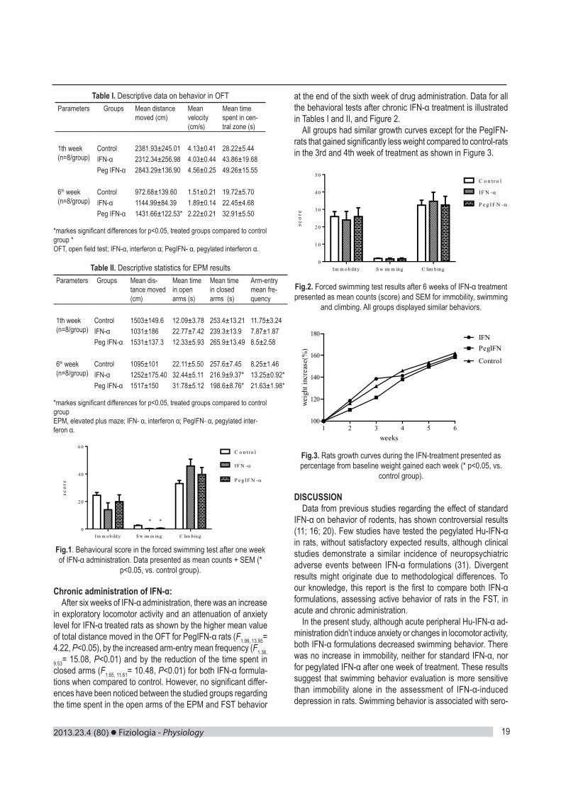
2013.23.4 (80) Fiziologia - Physiology 19
Table I. Descriptive data on behavior in OFTParameters Groups Mean distance
moved (cm)Mean velocity (cm/s)
Mean time spent in cen-tral zone (s)
1th week(n=8/group)
Control 2381.93±245.01 4.13±0.41 28.22±5.44IFN-α 2312.34±256.98 4.03±0.44 43.86±19.68Peg IFN-α 2843.29±136.90 4.56±0.25 49.26±15.55
6th week(n=8/group)
Control 972.68±139.60 1.51±0.21 19.72±5.70IFN-α 1144.99±84.39 1.89±0.14 22.45±4.68Peg IFN-α 1431.66±122.53* 2.22±0.21 32.91±5.50
*markes significant differences for p<0.05, treated groups compared to control group *OFT, open field test; IFN-α, interferon α; PegIFN- α, pegylated interferon α.
Table II. Descriptive statistics for EPM resultsParameters Groups Mean dis-
tance moved (cm)
Mean time in open arms (s)
Mean time in closed arms (s)
Arm-entry mean fre-quency
1th week(n=8/group)
Control 1503±149.6 12.09±3.78 253.4±13.21 11.75±3.24IFN-α 1031±186 22.77±7.42 239.3±13.9 7.87±1.87Peg IFN-α 1531±137.3 12.33±5.93 265.9±13.49 8.5±2.58
6th week(n=8/group)
Control 1095±101 22.11±5.50 257.6±7.45 8.25±1.46IFN-α 1252±175.40 32.44±5.11 216.9±9.37* 13.25±0.92*Peg IFN-α 1517±150 31.78±5.12 198.6±8.76* 21.63±1.98*
*markes significant differences for p<0.05, treated groups compared to control groupEPM, elevated plus maze; IFN- α, interferon α; PegIFN- α, pegylated inter-feron α.
Fig.1. Behavioural score in the forced swimming test after one week of IFN-α administration. Data presented as mean counts + SEM (*
p<0.05, vs. control group).
Chronic administration of IFN-α:After six weeks of IFN-α administration, there was an increase
in exploratory locomotor activity and an attenuation of anxiety level for IFN-α treated rats as shown by the higher mean value of total distance moved in the OFT for PegIFN-α rats (F1.99, 13.95= 4.22, P<0.05), by the increased arm-entry mean frequency (F1.36,
9.53= 15.08, P<0.01) and by the reduction of the time spent in closed arms (F1.65, 11.61= 10.48, P<0.01) for both IFN-α formula-tions when compared to control. However, no significant differ-ences have been noticed between the studied groups regarding the time spent in the open arms of the EPM and FST behavior
at the end of the sixth week of drug administration. Data for all the behavioral tests after chronic IFN-α treatment is illustrated in Tables I and II, and Figure 2.
All groups had similar growth curves except for the PegIFN-rats that gained significantly less weight compared to control-rats in the 3rd and 4th week of treatment as shown in Figure 3.
Fig.2. Forced swimming test results after 6 weeks of IFN-α treatment presented as mean counts (score) and SEM for immobility, swimming
and climbing. All groups displayed similar behaviors.
1 2 3 4 5 6100
120
140
160
180 IFNPegIFN
Control
weeks
wei
ght i
ncre
ase(
%)
Fig.3. Rats growth curves during the IFN-treatment presented as percentage from baseline weight gained each week (* p<0.05, vs.
control group).
DISCUSSIONData from previous studies regarding the effect of standard
IFN-α on behavior of rodents, has shown controversial results (11; 16; 20). Few studies have tested the pegylated Hu-IFN-α in rats, without satisfactory expected results, although clinical studies demonstrate a similar incidence of neuropsychiatric adverse events between IFN-α formulations (31). Divergent results might originate due to methodological differences. To our knowledge, this report is the first to compare both IFN-α formulations, assessing active behavior of rats in the FST, in acute and chronic administration.
In the present study, although acute peripheral Hu-IFN-α ad-ministration didn’t induce anxiety or changes in locomotor activity, both IFN-α formulations decreased swimming behavior. There was no increase in immobility, neither for standard IFN-α, nor for pegylated IFN-α after one week of treatment. These results suggest that swimming behavior evaluation is more sensitive than immobility alone in the assessment of IFN-α-induced depression in rats. Swimming behavior is associated with sero-
Table II. Descriptive statistics for EPM results Parameters Groups
Mean distance moved (cm)
Mean time in open arms (s)
Mean time in closed arms (s)
Arm-entry mean frequency
1th week (n=8/group)
Control 1503±149.6 12.09±3.78 253.4±13.21 11.75±3.24 IFN-α 1031±186 22.77±7.42 239.3±13.9 7.87±1.87 Peg IFN-α 1531±137.3 12.33±5.93 265.9±13.49 8.5±2.58
6th week (n=8/group)
Control 1095±101 22.11±5.50 257.6±7.45 8.25±1.46 IFN-α 1252±175.40 32.44±5.11 216.9±9.37* 13.25±0.92* Peg IFN-α 1517±150 31.78±5.12 198.6±8.76* 21.63±1.98*
*markes significant differences for p<0.05, treated groups compared to control group EPM, elevated plus maze; IFN- α, interferon α; PegIFN- α, pegylated interferon α.
I m m o b ility S w im m in g C lim b in g0
2 0
4 0
6 0
sco
re
C o n t r o l
IF N -
P e g IF N -
* *
Fig.1. Behavioural score in the forced swimming test after one week of IFN-α administration. Data presented as mean counts + SEM (* p<0.05, vs. control group). Chronic administration of IFN-α: After six weeks of IFN-α administration, there was an increase in exploratory locomotor activity and an attenuation of anxiety level for IFN-α treated rats as shown by the higher mean value of total distance moved in the OFT for PegIFN-α rats (F1.99, 13.95= 4.22, P<0.05), by the increased arm-entry mean frequency (F1.36, 9.53= 15.08, P<0.01) and by the reduction of the time spent in closed arms (F1.65, 11.61= 10.48, P<0.01) for both IFN-α formulations when compared to control. However, no significant differences have been noticed between the studied groups regarding the time spent in the open arms of the EPM and FST behavior at the end of the sixth week of drug administration. Data for all the behavioral tests after chronic IFN-α treatment is illustrated in Tables I and II, and Figure 2. All groups had similar growth curves except for the PegIFN-rats that gained significantly less weight compared to control-rats in the 3rd and 4th week of treatment as shown in Figure 3.
I m m o b ility S w im m in g C lim b in g0
1 0
2 0
3 0
4 0
5 0
sco
re
C o n t r o l
IF N -
P e g IF N -
Fig.2. Forced swimming test results after 6 weeks of IFN-α treatment presented as mean counts (score) and SEM for immobility, swimming and climbing. All groups displayed similar behaviors.
1 2 3 4 5 6100
120
140
160
180 IFNPegIFN
Control
weeks
wei
ght i
ncre
ase(
%)
Fig.3. Rats growth curves during the IFN-treatment presented as percentage from baseline weight gained each week (* p<0.05, vs. control group). DISCUSSION Data from previous studies regarding the effect of standard IFN-α on behavior of rodents, has shown controversial results (11; 16; 20). Few studies have tested the pegylated Hu-IFN-α in rats, without satisfactory expected results, although clinical studies demonstrate a similar incidence of neuropsychiatric adverse events between IFN-α formulations (31). Divergent results might originate due to methodological differences. To our knowledge, this report is the first to compare both IFN-α formulations, assessing active behavior of rats in the FST, in acute and chronic administration. In the present study, although acute peripheral Hu-IFN-α administration didn‟t induce anxiety or changes in locomotor activity, both IFN-α formulations decreased swimming behavior. There was no increase in immobility, neither for standard IFN-α, nor for pegylated IFN-α after one week of treatment. These results suggest that swimming behavior evaluation is more sensitive than immobility alone in the assessment of IFN-α-induced depression in rats. Swimming behavior is associated with serotonergic system activity and is enhanced by selective serotonin reuptake inhibitors (8). It is known that IFN-α upregulates the serotonin (5-HT) uptake, by increasing 5-HT transporter expression (2), and down regulates 5-HT synthesis by activating the enzyme indolamine 2,3deoxigenase (IDO) that can metabolize tryptophan, 5-HT precursor, to kynurenine, leading to tryptophan depletion (13), reduced 5-HT levels (30) and synthesis of neurotoxic metabolites (22). Why in our study Hu-IFN-α didn‟t increased the immobility in the FST remains an open question. Our findings are in contrast with two previous studies, one on Wistar rats and another on mice, that used a

Fiziologia - Physiology 2013.23.4 (80)20
tonergic system activity and is enhanced by selective serotonin reuptake inhibitors (8). It is known that IFN-α upregulates the serotonin (5-HT) uptake, by increasing 5-HT transporter expres-sion (2), and down regulates 5-HT synthesis by activating the enzyme indolamine 2,3deoxigenase (IDO) that can metabolize tryptophan, 5-HT precursor, to kynurenine, leading to tryptophan depletion (13), reduced 5-HT levels (30) and synthesis of neu-rotoxic metabolites (22).
Why in our study Hu-IFN-α didn’t increased the immobility in the FST remains an open question. Our findings are in contrast with two previous studies, one on Wistar rats and another on mice, that used a similar dose of non-pegylated Hu-IFN-α and reported an increase of the immobility of FST, but after a single intravenous dose, 15min prior to testing (9; 6). The increase in immobility seemed to be mediated through central µ-opioid receptors, and not by IFN-α receptors, the effect being reversed by naloxone injection (6). We must emphasize that in our study all the behavioral tests were done 24 hours after the last drug administration and repeated doses.
Another study that developed a murine model of depression for repeated administration of non-pegylated Hu-IFN-α, and not a single dose, showed that a 400-1600 UI Hu-IFN-α /g/day with daily administration for 5 days increases immobility in the FST (5). The dose of Hu-IFN-α we used was similar to the doses administered in patients with chronic hepatitis C (50UI/g/day), and thus, lower than those from Siddegowda et al study (5).
In our study, the FST results of the Hu-IFN-α treated rats showed that the reduction of swimming counts was associ-ated to an increase in climbing behavior and not to increase immobility, without reaching a significant level compared to control group, neither for climbing, nor for immobility counts. The climbing behavior is known to be mediated by enhancement of norepinephrine neurotransmission (8). Acute administration of IFN-α increases plasma concentration of norepinephrine, epinephrine and cortisol in humans (17), by causing a marked activation of hypthalamo-pituitary-aderenal axis (3). In rats, it was shown that administration of IFN-α for 7 days increases norepinephrine levels in the cerebral cortex, hypothalamus and medulla oblongata (17).
Cognitive and behavioral side effects of IFN-α therapy usually develop after a several weeks of treatment (6,29). There are few animal studies with chronic IFN-α exposure. A study, using standard IFN-α, observed increased anxiety behavior, without cognitive impairment, in rats treated for 5 weeks (21). In our study, there were no changes in behavioral parameters after 6 weeks of administration of both IFN-α formulations. As we used recombinant human IFN-α, we can’t exclude the formation of anti-IFN-α antibodies after 4-5 weeks of treatment. Unfortunately, anti-IFN-α antibodies dosage wasn’t accessible at that moment.
CONCLUSIONOur study provides data demonstrating that acute administra-
tion of both Hu-IFN-α formulations, at low doses compared to other studies, determines changes in active behaviors during the forced swimming test in rats (swimming). Further studies
are needed to confirm that the serotonergic system is indeed involved in the reduction of swimming behavior induced by IFN-α administration.
Whether the chronic treatment might influence active behav-iors in FST is still an open question.
Because histological and immunohistochemical studies sug-gest a possible neurodegenerative effect of IFN-α, is necessary the improvement of animal models of IFN-α-induced depression, to better understand the pathophysiological processes involved in it and to test possible prophylactic or therapeutic drugs.
DISCLOSURE STATEMENTThis is not an industry supported study. All authors have
indicated no financial conflicts of interest.
AcknowledgementThis work was supported by a CNCSIS grant: BD180/2008.
REFERENCES1. Alesci S, Martinez PE, Kelkar S, et al. Major depression is associ-ated with significant diurnal elevations in plasma interleukin-6 levels, a shift of its circadian rhythm, and loss of physiological complexity in its se-cretion: clinical implications. J ClinEndocrinolMetab 2005; 90: 2522-30. 2. Capuron L, Ravaud A, Neveu PJ, et al. Association between de-creased serum tryptophan concentrations and depressive symptoms in cancer patients undergoing cytokine therapy.Mol Psychiatry 2002; 7, 468-73.3. Capuron L, Raison CL, Musselman DL, et al. Association of exag-gerated HPA axis response to the initial injection of interferon-alpha with development of depression during interferon-alpha therapy.Am J Psychiatry 2003; 160: 1342-5.4. Capuron L, Neurauter G, Musselman DL, et al. Interferon-alpha-induced changes in tryptophan metabolism: relationship to depression and paroxetine treatment.Biol Psychiatry 2003; 54: 906-14.5. Corssmit EP, Heijligenberg R, Endert E, et al. Endocrine and metabolic effects of interferon alpha in humans. J ClinEndocrinolMetab 1996; 81: 3265-3269.6. Crawley JN, Gerfen CR, McKay R, et al. Current Protocols in Neuroscience, John Wiley & Sons Inc, Ed. 20037. De La Garza R, Asnis GM, Pedrosa E, et al. Recombinant human interferon-alpha does not alter reward behavior, or neuroimmune and neuroendocrine activation in rats.ProgNeuropsychopharmacolBiol Psychiatry 2005;29: 781-92.8. Detke MJ, Rickels M, Lucki I. Active behaviors in the rat forced swimming test differentially produced by serotonergic and noradrenergic antidepressants. Psychopharmacology 1995; 121: 66-72.9. Dieperink E, Willenbring M, Ho SB. Neuropsychiatric Symptoms Associated With Hepatitis C and Interferon Alpha: A Review. Am. J. Psychiatry 2000; 157: 867-76.10. Dunn AJ, Swiergiel AH, Renaud de Beaurepaire. Cytokines as mediators of depression: what can we learn from animal studies? Neuroscience &Biobehavioral Reviews 2005; 29: 891-909.11. Fahey B, Hickey B, Kelleher D, et al. The widely-used anti-viral drug interferon-alpha induces depressive- and anxiogenic-like effects in healthy rats.Behav Brain Res 2007; 182: 80-7.12. Glue P, Fang JW, Rouzier-Panis R, et al. Pegylated interferon-alpha2b: pharmacokinetics, pharmacodynamics, safety, and preliminary efficacy data. Hepatitis C Intervention Therapy Group.ClinPharmacol-Ther2000; 68: 556-67.

2013.23.4 (80) Fiziologia - Physiology 21
13. Guillemin GJ, Smythe G, Takikawa O, Brew BJ.Expression of in-doleamine 2,3-dioxygenase and production of quinolinic acid by human microglia, astrocytes, and neurons.Glia 2005; 49: 15-23.14. Horsmans Y. Interferon-induced depression in chronic hepatitis C. J AntimicrobChemother 2006; 58: 711-713.15. Janicki PK. Binding of human alpha-interferon in the brain tissue membranes of rat. Res CommunChemPatholPharmacol 1992; 75: 117-120.16. Kentner AC, Miguelez M, James JS, Bielajew C. Behavioral and physiological effects of a single injection of rat interferon-α on male Sprague–Dawley rats: A long-term evaluation. Brain Research 2006; 1095: 96-106. 17. Kumai T, Tateishi T, Tanaka M, et al. Effect of interferon-alpha on tyrosine hydroxylase and catecholamine levels in the brain of rats. Life Sci 2000; 67: 663-669.18. Levine J, Barak Y, Chengappa KN, et al. Cerebrospinal cytokine levels in patients with acute depression. Neuropsychobiology1999; 40: 171-6.19. Loftis MJ, Hauser P, Macey TA, Lowe JD. Can rodents be used to model interferon-alpha-induced depressive symptoms? Letter to the Editor. Progress in Neuro-Psychopharmacology & Biological Psychiatry 2006; 30: 1364-1365.20. Loftis JM, Wall JM, Pagel RL, Hauser P. Administration of pegylated interferon-alpha-2a or -2b does not induce sickness behavior in Lewis rats. Psychoneuroendocrinology2006; 31: 1289-94.21. Makino M, Kitano Y, Komiyama C, et al. Involvement of central opioid systems in human interferon-alpha induced immobility in the mouse forced swimming test.Br J Pharmacol 2000; 130: 1269-74.22. Makino M, Kitano Y, Komiyama C, Takasuna K. Human interferon-alpha increases immobility in the forced swimming test in rats.Psycho-pharmacology 2000; 148: 106-10.23. Myint AM, O’Mahony S, Kubera M, et al. Role of paroxetine in interferon-alpha-induced immune and behavioural changes in male
Wistarrats.J Psychopharmacol 2007; 21: 843-50.24. Porsolt RD, Bertin A, Jalfre M. Behavioral despair in mice: a pri-mary screening test for antidepressants. Archives Internationales de Pharmacodynamieet de Therapie 1977; 229: 327-336.25. Raison CL, Capuron L, Miller A. Cytokines sing the blues: inflamma-tion and the pathogenesis of depression. Review. Trends in Immunology 2006; 27: 24-31. 26. Raison CL, Demetrashvili M, Capuron L, Miller AH. Neuropsychiatric adverse effects of interferon-α: recognition and management. CNS Drugs 2005; 19: 105-123.27. Renault PF, Hoofnagle JH, Park Y. Psychiatric complications of long term interferon alpha therapy. Arch. Intern. Med. 1987; 147: 1577-80. 28. Sato T, Suzuki E, Yokoyama M, et al. Chronic intraperitoneal injec-tion of interferon-α reduces serotonin levels in various regions of rat brain, but does not change levels of serotonin transporter mRNA, nitrite or nitrate. Psychiatry and Clinical Neurosciences 2006; 60: 499-506.29. Siddegowda NK, Rao NSK, Andrade C. Development of a murine animal model of depression or repeated dosing with human interferon alpha. Indian J Psychiatry 2011; 53: 239-243.30. Tsao CW, Lin YS, Cheng JT, et al. Interferon-alpha-induced sero-tonin uptake in Jurkat T cells via mitogen-activated protein kinase and transcriptional regulation of the serotonin transporter.J Psychopharma-col 2008; 22: 753-60.31. Tsao CW, Lin YS, Cheng JT, et al. Serotonin transporter mRNA expression is decreased by lamivudine and ribavirin and increased by interferon in immune cells.Scand J Immunol 2006; 63: 106-15.32. Wang J, Dunn AJ, Roberts AJ, Zhang H.Decreased immobility in swimming test by homologous interferon-α in mice accompanied with increased cerebral tryptophan level and serotonin turnover.NeurosciLett. 2009; 452: 96-100.33. Wichers MC, Kenis G, Koek GH, et al. Interferon-alpha-induced depressive symptoms are related to changes in the cytokine network but not to cortisol.J Psychosom Res 2007; 62: 207-14.
EFECTELE INTERFERONULUI-ALFA RECOMBINANT UMAN ASUPRA COMPORTAMENTELOR ACTIVE LA TESTUL INOTULUI FORTAT, LA SOBOLAN
REZUMATDepresia indusă de cytokine proinflamatorii poate fi reprodusă pe modele animale prin administrarea de interferon alfa (IFN-α). Cele mai utilizate teste comportamentale pentru evaluarea depresiei pe modele animale sunt testul înotului forţat (FST) şi testul suspendării de coadă.Scopul studiului a fost să observe efectele administrării acute şi cornice de interferon-alpha recombinant uman (Hu-IFN-α) asupra comportamentelor active la FST. Şobolani Wistar, masculi, au fost alocaţi aleatoriu în trei grupuri experimentale: control, trataţi cu IFN-α nepegylat şi trataţi cu IFN-α pegylat. La sfârşitul primei şi a celei de-a şasea săptămâni de tratament, şobolanii au fost evaluaţi printr-un set de teste comportamentale. La tratamentul acut cu IFN-α, indiferent de tipul de preparat, s-a constat la FST scăderea semnificativă a frecvenţei miscărilor de înot, însă fără diferenţe pentru imobilitate sau încercările de a escalada pereţii cuvei de înot (căţărare; eng. climbing”). La sfârşitul perioadei de şase săptămâni nu au mai fost constatate diferenţe în ceea ce priveşte comportamentele active la FST.Studiul nostru a arătat că administrarea acută de IFN-α induce modificări ale comportamentelor active la FST. Studii ulterioare sunt necesare pentru a demonstra implicarea sistemului neurotransmiţător serotoninergic în modificările comportamentale constatate.Cuvinte cheie: interferon-alfa, depresie, citokine, testul inotului fortat, comportamente active.
Fiziologia - Physiology 2013.23.4 (80)22
CORTICAL ACTIVITY AND HEART RATE CHANGES AS MARKERS OF NOXIOUS STIMULATION RESPONSE DURING CHLORAL HYDRATE ANESTHESIA
PAVEL B, DĂNEASA A, CĂLIN A, RĂDULESCU AM , ROSCA AE, BRAGA RI, ZAHIU D, ZĂGREAN LDivision of Physiology and Fundamental Neurosciences, “Carol Davila” University of Medicine and Pharmacy, Bucharest, Romania
ABSTRACTThe aim of our study was to evaluate the importance and applicability of cortical activity and heart rate changes as markers of noxious stimulation response during chloral hydrate anaesthesia in rats.In this study we used a group of 11 Wistar rats, which were chronically implanted, with electrodes being placed on the dura mater of the right hemisphere, over the olfactory cortex (reference), the frontal and the parietal lobes. A noxious mechanical stimulus was applied on the left hindpaw during chloral hydrate anesthesia, at an anesthetic depth of 2-3 Hz, estimated using the median frequency. We assessed the electrical activity of the cortical areas (evaluated through median frequency, delta power and cortical connectivity between frontal and parietal cortex) and heart rate response to mechanical noxious stimulation.After data processing and analysis we observed an increase of median frequency and a decrease of delta power during the application of the noxious stimulus in both areas (frontal and parietal). No difference between fronto-parietal connectivity during baseline and stimulation tracks was noted. Tachogram analysis showed a mild increase in heart rate during noxious stimulation, but it did not reach statistic significance.Monitoring cortical electric activity is a better choice than ECG monitoring for noxious stimulation response assessment during chloral hydrate anesthesia in rat.Key words: anesthesia, cortical connectivity, nociception, electrocorticogram
Received September 10th 2013. Accepted October 12th 2013. Address for correspondence: Pavel B, MD, Division of Physiology and Fundamental Neurosciences, University of Medicine and Pharmacy “Carol Davila”, Bucharest, Romania, phone: +40213180762; e-mail: [email protected]
INTRODUCTIONThe monitoring of the anesthetic depth has been a primary
concern for physicians since the dawn of anesthesia. In 1937 anesthetic depth was quantified relying on pupil diameter changes, respiratory rate, heart rate, and blood pressure (1). In the same year, the electroencephalogram (EEG) was proposed as a method for the assessment of anesthetic depth (2). Currently, EEG signal analysis is used for the monitoring of the hypnotic effect (3); fa-cial electromyogram, analysis of heart rate variability, cutaneous conductance are among the methods, cited in scientific literature, that are used to measure the reaction to noxious stimuli (4,5,6,7). Although some data on the subject of EEG changes during noxious stimulation can be found in scientific literature (8, 9), the main dif-ficulty in using EEG changes as markers of noxious stimulation lies in the occurrence of the phenomenon called paradoxical arousal, which consists of an increase in anesthesia depth following nox-ious stimulation, represented by an increase in slow wave activity (10). New data shows that a decrease in cortical connectivity is present during anesthesia (11,12). Cortical connectivity decreases especially between frontal and parietal areas during anesthesia in studies performed in humans and animals. Methods as fMRI and EEG signals correlation algorithms are cited in cortical connectivity assessment (13,14,15). Recent study investigating simultaneous cortical connectivity assessment using fMRI and EEG has proved a similar change trend (16).
The main aim of this study is to investigate if changes in the functional connectivity between frontal and parietal areas could be used as marker of a reaction to noxious stimulation. As far as we know such an inquiry has not been yet attempted. Other aims of this study are to investigate if the activations of the frontal and parietal areas differ one from another and to find out what relationship exists between cortical activation and heart rate changes during noxious stimulation.
For these issues we used a chronically implanted rat model, who received a painful stimulus under chloral hydrate anesthesia. Cortical area (frontal and parietal) activity was measured as median frequency (MEF) and delta power variation, during stimulation. For the evaluation of the functional connectivity, electrocorticogram (ECoG) synchronicity between cortical areas, using correlation coefficient was performed (17). The electrocardiogram (ECG) was recorded and heart rate changes were analyzed.
MATERIALS AND METHODSAnimals
In this study a group (n=11) of Wistar rats was used, with weights ranging from 250 to 300 grams, which were kept in a room with con-stant temperature (230 Celsius) and 12-h light-dark cycle (lights on at 07:00 h), with free access to food and water. Rats were chronically implanted, under chloral hydrate (Sigma-Aldrich Gmbh, Munich, Germany) anesthesia (400 mg/kg, injected intraperitoneal), using

2013.23.4 (80) Fiziologia - Physiology 23
Nickel-Chrome (Ni80Cr20, Ø 0.15 mm, Goodfellow, UK) electrodes which were placed directly on the dura mater, through burr holes obtained by trephination, and were finally fixed with cement. Three electrodes were implanted, one reference electrode was placed over the olfactory cortex (7 mm anterior to bregma and 1 mm lateral to the frontal suture) and two active electrodes were placed over the right hemisphere, in the frontal lobe (3 mm anterior to bregma and 3 mm lateral to the frontal suture) corresponding to the motor cortex, and in the parietal lobe (3 mm posterior to bregma and 3 mm lateral to the sagital suture), corresponding to the somatosensory areas of the left hindpaw (18). The rats underwent experimentation after a minimum period of 7 days after the intervention. This study has been approved by the Ethics Committee of the University of Medicine and Pharmacy Carol Davila Bucharest.
Anesthesia and stimulation procedureAnesthesia was induced and maintained with chloral hydrate
(300 mg/kg administrated intraperitoneally). After a steady state of 20 minutes from the induction, we started recording, but only when the median frequency of the ECoG, recorded in the frontal lobe, reached 2 – 3 Hz, indicating an appropriate anesthetic depth. The stimulus consisted in placing the skin from the posterior left hindpaw between the arms of a Pean type forceps, which was then closed, locking only the first ratchet. It has already proven in human and animal that peripheral noxious stimulation induced predominantly right hemisphere activation, phenomena called lateralization (19, 20). So, we chose the left hindpaw to be stimulated and right hemi-sphere for ECoG recordings. The mechanical noxious stimulus was applied for 10 seconds. During the experiment rats temperature was maintained at 370-380 Celsius using a heating pad.Data acquisition and signal processing
The electrocortigram (ECoG) signal acquisition was made using BIOPAC MP-150 system (Biopac Systems, CA, USA) at a sample rate of 1000 Hz, a gain of 1000, with filters set at 0.5 Hz (high pass) and 30 Hz (low pass). Median frequency and delta power of the spectrum were computed for each channel sepa-rately, using AcqKnowledge 4.2 software. Epoch length was set at 10 seconds. Functional connectivity between the ECoG signals from the two areas was computed using correlation coefficient function provided by the AcqKnowledge 4.2. The analyzed data consisted of 1 minute baseline signal and 10 seconds of signal corresponding to the noxious stimulation. Because the acquisi-tion frequency was set at 1000 Hz, we obtained 60.000 points of data for one baseline track of 1 minute, summing 660.000 points of data for 11 rats. This large amount of data was not supported by our statistics program, so we decided to compute a correlation coefficient for each of the 11 tracks and calculate the mean. We compared the mean values of the correlation coefficients during baseline track versus stimulation track (17). ECG signal acquisition was also made using BIOPAC MP-150 system at the same sample rate and using the same filters as for the ECoG, but with a gain of 500.Statistical analysis
Data were presented as mean value ± standard deviation (SD). Statistical analysis was performed with the Wilcoxon rank
sum test, using SPSS (version 11.0, SPSS Inc., Chicago, IL). P values ˂ 0.05 were considered statistically significant.
RESULTSAn increase of the median frequency in the frontal area during
noxious stimulation was observed, from a value of 2.66 ± 1.06 Hz to 5.37 ± 1.7 Hz (p<0.05)(Figure 1). Parietal somatosensory area activation follows a similar trend during noxious stimulation, median frequency rose from 2.92 ± 1.01 Hz during baseline to 5.11 ± 1.78 Hz (p <0.05) (Figure 2).
Delta power (μV2/Hz) followed an inverse trend, resulting in a decrease in delta power during noxious stimulation in both areas, as a consequence of the increase in cortical activity. So, we got a decrease of delta power from 8.65 ± 6.77 during baseline to 4.36 ± 1.90 (p<0.05) during mechanical clamp, in frontal area (Figure 3). In parietal area delta power decrease from a baseline of 17.84 ± 14.27 to 8.42 ± 5.56 during noxious stimulation (Figure 4).
Analysis of the functional connectivity between frontal and pa-rietal lobe did not yield statistically significant results, correlation coefficient was 0.63 ± 0.17 during baseline recording and 0.61 ± 0.16 during noxious stimulation with a p = 0.7. Heart rate maintained a steady value of 394.8 ± 44.15 beats per minute (bpm) during baseline recording and a value of 399 ± 46.18 bpm during stimulation with a p = 0.4.
DISCUSSIONCortical activation and connectivity
Change of cortical electric activity during noxious stimulation
Fig. 1. Median frequency (Hz) variation during mechanical
noxious stimulation compared with baseline, in the frontal
lobe (p<0.05)
Fig. 3. Delta power (μV2/Hz) variation during mechanical
noxious stimulation compared with baseline, in the frontal
lobe (p<0.05)
Fig. 2. Median frequency (Hz) variation during mechanical
noxious stimulation compared with baseline, in the parietal
lobe (p<0.05)
Fig. 4. Delta power (μV2/Hz) variation during mechanical
noxious stimulation compared with baseline, in the parietal
lobe (p<0.05)
Fiziologia - Physiology 2013.23.4 (80)24
are reported in humans, as well as in animals (8, 21, 22). The novelty of our study is represented by the analysis of cortical
connectivity, using ECoG signals, during noxious stimulation under chloral hydrate anesthesia, which has not been reported until now.
Following data processing and analysis, an increase in cortical electric activity in parietal and frontal lobe is noticed. Similarly, median frequency increases in the parietal and the frontal lobe compared with baseline. Increase in median frequency following mechanical noxious stimulation is not a constant finding during anesthesia. Two other studies where mechanical stimulation was realized by clamp-ing, under halothane anesthesia, revealed two different responses (23, 24). In one study MEF increased and in the other one it did not. The explanation for these contradicting results could be found by looking at the types of stimulation used, because MEF increases during oscillating clamp application compared with stationary clamp, which does not produce MEF changes. In our study we obtained an increase of MEF during stationary clamping. The factors in our study that could account for this result are a longer stimulation time, namely 10 seconds versus the 5 seconds used in the previous study, and the different anesthetic that was used.
Delta power analysis showed a similarly decrease during noxious stimulation compared with baseline in the parietal lobe and in the frontal lobe. This data is not concordant with previ-ous findings, in which a greater decrease in delta power was observed in the frontal lobe(8), in a study performed in humans under isofluran anesthesia. We interpret these different results as being specific to chloral hydrate anesthesia.
Some studies maintained the idea that MEF increase reflects the noxious character of the stimulus, in horse under halothane anesthesia (25) and total power spectrum decreases following noxious stimulation as a consequence of anesthetic depth inadequacy (24, 26). We observed an increase in MEF and a decrease in delta power during noxious stimulation in both cortical areas, therefore, following the same line of reasoning as above, we could conclude that the observed cortical activation is due to a combination of supramaximal noxious stimulation and inadequate anesthetic depth.
Recent studies have proven that anesthetics reduce corti-cal and cortical-subcortical connectivity during anesthesia (11, 12), including fronto-parietal connectivity (27). In order to evaluate the connectivity between the two areas we measured the functional cortical connectivity, analyzing the synchronicity between the two cortical electrical signals (ECoG) using correla-tion coefficient (17). We did not obtain a statistically significant difference between correlation coefficient during stimulation compared with baseline. We considered that noxious stimulation was supramaximal and as such it reduced anesthetic depth, as was indicated by the MEF and delta Power changes, therefore a higher than expected connectivity between the parietal and frontal lobe can be explained. The reason for which we obtained a correlation coefficient during baseline that was very similar to the one during stimulation, in spite of a significant change in cortical electrical activity (assessed by MEF and delta power) is an open question for future studies. A low specificity of the correlation coefficient in the functional connectivity assessment
could partially explain this finding.
ECG changesIt is known that noxious stimulation leads to change in heart rate,
change which is present in the awake as well as in the anesthetized patient; nevertheless a tight correlation between the change in heart rate and noxious stimulation during anesthesia could not be estab-lished. A higher sensitivity parameter derived from ECG analysis, is the heart rate variability (HRV), which is indicative of the sympathetic – parasympathetic balance. In human studies, a good correlation between noxious stimulation during anesthesia and the sympathetic – parasympathetic balance has been established. Despite of this good correlation between anesthesia and ECG changes in humans, ECG changes during anesthesia are poorly understood in rats (28). We have chosen for heart rate analysis instead of HRV analysis because the rats in our study where breathing spontaneously and therefore the effects of sinus arrhythmia could not be excluded. It is worth mentioning that chloral hydrate, compared to other anesthetics (ketamine, pentobarbital), has no known pharmacodynamic effect on the myocardium (29), so it seems to be the ideal anesthetic for heart rate variation assessment during noxious stimulation. Tachogram analysis showed that heart frequency mildly varies during noxious stimulation, without reaching statistical significance. How noxious stimuli can activate the cerebral cortex but not the sympathetic nervous system, knowing there is a strong correlation between frontal cortex activity and sympathetic activity in human (30) is still a matter of debate.
CONCLUSIONSCortical activity increases in both areas during noxious stimula-
tion, most likely due to a combination of supramaximal noxious stimulation and inadequate anesthetic depth. Our hypothesis regarding the change of cortical functional connectivity during noxious stimulation, by measuring correlation coefficient, is not confirmed by our results. Heart rate does not change during stimulation, so we conclude that monitoring cortical electric activ-ity is a better choice than ECG monitoring for noxious stimulation response assessment during chloral hydrate anesthesia in rat.
REFERENCES1. Guedel A. Inhalation anesthesia: a fundamental guide New York, NY: Macmillan, 19372. Gibbs FA et al. Effect on the electroencephalogram of certain drug wich influence nervous activity Arch Intern Med 1937; 60:154-160.3. Singh H. Bispectral index (BIS) monitoring during propofol-induced sedation and anaesthesia. Eur J Anaesthesiol. 1999; 16(1): 31-6.4. Jellish WS, Leonetti JP, Buoy CM, Sincacore JM, Sawicki KJ, Macken MP. Facial nerve electromyographic monitoring to predict movement in patients titrated to a standard anesthetic depth.5. Anesth Analg. 2009;109(2): 551-8.6. Fabrizi L, Worley A, Patten D et al. Electrophysiological measurements and analysis of nociception in human infants. J Vis Exp. 2011; 20: 58. 7. Ledowski T, Albus S, Stein J, Macdonald B. Skin conductance for monitoring of acute pain in adult postoperative patients: influence of electrode surface area and sampling time. J Clin Monit Comput. 2011; 25(6): 371-6.

2013.23.4 (80) Fiziologia - Physiology 25
8. Jeanne M, Logier R, De Jonckheere J, Tavernier B. Validation of a graphic measurement of heart rate variability to assess analgesia/nociception balance during general anesthesia. Conf Proc IEEE Eng Med Biol Soc. 2009; 2009: 1840-3.9. Kochs E, Bischoff P, Pichlmeier U, Schulte am Esch J. Surgical stimulation induces changes in brain electrical activity during isoflurane/nitrous oxide anesthesia. A topographic electroencephalographic analysis. Anesthesiology 1994; 80:1026-34.10. Rundshagen I, Schröder T, Prichep LS, John ER, Kox WJ. Changes in cortical electrical activity during induction of anaesthesia with thiopental/fentanyl and tracheal intubation: a quantitative electroencephalographic analysis. Br J Anaesth. 2004; 92(1): 33-8.11. Hagihira S, Takashina M, Mori T, Ueyama H, Mashimo T. Electroencephalographic bicoherence is sensitive to noxious stimuli during isoflurane or sevoflurane anesthesia. Anesthesiology. 2004; 100(4): 818-25.12. Ferrarelli F, Massimini M, Sarasso S et al. Breakdown in cortical effective connectivity during midazolam-induced loss of consciousness. Proc Natl Acad Sci U S A. 2010;107(6): 2681-6. 13. Mhuircheartaigh RN, Rosenorn-Lanng D, Wise R, Jbabdi S, Rogers R, Tracey I. Cortical and subcortical connectivity changes during decreasing levels of consciousness in humans: a functional magnetic resonance imaging study using propofol. J Neurosci. 2010;30(27): 9095-102.14. Liu X, Pillay S, Li R, Vizuete JA, Pechman KR, Schmainda KM, Hudetz AG. Multiphasic modification of intrinsic functional connectivity of the rat brain during increasing levels of propofol. Neuroimage. 2013; 83: 581-92. 15. Tu Y, Yu T, Fu XY, Xie P, Lu S, Huang XQ, Gong QY. Altered thalamocortical functional connectivity by propofol anesthesia in rats. Pharmacology. 2011; 88(5-6): 322-6. 16. Rowland NC, Goldberg JA, Jaeger D. Cortico-cerebellar coherence and causal connectivity during slow wave activity. Neuroscience. 2010; 166(2): 698-711. 17. Jordan D, Ilg R, Riedl V, Schorer A, Grimberg S, Neufang S, Omerovic A, Berger S, Untergehrer G, Preibisch C, Schulz E, Schuster T, Schröter M, Spoormaker V, Zimmer C, Hemmer B, Wohlschläger A, Kochs EF, Schneider G. Simultaneous electroencephalographic and functional magnetic resonance imaging indicate impaired cortical top-down processing in association with anesthetic-induced unconsciousness. Anesthesiology. 2013; 119(5): 1031-42.18. Bonita J, Albano A, Ambolode LC, Rappc P. Study of Correlations of Multichannel Human EEG. Proceedings of the 11th SPVM National Physics Conference and Workshop 2009 SPVM. ISSN 1655-4620.
19. Paxinos G, Watson C. The Rat Brain in Stereotaxic Coordinates, Sixth Edition [Spiral-bound]. 2009 20. Geschwind N, Galaburda A. Cerebral lateralization. Biological mechanisms, associations and pathology: I. A hypothesis and aprogram for research. Arch Neurol 1985; 42: 428-59.21. de Celis Alonso B, Sergeyeva M, Brune K, Hess A. Lateralization of responses to vibrissal stimulation: connectivity and information integration in the rat sensory-motor cortex assessed with fMRI.22. Neuroimage. 2012; 62(3): 2101-9. 23. Diesch TJ, Mellor DJ, Johnson CB, Lentle RG. Electroencephalographic responses to tail clamping in anaesthetized rat pups. Lab Anim. 2009; 43(3): 224-31. 24. Diesch TJ, Mellor DJ, Johnson CB, Lentle RG. Developmental changes in the electroencephalogram and responses to a noxious stimulus in anaesthetized tammar wallaby joeys (Macropus eugenii eugenii). Lab Anim. 2010; 44(2): 79-87.25. Orth M, Barter L, Dominguez C, Atherley R, Carstens E, Antognini JF. Halothane and propofol differentially affect electroencephalographic responses to noxious stimulation. Br J Anaesth. 2005; 95(4): 477-84. 26. Murrell JC, Mitchinson SL, Waters D, Johnson CB. Comparative effect of thermal, mechanical, and electrical noxious stimuli on the electroencephalogram of the rat. Br J Anaesth. 2007; 98(3): 366-71.27. Murrell J, Johnson C, White K, Taylor P, Haberham Z, Waterman-Pearson AE. Changes in the EEG during castration in horses and ponies anaesthetized with halothane. Vet Anaesth Analg 2003; 30: 138-46.28. Traast HS, Kalkman CJ. Electroencephalographic characteristics of emergence from propofol/sufentanil total intravenous anaesthesia. Anaesth and Analg 1995; 81: 366-71.29. Ku SW, Lee U, Noh GJ, Jun IG, Mashour GA. Preferential inhibition of frontal-to-parietal feedback connectivity is a neurophysiologic correlate of general anesthesia in surgical patients. PLoS One. 2011; 6(10): e25155. 30. Moldovan M, Spulber S, Saravan V, Iosifescu R, Zăgrean AM, Zăgrean L. The relationship between respiratory sinus arrhythmia and heart rate during anesthesia in rat. Rom J Physiol. 2004; 41(1-2): 31-9.31. Jiang X, Gao L, Zhang Y et al. A comparison of the effects of ketamine, chloral hydrate and pentobarbital sodium anesthesia on isolated rat hearts and cardiomyocytes. J Cardiovasc Med 2011; 12(10): 732-5.32. Tanida M, Sakatani K, Takano R, Tagai K. Relation between asymmetry of prefrontal cortex activities and the autonomic nervous system during a mental arithmetic task: near infrared spectroscopy study. Neurosci Lett. 2004; 369(1): 69-74.
MODIFICARILE ACTIVITATII CORTICALE SI ALE RITMULUI CARDIAC CA MARKERI AI RASPUNSULUI LA STIMULAREA NOCICEPTIVA IN TIMPUL ANESTEZIEI CU CLORALHIDRAT
REZUMATScopul studiului nostru a fost sa evaluam importanta si aplicabilitatea utilizarii modificarilor activitatii corticale si ale ritmului cardiac, ca markeri de raspuns la stimularea nociceptiva in timpul anesteziei cu cloralhidrat la sobolani.In acest studiu am folosit 11 sobolani Wistar, care au fost implantati cronic, cu electrozi plasati pe duramater la nivelul emisferului drept, deasupra cortexului olfactiv (referinta), a lobului frontal si a lobului parietal. In timpul anesteziei cu cloralhidrat, la o profunzime de 2-3 Hz, estimata prin frecventa mediana, a fost aplicat un stimul nociceptiv mecanic pe laba posterioara stanga a sobolanului. Noi am evaluat activitatea electrica a ariilor corticale (evaluata prin frecventa mediana, puterea delta si conectivitatea corticala intre cortexul frontal si parietal) si raspunsul frecventei cardiace la stimularea nociceptiva.Dupa procesarea si analiza datelor am constatat o crestere a frecventei mediane si o scadere a puterii delta in timpul aplicarii stimu-lului nociceptiv in ambele arii (frontal si parietal). Nu s-a observat nici o modificare a conectivitatii fronto-parietale in timpul stimularii fata de traseul martor. Analiza tahogramei a evidentiat o usoara crestere a frecventei cardiace in timpul stimularii nociceptive dar fara semnificatie statistica.Monitorizarea activitatii electrice corticale este o optiune mai buna decat monitorizarea EKG pentru evaluarea raspusului la stimularea nociceptiva in timpul anesteziei cu cloralhidrat la sobolan.Cuvinte cheie: anestezie, conectivitate corticala, nociceptie, electrocorticograma.

Fiziologia - Physiology 2013.23.4 (80)26
ARGUMENTS FOR USING THROMBELASTOGRAPHY IN THE STUDY OF HEMOSTASIS COMPARED TO OTHER LABORATORY TESTS
FLORINA-MADALINA ONICEANU, ILEANA ION, CECILIA ADUMITRESI, NINELA RADULESCU, CRISTINA FARCAS, ANABELLA-CRISTIANA STEFANESCU, CHIRICA RFaculty of Medicine, “Ovidius” University Constanta
ABSTRACTThe authors showcase clinical manifestations and lab work in the evolution of haemostatic pathology in children. Classic haemostasis tests were used, along with more specific and sensitive ones, like determination of some coagulation factors and thrombelastography. The authors aimed to identify the mechanisms involved in this pathology: immunological mechanisms, deficiency of production, thrombocytopenia produced by medication, endothelial lesions due to capillary fragility, but some mechanisms remained unrevealed. Also, the authors compared basic coagulation test with thrombelastography to emphasize the utility and rapidity of this last test to evaluate the hypo or hypercoagulable state in children. For all the patients, there were determined complete blood count, prothrombin time (PT), activated partial thromboplastin time (APTT), International Normalised Ratio (INR), total calcium level, ionized calcium, fibrinogen. The objective was to evaluate the risk for thrombotic or bleeding manifestations using thrombelastography. Classical methods in the study of haemostasis are not able to highlight clot formation or the changes produced after thrombus formation and also they cannot suggest predisposition for thrombosis the way thrombelastography does. Key words: children, thrombelastography, haemostasis
Received 15th of September 2013.Accepted 25th of October 2013. Address for correspondence: Adumitresi Cecilia, MD, PhD, Department of Physiology, Faculty of Medicine, “Ovidius” University Constanta,Universităţii Street, Campus 2 Constanţa, Romania, phone / fax: 0241- 605000; email:[email protected]
INTRODUCTION
Bleeding syndromes in children represent an extensive pathol-ogy in medical practice. Lab work in these cases represents, along with precise clinical examination, the key in ascertainment of the proper diagnosis. Based on standard coagulation tests, lab work includes also more specific and sensitive analysis, like thrombelastography, a method which is not used in Romania, but applied in international clinics and research fields, a method that enhances the diagnosis in bleeding, but also thrombotic pathology (1).
Using thrombelastography for these patients, the authors want to highlight the utility and the necessity of this method in clinic to establish a tendency towards hypo or hypercoagulation in several bleeding or thrombotic syndromes in children.
MATERIALS AND METHOD
The present study was performed from January 2010 to January 2013 on a group of 36 patients admitted in the Onco-Hemathology Department, Paediatric Clinic, Constanta County Hospital. From this group of patients, 12 were admitted for thrombotic events and 24 of them had hemorrhagic manifesta-tions. This last subgroup present characteristic clinical manifesta-tions for thrombocytopenia, like petechiaes, bruising, epistaxis, gingival bleeding, metrorrhagia,hematuria. The authors tried to
identify the mechanisms involved in this pathology: immunologi-cal mechanisms, deficiency of production, thrombocytopenia pro-duced by medication, endothelial lesions with capillary fragility, but some mechanisms remained unrevealed (2).
Also, the authors compared basic coagulation test with throm-belastography to emphasize the utility and rapidity of this last test to evaluate the hypo or hypercoagulable state in children.
For all the patients, we determined complete blood count, prothrombin time (PT), activated partial thromboplastin time (APTT), International Normalised Ratio (INR), serum calcium level and ionic part, fibrinogen. We tried to evaluate the risk for thrombotic or bleeding manifestations using thrombelastography.
The results were shown in tables and graphics.
RESULTS
Thrombotic manifestations were represented by stroke, with an incidence of 16.6%, deep venous thrombosis (DVT) representing 50%, and the rest of 33.3% are patients with high risk for throm-botic events. Hemorrhagic manifestations were represented by clinical characteristic manifestations for thrombocytopenia, like petechiaes, bruising, epistaxis, gingival bleeding, metrorrhagia, hematuria. Among this patients, 7 had immunological thrombo-cytopenic purpura (PTI), representing 29% of this subgroup; the mechanism that produces thrombocytopenia is immunological, with shortage of the platelets life span.
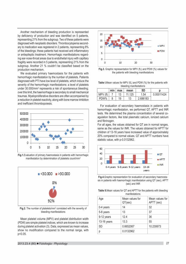
2013.23.4 (80) Fiziologia - Physiology 27
Another mechanism of bleeding production is represented by deficiency of production and was identified on 5 patients, representing 21% from the subgroup. Two of these patients were diagnosed with neoplastic disorders. Thrombocytopenia second-ary to medication was registered in 2 patients, representing 8% of the bleedings; these patients had received anti inflammatory or antiepileptic treatment. Hemorrhagic manifestations regard-ing ear-nose-throat areas due to endothelial injury with capillary fragility were recorded in 5 patients, representing 21% from the subgroup. Another 21 % couldn’t be classified based on the production mechanism.
We evaluated primary haemostasis for the patients with hemorrhagic manifestation by the number of platelets. Patients diagnosed with PTI have low level of platelets, which induce the severity of the hemorrhagic manifestations: a level of platelets under 30.000/mm3 represents a risk of spontaneous bleeding; over this limit, the haemorrhage is secondary to small mechanical traumas. Myeloproliferative disorders are often accompanied by a reduction in platelet reactivity, along with bone marrow inhibition and inefficient thrombopoiesis.
Fig.1.Evaluation of primary haemostasis in patients with hemorrhagic manifestation by determination of platelets count/mm3
Fig.2. The number of platelets/mm3 correlated with the severity of bleeding manifestations
Mean platelet volume (MPV) and platelet distribution width (PDW) are simple platelet indices, which are known to increase during platelet activation (3). Data, expressed as mean values, show no modification compared to the normal range, with p<0.05.
Fig.3. Graphic representation for MPV (fL) and PDW (%) values for the patients with bleeding manifestations
Table I.Mean values for MPV (fL) and PDW (%) for the patients with bleeding manifestations
min max mean SD pMPV (fl) 7 13 11.125 1.54 0.00011424PDW% 8 19 13 2.686183
For evaluation of secondary haemostasis in patients with hemorrhagic manifestation, we performed QT, APTT and INR tests. We determined the plasma concentration of several co-agulation factors, like total plasmatic calcium, ionized calcium and fibrinogen. For all ages, the values obtained for QT are in normal ranges, same as the values for INR. The values obtained for APTT for children of 13-16 years have increased value of approximately 20% compared to normal values. QT and APTT numbers have statistic value, with p-0.0132962.
Fig.4.Graphic representation for evaluation of secondary haemosta-sis in patients with haemorrhagic manifestation using QT (sec), APTT
(sec) and INR
Table II.Mean values for QT and APTT for the patients with bleeding manifestations
Age Mean values for QT(sec)
Mean values for APTT (sec)
0-4 years 14 325-8 years 13 379-12 years 12.4 3613-16 years 13.3 55SD 0.6652067 10.230673p 0.0132962

Fiziologia - Physiology 2013.23.4 (80)28
Fig.5.Graphic representation of mean values for fibrinogen (mg/dl), total calcium (mg/dl) and ionized calcium (mg/dl) on age groups
High numbers for mean values of fibrinogen along with increas-ing age can be observed. This can be explained maybe due to the increased incidence of infectious inflammatory diseases in age group 9-12 years and 13-16 years.
Total and ionized calcium levels are in normal range for all group age, suggesting that calcium as coagulation factor doesn’t influence bleeding disorders for the patients in the study group. The numbers have statistical significance.
Table III.Mean values of total and ionized calcium on age groups for the patients with haemorrhage
Age group Tota l Calc ium (mg/dl)
Ionized Calcium (mg/dl)
0-4 y 9.3 3.95-8 y 9.2 3.69-12 y 7.8 3.813-16 y 9.1 4.3SD 0.7047458 0.294392p 0.0001995
For the patients diagnosed with thrombotic manifestation, we evaluated primary haemostasis using platelet count, MPV and PDW.
Fig.6. Graphic representation for platelet count (/mm3) in patients with thrombotic manifestations
Trombocytopeniaandhypocalcemia enhance coagulation dis-orders, characterised by cutaneous and mucosal manifestations, also cerebral involvement which can be fatal for the patients’ life.
Fig.7.Graphic representation for mean MPV and PDW values for the patients with thrombotic events
For MPV, we calculated a minimum of 8.6 fL, a maximum of 13.4 fL and mean value 10.558 fL. For PDW, the minimum value is 9.2%, maximum is 13.4%, with mean value 11.141%.
Table IV. Mean values for MPV and PDW in patients with thrombotic manifestations
min max mean SD pMPV (fL) 8.6 13.4 10.558 1.34 0.32607PDW(%)
9.2 13.4 11.141 1.49
For the patients with thrombotic events, we evaluated sec-ondary haemostasis by testing QT, APTT, INR and also several coagulation factors: calcium and fibrinogen.
Fig.8. Graphic representation for mean vales of QT (sec), APTT (sec) and INR for the patients with thrombotic manifestations
For age group 0-4 years, QT values are found in normal range, same as in INR, but with a slight increase compared to the mean values for the other age groups.
APTT numbers for all age groups are in normal range. The values have no statistic significance.

2013.23.4 (80) Fiziologia - Physiology 29
Table V.Mean values for QT and AP in patients with thrombotic manifestations
Age group QT (sec) APTT (sec)0-4years 14.1 33.95-8 years 12.6 33.59-12 years 12.6 33.913-16 years 13.3 34.4SD 0.7141428 0.3685557
p 2.081
Evaluation of secondary haemostasis using coagulation factors calcium and fibrinogen point out the same phenomena recorded in patient with bleeding disorders: the rising of mean value in fibrinogen as the children grow old.
Fig.9.Graphic representation for mean values of fibrinogen, ionized calcium, total calcium in patients with thrombotic events
Calcium values, both ionized and total are in normal ranges, suggesting that this coagulation factor doesn’t influence the haemostatic equilibrium for the patients in the study group.
Table VI. Mean values for total and ionized calcium in patients with thrombotic events
Age group Ionized Calcium(mg/dl) Total Calcium(mg/dl)
0-4 years 4.4 10.3
5-8 years 4.5 10.2
9-12 years 4.4 9.2
13-16 years 4.8 9.8
SD 0.1892969 0.499166
p 4.932
To evaluate the risk for thrombotic events or bleeding, we per-formed thrombelastography for all patients in the two subgroups.
Fig.10. Graphic representation comparing mean values for the thrombelastographic parameters R, K, MA and α-angle in the two
subgroups
Mean value for parameter R in patients with bleedings is 36.5 mm. This number is 2.6 times bigger compared to the normal values. This can be a sign that haemorrhage condition is about to happen and soon it will be clinically distinguished.
The same increase is recorded for the mean value for K parameter, approximately three times the normal range; this suggests a prolonged time for clot formation in patients with haemorrhagic manifestations.
For the subgroup with thrombotic tendencies, we can observe prolonged reaction time and prolonged clot formation time, but not with the same amplitude as in the first subgroup.
The value for maximum amplitude for the patients with hemor-rhagic manifestations is 46.5 and suggests lack of elasticity for the forming clot.
The same friable clot is formed in the patients with thrombotic manifestations, with high risk of an acute event. The accidental thrombosis can have the origin at the level of endothelial lesions which can possibly trigger coagulation cascade in this case, le-sions produced by pre-existing hypercoagulable state.
CONCLUSION
Haemostasis testing using classical methods doesn’t enhance changes after thrombotic events. Thrombelastography evaluates very accurately tendencies towards hypo or hypercoagulable state.
Classical methods in the study of haemostasis are not able to highlight clot formation or the changes produced after throm-bus formation and also they cannot suggest predisposition for thrombosis.
Primary hypercoagulable state remains an extremely sensitive lab diagnosis, referring to the high specialization for the personnel working in the lab, but also regarding the costs.
In such pathology, we should always consider the factors that could trigger thrombotic events, such as: severe varices, oncologic and hematologic pathology which provides hypervis-cosity, sevre infections, nephrotic syndrome, congestive heart failure, severe haemorrhage, dehydration. Thromboprophylaxis must be initiated knowing all these aspects, including the risk for adverse reactions.

Fiziologia - Physiology 2013.23.4 (80)30
REFERENCES1. Serban M, Schramm W. Hemostazeologieclinica, Ed. Brumar, Timisoara, 20012. Montgomery RR, Scott JP. Hemoragic and thrombotic disease. In
Nelson Textbook of Pediatrics, ed. 17, 20043. Vagdatli E, Gounari E, Lazaridou E, et al. Platelet distribution width: a simple, practical and specific marker of activation of coagula-tion. Hippokratia. 2010; 14(1): 28-32.
ARGUMENTE PENTRU UTILIZAREA TROMBELASTOGRAFIEI IN STUDIUL HEMOSTAZEI COMPARATIV CU ALTE TESTE DE LABORATOR
REZUMATAutorii prezinta manifestarile clinice si testele de laborator in evolutia sindroamelor hemoragipare la copii. Au fost utilizate teste clasice de hemostaza, impreuna cu alte teste specifice, precum determinarea unor factori ai coagularii si trombelastografia. S-a incercat identificarea mecanismelor implicate in aceasta patologie: mecanisme imunologice, deficite de productie, trombocitopenia indusa medicamentos, leziuni endoteliale secundare fragilitatii capilare, dar unele mecanisme au ramas neidentificate. De aseme-nea, autorii au comparat teste clasice de hemostaza cu trombelastografia pentru a evidential utilitatea si rapiditatea acestui ultim test pentru evaluarea statusului hipo sau hipercoagulabil la copii. Pentru toti acesti pacienti, am determinat hemograma, timpul de protrombina, timpul de tromboplastina partial activata, INR, calciu total si ionic, fibrinogenul. Am incercat sa evaluam riscul pentru evenimente hemoragice sau trombotice folosind trombelastografia. In concluzie, metodeleclasice de studiu ale hemostazei nu pot evidential formarea cheagului si modificarile produse dupa formarea acestuia; de asemenea, ele nu pot sugera predispozitia pentru tromboza asa cum o face trombelastografia. Cuvinte cheie: copii, trombelastografie, hemostaza.

2013.23.4 (80) Fiziologia - Physiology 31
PATIENT RELATED PROGNOSTIC FACTORS AND THERAPEUTIC STRATEGY IN LARYNX SQUAMOUS CELL CARCINOMA
MIHAELA PRODEA1,2,3, DELIA HORHAT3,4, NICOLAE BALICA1,3, OVIDIU BORUGA3,5, MARIOARA POENARU1,3
1MD PhD, E.N.T. Department, Municipal Emergency Hospital Timişoara2MD PhD Student UMF Victor Babes Timişoara3UMF Victor BabeşTimişoara4MD PhD, E.N.T. Department, Emergency Hospital Timişoara County 5MD PhD, Ophtalmology Department, Municipal Emergency Hospital Timişoara
ABSTRACTAim: This report is trying to achieve a detailed analyze about oncological and functional results of total and reconstructive surgery of larynx cancer. Identifying certitude and probable prognostic factors (clinic, pathologic and biologic) in head and neck squamous cell carcinoma (HNSCC) is a big step in the patient evaluation. A modern oncologic vision provides a more sophisticated and advanced diagnosis evaluation which is correlated with the therapy indications often integrated and multimodal. The initiative to find certitude prognostic markers which do not need advanced molecular biology techniques is useful in making a more accurate “classification of those factors”. This will give the opportunity of a multidisciplinary approach, with a tight collaboration between otolaryngologist, pathologist, radiotherapist and oncologist, to improve local control, survival rate and quality of life in HNSCC patients (1,2). These factors may be characterized as follows: prognostic factors related to patient (age, gender, associated pathology etc.); prognostic factors related to the primary tumor (staging, cervical metastatic lymph nodes, capsular integrity, grading, molecular biology etc.); prognostic factors related to therapy.Methods: In our retrospective study on 332 patients treated in the ENT Clinic Timişoara, from 2008-2010, we took into consideration patient related factors such as age, tobacco intake, alcohol intake and localization of the tumor (supraglottic, glottic, and transglot-tic), histopathological differentiation of tumor, T stage, subglottic extension, anterior commissure invasion, vocal cord mobility, the surgical margins, the type of surgery and the post-operative radiotherapy (pRT) applied. Local control and 3-year survival rates were estimated. The local control was calculated in the same way as the survival using the life table method. In order to measure the statistical comparison between local recurrence and the data of the 213 patients with primary surgery and we used a Chi-square test. All the analyses were made with MedCalc for windows. Statistical significance was set at the P < 0.05 level. Results: In this period we performed as a first choice treatment modality: 73 total laryngectomies, 5 horizontal supraglotticlaryngectomies with epiglottectomy, 9 vertical hemi-laryngectomies, 6 supracricoid partial laryngectomies 25 classic cordectomies, 62 endoscopic laser CO2 microsurgical interventions, 5 suspended microlaringoscopies with excision, in 28 patients we performed tracheostomy followed by radiotherapy or salvation surgery and RT. In 44 patients we performed tracheostomy without other treatment in our unit and the patientswere lost for follow up, in 62 cases we performed microlaringoscopy with biopsy of the tumor, the patients being lost for follow the treatment options, 13 patients of these refused any treatment modality, they were also excluded from the study. After primary surgery we performed 20 re-interventions, due to local and lymph node recurrences, 11 total laryngectomies, 6 tracheostomy, 2 fronto-lateral laryngectomies, 1 radical neck dissection and in one case a phonator prostheses was inserted. In our study we had one case with pulmonary metastases which followed radio-chemotherapy. Conclusions: In our series, of the 213 patients managed with surgi-cal treatment, 20 developed local recurrence (9.38%). The average time for the local recurrence was 10.5 months (1–21 months). Glottic cancer is the most common form of larynx cancer consists of 60% of cases, followed by supraglottic cancer (40%). In our series, we found 137 (64%) cases with glottis localization, 60 (28%) cases of supraglottic cancer and 16 (8%) of subglottic cancer. We used Kaplan-Mayer method to calculate free of disease survival in our series for three years period and we had in the first 12 months 96.24% survival, in 24 months 92.44% and in 36 months 90.14% free of disease survival.Key words: larynx cancer, prognostic factors, partial and reconstructive larynx surgery, radiotherapy.
Received 15th of September 2013.Accepted 25th of October 2013. Address for correspondence: MihaelaProdea, MD, PhD Student, ENT Department, “Victor Babes” University of Medicine and Pharmacy Timisoara, EftimieMurgu Square No. 2A, RO-300041, Timisoara, Romania; phone: +40723036805; e-mail: [email protected]
INTRODUCTION
Despite of the surgical techniques improvement, comple-mentary therapies and modern treatments, the Head and Neck Squamous Cell Carcinoma (HNSCC) survival has been almost
the same in the last 25 years. It is difficult to explain why similar tumors for stage and site have different clinical outcome. In fact, nowadays there are not certain predictive factors that indicate the prognosis for one type of cancer stage (1).
In the light of these considerations, studies of molecular biology

Fiziologia - Physiology 2013.23.4 (80)32
have been developed new markers useful in addition to the traditional ones to indicate the prognosis, to orientate the therapeutic strategy and improve the outcome. These new markers should be able to identify cancer subgroups with particular evolution and provide major information on the cancerous cell development in order to upgrade the therapeutic strategies for preventive and specific treatment based on an accurate biological characteristic definition (2).
Identifying certitude and probable prognostic factors (clinic, pathologic and biologic) in larynx cancer is a big step in patient evaluation. A modern oncologic vision provides a more sophis-ticated and advanced diagnosis evaluation which is correlated with the therapy indications often integrated and multimodal. The initiative to find certitude prognostic markers which do not need advanced molecular biology techniques is useful in making a more accurate “classification of those factors”. This will give the opportunity of a multidisciplinary approach, with a tight col-laboration between otolaryngologist, pathologist, radiotherapist and oncologist, to improve local control, survival rate and quality of life in larynx cancer patients (1,2).
These factors may be characterized as follows: prognostic fac-tors related to patient (age, gender, associated pathology etc.); prognostic factors related to the primary tumor (staging, cervical metastatic lymph nodes, capsular integrity, grading, molecular biology etc.); prognostic factors related to therapy.
In our retrospective study on 332 patients treated in the ENT Clinic Timişoara, from 2008-2010, we took into consideration patient related factors such as age, tobacco intake, alcohol intake and localization of the tumor (supraglottic, glottic, and transglottic), histopathological differentiation of tumor, T stage, subglottic extension, anterior commissure invasion, vocal cord mobility, the surgical margins, the type of surgery and the post-operative radiotherapy (pRT) applied. Local control and 3-year survival rates were estimated. The local control was calculated in the same way as the survival using the life table method. In order to measure the statistical comparison between local recurrence and the data of the 213 patients with primary surgery and we used a Chi-square test. All the analyses were made by MedCalc for windows. Statistical significance was set at the P < 0.05 level.
MATERIALS AND METHODS
Prognostic factors related to patientDemographic parameters
Age: the average age in these types of tumors is 63 years old, it is known to influence the outcome in certain types of cancers and therefore to be considered a negative prognostic factor in numerous studies (1,2). Patient age and associated diseases may influence the systemic immune response and patient ability to tolerate a maximal therapy. In our patients these tumors were more frequent in the interval 55-64 years old and the average age is 59.8 years old.
Gender: In our study of patients with primary surgery, gender was taken into consideration, and we had 206 (97%) males and 7 (3%) females treated in Timişoara ENT Clinic.
Alcohol and tobacco consumption: 88.26% (188) of our pa-tients wereusing alcohol and tobacco, which has been recognized as important risk factors for the development of HNSCC. Exposure to these two carcinogens produces specific molecular insults that promote neoplasia. Cigarette smoking has been associated with over expression of the proto-oncogene bcl-2, a protein that inhibit apoptosis (2) and the association of cigarette smoking and alcohol consumption has been associated with higher rate of nonspecific mutations in the tumor suppressor gene p53 (3).
Comorbidities: The patients with HNSCC have often other diseases, some of them correlated to the use of alcohol and tobacco. Rogers et al used the Adult Comorbidity Evaluation (ACE-27) and found that 53% of 157 patients with HNSCC had at least one comorbidity (4). Piccirillo et al studied the comorbidity of 7131 HNSCC patients from the Surveillance Epidemiology and End Results (SEER) database in the U.S.A. and found preva-lence rates of comorbidity to be 34.6%, 21.2%, 12.7%, 9.5%, and 22.1%, respectively for CCI (Charlson comorbidity index) scores of 0, 1, 2, 3 and 4, respectively (5). Cardiovascular and pulmonary diseases are the most common comorbid conditions in data on Caucasians, other frequent comorbid condition was liver disease, followed by gastrointestinal disease (6). Most frequent in our patients were cardiovascular diseases, liver disease and pulmonary affections.
Socio-economic status: Almost 44% (146 patients) of our patients were from rural area, which could explain the fact that their presentation to the doctor and actual diagnosis was delayed compared to patients with medium and high level of education.
Nutrition status: Our patients who developed squamous cell carcinoma of the larynx were often malnourished because of poor dietary habits (72%), excessive alcohol consumption (92.77%), local tumor effects, tumor-induced cachexia (4%). Logistic re-gression analysis identified a weight loss of more than 10% to be the most prominent predictive parameter for the occurrence of major postoperative complications (7).
Prognostic factors related to the therapy: Surgery, radioteraphy(RT) and chemotherapy (CT)
In this period we performed as a first choice treatment modal-ity: 73 total laryngectomies, 5 horizontal supraglotticlaryngec-tomies with epiglottectomy, 5 vertical hemilaryngectomies, 6 supracricoid partial laryngectomies 25 classic cordectomies, 62 endoscopic laser CO2 microsurgical interventions, 5 suspended microlaryngoscopies with excision, in 57 patients we performed tracheostomy but the patients didn’t followed further treatment in our unit, in 62 cases we performed microlaringoscopy with biopsy of the tumor, the patients being lost for follow the treat-ment options, and 13 patients of these refused any treatment modality, they were also excluded from the study.
The overall objective of all surgical oncology procedures is to excise all tumor extensions with sufficient surgical margins. In our study we had positive margins only in two cases, both treated with postoperative radiotherapy. Oral cavity and salivary glands tumors, which are accessible and relatively non-sensitive

2013.23.4 (80) Fiziologia - Physiology 33
to radiation, are managed surgically. Patients with HNC have a 4% risk per year of developing a second primary tumor even after complete tumor ablation. Prevention (smoking and alcohol cessation) and early detection should be a principal objective of postoperative patient care (8). Computed tomography, MRI and PET imaging can be used for early detection of recurrence in this population (9). To follow-up our patients we performed routine head and neck examination and yearly chest X-ray imaging and abdominal ultrasonography after surgery.
After primary surgery we performed 20 re-interventions, due to local and lymph node recurrences, 11 total laryngectomies, 6 tracheostomy, 2 fronto-lateral laryngectomies, 1 radical neck dissection and in one case a phonator prostheses was inserted. In our study we had one case with pulmonary metastases which followed radio-chemotherapy.
Surgery after (chemo)radiation (RCTX/RTX) therapy is felt to be plagued with a high incidence of wound complications as the consequence of radiation induced wound bed changes (10). Salvage laryngectomy was more frequently associated with postoperative complications after chemoradiotherapy compared with primary total laringectomy. Problems related to local wound healing, especially the development of pharyngocutaneous fis-tula, constituted the most common postoperative complication in these patients. Multivariate analysis showed that primary chemoradiotherapy was an independent predictor of local wound complications and pharyngocutaneous fistula (11).Major peri- and postoperative complications upon surgery after RCTX or RTX are reported to be up to 73% for i.e. salvage laryngectomies (12).
Prognostic factors related to Radiotherapy and Chemo-therapy
Studies in the last 25 years tried to define precise indications for Radiotherapy (RT) and Radio-chemotherapy. This technology advanced, radiant substances doses were adjusted and new pharmaceuticals were developed. It is obvious that RT and CT with improper indication or inadequate doses will not ensure the best local control and becomes a negative prognostic factor.In our series we had 20 cases of postoperative radiotherapy and 2 cases of radio-chemotherapy.
RESULTS AND DISCUSSIONS
In our series, of the 213 patients managed with surgical treat-ment, 20 developed local recurrence (9.38%). The average time for the local recurrence was 10.5 months (1–21 months).
Glottic cancer is the most common form of larynx cancer consists of 60% of cases, followed by supraglottic cancer (40%). In our series, we found 137 (64%) cases with glottis localization, 60 (28%) cases of supraglottic cancer and 16 (8%) of subglottic cancer. Dufour (13) and Gallo (14) reported that localization of tumor does not correlate with local recurrence and our results also support their theory.
We used Kaplan-Mayer method to calculate free of disease
in our series for three years period and we had in the first 12 months 96.24% survival, in 24 months 92.44% and in 36 months 90.14% free of disease survival.
CONCLUSIONS
The increasing knowledge about clinical oncology and biology of head and neck carcinomas, together with the improvement of statistical methods for studying the prognostic role of clinical and biological factors, have outlined several prognostic factors, based on scientific data. These factors can have a routine clinical application in head and neck oncology.
Moreover clinical and histopathological factors (i.e. age, stage, concomitant diseases, data obtained from pTNM as pN+, capsu-lar breakings, intra-vascular emboli, etc) have been more clearly defined and reinforced. As a result it has been accepted that the prognostic factors listed above might have a practical applica-tion in the case history of these patients. Studies demonstrate that different prognostic factors may influence the choice of the therapeutic treatment.
Owing to the well documented negative effects of tobacco and alcohol use, in addition to data suggesting that abstinence from these substances may improve outcomes. Substance abuse counseling and cessation programs should be integrated into the care of patients with larynx cancer.
Lavaf A et al. reported in their large analysis of adjuvant RT in patients with locally advanced HNSCC, that adjuvant RT resulted in an approximately 10% absolute increase in 5-year cancer-specific survival and overall survival for patients with positive lymph nodes HNSCC compared with surgery alone. Despite combined surgery and adjuvant RT, outcomes in this high-risk population remain suboptimal, emphasizing the need for continued investigation of innovative treatment approaches (15).
Besides, new research on biological factors may acknowledge on the tumor sensitivity to radiotherapy and radiochemotherapy.
Treatment of head and neck cancer requires accurate risk stratification in order to determine its type and expected clinical outcome. Physical examination, diagnostic imaging studies, and pathologic review enable the clinician to determinate the primary tumor size and extent, lymph nodes status and distant metastases (2).
Further studies of these prognostic factors are required for prevention, early detection and to provide a complex treatment recommendation for patients with head and neck squamous cell cancer to improve local control and patient life quality.
REFERENCES1. Cortesina G, et al. Oncogenes and carcinogenesis of head and neck carcinomas, Ed. 2002. ISBN 88-7711-424-X2. Gallo O, Bianchi S, Porfirio B. Bcl-2 overexpression and smoking his-tory in head and neck cancer [letter]. J Natl Cancer Inst 1995; 87: 1024-5.3. Brennan JA, Boyle JO, Koch WM, Goodman SN, Hruban RH, Eby YJ, Couch MJ, Forastiere AA, Sidransky D. Association between cigarette smoking and mutation of the p53 gene in squamous-cell car-cinoma of the head and neck. N Engl J Med. 1995; 332(11): 712-717.

Fiziologia - Physiology 2013.23.4 (80)34
4. Rogers SN, Aziz A, Lowe D, Husband DJ. Feasibility study of the retrospective use of the Adult Comorbidity Evaluation index (ACE-27) in patients with cancer of the head and neck who had radiotherapy. Br J Oral MaxillofacSurg 2006; 44: 283-8.5. Piccirillo JF, Spitznagel EL, Vermani N, Costas I, Schnitzler M. Comparison of comorbidity indices for patients with head and neck cancer. Med Care 2004; 42: 482-6.6. Liu CT, Chiu TJ, Huang TL, Chien CY, Fang FM. Impact of comor-bidity on survival for locally advanced head and neck cancer patients treated by radiotherapy or radiotherapy plus chemotherapy. Chang Gung Med J. 2010; 33(3): 283-91. 7. vanBokhorst-de van der Schueren MA1, van Leeuwen PA, Sau-erwein HP, Kuik DJ, Snow GB, Quak JJ. Assessment of malnutrition parameters in head and neck cancer and their relation to postoperative complications. Head Neck 1997;19(5):419-25. 8. Gil Z, Fliss DM. Contemporary management of head and neck cancers. Isr Med Assoc J. 2009; 11(5): 296-300. 9. Gil Z, Even-Sapir E, Margalit N, Fliss DM. Integrated PET/CT system for staging and surveillance of skull base tumors. Head Neck
2007; 29(6): 537-45.10. Schultze-Mosgau S, Grabenbauer GG, Radespiel-Troger M, et al. Vascularization in the transition area between free grafted soft tissues and pre-irradiated graft bed tissues following preoperative radiotherapy in the head and neck region. Head Neck 2002, 24: 42-51.11. Ganly I, Patel S, Matsuo J, et al. Postoperative complications of salvage total laryngectomy. Cancer 2008; 103:2073-2081. 12. Relic A, Scheich M, Stapf J, et al. Salvage surgery after induction chemotherapy with paclitaxel/cisplatin and primary radiotherapy for advanced laryngeal and hypopharyngeal carcinomas. Eur Arch Oto-rhinolaryngol 2009, 266: 1799-1805.13. Dufour X, Hans S, De Mones E, et al. Local control after supracricoid partial laryngectomy for ‘advanced’ endolaryngeal SCC classiWed as T3. Arch Otolaryngol Head Neck Surg 2004; 130(9): 1092-99.14. Gallo A, Manciocco V, Tropiano ML, et al. Prognostic value of resection margins in supracricoidlaryngectomy. Laryngoscope 2004; 114: 616-621.15. Lavaf A, Genden EM, Cesaretti JA, Packer S, Kao J. Adjuvant radio-therapy improves overall survival for patients with lymph node-positive head and neck squamous cell carcinoma. Cancer 2008; 112(3): 535.
FACTORI DE PROGNOSTIC LEGAŢI DE PACIENT ŞI STRATEGII TERAPEUTICE ÎN CARCINOMUL LARINGIAN CU CELULE SCUAMOASE
REZUMATScop: În acest studio ne propunem să analizăm rezultatele oncologice şi funcţionale în cazul intervenţiilor chirurgicale totale şi reconstructive, ale laringelui. Identificarea factorilor prognostici probabili şi de certitudine (clinici, biologici şi morfopatologici) în cancerele capului şi gâtului reprezintă un pas important în evaluarea pacienţilor. O viziune modern prevede o evaluare diagnostic mai avansată, corelată cu indicaţiile de terapie multimodală. Iniţiativa de a găsi markeri de prognostic de certitudine care nu necesită tehnici avansate de biologie molecular este utilă în a face o mai corectă clasificare a acestor factori. Acest lucru oferă posibilitatea de abordare multidisciplinară, cu o colaborare strânsă între otorinolaringolog, patolog, radioterapeut, şi oncolog, pentru a îmbunătăţi controlul local, rata de supravieţuire şi calitatea vieţii la aceşti pacienţi. Aceşti factori pot fi caracterizaţi după cum urmează: factori de prognostic legaţi de pacient (vârsta, sex, patologie asociată etc); factori de prognostic legaţi de tumora primară (stadiu, ganglion limfatici metastatici, integritatea capsulară, clasificare, biologie molecular etc); factori de prognostic legaţi de tratament. Material şi metodă: Am realizat un studio retrospective pe 332 de pacienţi trataţi în Clinica ORL Timişoara, din perioada 2008-2010, am luat în consid-erare factorii prognostici legaţi de pacient, cum ar fi vârsta, consumul de tutun, consumul de alcool şi localizarea tumorii (supraglotică, glotică, şi subglotică) , diferenţierea histopatologică a tumorii, stadiul T, prelungirea subglotică, invazia comisurii anterioare, mobilitatea corzilor vocale, marginile chirurgicale, tipul de chirurgie şi radioterapia postoperatorie (pRT) aplicată. Au fost evaluate controlul local şi supravieţuirea la 12, 24 si 36 de luni. Controlul local a fost calculate în acelaşi mod ca supravieţuirea folosind metoda Kaplan-Mayer. Pentru a efectua comparaţia statistic între recidiva locală şi datele obţinute de la cei 213 de pacienţi cu intervenţii chirurgicale primare, am folosit un test Chi-pătrat. Toate analizele au fost efectuate cu programul Med Calc pentru Windows. Semnificaţie statistică a fost stabilită P< 0,05. Rezultate: În această perioadă s-au realizat ca tratament de primă alegere: 73 laringectomii totale, 5 laringectomii orizontale supraglotice cu epiglotectomie, 9 hemilaringectomii verticale, 6 laringectomii parţiale supracricoidiene, 25 cordectomii clasice, 62 de intervenţii microchirurgicale endoscopice cu laser CO2, 5 microlaringoscopii suspendate cu excizia tumorii, la 28 de pacienţi s-a practicat mai întâi traheostomie de necessitate şi ulterior intervenţia chirurgicală curativă. Au fost excluşi din studiu 44 de pacienţi la care s-a practicat traheostomie fără ca aceştia să mai urmeze tratamentul în unitatea noastră şi au fost pierduţi din evidenţă, în 62 de cazuri s-au efectuat microlaringoscopii cu biopsie a tumorii, fără ca aceştia să mai urmeze alt tratament, dintre aceştia, 13 pacienţi au refuzat orice modalitate de tratament, acestea au fost, de asemenea, excluşi din studiu. După intervenţia chirurgicală primară s-au efectuat 20 de reintervenţii, datorită recidivelor locale şi metastazelor limfonodale: 11 laringectomii totale, 6 traheostomii de necesitate, 2 laringectomii fronto-laterale, 1 evidare ganglionară radical şi într-un caz inserţia de proteză fonatorie. În studiul efectuat, metastazele pulmonare au fost evidenţiate doar într-un caz, care a urmat ulterior tratament radio-chimioterapic. Concluzii: Din cei 213 de pacienţi cu tratament chirurgical, 20 au dezvoltat recurenţe (9,38%). Intervalul de apariţie a recurenţelor locale a fost de 10,5 luni (1-21 luni). Cancerul cu localizare glotică este forma cea mai des întâlnită, 60% din cazuri, urmat de tumori cu localizare supraglotică (40%). În seria noastră, am avut 137 (64%) cazuri cu localizare la nivelul glotei, 60 (28%) de cazuri de cancer supraglotic şi 16 (8%) de cancer subglotic. Am folosit metoda Kaplan-Mayer pentru a calcula supravietuirea fara recidiva pe perioada de trei ani şi am avut în primele 12 luni 96,24% supravieţuire, la 24 de luni 92,44% şi 90,14% la 36 luni.Cuvinte cheie: cancer de laringe, factori de prognostic, laringectomie parţială şi reconstructivă, radioterapie.

2013.23.4 (80) Fiziologia - Physiology 35
IN VITRO CHARACTERISATION OF TUMOR-ASSOCIATED FIBROBLASTS FROM DIFFERENT SOLID TUMORS
LAURA CERNAT1, FLORINA BOJIN1, MEDA DEICA1, DIANA ANASTASIU1, GABRIELA TANASIE1, RAZVAN ILINA2, VIRGIL PAUNESCU1, CARMEN PANAITESCU1
1Department of Functional Sciences, “Victor Babes” University of Medicine and Pharmacy Timisoara3Oncologic Surgery Department, Municipal Emergency Hospital Timisoara
ABSTRACTDespite the uncertain origin, tumor-associated fibroblasts (TAFs) are crucial stromal cellular components of solid tumors. Their role in tumor development, progression and metastasis is well known. Establishment of molecular phenotype of TAFs and some specific markers and also the examination of behavior of TAFs derived from different solid tumors could provide priceless information for considering these cells as serious targets for novel therapeutic approaches. In our experiment we attended characterization by flow cytometry of TAFs derived from 2 types of solid tumor (uterus and breast) and make a comparison regarding markers expressed. Both types of TAFs expressed CD90, CD73, CD44, CD29 and CD26 (in small extent). Results of this study revealed some similari-ties between the two types of isolated TAFs, suggesting that it is possible to materialize a possible common phenotype of TAFs and make them really efficient targets for therapy. Key words: TAFs, microenvironment, flow cytometry, markers, FAP
Received 21st September 2013.Accepted 15th October 2013.Address for correspondence: Laura Cernat, MD, PhD student, Department of Functional Sciences, “Victor Babes” University of Medicine and Pharmacy Timisoara, EftimieMurgu Square No. 2A, RO-300041, Timisoara, phone/fax: +40256220479; e-mail: [email protected]
INTRODUCTIONIn the complex research of cancer disease, last decades are
characterised by studies of elaborated molecular mechanisms which underlie the cancer pathogenesis. Heterogeneous and intricated biological networks are involved in neoplasm initia-tion, progression, invasion and metastasis. Among these, the complicated tumor-stroma interactions are primarily, because we know now that the involvement of microenvironmental elements are crucial for disease. There are multiple environmental stimuli which produce effects in cells and are capable to convert cells fate by phenotypic changes. During tumorigenesis, stromal cellular components are active players and deeply implicated in cancer evolution. Researchers put questions about the capability of tumoral stromal cells of being potential therapeutical target and markers of prognosis. Accordingly, they focused on this tumor compartment, especially on tumor-associated fibroblasts (TAFs), cells which appear to be an active and powerful anchor for tumoral cells.
Tumor stroma contains many cellular types (fibroblasts, im-mune cells, inflammatory cells, smooth muscle cells). Multiple in-teractions between them results in changes in intercellular media (ECM, extracellular matrix), creating a permissive environment for tumoral develop and progression, so-called „stromogenesis” (1). Multiple alterations in this stroma are giving the state of phenotipically activated stroma(2,3) which directly contributes to cancer stages of progression. It seems that,althoughfibroblasts are the preponderant population in tumor stroma, they are found in various proportions in different types of carcinomas. The origin of TAFs is still uncertain, many studies are assigning them to
different sort of cells: resident activated fibroblasts, MSC-derived TAFs, trans-differentiating epithelial/endothelial cells through epigenetic transitions (4,5,6).
This is an important reason of difficulties in TAFs phenotypical characterization, especially when considering the tissue from which the tumor originates and also the histological type of tumor. The marked heterogeneity of TAFs phenotype arise serious questions and issues for considering these cells as potential therapeutic targets. Molecular markers of activated state of stromal fibroblasts are required in order to identify these cells and to find targeted therapies. Within different tumors there are several subpopulations of TAFs. The expression of activation markers of these subpopulations is partial (4). Some studies identified fibroblast-specific protein (FSP), PDGF receptor β and fibroblast activation protein (FAP) as overexpressed markers in TAFs from some solid tumors (7,8), along with expression of α-SMA expression as myofibroblast marker.
FAP is a dipeptidyl peptidase of the serine protease family which possess a restrictive motif of protein expression and activ-ity (in stromal fibroblasts and tisular fibroblasts during granulation process, but not in fibroblasts from normal stroma) (9,10,11) It seems that FAP is involved in tumorigenesis in animal model and the treatment with anti-FAP directed antibodies diminished FAP-dependent tumor growth(10).
In our study, we intended to isolate and phenotypical charac-terise TAFs from solid tumors derived from breast and uterus and to investigate whether TAFs are expressing FAP and other markers and if there is a pattern regarding the origin tissue of tumor or an interindividual variation of marker expression.
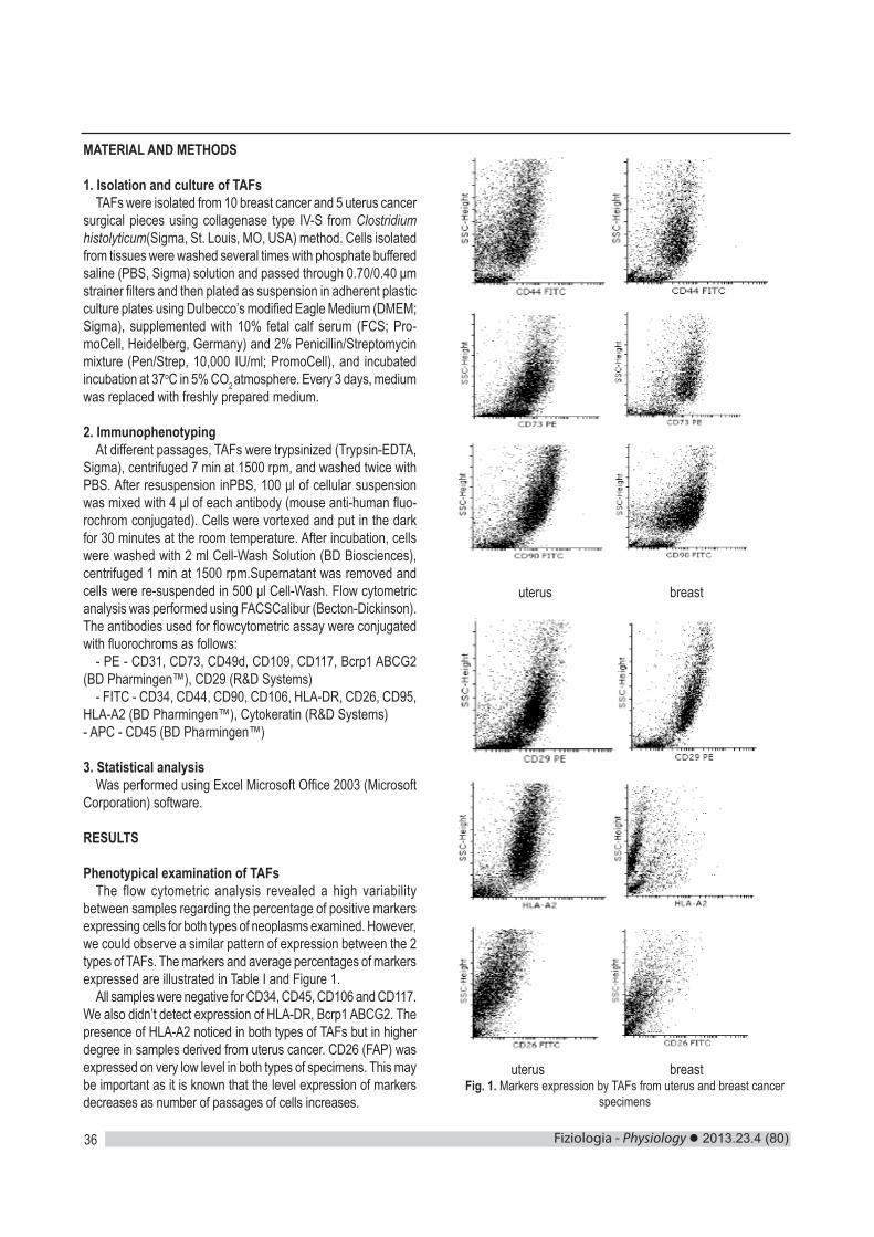
Fiziologia - Physiology 2013.23.4 (80)36
MATERIAL AND METHODS
1. Isolation and culture of TAFsTAFs were isolated from 10 breast cancer and 5 uterus cancer
surgical pieces using collagenase type IV-S from Clostridium histolyticum(Sigma, St. Louis, MO, USA) method. Cells isolated from tissues were washed several times with phosphate buffered saline (PBS, Sigma) solution and passed through 0.70/0.40 μm strainer filters and then plated as suspension in adherent plastic culture plates using Dulbecco’s modified Eagle Medium (DMEM; Sigma), supplemented with 10% fetal calf serum (FCS; Pro-moCell, Heidelberg, Germany) and 2% Penicillin/Streptomycin mixture (Pen/Strep, 10,000 IU/ml; PromoCell), and incubated incubation at 37oC in 5% CO2 atmosphere. Every 3 days, medium was replaced with freshly prepared medium.
2. ImmunophenotypingAt different passages, TAFs were trypsinized (Trypsin-EDTA,
Sigma), centrifuged 7 min at 1500 rpm, and washed twice with PBS. After resuspension inPBS, 100 μl of cellular suspension was mixed with 4 μl of each antibody (mouse anti-human fluo-rochrom conjugated). Cells were vortexed and put in the dark for 30 minutes at the room temperature. After incubation, cells were washed with 2 ml Cell-Wash Solution (BD Biosciences), centrifuged 1 min at 1500 rpm.Supernatant was removed and cells were re-suspended in 500 μl Cell-Wash. Flow cytometric analysis was performed using FACSCalibur (Becton-Dickinson). The antibodies used for flowcytometric assay were conjugated with fluorochroms as follows:
- PE - CD31, CD73, CD49d, CD109, CD117, Bcrp1 ABCG2 (BD Pharmingen™), CD29 (R&D Systems)
- FITC - CD34, CD44, CD90, CD106, HLA-DR, CD26, CD95, HLA-A2 (BD Pharmingen™), Cytokeratin (R&D Systems)- APC - CD45 (BD Pharmingen™)
3. Statistical analysisWas performed using Excel Microsoft Office 2003 (Microsoft
Corporation) software.
RESULTS
Phenotypical examination of TAFsThe flow cytometric analysis revealed a high variability
between samples regarding the percentage of positive markers expressing cells for both types of neoplasms examined. However, we could observe a similar pattern of expression between the 2 types of TAFs. The markers and average percentages of markers expressed are illustrated in Table I and Figure 1.
All samples were negative for CD34, CD45, CD106 and CD117. We also didn’t detect expression of HLA-DR, Bcrp1 ABCG2. The presence of HLA-A2 noticed in both types of TAFs but in higher degree in samples derived from uterus cancer. CD26 (FAP) was expressed on very low level in both types of specimens. This may be important as it is known that the level expression of markers decreases as number of passages of cells increases.
uterus breast
uterus breastFig. 1. Markers expression by TAFs from uterus and breast cancer
specimens

2013.23.4 (80) Fiziologia - Physiology 37
Table I. Average expression of TAF markersTAFs derived from
uterus cancer samples (%) (n=5)
TAFs derived from breast cancer samples
(%) (n=10)CD90 37.24 25.03CD73 47.27 38.21CD44 21.5 12.8CD29 43.45 38.38CD26 2.51 2.33
HLA-A2 27.085.22
DISCUSSION. CONCLUSIONS
TAFs affect in an important degree cancer development and progression. As key players in this complex process, TAFs are needed to be fully understand and characterised. This means that TAFs are an attractive therapeutic target and identification of specific markers of TAFs and signaling pathways involved in their activation and function within the tumor has to be an esential prerogative. Ideally, it would be valuable to find some universal „badges” of TAFs, which could be reachable in any tumor, re-gardless of histologic type. In our work we tried to characterise, by flow cytometry, TAFs derived from 2 types of solid tumor and make a comparison. We found some similarities regarding the type of markers expressed, even there were quantitative variations between samples.
1. CD90 (Thy-1) is suggested as biomarker for several types f cancer and cancer stem cells. It is expressed ont only by TAFs, but also by bone-marrow derived stem cells, T-cells, endothelial cells (12,13). CD90 seems to be an interesting fac-tor for distinguishing TAFs from benign stromal cells in prostate cancer (14).
1. CD73 (5’-nucleotidase) is a potent immunosuppres-sive molecule and is overexpressed in different types of cancer (leukemia, melanoma, breast, colon) (15,16). CD73 positive cells are capable to evade anti-tumor immune mechanisms. The prognostic value of this marker is still controversial, especially for breast cancer.
2. CD44 is a glycoprotein involved in cell proliferation, migration, angiogenesis, cell survival, tumor metastasis – es-sential aspects in cancer progression. CD44 expression by TAFs may be engaged in sustenance of cancer stem population inside the tumor (17).
3. CD29 (Integrin beta-1) is involved in cell adhesion, immune response and metastasis.
4. HLA-A2 is one of the most frequent HLA class I allo-specificities and is considered a potential prognostic factor in some malignancies. The presence of HLA-A2 antigen is crucial for tumor recognition by T-cells; down-regulation of this antigen expression delineates one of tumor strategies of escape from host immune defense.
5. CD26 (FAP) seems to be restricted as expression to TAFs, granulation tissue of healing wounds, soft tissues sarco-
mas, although the biological role in tumor microenvironment is not well known (it is thought to be involved in EMT during embryogen-esis and carcinogenesis). Some authors revealed that silencing of FAP induced the inhibition of TAFs growth and decreased the tumor growth in ovarian cancer xenograft models (18).
6. Although the markers mentioned above are highly seductive candidates for therapeutically targeting in cancer, most of them are not specific for TAFs; many types of cells (normal cells from different origins, tumoral cells, immune cells etc.) are expressing these markers. CD26 could be a serious nominee as a specific TAF marker. However, our experiment revealed a weak expression of this marker by TAFs derived from both uterus cancer and breast cancer samples. Further studies are required for establish the conditions in which it could be determined if this marker is constantly expressed by TAFs in solid tumors. Furthermore, targeting TAFs with active functional CTLs against CD26 protein could be an interesting approach.
AcknowledgementsThis work was supported by the Sectorial Operational Pro-
gramme for Human Resources Development, financed from the European Social Fund, FSE POSDRU 107/1.5/S/78702 and by UEFISCDI, PNII-Idei, Project No. 318/2011.
REFERENCES1. Tuxhorn JA, Ayala GE, Rowley DR. Reactive stroma in prostate cancer progression. J Urol 2001;166(6): 2472-83.2. De Wever O, Mareel M. Role of tissue stroma in cancer cell invasion. J Pathol 2003;200: 429-47.3. Weaver VM, Gilbert P. Watch thy neighbor: cancer is a communal affair. J Cell Sci 2004;117:1287-90.4. Cirri P, Chiarugi P. Cancer associated fibroblasts: the dark side of the coin. Am J Cancer Res 2011; 1(4): 482-497.5. Anderberg C, Pietras K. On the origin of cancer-associated fibroblasts. Cell Cycle 2009; 8(10):1461-2.6. Paunescu V, Bojin FM, Tatu CA, Gavriliuc OI, Rosca A, Gruia AT, Tanasie G, Bunu C, Crisnic D, Gherghiceanu M, Tatu FR, Tatu CS, Ver-mesan S. Tumour-associated fibroblasts and mesenchymal stem cells: more similarities than differences. J Cell Mol Med 2011; 15(3):635-646.7. Kalluri R, Zeisberg M Review. Fibroblasts in cancer. Nat Rev Cancer 2006; 6(5):392-401.8. Micke P, Ostman A. Review: Tumour-stroma interaction: cancer-associated fibroblasts as novel targets in anti-cancer therapy? Lung Cancer 2004; 45(2):S163-75.9. Kunz-Schughart LA, Knuechel R. Tumor-associated fibroblasts (part I): active stromal participants in tumor development and progression. Histol Histopathol 2002;17: 599-621.10. Cheng JD, Dunbrack Jr RL, Valianou M, Rogatko A, Alpaugh RK, Weiner LM. Promotion of tumor growth by murine fibroblast activation protein, a serine protease, in an animal model. Cancer Res 2002;62: 4767-72.11. Huber MA, Kraut N, Park JE, Schubert RD, Rettig WJ, Peter RU, et al. Fibroblast activation protein: differential expression and serine protease activity in reactive stromal fibroblasts of melanocytic skin tumors. J Invest Dermatol 2003;120: 182-8.12. Dennis JE, Esterly K, Awadallah A, Parrish CR, Poynter GM et al. Clinical-scale expansion of a mixed population of bone-marrow-derived stem and progenitor cells for potential use in bone-tissue regeneration.

Fiziologia - Physiology 2013.23.4 (80)38
Stem Cells 2007; 25: 2575-82.13. Herrera MB, Bruno S, Buttiglieri S, Tetta C, Gatti S et al. Isolation and characterization of a stem cell population from adult human liver. Stem Cells 2006; 24: 2840-50.14. True LD, Zhang H, Ye M, Huang CY, Nelson PS, von Haller PD, Tjoelker LW, Kim JS, Qian WJ, Smith RD, Ellis WJ, Liebeskind ES, Liu AY. CD90/THY1 is overexpressed in prostate cancer-asso-ciated fibroblasts and could serve as a cancer biomarker. Mod Pathol2010;23(10):1346-56.15. Serra S, Horenstein AL, Vaisitti T, et al. CD73-generated extracellular adenosine in chronic lymphocytic leukemia creates local conditions
counteracting drug-induced cell death. Blood 2011; 118: 6141-52.16. Spychala J, Lazarowski E, Ostapkowicz A, Ayscue LH, Jin A, Mitchell BS. Role of estrogen receptor in the regulation of ecto-5′-nucleotidase and adenosine in breast cancer.Clinical Cancer Research 2004; 10: 708-717.17. Kinugasa Y, Matsui T, Takakura N. CD44 expressed on cancer-asso-ciated fibroblasts is a functional molecule supporting the stemness and drug resistance of malignant cancer cells in the tumor microenvironment. Stem Cells 2014;32(1):145-56.18. Lai Ma, Fangyuan Wang. Fibroblast activation protein regulates tumor-associated fibroblasts and epithelial ovarian cancer cells.Int J Oncol 2012; 41: 541-550.
CARACTERIZARE IN VITRO A FIBROBLASTELOR PERI-TUMORALE IZOLATE DIN DIFERITE TUMORI SOLIDE
REZUMATCu toate ca originea acestora nu este deplin lamurita, fibroblastele peritumorale sunt componentele celulare esentiale ale stromei peri-tumorale. Este cunoscut deja rolul acestora in initierea, dezvoltarea si metastazarea tumorala. Stabilirea unui profil molecular al fenotipului si a unor markeri specifici ai fibroblastelorperitumorale izolate din tumori solide cu origini diferite ar aduce informatii esentiale privind considerarea acestor celule ca tinte terapeutice de succes. In experimentul nostru am tentat caracterizarea prin flow citometrie a fibroblastelor peritumorale izolate din piese rezecate chirurgical de cancer de uter si de san, pentru a depista eventuale similitudini privind markerii exprimati. Ambele tipuri de fibroblaste peritumorale au exprimat CD90, CD73, CD44, CD29 si CD26 (FAP)(acesta din urma in grad foarte redus). Rezultatele au relevat similaritati in ceea ce priveste tipurile de markeri exprimati, ceea ce ar putea sugera posibilitatea stabilirii unui fenotip general al fibroblastelor peritumorale aplicabil indiferent de originea acestora. Cuvinte cheie: fibroblaste peritumorale, microclimat tumoral, markeri de expresie, FAP

2013.23.4 (80) Fiziologia - Physiology 39
COMORBIDITIES AND RISK FACTORS IN HEART FAILURE WITH PRESERVED EJECTION FRACTION
ADELINA MAVREA, ADINA BUCUR, MIRELA TOMESCUUniversity of Medicine and Pharmacy “Victor Babes“ Timisoara
ABSTRACT Almost half of heart failure patients have heart failure with preserved ejection fraction (HFPEF). Patients are elderly with multiple comorbidities. We aimed to evaluate the incidence of comorbidities and their influence on risk factors and clinical status, in patients with heart failure with preserved ejection fraction (HFPEF).Material and methods:We included 78 patients who were diagnosed for the first time with HFPEF. We evaluated the incidence of comorbidities and their connection with prognostic markers (represented by pro-BNP), with inflammatory status (represented by TNF - tumor necrosis factor) and with clinical status (represented by quality of life and exercise capacity) at baseline and after 1 year. Results:There were statistically significant differences between the amount of pro-BNP in patients with chronic kidney disease both at baseline and after 1 year in patients, in patients with coronary heart disease and metabolic syndrome after 1 year of follow up, and for diabetes patients between pro-BNP values at baseline. In any category of patients were not clinically significant differences in relation to clinical status.Conclusion: Comorbidities in HFPEF can influence patient evolution through influence on prognostic factors.Key words: comorbidities, HFPEF, prognostic markers
Received 21st September 2013.Accepted 15th October 2013. Address for correspondence: Adina Bucur, MD, PhD, Department of Functional Sciences, “Victor Babes” University of Medicine and Pharmacy Timisoara, EftimieMurgu Square No. 2A, RO-300041, Timisoara, phone/fax: +40256490507; e-mail: [email protected]
INTRODUCTION
During the last 15 years it has increasingly recognized that many patients with heart failure (HF) have normal left ventricular ejection fraction. Current studies have reported a prevalence of heart failure with preserved ejection fraction (HFPEF) ranging from 30% to 70% among patients with heart failure. Actually, the prevalence of HFPEF relative to heart failure with reduce ejec-tion fraction (HFREF) is rising at a rate of approximately 1% per year (1).The investigators concluded that distinct risk profiles for HFPEF and HFREF exist, which would support differential patho-physiological mechanism for both HF subtypes. High prevalence of comorbidities in patient with heart failure suggested common risk factors or a causal relation. Compared with HFREF patients, those with HFPEF were older, and had a higher prevalence of hy-pertension, diabetes, chronic renal disease, ischemic coronaries disease (2).HFPEF is a syndrome disease with multiple etiologies and phenotypic expressions. Left ventricular diastolic dysfunction is a key pathophysyological element and relates to increased myocytes stiffness and extracellular matrix changes. Systolic abnormalities, left atrial dysfunction, chronotropic incompetence, vascular stiffening, pulmonary hypertension, contribute to HFPEF (3).Exercise incompetence of affected patientscan be related to striking hemodynamic abnormalities in HFPEF at rest, and even more pronounced, during exercise. Common comorbidities such as diabetes, hypertension, renal dysfunction, may aggravate the clinical picture and play a role in precipitating the disease (4).
We aimed to compare the presence of comorbidities with clinical status, with prognosis marker and with clinical evolution for patients diagnosed with HFPEF.
MATERIAL AND METHODS
Seventy-eight patients admitted to the Ascar Cardiology Clinic in 2011/2012 years, whohad clinical signs of heart failureand primary diagnosis of heart failure with preserved ejection fraction, were included in the present study.
Inclusion criteria:̶ Sinus rhythm̶ Clinical signs and symptoms of heart failure, according to Framingham criteria̶ FE ≥ 45 %̶ E/E´ ≥ 15̶ Pro-BNP >150pg/mlExclusion criteria:̶ Atrialfibrillation̶ Acute coronary syndrome within the last 30 days̶ Cardiac stimulator implant̶ Severe valvular heart diseases̶ Severe respiratory dysfunction ̶ Chronic kidney disease ≥4 KDOQI̶ Mental disorders̶ Cancer

Fiziologia - Physiology 2013.23.4 (80)40
Presence of sinus rhythm was considered as mandatory criteri-onin order to eliminate the effect of atrial fibrillation on cardiac function (the diastolic one, especially), clinical symptomatology andheart failure evolution.All the patients have signed Informed Consent forms.Laboratory constants were determined at pa-tients’ admission to clinic.BNP was determined by MEIA Abbott method. Ultrasound evaluation has been performed usinga Vivid S5 cardiac ultrasound system, as follows:assessment of LVTD, LVTS and LV wall thickness in M-Mode; measurement of ejection fraction by Simpson method; assessment of transmitral diastolic flow (E and A waves, e/a ratio, EWDT- E wave deceleration time, IVRT - isovolumetric relaxation time), assessment of pulmonary venous flow; tissue Doppler with recording of early and late diastolic mitral annulus velocities (E´ - maximum early diastolic velocity and A´ - maximum late diastolic velocity); assessment of left atrium sizes – determination of LA area in apical 4-chamberOne year after patients’ inclusion, inflammatorymarkers, along withproBNPand clinical status components were re-assessed. Minnesota quality of life questionnaire was filled in by patients the day before discharge. Twelve months after patients’ inclu-sion, inflammatory markers, along with proBNP, NT-proBNP and clinical status components were re-assessed.
RESULTS
Of all patients, 50 (64.10%) patients were diagnosed with isch-emic heart disease (IC), 29 (37.17%) patients with chronic kidney disease (KCD) and 25(32%) with diabetes(D). From coronary patients, 14 patients (28%) are diagnosed with chronic kidney disease, and 12 patients (24%) are diabetic. Only 5 patients (6.4%) are diagnosed with all three comorbidities (Figure 1).
Fig.1.Prevalence of comorbidities
Two groups of patients were formed, according to NYHA class: NYHA III (group1) and NYHA II(group2). We must mention that no NYHA I or NYHA IV patients were included in the studied group.
Table I shows the features of the two patient groups.As one can see, there are no statistically significant differences between the two groups, except the presence of metabolic syndrome (p=0.0123) and chronic kidney disease (p=0.0066).
Table I.Features of patient groups according to NYHA class
Grup IN=31
Grup IIN=47
p
Age 65 ±9.0 66± 9.2 0.63Male sex 8 (26%) 15 (32%) 0.75CI 17 (55%) 33(70%) 0.26HTA 29 (94%) 42 (89%) 0.72DZ 9 (29%) 16 (34%) 0.82Metabolic syndrome 23 (74%) 20 (42.5%) 0.0123BCR 10 (32%) 19 (40%) 0.0066Regarding the patients diagnosed with diabetes and those
who are not diabetic, has revealed a statistically significant dif-ference in BNP at patients inclusion in the study (p=0.046, 95% confidence interval of the difference). We are not able to certify the same about proBNP value after 1 year (pro-BNP1), because there are not statistically significant differences between patients with diabetes and those without diabetes (p =0.09)(Figure 2).
Fig. 2. Pro-BNP evolution for diabetic patients
In patients diagnosed with ischemic heart disease were sta-tistically significant differences for proBNP after 1 year of follow up (p=0.03). Moreover, it can be observed that in patients with ischemic heart disease, the pro-BNP is growing after 1 year follow-up compared with patients who were not diagnosed with the disease (Figure 3).
Fig. 3. Pro-BNP evolution for patient diagnosed with ischemic cardiopathy

2013.23.4 (80) Fiziologia - Physiology 41
Statistically significant differences are noted in pro-BNP value both initially (p=0,028) and after 1 year of follow-up (p=0,048) in patients with chronic kidney disease (GFR<60ml/kgc/min) (Fig.4).
In case of metabolic syndrome were not significant differences of biomarkers and clinical status in patients included in the study.
Fig. 4. Pro-BNP evolution for patient diagnosed with renal chronic disease
After one year of monitoring, there were significant differences for proBNP value (p <0.0001) and TNF value (p = 0.004) (Figure 5).
Fig.5. Pro-BNP and TNF value for patients diagnosed with metabolic syndrome, after 1 year.
DISCUSSION
It is expected that the incidence and prevalence of heart failure, as well as of comorbidities, will increase exponentially in the following decades due to an aging population. An increased incidence ofcomorbiditiesresulted in an increasein the number ofhospitalizationsdue to non-cardiovascular andnon-HF hospitalization (5).
We can see that there are non-cardiovascular comorbidities (diabetes, metabolic syndrome), which have significant differences in prognostic markers after one year of follow up, which may negatively influence the evolution of these patients.
Although a short period of monitoring, these comorbidities did not influence the clinical status of patients; patients with more comorbidityare more likely to have clinical signs of congestion. Systemically raised venous pressure with ancillary neurohormonal responses may negatively impact non-cardiac tissues and organs, thus facilitating the occurrence of so called comorbidities.Therefore, comorbidities and heart failure are conditions that can influence each other in terms of their evolution.
Over the past decade, myocardial structure, cardiomyocyte function, and intramyocardial signaling were shown to be specifically altered in heart failure with preserved ejection fraction (HFPEF). A new paradigm for HFPEF development is therefore proposed, which identifies a systemic pro-inflammatory state induced by comorbidities as the cause of myocardial structural and functional alterations. (6)
CONCLUSION
The new HFPEF paradigm proposes comorbidities, plasma markers of inflammation, or vascular hyperemic responses to be included in diagnostic algorithms .
REFERENCES1. Owan TE, Hodge DO, Herges RM, Jacobsen SJ, Roger VL, Redfield MM. Trends in prevalence and outcome of heart failure with preserved ejection fraction. N Engl. J Med 2006; 355: 251-259.2. Brouwers FP, de Boer RA, van der Harst P, Voors AA, Gansevoort RT, Bakker SJ, Hillege HL, van Veldhuisen DJ, van Gilst WH. Incidence and epidemiology of new onset heart failure with preserved vs. reduced ejection fraction in a community-based cohort: 11-year follow-up of PREVEND. Eur Heart J 2013; 34:1424-31.3. Borlaug PA. Heart failure with preserved and reduced ejection fraction: different risk profiles for different disease. Eur Heart J. 2013; 34: 1393-95.4. Braunstein JB, Anderson GF, Gerstenblith G, Weller W, Niefeld M, Herbert R, Wu AW. Non-cardiac comorbidity increases preventable hospitalizations and mortality among Medicare beneficiaries with chronic heart failure. J AmmCollCardiol. 2003; 42: 1226-35.5. Ather S, Chan W, Bozkurt B, Aguilar D, Ramasubbu K, Zachariah AA, Wehrens XH, Deswal A. Impact of non-cardiac comorbidities on morbidity and mortality in a predominantly male population with heart failure and preserved versus reduced ejection fraction. J AmmColl Cardio 2012; 59: 998-1005.6. Borbely A, van der Velden J, Papp Z. Cardiomyocyte stiffness in diastolic heart failure. Circulation 2005; 111: 774-781.

Fiziologia - Physiology 2013.23.4 (80)42
COMORBIDITĂŢI ŞI FACTORII DE RISC ÎN INSUFICIENŢA CARDIACĂ, CU FRACŢIE DE EJECŢIE PREZERVATĂ
REZUMATAproape jumătate din pacienţii cu insuficienţă cardiacă au insuficienţă cardiacă cu fracţie de ejecţie prezervată (ICFEP).Pacientii sunt în vârstă, cu mai multe comorbidităţi.Ne-am propus pentru a evalua incidenţa comorbidităţilor şi influenţa lor asupra factorilor de risc şi asupra stării clinice la pacienţii cu insuficienţă cardiacă cu fractie de ejectieprezervată (ICFEP).Material şimetode: Am inclus 78 de pacienti, care au fost diagnosticaţi pentru prima dată cu ICPEF. Am evaluat incidenţa comorbidităţilor şi legătura lor cu markeri de prognostic (pro - BNP), cu statusul inflamator (TNF-factor de necroză tumorală) şi cu starea clinică, reprezentată de calitatea vieţii şi capacitatea de efort, la includere şi după 1 an de urmărire.Rezultate: Au fost diferenţe semnificative statistic între valoarea pro-BNP la pacienţii cu boală cronică de rinichi, atât la momentul iniţial şi după 1 an, la pacientii cu boala coronariana si sindrom metabolic după 1 an de urmărire, precum şi pentru pacienţii cu diabet între valoarea pro-BNP la momentul iniţial. În orice categorie de pacienţi nu au fost diferenţe semnificative în ceea ce priveşte statusul clinic. Concluzie: Comorbidităţile în ICFEP pot influenţa evoluţia pacienţilor prin influenţa asupra factorilor de prognostic.Cuvinte cheie: comorbidităţi, ICFEP, markeri de prognostic.