Embed Size (px)
Citation preview

Available online at www.sciencedirect.com
ARTICLE IN PRESS
Consciousness
Consciousness and Cognition xxx (2008) xxx–xxx
andCognition
www.elsevier.com/locate/concog
EEG paroxysmal gamma waves during BhramariPranayama: A yoga breathing technique
Franc�ois B. Vialatte a,*, Hovagim Bakardjian a,Rajkishore Prasad b, Andrzej Cichocki a
a RIKEN Brain Science Institute, Laboratory for Advanced Brain Signal Processing, 2-1 Hirosawa, Wako-Shi, Saitama-Ken 351-0198, Japanb Machine Integrated Systems Lab, University of Electro-Communications, Chofu City, Tokyo 182-8585, Japan
Received 11 May 2007
Abstract
Here we report that a specific form of yoga can generate controlled high-frequency gamma waves. For the first time, par-oxysmal gamma waves (PGW) were observed in eight subjects practicing a yoga technique of breathing control called Bhra-mari Pranayama (BhPr). To obtain new insights into the nature of the EEG during BhPr, we analyzed EEG signals using time-frequency representations (TFR), independent component analysis (ICA), and EEG tomography (LORETA). We found thatthe PGW consists of high-frequency biphasic ripples. This unusual activity is discussed in relation to previous reports on yogaand meditation. It is concluded this EEG activity is most probably non-epileptic, and that applying the same methodology toother meditation recordings might yield an improved understanding of the neurocorrelates of meditation.� 2008 Elsevier Inc. All rights reserved.
Keywords: Electroencephalography; Meditation; Yoga; Epilepsy; Temporal lobe; Brain mapping; Signal processing; Gamma
1. Introduction
Meditation is a psychologically induced, altered state of consciousness (Vaitl et al., 2005), and its study pro-vides insights into cognitive and emotional brain correlates of consciousness (e.g. Lou, Nowak, & Kjaer,2005). Analysis of such correlates is crucial for contemporary investigations of consciousness (Zeman,2005). However, despite almost 50 years of study, a comprehensive empirical and theoretical foundationfor meditation is still only now just emerging, and studies of its clinical impact are required (Cahn & Polich,2006). Meditation techniques such as transcendental meditation (e.g. Yamamoto, Kitamura, Yamada, Naka-shima, & Kuroda, 2006); Zen or other Buddhist meditations (Lutz, Greischar, Rawlings, Ricard, & Davidson,
1053-8100/$ - see front matter � 2008 Elsevier Inc. All rights reserved.doi:10.1016/j.concog.2008.01.004
* Corresponding author. Fax: +81 (0) 48 467 9694.E-mail address: [email protected] (F.B. Vialatte).
Please cite this article in press as: Vialatte, F. B. et al., EEG paroxysmal gamma waves during Bhramari ..., Conscious-ness and Cognition (2008), doi:10.1016/j.concog.2008.01.004
2 F.B. Vialatte et al. / Consciousness and Cognition xxx (2008) xxx–xxx
ARTICLE IN PRESS
2004); yoga type meditation practices such as pranayama,1 dhyana2 or samadhi3 (e.g. Yoga Nidra in Lou et al,1999; or Lou et al., 2005); and shamanic trances (Oohashi et al., 2002), etc. These can be divided into thosepractices involving movements, like walking, dancing, and singing, and the ‘‘silent” meditation methods,which are usually practiced in a characteristic sitting position (Vaitl et al., 2005). This considerable diversitycomplicates the description of a uniform theory of meditation. Usually, most studies concerning meditationfocus on transcendental meditation, because the technique is comparatively simple and hence easilyreproducible.
BhPr is a pranayama technique, therefore a technique of breathing control. However, because of its hyp-notic and repetitive aspect it is also very close to and to some degree overlapping with mantra repetition tech-niques. Finally, BhPr changes the breathing rhythm, with very long exhalations and short inhalations, whichmay have a physiological effect. BhPr practiced for 5 to 10 consecutive minutes induces subjective feelings ofmind refreshment and blissfulness, and even sometimes a state close to dhyana. Therefore, BhPr is a pranay-ama technique, but also a meditation technique.4 Very few scientific studies on the effects of this techniquehave been done. It has been claimed that BhPr may reduce hormonal imbalance manifestation such as hyper-tension, anxiety, and abnormal blood pressure (Singh, 1995). It has also been said to have a calming effect,which was used in a program for substance dependence recovery (Nespor, 2000).
A dramatic increase of activity in the gamma band in association with meditation—visible in the rawEEG—was reported in a study using trained practitioners of meditation (Lutz et al., 2004). This activityhas been hypothesized to be representative of epilepsy (Nicholson, 2006), but this interpretation has beenrejected by other studies (St. Louis & Lansky, 2006).
Using high-density array EEG (Biosemi system, 128 electrodes) and video control, we investigated the EEGin eight subjects performing Bhramari Pranayama (BhPr) yoga meditation (one trained for a 4 month period,six trained for 1 month, and one beginner). All displayed a similar, high-frequency pattern during BhPr.
2. Methods
2.1. Bhramari Pranayama
Subjects used the BhPr yoga technique, which involves producing a vibrating sound while exhaling strictlythrough the nasal airways. The oral cavity was closed at the lips, the ear canals were closed by depressing thetragus with fingers, and the eyelids were closed. The generated sound may be described as emulating the buzz-ing of bumblebees, having a constant pitch. The index finger was placed onto the forehead along the eyebrows,middle fingers were placed at the base of the nose near the corners of the eyes, and the little fingers were placedalong the nose such that they lay next to the nostrils. Elbows were raised horizontally. As subjects sat on a thincushion on the floor of the experimental room, their legs were crossed at the knees with their ankles placed ontop of their thighs. While performing BhPr, subjects concentrated on an imaginary point located between theireyebrows. This yoga technique is demanding: (a) the humming sound must come from the nose and shouldremain constant; (b) the leg posture requires flexibility in the hips; and (c) the shoulders tire quickly in thisposture.
For this study, we recruited eight volunteer subjects (all male and right-handed) complying with the con-straints of BhPr yoga (able to maintain the yoga posture and produce the humming sound):
1. Subject B1 (beginner): never practiced BhPr before the recording session;
1 Breath control technique.2 Dhyana in Sanskrit or jhana in Pali refers to a type or aspect of meditation, when the mind attains the ability to sustain its attention
without getting distracted.3 Samadhi is a Hindu and Buddhist term that describes a non-dualistic state of consciousness in which the consciousness of the
experiencing subject becomes one with the experienced object, and in which the mind becomes still though the person remains conscious.4 We insist however on the fact the BhPr is a specific technique that should not be confused with other meditation techniques, such as,
for instance, Zen meditation or transcendental meditation.
Please cite this article in press as: Vialatte, F. B. et al., EEG paroxysmal gamma waves during Bhramari ..., Conscious-ness and Cognition (2008), doi:10.1016/j.concog.2008.01.004
F.B. Vialatte et al. / Consciousness and Cognition xxx (2008) xxx–xxx 3
ARTICLE IN PRESS
2. Subject I1–I6 (intermediate): practiced BhPr (two sessions per day) for 31–34 days before the recordingsession;
3. Subject E1 (expert): practiced BhPr (two sessions per day) every day for 4 months before the recordingsession.
The Ethical Committee of Riken Brain Science Institute (BSI) in Wako-Shi, Japan, approved the project,and written informed consent was obtained from the subjects. The subjects had no history of neurological,psychiatric, (or) epileptic (diseases) or other severe diseases. All experiments were performed with theinformed and explicit consent of each participant, in line with the code of Ethics of the World Medical Asso-ciation (Declaration of Helsinki) and the standards established by the Riken BSI’s Institutional Review Board.
2.2. EEG recording
Recordings were performed in an electrically shielded room. EEG was recorded with 128 active electrodes(Biosemi system) at a sampling frequency of 2048 Hz. Signals were analog bandpass filtered between 1 and300 Hz, and notch filters were applied at 50 Hz and at every harmonic of 25 Hz to substantially removeany external noise related to line power frequencies.
Postural electromyographic (EMG) noise was monitored and controlled by recording EEG during falseBhPr. In false BhPr, the subjects assumed the BhPr position, but did not produce the humming noise. Instead,they attempted to mimic the BhPr respiratory rhythm to reveal the potential effects of hypoventilation.5 Respi-ratory patterns were controlled by recording respiration with a respiratory belt. In three intermediate subjects(I2, I4, and I6),6 we also recorded three EMG channels from the temporalis (forefront), masseter (cheek), andsternocleidomastoideus (SCM, neck) muscles.
BhPr was recorded in one or two consecutive sessions in which each subject performed approximately 20breathing episodes. EEG was also recorded before and after BhPr in the resting/eyes-closed condition. Sub-jects were video monitored with a security camera. Trials consisted of an inhalation period and exhalationperiod (humming period).
2.3. Software programs
Signal analysis was performed using MATLAB�, EEGLAB (Delorme & Makeig, 2004), SigmaStat�,LORETA (Pascual-Marqui, 1999; Pascual-Marqui, Michel, & Lehmann, 1994), and ICALAB (Cichockiet al., online toolbox; Cichocki & Amari, 2003). MATLAB� was used for the wavelet transforms and prepro-cessing of signals. EEGLAB was used for topographic mapping, ICALAB for independent component anal-ysis, and SigmaStat� for statistical tests. LORETA was used to compute EEG low-resolution tomographysources.
2.4. Signal processing
2.4.1. Independent component analysisLet us consider the case of multiple EEG channel sampling of brain activity over time. If the signals from
each channel form the rows of the data matrix D, then each column of D is a time point. The problem is tofind the original brain sources, which were mixed in the EEG channels. This is a typical problem of blindsource separation (BSS). Independent components analysis (ICA) is a valid solution to BSS in the context ofEEG recordings (Makeig, Bell, Jung, & Sejnowski, 1996; Tang, Sutherland, & McKinney, 2005; Cichockiet al., 2005), and finds the unmixing square matrix W (n = m = the number of channels) such thatW.D = C (Brown, Yamada, & Sejnowski, 2001). The rows of C are called ‘‘independent components”
because they are forced to be as independent as possible and are the sources being searched. It was shown
5 Imitating the posture of BhPr is not enough, because this technique alters the breathing rhythm (which may generate both EMG andhypoventilation).
6 Because EMG reduces subject’s concentration, only three subjects used it.
Please cite this article in press as: Vialatte, F. B. et al., EEG paroxysmal gamma waves during Bhramari ..., Conscious-ness and Cognition (2008), doi:10.1016/j.concog.2008.01.004
4 F.B. Vialatte et al. / Consciousness and Cognition xxx (2008) xxx–xxx
ARTICLE IN PRESS
in Delorme, Sejnowski, and Makeig (2007) that preprocessing EEG data using ICA allows effective artifactseparation. The SOBI algorithm, in particular, is especially well suited for analysis of high-density EEG(Tang et al., 2005).
2.4.2. Spectral analysis
A digital FFT-based power spectrum analysis (Welch technique, Hanning windowing function, no phaseshift) computed the power density of EEG rhythms with windows of 500 msec. The resulting values wereafterwards normalized into a relative power. We used fixed but narrow bands for the theta (4–8 Hz), alpha(8–12 Hz), beta (12–30 Hz), and gamma (30–80 Hz) ranges. The use of fixed frequency bands allowed a bettercomparison with previous literature, a more direct comprehension of results, and an enhancement of evenslight differences.
2.4.3. Wavelets
There is a wide variety of wavelets. In the present study, complex Morlet wavelets(Kronland-Martinet, Morlet, & Grossmann, 1987) were used:
Pleaness
WðkÞ ¼ A: exp�k2
2r2t
� �: expð2ipf0kÞ;
where rt2 and f0 jointly determine the number of oscillations in the wavelet. In the present investigation, the
wavelet family defined by 2p rtf = 7 was chosen, as described in (Tallon-Baudry, Bertrand, Delpuech, & Per-nier, 1996). Complex Morlet wavelets are appropriate for time–frequency analysis of electroencephalographicsignals (Caplan, Madsen, Raghavachari, & Kahana, 2001; Duzel et al., 2003; Martin, Gervais, Hugues, Mess-aoudi, & Ravel, 2004; Ohara, Crone, Weiss, & Lenz, 2004; Tallon-Baudry & Bertrand, 1999; Tallon-Baudryet al., 1996). They produce a precise time–frequency representation for the analysis of EEG signals, because oftheir symmetric shape in both time and frequency. The (continuous) wavelet transform W of a time series x isobtained by:
Wðk; sÞ¼DX
l
xðlÞW� l� k
s
� �;
where w(k) is the (complex) ‘‘mother”’ wavelet and s is a scaling factor.
2.4.4. LORETA
We employed LORETA (Pascual-Marqui et al., 1994; Pascual-Marqui, 1999) to compute 3-D linear solu-tions (LORETA solutions) for the EEG inverse problem within a three-shell spherical head model includingscalp, skull, and brain compartments. LORETA is presently used by independent laboratories worldwide forEEG source analysis. We used a two-step approach, combining LORETA and ICA (Marco-Pallares, Grau, &Ruffini, 2005), which brings further information for identified specific EEG patterns. The brain compartmentwas restricted to the cortical gray matter/hippocampus and was coregistered into the Talairach probabilitybrain atlas, digitized at the Brain Imaging Center of the Montreal Neurologic Institute (Talairach & Tour-noux, 1988). This compartment included 2394 voxels (7-mm resolution), each voxel containing an equivalentcurrent dipole. LORETA solutions consisted of voxel current density values able to model EEG spectralpower density at scalp electrodes.
3. Results
3.1. Basic traits
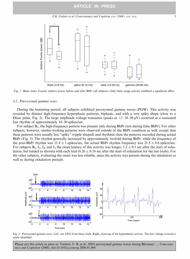
We compared relative Fourier power before and after BhPr (Fig. 1). Low-frequency power was diminished,significantly for the theta range (4–8 Hz, p < .05) and non-significantly for the alpha range. We also analyzedthe evolution of this effect as compared to the subject’s training. No significant effect could be observed in rela-tion to the training (Pearson R correlation test, p > > .10).
se cite this article in press as: Vialatte, F. B. et al., EEG paroxysmal gamma waves during Bhramari ..., Conscious-and Cognition (2008), doi:10.1016/j.concog.2008.01.004
theta (4-8 Hz) alpha (8-12 Hz) beta (12-30 hz) gamma (30-80 Hz)
rela
tive
Fou
rier
pow
er (
dB)
*
Fig. 1. Basic traits. Fourier relative power before and after BhPr (all subjects). Only theta range activity exhibited a significant effect.
F.B. Vialatte et al. / Consciousness and Cognition xxx (2008) xxx–xxx 5
ARTICLE IN PRESS
3.2. Paroxysmal gamma wave
During the humming period, all subjects exhibited paroxysmal gamma waves (PGW). This activity wasrevealed by distinct high-frequency hyperphasic patterns, biphasic, and with a very spiky shape (close to aDirac pulse, Fig. 2). The large amplitude voltage transients (peaks at +/- 10–30 lV) occurred at a sustainedfast rhythm of approximately 10–30 spikes/sec.
For subject B1, the high-frequency pattern was present only during BhPr (not during false BhPr). For othersubjects, however, similar-looking patterns were observed outside of the BhPr condition as well, except thatthese patterns were usually less ‘‘spiky” (ripple shaped) and rhythmic than the patterns recorded during actualBhPr (Fig. 3). The rhythm generally increased by approximately twofold during BhPr: while the frequency ofthe post-BhPr rhythm was 11.8 ± 1 spikes/sec, the actual BhPr rhythm frequency was 21.5 ± 0.6 spikes/sec.For subjects B1, I2, I4, and I5 the onset latency of this activity was longer, 1.2 ± 0.1 sec after the start of exha-lation, but tended to shorten with each trial (0.26 ± 0.16 sec after the start of exhalation for the last trials). Forthe other subjects, evaluating the onset was less reliable, since the activity was present during the inhalation aswell as during exhalation periods.
Fig. 2. Paroxysmal gamma wave. Left, raw EEG from three trials. Right, close-up of the hyperphasic activity. The fast voltage reversal iseasily identified.
Please cite this article in press as: Vialatte, F. B. et al., EEG paroxysmal gamma waves during Bhramari ..., Conscious-ness and Cognition (2008), doi:10.1016/j.concog.2008.01.004
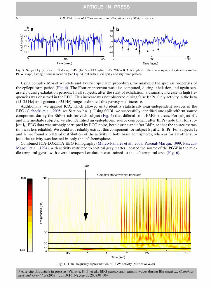
Fig. 3. Subject E1. (a) Raw EEG during BhPr. (b) Raw EEG after BhPr. When ICA Is applied to these raw signals, it extracts a similarPGW shape, having a similar location (see Fig. 5), but with a less spiky and rhythmic pattern.
6 F.B. Vialatte et al. / Consciousness and Cognition xxx (2008) xxx–xxx
ARTICLE IN PRESS
Using complex Morlet wavelets and Fourier spectrum procedures, we analyzed the spectral properties ofthe epileptiform period (Fig. 4). The Fourier spectrum was also computed, during inhalation and again sep-arately during exhalation periods. In all subjects, after the start of exhalation, a dramatic increase in high fre-quencies was observed in the EEG. This increase was not observed during false BhPr. Only activity in the beta(15–35 Hz) and gamma (>35 Hz) ranges exhibited this paroxysmal increase.
Additionally, we applied ICA, which allowed us to identify statistically near-independent sources in theEEG (Cichocki et al., 2005, see Section 2.4.1). Using SOBI, we successfully identified one epileptiform sourcecomponent during the BhPr trials for each subject (Fig. 5) that differed from EMG sources. For subject E1,and intermediate subjects, we also identified an epileptiform source component after BhPr (note that for sub-ject I6, EEG data was strongly corrupted by ECG noise, both during and after BhPr, so that the source extrac-tion was less reliable). We could not reliably extract this component for subject B1 after BhPr. For subjects I5
and I6, we found a bilateral distribution of the activity in both brain hemispheres, whereas for all other sub-jects the activity was located in only the left hemisphere.
Combined ICA-LORETA EEG tomography (Marco-Pallares et al., 2005; Pascual-Marqui, 1999; Pascual-Marqui et al., 1994), with activity restricted to cortical gray matter, located the source of the PGW in the mid-dle temporal gyrus, with overall temporal evolution constrained to the left temporal area (Fig. 6).
Fig. 4. Time–frequency representation of PGW activity (Morlet wavelet).
Please cite this article in press as: Vialatte, F. B. et al., EEG paroxysmal gamma waves during Bhramari ..., Conscious-ness and Cognition (2008), doi:10.1016/j.concog.2008.01.004
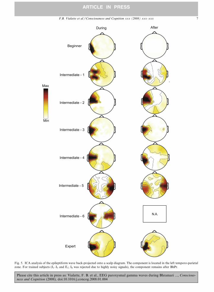
Fig. 5. ICA analysis of the epileptiform wave back-projected onto a scalp diagram. The component is located in the left temporo-parietalzone. For trained subjects (I1–I5 and E1; I6 was rejected due to highly noisy signals), the component remains after BhPr.
F.B. Vialatte et al. / Consciousness and Cognition xxx (2008) xxx–xxx 7
ARTICLE IN PRESS
Please cite this article in press as: Vialatte, F. B. et al., EEG paroxysmal gamma waves during Bhramari ..., Conscious-ness and Cognition (2008), doi:10.1016/j.concog.2008.01.004
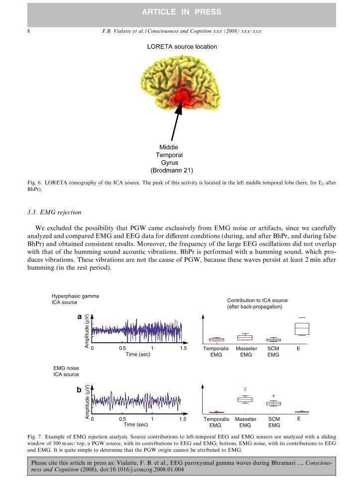
Fig. 6. LORETA tomography of the ICA source. The peak of this activity is located in the left middle temporal lobe (here, for E1 afterBhPr).
8 F.B. Vialatte et al. / Consciousness and Cognition xxx (2008) xxx–xxx
ARTICLE IN PRESS
3.3. EMG rejection
We excluded the possibility that PGW came exclusively from EMG noise or artifacts, since we carefullyanalyzed and compared EMG and EEG data for different conditions (during, and after BhPr, and during falseBhPr) and obtained consistent results. Moreover, the frequency of the large EEG oscillations did not overlapwith that of the humming sound acoustic vibrations. BhPr is performed with a humming sound, which pro-duces vibrations. These vibrations are not the cause of PGW, because these waves persist at least 2 min afterhumming (in the rest period).
Temporalis EMG
Masseter EMG
SCMEMG
E
Temporalis EMG
Masseter EMG
SCMEMG
E0 0.5 1 1.5
Hyperphasic gamma ICA source
0 0.5 1 1.5Time (sec)
Am
plitu
de (
μV)
EMG noise ICA source
Am
plitu
de (
μV)
Time (sec)
Contribution to ICA source (after back-propagation)
a
b
Fig. 7. Example of EMG rejection analysis. Source contributions to left-temporal EEG and EMG sensors are analyzed with a slidingwindow of 500 msec: top, a PGW source, with its contributions to EEG and EMG; bottom, EMG noise, with its contributions to EEGand EMG. It is quite simple to determine that the PGW origin cannot be attributed to EMG.
Please cite this article in press as: Vialatte, F. B. et al., EEG paroxysmal gamma waves during Bhramari ..., Conscious-ness and Cognition (2008), doi:10.1016/j.concog.2008.01.004
F.B. Vialatte et al. / Consciousness and Cognition xxx (2008) xxx–xxx 9
ARTICLE IN PRESS
For the three subjects with EMG recordings (I2, I4, and I6), we applied additional control of the signalsafter BhPr and removed artifact periods prior to further analysis. Only the data from subject I6 could notbe cleared of artifacts due to strong and continuous ECG noise corruption of the signal during and after BhPr.
During BhPr, the presence of EMG noise is to be expected due to the nature of the experimental taskrequiring muscle activity and to glossokinetic artifacts elicited by the humming sound. Our challenge underthese conditions was to identify if the PGW originated from the brain. We computed the ICA sources usingthe SOBI algorithm, while including also the available EMG channels into the data matrix. After identifica-tion of the PGW source, the relative contribution of EEG and EMG to the source was computed. After thispreprocessing, EMG noise originating from the muscles should have a higher EMG amplitude as compared tothe EEG. For all three subjects, PGW were attributed to the EEG (Fig. 7).
4. Discussion
The basic trait we observed for BhPr is an increase theta range activity, which is similar to results obtainedwith other meditation techniques (Hebert & Lehmann, 1977; Aftanas & Golocheikine, 2001; Aftanas, Varla-mov, Pavlov, Makhnev, & Reva, 2001; Vaitl et al., 2005). In these previous reports, the theta activity corre-lated with subjective reports of ‘‘blissful mental quiescence”, described as a state in which thoughts are absentbut consciousness remains. We cannot find comparisons of our results with previous reports specific to BhPr,as to the best of our knowledge no previous scientific investigation of this technique using EEG has beenpublished.
We also reported a biphasic hypersynchronous activity, in the high gamma range. The three main possibleinterpretations of this PGW phenomenon are as follows:
1. Epileptic activity. We consider this interpretation to be very unlikely. Finding an EEG waveform with cer-tain characteristics in common with epileptic EEG patterns does not necessarily link these patterns to epi-lepsy, any more than, for instance, finding symptoms of fever in a patient indicates a link to tuberculosis,even though fever accompanies tuberculosis. Since our subjects had no history whatsoever of neurological,psychiatric, or epileptic diseases, a link with epilepsy on such evidence is excessively speculative. Recently, aputative role for meditation practice in the generation of epileptic seizures has been hypothesized (see forinstance Nicholson, 2006; Persinger, 1993; St. Louis & Lansky, 2006), which has since become a subject ofcontroversy and scientific debate (e.g. Orme-Johnson, 2006). High-frequency epileptiform EEG hasreceived experimental attention only recently (Fisher, Webber, Lesser, Arroyo, & Uematsu, 1992; Jirschet al., 2006), most likely due to the poor representation and limited possibilities for reliable signal analysisin conventional scalp EEG for frequencies above the beta range. The BhPr activity we observed thus mightrepresent epileptic activity. This interpretation would be consistent with the spike shape of the recorded sig-nals (Figs. 2 and 3) and the temporal lobe location of the BhPr-associated activity (Figs. 5 and 6), both ofwhich are similar in high-frequency epilepsy (Fisher et al., 1992; Jirsch et al., 2006), and musicogenic epi-lepsy (Brien & Murray, 1984). However, the isolated scalp location of the BhPr-associated activity did notpropagate to other brain areas during or after BhPr, which is not consistent with epilepsy (as has beenreported by St. Louis & Lansky, 2006, for other types of meditation). Furthermore, simultaneous videorecording did not identify any epileptic behavior in either subject. Moreover, the subjects subjectivelyreported only a feeling of peacefulness. Finally, the EEG waveform remained temporally stable, withoutshowing epileptiform activity in low-frequency ranges (<15 Hz). Therefore, while this activity resemblesepileptic fast ripples, it is more likely to be a normal variant7 and should not be interpreted as conventionalepilepsy.
2. Anti-epileptic activity. Similar investigations (meditation on self-generated sound patterns) have shown asignificant activation in the same left temporal lobe location (Lazar et al., 2000; Lehmann et al., 2001),but did not report any epileptic-like behavior. In population studies, meditation and yoga appeared notto induce but rather reduce epilepsy (Orme-Johnson, 2006; Yardi, 2001; Sathyaprabha et al., 2007), even
7 Normal variant: epileptiform EEG not related to epilepsy (e.g., phantom spike waves, SREDA, wicket spikes).
Please cite this article in press as: Vialatte, F. B. et al., EEG paroxysmal gamma waves during Bhramari ..., Conscious-ness and Cognition (2008), doi:10.1016/j.concog.2008.01.004
10 F.B. Vialatte et al. / Consciousness and Cognition xxx (2008) xxx–xxx
ARTICLE IN PRESS
in individuals with drug-resistant chronic epilepsy (Rajesh, Jayachandran, Mohandas, & Radhakrishnan,2006). The ‘‘anti-epileptic” effect of yoga on epileptic seizures could be attributed to high-frequency hyper-synchrony: it is well known that regular high-frequency stimulation in the limbic area reduces epilepsy (e.g.
Lee et al., 2005). This activity may also ultimately produce high-frequency stimulation of the temporo-lim-bic system. Conceivably, we can infer that this type of high-frequency stimulation could then induce a Heb-bian8 process, which in turn would protect the temporo-limbic zone from low-frequency epileptogenicactivity (e.g., typical absence seizures at 3 Hz). Nevertheless, however attractive this interpretation maybe, it remains speculative, as no direct observation of such a process was made: only one recent study(Lazar et al., 2005) reported increase of cortical thickness in long-term meditationers, but unfortunately,on another type of meditation, and with a location inconsistent with the one in this study. Moreover, BhPrrequires a nasal sound during exhalation, which is not common even among meditative breathing tech-niques, much less among most yoga or classical meditation techniques. Therefore, we remind the readerthat despite the possibility that future work may indicate these results to be more generally applicable,the present investigation reports only results specific to the BhPr technique. These results may or maynot be relevant to the question of whether other specific meditation techniques (which do not use anybreathing technique, nor humming) may be protective against epilepsy.
3. Hyperphasic-meditation wave. It was already reported in a previous study that trained practitioners of Bud-dhist meditation (Lutz et al., 2004) display high-frequency gamma waves of unusually strong amplitude.We studied here a yoga meditation type and found a similar trait. It is logical then to infer that these activ-ities might be of similar nature. There could be a link between these two different kinds of meditation, andtherefore it could represent an interesting candidate for unifying traits of the different types of meditation.However, it is to be noted that the PGW signature identification method was developed here for the firsttime; and that before advancing such a theory, replication of this method on other type of meditationrecordings should first be performed. Out of the three interpretive hypotheses provided here, this thirdone is the easiest to test, by applying similar methods of signal processing to other datasets in order toinvestigate PGW activity.
In summary, we consider the epileptiform-like waves measured during BhPr to be representative of non-epi-leptic hypersynchrony. One month of training appeared sufficient to allow this activity to remain stable severalminutes after BhPr. The interpretation of how such an activity could influence the brain, and thereby induce asubjective feeling such as ‘‘bliss”, is highly speculative.9 In the absence of further information, we will thereforeleave this as an open question.
Acknowledgments
We thank our collaborators from LABSP for helping in the experimental design and for constructive crit-ical discussions. Pacific Edit reviewed the manuscript prior to the final submission.
References
Aftanas, L. I., & Golocheikine, S. A. (2001). Human anterior and frontal midline theta and lower alpha reflect emotionally positive stateand internalised attention: High-resolution EEG investigation of meditation. Neuroscience Letters, 310, 57–60.
Aftanas, L. I., Varlamov, A. A., Pavlov, S. V., Makhnev, V. P., & Reva, N. V. (2001). Affective picture processing: Event-relatedsynchronization within individually defined human theta band is modulated by valence dimension. Neuroscience Letters, 303, 115–118.
Brien, S. E., & Murray, T. J. (1984). Musicogenic epilepsy. Canadian Medical Association Journal, 131, 1255–1258.Brown, G. D., Yamada, S., & Sejnowski, T. J. (2001). Independent component analysis at the neural cocktail party. Trends in
Neurosciences, 24, 54–63.Cahn, B. R., & Polich, J. (2006). Meditation states and traits: EEG, ERP, and neuroimaging studies. Psychological Bulletin, 132(2),
180–211.
8 The temporo-limbic system would become accustomed to self-produced gamma waves.9 For instance, a previous reductionist hypothesis (Persinger, 1983) tried to assimilate all religious and mystical experiences to temporal
electrical microseizures in temporal areas, but this would be an extrapolation going too far from our observed facts.
Please cite this article in press as: Vialatte, F. B. et al., EEG paroxysmal gamma waves during Bhramari ..., Conscious-ness and Cognition (2008), doi:10.1016/j.concog.2008.01.004
F.B. Vialatte et al. / Consciousness and Cognition xxx (2008) xxx–xxx 11
ARTICLE IN PRESS
Caplan, J. B., Madsen, J. R., Raghavachari, S., & Kahana, MJ. (2001). Distinct patterns of brain oscillations underlie two basicparameters of human maze learning. Journal of Neurophysiology, 86, 368–380.
Cichocki A, Amari S, Siwek K, Tanaka T, Phan AH et al., ICALAB Toolboxes, Available from http://www.bsp.brain.riken.jp/ICALAB.Cichocki, A., & Amari, A. (2003). Adaptive blind signal and image processing: Learning algorithms and applications. Wiley.Cichocki, A., Shishkin, S. L., Musha, T., Leonowicz, Z., Asada, T., & Kurachi, T. (2005). EEG filtering based on blind source separation
(BSS) for early detection of Alzheimer’s disease. Clinical Neurophysiology, 116(3), 729–737.Delorme, A., & Makeig, S. (2004). EEGLAB: An open source toolbox for analysis of single-trial EEG dynamics including independent
component analysis. Journal of Neuroscience Methods, 134, 9–21.Delorme, A., Sejnowski, T., & Makeig, S. (2007). Enhanced detection of artifacts in EEG data using higher-order statistics and
independent component analysis. Neuroimage, 34(4), 1443–1449.Duzel, E., Habib, R., Schott, B., Schoenfeld, A., Lobaugh, N., McIntosh, A. R., et al. (2003). A multivariate, spatiotemporal analysis of
electromagnetic time–frequency data of recognition memory. NeuroImage, 18, 185–197.Fisher, R. S., Webber, W. R. S., Lesser, R. P., Arroyo, S., & Uematsu, S. (1992). High-frequency EEG activity at the start of seizures.
Journal of Clinical Neurophysiology, 9(3), 441–448.Hebert, R., & Lehmann, D. (1977). Theta bursts: An EEG pattern in normal subjects practising the transcendental meditation technique.
Electroencephalography and Clinical Neurophysiology, 42, 397–405.Jirsch, J. D., Urrestarazu, E., LeVan, P., Olivier, A., Dubeau, F., & Gotman, J. (2006). High- frequency oscillations during human focal
seizures. Brain, 129(Pt 6), 1593–1608.Kronland-Martinet, R., Morlet, J., & Grossmann, A. (1987). The wavelet transform. In C. H. Chen (Ed.), Expert systems and pattern
analysis (pp. 97–126). World Scientific.Lazar, S. W., Bush, G., Gollub, R. L., Fricchione, G. L., Khalsa, G., & Benson, H. (2000). Functional brain mapping of the relaxation
response and meditation. Neuroreport, 11(7), 1581–1585.Lazar, S. W., Kerr, C. E., Wasserman, R. H., Gray, J. R., Greve, D. N., Treadway, M. T., et al. (2005). Meditation experience is
associated with increased cortical Thickness. Neuroreport, 16(17), 1893–1897, November 28.Lee, K. H., Hitti, F. L., Shalinsky, M. H., Kim, U., Leiter, J. C., & Roberts, D. W. (2005). Abolition of spindle oscillations and 3-Hz
absence seizurelike activity in the thalamus by using high-frequency stimulation: Potential mechanism of action. Journal of
Neurosurgery, 103(3), 538–545.Lehmann, D., Faber, P. L., Achermann, P., Jeanmonod, D., Gianotti, L. R., & Pizzagalli, D. (2001). Brain sources of EEG gamma
frequency during volitionally meditation-induced, altered states of consciousness, and experience of the self. Psychiatry Research,
108(2), 111–121.Lou, H. C., Kjaer, T. W., Friberg, L., Wildschiodtz, G., Holm, S., & Nowak, M. (1999). A 15O-H2O PET study of meditation and the
resting state of normal consciousness. Human Brain Mapping, 7, 98–105.Lou, H. C., Nowak, M., & Kjaer, T. W. (2005). The mental self. Progress in Brain Research, 150, 197–204.Lutz, A., Greischar, L. L., Rawlings, N. B., Ricard, M., & Davidson, R. J. (2004). Long-term meditators self-induce high-amplitude
gamma synchrony during mental practice. Proceedings of the National Academy of Sciences of the United States of America, 101(46),16369–16373.
Makeig, S., Bell, A. J., Jung, T. P., & Sejnowski, T. J. (1996). Independent component analysis of electroencephalographic data. In D.Touretzky, M. Mozer, & M. Haselmo (Eds.), Advances in neural information processing systems. vol. 8 (pp. 145–151). Cambridge, MA:MIT Press.
Marco-Pallares, J., Grau, C., & Ruffini, G. (2005). Combined ICA-LORETA analysis of mismatch negativity. NeuroImage, 25(2),471–477.
Martin, C., Gervais, R., Hugues, E., Messaoudi, B., & Ravel, N. (2004). Learning modulation of odor-induced oscillatory responses in therat olfactory bulb: A correlate of odor recognition ? Journal of Neuroscience, 24, 389–397.
Nespor, K. (2000). Yoga in addictive diseases–practical experience. Yoga Magazine.Nicholson, P. (2006). Does meditation predispose to epilepsy? EEG studies of expert meditators self-inducing simple partial seizures.
Medical Hypotheses, 66(3), 674–676.Ohara, S., Crone, N. E., Weiss, N., & Lenz, F. A. (2004). Attention to a painful cutaneous laser stimulus modulates electrocorticographic
event-related desychronization in humans. Clinical Neurophysiology, 115, 1641–1652.Oohashi, T., Kawai, N., Honda, M., Nakamura, S., Morimoto, M., Nishina, E., et al. (2002). Electroencephalographic measurement of
possession trance in the field. Clinical Neurophysiology, 113, 435–445.Orme-Johnson, D. (2006). Evidence that the Transcendental Meditation program prevents or decreases diseases of the nervous system and
is specifically beneficial for epilepsy. Medical Hypotheses, 67(2), 240–246.Pascual-Marqui, R. D., Michel, C. M., & Lehmann, D. (1994). Low resolution electromagnetic tomography: A new method for localizing
electrical activity in the brain. International Journal of Psychophysiology, 18, 49–65.Pascual-Marqui, R. D. (1999). Review of methods for solving the EEG inverse problem. International Journal of Bioelectromagnetism, 1,
75–86.Persinger, M. A. (1983). Religious and mystical experiences as artifacts of temporal lobe function: a general hypothesis. Percepual and
Motor Skills, 57, 1255–1262.Persinger, M. A. (1993). Transcendental meditation and general meditation are associated with enhanced complex partial epileptic-like
signs: evidence for ‘‘cognitive” kindling? Percepual and Motor Skills, 76(1), 80–82.Rajesh, B., Jayachandran, D., Mohandas, G., & Radhakrishnan, K. (2006). A pilot study of a yoga meditation protocol for patients with
medically refractory epilepsy. Journal of Alternative and Complementary Medicine, 12(4), 367–371.
Please cite this article in press as: Vialatte, F. B. et al., EEG paroxysmal gamma waves during Bhramari ..., Conscious-ness and Cognition (2008), doi:10.1016/j.concog.2008.01.004
12 F.B. Vialatte et al. / Consciousness and Cognition xxx (2008) xxx–xxx
ARTICLE IN PRESS
Sathyaprabha, T. N., Satishchandra, P., Pradhan, C., Sinha, S., Kaveri, B., Thennarasu, K., et al. (2007). Modulation of cardiacautonomic balance with adjuvant yoga therapy in patients with refractory epilepsy. Epilepsy Behaviour, epub ahead of print..
Singh, V. (1995). The Role of Brahmari in Pregnancy. Yoga Magazine.St. Louis, E. K., & Lansky, E. P. (2006). Meditation and epilepsy: A still hung injury. Medical Hypotheses, 67(2), 247–250.Talairach, J., & Tournoux, P. (1988). Co-Planar Stereotaxic Atlas of the Human Brain. Stuttgart: Thieme.Tallon-Baudry, C., Bertrand, O., Delpuech, C., & Pernier, J. (1996). Stimulus specificity of phase locked and non-locked 40 hz visual
responses in human. Journal of Neurosience, 16, 4240–4249.Tallon-Baudry, C., & Bertrand, O. (1999). Oscillatory gamma activity in humans and its role in object representation. Trends in Cognitive
Science, 3, 151–162.Tang, A. C., Sutherland, M. T., & McKinney, C. J. (2005). Validation of SOBI components from high-density EEG. NeuroImage, 25(2),
539–553.Vaitl, D., Birbaumer, N., Gruzelier, J., Jamieson, G. A., Kotchoubey, B., Kubler, A., et al. (2005). Psychobiology of altered states of
consciousness. Psychological Bulletin, 131(1), 98–127.Yamamoto, S., Kitamura, Y., Yamada, N., Nakashima, Y., & Kuroda, S. (2006). Medial prefrontal cortex and anterior cingulate cortex
in the generation of alpha activity induced by transcendental meditation: A magnetoencephalographic study. Acta Medica Okayama,
60(1), 51–58.Yardi, N. (2001). Yoga for control of epilepsy. Seizure, 10, 7–12.Zeman, A. (2005). What in the world is consciousness? Progress in Brain Research, 150, 1–10.
Please cite this article in press as: Vialatte, F. B. et al., EEG paroxysmal gamma waves during Bhramari ..., Conscious-ness and Cognition (2008), doi:10.1016/j.concog.2008.01.004