Embed Size (px)
Citation preview
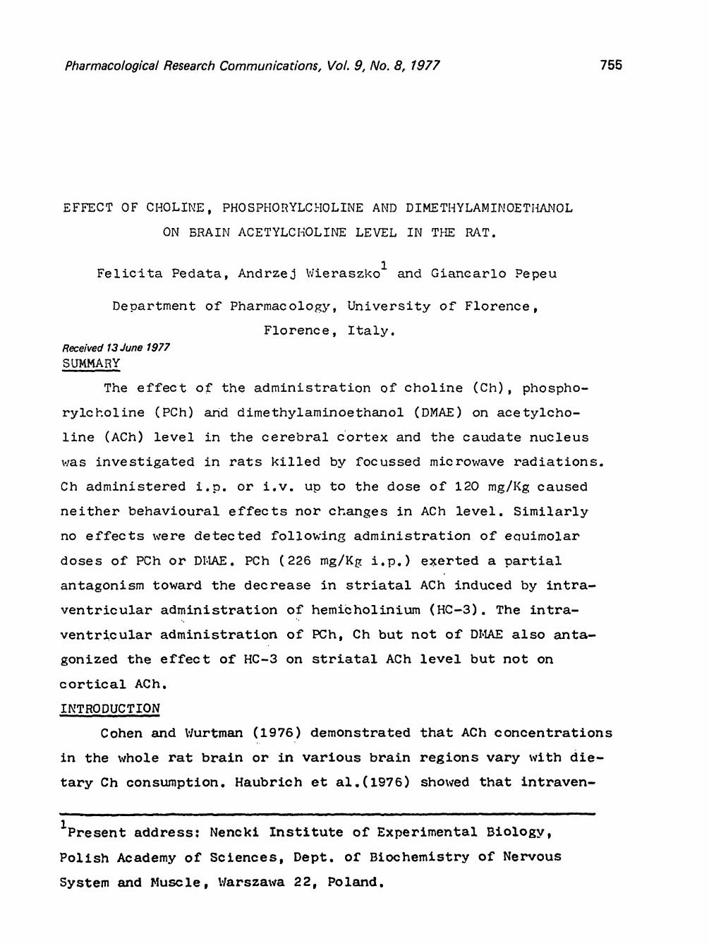
Pharmacological Research Communications, Vol. 9, No. 8, 1977 755
EFFECT OF CHOLINE, PHOSPHORYLCHOLINE AND DIMETHYLAMINOET]L4NOL
ON BRAIN ACETYLCHOLINE LEVEL IN THE RAT.
1 Felici-ta Pedata, Andrzej Wieraszko and Giancarlo Pepeu
Department of PharmacoloKy, University of Florence,
Florence, Italy. Received 13 June 1977 SUMMARY
The effect of the administration of choline (Ch), phospho-
rylcholine (PCh) and dimethylaminoethanol (DMAE) on acetylcho-
line (ACh) level in the cerebral Cortex and the caudate nucleus
was investigated in rats killed by focussed microwave radiations.
Ch administered i.p. or i.v. up to the dose of 120 mg/]{g caused
neither behavioural effects nor changes in ACh level. Similarly
no effects were detected following administration of ecuimolar
doses of PCh or DI:LkE. PCh (228 mg/K~_ i.p.) exerted a partial
antagonism toward the decrease in striatal ACh induced by intra-
ventricular administration of hemicholinium (HC-3). The intra-
administration of PCh, Ch but not of DI, LkE also anta-
effect of HC-3 on striatal ACh level but not on
ventric ular
gonized the
cortical ACh.
INTRODUCTION
Cohen and Wurtman (1976) demonstrated that ACh concentrations
in the whole rat brain or in various brain regions vary with die-
tary Ch consumption. Haubrich et ai.(1976) showed that intraven-
- - - j I _ _ I i m - - | i I I , - - . _ - - . . . . . . . . .
1present address: Nencki Institute of Experimental Biology,
Polish Academy of Sciences, Dept. of Biochemistry of Nervous
System a n d Muscle, Warszawa 22, Poland.
756 Pharmacological Research Communications, Vol.. 9, No. 8, 1977
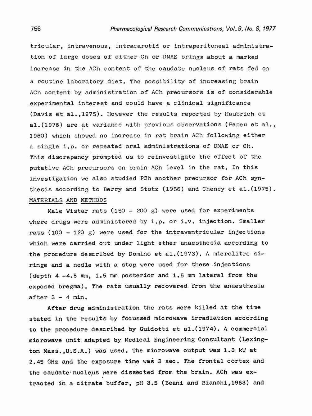
tricular, intravenous, intracarotid or intraperitoneal administra-
tion of large doses of eitller Ch or DMAE brings about a marked
increase in the ACh content of the caudate nucleus of rats fed on
a routine laboratory diet. The possibility of increasing brain
ACh content by administration of ACh precursors is of considerable
experimental interest and could have a clinical significance
(Davis et al.,1975). However the results reported by Haubrich et
a1.(1976) are at variance with previous observations (Pepeu et al.,
1960) which showed no increase in rat brain ACh following either
a single i.p. or repeated oral administrations of DMAE or Ch.
This discrepancy prompted us to reinvestigate the effect of the
putative ACh precursors on brain ACh level in the rat. In this
investigation we also studied PCh another precursor for ACh syn-
thesis according to Berry and Stotz (1956) and Cheney et a1.(1975).
MATERIALS AND METHODS
Male Wistar rats (150 - 200 ~) were used for experiments
where drugs were administered by i.p. or i.v. injection. Smaller
rats (I00 - 120 g) were used for the intraventricular injections
which were carried out under light ether anaesthesia according to
the procedure described by Domino et ai.(1973). A microlitre si-
ringe and a nedle with a stop were used for these injections
(depth 4 -4.5 mm, 1.5 mm posterior and 1.5 mm lateral from the
exposed bregma). The rats usually recovered from the anaesthesia
after 3 - 4 rain.
After drug administration the rats were killed at the time
stated in the results by focussed microwave irradiation according
to the procedure described by Guidotti et ai.(1974). A commercial
microwave unit adapted by Medical Engineering Consultant (Lexing-
ton Mass. ,U.S.A.) was used. The microwave output was 1.3 kW at
2.45 GHz and the exposure time was 3 sec. The frontal cortex and
the caudate nucleus were dissected from the brain. ACh was ex- !
tracted in a citrate buffer, pH 3.5 (Beanl and Bianchi,1968) and

Pharmacological Research Communications, Vol 9, No 8, 1977 757
the content of the extracts was determined by bioassay on the frog
rectus abdomini s.
DMAE was supplied by Riker Laboratories Inc. as the p-acetyl-
aminobenzoate under the trade mark name of Deaner; PCh vsas sup-
plied by Geigy as chloride. The other drugs used were from corn- / /"
mercial sources. All~drugs were dissolved, in saline or distilled • t
water and ' i n j e c t e d i n a volume neve r exceedin~,r 0 .5 ml.
RESULTS
When Ch was injected i.v. or i.p. at doses of 30, 60 and 120
mg/Kg and the rats were killed~ 20~40 or 60 min later, no behav-
ioural changes were observed ~or Were ACh levels in the caudate
nucleus and in the Cerebral cortex increased. Similarly no effects
were detected following administration of equimolar doses of PCh
and D~LAE.
Table 1
Effect of choline (Ch), phosphorylcholine (PCh) and dimethyl-
aminoethanol (DI, LAE) on brain ACh level.
m ~ n n n n n I m I IIII I i I
Treatment Dose N o ACh level
mg/Kg i.p. of rats nmol/g + S.E.
cortex caudate n. | i ml m
Saline - 34 20.6 + 1.0 66.2 + 3.2
Ch 120 4 18.7 + 0.9 65.0 + 4.0
PCh 226 4 1 6 . 2 + 0 . 9 6 6 . 9 + 2 . 3
DMAE 2 3 3 4 2 1 . 7 + 0 . 7 6 7 . 8 4, 2 . 0
Scopolamine 0.5 6 11.3 + 1.6 + 46.3 + 4.3 +
Ch + 120 4 11.5 + 2.4 + 50.I + 1.4 +
Scopolamine 0.5 -- --
PCh + 226 2 9 . 8 5 0 . 4
S~opo famine 0.5
- - _ n mUll I n ¿ u m I - - _ . . . .
The rats were killed 40 rain after the administration of Ch and its
precursors and 30 rain after scopolamine. ÷
This value differs from saline with P<O.OI
758 Pharmacolog/cal Research Communications, Vol. 9, No 8, 1977
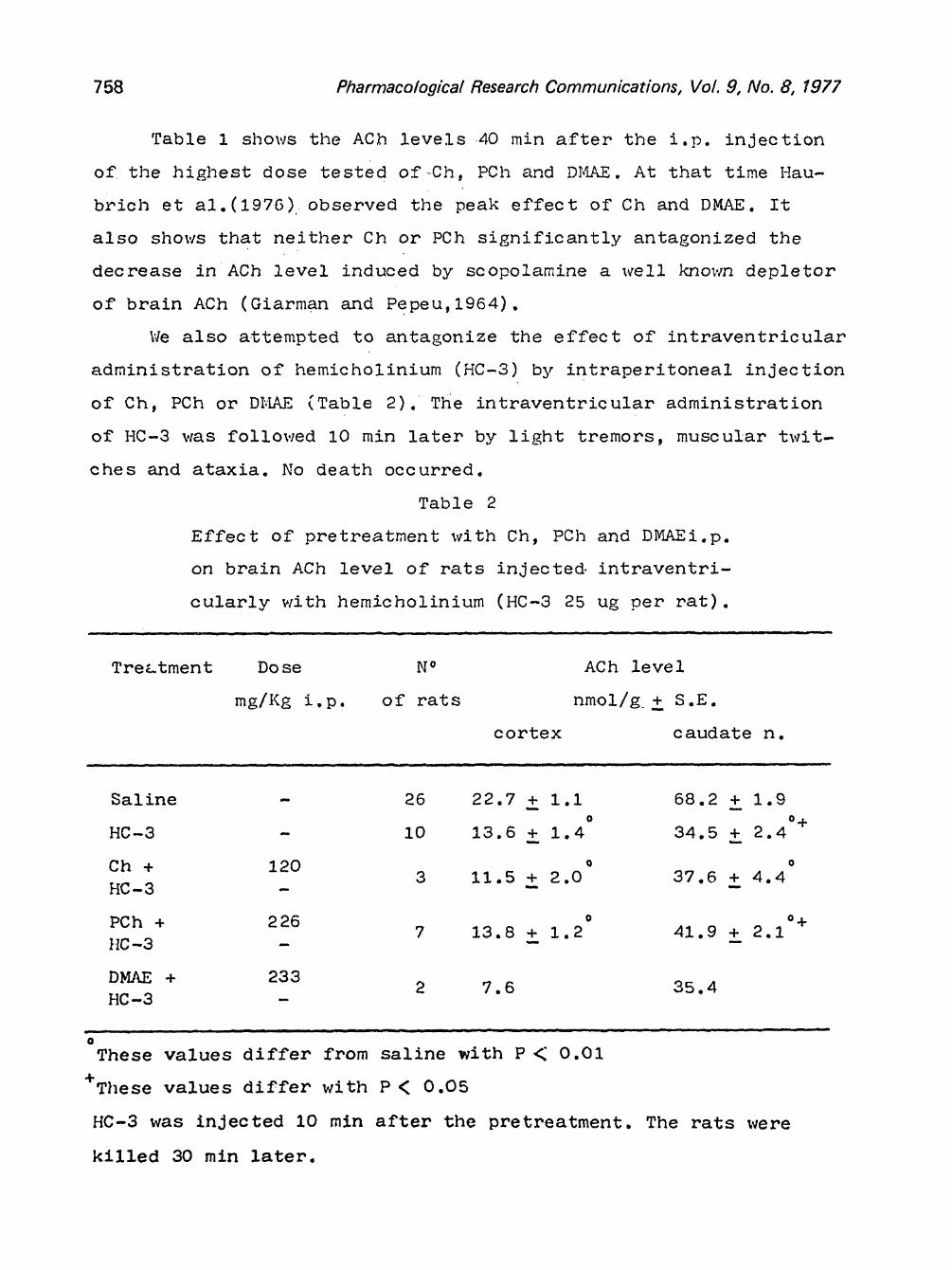
Table 1 s h o w s the ACh levels 4 0 rain after the i.p. injection
of the higluest dose tested of Ch, PCh and D~,IAE. At that time Hau-
brich et a1.(1976)observed the peak effect of Ch and DMAE. It
also shows that neither Ch or PCh significantly antagonized the
decrease in ACh level induced by scopolamine a ~¢ell kno~vn depletor
of brain ACh (Giarman and Pepeu, 1964).
We also attempted to antagonize the effect of intraventricular
administration of hemicholinium (HC-3) by intraperitoneal injection
of Ch, PCh or DI:DiE (Table 2). The intraventricular administration
of HC-3 was followed I0 rain later by light tremors, muscular twit-
ches and ataxia. No death occurred.
Table 2
Effect of pretreatment with Ch, PCh and DMAEiop.
on brain ACh level of rats injected intraventri-
cularly with hemicholinium (HC-3 25 ug per rat).
Treatment Dose N o ACh level
rag/KS i.p. of rats nmol/g + S.E.
cortex c audate n.
Saline - 26 22.7 + i.i 68.2 + 1.9 o o+
HC-3 - i0 13.6 + 1.4 34.5 + 2.4
Ch + 120 o o 3 1 1 . 5 + 2 . 0 3 7 . 6 + 4 . 4
H C - 3 - - - - -
PCh + 2 2 6 o o+ 7 1 3 . 8 + 1 . 2 4 1 . 9 + 2 . 1
HC - 3 - - - - -
DMAE + 2 3 3 2 7 . 6 3 5 . 4
HC-3
These values differ from saline with P < 0.01
+These values differ with P < 0.05
HC-3 was injected I0 mln after the pretreatment. The rats were
killed 30 rain later.
Pharmacological Research Communications, Vol. 9, No. 8, 1977 759
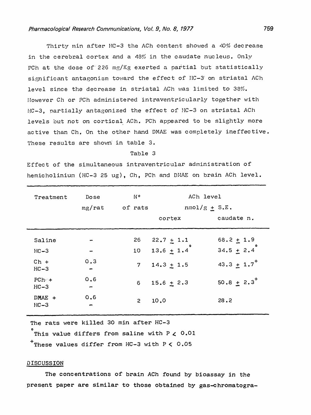
Thirty rain after HC-3 the ACh content showed a ~D% decrease
in the cerebr, al cortex and a 48% in the oaudate nucleus. 0nly
PCh at the dose of 226 m~/](g exerted a martial but statistically
significant antagonism to~,/ard the effect of IIC-3" on striatal ACh
level since the decrease in striatal ACh was limited to 38%.
However Ch or PCh administered intraventricularly together with
HC-3, martially anta.gonized the effect of HC-3 on striatal ACh
levels but not on cortical ACh. PCh appeared to be slightly more
active than Ch. On the other hand DMAE was completely ineffective.
These results are shovrn in table 3.
Table 3
Effect of the simultaneous intraventricular administration of
hemicholinium (HC-3 25 ug), Ch, PCh and DHAE on brain ACh level.
Treatment Do se N o ACh level
mg/rat of rats nmol/~ + S.E.
cortex caudate n.
Saline - 26 22.7 + I.I 68.2 + 1.9 O
H C - 3 - 1 0 1 3 . 6 + 1 . 4 3 4 . 5 + 2 . 4
C h O . 3 + 7 1 4 . 3 + 1 . 5 4 3 . 3 + 1 . 7 +
H C - 3 - - " - -
P C h , + 0 . 6 6 1 5 . 6 + 2 . 3 5 0 . 8 + 2 . 3 +
H C - 3 - - "
D r,~LE + 0 . 6 2 1 0 . 0 2 8 . 2
H C - 3
. . . . . ~ " . . . . . . n i lm n
The rats were killed 30 min after HC-3 @
This value differs from saline with P K 0.01
+These values differ from HC-3 with P < 0.05
DISCUSSION - - ,|
The concentrations of brain ACh found by bloassay in the
present paper are similar to those obtained by gas-chromatogra-
760 Pharmacological Research Communications, Vol. 9, No. 8, 1977
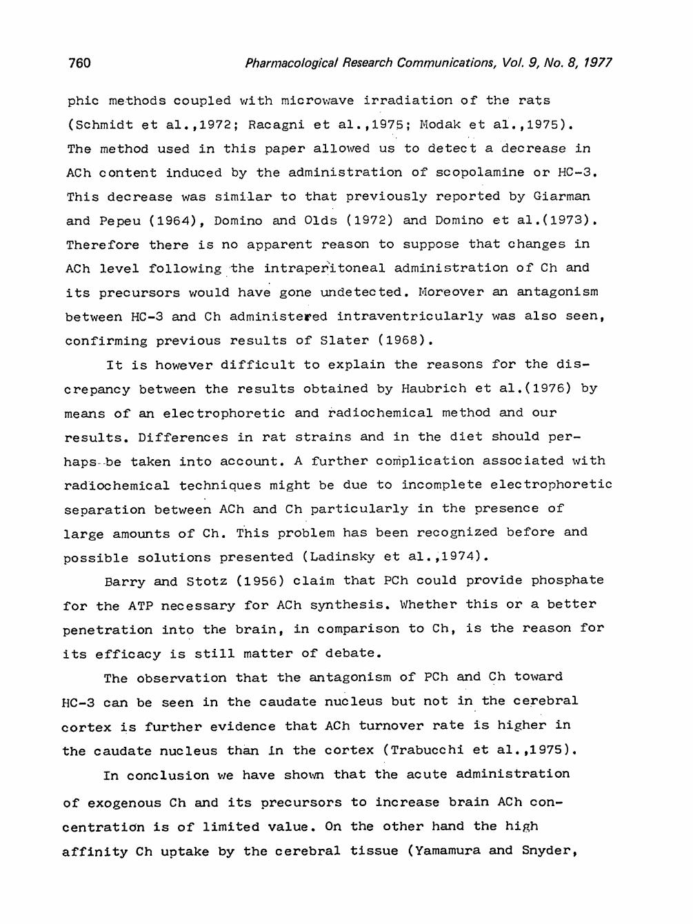
phic methods coupled with microwave irradiation of the rats
(Schmidt et al.,1972; Racagni et al.,1975; Modak et al.,1975).
The method used in this paper allowed us to detect a decrease in
ACh content induced by the administration of scopolamine or HC-3.
This decrease was similar to that previously reported by Giarman
and Pepeu (1964), Domino and Olds (1972) and Domino et ai.(1973).
Therefore there is no apparent reason to suppose that changes in
ACh level following the intraper?'itoneal administration of Ch and
its precursors would have gone undetected. Moreover an antagonism
between HC-3 and Ch administered intraventricularly was also seen,
confirming previous results of Slater (1968).
It is however difficult to explain the reasons for the dis-
crepancy between the results obtained by Haubrich et ai.(1976) by
means of an electrophoretic and radiochemical method and our
results. Differences in rat strains and in the diet should per-
haps be taken into account. A further complication associated with
radiochemical techniques might be due to incomplete electrophoretic
separation between ACh and Ch particularly in the presence of
large amounts of Ch. This problem has been recognized before and
possible solutions presented (Ladinsky et ai.,1974).
Barry and Stotz (1956) claim that PCh could provide phosphate
for the ATP necessary for ACh synthesis. Whether this or a better
penetration into the brain, in comparison to Oh, is the reason for
its efficacy is still matter of debate.
The observation that the antagonism of PCh and Ch toward
HC-3 can be seen in the caudate nucleus but not in the cerebral
cortex is further evidence that ACh turnover rate is higher in
the caudate nucleus than in the cortex (Trabucchi et ai.,1975).
In conclusion we have sho~au that the acute administration
of exogenous Ch and its precursors to increase brain ACh con-
centration is of limited value. On the other hand the high
affinity Ch uptake by the cerebral tissue (Yamamura and Snyder,
Pharmacological Research Communications, VeL 9, No. 8, f977 761
1973) is easily saturated and therefore should represent an
obstacle to large increase in ACh level•
AKN0 WLE D GE ME NT S
The present work was supported partly by grant n o 7 5 . 0 0 6 6 7
from the Consiglio Nazionale delle Ricerche and by a grant from
the Istituto Nazionale Ricerca e Cura Anziani (INRCA). F.Pedata
was the recepient of an INRCA research fellowship.
RE FE RE NCE S i 11 .i
J.Pharmac Cohen,E.L Davis,K.L 152, 1975 Domino,E.
Beani, L. B erry,J.F. £~ Stotz,E. J.Biol.Chem. 218: 871-874, 1956. Cheney,D.L•,Costa,E.,Hanin,I.,Trabucchi,M. & Wang,C.T.
•exp. Ther. 192: 288-296, 1975. L J"
• & Wurtman,R.J. Science 191: 561-562, 1976. .,Berger,P.A. & Hollister,L.E. New Engl. J.Med.
& Bianchi,C. J.Pharm.Pharmac. 15: 281-282, 1963.
17:
1-16, 1972. Neuro-
1964. &
Brain Res.
1973.
F. & Olds,M.E. Psychopharmacol.(Berl.) 23: Domino,E.F.,Mohrman,M.E.,Wilson,A.E. & Haarstad,V.B. pharmac. 12: 549-561, 1973. Giarman,N.J. & Pepeu,G. Brit. J.Pharmac. 23: 123-130, Guidotti ,A. ,Chenev ,D. L. Trabucchi ,M., Doteuchi ,M., Wang ,C Hawkings,A.R. Neuropharmac. 13: 1115-1122, 1974. Haubrich,D.R.,Wang,P.F.L.,Clody,D.E. & Wedeking,P.W. Life Sci. 17: 975-980, 1976. Ladinsky,H & Consolo,S. In: (Hanin,I. ed.) pp. 1-17 New Modak,A.T. ,Stavinoha,W.B. & Ther. 217: 298-301, 1975. Pe peu, G., Freedman, D.K. & Giarman ,N. J. J. Pharmac. exp. Ther. 291-295, 1960. Racagni,G.,Trabucchi,M. & Cheney,D.L. Pharmac. 29D: 99-105, 1975. Schmidt,D.E.,Speth,R.C.,l'.relsch,F.& Schmidt,M•J. 38; 377-389, 1972.
f l Slater,P. Int.J.Neuropharmac• 7: 421-427, 1968. Trabucchi,M•,Cheney,D.L.,Racagni,G. & Costa,E. Brain Res• 85: 130-134, 1975 Yamamura,H.I. & Snyder,S.H• J• Neurochem. 21: 1355-1374,
129: h
Naunyn-Sc hmie d. Arch.
H a n d b o o k of Chemical Assay Methods. York, Raven Press, 1 9 7 4 . We intraub, S. T. Arch. int. Pharmac odyn.