Embed Size (px)
Citation preview

International Journal of Agricultural Policy and Research Vol.5 (12), pp. 192-200, December 2017 Available online at https://www.journalissues.org/IJAPR/ https://doi.org/10.15739/IJAPR.17.023 Copyright © 2017 Author(s) retain the copyright of this article ISSN 2350-1561
Original Research Article
Effect of drying plantain (Musa spp.) suckers prior to in vitro culture on reduction of lethal browning
Received 3 November, 2017 Revised 5 December, 2017 Accepted 18 December, 2017 Published 29 December, 2017
Eustache T. Ade-Eyitatyo F. Agbadje*1,
Arnaud Agbidinoukoun1, Martine Zandjanakou-Tachin2,
Gilles Todjro Habib Cacaï1, Sètondji Serge Houedjissin1,
and Corneille Ahanhanzo1
1Central Laboratory of plant
Biotechnology and plants breeding, University of Abomey-Calavi(UAC),
Benin. 2School of Horticulture and Green
Space Management (EHAEV), National University of Agriculture
(UNA), Benin.
*Corresponding Author Email: [email protected]
Tel.: +229 97014050
This work aimed to evaluate the effect of drying plantain suckers on explants size, survival rate and browning intensity of plantain apices during in vitro culture. Three sets of suckers were used in the experiment. The first set used as a control and was not dried. The second set was dried under shade for 7 days and the third set was dried under shade for 14 days. Then explants were isolated. Comparative study was undertaken based on the immersion of explants in cysteine solution before in vitro culture. According to their size, two kinds of explants were isolated from each set (0.5 cm × 0.5 cm and 1 cm × 1 cm). The explants of 0.5 cm × 0.5 cm were introduced directly in culture after isolation. The explants of 1 cm × 1 cm were divided into two sub-sets. The explants isolated from the first sub-set were briefly immersed in 50 mg/l cysteine solution before in vitro culture. The second sub-set was introduced directly in the culture. The results showed that when the suckers are dried under shade for both of 7 and 14 days it reduced tissue browning and increased number of apices survived than the brief immersion of explants in 50 mg/l solution of cysteine. The response of explants varied with their sizes (0.5cm × 0.5 cm vs 1cm × 1 cm). Thus, explants with the size of 1 cm × 1 cm isolated from suckers dried for 7 days was the best treatment as 100% survival rate of material was observed during the four weeks of culture. As a result first subculture can be carried out after 4 weeks of culture instead of one to two weeks as recommended under other conditions. This method consumes less time and material, easy to perform and reduces the risk of loss of material during subcultures. Key words: browning intensity, suckers drying, banana and plantain tissue culture, apices survival
INTRODUCTION Bananas and plantains are among the most important food crops in Central and Western Africa. They play an essential role in contributing to food security, generation of employment, diversification of income sources in rural and urban areas, gross domestic product (GDP) and poverty reduction (Nkendah and Akyeampong, 2003). Most of banana cultivars produced seedless fruits. The traditional planting material consists of suckers produced by the mother plant in the fields. This natural propagation pathway is slow, laborious and produces small quantity and especially low phytosanitary planting material (Gandonou
et al., 2012). In vitro propagation makes it possible to remove these constraints through mass production of plantlets (Rahman et al., 2013). Compared to conventional suckers, micro plantlets are more rapidly growing, larger, uniform and have a shorter production cycle and give a higher yield (Sheela and Nair, 2006; Bhanusree et al., 2015).
Main problems in banana and plantain tissue culture are browning/darkening of culture medium and the cut surfaces of the explants, from initiation phase and during subcultures (Vuylsteke, 1998), browning of young leaves,

Int. J. Agric. Pol. Res. 193 shoot necrosis and plantlet death during proliferation and rooting (Martin et al., 2007). These phenomena are attributed mainly to the oxidation of phenolic compounds (Ngomuo et al., 2014a). Banana tissues are known to contain large amount of latex and phenolic compounds (Waman et al., 2015). These compounds are released into the culture medium by the explant and accumulate therein. Their oxidation by polyphenol oxidase (PPO) produces quinones and highly reactive forms of oxygen (Ahmad et al., 2013). These oxidation products of phenolic compounds gradually penetrate the tissues and repress the enzymatic activities. As a result, they are very toxic to the plant (Ssekiwoko et al., 2014). Moreover, they form a black layer around the explant, that prevents nutrients uptake (Chikezie, 2012). Thus, oxidation of phenolic compounds and other exudates negatively influences explant survival, plantlets proliferation and growth during tissue culture (Ndakidemi et al., 2014; Ssekiwoko et al., 2014).
Factors influencing tissue browning during the in vitro culture phase of banana and plantain include the size of the explant, the season when the sucker is harvested, and the means used to prevent or reduce the oxidation of phenolic compounds. As stated by Vuylsteke, (1989), large explants are more susceptible to browning than smaller ones. Likewise, explants from suckers harvested in wet seasons are more vulnerable than those from suckers harvested in the dry season (Waman et al., 2015). The control of the oxidation of phenolic compounds during the in vitro culture is commonly carried out by either adding antioxidants (ascorbic acid, citric acid, cysteine) to the culture medium (Ko et al., 2009; Chikezie, 2012; Ngomuo et al., 2014b) or using pretreatment solution on explants prior to in vitroculture (Titov et al., 2006; Ngomuo et al., 2014a). Buah et al., (2010) reported that drying suckers under shade for 14 days, reduces phenolic content in the sucker. But to our knowledge, no study has evaluated the effect of such a drying on the browning of tissues during the in vitro culture phase. In contrast, Kone et al. (2010) showed that the suckers harvested for several days before explant isolation increased the infection rate during tissue culture and therefore, not favourable for the survival of the explant. Based on the above findings, the present study was initiated with the following specific objectives:
To determine the effect of drying duration and size of explant on survival and browning intensity during the in vitro culture phase
To determine the effect of the system drying of suckers-immersing explants in a 50 mg/l cysteine solution prior to in vitro culture on lethal browning. MATERIALS AND METHODS Establishment of explants The plant material consisted of young Musa suckers belonging to the cultivar aloga. The suckers harvested were divided into three sets. The suckers of the first set did not
undergo drying while suckers of set 2 and set 3 were dried under shade for 7 days and 14 days respectively before explant isolation. Prior to extraction of explants, the tissue blocks were double disinfected by immersing in 15% bleach solution (3.6% active chlorine) for 15 minutes. After each disinfection, size reduction was performed to produce two types of explants; size of 0.5 cm x 0.5 cm and of 1 cm × 1 cm. The large explants 1 cm × 1 cm were divided into two sub-sets. The explants of first sub-set were directly introduced to in vitro culture after their isolation. The explants in the second sub-set were pretreated briefly by immersing in a 50 mg/l of cysteine solution. The initiating medium was MS medium (Murashige, 1962), to which sucrose (30 g/l), BAP (2 mg/l), bleach (1 ml/l) and agar (7 g/l) were added. The pH was adjusted to 5.7. Treatments and data collection For the determination of the effects of suckers drying and explant size on browning and survival, 6 combinations of drying x explant size were used; no drying vs 0.5 cm × 0.5 cm size, no drying vs 1 cm x 1 cm size, drying for 7 days vs 0.5 cm × 0.5 cm size, drying for 7 days vs 1 cm ×1 cm size, drying for 14 days vs 0.5 cm x 0.5 cm size and drying for 14 days vs 1 cm × 1 cm size).
For the assessment of the effect of drying suckers and immersing in cysteine on browning, explants of 1 cm x 1 cm were used in the following combinations: no dried and immersion in cysteine, drying for 7 days and not immersing, drying for 14 days and not immersing, no drying and not immersing (control sample).
The last treatment was combinations of drying and immersing of suckers; explant size 1 × 1 cm size dried for 7 days immersed in cysteine vs direct in vitro culture, explant size 1 × 1 cm size dried for 14 days immersed in cysteine vs direct in vitro culture, explant size 1 × 1 cm size not dried immersed in cysteine vs direct in vitro culture.
Parameters assessed were; necrosis (explants which tissues are died), infection (explants with microbial contamination), survival (swelling explants), and browning intensity. This last parameter was assessed using a scale of 4 levels: no (any change in the colour of the explant), low (explant become brown), medium (explant become dark but not affect the culture medium) and high (explant and medium become dark). Research design and data analysis The experimental design is randomized plots. Eighteen repetitions were performed by treatment. The data were analysed by performing Logistic regression using the XLSTAT version 2014.5.03 software to determine the effect of drying and explant size on browning. The effect of drying and explant size on survival parameters, effect of immersing in cysteine vs direct in vitro culture were analysed using Analysis of Variance (ANOVA). The means were separated using the Student Newman-Keuls (SNK) test.

Agbadje et al. 194
Figure 1: Evolution of browning intensity with drying duration of suckers
RESULTS Effects of sucker drying duration and culture duration on browning intensity The browning intensity was influenced (p <0.0001) by both drying duration and culture duration. Compared to the
undried material, the dried material had a lower degree of browning throughout the duration of the culture (Figure 1). After the first week of culture, the majority of explants from undried set had a medium degree of browning (66.7%), the remainder having low (27.8%) and high (5.6%) degree of browning. On the other hand, explants resulting from dried suckers have low and medium degree of browning, with a

Int. J. Agric. Pol. Res. 195
Table1. Browning intensity (%) with explants size
Week 1 Week 2 Week 3 Week 4
0.5 × 0.5 1 × 1 0.5 × 0.5 1 × 1 0.5 × 0.5 1 × 1 0.5 × 0.5 1 × 1
No drying Low 33.33±11.4 22.22±10.1 0.00±00 0.00±00 0.00±00 11.11±7.6 0.00±00 11.11±7.6 Medium 55.56±12.1 77.78±10.1 11.11±7.6 16.67±9.0 0.00±00 22.22±10.1 0.00±00 38.89±11.8 High 11.11±7.6 0.00±00 88.89±7,6 83.33±9.0 100.00±00 66.67±11.4 100.00±00 50.00±12.1
Drying 7 days
Low 66.67±11.4 94.44±5.6 50.00±12.1 22.22±10.1 44.44±12.1 33.33±11.4 33.33±11.4 50.00±12.1
Medium 33.33±11.4 5.56±5.6 33.33±11.4 61.11±11.8 27.78±10.9 61.11±11.8 38.89±11.8 50.00±12.1 High 0.00±00 0.00±00 16.67±9.0 16.67±9.0 27.78±10.9 5.56±5.56 27.78±10.9 0.00±00
Drying 14 days
Low 72.22±10,9 77.78±10,1 77.78±10,1 72.22±10.9 44.44±12.1 61.11±11.8 33.33±11.4 44.44±12.1
Medium 27.78±10,9 22.22±10,1 16.67±9.0 27.78±10.9 50.00±12.1 38.89±11.8 50.00±12.1 55.56±12.1 High 0.00±00 0.00±00 5.56±5.56 0.00±00 5.56±5,56 0.00±00 16.67±9,0 0.00±00
Figure 2 : Classification of averages of browning intensity with explants
predominance of low degree (80.6% and 64% respectively) for 7 days and 14 days dried suckers. There were no explants having high intensity of browning when dried either for 7 days or 14 days in the first week of culture. With the culture duration, the intensity of browning increased for all sets. Thus, percentage of medium degree of browning in undried set decreased in favour of high degree from 5.6% at the end of the first week to 75% at the end of the 4th week. In the dried sets, the low degree of browning decreased in favour of the medium and high degrees, which reach respectively 44.4% and 13.9% in the set dried for 7 days, 52.8% and 8.3% in the set dried during 14 days. At the end of 4 weeks of culture, the majority of material resulting from undried set had a high degree of browning whereas the majority of material resulting from the dried suckers had a low and medium degrees of browning. The difference in degree of browning between the set of undried suckers and those of dried suckers was significant. In contrast, the differences observed between the 2 sets of dried suckers were not significant. Effects of explant size on browning intensity Browning intensity increased with culture duration for all sets but the increase was more prominent in the small size explants (0.5cm × 0.5 cm) compared to those of larger size
(1 cm ×1 cm). In fact, the Table 1 showed that at the third week of culture, all the small explants from the undried set had a higher degree of browning intensity, whereas in the large explants it was only 67% of browning. Similarly, at the end of the 4 weeks of culture, a high degree of browning was practically not observed with large explants resulting from dried material whereas this degree of browning was observed among the small explants (27.78% for 7 days drying set and 16.67% for 14 days drying material). The percentage of low degree of browning was greater at the end of culture in the large explants of the two sets of dried material; 50% for large explants against 33.33% for small explants dried for 7 days and 44.44% against 33.33% for the set dried for 14 days. The influence of the explant size on browning degree was highly significant but the difference between the two size of explant (0.5 cm × 0.5 cm and 1 cm × 1cm) was not significant. Comparative study of effects of suckers drying and explant immersion in cysteine on browning intensity Figure 2 present the means of browning degree for different types of pretreatment of explants (immersing in cysteine, dried for 7 days, dried for 14 days) and the control set without pretreatment(neither dried nor immersed). The untreated control group and the immersing in cysteine set

Agbadje et al. 196
Figure 2 : Browning intensity
a : low browning – b : medium browning – c : high browning
exhibited the highest means (2,53 and 2,19 respectively) while the two dried sets (7 days and 14 days of drying) had the lowest means (1,618 and 1,431 respectively). No significant difference was noted between the immersing in cysteine set and the control. In contrast, significant difference was observed between 7 days dried set (intermediate browning degree) and 14 days dried set (lower browning degree). Effects of drying duration of suckers and explant size on infection, necrosis and apices survival rates The rate of infection was influenced by interaction of the drying duration of suckers and the size of explant. The infection rate was zero for small explants regardless of drying duration of material. For explants of size 1 cm × 1 cm, the infection rate was zero for material from undried suckers and those dried for 7 days. On the other hand, an infection rate of 22.22% was recorded in samples from dried suckers for 14 days (Figure 4A). Suckers drying for 14 days for 1 cm × 1 cm explants forms a group that differed significantly from all other treatments. The 1 cm × 1 cm explants provided by suckers drying for 14 days was therefore the most vulnerable treatment for infection.
The necrosis rate was influenced by explant size (p <0.000) and suckers drying duration (p = 0.003). In all sets, small explants (0.5 cm × 0.5 cm) were more necrotic than large (1 cm × 1 cm) explants, and explants from dried material were less necrotic than those from the material undried. Thus, the necrosis rate was 55.56% in undried set for small explants compared to 33.33% for large explants of the same set. In 7 days and 14 days dried sets, the necrosis rate was 27.78% and 38.89% respectively for small explants versus a zero rate for large explants (Figure 4B). The 1 cm × 1 cm explants from the two dried sets formed a group that was significantly different from all other combinations. They were less vulnerable to necrosis than other treatments.
The survival rate was influenced by drying duration (p = 0.013) and the size of explant (p = 0.004). For each size category of explant, the survival rate was better in dried sets compared to the undried set. For small explants, it was 44.44% compared to 72.22% and 61.11% respectively in undried control set, 7 days and 14 days dried sets. For large explants, the values obtained for undried control set, 7 days and 14 days dried sets were respectively: 66.67%, 100% and 77.78%. Explants of size 1 cm × 1 cm from 7 days dried without infection or necrosis formed the group with the

Int. J. Agric. Pol. Res. 197
Figure 3 : Infection, necrosis and survival rates with drying duration and size of explant
best survival rate. Conversely, the 0.5 cm × 0.5 cm size explants from the undried set formed a second group
characterized by the lowest survival rates. All other treatments formed an intermediate group (Figure 4C).
Agbadje et al. 198
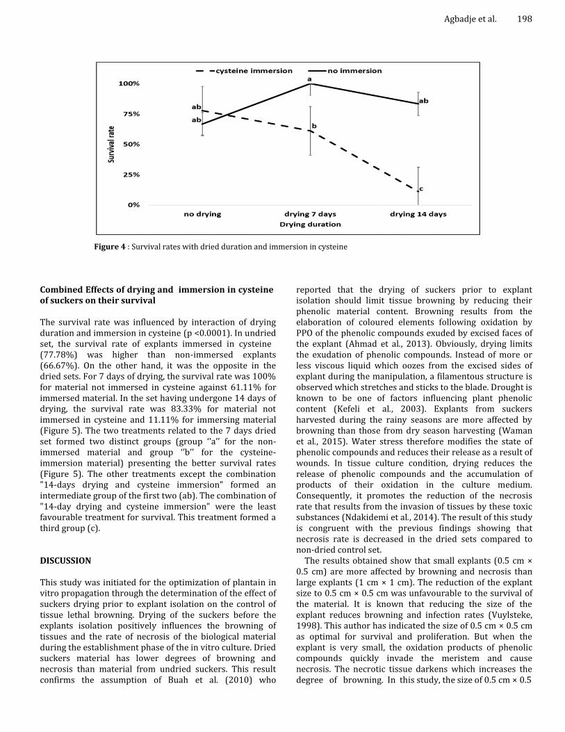
Figure 4 : Survival rates with dried duration and immersion in cysteine
Combined Effects of drying and immersion in cysteine of suckers on their survival The survival rate was influenced by interaction of drying duration and immersion in cysteine (p <0.0001). In undried set, the survival rate of explants immersed in cysteine (77.78%) was higher than non-immersed explants (66.67%). On the other hand, it was the opposite in the dried sets. For 7 days of drying, the survival rate was 100% for material not immersed in cysteine against 61.11% for immersed material. In the set having undergone 14 days of drying, the survival rate was 83.33% for material not immersed in cysteine and 11.11% for immersing material (Figure 5). The two treatments related to the 7 days dried set formed two distinct groups (group ‘’a’’ for the non-immersed material and group ‘’b’’ for the cysteine-immersion material) presenting the better survival rates (Figure 5). The other treatments except the combination "14-days drying and cysteine immersion" formed an intermediate group of the first two (ab). The combination of "14-day drying and cysteine immersion" were the least favourable treatment for survival. This treatment formed a third group (c). DISCUSSION This study was initiated for the optimization of plantain in vitro propagation through the determination of the effect of suckers drying prior to explant isolation on the control of tissue lethal browning. Drying of the suckers before the explants isolation positively influences the browning of tissues and the rate of necrosis of the biological material during the establishment phase of the in vitro culture. Dried suckers material has lower degrees of browning and necrosis than material from undried suckers. This result confirms the assumption of Buah et al. (2010) who
reported that the drying of suckers prior to explant isolation should limit tissue browning by reducing their phenolic material content. Browning results from the elaboration of coloured elements following oxidation by PPO of the phenolic compounds exuded by excised faces of the explant (Ahmad et al., 2013). Obviously, drying limits the exudation of phenolic compounds. Instead of more or less viscous liquid which oozes from the excised sides of explant during the manipulation, a filamentous structure is observed which stretches and sticks to the blade. Drought is known to be one of factors influencing plant phenolic content (Kefeli et al., 2003). Explants from suckers harvested during the rainy seasons are more affected by browning than those from dry season harvesting (Waman et al., 2015). Water stress therefore modifies the state of phenolic compounds and reduces their release as a result of wounds. In tissue culture condition, drying reduces the release of phenolic compounds and the accumulation of products of their oxidation in the culture medium. Consequently, it promotes the reduction of the necrosis rate that results from the invasion of tissues by these toxic substances (Ndakidemi et al., 2014). The result of this study is congruent with the previous findings showing that necrosis rate is decreased in the dried sets compared to non-dried control set.
The results obtained show that small explants (0.5 cm × 0.5 cm) are more affected by browning and necrosis than large explants (1 cm × 1 cm). The reduction of the explant size to 0.5 cm × 0.5 cm was unfavourable to the survival of the material. It is known that reducing the size of the explant reduces browning and infection rates (Vuylsteke, 1998). This author has indicated the size of 0.5 cm × 0.5 cm as optimal for survival and proliferation. But when the explant is very small, the oxidation products of phenolic compounds quickly invade the meristem and cause necrosis. The necrotic tissue darkens which increases the degree of browning. In this study, the size of 0.5 cm × 0.5

Int. J. Agric. Pol. Res. 199 cm seems very small and more vulnerable to necrosis than size 1 cm × 1 cm. On the other hand, the small explants are not affected by the infection which confirms the rule in this matter.
Drying of suckers requires their storage after harvest for several days before their introduction in vitro. This storage is a factor that increases the rate of infection after initiation (Kone et al., 2010). To limit contamination during this experiment, bleach (1ml/l) was added to the medium as recommended in the technical reference book (Cides, 1999). Despite this precaution, cases of infection appeared in the 1 cm × 1 cm size samples dried for 14 days at a rate of 22.22% compared to a zero rate for the other two sets. The difference in the infection rate between sets is significant and confirms that storage favours contamination. However, in this study, there was no contamination after 7 days regardless of the size of explant as in the non-storage control group, whereas an increase in contamination only after 2 to 3 days of storage was noticed in other work (Kone et al., 2010). The absence of infection after seven days of storage in this study could be attributed to the action of bleach added to medium.
The effectiveness of the drying of the suckers in reducing the browning and improving the survival rate was better than those of the immersion of the explants in a 50 mg / l solution of cysteine. Immersion in a solution of cysteine or other antioxidants (ascorbic acid, citric acid or citric acid and potassium citrate mixture) is a classic way to control lethal tissue browning during tissue culture (Strosse et al., 2004; Onuoha et al., 2011; Safwat et al., 2016). Antioxidants are electron donors (reducing agents) that inhibit the oxidation of labile substrates (George, 1996). During the immersion period, the antioxidants are able to scavenge the oxygen radicals produced by the damaged tissues and thus protect the cells against oxidation. But after initiation, the exudation of phenolic compounds continues, causing browning to resume (Titov et al., 2006). Due to the continued release of phenolic compounds for several days, frequents subcultures are recommended during the establishment phase until the cut end of the explant become sealed up and the leaching of phenolic stop (Ahmad et al., 2013). Thus, the immersion of the explants in a solution of antioxidants does not reduce the exudation of phenolic compounds but acts on the compounds released during the immersion. On the other hand, drying drastically limits the release of phenolic compounds and thus offers long-term protection against the lethal browning of tissues. The results show that explants with both drying and cysteine immersion had lower survival rates than other treatments. The combination of both treatments is fatal for the biological material. Drying makes the plant material hypotonic with respect to the culture medium and thus creates a concentration gradient that promotes passive absorption of nutrients (Buah et al., 2010). Thatosmotic state of dried material should be one of the factor that promote the increase of the death of meristem cells in presence of cysteine. Further studies are needed to verify this hypothesis.
Conclusion Drying under shade of sucker prior to explant isolation positively influences the tissue browning intensity and in vitro survival of material. Its effectiveness is greater than the brief immersion of explants in 50 mg / l solution of cysteine. The result is better for explants of 1 cm × 1 cm compared to those of 0.5 cm × 0.5 cm. Thus, explants with the size of 1 cm × 1 cm isolated from suckers dried for 7 days was the best treatment as 100% survival rate of material was observed during the four weeks of culture. As a result first subculture can be carried out after 4 weeks of culture instead of one to two weeks as recommended under other conditions. This method consumes less time and material, easy to perform and reduces the risk of loss of material during subcultures. In the absence of other means of controlling the lethal browning of tissues during the in vitro establishment of the apices of bananas and plantains, the drying under shade of suckers for 7 days and isolation of 1 cm × 1 cm explants can be recommended. Acknowledgments The authors thank the programme Vitroplant of CBRSI (Centre Béninois de la Recherche Scientifiqueet de l’Innovation) for supporting the laboratory activities cost. We are also very grateful to all the farmers for their full. collaboration. REFERENCES Ahmad I, Hussain T, Ashraf I, Nafees M, Maryam RM, Iqbal
M (2013). Lethal effects of secondary metabolites on plant tissue culture. Am. Eurasian J. Agric. Environ. Sci. 13: 539-547.
Bhanusree M, Kumar KR, Suresh C, Shukla G, Chakravarty S (2015). Comparative studies on tissue culture plantlet versus conventional sucker var. Grand Naine banana. Plant Archives 15(2): 785-788.
Buah J, Danso E, Taah K, Abole E, Bediako E, Asiedu J, Baidoo R (2010). The effects of different concentrations cytokinins on the in vitro multiplication of plantain (Musa sp.). Biotechnology 9(3): 343-347.
Chikezie U (2012). Effect of ascorbic acid on blackening and sprouting of Musa spp shoot tips. ISABB. J. Biotech. Bioinform. 2(2): 11-17.
CIDES (1999). Micropropagation en entreprise - cahier de références techniques. (Ed CIDES).
Gandonou G, Ahanhanzo C, Agbangla C, Agbidinoukoun A, Doussoh A, Cacai G, Dossoukpevi R (2012). Micropropagation in vitro de la variété locale «Aloga» du bananier plantain (Musa x paradisiaca L.) au Bénin. Int. J. Biol. Chem. Sci 6(3): 1102-1111.
George EF (1996). Plant Propagation by Tissue Culture,” Parts 1 and 2. (Ed W. Edington, Exegetics Ltd.). Eversley.
Kefeli VI, Kalevitch MV, Borsari B (2003). Phenolic cycle in

plants and environment. J. Cell Mol. Biol 2(1): 13-18.
Ko W, Su C, Chen C, Chao C (2009). Control of lethal browning of tissue culture plantlets of Cavendish banana cv. Formosana with ascorbic acid. Plant Cell Tiss Organ Cult 96(2): 137-141.
Kone T, Kone M, Kone D, Kouakou TH, Traore S, Kouadio Y (2010). Effet de la photopériode et des vitamines sur la micropropagation du bananier plantain (Musa AAB) à partir de rejets écailles de rang1. J. Appl. Biosci. 26: 1675-1686.
Martin KP, Zhang C-L, Slater A, Madassery J (2007). Control of shoot necrosis and plant death during micro-propagation of banana and plantains (Musa spp.). Plant Cell Tiss Organ Cult 88(1): 51-59.
Murashige T, Skoog F ( 1962). A revised medium for rapid growth and bioassay with tobacco tissue cultures. Physiol. Plant 15: 473–497.
Ndakidemi CF, Mneney E, Ndakidemi PA (2014). Effects of ascorbic acid in controlling lethal browning in in vitro culture of Brahylaena huillensis using nodal segments. Am. J. Plant Sci 5(1): 187.
Ngomuo M, Mneney E, Ndakidemi P (2014a). Control of lethal browning by using ascorbic acid on shoot tip cultures of a local Musa spp.(Banana) cv. Mzuzu in Tanzania. Afr. J. Biotechnol. 13(16):1721-1725.
Ngomuo M, Mneney E, Ndakidemi PA (2014b). The in vitro propagation techniques for producing banana using shoot tip cultures. Am. J. Plant Sci 5(11): 1614.
Nkendah R, Akyeampong E (2003). Données socioéconomiques sur la filière plantain en Afrique Centrale et de l’Ouest. InfoMusa 12(1): 8-12.
Onuoha C, Eze C, Unamba C, UGOCHUKWU C (2011). In vitro prevention of browning in plantain culture. ROM. J. BIOL. – PLANT BIOL 56(2): 123-130.
Agbadje et al. 200 Rahman S, Biswas N, Hassan MM, Ahmed MG, Mamun A,
Islam MR, Moniruzzaman M, Haque ME (2013). Micropropagation of banana (Musa sp.) cv. Agnishwar by in vitro shoot tip culture. Intl. Res. J. Biotech 4(4): 83-88.
Safwat G, Abdul-Rahman F, El Sharbasy S (2016). The effect of some antioxidants on blackening and growth of in vitro culture of banana (Musa spp. cv. Grand naine). Egypt. J. Genet. Cyto. 44(1).
Sheela V, Nair SR (2006). Growth, flowering and yield potential of tissue culture banana (Musa AAB cv. Nendran). J. Trop. Agr. 39(1): 1-4.
Ssekiwoko F, Talengera D, Kiggundu A, Namutebi M, Karamura E, Kunert K (2014). In-vitro proliferation of Musa balbisiana improves with increased vitamin concentration and dark culturing. J. Ap. Biol. Biotech. 2(3): 001-007.
Strosse H, Van den Houwe I, Panis B (2004). Banana cell and tissue culture-review in Banana improvement : cellular, molecular biology and induced mutations. 1-12 (Eds S. M. Jain R. Swennen). Science Publishers, Inc.
Titov S, Bhowmik SK, Mandal A, Alam MS, Uddin SN (2006). Control of phenolic compound secretion and effect of growth regulators for organ formation from Musa spp. cv. Kanthali floral bud explants. Am. J. Biochem. Biotechnol 2(3): 97-104.
Vuylsteke D (1998). Shoot-tip culture for the propagation, conservation and distribution of Musa germplasm. IITA.n
Waman A, Bohra P, Sathyanarayana B, Umesha K, Mukunda G, Ashok T, Gowda B (2015). Optimization of factors affecting in vitro establishment, ex vitro rooting and hardening for commercial scale multiplication of silk banana (Musa AAB). Erwerbs-Obstbau 57(3): 153-164.