Embed Size (px)
Citation preview
Int Adv Otol 2014; 10(3): 251-5 • DOI: 10.5152/iao.2014.276
Original Article
Effect of Nasal Septal Deviation on Pneumatization of the Mastoid Air Cell System: 3D Morphometric Analysis of Computed Tomographic Images in a Pediatric Population
Dong-Hee Lee, Kyung-Suk JinDepartment of Otolaryngology - Head and Neck Surgery, The Catholic University of Korea, Seoul, Republic of Korea (DL, KJ)
OBJECTIVE: To evaluate the association between nasal septal deviation (NSD) and the volume of mastoid air cell pneumatization and compare it with the volume of maxillary sinus in a pediatric population.
MATERIALS and METHODS: This retrospective cross-sectional study was conducted at a university-based, secondary referral hospital. Paranasal sinus CT imaging data of 59 children were reconstructed to the 3-dimensional model, and subsequently, we measured the volume of the maxillary sinus and mastoid air cell. On coronal images, nasal septal angle (NSA) and NSD/NC (nasal septal deviation/nasal cavity) ratio were measured.
RESULTS: Mastoid air cell volume, as well as maxillary sinus volume, of the deviated side was smaller than that of the contralateral side, but these were not statistically significant. There was no correlation between NSA and volumes of mastoid air cell and the maxillary sinus. There also was no correlation between NSD/NC ratio and mastoid air cell and maxillary sinus volumes. Significant linear and growth regression was found between age and volume of the mastoid air cell and maxillary sinus but not between age and NSA and NSD/NC ratio.
CONCLUSION: Mastoid air cell volume and maxillary sinus volume of the deviated side tended to be smaller than those of the contralateral side, which suggests that NSD can influence both aerations. However, because the degree of NSD did not correlate with the volumes of the mastoid air cell and maxillary sinus in this study, we should consider that further possible factors may be involved in both aerations.
KEY WORDS: Nasal septal deviation, mastoid, paranasal sinus, 3-dimensional imaging, computed tomography, pediatric population
INTRODUCTIONThe mastoid air cell system is an air reservoir for the middle ear, and it has been suggested that it plays an important role as a protector of inner ear structures from external temperature changes and as a pressure regulator.[1, 2] Two hypotheses for the inter-individual variation of mastoid pneumatization have been proposed.[1] The first is the genetic theory-that is, the degree of mastoid pneumatization is genetically determined. According to the environmental theory, the degree of mastoid pneumatiza-tion is determined by postnatal pathologic involvement of the middle ear. Up to date, there is considerable evidence to indicate that the genetically controlled normal mastoid pneumatization can be changed in varying degrees by postnatal environmental factors.[3-5]
It has been studied if the paranasal sinus (PNS) could influence the degree of mastoid pneumatization postnatally. Its background is that PNSs drain backward from the superior and middle meatus to the nasopharynx, and the ventilation/drainage outlet of the middle ear-mastoid pneumatization systems opens into the nasopharynx. These two openings are close enough to influence each other.[6, 7] Lee et al.[8] reported that no significant interaction existed between 3 PNSs (frontal, maxillary, and sphenoid) and mastoid air cell pneumatization in a pediatric population and that their size change had a significant linear regression relationship with age. They suggested that environmental factors could influence postnatal pneumatization of 3 PNSs and mastoid air cell pneumatiza-tion and that their effect was greater in MAC pneumatization.
Besides PNSs, the nasal septum can influence the pressure of the middle ear-mastoid pneumatization systems. The nasal structures attempt to equalize the amount of air passing through both nasal cavities. Nasal septal deviation (NSD) has been known to jeopar-dize the nasal aerodynamics and diminish the amount of nasal airflow at the convex side.[9] This pressure change in the nasopharynx can influence the pressure of the middle ear-mastoid pneumatization systems and consequent postnatal pneumatization of the mastoid air cell.
Corresponding Address:Dong-Hee Lee, Department of Otolaryngology - Head and Neck Surgery, The Catholic University of Korea, Seoul, Republic of KoreaPhone: +82-10-9773-8990; E-mail: [email protected]: 19.08.2014 Accepted: 30.08.2014Copyright 2014 © The Mediterranean Society of Otology and Audiology 251
The purpose of this study was to determine the association between nasal septal deviation and the volume of the mastoid air cell in a pe-diatric population. To analyze the effect of nasal septal deviation on the volume of the mastoid air cell as aging, it was compared with the volume of the maxillary sinus.
MATERIALS and METHODSAll of the PNS CT scans that were taken in the department of oto-laryngology-head and neck surgery at a university-based, secondary referral hospital during the last 5 years were used in the study. The inclusion criteria were as follows: (1) age under 18 years; (2) cases without previous sinonasal or middle ear/mastoid surgery; (3) cases without nasal polyp or perforated/adhesive tympanic membrane; and (4) cases without cleft palate.
The continuous non-overlapping sections of PNS CT scans were used with acquisition parameters of 1.0-mm slice thickness, 100-140 kV (according to age), and 80-200 mAs (according to age). Subsequent to storing the imaging data in a DICOM (Digital Imaging and Commu-nication in Medicine) file, they were imported to a personal comput-er running VworksTM 4.0 software (Cybermed Inc, Seoul, Korea). When reconstructed as a volume-rendering algorithm, a lower threshold of –1024 Hounsfield units (HU) and an arbitrary upper threshold of -318 HU were used to represent the air space only. The volume of the max-illary sinus and mastoid air cell was automatically and directly calcu-lated using the 3-dimensional reconstruction software. The volume of and mastoid air cell was defined as the sum of volumes of air cells behind the aditus ad antrum within a mastoid bone.
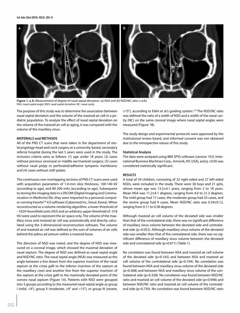
The direction of NSD was noted, and the degree of NSD was mea-sured on a coronal image, which showed the maximal deviation of nasal septum. The degree of NSD was defined as nasal septal angle and NSD/NC ratio. The nasal septal angle (NSA) was measured as the angle between a line drawn from the superior insertion of the nasal septum at the crista galli to the inferior insertion of the septum at the maxillary crest and another line from the superior insertion of the septum at the crista galli to the maximally deviated point of the convex nasal septum (Figure 1A). Patients with NSD were grouped into 3 groups according to the measured nasal septal angle as group I (mild, <9°), group II (moderate, ≥9° and <15°), or group III (severe,
≥15°), according to Elahi et al.’s grading system.[10] The NSD/NC ratio was defined the ratio of a width of NSD and a width of the nasal cav-ity (NC) on the same coronal image where nasal septal angles were measured (Figure 1B).
The study design and experimental protocols were approved by the institutional review board, and informed consent was not obtained due to the retrospective nature of this study.
Statistical AnalysisThe data were analyzed using IBM SPSS software (version 19.0, Inter-national Business Machines Corp., Armonk, NY, USA), and p <0.05 was considered statistically significant.
RESULTSA total of 59 children, consisting of 32 right-sided and 27 left-sided NSDs, were included in the study. There were 38 boys and 21 girls, whose mean age was 13.2±4.1 years, ranging from 2 to 18 years. Mean NSA was 11.2±4.1 degrees, ranging from 4.0 to 21.3 degrees. The mild group had 17 cases, the moderate group had 33 cases, and the severe group had 9 cases. Mean NSD/NC ratio was 0.34±0.12, ranging from 0.11 to 0.58 degrees.
Although mastoid air cell volume of the deviated side was smaller than that of the contralateral side, there was no significant difference of maxillary sinus volume between the deviated side and contralat-eral side (p=0.052). Although maxillary sinus volume of the deviated side was smaller than that of the contralateral side, there was no sig-nificant difference of maxillary sinus volume between the deviated side and contralateral side (p=0.671) (Table 1).
No correlation was found between NSA and mastoid air cell volume of the deviated side (p=0.165) and between NSA and mastoid air cell volume of the contralateral side (p=0.104). No correlation was found between NSA and maxillary sinus volume of the deviated side (p=0.408) and between NSA and maxillary sinus volume of the con-tralateral side (p=0.328). No correlation was found between NSD/NC ratio and mastoid air cell volume of the deviated side (p=0.948) and between NSD/NC ratio and mastoid air cell volume of the contralat-eral side (p=0.730). No correlation was found between NSD/NC ratio
252
Int Adv Otol 2014; 10(3): 251-5
Figure 1. a, b. Measurement of degree of nasal septal deviation. (a) NSA and (b) NSD/NC ratio (=a/b)NSA: nasal septal angle; NSD: nasal septal deviation; NC: nasal cavity
a b
and maxillary sinus volume of the deviated side (p=0.730) and be-tween NSD/NC ratio and maxillary sinus volume of the contralateral side (p=0.429). Mastoid air cell volume of the deviated side and of the contralateral side was not significantly different between groups I, II, and III (p=0.355 and 0.125, respectively). The maxillary sinus volume of the deviated side and of the contralateral side was not significantly different between groups I, II, and III (p=0.331 and 0.446, respective-ly) (Table 1).
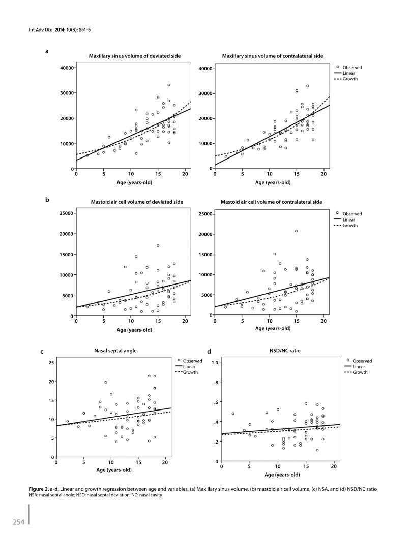
Significant linear and growth regression was found between age and mastoid air cell volume of the deviated side (p=0.008 for linear and p=0.002 for growth) and between age and maxillary sinus volume of the deviated side (p<0.0001 for linear and growth). Significant linear and growth regression was also found between age and mastoid air cell volume of the contralateral side (p=0.007 for linear and p=0.002 for growth) and between age and maxillary sinus volume of the con-tralateral side (p<0.0001 for linear and growth). However, linear and growth regression was not found between age and NSA (p=0.091 for linear and p=0.170 for growth) or between age and NSD/NC ratio (p=0.248 for linear and p=0.325 for growth) (Figure 2).
In order to eliminate the influence of individual variation, the ratio of the volumes between the sides (volume of contralateral side/volume of deviated side) were calculated and analyzed for correlation with NSA and the NSD/NC ratio. No correlation was found between NSA and the ratio of mastoid air cell volumes (p=0.623) or between NSA and the ratio of maxillary sinus volumes (p=0.546). No correlation was found between the NSD/NC ratio and ratio of mastoid air cell vol-umes (p=0.524) or between the NSD/NC ratio and ratio of maxillary sinus volumes (p=0.334). The ratio of mastoid air cell volumes was also not significantly different between groups I, II, and III (p=0.080). The ratio of maxillary sinus volumes was not significantly different between groups I, II, and III (p=0.345)(Table 1).
DISCUSSIONThe possibility that PNSs may influence the development of the mid-dle ear-mastoid systems has been suggested, because the openings of both are close enough to influence each other. One of the authors
(Lee-D.H.) reported that there was no interaction in the pneumatiza-tion of three PNSs (frontal, maxillary, and sphenoid) and mastoid air cells and that the growth of both was influenced by age in a pediatric population.[8]
Karakas and Kavakli [11] reported that the volumes of PNSs and mas-toid air cells increased with age and found a positive correlation be-tween right-left and ipsilateral PNSs and mastoid air cells. Firat et al.[9] evaluated the effect of NSD on ethmoid cell volume and determined whether there was any correlation between NSD grade and ethmoid cell volume in adult population. They failed to find any difference between deviated and contralateral total ethmoid cell volume but found that the ratio of total ethmoid cell volume of the deviated side compared with the contralateral side decreased as the degree of NSD increased and concluded that NSD affects the total ethmoid cell vol-ume. Kim et al.[12] found a positive correlation between the pneumati-zation of mastoid air cells and of the sphenoid sinus but not between the volume of mastoid air cells and of the maxillary sinuses. Kapusuz Gencer et al.[13] studied the possible role of NSD on the volume of maxillary sinuses in adult population. They revealed significant dif-ferences between deviated and contralateral maxillary sinus volume and concluded that maxillary sinus volume tend to be higher at the contralateral side of severe NSD in adults. Kapusuz Gencer et al.[14] evaluated the role of NSD on the volume of mastoid air cells and a possible relationship to chronic otitis, which was conducted in an adult population. They concluded that mastoid cell volumes tend to be larger at the contralateral side of severe NSD in adults.
However, the studies above were conducted in adult populations. The strength of this study was that it was performed in a pediatric population under 18 years old, and it better demonstrated the devel-opment of PNSs and mastoid air cells. Considering that major devel-opment and pneumatization of PNSs and mastoid air cells occur in children, the study should have been performed in a pediatric popu-lation. During the growth of PNSs, there are two active pneumatiza-tion periods: the first is between birth and 4 years of age, and the sec-ond is between 8 and 12 years of age. There are slow growth phases between these periods.[15] Similar to the growth of PNSs, the mastoid
253
Lee and Jin. Effect of Nasal Septal Deviation on Mastoid Volume
Deviated side Contralateral side p value
Maxillary sinus volume (cm3) 16.1±6.2 16.3±6.5 0.671
Mastoid air cell volume (cm3) 6.1±3.7 6.6±4.1 0.053
group I group II group III p value
Maxillary sinus volume of deviated side (cm3) 14.3±5.1 16.6±6.4 17.7±7.0 0.331
Maxillary sinus volume of contralateral side (cm3) 14.9±5.1 16.5±7.1 18.8±6.9 0.446
Mastoid air cell volume of deviated side (cm3) 5.7±4.1 5.9±3.6 7.5±3.3 0.355
Mastoid air cell volume of contralateral side (cm3) 6.0±4.3 6.2±4.1 9.1±3.1 0.124
NSA (°) 0.2±0.1 0.4±0.1 0.5±0.1
NSD/NC ratio 6.6±1.5 11.7±1.7 18.0±2.4
Ratio of maxillary sinus volumes 1.1±1.5 1.0±0.2 1.1±0.2 0.345
Ratio of mastoid air cell volumes 1.1±0.3 1.1±0.4 1.4±0.7 0.080
Group I (mild, <9°), group II (moderate, ≥9°and <15°), or group III (severe, ≥15°) according to Elahi et al.’s grading system. NSA: nasal septal angle; NSD: nasal septal deviation; NC: nasal cavity Ratio of maxillary sinus or mastoid air cell volumes = volume of contralateral side/volume of deviated side
Table 1. Characteristics of parameters
254
Int Adv Otol 2014; 10(3): 251-5
Figure 2. a-d. Linear and growth regression between age and variables. (a) Maxillary sinus volume, (b) mastoid air cell volume, (c) NSA, and (d) NSD/NC ratioNSA: nasal septal angle; NSD: nasal septal deviation; NC: nasal cavity
a
b
c d
Maxillary sinus volume of deviated side
0 5 10 15 20
0 5 10 15 20
0 5 10 15 20
0 5 10 15 20
0 5 10 15 20
0 5 10 15 20
Mastoid air cell volume of deviated side
Nasal septal angle
Age (years-old)
Age (years-old)
Age (years-old)
Age (years-old)
Age (years-old)
Age (years-old)
NSD/NC ratio
Maxillary sinus volume of contralateral side
ObservedLinearGrowth
ObservedLinearGrowth
ObservedLinearGrowth
ObservedLinearGrowth
40000
30000
20000
10000
0
25000
20000
15000
10000
5000
0
25
20
15
10
5
0
1.0
.8
.6
.4
.2
.0
25000
20000
15000
10000
5000
0
40000
30000
20000
10000
0
Mastoid air cell volume of contralateral side
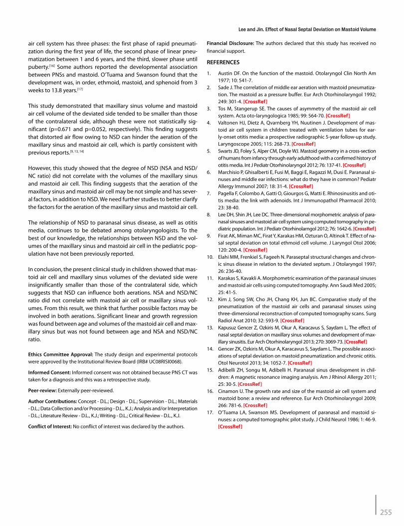
air cell system has three phases: the first phase of rapid pneumati-zation during the first year of life, the second phase of linear pneu-matization between 1 and 6 years, and the third, slower phase until puberty.[16] Some authors reported the developmental association between PNSs and mastoid. O’Tuama and Swanson found that the development was, in order, ethmoid, mastoid, and sphenoid from 3 weeks to 13.8 years.[17]
This study demonstrated that maxillary sinus volume and mastoid air cell volume of the deviated side tended to be smaller than those of the contralateral side, although these were not statistically sig-nificant (p=0.671 and p=0.052, respectively). This finding suggests that distorted air flow owing to NSD can hinder the aeration of the maxillary sinus and mastoid air cell, which is partly consistent with previous reports.[9, 13, 14]
However, this study showed that the degree of NSD (NSA and NSD/NC ratio) did not correlate with the volumes of the maxillary sinus and mastoid air cell. This finding suggests that the aeration of the maxillary sinus and mastoid air cell may be not simple and has sever-al factors, in addition to NSD. We need further studies to better clarify the factors for the aeration of the maxillary sinus and mastoid air cell.
The relationship of NSD to paranasal sinus disease, as well as otitis media, continues to be debated among otolaryngologists. To the best of our knowledge, the relationships between NSD and the vol-umes of the maxillary sinus and mastoid air cell in the pediatric pop-ulation have not been previously reported.
In conclusion, the present clinical study in children showed that mas-toid air cell and maxillary sinus volumes of the deviated side were insignificantly smaller than those of the contralateral side, which suggests that NSD can influence both aerations. NSA and NSD/NC ratio did not correlate with mastoid air cell or maxillary sinus vol-umes. From this result, we think that further possible factors may be involved in both aerations. Significant linear and growth regression was found between age and volumes of the mastoid air cell and max-illary sinus but was not found between age and NSA and NSD/NC ratio.
Ethics Committee Approval: The study design and experimental protocols were approved by the Institutional Review Board (IRB# UC08RISI0068).
Informed Consent: Informed consent was not obtained because PNS CT was taken for a diagnosis and this was a retrospective study.
Peer-review: Externally peer-reviewed.
Author Contributions: Concept - D.L.; Design - D.L.; Supervision - D.L.; Ma terials - D.L.; Data Collection and/or Processing - D.L., K.J.; Analysis and/or In terpretation - D.L.; Literature Review - D.L., K.J.; Writing - D.L.; Critical Review - D.L., K.J.
Conflict of Interest: No conflict of interest was declared by the authors.
Financial Disclosure: The authors declared that this study has received no financial support.
REFERENCES
1. Austin DF. On the function of the mastoid. Otolaryngol Clin North Am 1977; 10: 541-7.
2. Sade J. The correlation of middle ear aeration with mastoid pneumatiza-tion. The mastoid as a pressure buffer. Eur Arch Otorhinolaryngol 1992; 249: 301-4. [CrossRef]
3. Tos M, Stangerup SE. The causes of asymmetry of the mastoid air cell system. Acta oto-laryngologica 1985; 99: 564-70. [CrossRef]
4. Valtonen HJ, Dietz A, Qvarnberg YH, Nuutinen J. Development of mas-toid air cell system in children treated with ventilation tubes for ear-ly-onset otitis media: a prospective radiographic 5-year follow-up study. Laryngoscope 2005; 115: 268-73. [CrossRef]
5. Swarts JD, Foley S, Alper CM, Doyle WJ. Mastoid geometry in a cross-section of humans from infancy through early adulthood with a confirmed history of otitis media. Int J Pediatr Otorhinolaryngol 2012; 76: 137-41. [CrossRef]
6. Marchisio P, Ghisalberti E, Fusi M, Baggi E, Ragazzi M, Dusi E. Paranasal si-nuses and middle ear infections: what do they have in common? Pediatr Allergy Immunol 2007; 18: 31-4. [CrossRef]
7. Pagella F, Colombo A, Gatti O, Giourgos G, Matti E. Rhinosinusitis and oti-tis media: the link with adenoids. Int J Immunopathol Pharmacol 2010; 23: 38-40.
8. Lee DH, Shin JH, Lee DC. Three-dimensional morphometric analysis of para-nasal sinuses and mastoid air cell system using computed tomography in pe-diatric population. Int J Pediatr Otorhinolarngol 2012; 76: 1642-6. [CrossRef]
9. Firat AK, Miman MC, Firat Y, Karakas HM, Ozturan O, Altinok T. Effect of na-sal septal deviation on total ethmoid cell volume. J Laryngol Otol 2006; 120: 200-4. [CrossRef]
10. Elahi MM, Frenkiel S, Fageeh N. Paraseptal structural changes and chron-ic sinus disease in relation to the deviated septum. J Otolaryngol 1997; 26: 236-40.
11. Karakas S, Kavakli A. Morphometric examination of the paranasal sinuses and mastoid air cells using computed tomography. Ann Saudi Med 2005; 25: 41-5.
12. Kim J, Song SW, Cho JH, Chang KH, Jun BC. Comparative study of the pneumatization of the mastoid air cells and paranasal sinuses using three-dimensional reconstruction of computed tomography scans. Surg Radiol Anat 2010; 32: 593-9. [CrossRef]
13. Kapusuz Gencer Z, Ozkiris M, Okur A, Karacavus S, Saydam L. The effect of nasal septal deviation on maxillary sinus volumes and development of max-illary sinusitis. Eur Arch Otorhinolaryngol 2013; 270: 3069-73. [CrossRef]
14. Gencer ZK, Ozkiris M, Okur A, Karacavus S, Saydam L. The possible associ-ations of septal deviation on mastoid pneumatization and chronic otitis. Otol Neurotol 2013; 34: 1052-7. [CrossRef]
15. Adibelli ZH, Songu M, Adibelli H. Paranasal sinus development in chil-dren: A magnetic resonance imaging analysis. Am J Rhinol Allergy 2011; 25: 30-5. [CrossRef]
16. Cinamon U. The growth rate and size of the mastoid air cell system and mastoid bone: a review and reference. Eur Arch Otorhinolaryngol 2009; 266: 781-6. [CrossRef]
17. O’Tuama LA, Swanson MS. Development of paranasal and mastoid si-nuses: a computed tomographic pilot study. J Child Neurol 1986; 1: 46-9. [CrossRef]
255
Lee and Jin. Effect of Nasal Septal Deviation on Mastoid Volume