Embed Size (px)
Citation preview

EFFECT OF NITRIC OXIDE (NO) ON ORTHODONTIC
TOOTH MOVEMENT IN RATS
By
ARVIND KENNETH VAKANI
A THESIS PRESENTED TO THE GRADUATE SCHOOL OF THE UNIVERSITY OF FLORIDA IN PARTIAL FULFILLMENT
OF THE REQUIREMENTS FOR THE DEGREE OF MASTER OF SCIENCE
UNIVERSITY OF FLORIDA
2003

ACKNOWLEDGMENTS
I thank my family (my mother, father, and brother) for all of their help and support.
I know that none of my accomplishments would have been possible without their
patience and the sacrifices that they made over the years.
I also thank the members of my supervisory committee (Drs. Wheeler, Dolce, and
Holliday). They have been instrumental in the success of my clinical training. I also
acknowledge Linda Archer, laboratory technician, for her help with the laboratory
portion of the project.
ii

TABLE OF CONTENTS page ACKNOWLEDGMENTS .................................................................................................. ii
LIST OF FIGURES ........................................................................................................... iv
ABSTRACT.........................................................................................................................v
CHAPTER
1 INTRODUCTION ........................................................................................................1
2 MATERIALS AND METHODS .................................................................................7
Animals.........................................................................................................................7 Radiographic Examination ...........................................................................................8 Histological Examination .............................................................................................9 Statistical Analysis........................................................................................................9
3 RESULTS...................................................................................................................11
4 DISCUSSION.............................................................................................................15
LIST OF REFERENCES...................................................................................................18
BIOGRAPHICAL SKETCH .............................................................................................20
iii

LIST OF FIGURES
Figure page 1. Synthesis and metabolism of NO ...............................................................................6
2. Change in body weight (gm) of rats receiving L-NAME at three distinct time points: surgery, activation and sacrifice ...................................................................13
3. Change in body weight (gm) of rats not receiving L-NAME at three distinct time points: surgery, activation and sacrifice ...................................................................13
4. Consumption of L-NAME (ml) by the control and springs groups .........................13
5. Orthodontic tooth movement from Day 1-10 of spring rats vs. spring rats receiving the NOS inhibitor .....................................................................................13
6. Orthodontic tooth movement from Day 1-10 of molar drift rats vs. molar drift rats receiving the NOS inhibitor...............................................................................14
7. Photomicrographs showing the distal root of the maxillary first molar...................14
iv

Abstract of Thesis Presented to the Graduate School
of the University of Florida in Partial Fulfillment of the Requirements for the Degree of Master of Science
EFFECT OF NITRIC OXIDE (NO) ON ORTHODONTIC TOOTH MOVEMENT IN RATS
By
Arvind Kenneth Vakani
May 2003
Chair: Calogero Dolce Major Department: Orthodontics
Applying force to a tooth produces strains in the periodontal ligament (PDL) and
the surrounding bone. The strain initiates an acute inflammatory response that induces
the secretion of prostaglandins, cytokines, and growth factors. These factors in turn
produce an uncoupling in the normal bone remodeling process, which then produces
tooth movement. Nitric oxide (NO), a short-lived free radical, is produced by enzymes
called NO synthases (NOS). Nitric oxide has important effects in bone remodeling. The
purpose of this study was to examine the effect of nitric oxide on orthodontic tooth
movement in rats. Maxillary first molars of male Sprague-Dawley rats were moved via a
closed-coil spring for 10 days. Systemic administration of N-nitro-L-arginine methyl
ester (L-NAME), a general inhibitor of nitric oxide synthases, significantly reduced tooth
movement. These results suggest that NO levels may influence orthodontic tooth
movement.
v

CHAPTER 1 INTRODUCTION
Among the many goals of the specialty of orthodontics is for the clinician to
achieve tooth movement in an effective and expedient manner. It is quite difficult to
move a tooth spatially without having an impact on neighboring teeth; and so the most
essential element to the practice of orthodontics is anchorage. Absolute anchorage would
imply that one tooth is moved without any untoward effects on the adjacent teeth being
used for that particular movement. Until now, clinicians have relied heavily on
laboratory-constructed devices such as the transpalatal arch (TPA) or Nance to maintain
anchorage within the dental arch. Additionally, clinicians have sought the help of
biologically invasive dental implants. With both approaches, the results have been less
than satisfactory. In the quest for more complete anchorage, research has focused on the
field of pharmacology. Recently, bisphosphonates have been used as a way to prevent
tooth movement (Igarashi, Mitani, Adachi, Sinoda 1994). Histologic examination has
shown that with the application of bisphosphonates in experimental animals, fewer
osteoclasts appeared on the alveolar bone surface and that bone resorption was inhibited
(Igarashi et al.1994). Thus, a pharmacological approach seems promising in the
endeavor to gain pure control of tooth movement.
Orthodontic tooth movement occurs as a response to mechanical forces placed on
the tooth that disperse throughout the periodontal ligament and then initiate bone
remodeling. This sequence of events has previously been described by the
pressure:tension hypothesis (that bone is resorbed in areas of pressure and is formed in
1
2
areas of tension) (Sandy, Farndale, Meikle 1993). Applying force to a tooth disrupts the
equilibrium between bone formation and resorption. This results in an overwhelmingly
greater amount of bone resorption than formation on the pressure side; and the reverse on
the tension side (Sandy et al.1993).
Orthodontic tooth movement actually involves three unique phases (King, Keeling,
Wronski 1991). As an orthodontic force is applied to a tooth, the initial phase allows for
some movement of the tooth. However, this movement is strictly compressive
deformation of the periodontal ligament on the pressure side. In the second phase, the lag
phase, there is no tooth movement. Here, necrotic tissue that is accumulated in the
compressed area of the PDL is eliminated (King, Keeling, McCoy, Ward, 1991).
Essentially, hyalinization occurs in almost all cases. Elimination of the hyalinized tissue
is associated with undermining bone resorption (King et al., 1991). The final phase is
when true orthodontic tooth movement occurs. Osteoclasts within the PDL spaces begin
to resorb the adjacent alveolar bone and permit the movement of teeth (King et al., 1991).
Mechanical stress is believed to influence both bone formation and resorption.
The immediate effect of force on a tooth is movement of the tooth within the PDL space
that stimulates nerves and causes blood-flow changes (Ehrlich, Lanyon 2002). Any of
these events could transform the physical force into a message that activates the
biological system. Damaged tissue releases chemo-attractants that elicit the response of
macrophages and PMNs (to remove the necrotic debris). These cells release factors
(called chemokines and cytokines) that allow them to communicate with one another.
These factors may cause pleuripotent stem cells to differentiate and thus augment the
bone remodeling process (Norton 2000).

3
Bone remodeling occurs through the activation of specific signaling pathways.
Inflammatory cytokines produced by mechanically activated cells; and a variety of local
mediators have been implicated in modulating the activities of osteoclasts and
osteoblasts. However, the signal-transduction pathways of orthodontic mechanical
stimuli have yet to be elucidated (Hayashi, Igarashi, Miyoshi, Shinoda, Mitani 2002). It
is quite possible that the mechanical signal itself may be stimulatory. Osteoblasts and
osteocytes may themselves act as mechanical sensors for bone remodeling (Hayashi et al.
2002). It has been previously shown that osteoblasts lay down new bone matrix in the
form of osteoid in areas formerly occupied by osteoclasts (King et al.1991). Frequently,
osteoblasts become trapped within the bone matrix; and differentiate into osteocytes.
These osteocytes communicate with each other and with cells on the bone surface
through cytoplasmic processes throughout the bone matrix. It is thought that osteocytes
act as sensors of mechanical stress by detecting and responding to changes in fluid flow
(Van’T Hof, Ralston 2001). From in vitro and in vivo studies, the orthodontic force
(mechanical stress) appears to evoke an acute inflammatory response mediated by the
release of prostaglandins, cytokines and growth factors (Sandy et al.1993). These factors,
in turn, regulate bone remodeling.
Nitric Oxide (NO) is a short-lived, reactive molecule that serves as a sensitive
mediator in the nervous, vascular and immune systems. It is produced by nitric oxide
synthase (NOS) from the L-arginine amino acid and yields L-citrulline as a co-product
(Figure 1) (Chae et al.1997). Many cell types within the bone microenvironment are
potentially capable of producing NO (Evans, Ralston 1996). Nitric oxide synthesis can

4
be induced in osteoblasts and osteocytes when exposed to mechanical strain and/or shear
stress (Van’T Hof et al. 2001).
Three forms of NOS are currently recognized: neuronal (nNOS), endothelial
(eNOS), and inducible (iNOS) (Evans et al. 1996). The names reflect the tissues in
which these enzymes were first located yet they may be found in a variety of tissues and
cell types. The neuronal and endothelial forms are constitutively expressed and their
activity depends on elevated levels of intracellular calcium (Ralston et al. 1995). The
inducible pathway of NO production is regulated at the transcriptional level by
proinflammatory cytokines and endotoxin; and has potent effects on bone resorption
(Evans et al. 1996).
Studies have shown that calcium, cGMP, and cAMP act as key mediators or second
messengers in the function of many drugs and hormones (Davidovitch, Shanfeld 1975).
Intracellular second messengers play an important role in the differentiation of
osteoclasts from their precursors (monocytes) in bone resorption during mechanical force
application (King et al. 1991). Some studies have shown that low concentrations of NO
inhibit osteoclast function and that this action is cGMP dependent (Holliday, Dean, Lin,
Greenwald, Gluck 1997). In addition, osteoblast function is governed by a nitric oxide
dependent pathway that is mediated by the second messenger cGMP (Mancini et al.
2000). It is well known that the local microenvironment is crucial to the regulation of
osteoclast activity; and that a number of factors produced by osteoblasts modulate the
proliferation and differentiation of osteoclast precursors and the function of mature cells
(Mancini et al. 1998). Nitric oxide is of major importance as one of the local factors that
regulates bone metabolism. Nitric oxide is produced by osteoblasts; and inhibits the

5
function of mature osteoclastic cells (Mancini et al. 1998). Therefore, cytokine-induced
NO released from osteoblasts can act to down-regulate osteoclast formation and activity,
thereby identifying NO as a potentially important osteoblast-osteoclast coupling factor
(Van’T Hof, Ralston 1997).
Nitric oxide has been suggested to have a biphasic effect on bone remodeling. It
has been shown that high NO concentrations inhibit resorption by mature osteoclasts and
also inhibit the production of osteoclasts from its precursor cells. On the contrary, it has
been suggested that low concentrations of NO stimulate osteoblast growth and cytokine
production (Collin-Osdoby, Nickols, Osdoby 1995). Recently, however, it has been
shown that low concentrations of NO actually inhibit osteoclast formation (Holliday et
al., 1997). Thus, the effects of NO on bone remodeling appear to be dependent on dosage
and the specific nature of the system. For instance, the role of NO in remodeling long
bones may be different from that in calvaria. Therefore, it was not possible to predict the
role, if any, on NO in orthodontic tooth movement. To test for the role of NO in OTM,
we used the general NO synthase inhibitor L-nitro-arginine-methyl ester (L-NAME).
The formation of nitric oxide can be inhibited by substituting arginine analogues
such as L-N-monomethyl arginine (L-NMMA) and L-nitro-arginine-methyl ester
(L−NAME) (Figure 1). Our aim was to demonstrate the effect of nitric oxide inhibition
on orthodontic tooth movement using L-NAME.
Data from this study provide information regarding the possibility of manipulating
OTM by pharmacological intervention of NO signaling. Since agents are attractive target
for bone specific pharmaceuticals.

6
L-Arginine + O2 iNOS
Arginine analogues
(L-NAME)
L-Citrulline + NO
Nitric oxide synthase eNOS
nNOS NOS stimulus
NO donors (+NO) NOS inhibitor (-NO)
ADP
ribosylation
Guanylyl cyclase Phosphodiesterase cGMP Protein kinase G Cell effects Figure 1. Synthesis and metabolism of NO

CHAPTER 2 MATERIALS AND METHODS
Animals
Experiments were performed according to methods described by King et al. (1991).
Two hundred fourteen male Sprague-Dawley rats (20 to 30 days old) were used because
of availability, cost, genetic homogeneity, and (at this age) the ability to recover rapidly
from the surgeries required to place the appliances. Male rats were chosen to eliminate
the hormonal changes associated with estrus. Upon their arrival they were acclimated for
5 to 7 days under experimental conditions.
Orthodontic Treatment
The rats were weighed and profoundly anesthetized using intra-muscular injections
of ketamine (87 mg/kg) and xylazine (13 mg/kg). The occlusal surfaces of both
maxillary first molars were prepared by roughening the surface and modified orthodontic
cleats were bonded. The opposing mandibular first molars were extracted and all four
incisors pinned to prevent further eruption. The animals were then allowed to recover for
3 weeks, while monitoring wound healing and weight gain.
Upon their recovery, the rats were randomly divided among four experimental
groups:
• Control/drift with drug (CD) • Control/drift without drug (C) • Springs with drug (SD) • Springs without drug (S)
7
8
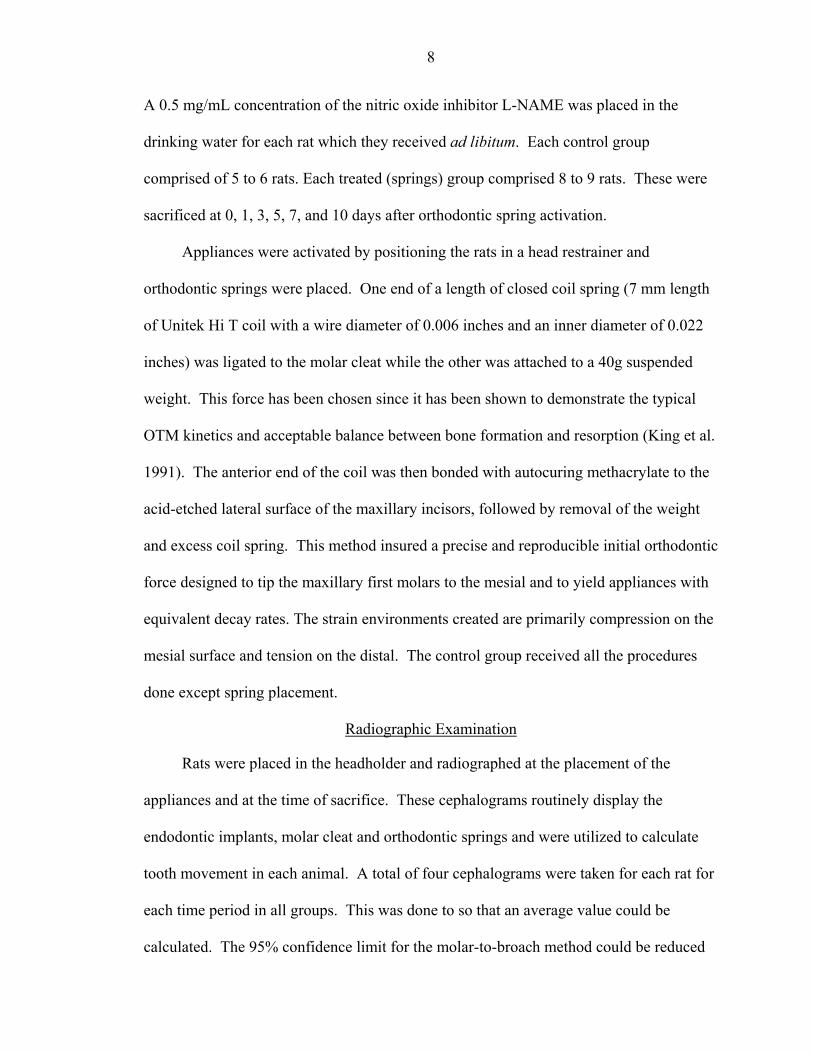
A 0.5 mg/mL concentration of the nitric oxide inhibitor L-NAME was placed in the
drinking water for each rat which they received ad libitum. Each control group
comprised of 5 to 6 rats. Each treated (springs) group comprised 8 to 9 rats. These were
sacrificed at 0, 1, 3, 5, 7, and 10 days after orthodontic spring activation.
Appliances were activated by positioning the rats in a head restrainer and
orthodontic springs were placed. One end of a length of closed coil spring (7 mm length
of Unitek Hi T coil with a wire diameter of 0.006 inches and an inner diameter of 0.022
inches) was ligated to the molar cleat while the other was attached to a 40g suspended
weight. This force has been chosen since it has been shown to demonstrate the typical
OTM kinetics and acceptable balance between bone formation and resorption (King et al.
1991). The anterior end of the coil was then bonded with autocuring methacrylate to the
acid-etched lateral surface of the maxillary incisors, followed by removal of the weight
and excess coil spring. This method insured a precise and reproducible initial orthodontic
force designed to tip the maxillary first molars to the mesial and to yield appliances with
equivalent decay rates. The strain environments created are primarily compression on the
mesial surface and tension on the distal. The control group received all the procedures
done except spring placement.
Radiographic Examination
Rats were placed in the headholder and radiographed at the placement of the
appliances and at the time of sacrifice. These cephalograms routinely display the
endodontic implants, molar cleat and orthodontic springs and were utilized to calculate
tooth movement in each animal. A total of four cephalograms were taken for each rat for
each time period in all groups. This was done to so that an average value could be
calculated. The 95% confidence limit for the molar-to-broach method could be reduced

9
to 23 µm when the average of four independent determinations was used (King et al.
1991). The cephalograms were then digitized at 600 dpi and analyzed using the NIH
imaging program ImageJ. OTM was measured from the molar cleat to the distal pin on
the incisor. The difference between these measurements made on appliance placement
and radiographs taken at sacrifice was calculated and reported as tooth movement (King
et al. 1991).
Histological Examination
In order to study the histological changes in bone and the tissues surrounding the
tooth, the posterior portion of the maxilla containing teeth, bone and tissue was dissected
and fixed in a paraformaldehyde fixative solution (Shirazi, Nilforoushan, Alghasi,
Dehpour 2002). The soft tissue was then removed and the hard tissue was placed in a
demineralization solution. Thereafter, the bone was dehydrated in ethanol and xylene
and then infiltrated with paraffin and xylene prior to being embedded in paraffin. 4µm
thick samples were treated with 0.1% pepsin for enzymatic digestion that allowed for
better solution infiltration and embedded in paraffin. A series of antibodies to NOS
(Oncogene Research Products) were used to bind to the exposed epitope on the tissue
samples. They were finally stained with 3,3’ Diaminobenzidine tetrahydrochloride
(DAB). The intensity of staining of the PDL around the distal root of the maxillary first
molar was the criteria for comparison among the samples.
Statistical Analysis
The means and standard errors of molar movement were calculated for each time
point. Statistical evaluation of the data was done with the analysis of variance

10
(ANOVA). The paired t test was used to evaluate the significance of difference in tooth
movement between the rats treated with and without study drug (L-NAME).

CHAPTER 3 RESULTS
The body weight of all of the rats in the four groups was recorded for three
different time points: surgery, activation and sacrifice. This was done to observe any
irregular eating habits due to trauma caused by surgery, activation of springs or the effect
of L-NAME (Figure 2) on body weight. The weight of the rats increased substantially
after surgery in all 4 groups. However it is evident that the rats may have had some
discomfort during food consumption due to intraoral appliances since their body weight
decreased slightly from the day of springs activation to sacrifice (Figures 2 and 3).
The amount of L-NAME consumption was noted for both the molar drift and
springs groups. Both groups consumed approximately the same amount of NOS inhibitor
(L-NAME) (Figure 4).
As a note, the data for Day 1 of OTM with L-NAME are missing and therefore not
included in Figure 5 or the discussion to follow. Tooth movement in the OTM groups
exhibited the three typical phases of tooth movement. An initial compression of the
periodontal ligament accounts for the 0.4 mm of molar movement from Day 0 to 1. A lag
phase with no tooth movement was noted thereafter (from Day 1 to 5). This is the period
where necrotic tissue accumulated in the compressed area of the PDL is eliminated.
Finally, orthodontic tooth movement occurs from Day 5 to 10. The OTM group with L-
NAME showed similar tooth movements as the OTM control group until Day 5.
Throughout the period of Day 5 to 10 the OTM with L-NAME group showed remarkably
less tooth movement than the control. On the final day it was noted that molar movement
11

12
in the OTM with L-NAME group was approximately 0.2 mm less than that of the OTM
control group and was statistically significant at P<0.0001.
L-NAME caused a significant reduction in tooth movement in the distal drift
groups (after Day 3) as compared to the orthodontically treated groups (after Day 5).
Perhaps the orthodontic appliances overpowered the effects of the NOS inhibitors on the
biological systems of the rats.
The distal drift control and distal drift with L-NAME groups showed some
dissimilarities (Figure 6). From activation to Day 2, the distal drift with L-NAME group
showed more molar movement than the control group. After Day 2 until Day 10, the
distal drift control group showed more molar movement than the distal drift with L-
NAME group. The difference in tooth movement between the distal drift and the drift
with L-NAME groups was statistically significant for Days 5 (P<0.0002) and 7
(P<0.001). On the final day it was noted that molar movement in the distal drift with L-
NAME group was approximately 0.3 mm less than that of the distal drift control group
and was statistically significant at P<0.022.
The periodontal ligament space around the distal root of the maxillary first molar
was carefully examined for the intensity of DAB staining that occurs as an indication of
NOS expression. The results show less intensity of staining around the roots of those rats
treated with L-NAME (Figure 7) as compared to control rats. These findings are
consistent with our hypothesis that L-NAME would serve to block the enzymatic activity
of nitric oxide synthase in rat specimens. In addition, there was no difference in gross
morphology of the roots between L-NAME treated rats and control rats.

13
0
100
200
300
400
Control with L-NAME Springs with L-NAME
(gra
ms) Surgery
Activation
Sacrifice
Figure 2. Change in body weight (gm) of rats receiving L-NAME at three distinct time
points: surgery, activation and sacrifice
0
50
100
150
200
250
300
350
400
Control w/o L-NAME Springs w/o L-NAME
(gra
ms)
Surgery
Activation
Sacrifice
Figure 3. Change in body weight (gm) of rats not receiving L-NAME at three distinct
time points: surgery, activation and sacrifice
05
10152025303540
1
( 0 . 5 mg/ ml c onc . )
Cont rol wit h L-NAME
Springs wit h L-NAME
Figure 4. Consumption of L-NAME (ml) by the control and springs groups
00.10.20.30.40.50.60.70.8
Activation 1 3 5 7 10Time (days)
mm
(se)
OTM
OTM & L-NAME
*
p<.0001
Figure 5. Orthodontic tooth movement from Day 1-10 of spring rats vs. spring rats
receiving the NOS inhibitor

14
-0.8-0.7-0.6-0.5-0.4-0.3-0.2-0.1
0 Activation
1 3 5 7 10
Time (days)
mm
(se)
Drift
Drift & L-NAME
p<.0002*
p<.001*
p<.022*
Figure 6. Orthodontic tooth movement from Day 1-10 of molar drift rats vs. molar drift
rats receiving the NOS inhibitor
A B Figure 7. Photomicrographs showing the distal root of the maxillary first molar. A)
From a rat treated with L-NAME. B) From a control rat. Slides were stained with DAB

CHAPTER 4 DISCUSSION
The results of the present study highly suggest that nitric oxide (NO) has a
significant modulatory effect on tooth movement. Systemic administration of L-NAME,
a nitric oxide synthase inhibitor, in rats led to a decrease in orthodontic tooth movement
and distal molar drift compared to controls. Thus, it appears that nitric oxide production
is essential for an optimal response by the periodontal ligament upon application of
orthodontic forces in the rat model.
The present results clearly demonstrate that the systemic administration of L-
NAME caused a significant reduction in tooth movement in the treated and control/drift
groups. All groups showed similar rates of tooth movement from day 0 to day 1,
however, in the final phase of bone remodeling from day 3 to day 10, the L-NAME
treated groups exhibited less tooth movement than the OTM/drift control groups (Figures
5 and 6).
Fourteen rats died among the L-NAME treated groups whereas only 2 died in the
non-drug treated groups. Rats may suffer from high blood pressure secondary to
systemic L-NAME administration (Tucker, Ledingham, Zheng, Laverty 2000). Since
nitric oxide (NO) is a signaling molecule in the cardiovascular, immune and nervous
systems, it has a wide range of effects throughout the body. Nitric oxide influences blood
pressure not only via vascular endothelial release, but also by central cardiovascular
control. Hypertension is a multifactorial disease that may be defined as a sustained
elevation in blood pressure resulting in target organ damage. In essential hypertension,
15

16
the majority of evidence indicates that there is an abnormality in the L-arginine/NO
pathway (Tucker et al. 2000). Some studies have shown that general NOS inhibition
elicited a sustained blood pressure increase, a decrease in heart rate, cardiac hypertrophy
and an increase in wall thickness of the coronary and carotid artery (Gerova 2000). It is
quite possible that the increase in blood pressure among the rats in all four groups may
have affected the results of the study since they had systemic complications secondary to
L-NAME administration. Therefore, the tooth movement effects noted may not be due
to NOS inhibition and strictly a consequence of the effects on the biological system
induced by high blood pressure. In future studies, a more local administration of the
NOS inhibitor or a reduced concentration may prevent these untoward effects.
Currently, an applicable approach for local delivery of the NOS inhibitor L-NAME
is through Elvax 40. This is a non-biodegradable, noninflammatory sustained release
polymer that has been used as a vector to deliver pharmacologic agents to a precise
location. Elvax would serve to aid in the elimination of the adverse systemic effects of
nitric oxide that have been documented.
Perhaps locating a cNOS inhibitor would be more effective since we would not be
affecting iNOS. It would also be beneficial to investigate the effects of nitric oxide using
a NOS stimulus (e.g. sodium nitroprusside) as opposed to a NOS inhibitor as was done in
the present study. This would prevent the increase in blood pressure since nitric oxide
production would not be attenuated. However, it may also serve to lower blood pressure
which could also be a problem. The application of a phosphodiesterase V inhibitor (e.g.
Zaprinest, Sildenafil) may be used in order to prevent the breakdown of cGMP.
Therefore, cGMP levels are higher as though there is more NO present (Figure 1).

17
Recently, other researchers have demonstrated the effects of the involvement of
nitric oxide in orthodontic tooth movement in rats. Hayashi et al. noted results similar to
those of the present study. Our results confirm their findings that the administration of
L−NAME, a nitric oxide synthase inhibitor, significantly reduced tooth movement.
However, they found that the local administration of L-NIL (N-1-iminoethyl-L-lysine), a
selective inhibitor of iNOS, did not affect orthodontic tooth movement (Hayashi 2002).
In conclusion, our results indicate that L-NAME decreases orthodontic tooth
movement compared to control subjects. This is an exciting finding in that it may be of
great use in the field of orthodontics. Local application of L-NAME and its subsequent
effects on tooth movement may allow the clinician greater control of orthodontic forces.
These results suggest that nitric oxide is instrumental in orthodontic tooth movement and
our study has shown that blocking NOS systemically significantly reduces tooth
movement. However, concerns that hypertension resulting from systemic NOS inhibition
must be addressed. If NOS inhibitors prove to be specifically blocking OTM, they may
be advantageous in regards to anchorage control and, more importantly, the management
of relapse. In the future, NOS inhibitors may prove to serve as a pharmacological means
of providing anchorage for clinicians. Administering L-NAME locally may allow the
orthodontist to gain full control of the dentition in specific areas during tooth movement
thereby eliminating many appliances needed for anchorage control. Moreover, it may be
of benefit in preventing a relapse of tooth movement and permit a more sustained
orthodontic result.
LIST OF REFERENCES
Chae H-J, Park R-K, Chung H-T, Kang J-S, Kim M-S, Choi D-Y, Bang B-G, Kim H-R. Nitric Oxide is a Regulator of Bone Remodeling. J Pharm Pharmacol 1997;49:897-902.
Collin-Osdoby P, Nickols GA, Osdoby P. Bone Cell Function, Regulation, and Communication: A Role for Nitric Oxide. J Cell Biochem 1995;57:399-408.
Davidovitch Z, Shanfeld JL. Cyclic AMP Levels in the Alveolar Bone of Orthodontically Treated Cats. Arch Oral Biol 1975;20:567-574.
Ehrlich PJ, Lanyon LE. Mechanical Strain and Bone Cell Function: A Review. Osteoporosis Int 2002;13:688-700.
Evans DM, Ralston SH. Nitric Oxide and Bone. J Bone Miner Res 1996;11:300-305.
Gerova M. Nitric Oxide-Compromised Hypertension: Facts and Enigmas. Physiol Res 2000;49(1):27-35.
Hayashi K, Igarashi K, Miyoshi K, Shinoda H, Mitani H. Involvement of Nitric Oxide in Orthodontic Tooth Movement in Rats. Am J Orthod Dentofacial Orthop 2002;122:306-9.
Holliday LS, Dean AD, Lin RH, Greenwald JE, Gluck SL. Low NO Concentrations Inhibit Osteoclast Formation in Mouse Marrow Cultures by cGMP-dependent Mechanism. Am J Physiol 1997;272:F283-F291.
Igarashi K, Mitani H, Adachi H, Shinoda H. Anchorage and Retentive Effects of a bisphosphonate (AHBuBP) on Tooth Movement in Rats. Am J Orthod Dentofacial Orthop 1994;106(3):279-89.
King GJ, Keeling SJ, McCoy EA, Ward TH. Measuring Dental Drift and Orthodontic Tooth Movement in Response to Various Initial Forces in Adult Rat. Am J Orthod Dentofacial Orthop 1991;99:456-465.
King GJ, Keeling SD, Wronski, TJ. Histomorphometric Study of Alveolar Bone Turnover in Orthodontic Tooth Movement. Bone. 1991;12:401-9.
Mancini L, Moradi-Bidhendi N, Becherini L, Martineti V, MacIntyre I. The Biphasic Effects of Nitric Oxide in Primary Rat Osteoblasts are cGMP Dependent. Biochem Biophys Res Comm 2000;274:477-481.
18

19
Mancini L, Moradi-Bidhendi N, Brandi ML, MacIntyre I. Nitric Oxide Superoxide and Peroxynitrite Modulate Osteoclast Activity. Biochem Biophys Res Comm 1998;243:785-790.
Norton L. Fundamental Principles of the Biology of Tooth Movement. Seminars in Orthodontics 2000;6(3):139-144.
Ralston S, Ho L, Helfrich M, Grabowski P, Johnston P, Benjamin N. Nitric Oxide: A Cytokine-Induced Regulator of Bone Resorption. J Bone Miner Res 1995;10:1040-1049.
Sandy JR, Farndale RW, Meikle MC. Recent Advances in Understanding Mechanically Induced Bone Remodeling and Their Relevance to Orthodontic Theory and Practice. Am J Orthod Dentofacial Orthop 1993;103(3):212-22.
Shirazi M, Nilforoushan D, Alghasi H, Dehpour A-R. The Role of Nitric Oxide in Orthodontic Tooth Movement in Rats. Angle Orthod 2002;72:211-215.
Tucker EJ, Ledingham JM, Zheng Y, Laverty R. Effects of Chronic Inhibition of Nitric Oxide Synthase in the Genetically Hypertensive Rat. Clin Exp Pharmacol Physiol 2000;Aug;27(8):647-9.
Van’T Hof RJ, Ralston SH. Cytokine-Induced Nitric Oxide Inhibits Bone Resorption by Inducing Apoptosis of Osteoclast Progenitors and Suppressing Osteoclast Activity. J Bone Miner Res 1997;12:1797-1804.
Van’T Hof RJ, Ralston SH. Nitric Oxide and Bone. Immunology 2001;103:255-261.

BIOGRAPHICAL SKETCH
Arvind Kenneth Vakani was born in New York and raised in South Florida. He
attended both Rollins College and the University of South Florida for his undergraduate
studies; and then the University of Florida for his dental education. Dr. Vakani graduated
with High Honors from the University of Florida in 2000, obtaining a Doctor of Dental
Medicine degree. After graduation, Dr. Vakani furthered his dental education at the
University of Florida to complete a degree of Master of Science with a certificate in
orthodontics.
20