Embed Size (px)
Citation preview

Purdue UniversityPurdue e-PubsWeldon School of Biomedical Engineering FacultyPublications Weldon School of Biomedical Engineering
1978
Effect of pentobarbital anesthesia on ventriculardefibrillation threshold in dogsCharles F. BabbsPurdue University, [email protected]
Follow this and additional works at: http://docs.lib.purdue.edu/bmepubs
Part of the Biomedical Engineering and Bioengineering Commons
This document has been made available through Purdue e-Pubs, a service of the Purdue University Libraries. Please contact [email protected] foradditional information.
Recommended CitationBabbs, Charles F., "Effect of pentobarbital anesthesia on ventricular defibrillation threshold in dogs" (1978). Weldon School ofBiomedical Engineering Faculty Publications. Paper 101.http://docs.lib.purdue.edu/bmepubs/101
1
Effect of pentobarbital anesthesia on ventricular
defibrillation threshold in dogs
Charles F. Babbs, M.D., M.S.
Biomedical Engineering Center, Purdue University, West Lafayette, Indiana, USA.
[American Heart Journal, Volume 95, Issue 3, March 1978, Pages 331-337]
ABSTRACT
The effect of pentobarbital anesthesia upon the minimal voltage and current required for
electrical ventricular defibrillation (the defibrillation threshold) was investigated in dogs.
Threshold current, energy, and charge in five dogs averaged 2 per cent, 13 per cent, and 6 per
cent less under surgical levels of pentobarbital anesthesia than thresholds in the same animals in
the awake, unanesthetized state. In dogs given sufficient pentobarbital to produce apnea and
supported by mechanical ventilation, threshold current, energy, and charge averaged 3 per cent,
17 per cent, and 2 per cent less than comparable awake values. These differences were far from
statistically significant. In a second study, five dogs were kept for 8 to 10 hours at a surgical
level of anesthesia with pentobarbital sodium. Defibrillation threshold current, determined at
hourly intervals, did not drift outside ± 10 per cent limits. Arterial blood gas measurements
revealed a stable, compensated metabolic acidosis in all animals (pH 7.36 ± 0.06, pCO2 33 ± 4
mm. Hg, pO2 71 ± 9 mm. Hg). These data support the validity of defibrillation studies using
animals anesthetized with pentobarbital and indicate the stability of the defibrillation threshold
under controlled experimental conditions.
Key words: animal model, cardiac arrest, confounding variable, stability, validity, ventricular
fibrillation
Supported in part by grant HL-17855-03 from the National Heart, Lung and Blood Institute,
National Institutes of Health, Bethesda, Md. Dr. Babbs was supported by Fellowship Grant from
the American Heart Association, Indiana Affiliate, Inc.
2
INTRODUCTION
Electric shock applied across the chest or directly to the heart is the only practicable means for
termination of ventricular fibrillation, an otherwise lethal cardiac arrhythmia. [1] The shock
strength required for defibrillation of the ventricles depends upon body weight in animals and in
man, [2-5] but also may be influenced by physiologic factors such as myocardial ischemia,
electrolyte imbalance, drug action, and body temperature. [6-8] Knowledge of the factors which
determine the shock strength necessary for defibrillation is important because a shock which is
too weak may fail to defibrillate, and a shock which is too strong may damage the heart. [9-10]
All controlled experimental studies of electrical ventricular defibrillation reported to date have
used anesthetized animals as subjects--in most cases barbiturate anesthetized dogs. In contrast,
virtually all clinical ventricular defibrillations outside the operating room are carried out in
unanesthetized patients in settings such as the coronary care unit or emergency room. Recently
the barbiturate anesthetized dog has been criticized as a model of normal cardiovascular
physiology. [11] Since no study can be found in the literature reporting the influence of
anesthesia on the efficacy of electrical defibrillation, a reasonable question may be raised about
the validity of anesthetized animal models used for defibrillation studies.
The author has reviewed 58 reports, dated 1899 to 1975, concerning the efficacy of electric
shock for the termination of cardiac fibrillation in animals. In only one study was no anesthesia
employed routinely. In 37 of 58 studies injectable pentobarbital sodium was the only anesthetic
used in a particular species. In 10 of the studies thiopental sodium was employed, either alone or
in combination with inhalation anesthetics and muscle relaxants. In six of the studies a mixture
of halothane, nitrous oxide, and oxygen was used, often after the induction of anesthesia with
intravenous thiopental sodium. The use of a variety of other anesthetic agents for defibrillation
studies has been reported, including sodium barbital, glycerol guaiacolate, morphine, and
chloralose. In 52 of the 58 studies dogs were used as experimental subjects. In particular the
pentobarbital anesthetized dog was used in 36 (62 per cent) of the defibrillation studies.
Unquestionably, anesthetic agents of all kinds have direct effects upon excitable biologic
membranes. Although the central nervous system is the most important site of action of general
anesthetic agents, many anesthetics, including pentobarbital, are known to affect the mechanical
and electrical performance of cardiac muscle. The amplitude and strength of myocardial
contraction are depressed by cyclopropane, diethyl ether, nitrous oxide, and halothane, as well as
by barbiturates including pentobarbital, secobarbital, and thiopental. [12-16] Hydrocarbon
anesthetics may depress the rate of activity of the sinus node pacemaker as well as the speed of
AV and intraventricular conduction. [13, 17] Significant differences in the response of the dog
heart to the AV blocking and arrhythmogenic actions of digitalis glycosides have been reported
for animals anesthetized with pentobarbital vs. halothane, vs. methoxyflurane. [18]
Cyclopropane, halothane, and to a lesser extent thiopental are known to sensitize the
myocardium to the arrhythmogenic effects of epinephrine. [19] Accordingly, it is quite
conceivable that anesthetics could alter those parameters of cardiac electrophysiology which
determine the success or failure of electrical defibrillation.
3
It is generally accepted that during the induction of anesthesia the relative lipid solubility of
general anesthetics and the generous blood flow to the brain cause initial concentration of these
drugs in the central nervous system. After maintenance of anesthesia for several minutes to
several hours, however, these drugs become widely redistributed in peripheral tissues, including
the myocardium. [20-22] If there were a significant effect of anesthetics upon the determinants
of electrical defibrillation, one might reasonably expect a gradual drift of the threshold voltage
and current for defibrillation over the course of experiments using anesthetized animals as
subjects. Control studies demonstrating the presence or absence of such a drift in defibrillation
threshold over time intervals greater than one hour have not been reported to date. Accordingly,
the following studies were undertaken to determine (1) if the induction of anesthesia with
intravenous pentobarbital sodium in the dog alters transchest defibrillation threshold, and (2) if
the maintenance of a stable surgical level of anesthesia with pentobarbital sodium for 8 to 10
hours is associated with a change in the defibrillation threshold.
METHODS
Study 1. Effect of pentobarbital anesthesia.
Five dogs of mixed breed, weighing 6 to 14 kilo grams, served as subjects. Initially each dog was
anesthetized with injectable pentobarbital sodium (Nembutal, 50 mg./ml. in a 10 per cent
alcohol, 40 per cent propylene glycol vehicle, 25 to 30 mg./Kg. intravenously). No preanesthetic
medication was given. A bipolar catheter electrode was placed in the right ventricle of the heart
via a right jugular venous cut-down, using sterile technique. Position of the catheter electrode
within the heart was verified by recording the catheter tip electrogram and comparing its timing
with the electrocardiogram (ECG) Lead II. The catheter was stabilized in the jugular vein and the
wound was closed with 2-0 silk sutures. The external portion of the catheter was protected with
an adhesive elastic bandage placed around the neck and a soft collar 8 cm. in width.
After a recovery period of 36 to 72 hours the defibrillation threshold was determined before and
after induction of anesthesia with pentobarbital. These investigations in unanesthetized and
anesthetized subjects were carried out in accordance with National Institutes of Health and
institutional guidelines for the use of laboratory animals. [3, 24]. Defibrillation threshold in
awake, unrestrained animals was measured as follows: ECG Lead II electrodes were applied to
the limbs and the position of the catheter electrode was confirmed by recording the catheter tip
electrogram. Defibrillating electrodes, held in position with rubber straps, were applied to the
shaved skin of the thorax with electrolytic jelly, one centered over the apex beat area and the
other in the opposite position on the right chest wall. The defibrillating electrodes were stainless
steel discs, 2 mm. thick and 8 to 10 cm. in diameter (20 per cent of the animal's chest
circumference ± 1cm.). The standard location of each electrode was outlined in ink on the thorax.
Ventricular fibrillation was produced by the application of a 1 second train of 60 Hz, 2 msec.
duration rectangular electrical pulses of 5 to 15 volts intensity via the right ventricular catheter.
Ventricular fibrillation was confirmed by the presence of random waves in the electrocardiogram

4
and by the descending level of consciousness of the subject. As the animal lost consciousness it
was placed in dorsal recumbency.
The defibrillator employed contained a 16 microfarad capacitance, a 44 milihenry inductance,
and a 7 ohm internal resistance in series with the subject. A 1.00 ohm, 100 watt resistor in series
with one electrode was used for measuring current output. The peak output voltage of the
defibrillator could be varied continuously from 0 to 7,000 volts. The duration of the delivered
current pulse, slightly dependent upon subject resistance, was typically 4 to 5 msec. The wave
form of the current pulse was a heavily damped sinusoid.
As soon as possible after the confirmation of ventricular fibrillation, (15 to 45 seconds after
endocardial stimulation), a defibrillator shock, calculated to be adequate for defibrillation on the
basis of the dog's body weight as described by Geddes and colleagues [2] was delivered at the
time of end-expiration. The voltage and current applied to the subject were measured using a
Tektronics model D-11 dual channel storage oscilloscope. Defibrillation was confirmed by return
of the femoral pulse and QRS complexes in the electrocardiogram. With return of consciousness
the animal was allowed to right itself. After a recovery period of about 2 minutes, the heart was
refibrillated and defibrillation was attempted with a voltage setting 5 to 10 per cent less than that
of the previous trial. This procedure was repeated until the animal was not defibrillated by the
first shock, whereupon a stronger shock was applied immediately to restore cardiac pumping
action.
Threshold voltage and current were defined as the lowest values able to defibrillate the
ventricles. Only data from the first shock delivered after the onset of fibrillation were used in
calculation of threshold. In this study threshold values were considered adequately precise if they
differed no more than 10 per cent from values unable to defibrillate the ventricles. Delivered
energy and charge were calculated as described by Babbs and Whistler. [25] Twenty minutes
after the defibrillation threshold was determined in the unanesthetized animal, intravenous
pentobarbital sodium (25 to 30 mg./Kg.), was given to produce surgical anesthesia and the
defibrillation threshold measurement was repeated with the animal in dorsal recumbency. After
a three day recovery period, another set of threshold determinations was made in the awake and
anesthetized states.
On the final day of testing, the defibrillation threshold was determined following larger doses of
pentobarbital. After the routine threshold determination under surgical anesthesia, sufficient
intravenous pentobarbital was given to produce apnea and the threshold measurement was
repeated. Then sufficient intravenous pentobarbital was given to produce circulatory shock
(defined as systolic blood pressure less than 50 mm. Hg measured via a catheter placed in the
abdominal aorta) and a final threshold determination made within 10 minutes. During apnea and
shock the animal was maintained using mechanical ventilation sufficient to produce a respiratory
minute volume, measured with a Wright respirometer, roughly equal to that measured under
surgical anesthesia.

5
Study 2. Stability of defibrillation threshold under pentobarbital anesthesia.
Five dogs of mixed breed, 5 to 16 kilograms in weight, conditioned in captivity for a period of at
least two weeks, and disease-free by physical examination, were used in this study. Each animal
was anesthetized with intravenous pentobarbital sodium (25 to 30 mg./Kg.). No other drug,
except normal saline, was administered at any time. The trachea was intubated and the animal
placed in dorsal recumbency for the duration of the study. The urinary bladder was catheterized
with a No. 8 French filiform catheter connected to a closed volumetric drainage bottle. Mean
aortic blood pressure was measured using a mercury manometer, and respiratory minute volume
was measured with a Wright respirometer. Arterial blood pH, pCO2, and pO2 were monitored
using an Instrumentation Laboratories Model 213 blood gas analyzer. Disc electrodes of 8, 10, or
12 cm. in diameter (20 per cent of the chest circumference ± 1 cm.) were applied to the shaved
skin of the right and left hemithoraces with electrolytic jelly and sutured in place. One electrode
was centered over the apex beat of the heart and the other was located at a corresponding
position on the right chest wall, 3 cm. cephalad of the left electrode. Defibrillation threshold was
determined once every hour using the method described for Study 1. The values of mean aortic
blood pressure, respiratory minute volume, urine output, arterial blood gases, and esophageal
temperature were recorded at half-hour intervals. A stable level of surgical anesthesia was
maintained in each animal by the intravenous administration of 2 to 5 mg./Kg. maintenance
doses of pentobarbital sodium each hour. Saline solution, 0.9 per cent, was given in quantities of
1 to 2 ml./Kg./hr. by vein. Esophageal temperature was maintained in the range of 36 to 39° C.
with the aid of warm overhead lights.
RESULTS
Study 1. Effect of pentobarbital anesthesia.
In all five dogs comparisons could be made between defibrillation thresholds in the awake and
the anesthetized states. In one dog, three successive comparisons of awake vs. anesthetized
threshold values were made during a 12-day period. Threshold current data for this animal are
plotted in Fig. 1. The ratios of the threshold peak current for all dogs at all levels of anesthesia to
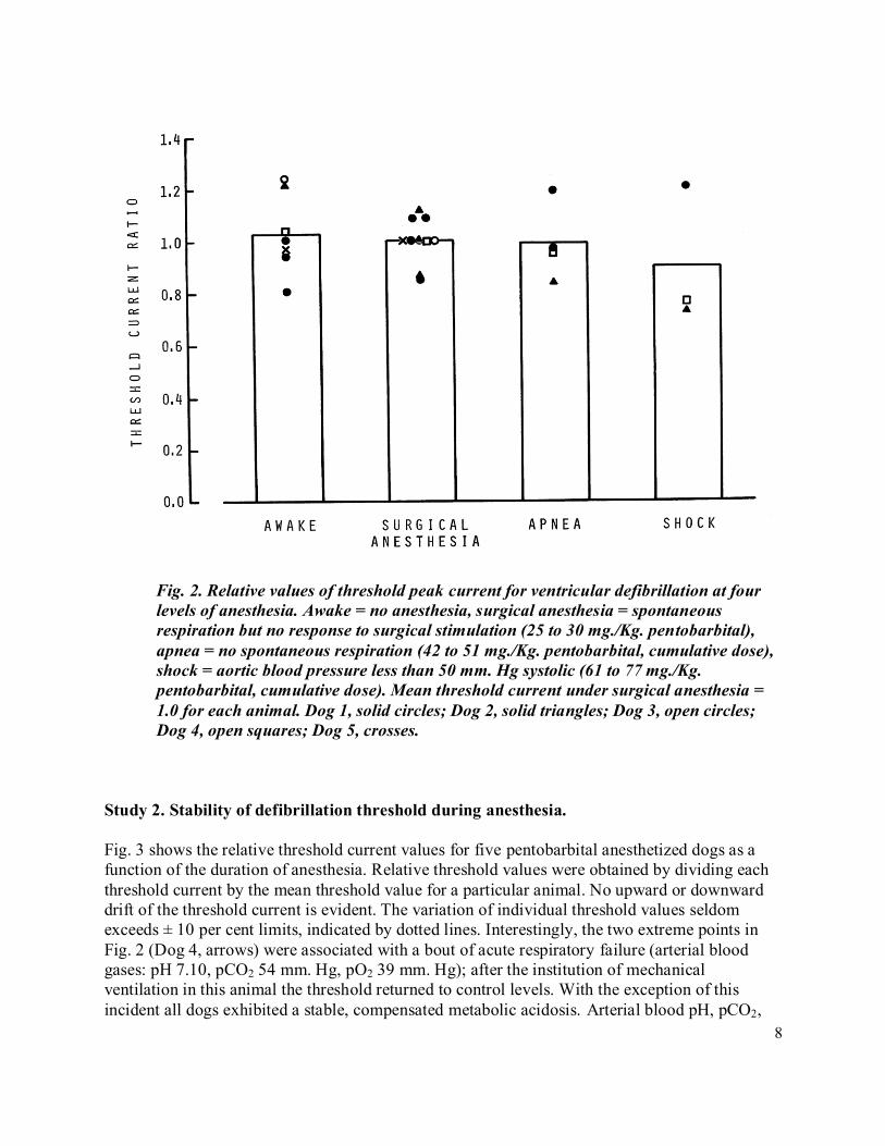
the average threshold under surgical anesthesia for each animal, are plotted in Fig. 2. No
consistent effect of pentobarbital anesthesia on the ventricular fibrillation threshold is evident.
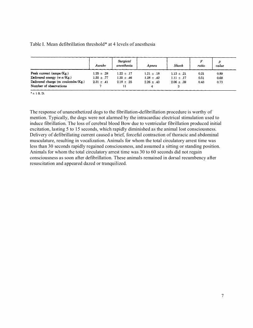
Mean values of threshold shock strength (on the final day of testing) in terms of peak current,
delivered energy, and delivered charge per kilogram of body weight are given in Table I. One-
way analyses of variance indicate that the observed effects of anesthesia level upon threshold
current, energy, and charge are far from statistically significant, as indicated by the p values in
the table.
6
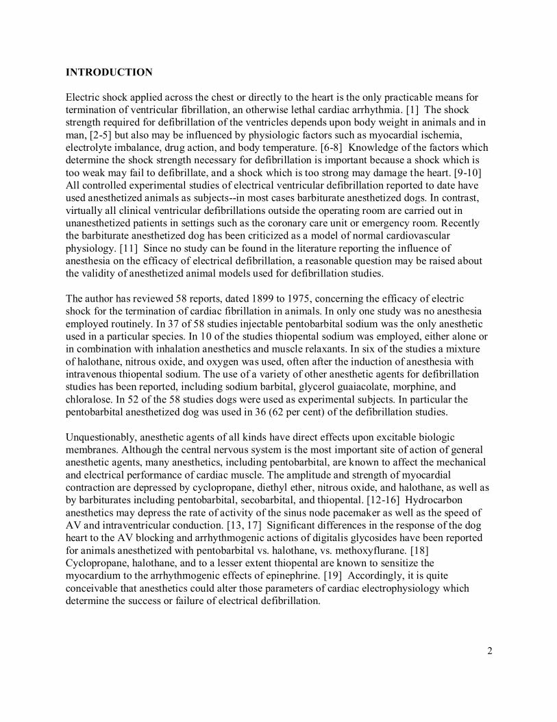
Fig. 1. Threshold peak current for ventricular defibrillation in unanesthetized and
anesthetized states on successive trials in the same animal. On three successive trials
the threshold current in the unanesthetized animal was greater than, equal to, and less
than the threshold current after induction of pentobarbital anesthesia. Mean threshold
before anesthesia = 1.02 A/Kg; mean threshold after anesthesia = 1.08 A/Kg.
Threshold data for the unanesthetized dog were reproducible within ± 10 per cent
limits.
7
Table I. Mean defibrillation threshold* at 4 levels of anesthesia
The response of unanesthetized dogs to the fibrillation-defibrillation procedure is worthy of
mention. Typically, the dogs were not alarmed by the intracardiac electrical stimulation used to
induce fibrillation. The loss of cerebral blood Bow due to ventricular fibrillation produced initial
excitation, lasting 5 to 15 seconds, which rapidly diminished as the animal lost consciousness.
Delivery of defibrillating current caused a brief, forceful contraction of thoracic and abdominal
musculature, resulting in vocalization. Animals for whom the total circulatory arrest time was
less than 30 seconds rapidly regained consciousness, and assumed a sitting or standing position.
Animals for whom the total circulatory arrest time was 30 to 60 seconds did not regain
consciousness as soon after defibrillation. These animals remained in dorsal recumbency after
resuscitation and appeared dazed or tranquilized.
8
Fig. 2. Relative values of threshold peak current for ventricular defibrillation at four
levels of anesthesia. Awake = no anesthesia, surgical anesthesia = spontaneous
respiration but no response to surgical stimulation (25 to 30 mg./Kg. pentobarbital),
apnea = no spontaneous respiration (42 to 51 mg./Kg. pentobarbital, cumulative dose),
shock = aortic blood pressure less than 50 mm. Hg systolic (61 to 77 mg./Kg.
pentobarbital, cumulative dose). Mean threshold current under surgical anesthesia =
1.0 for each animal. Dog 1, solid circles; Dog 2, solid triangles; Dog 3, open circles;
Dog 4, open squares; Dog 5, crosses.
Study 2. Stability of defibrillation threshold during anesthesia.
Fig. 3 shows the relative threshold current values for five pentobarbital anesthetized dogs as a
function of the duration of anesthesia. Relative threshold values were obtained by dividing each
threshold current by the mean threshold value for a particular animal. No upward or downward
drift of the threshold current is evident. The variation of individual threshold values seldom
exceeds ± 10 per cent limits, indicated by dotted lines. Interestingly, the two extreme points in
Fig. 2 (Dog 4, arrows) were associated with a bout of acute respiratory failure (arterial blood
gases: pH 7.10, pCO2 54 mm. Hg, pO2 39 mm. Hg); after the institution of mechanical
ventilation in this animal the threshold returned to control levels. With the exception of this
incident all dogs exhibited a stable, compensated metabolic acidosis. Arterial blood pH, pCO2,
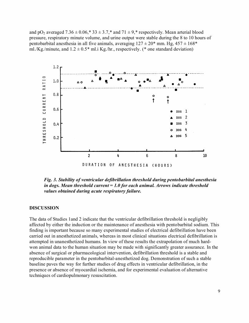
9
and pO2 averaged 7.36 ± 0.06,* 33 ± 3.7,* and 71 ± 9,* respectively. Mean arterial blood
pressure, respiratory minute volume, and urine output were stable during the 8 to 10 hours of
pentobarbital anesthesia in all five animals, averaging 127 ± 20* mm. Hg, 457 ± 168*
ml./Kg./minute, and 1.2 ± 0.5* ml.i Kg./hr., respectively. (* one standard deviation)
Fig. 3. Stability of ventricular defibrillation threshold during pentobarbital anesthesia
in dogs. Mean threshold current = 1.0 for each animal. Arrows indicate threshold
values obtained during acute respiratory failure.
DISCUSSION
The data of Studies 1and 2 indicate that the ventricular defibrillation threshold is negligibly
affected by either the induction or the maintenance of anesthesia with pentobarbital sodium. This
finding is important because so many experimental studies of electrical defibrillation have been
carried out in anesthetized animals, whereas in most clinical situations electrical defibrillation is
attempted in unanesthetized humans. In view of these results the extrapolation of much hard
won animal data to the human situation may be made with significantly greater assurance. In the
absence of surgical or pharmacological intervention, defibrillation threshold is a stable and
reproducible parameter in the pentobarbitalanesthetized dog. Demonstration of such a stable
baseline paves the way for further studies of drug effects in ventricular defibrillation, in the
presence or absence of myocardial ischemia, and for experimental evaluation of alternative
techniques of cardiopulmonary resuscitation.

10
REFERENCES
1. Geddes, L. A.: Electrical ventricular defibrillation, Cardiovasc. Res. Center Bull. 10:3,
1971.
2. Geddes, L.A., Tacker, W. A., Rosborough, J.P., Moore, A.,G., and Cabler, P. S.:
Electrical dose for ventricular defibrillation of large and small animals using precordial
electrodes, J. Clin. Invest. 53:310, 1974.
3. Geddes, L.A., Tacker, W. A., Rosborough, J.P., Moore, A.,G., Cabler, B. S., Bailey, M.,
McCrady, J. D., and Witzel, D.: The electrical dose for ventricular defibrillation with electrodes
applied directly to the heart, J. Thorac. Cardiovasc. Surg. 68:593, 1974.
4. Tacker, W. A., Galioto, F. M., Giuliani, E., Geddes, L.A., and McNamara, D. G.: Energy
dose for human transchest electrical ventricular defibrillation, N. Engl. J. Med. 290:214, 1974.
5. Gutgesell, H. P., Tacker, W. A., Geddes, L. A., Davis, J.D., Lie, J. T., and McNamara, D.
G.: Energy dose for ventricular defibrillation in children, Pediatrics 58:898, 1977.
6. Tacker, W. A., Geddes, L. A., Cabler, P. S., and Moore, A.G.: Electrical threshold for
defibrillation of canine ventricles following myocardial infarction, AM. HEART J. 88:476, 1974.
7. Tacker, W. A., Geddes, L. A., Kline, B., and Borton, C.: Alteration of electrical
defibrillation threshold by the cardiac glycoside, ouabain; Proceedings of the Cardiac
Defibrillation Conference, Purdue University, West Lafayette, Indiana, 1975, pp. 129-133.
8. Babbs, C. F., Abendschein, D. R., Tacker, W. A., and Geddes, L.A.: Subject-dependent factors
in ventricular defibrillation, Medical Instrumentation 10:52, 1976.
9. Davis, J. S., Lie, J. T., Bentinch, D. C., Titus, J. L., Tacker, W. A., and Geddes, L.A.:
Cardiac damage due to electric current and energy; Proceedings of the Cardiac Defibrillation
Conference, Purdue University, West Lafayette, Indiana, 1975, pp. 27-32.
10. Dahl, C. F., Ewy, G. A., Warner, E. D., and Thomas, E. D.: Myocardial necrosis from
direct current countershock: Effect of paddle electrode size and time interval between discharges,
Circulation 50:956, 1974.
11. Priano, L. L., Traber, D. L., and Wilson, R. D.: Barbiturate anesthesia: an abnormal
physiologic situation, J. Pharmacol. Exp. Ther. 165:126, 1969.
12. Price, H. L., and Helrich, M.: The effect of cyclopropane, diethyl ether, nitrous oxide,
thiopental, and hydrogen ion concentration on the myocardial function of the dog heart-lung
preparation, J. Pharmacol. Exp. Ther. 115:206, 1955.
11
13. Flacke, W., and Alper, M. H.: Actions of halothane and norepinephrine in the isolated
mammalian heart, Anesthesiology 23:793, 1962.
14. Hardman, H. F., Moore, J. I., and Lum, B. K. B.: A method for analyzing the effect of pH
and the ionization of drugs upon cardiac tissue with special reference to pentobarbital, J.
Pharmacol. Exp. Ther. 126:136, 1959.
15. Effendi, H., Versprille, A., and Wise, M. E.: The negative inotropic effect of barbiturates on
the heart of newborn and adult guinea-pigs, Arch. Int. Pharmacodyn. 209:127, 1973.
16. Etsten, B., and Li, T. H.: Hemodynamic changes during thiopental anesthesia in humans:
cardiac output, stroke volume, total peripheral resistance, and intrathoracic blood volume, J.
Clin. Invest. 34:500, 1955.
17. Smith, S. L., Webb, W. R., Sabian, L. W., and Hagaman, V. D.: Cardiac excitability in
ether, cyclopropane and halothane anesthesia, Anesthesiology 23:766, 1962.
18. Morrow, D. H., Haley, J. V., and Logic, J. R.: Anesthesia and digitalis VII. The effect of
pentobarbital, halothane, and methoxyflurane on the A-V conduction and inotropic responses to
ouabain, Anesth. Analg. 51:430, 1972.
19. Dresel, P. E.: Cyclopropane-epinephrine cardiac arrhythmias, in Price, H. L., and Cohen,
P. J., Effects of anesthetics on the circulation, Springfield, Illinois, 1964, Charles C. Thomas.
20. Brodie, B. B., Borns, J. J., More, L. C., Lief, P. A., Burnstein, E., and Papper, E. M.: The
fate of pentobarbital in man and dog and a method for its estimation in biological material, J.
Pharmacol Exp. Ther. 109:26, 1953.
21. Price, H. L., Kounat, P. J., Safer, J. N., Couner, E. H., and Price, M. L.: The uptake of
thiopental of body tissues and its relation to the duration of narcosis, Clin. Pharmacol. Ther.
1:16, 1960.
22. Goldstein, A., and Aronow, L.: The durations of action of thiopental and pentobarbital, J.
Pharmacol. Exp. Ther. 128:1, 1960.
23. Guide for the care and use of laboratory animals, U. S. Department of Health, Education
and Welfare, DHEW Pub. No. (NIH) 73-23, Revised 1972.
24. Regulations for animal facilities and care, Purdue University, May, 1972.
25. Babbs, C. F., and Whistler, S. J.: Evaluation of the operating internal resistance,
inductance, and capacitance of intact damped sine wave defibrillators, Medical Instrumentation.
Medical Instrumentation 12:34, 1978.