Embed Size (px)
Citation preview
lable at ScienceDirect
Journal of Pharmaceutical Sciences 106 (2017) 3486-3498
Contents lists avai
Journal of Pharmaceutical Sciences
journal homepage: www.jpharmsci .org
Pharmaceutical Biotechnology
Effect of Polysorbate 20 and Polysorbate 80 on the Higher-OrderStructure of a Monoclonal Antibody and Its Fab and Fc FragmentsProbed Using 2D Nuclear Magnetic Resonance Spectroscopy
Surinder M. Singh 1, Swati Bandi 1, David N.M. Jones 2, 3, Krishna M.G. Mallela 1, 3, *
1 Department of Pharmaceutical Sciences and Center for Pharmaceutical Biotechnology, Skaggs School of Pharmacy and Pharmaceutical Sciences,University of Colorado Anschutz Medical Campus, Aurora, Colorado 800452 Department of Pharmacology, School of Medicine, University of Colorado Anschutz Medical Campus, Aurora, Colorado 800453 Program in Structural Biology and Biochemistry, University of Colorado Anschutz Medical Campus, Aurora, Colorado 80045
a r t i c l e i n f o
Article history:Received 6 April 2017Revised 15 August 2017Accepted 17 August 2017Available online 24 August 2017
Keywords:proteinsexcipientssurfactantsstabilityprotein aggregationprotein structureprotein formulationstabilizationprotein bindingphysical characterization
Abbreviations used: CD, circular dichroism; DSC, detry; Fab, antigen-binding fragment; Fc, crystallizabtitration calorimetry; Kd, dissociation constant; mAb,nuclear magnetic resonance; PS, polysorbate; PS20,sorbate 80; Tonset, onset temperature.The authors Surinder M. Singh and Swati Bandi contrThis article contains supplementary material availableor via the Internet at https://doi.org/10.1016/j.xphs.20* Correspondence to: Krishna M.G. Mallela (T
Fax: þ303-724-7266).E-mail address: [email protected] (K.
http://dx.doi.org/10.1016/j.xphs.2017.08.0110022-3549/© 2017 American Pharmacists Association
a b s t r a c t
We examined how polysorbate 20 (PS20; Tween 20) and polysorbate 80 (PS80; Tween 80) affect thehigher-order structure of a monoclonal antibody (mAb) and its antigen-binding (Fab) and crystallizable(Fc) fragments, using near-UV circular dichroism and 2D nuclear magnetic resonance (NMR). Bothpolysorbates bind to the mAb with submillimolar affinity. Binding causes significant changes in thetertiary structure of mAb with no changes in its secondary structure. 2D 13C-1H methyl NMR indicatesthat with increasing concentration of polysorbates, the Fab region showed a decrease in crosspeakvolumes. In addition to volume changes, PS20 caused significant changes in the chemical shifts comparedto no changes in the case of PS80. No such changes in crosspeak volumes or chemical shifts wereobserved in the case of Fc region, indicating that polysorbates predominantly affect the Fab regioncompared to the Fc region. This differential effect of polysorbates on the Fab and Fc regions was becauseof the lesser thermodynamic stability of the Fab compared to the Fc. These results further indicate thatPS80 is the preferred polysorbate for this mAb formulation, because it offers higher protection againstaggregation, causes lesser structural perturbation, and has weaker binding affinity with fewer bindingsites compared to PS20.
© 2017 American Pharmacists Association®. Published by Elsevier Inc. All rights reserved.
Introduction
Conformational integrity of therapeutic proteins encountersnumerous challenges from manufacturing through eventual in-jection in intended patient which include exposure to various in-terfaces (air-liquid, liquid-liquid, and liquid-solid). Many processessuch as filtration,1 storage,2 freeze-thaw,3 vial filling,4 agitation,5
lyophilization,6 and final injection7 expose therapeutic proteins to
ifferential scanning calorim-le fragment; ITC, isothermalmonoclonal antibody; NMR,polysorbate 20; PS80, poly-
ibuted equally.from the authors by request17.08.011.elephone: þ303-724-3576;
M.G. Mallela).
®. Published by Elsevier Inc. All rig
denaturing interfaces resulting in significant protein aggregation.One strategy to minimize interface-induced protein aggregation isto include polysorbates, such as polysorbate 20 (PS20; Fig. 1a) orpolysorbate 80 (PS80; Fig. 1b) in protein formulations. Polysorbatesare a class of nonionic detergents that protect biotherapeuticsagainst destabilizing environments at various interfaces.8-13 Owingto large fatty acid aliphatic chains, critical micelle concentration(CMC) for polysorbates, which is the maximum concentration ofmonomeric polysorbate available in solution, is very low. Theapproximate CMC values for PS20 and PS80 are 55 mM and 13 mM,respectively.14,15 Increased hydrophobicity of longer fatty acid(monooleate vs. monolaureate) underlies the lower CMC of PS80(13 mMor 0.001% w/v) when compared with PS20 (CMC¼ 55 mMor0.006% w/v). Therapeutic protein formulations contain poly-sorbates in the range of 0.003%-0.8% w/v,16-18 for example, Humira(adalimumab) contains 0.1% w/v PS80,17 Raptiva (efalizumab)contains 0.2% PS20,17 and Tecentriq (atezolizumab) contains 0.8%w/v PS20 (https://www.accessdata.fda.gov/drugsatfda_docs/label/2016/761034s000lbl.pdf). Although the protection of proteinbiotherapeutics by polysorbates against interface-induced
hts reserved.
Polysorbate 20
Polysorbate 80
Antibody
O
OHO
O
( )
O
OOH
OH
O
O
)(z y
)( x
)(w
O
OHO
O
( )
O
OOH
OH
O
O
)(z y
)( x
)(w
w+x+y+z=20
w+x+y+z=20
NN N
N
C C
C C
CH2CH2
CH3CH3
CH1CH1
VH VH
CL CL
VLVL
Fab
Fc
-S-S--S-S-
-S-S--S-S-
Papaincleavage
site
Particle Diameter (μm)2 3 4 5 6 7 8 9 10
# of
par
ticle
s/m
l
0
500
1000
20000
40000
60000
80000mAbmAb + PS20mAb + PS80
2 3 4 5 6 7 8 9 100
10
20
30
40
a
b
c
d
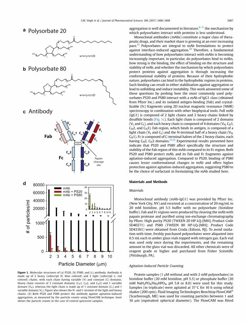
Figure 1. Molecular structures of (a) PS20, (b) PS80, and (c) antibody. Antibody ismade up of 2 heavy (subscript H; blue colored) and 2 light (subscript L; redcolored) chains, with each chain having variable (V) and constant (C) domains.Heavy chain consists of 3 constant domains (CH1, CH2, and CH3) and 1 variabledomain (VH), whereas the light chain is made up of 1 constant domain (CL) and 1variable domain (VL). Figure also shows the N- and C-termini of the light and heavychains. (d) Both PS20 and PS80 protect the antibody against agitation-inducedaggregation, as measured by the particle counts using FlowCAM technique. Insetshows the particle counts in the case of control quiescent samples.
S.M. Singh et al. / Journal of Pharmaceutical Sciences 106 (2017) 3486-3498 3487
aggregation is well documented in literature,8-11 the mechanism bywhich polysorbates interact with proteins is less understood.
Monoclonal antibodies (mAbs) constitute a major class of thera-peutic drugs, and their market share is growing at an ever increasingpace.19 Polysorbates are integral to mAb formulations to protectagainst interface-induced aggregation.20 Therefore, a fundamentalunderstanding of how polysorbates interact with mAbs is becomingincreasingly important, in particular, do polysorbates bind to mAbs,how strong is the binding, the effect of binding on the structure andstability of mAb, andwhether themechanism bywhich polysorbatesprotect proteins against aggregation is through increasing theconformational stability of proteins. Because of their hydrophobicnature, polysorbates can bind to the hydrophobic regions in proteins.Such binding can result in either stabilization against aggregation orlead to unfolding and induce instability. This work answered some ofthese questions by probing how the most commonly used poly-sorbates PS20 and PS80 interact with a mAb of IgG1 class (obtainedfrom Pfizer Inc.) and its isolated antigen-binding (Fab) and crystal-lizable (Fc) fragments using 2D nuclear magnetic resonance (NMR)spectroscopy in combination with other biophysical tools. Full mAb(IgG1) is composed of 2 light chains and 2 heavy chains linked bydisulfide bonds (Fig. 1c). Each light chain is composed of 2 domains(VL and CL), and each heavy chain is composed of 4 domains (VH, CH1,CH2, and CH3). Fab region, which binds to antigen, is composed of alight chain (VL and CL) and the N-terminal half of a heavy chain (VH,
CH1). Fc is composed of C-terminal halves of the 2 heavy chains, eachhaving CH2, CH3 domains.21,22 Experimental results presented hereindicate that PS20 and PS80 affect specifically the structure andstability of the Fab region of this mAb compared to its Fc region. BothPS20 and PS80 protect mAb, and its Fab and Fc fragments againstagitation-induced aggregation. Compared to PS20, binding of PS80causes lesser conformational changes in mAb and offers higherprotection against agitation-induced aggregation, suggesting PS80 tobe the choice of surfactant in formulating the mAb studied here.
Materials and Methods
Materials
Monoclonal antibody (mAb-IgG1) was provided by Pfizer Inc.(New York City, NY) and received at a concentration of 20 mg/mL in20 mM histidine, pH 5.5 buffer with no polysorbate (histidinebuffer). Fab and Fc regionswere produced by cleaving themAbwithpapain protease and purified using ion-exchange chromatographyby Pfizer. High purity PS20 (TWEEN 20 HP-LQ-[MH], Product CodeSD40271) and PS80 (TWEEN 80 HP-LQ-[MH]; Product CodeSD43361) were obtained from Croda (Edison, NJ). To avoid oxida-tion with time, freshly purchased polysorbates were aliquoted into0.5 mL each in amber glass vials topped with nitrogen gas. Each vialwas used only once during the experiments, and the remainingamount in the glass vial was discarded. All other chemicals were ofreagent grade or higher and purchased from Fisher Scientific(Pittsburgh, PA).
Agitation-Induced Particle Counting
Protein samples (1 mM without and with 2 mM polysorbates) inhistidine buffer (20 mM histidine, pH 5.5) or phosphate buffer (20mM NaH2PO4/Na2HPO4, pH 5.8 or 8.0) were used for this study.Samples (in triplicate) were agitated at 37�C for 16 h using orbitalshaker (200 rpm). Fluid Imaging Technologies Benchtop FlowCAM®
(Scarborough, ME) was used for counting particles between 1 and10 mm (equivalent spherical diameter). The FlowCAM was fitted
0 2 4 6 8 10 12 14
-0.70
-0.60
-0.50
-0.40
-0.30
-0.20
-0.10
0.00
-1.60
-1.40
-1.20
-1.00
-0.80
-0.60
-0.40
-0.20
0.00
0 10 20 30 40 50 60 70
Time (min)
ces/lacµ
Molar Ratio
tnatcejni fo elom/lack
0 2 4 6 8 10 12 14-0.40
-0.35
-0.30
-0.25
-0.20
-0.15
-0.10
-0.05
0.00
-0.60
-0.50
-0.40
-0.30
-0.20
-0.10
0.00
0 10 20 30 40 50 60 70
Time (min)
ces/lacµMolar Ratio
tnatcejni fo elom/lack
08SP 02SP
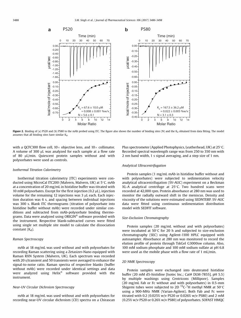
Kd = 67.6 ± 10.0 μM = 0.008 ± 0.001 %w/vN = 5.6 ± 0.1
Kd = 167.5 ± 36.2 μM = 0.022 ± 0.005 %w/vN = 3.1 ± 0.3
a b
Figure 2. Binding of (a) PS20 and (b) PS80 to the mAb probed using ITC. The figure also shows the number of binding sites (N) and the Kd obtained from data fitting. The modelassumes that all binding sites have similar Kd.
S.M. Singh et al. / Journal of Pharmaceutical Sciences 106 (2017) 3486-34983488
with a QCFC300 flow cell, 10� objective lens, and 10� collimator.A volume of 300 mL was analyzed for each sample at a flow rateof 80 mL/min. Quiescent protein samples without and withpolysorbates were used as controls.
Isothermal Titration Calorimetry
Isothermal titration calorimetry (ITC) experiments were con-ducted using MicroCal ITC200 (Malvern, Malvern, UK) at 5�C. mAbat a concentration of 20 mg/mL in histidine buffer was titrated with10 mM polysorbates. Except for the first injection (0.2 mL), injectionvolume for the remaining 12 injections was 3 mL each. Each injec-tion duration was 6 s, and spacing between individual injectionswas 300 s. Blank ITC thermograms (titration of polysorbate intohistidine buffer without mAb) were recorded under similar con-ditions and subtracted from mAb-polysorbate binding thermo-grams. Data were analyzed using ORIGIN® software provided withthe instrument. Respective blank-subtracted curves were fittedusing single set multiple site model to calculate the dissociationconstant (Kd).
Raman Spectroscopy
mAb at 18 mg/mL was used without and with polysorbates forrecording Raman scattering using a Zetasizer-Nano equipped withRaman RXN System (Malvern, UK). Each spectrum was recordedwith 20 s/transient and 50 transients were averaged to enhance thesignal-to-noise ratio. Raman spectra of respective blanks (bufferwithout mAb) were recorded under identical settings and datawere analyzed using Helix® software provided with theinstrument.
Near-UV Circular Dichroism Spectroscopy
mAb at 18 mg/mL was used without and with polysorbates forrecording near-UV circular dichroism (CD) spectra on a Chirascan
Plus spectrometer (Applied Photophysics, Leatherhead, UK) at 25�C.Recorded spectral wavelength range was from 250 to 350 nmwith2 nm band width, 1 s signal averaging, and a step size of 1 nm.
Analytical Ultracentrifugation
Protein samples (1 mg/mL mAb in histidine buffer without andwith polysorbates) were subjected to sedimentation velocityanalytical ultracentrifugation (SV-AUC) experiment on a BeckmanXL-A analytical centrifuge at 25�C. Two hundred scans wererecorded at 42,000 rpm. Protein absorbance at 280 nmwas used tomonitor the radially outward shift in the meniscus. Density andviscosity of the solutions were estimated using SEDNTERP. SV-AUCdata were fitted using continuous sedimentation distributionmodel with SEDFIT software.
Size-Exclusion Chromatography
Protein samples (20 mg/mL without and with polysorbates)were incubated at 50�C for 20 h and subjected to size-exclusionchromatography (SEC) using Agilent-1100 HPLC equipped withautosampler. Absorbance at 280 nm was monitored to record theelution profile of protein through TskGel G3000sw column. Also,100 mM sodium phosphate and 100 mM sodium sulfate at pH 6.8were used as the mobile phase with a flow rate of 1 mL/min.
2D NMR Spectroscopy
Protein samples were exchanged into deuterated histidinebuffer (20 mM d5-histidine [Isotec Inc.; Cat# DLM-7855], pH 5.5)by multiple washings using Centricons (Millipore). Samples(20 mg/mL Fab or Fc without and with polysorbates) in 0.5-mmShigemi tubes were subjected to 2D 13C-1H methyl NMR at 50�Cusing a 900-MHz NMR (Varian-Agilent). Both Fab and Fc weretitrated with 0.2 (0.025% w/v PS20 or 0.026% w/v PS80) and 2 mM(0.25% w/v PS20 or 0.26% w/v PS80) of polysorbates. SOFAST HMQC

1500 1600 1700-0.5
0.0
0.5
1.0 mAbmAb+2 mM PS20mAb+2 mM PS80
Raman shift, Δν (cm-1)
Ram
an S
catte
ring
Inte
nsity
(Nor
mal
ized
)
ba
Sedimentation coefficient (S)0 5 10 15 20 25
c(s)
0.0
0.5
1.0
1.5
2.0
2.5
0 5 10 15 200
50
100
150
Milli
Abs
orba
nce
@ 2
80 n
m
Retention time (min)
dcmAbmAb+2 mM PS20mAb+2 mM PS80
mAbmAb+2 mM PS20mAb+2 mM PS80
e
Temperature (oC)30 45 60 75 90
Mea
n R
esid
ue e
llipt
icity
(deg
cm
2 dm
ol-1)
-100
-80
-60
-40
mAbmAb+PS20mAb+PS80
mAbmAb+2 mM PS20mAb+2 mM PS80
Mea
n R
esid
ue e
llipt
icity
(deg
cm
2 dm
ol-1)
-120
-80
-40
0
40
Wavelength (nm)250 300 350
Figure 3. Effect of polysorbate binding on (a) the secondary structure of mAb as probed by Raman scattering, and (b) the tertiary structure of mAb as probed by near-UV CD. Both (c)AUC and (d) SEC indicate that mAb is a monomer in the absence and presence of polysorbates. The peak in the SEC chromatogram (panel d) at the elution time of 11 min correspondto the histidine present in the buffer. (e) Change in the near-UV CD signal as a function of increasing solution temperature. Both PS20 and PS80 affect the partial protein unfoldingwith relatively no change in the global unfolding of mAb. In all the panels, black, red, and green curves represent the data in the absence of polysorbates, with PS20, and with PS80,respectively. In panel (b), triplicate data have been shown for each experimental condition to indicate the statistical significance of the changes in spectra with the addition ofpolysorbates.
S.M. Singh et al. / Journal of Pharmaceutical Sciences 106 (2017) 3486-3498 3489
pulse sequence from VNMRJ 4.0 Biopack was used for recording theNMR spectra after optimizing the required parameters. Spectrawere collected with a spectral width of 14,044.9 Hz in 1H dimen-sion and 7239.06 Hz in 13C dimension. Number of transients in thesecond dimension was 128. Raw data (FID) were processed usingNMRPipe software with zero filling to twice the real points, solventfiltered, apodized using SP (Sine-bell), GMB (Gaussian), and Poly(baseline correction) function. Changes in chemical shifts and peakvolumes were analyzed using NMRPipe.
Chemical Denaturation Melts
For denaturation melts of the mAb, Fab, and Fc, 1 mM protein inhistidine buffer (pH 5.5) was used. Guanidinium chloride (GdmCl)was used as the denaturant. Protein samples were prepared atvarying GdmCl concentrations and equilibrated for 1 h. Changes inthe intrinsic protein fluorescence of aromatic amino acids (excita-tion at 280 nm, emission at 355 nm) were monitored as a functionof increasing GdmCl concentration.

S.M. Singh et al. / Journal of Pharmaceutical Sciences 106 (2017) 3486-34983490
Differential Scanning Calorimetry
Excess heat thermograms for thermal denaturation of mAb, Fab,and Fc were recorded using MicroCal VP-Capillary DSC equippedwith an autosampler in either histidine buffer or phosphate buffer.Protein concentrationwas 0.5mg/mL. Thermogramswere recordedfrom 25�C to 100�C with a thermal ramp of 90�C/h. Thermogramsof respective blanks (buffer without protein) were recorded underidentical settings. ORIGIN® analysis software provided with theinstrument was used to analyze protein denaturationthermograms.
Results
PS20 and PS80 Protect mAb Against Agitation-Induced Aggregation
mAb (without and with PS20 or PS80) was subjected to over-night agitation at 37�C and the number of microparticles wascounted using FlowCAM. Number of particles (>1 mm) was signif-icantly reduced by ~100 times in the presence of PS20 whencompared with no polysorbate in the solution (Fig. 1d). With PS80,the number of microparticles were much less compared to PS20,indicating that PS80 provided better protection against agitation-induced aggregation. Number of particles were less than 30 forquiescent samples (inset of Fig.1d). These particle data clearly showthat both polysorbates PS20 and PS80 protect mAb againstagitation-induced aggregation.
PS20 and PS80 Bind to mAb With a Submillimolar Kd
ITC was used for measuring binding. PS20 or PS80 was titratedwith mAb in small successive installments, thereby saturating thebinding sites on the protein.23 The heat transfer data associatedwith ligand binding was fitted to obtain the Kd and the reactionstoichiometry (N) (Fig. 2). PS20 was bound to mAbwith a Kd of 67.6± 10.0 mM (0.008 ± 0.001%w/v) and N¼ 5.6 ± 0.1 molecules of PS20binding to 1 molecule of mAb (Fig. 2a). Correspondingly, PS80 wasbound to themAbwith a Kd of 167.5± 36.2 mM (0.022± 0.005%w/v)and N ¼ 3.1 ± 0.3 molecules of PS80 binding to 1 molecule of mAb(Fig. 2b). These Kd values are within the polysorbate concentrationrange used in protein formulations (0.003%-0.8% w/v). The lessernumber of binding sites for PS80 compared to PS20might be due toits larger hydrophobic group (oleate) and a kink in the aliphaticsidechain because of cis-double bond causing more stearichindrance when compared with PS20 (monolaurate).
The above Kd values were obtained using the total polysorbateconcentration used for isothermal titration. However, polysorbatesexist as monomers as well as micelles in solution. Previouslypublished data suggest that proteins do not bind to polysorbatemicelles because of the hydrophilic nature of the micelle surface.24
Therefore, if Kd values were to be calculated based on the availablemonomer concentration, the true Kd values would be much lowerthan the above estimated Kd values from ITC measurements. Thespecific binding of proteins to polysorbate monomers could alsopartly explain the previously observed increase in the apparentCMC of polysorbates in the presence of proteins,25 because thepresence of proteins depletes the concentration of free monomersavailable for micelle formation by binding to the monomeric formof polysorbates.
PS20 and PS80 Perturb the Tertiary Structure of mAb, but Not ItsSecondary Structure
Raman scattering and near-UV CD were used for measuring theeffect of polysorbates on the secondary and tertiary structures of
mAb, respectively. Far-UV CD could not be used for these samplesbecause histidine present in the buffer significantly absorbs in thiswavelength range. Amide I region in the Raman spectrum is sen-sitive to changes in the secondary structure of a protein.26 Anti-bodies exhibit a Raman amide I band at 1674 cm�1 characteristic oftheir b-structure (Fig. 3a).26 Addition of polysorbates did not causesignificant changes in the Raman spectrum, indicating that PS20 orPS80 did not perturb the secondary structure of mAb.
Near-UV CD is a highly sensitive method to evaluate the impactof various buffers and processes on the tertiary structure of bio-therapeutic proteins.24,27 Binding of polysorbates caused significantchanges in the near-UV CD spectrum (Fig. 3b), indicating that thePS20 and PS80 affected the tertiary structure of mAb. Figure 3bshows the triplicate data for each experimental condition to indi-cate the statistical significance of the changes in spectra with theaddition of polysorbates. Quantitative spectral similarity wascompared using the weighted spectral difference (WSD) parameterdescribed earlier.28 Spectral variation between samples underdifferent solution conditions (WSD ¼ 61% for PS20 and WSD ¼ 12%for PS80, with respect to the control sample with no polysorbates)is much higher than the spectral variation between differentsamples under identical solution conditions (WSD <5%). Near-UVCD measures chirality of the structural environment around thesidechains of aromatic amino acids (Phe, Tyr, and Trp). Changes inthe intensity and spectral pattern around 295 nm indicate that theenvironment around tryptophan residues is altered upon theaddition of polysorbates. Although the pattern of spectral regionaround 260 nm is similar, the intensity is significantly changedupon the addition of polysorbates, which is indicative of the ter-tiary structural changes around tyrosines and phenylalanine resi-dues as well. Furthermore, mAb tertiary structure perturbation ismore pronounced in the case of PS20 compared to PS80, which isconsistent with the higher binding affinity and stoichiometry ofPS20 when measured by ITC (Fig. 2).
It was further confirmed that the above changes in the tertiarystructure of mAb upon the addition of polysorbates is not due tooligomerization. Both AUC (Fig. 3c) and SEC (Fig. 3d) showed thatthe mAb was a monomer in the absence and in the presence ofpolysorbates. These AUC and SEC data also indicated the purity ofmAb samples received from Pfizer. The small peak observed aroundthe retention time of 11 min in the SEC chromatogram is from thehistidine present in the buffer.
The effect of polysorbates on the tertiary structure of mAb hasalso been observed in its partial unfolding with no effect on itsglobal unfolding (Fig. 3e). The new tertiary structure that is formedin the presence of polysorbates unfolds at lower temperaturescompared to global unfolding at higher temperatures, as indicatedby the appearance of sloped native baseline in the presence ofpolysorbates (Fig. 3e). Sloped baselines in thermal melts can orig-inate either from the presence of partially unfolded states orbecause the signal of the native state changes as a function of so-lution temperature.29-32 Because sloped native baseline was notobserved in the absence of polysorbates, the sloped baselines in thepresence of polysorbates can be attributed solely to the presence ofpartially unfolded states.32 Compared to the effect of polysorbateson partial protein unfolding, polysorbates did not significantlyaffect the global unfolding of mAb, as evident from minimalchanges in the midpoint temperature of the main transition (closeto 66�C) in the near-UV CD thermal melts (Fig. 3e).
PS20 and PS80 Specifically Perturb the Tertiary Structure of FabRegion When Compared to the Fc Region
The main goal of this project was to determine which specificregions in the mAb interact with the polysorbates. Because of the
Retention time (min)0 5 10 15 20M
illi A
bsor
banc
e @
280
nm
0
50
100
150
0 5 10 15 200
50
100
150
Milli
Abs
orba
nce
@ 2
80 n
m
Retention time (min)
Fab Fab+2 mM PS20 Fab+2 mM PS80
Fc Fc+2 mM PS20Fc+2 mM PS80
Mea
n R
esid
ue e
llipt
icity
(deg
cm
2 dm
ol-1)
-100
-50
0
50
100
Fab Fab + 2 mM PS20 Fab + 2 mM PS80
Wavelength (nm)250 300 350
Mea
n R
esid
ue e
llipt
icity
(deg
cm
2 dm
ol-1)
-200
-150
-100
-50
0
50
Wavelength (nm)250 300 350
Fc Fc + 2 mM PS20Fc + 2 mM PS80
ba
dc
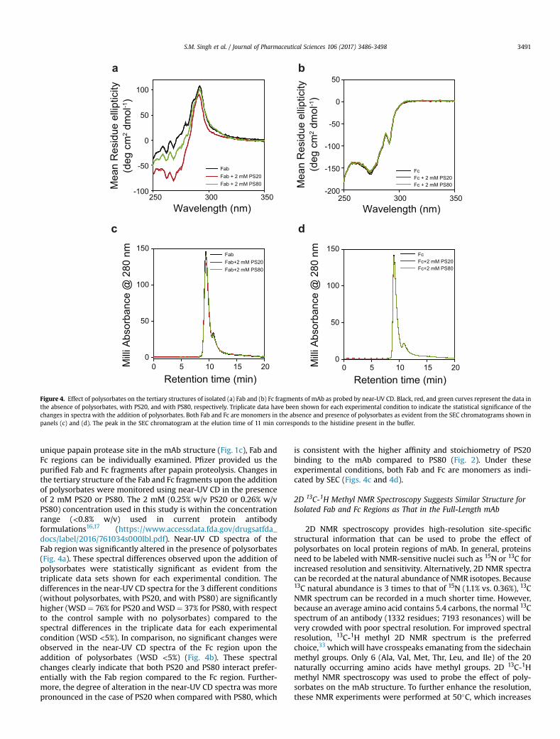
Figure 4. Effect of polysorbates on the tertiary structures of isolated (a) Fab and (b) Fc fragments of mAb as probed by near-UV CD. Black, red, and green curves represent the data inthe absence of polysorbates, with PS20, and with PS80, respectively. Triplicate data have been shown for each experimental condition to indicate the statistical significance of thechanges in spectra with the addition of polysorbates. Both Fab and Fc are monomers in the absence and presence of polysorbates as evident from the SEC chromatograms shown inpanels (c) and (d). The peak in the SEC chromatogram at the elution time of 11 min corresponds to the histidine present in the buffer.
S.M. Singh et al. / Journal of Pharmaceutical Sciences 106 (2017) 3486-3498 3491
unique papain protease site in the mAb structure (Fig. 1c), Fab andFc regions can be individually examined. Pfizer provided us thepurified Fab and Fc fragments after papain proteolysis. Changes inthe tertiary structure of the Fab and Fc fragments upon the additionof polysorbates were monitored using near-UV CD in the presenceof 2 mM PS20 or PS80. The 2 mM (0.25% w/v PS20 or 0.26% w/vPS80) concentration used in this study is within the concentrationrange (<0.8% w/v) used in current protein antibodyformulations16,17 (https://www.accessdata.fda.gov/drugsatfda_docs/label/2016/761034s000lbl.pdf). Near-UV CD spectra of theFab regionwas significantly altered in the presence of polysorbates(Fig. 4a). These spectral differences observed upon the addition ofpolysorbates were statistically significant as evident from thetriplicate data sets shown for each experimental condition. Thedifferences in the near-UV CD spectra for the 3 different conditions(without polysorbates, with PS20, and with PS80) are significantlyhigher (WSD¼ 76% for PS20 andWSD¼ 37% for PS80, with respectto the control sample with no polysorbates) compared to thespectral differences in the triplicate data for each experimentalcondition (WSD <5%). In comparison, no significant changes wereobserved in the near-UV CD spectra of the Fc region upon theaddition of polysorbates (WSD <5%) (Fig. 4b). These spectralchanges clearly indicate that both PS20 and PS80 interact prefer-entially with the Fab region compared to the Fc region. Further-more, the degree of alteration in the near-UV CD spectra was morepronounced in the case of PS20 when compared with PS80, which
is consistent with the higher affinity and stoichiometry of PS20binding to the mAb compared to PS80 (Fig. 2). Under theseexperimental conditions, both Fab and Fc are monomers as indi-cated by SEC (Figs. 4c and 4d).
2D 13C-1H Methyl NMR Spectroscopy Suggests Similar Structure forIsolated Fab and Fc Regions as That in the Full-Length mAb
2D NMR spectroscopy provides high-resolution site-specificstructural information that can be used to probe the effect ofpolysorbates on local protein regions of mAb. In general, proteinsneed to be labeled with NMR-sensitive nuclei such as 15N or 13C forincreased resolution and sensitivity. Alternatively, 2D NMR spectracan be recorded at the natural abundance of NMR isotopes. Because13C natural abundance is 3 times to that of 15N (1.1% vs. 0.36%), 13CNMR spectrum can be recorded in a much shorter time. However,because an average amino acid contains 5.4 carbons, the normal 13Cspectrum of an antibody (1332 residues; 7193 resonances) will bevery crowded with poor spectral resolution. For improved spectralresolution, 13C-1H methyl 2D NMR spectrum is the preferredchoice,33 which will have crosspeaks emanating from the sidechainmethyl groups. Only 6 (Ala, Val, Met, Thr, Leu, and Ile) of the 20naturally occurring amino acids have methyl groups. 2D 13C-1Hmethyl NMR spectroscopy was used to probe the effect of poly-sorbates on the mAb structure. To further enhance the resolution,these NMR experiments were performed at 50�C, which increases
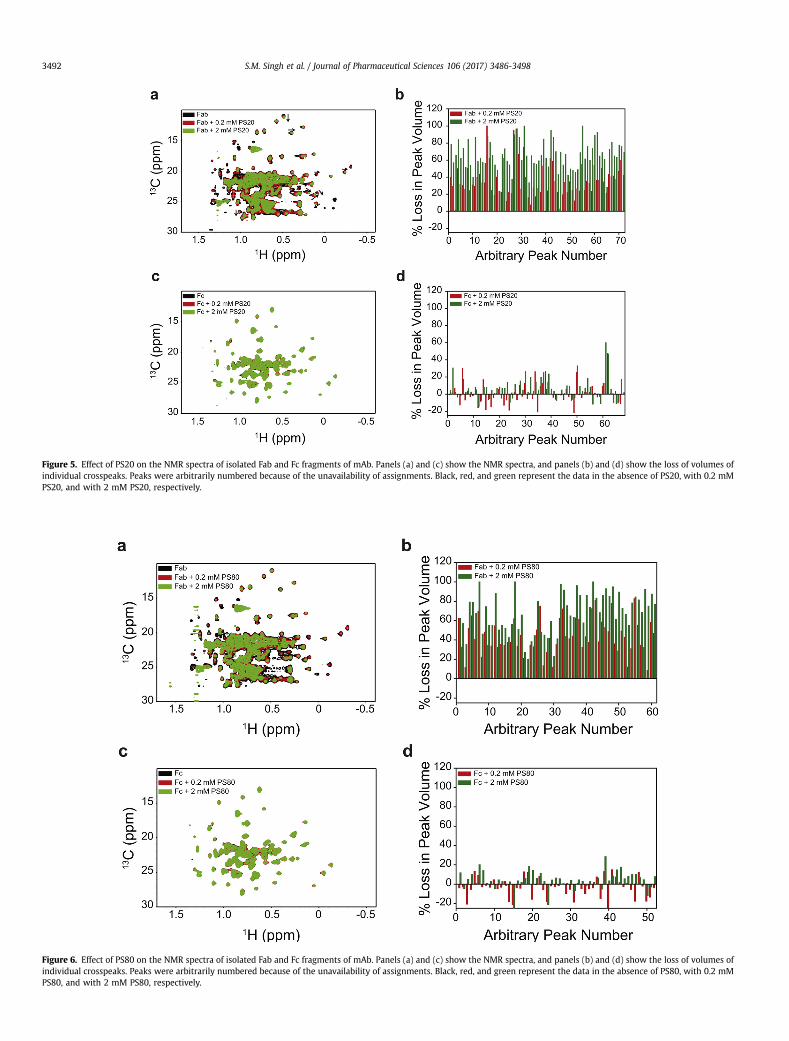
Figure 5. Effect of PS20 on the NMR spectra of isolated Fab and Fc fragments of mAb. Panels (a) and (c) show the NMR spectra, and panels (b) and (d) show the loss of volumes ofindividual crosspeaks. Peaks were arbitrarily numbered because of the unavailability of assignments. Black, red, and green represent the data in the absence of PS20, with 0.2 mMPS20, and with 2 mM PS20, respectively.
Figure 6. Effect of PS80 on the NMR spectra of isolated Fab and Fc fragments of mAb. Panels (a) and (c) show the NMR spectra, and panels (b) and (d) show the loss of volumes ofindividual crosspeaks. Peaks were arbitrarily numbered because of the unavailability of assignments. Black, red, and green represent the data in the absence of PS80, with 0.2 mMPS80, and with 2 mM PS80, respectively.
S.M. Singh et al. / Journal of Pharmaceutical Sciences 106 (2017) 3486-34983492
Temperature (oC)30 45 60 75 90
Cp
(cal
/mol
/0 C)
0
20000
40000
60000
80000 mAbFab Fc
Temperature (oC)30 40 50 60 70 80 90
Cp
(cal
/mol
e/o C
)
0
20000
40000
60000
80000
30 45 60 75 90
0
5000
10000
15000
20000
25000
30 45 60 75 90
0
10000
20000
30000
[GdmCl] (M)0 1 2 3 4 5
Nor
mal
ized
Flu
ores
cenc
e
0.0
0.5
1.0
mAb Fab Fc
Temperature (oC)
Cp
(cal
/mol
e/o C
)
Temperature (oC)
Cp
(cal
/mol
e/o C
)
mAbmAb+PS20 mAb+PS80
FabFab+PS20
FcFc+PS20 Fc+PS80
Fab+PS80
a b
c d
e
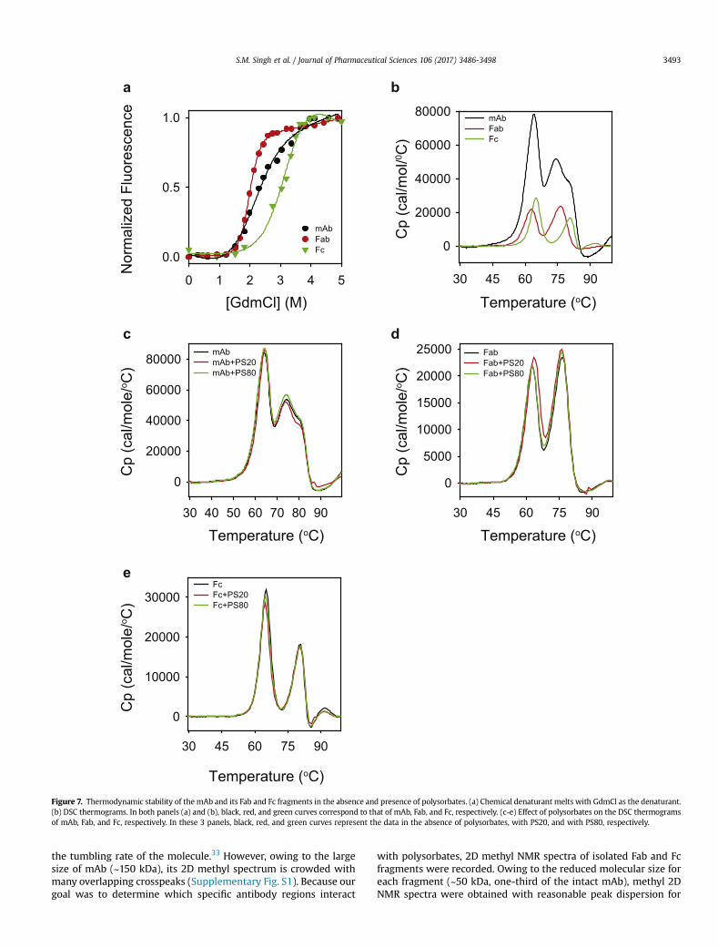
Figure 7. Thermodynamic stability of the mAb and its Fab and Fc fragments in the absence and presence of polysorbates. (a) Chemical denaturant melts with GdmCl as the denaturant.(b) DSC thermograms. In both panels (a) and (b), black, red, and green curves correspond to that of mAb, Fab, and Fc, respectively. (c-e) Effect of polysorbates on the DSC thermogramsof mAb, Fab, and Fc, respectively. In these 3 panels, black, red, and green curves represent the data in the absence of polysorbates, with PS20, and with PS80, respectively.
S.M. Singh et al. / Journal of Pharmaceutical Sciences 106 (2017) 3486-3498 3493
the tumbling rate of the molecule.33 However, owing to the largesize of mAb (~150 kDa), its 2D methyl spectrum is crowded withmany overlapping crosspeaks (Supplementary Fig. S1). Because ourgoal was to determine which specific antibody regions interact
with polysorbates, 2D methyl NMR spectra of isolated Fab and Fcfragments were recorded. Owing to the reduced molecular size foreach fragment (~50 kDa, one-third of the intact mAb), methyl 2DNMR spectra were obtained with reasonable peak dispersion for
S.M. Singh et al. / Journal of Pharmaceutical Sciences 106 (2017) 3486-34983494
both Fab and Fc fragments (Supplementary Fig. S1). More impor-tantly, combined NMR spectra of isolated Fab and Fc regions weresimilar to those of the intact mAb, implying that the isolated Faband Fc fragments retain similar structures as that in the full-lengthmAb.
PS20 and PS80 Predominantly Affect the Fab Region WhenCompared to the Fc
Fab and Fc fragments were titrated with PS20 or PS80 (0, 0.2,and 2mM), and the changes in 2D 13C-1Hmethyl NMR spectrawereshown in Figures 5 and 6. These NMR experiments were performedat 50�C. Increasing the solution temperature from 5�C to 50�C de-creases the CMC of polysorbates (from 90 to 48 mM [1.9 times] forPS20, and from 36 to 12 mM [3 times] for PS80).34 Because CMCdefines the maximum monomer concentration possible at equi-librium under micelle-forming conditions and because CMC de-creases with temperature, the Kd values at 50�C will be higher thanthose measured by ITC at 5�C,35 which implies that thepolysorbate-bound protein population will be lower at 50�C underNMR conditions compared to that at 5�C. Evenwith such decreasedpopulation of polysorbate-bound conformation of mAb, significanteffects of PS20 and PS80 on mAb NMR were observed. Whencompared to the Fc, the NMR crosspeak volumes of Fab showed asignificant decrease upon the addition of either PS20 (Figs. 5b vs.5d) or PS80 (Figs. 6b vs. 6d). The decrease in NMR crosspeak vol-umes of Fab upon the addition of polysorbates was not an artifact ofthe NMR experiment or data processing, as similar decrease wasnot seen in the case of Fc with identical protocols. Most changes inthe NMR crosspeak volumes of the Fc were random and werewithin the errors associated with NMR data processing and peakpicking (<10%). In that sense, Fc acts as a control for Fab in NMRexperiments. These results imply that PS20 and PS80 predomi-nantly affect the Fab region compared to the Fc region.
Decrease in NMR crosspeak volumes of Fab can be because of 5possible reasons: (1) increase in solutionviscosity upon the additionof polysorbates; (2) increase in the molecular size of Fab uponpolysorbate binding; (3) irreversible protein aggregation upon theadditionof polysorbates decreasing themonomer Fab concentrationin solution; (4) formation of transient interactions between Fabmonomers; and (5) increased protein dynamics in the presence ofpolysorbates. Addition of 2 mM polysorbates, which correspond to0.25% w/v PS20 or 0.26% w/v PS80, does not increase solution vis-cosity.36 Furthermore, Fc did not show decrease in crosspeak vol-umeswith the addition of polysorbates, indicating that the observedphenomenon is specific to Fab and is not due to the changes insolvent properties. However, specific binding of polysorbates to Fabcan cause an increase in microviscosity around Fab, whose effectcannot be completely ruled out. Increase in Fab size upon binding topolysorbates can be ruled out, because only a fewer number ofpolysorbate molecules (6 in the case of PS20 and 3 in the case ofPS80) bind tomAb. No visible aggregateswere observed in the NMRsample tube after the completion of the NMR experiments. Inaddition, the SEC chromatogram showed no oligomeric species(Supplementary Fig. S2), indicating the absence of irreversibleprotein aggregation. However, the presence of transient oligomerformation during the NMR experiments resulting in a decrease inNMR peak volumes cannot be completely ruled out. Increasedprotein dynamics30,37-40 in the presence of polysorbates populatingan ensemble of conformations with the average conformation closeto that in the absence of polysorbates can also lead to decreasedcrosspeak volumes. In addition, the pattern of the volume decreaseis not uniform across all the crosspeaks, indicating varying proteindynamics invarious regions of theprotein. Nonetheless, irrespectiveof the origin of the decrease in crosspeak volumes, the fact that
changes were observed only in the case of Fab, but not in Fc, in-dicates that polysorbates affect predominantly the Fab regioncompared to the Fc region.
In addition to the decrease in NMR crosspeak volumes, addi-tional effect was observed in the case of PS20. Some of the Fabcrosspeaks showed a clear change in their chemical shift positions(as indicated by the black arrows in Fig. 5a and in SupplementaryFigs. S3a and S3b), whereas no such changes were observed inthe case of Fc (Fig. 5c) or in the presence of PS80 (Figs. 6a, 6c, and inSupplementary Figs. S3c and S3d). These peak shifts indicate aconformational change in the case of Fab with PS20, compared toFab with PS80, or the Fc region with PS20 or PS80. This is quiteconsistent with the near-UV CD results which show significantstructural changes in the Fabwith the addition of PS20 (Fig. 4a), butnot in Fc (Fig. 4b).
Note that the near-UV CD (Fig. 4) and 2D 13C-1H methyl NMR(Figs. 5 and 6) measure the effect of polysorbate binding ondifferent levels of tertiary structure of proteins. Near-UV CD mon-itors the tertiary structure around the sidechains of aromatic aminoacids (Phe, Tyr, and Trp), whereas the methyl NMR monitors thetertiary structure around the methyl sidechains of 6 amino acids(Ala, Val, Met, Thr, Leu, and Ile) which constitute the hydrophobiccore of the protein.
Polysorbates Predominantly Affect the Fab Region Because of ItsLesser Thermodynamic Stability Compared to the Fc
Both Fab and Fc regions contain hydrophobic residues; however,predominant effect of polysorbates has been observed only on thestructure of Fab (Figs. 4-6). Possible explanation underlying thisdifferential effect of polysorbates is in the intrinsic thermodynamicstabilities of Fab and Fc regions. Thermodynamically lesser stabledomain is more vulnerable to structural perturbations, because ofits increased vulnerability to partial unfolding, compared to a morestable domain.37-39,41,42 When the stabilities of the Fab and Fc re-gions were measured using chemical denaturant melts with GdmClas the denaturant (Fig. 7a), Fab melted at a lower denaturant con-centration compared to the Fc region, indicating that Fab is lessstable than Fc. Being less stable, Fab is more prone to increasedunfolding populating partially unfolded states with exposed hy-drophobic residues to which polysorbates can bind, compared tothe Fc region.
The lesser stability of Fab with respect to the Fc region might bebecause of its variable domain. Fab contains 1 variable and 1 con-stant domain, whereas Fc contains 2 constant domains. Recentstudies have shown that the constant domains in multiple IgG1-mAbs have similar stability, and the difference in stabilities ofmAbs can be accounted by the varying stabilities of variable do-mains.43 Compared to the denaturant melts, differential scanningcalorimetry (DSC) can detect the unfolding of individual domains,because of the intrinsic differences in their heat capacities.43 DSCthermograms of the mAb, Fab, and Fc used in this study (Fig. 7b)indicate that the lesser stability of Fab compared to Fc is due to thedecreased stability of its variable domain, which unfolds at lowertemperatures around 62�C compared to the constant domain of Fabor the 2 constant domains of Fc. This speculation that Fab is lessstable because of its variable domain needs to be further confirmedby studying the properties of individual domains or obtaining high-resolution structural and stability data on intact mAb and its Faband Fc fragments. It is interesting to note that mAbs can sometimesshow changes in the structure and dynamics of constant domainswith variations in the complementarity-determining regions ofvariable domains,44 and the conformational changes can be prop-agated over long distances in the mAb structure.45 However, ourobservation that the Tm values of individual peaks in the DSC
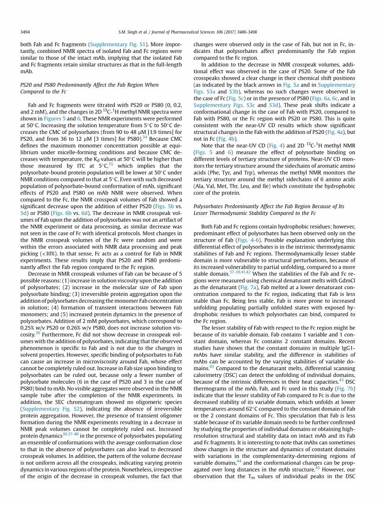
Figure 8. Role of conformational stability versus colloidal stability in mAb aggregation. (a) Aggregation of Fab (black) and Fc (red) regions at pH 5.5 (histidine buffer), as measuredby the particle counts using FlowCAM. (b) Aggregation of Fab in the absence of polysorbates (black), with PS20 (red), and with PS80 (green), respectively, as measured by FlowCAM.(c) Aggregation of Fc in the absence of polysorbates (black), with PS20 (red), and with PS80 (green), respectively, as measured by FlowCAM. (d) Aggregation of Fab at pH 5.8 (black)and pH 8.0 (red), respectively, as measured by FlowCAM. (e) Conformational stability and aggregation of Fab at pH 5.8 (black) and pH 8.0 (red). Far-UV CD measured the change inprotein conformation, whereas solution turbidity measured the aggregation. The scale on the left side of y-axis corresponds to far-UV CD, whereas the scale on the right side of y-axis corresponds to solution turbidity. (f) DSC thermograms of Fab at pH 5.8 (black) and pH 8.0 (red). Solid lines show the raw data, and the dashed lines show the deconvolution ofthe solid lines into unfolding of individual domains.
S.M. Singh et al. / Journal of Pharmaceutical Sciences 106 (2017) 3486-3498 3495
thermogram of intact mAb closely correspond to the Tm values ofindividual peaks in the thermograms of isolated Fab and Fc (Fig. 7b)indicates that the Fab and Fc of this mAb might have similar sta-bility as that in the intact mAb.
Polysorbates Do Not Significantly Affect the Global Stability of mAband Its Fab and Fc Regions
DSCwas used tomeasure the effect of polysorbates on the globalstabilities of mAb and its individual domains. DSC thermograms ofmAb and its Fab and Fc fragments were similar in the absence andpresence of polysorbates (Figs. 7c-7e), implying that the presenceof polysorbates did not significantly affect the overall stability ofmAb, Fab, and Fc. This is also consistent with the near-UV CD results(Fig. 3e), which showed minimal effect of polysorbates on theglobal unfolding transition. Similar conclusions were drawn earlierwhere DSC was unable to detect the effect of polysorbates on thestructural stabilities of local protein regions in mAbs.35,46
Despite lower thermodynamic stability, Fab aggregates lesserthan that of Fc. Chemical denaturation melts and DSC thermogramsshowed that the Fab region has lower thermodynamic stabilitythan that of Fc (Figs. 7a and 7b). How this difference in confor-mational stability translates into the aggregation propensity of Faband Fc regionswas further probed (Fig. 8). Upon overnight agitationat 37�C, Fc aggregated to a higher extent than that of Fab (Fig. 8a).Similar to mAb (Fig. 1d), both PS20 and PS80 protected Fab and Fcfrom agitation-induced aggregation (Figs. 8b and 8c). However, theextent of protection by PS20 or PS80 against protein aggregation is
lesser for Fab compared to Fc. This can be understood as follows.Both PS20 and PS80 protect proteins against interface-inducedaggregation by competing with proteins for interfaces. However,because PS20 and PS80 interact specifically with Fab causing itsunfolding (Figs. 4-6), Fab is more prone to aggregation compared toFc. Therefore, the observed effect of PS20 and PS80 on Fab aggre-gation (Fig. 8b) is the sum of decreased aggregation at interfacesand increased aggregation because of decreased conformationalstability, whereas in the case of Fc, the effect of PS20 and PS80 isjust the decreased aggregation at interfaces. This might be thereason why less protection was observed in the case of Fabcompared to Fc.
In addition, Fab aggregating lesser than that of Fc in the absenceof PS20 or PS80 despite its lower conformational stability impliesthat additional physical parameters contribute to the aggregationof Fab and Fc. In addition to conformational stability, colloidalstability, although relatively less appreciated, plays a big role inprotein aggregation.47,48 To a large extent, electrostatic interactionsbetween individual protein molecules resulting from the net sur-face charge distribution determine the colloidal stability. This is onereason why many proteins do not aggregate at pH values far awayfrom their pI. Theoretical pI values for the Fab and Fc regions usedin this study are 8.8 and 6.6, respectively. Because formulation pHwas 5.5 (histidine buffer), Fab is 3.3 units below its pI, whereas Fc is1.1 units below its pI. These values imply that Fab is relatively highlypositively charged compared to Fc, resulting in higher intermo-lecular repulsions leading to less aggregation. To test the role ofcolloidal stability in Fab aggregation, aggregation of Fab at 2
S.M. Singh et al. / Journal of Pharmaceutical Sciences 106 (2017) 3486-34983496
different solution pHs (5.8 and 8.0; phosphate buffer) was exam-ined. Both these pHs were below the theoretical pI for the Fab re-gion. Fab aggregation was higher at pH 8.0, closer to its pI of 8.8,compared to pH 5.8 (Fig. 8d). The conformational stability of Fab atthese 2 pH values was measured using far-UV CD thermal dena-turation melts (Fig. 8e). With increase in solution temperature, far-UV CD signal initially decreased followed by an increase.Comparing these changes in the far-UV CD signal with the DSCresults, the first transition during which the far-UV CD signaldecreased can be attributable to structure unfolding. Turbidity wasalso measured simultaneously to probe the Fab aggregation at the 2pH values. The second transition observed in the far-UV CD mea-surements during which the signal increased matched with that ofthe turbidity measurements, implying that the second transition infar-UV CD corresponds to the Fab aggregation. Comparing the firsttransition that corresponds to structural unfolding (Fig. 8e), Fab hadlower onset temperature (Tonset) of denaturation at pH 5.8compared to the Tonset at pH 8.0. Comparing the second transitionthat corresponds to Fab aggregation (Fig. 8e), Fab aggregated atlower temperatures at pH 8.0 compared to pH 5.8. These resultsimply that despite exhibiting lower Tonset for conformationalchanges at pH 5.8, Fab aggregates less at pH 5.8 compared to pH 8.0,indicating that the colloidal stability is playing a major role in Fabaggregation. These results are quite consistent with recent studieswhich indicated that the colloidal stability can account for the lackof a clear relationship between the conformational stability andaggregation of mAbs.49,50
To probe which domain of the Fab is affected by the pH change,it was subjected to DSC analysis at pH 5.8 and 8.0. Thermogram forFab at pH 5.8 exhibited 2 peaks (solid black line in Fig. 8f). As dis-cussed above, first peak at lower temperatures corresponds to thevariable domain, whereas the second peak corresponds to theconstant domain.43 However, at pH 8.0, DSC thermogram exhibiteda single peak (solid red line in Fig. 8f). Because this single peakconstitutes the unfolding of both the variable and constant domain,it could not be satisfactorily fitted into a 2-state single Tm transition.This single DSC peak at pH 8 was satisfactorily deconvoluted into 2peaks (broken red line in Fig. 8f). After deconvolution, a comparisonof Tm values clearly shows that a change in pH from 5.8 to 8.0 in-creases the stability of the variable domain.
Discussion
Polysorbates such as PS20 and PS80 are integral to antibodyand other protein-based formulations to prevent protein aggre-gation at interfaces.16 Proteins are polymers composed of varioushydrophilic and hydrophobic amino acids with diverse chemicalside chain moieties and therefore are highly surface-active com-pounds.51 This causes a quick adsorption of protein molecules atthe interfaces, which leads to their denaturation and aggrega-tion.51-53 Polysorbates being amphiphilic, compete with proteinsfor the interfaces and protect proteins from interface-inducedaggregation.54-61 Alternatively, polysorbates can also bind toexposed hydrophobic patches on proteins, thereby reducing thepropensity of hydrophobic interactions among protein moleculesleading to less aggregation.9,11,24,62-65 However, the specific natureof these polysorbate-protein interactions has not yet been clearlydemonstrated, in particular, whether polysorbates bind to mAbs,how strong is the binding, the effect of binding on the structureand stability of mAb, and whether the binding affects local orglobal protein structure. In this article, an attempt has been madeto address these questions on a mAb that is under drug develop-ment by Pfizer. First, it was confirmed that both PS20 and PS80protect mAb against agitation-induced aggregation (Fig. 1d). ITCdata showed that both polysorbates bind to the mAb with Kd in
the range of hundreds of micromolar (Fig. 2), which are in therange of polysorbate concentrations used in protein formulations.Similar attempts have been made previously on 2 mAbs todetermine their binding affinity to polysorbates.24,35 The Kd valueswere in the order of 1-10 mM, implying that the binding affinity ofpolysorbates to mAbs may depend on the nature of mAb. Bindingof polysorbates to mAbs is not very surprising; other nonionicsurfactants used in mAb formulations have been shown to bind tomAbs with a micromolar affinity.66 In our case, Raman scatteringshowed that binding of polysorbates has no impact on the sec-ondary structure of mAb (Fig. 3a). On the contrary, polysorbatessignificantly affect the tertiary structure of mAb as indicated bythe near UV-CD spectroscopy (Fig. 3b). Such changes in near-UVCD spectra upon polysorbate binding has been observed earlierin darbepoetin alfa.24 Ours is the first study on using near-UV CDto determine the effect of polysorbates on the structure of a mAb.Although it can be argued how these spectral changes reflect thechanges in the mAb structure, near-UV CD spectra have beenshown to be very useful in determining the effect of polysorbateson the tertiary structure of proteins (both in the case of darbe-poetin alfa24 and the mAb studied here). In addition to tertiarystructural changes, binding of polysorbates affected the partialunfolding of local protein regions rather than the global unfoldingof the entire mAb (Fig. 3e). To determine which region of mAbbinds to polysorbates, isolated Fab and Fc regions were subjectedto similar experiments. Near-UV CD of Fab and Fc indicated thatpolysorbates preferentially interact with the Fab region leading tostructural changes, with relatively no effect on the Fc (Figs. 4a and4b). To further confirm the near-UV CD results that polysorbatesaffect the local protein structures rather than the global proteinstructure, isolated Fab and Fc regions were subjected to 2D NMRspectroscopy of amino acid sidechain methyl groups. Applicationof NMR techniques to study antibodies has been feasible onlyrecently.33,67 Overall chemical shift pattern of the Fab remainedthe same with the addition of polysorbates except a few cross-peaks clearly showing chemical shift changes (Figs. 5a, 6a, andSupplementary Fig. S3), indicating that polysorbates affect thestructure of local protein regions rather than the global structureof Fab. In contrary to Fab, no changes were observed in thecrosspeak pattern or the chemical shifts of Fc with the addition ofpolysorbates. Using chemical denaturation melts and DSC, anattempt was made to address why the effect of polysorbates wasrestricted to Fab leaving Fc unaffected. Fab of the mAb studiedhere is less stable than its Fc (Fig. 7a). Compared to Fc, lesser stableFab will have increased population of partially unfolded stateswith hydrophobic residues exposed to solvent, which can interactwith polysorbates. Similar observations were made in our earlierstudies where the least stable region of a protein has been shownto be affected most by the excipients used in protein formula-tions.37-39,41 The lesser stability of Fab is due to its variable domain(Fig. 7b). Because mAb used in this study is an IgG1, its DSCthermogram was compared with the published thermograms of 3other IgG1-mAbs43 to identify the domain that unfolds at lowertemperatures. Differences in the DSC thermograms of various Fabsof mAbs arise from the differences in their variable domains, andthe mAb used in this study has less stable variable domaincompared to the other mAbs. This observation explains whypolysorbates affect predominantly the Fab region compared to theFc region in the mAb studied here.
Impact of polysorbates on the global and local stabilities of mAb,Fab, and Fc regions was also probed. Both near-UV CD (Fig. 3e) andDSC (Figs. 7c-7e) showed no significant changes in the globalunfolding, whereas near-UV CD (Fig. 3e) indicated the unfolding oflocal structural regions occurring at lower temperatures before theentire mAb unfolds at higher temperatures, as evident from the
S.M. Singh et al. / Journal of Pharmaceutical Sciences 106 (2017) 3486-3498 3497
appearance of sloped native baselines with the addition of poly-sorbates (Fig. 3e).
It was further examined how the conformational stability of theFab and Fc determines their aggregation behavior. Despite havinglower stability, Fab aggregates lesser compared to Fc (Fig. 8a). Byprobing the effect of pH on the conformational stability and ag-gregation (Figs. 8d-8f), colloidal stability seems to play a major rolein mAb aggregation. Fab is less stable at pH 5.8, but aggregates lesscompared to that at pH 8. These results further indicate that thevariations in conformational stability (controlled by intramolecularinteractions) and colloidal stability (controlled by intermolecularinteractions) of a protein may not be identical under different so-lution conditions. Similar results have been observed in 2 otherpublished studies. In the first study on the aggregation behavior of10 different IgG1 antibodies,49 there was no absolute trend be-tween the conformational stability and aggregation of the IgG1molecules, implying that colloidal stability plays a major role inIgG1 aggregation. In the second study,50 changing the solution pHfrom 6.5 to 4.5 reduced the thermal stability of IgG1; however, lessprotein aggregation was observed at pH 4.5, because of theincreased colloidal stability of the protein. Decreasing pH to acidicvalues increases the net charge on the protein surface leading toincreased repulsive interactions between individual protein mole-cules, thus leading to an increase in the colloidal stability of theprotein.
Although polysorbates protect proteins against interface-induced aggregation, they do not always lead to conformationalstabilization of proteins. In the case of mAb studied here, loss inNMR crosspeak volumes was observed for Fab upon the addition ofpolysorbates. Normally, this type of peak volume loss is attributedto protein aggregation. However, no protein aggregation wasobserved in our case. Other plausible explanation underlying peakvolume loss can be the presence of transient attractive intermo-lecular protein interactions in NMR samples. Although no signifi-cant changes were observed in the global conformational stabilityof mAb and its Fab and Fc fragments upon the addition of poly-sorbates, conformational destabilization of proteins by poly-sorbates have been observed in other proteins.68-71 For example,PS20 has been shown to reduce the thermodynamic stability ofhuman interferon-g and darbepoetin.24,69 Contrary evidence alsoexists where polysorbates can stabilize proteins. In the case ofalbutropin, PS20 and PS80 shifted the chemical denaturation meltsto higher denaturant concentration, which resulted in increasedthermodynamic stability of albutropin.62,69 In addition, poly-sorbates have been proposed to have dual effects on the stabilityand aggregation of proteins depending on the nature of protein andthe aggregation conditions.70 Therefore, polysorbates protectingproteins against interface-induced aggregation and the effect ofpolysorbates on the conformational stability of proteins can be 2independent phenomena. In protein formulations, polysorbates areused at a concentration where benefits of adding polysorbates tocompete with proteins for interfaces outweigh the possible disad-vantages involving protein conformational changes.
In summary, our study for the first time probed the detailednature of molecular interactions between the polysorbates andmAbs using high-resolution techniques such as 2D NMR. Publishedaccounts of the application of 2D NMR techniques to study anti-bodies are only 2 years old,33,67,72 and this study for the first timeshow the power of simple 2D NMR techniques in probing howexcipients in therapeutic formulations interact with mAbs. Futureavailability of NMR peak assignments for mAbs, similar to therecently solved backbone assignments for Fc,73 will further help inpinpointing the observed structural and dynamics changes tospecific local regions in mAbs. In addition to showing the applica-bility of 2D NMR methods to study polysorbate-mAb interactions,
our experimental data presented here indicate that PS80will be thepreferred choice over PS20 in formulating this mAb, because PS80offers higher protection against aggregation, causes lesser struc-tural perturbations, and has weaker binding affinity with fewerbinding sites compared to PS20.
Acknowledgments
We sincerely thank Ying Zhang, Pfizer Inc., and her team forproviding us the mAb, and isolated Fab and Fc fragments inpolysorbate-free buffer solutions. We thank Marguerite Areche-derra, Pfizer Inc. (currently at Waters Corporation), for initiatingthis project. We highly appreciate the help we received from the900-MHz Rocky Mountain NMR Facility and the Biophysics Core,University of Colorado Anschutz Medical Campus, in carrying outthe experiments. We acknowledge Marco Tonelli, National Mag-netic Resonance Facility at Madison, University of Wisconsin forperforming some of the initial NMR experiments. We also thankJohn Carpenter for many helpful discussions and for his criticalreading of themanuscript, and John Gabrielson regarding the use ofweighted spectral difference (WSD) parameter. This work wasfunded by a basic sciences research grant from Pfizer Inc.
References
1. Maa YF, Hsu CC. Investigation on fouling mechanisms for recombinant humangrowth hormone sterile filtration. J Pharm Sci. 1998;87:808-812.
2. McLeod AG, Walker IR, Zheng S, Hayward CP. Loss of factor VIII activity duringstorage in PVC containers due to adsorption. Haemophilia. 2000;6:89-92.
3. Eckhardt BM, Oeswein JQ, Bewley TA. Effect of freezing on aggregation ofhuman growth hormone. Pharm Res. 1991;8:1360-1364.
4. Cromwell ME, Hilario E, Jacobson F. Protein aggregation and bioprocessing.AAPS J. 2006;8:E572-E579.
5. Maa YF, Hsu CC. Protein denaturation by combined effect of shear and air-liquid interface. Biotechnol Bioeng. 1997;54:503-512.
6. Dong A, Prestrelski SJ, Allison SD, Carpenter JF. Infrared spectroscopic studies oflyophilization- and temperature-induced protein aggregation. J Pharm Sci.1995;84:415-424.
7. Tzannis ST, Hrushesky WJM, Wood PA, Przybycien TM. Irreversible inactivationof interleukin 2 in a pump-based delivery environment. Proc Natl Acad Sci U S A.1996;93:5460-5465.
8. Nema S, Washkuhn RJ, Brendel RJ. Excipients and their use in injectableproducts. PDA J Pharm Sci Technol. 1997;51:166-171.
9. Katakam M, Bell LN, Banga AK. Effect of surfactants on the physical stability ofrecombinant human growth hormone. J Pharm Sci. 1995;84:713-716.
10. Chang BS, Kendrick BS, Carpenter JF. Surface-induced denaturation of proteinsduring freezing and its inhibition by surfactants. J Pharm Sci. 1996;85:1325-1330.
11. Bam NB, Cleland JL, Yang J, et al. Tween protects recombinant human growthhormone against agitation-induced damage via hydrophobic interactions.J Pharm Sci. 1998;87:1554-1559.
12. Kim HL, McAuley A, McGuire J. Protein effects on surfactant adsorption suggestthe dominant mode of surfactant-mediated stabilization of protein. J Pharm Sci.2014;103:1337-1345.
13. Mehta SB, Lewus R, Bee JS, Randolph TW, Carpenter JF. Gelation of a mono-clonal antibody at the silicone oil-water interface and subsequent rupture ofthe interfacial gel results in aggregation and particle formation. J Pharm Sci.2015;104:1282-1290.
14. Mittal KL. Determination of CMC of polysorbate 20 in aqueous solution bysurface tension method. J Pharm Sci. 1972;61:1334-1335.
15. Patist A, Bhagwat SS, Penfield KW, Aikens P, Shah DO. On the measurement ofcritical micelle concentrations of pure and technical-grade nonionic surfac-tants. J Surfactants Deterg. 2000;3:53-58.
16. Kerwin BA. Polysorbates 20 and 80 used in the formulation of proteinbiotherapeutics: structure and degradation pathways. J Pharm Sci. 2008;97:2924-2935.
17. Warne NW. Development of high concentration protein biopharmaceuticals:the use of platform approaches in formulation development. Eur J Pharm Bio-pharm. 2011;78:208-212.
18. Martos A, Koch W, Jiskoot W, et al. Trends on analytical characterization ofpolysorbates and their degradation products in biopharmaceutical formula-tions. J Pharm Sci. 2017;106:1722-1735.
19. Ecker DM, Jones SD, Levine HL. The therapeutic monoclonal antibody market.MAbs. 2015;7:9-14.
20. Wang W, Singh S, Zeng DL, King K, Nema S. Antibody structure, instability, andformulation. J Pharm Sci. 2007;96:1-26.
21. Padlan EA. Anatomy of the antibody molecule. Mol Immunol. 1994;31:169-217.
S.M. Singh et al. / Journal of Pharmaceutical Sciences 106 (2017) 3486-34983498
22. Nelson DL, Cox MM. Lehninger Principles of Biochemistry. 7th ed. New York, NY:W.H. Freeman and Company; 2017.
23. Freyer MW, Lewis EA. Isothermal titration calorimetry: experimental design,data analysis, and probing macromolecule/ligand binding and kinetic in-teractions. Methods Cell Biol. 2008;84:79-113.
24. Deechongkit S, Wen J, Narhi LO, et al. Physical and biophysical effects ofpolysorbate 20 and 80 on darbepoetin alfa. J Pharm Sci. 2009;98:3200-3217.
25. Mahler HC, Senner F, Maeder K, Mueller R. Surface activity of a monoclonalantibody. J Pharm Sci. 2009;98:4525-4533.
26. Wen ZQ. Raman spectroscopy of protein pharmaceuticals. J Pharm Sci. 2007;96:2861-2878.
27. Li CH, Nguyen X, Narhi L, et al. Applications of circular dichroism (CD) forstructural analysis of proteins: qualification of near- and far-UV CD for proteinhigher order structural analysis. J Pharm Sci. 2011;100:4642-4654.
28. Dinh NN, Winn BC, Arthur KK, Gabrielson JP. Quantitative spectral comparisonby weighted spectral difference for protein higher order structure confirma-tion. Anal Biochem. 2014;464:60-62.
29. Harder ME, Deinzer ML, Leid ME, Schimerlik MI. Global analysis of three-stateprotein unfolding data. Protein Sci. 2004;13:2207-2222.
30. Latypov RF, Cheng H, Roder NA, Zhang J, Roder H. Structural characterization ofan equilibrium unfolding intermediate in cytochrome c. J Mol Biol. 2006;357:1009-1025.
31. Mayne L, Englander SW. Two-state vs. multistate protein unfolding studies byoptical melting and hydrogen exchange. Protein Sci. 2000;9:1873-1877.
32. Kuwajima K. Circular dichroism. In: Shirley B, ed. Protein Stability and Folding:Theory and Practice. Totowa, NJ: Humana Press; 1995:115-135.
33. Arbogast LW, Brinson RG, Marino JP. Mapping monoclonal antibody structureby 2D 13C NMR at natural abundance. Anal Chem. 2015;87:3556-3561.
34. Mohajeri E, Noudeh GD. Effect of temperature on the critical micelle concen-tration and micellization thermodynamic of nonionic surfactants: polyoxy-ethylene sorbitan fatty acid esters. J Chem. 2012;9:2268-2274.
35. Hoffmann C, Blume A, Miller I, Garidel P. Insights into protein-polysorbateinteractions analysed by means of isothermal titration and differential scan-ning calorimetry. Eur Biophys J. 2009;38:557-568.
36. Braun RJ, Parrott EL. Influence of viscosity and solubilization on dissolutionrate. J Pharm Sci. 1972;61:175-178.
37. Singh SM, Cabello-Villegas J, Hutchings RL, Mallela KMG. Role of partial proteinunfolding in alcohol-induced protein aggregation. Proteins. 2010;78:2625-2637.
38. Singh SM, Hutchings RL, Mallela KMG. Mechanisms of m-cresol-induced pro-tein aggregation studied using a model protein cytochrome c. J Pharm Sci.2011;100:1679-1689.
39. Hutchings RL, Singh SM, Cabello-Villegas J, Mallela KMG. Effect of antimicrobialpreservatives on partial protein unfolding and aggregation. J Pharm Sci.2013;102:365-376.
40. Kiefhaber T, Labhardt AM, Baldwin RL. Direct NMR evidence for an interme-diate preceding the rate-limiting step in the unfolding of ribonuclease A.Nature. 1995;375:513-515.
41. Bis RL, Singh SM, Cabello-Villegas J, Mallela KMG. Role of benzyl alcohol inthe unfolding and aggregation of interferon a-2a. J Pharm Sci. 2015;104:407-415.
42. Mehta SB, Bee JS, Randolph TW, Carpenter JF. Partial unfolding of a monoclonalantibody: role of a single domain in driving protein aggregation. Biochemistry.2014;53:3367-3377.
43. Ionescu RM, Vlasak J, Price C, Kirchmeier M. Contribution of variable domainsto the stability of humanized IgG1 monoclonal antibodies. J Pharm Sci. 2008;97:1414-1426.
44. Yan Y, Wei H, Fu Y, et al. Isomerization and oxidation in the complementarity-determining regions of a monoclonal antibody: a study of the modification-structure-function correlations by hydrogen-deuterium exchange massspectrometry. Anal Chem. 2016;88:2041-2050.
45. Majumdar R, Esfandiary R, Bishop SM, et al. Correlations between changes inconformational dynamics and physical stability in a mutant IgG1 mAb engi-neered for extended serum half-life. mAbs. 2015;7:84-95.
46. Garidel P, Hoffmann C, Blume A. A thermodynamic analysis of the bindinginteraction between polysorbate 20 and 80 with human serum albumins andimmunoglobulins: a contribution to understand colloidal protein stabilisation.Biophys Chem. 2009;143:70-78.
47. Chi EY, Krishnan S, Randolph TW, Carpenter JF. Physical stability of proteins inaqueous solution: mechanism and driving forces in nonnative protein aggre-gation. Pharm Res. 2003;20:1325-1336.
48. Goldberg DS, Bishop SM, Shah AU, Sathish HA. Formulation development oftherapeutic monoclonal antibodies using high-throughput fluorescence andstatic light scattering techniques: role of conformational and colloidal stability.J Pharm Sci. 2011;100:1306-1315.
49. Thiagarajan G, Semple A, James JK, Cheung JK, Shameem M. A comparison ofbiophysical characterization techniques in predicting monoclonal antibodystability. mAbs. 2016;8:1088-1097.
50. Kalonia C, Toprani V, Toth R, et al. Effects of protein conformation, apparentsolubility, and protein-protein interactions on the rates and mechanismsof aggregation for an IgG1 monoclonal antibody. J Phys Chem B. 2016;120:7062-7075.
51. Neurath H, Bull HB. The surface activity of proteins. Chem Rev. 1938;23:391-435.
52. Heitz F, Van Mau N. Protein structural changes induced by their uptake atinterfaces. Biochim Biophys Acta. 2002;1597:1-11.
53. Sharp JS, Forrest JA, Jones RA. Surface denaturation and amyloid fibrilformation of insulin at model lipid-water interfaces. Biochemistry. 2002;41:15810-15819.
54. Kreilgaard L, Jones LS, Randolph TW, et al. Effect of Tween 20 on freeze-thawing- and agitation-induced aggregation of recombinant human factorXIII. J Pharm Sci. 1998;87:1597-1603.
55. Thirumangalathu R, Krishnan S, Ricci MS, Brems DN, Randolph TW,Carpenter JF. Silicone oil- and agitation-induced aggregation of a monoclonalantibody in aqueous solution. J Pharm Sci. 2009;98:3167-3181.
56. Liu L, Qi W, Schwartz DK, Randolph TW, Carpenter JF. The effects of excipientson protein aggregation during agitation: an interfacial shear rheology study.J Pharm Sci. 2013;102:2460-2470.
57. Kerwin BA, Heller MC, Levin SH, Randolph TW. Effects of tween 80 and sucroseon acute short-term stability and long-term storage at �20�C of a recombinanthemoglobin. J Pharm Sci. 1998;87:1062-1068.
58. Joshi O, Chu L, McGuire J, Wang DQ. Adsorption and function of recombinantFactor VIII at the air-water interface in the presence of Tween 80. J Pharm Sci.2009;98:3099-3107.
59. Joshi O, McGuire J, Wang DQ. Adsorption and function of recombinant factorVIII at solid-water interfaces in the presence of Tween-80. J Pharm Sci. 2008;97:4741-4755.
60. Ludwig DB, Carpenter JF, Hamel JB, Randolph TW. Protein adsorption andexcipient effects on kinetic stability of silicone oil emulsions. J Pharm Sci.2010;99:1721-1733.
61. Petkov JT, Gurkov TD, Campbell BE, Borwankar RP. Dilatational and shearelasticity of gel-like protein layers on air/water interface. Langmuir. 2000;16:3703-3711.
62. Chou DK, Krishnamurthy R, Randolph TW, Carpenter JF, Manning MC. Effects ofTween 20® and Tween 80® on the stability of Albutropin during agitation.J Pharm Sci. 2005;94:1368-1381.
63. Bam NB, Randolph TW, Cleland JL. Stability of protein formulations: investi-gation of surfactant effects by a novel EPR spectroscopic technique. Pharm Res.1995;12:2-11.
64. Bam NB, Cleland JL, Randolph TW. Molten globule intermediate of recombinanthuman growth hormone: stabilization with surfactants. Biotechnol Prog.1996;12:801-809.
65. Jones LS, Cipolla D, Liu J, Shire SJ, Randolph TW. Investigation of protein-surfactant interactions by analytical ultracentrifugation and electron para-magnetic resonance: the use of recombinant human tissue factor as anexample. Pharm Res. 1999;16:808-812.
66. Budyak IL, Doyle BL, Weiss IV WF. Technical decision-making with higher orderstructure data: specific binding of a nonionic detergent perturbs higher orderstructure of a therapeutic monoclonal antibody. J Pharm Sci. 2015;104:1543-1547.
67. Arbogast LW, Brinson RG, Formolo T, Hoopes JT, Marino JP. 2D (1)H(N), (15)Ncorrelated NMRmethods at natural abundance for obtaining structural maps andstatistical comparability of monoclonal antibodies. Pharm Res. 2016;33:462-475.
68. Treuheit MJ, Kosky AA, Brems DN. Inverse relationship of protein concentrationand aggregation. Pharm Res. 2002;19:511-516.
69. Webb SD, Cleland JL, Carpenter JF, Randolph TW. A new mechanism fordecreasing aggregation of recombinant human interferon-gamma by a sur-factant: slowed dissolution of lyophilized formulations in a solution containing0.03% polysorbate 20. J Pharm Sci. 2002;91:543-558.
70. Wang W, Wang YJ, Wang DQ. Dual effects of Tween 80 on protein stability. Int JPharm. 2008;347:31-38.
71. Agarkhed M, O'Dell C, Hsieh MC, Zhang J, Goldstein J, Srivastava A. Effect ofpolysorbate 80 concentration on thermal and photostability of a monoclonalantibody. AAPS PharmSciTech. 2013;14:1-9.
72. Japelj B, Ilc G, Marusic J, Sencar J, Kuzman D, Plavec J. Biosimilar structuralcomparability assessment by NMR: from small proteins to monoclonal anti-bodies. Sci Rep. 2016;6:32201.
73. Yagi H, Zhang Y, Yagi-Utsumi M, et al. Backbone (1)H, (13)C, and (15)N reso-nance assignments of the Fc fragment of human immunoglobulin G glyco-protein. Biomol NMR Assign. 2015;9:257-260.

Figure S1. 2D 13C-1H HMQC NMR spectra of mAb (black) and its Fab (red) and Fc (green) fragments. Isolated Fab and Fc have similar structure as that in the intact mAb.
FabFc
mAb
1H (ppm)
-0.500.51.01.5
13C
(ppm
)
20
25
30
15
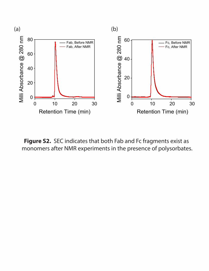
Figure S2. SEC indicates that both Fab and Fc fragments exist as monomers after NMR experiments in the presence of polysorbates.
(a) (b)
Retention Time (min)0 10 20 30M
illi A
bsor
banc
e @
280
nm
0
20
40
60
80Fab, Before NMRFab, After NMR
Retention Time (min)0 10 20 30M
illi A
bsor
banc
e @
280
nm
0
20
40
60 Fc, Before NMRFc, After NMR

Figure S3. Addition of PS20 caused chemical shift changes in the Fab region compared to PS80.
(a) (b)
(c) (d)
Fab + 0.2 mM PS20 Fab + 2 mM PS20
Fab
Fab + 0.2 mM PS20 Fab + 2 mM PS20
Fab
Fab + 0.2 mM PS80 Fab + 2 mM PS80
Fab
Fab + 0.2 mM PS80 Fab + 2 mM PS80
Fab
13C
(ppm
)
25
23
24
26
27
28
1H (ppm)0.50.6 0.4 0.3 0.2 0.1
13C
(ppm
)
25
23
24
26
27
28
1H (ppm)0.50.6 0.4 0.3 0.2 0.1
13C
(ppm
)
13
11
12
14
15
1H (ppm)0.50.6 0.4 0.3 0.2 0.10.70.8
13C
(ppm
)
13
11
12
14
15
1H (ppm)0.50.6 0.4 0.3 0.2 0.10.70.8