Embed Size (px)
Citation preview

Effect of Water Level on Kinematics of Healthy Horses
Walked on an Aquatic Treadmill Compared to Conventional
Rehabilitation Techniques
A THESIS SUBMITTED TO THE FACULTY OF THE GRADUATE SCHOOL OF THE
UNIVERSITY OF MINNESOTA
BY
Jose L. Mendez-Angulo
IN PARTIAL FULFILLMENT OF THE REQUIREMENTS FOR THE DEGREE OF
MASTER OF SCIENCE IN VETERINARY MEDICINE
Advisor: Troy N. Trumble
August 2012

© Jose L. Mendez-Angulo 2012

i
Acknowledgements
I want to thank my advisor Dr. Troy Trumble for his mentorship, guidance,
support, teaching and dedication throughout my graduate program and residency.
I also want to thank Drs. Erin Malone, Nicolas Ernst, and Anna Firshman for
being part of Master’s committee.
I would like to thank the veterinary students and technicians who helped handling
and grooming the horses of this project. In particular, Donna Groschen, Maggie
McQuestion, Sue Loly, Sara Wahlert, and Philip Kieffer.
I need to mention and thank my good friends Raffa and Luis, for being always
there, giving continuous support, encouragement and friendship, even after a long day of
work or when we were up working until late.
Obviously, I have to thank Sara for helping me with the statistics, writing and
editing of this study. Additionally, she has always been patient and understandable with
my crazy working hours, on-calls, and schedule.
I would like to acknowledge Talentia Scholarship program (Sevilla, Spain) for
supporting me financially during the first two years of my Master. Additionally, this
study was supported by a 2011 ACVS Foundation Surgeon-in-training award.
Finally, I would like to acknowledge the horses (Taco, Ella, Mr. Perfect, Legend,
Leche, Tropical, Añejo, Mesa, and Vaca) for allowing me to examine them and perform
the study.
ii
Dedication
This MS thesis is in part dedicated to my parents, Aniceto and Charo, for their
support, encouragement and love during the last 29 years, making possible the
accomplishment of this thesis. I also want to dedicate this work to Sara for her daily
understanding and love, and for following me blindly in all my professional adventures.
Finally, I want to dedicate this thesis to my brother Alberto for guiding me and protecting
me every day from somewhere.

iii
Abstract
Objectives- To calculate maximal flexion/extension of the carpal, tarsal, and fetlock
joints, as well as the stance and swing percentage of the stride in horses walking on the
underwater treadmill (UWTM) at four different water levels, and walking on three
conventional footings.
Animals- 9 clinically sound adult horses.
Procedures- Two-dimensional data was collected of horses walking on the UWTM at 4
water levels [<1cm of water (Baseline-B), fetlock (F), tarsus (T), and stifle deep (S)], and
on hard ground (HD), soft ground (SF), and a land treadmill (LT). Zinc oxide (for
UWTM) or retroreflective markers (for conventional surfaces) were used as skin markers
and placed at four specific anatomical locations on the left fore and hind limbs. Five gait
cycles of each horse for each surface were analyzed. Maximal flexion/extension angles,
and range of motion (ROM) were calculated for each joint.
Results-
Underwater treadmill- ROM was greater for all evaluated joints walking in water (F, T,
and S) compared to walking in no water (B), mainly due to an increase in flexion. The
greatest ROM for each joint was attained at the following water depths (in parentheses):
carpus (T), tarsus (S), fore fetlock (F and T), and hind fetlock (F and T).
Conventional surfaces- Maximal flexion of the tarsus and hind fetlock was greater on
LT and SF compared to HD, and carpus on LT compared to HD and SF. Maximal
extension of the carpus was greater on HD compared to SF and LT, tarsus on HD and SF
compared to LT, and both fetlocks on LT compared to HD and SF. The greatest overall
ROM of the carpus and fetlocks was achieved on LT, and tarsus on SF.
Conclusions and clinical relevance- These findings suggest that the UWTM is a useful
rehabilitation modality for increasing ROM of the distal limbs, and that the depth of
water should be considered. Additionally, conventional walking surfaces have a subtle
effect on flexion/extension of the distal limb. Therefore, data from this study could help
equine clinicians decide which footing surface/water depth is best for each individual
orthopedic patient in the early rehabilitation period.

iv
Table of Contents
Acknowledgements ..........................................................................................................i
Dedication ...................................................................................................................... ii
Abstract ......................................................................................................................... iii
List of Figures ................................................................................................................ vi
List of Tables............................................................................................................... viii
Chapter 1. Introduction and Literature Review .......................................................... 2
Hydrotherapy ....................................................................................................... 2
Introduction................................................................................................... 2
Water Properties ............................................................................................ 2
Swimming pools and underwater treadmills for horses .................................. 5
Evidence of the benefits of hydrotherapy in humans and small animals ......... 6
Claimed benefits of hydrotherapy in horses ................................................... 8
Range of Motion .................................................................................................. 9
Introduction................................................................................................... 9
Passive range of motion ................................................................................ 9
Active-assisted range of motion................................................................... 10
Active range of motion ................................................................................ 11
Kinematic analysis ............................................................................................. 11
Videographic systems ................................................................................. 12
Optoelectronic systems................................................................................ 13
Electrogoniometry ....................................................................................... 14
Chapter 2. Effect of water level on flexion and extension of the distal limb joints of
healthy horses walked on an underwater treadmill ................................................... 17
Chapter Summary ...........................................................................……………17
Introduction ....................................................................................................... 18
Material and Methods ........................................................................................ 19
Results ............................................................................................................... 25
Discussion.......................................................................................................... 29

v
Chapter 3. Comparison of flexion and extension of the distal limb joints of healthy
horses when walking on three conventional footing surfaces .................................... 36
Chapter Summary .............................................................................................. 36
Introduction ....................................................................................................... 37
Material and Methods ........................................................................................ 38
Results ............................................................................................................... 42
Discussion.......................................................................................................... 45
Chapter 4. Conclusions and future directions. .......................................................... 51
Conclusions ....................................................................................................... 51
Future directions ................................................................................................ 54
Bibliography .................................................................................................................. 56

vi
List of Figures
Figure 1. Photograph of a horse being recorded while walking on the UWTM with water
up to his stifle. A digital video camera can be observed on the left side of the image (red arrow)
that is positioned in front of the window that allowed visualization of the left forelimb.
Illumination is provided by a 300W halogen lamp positioned close to the window so the light did
not reflect back. ........................................................................................................................ 22
Figure 2. Photographs of a horse after being walked on the UWTM for data collection.
Observe the locations of the skin markers on the fore (A) and hind (B) limbs used to define the
body segments (blue lines) used to determine joint angles. Measured angles of each joint are
represented by a white line. ....................................................................................................... 23
Figure 3. Photograph of a horse on the UWTM with water up to his stifle. This
photograph is taken from the perspective of the camera being used to collect data (highlighted by
the red arrow in Figure 1) to demonstrate how well the markers could be identified. .................. 23
Figure 4. Maximal flexion (left-hand column) and extension (middle column) angles, and
ROM (right-hand column) of the carpus (1st row), tarsus (2
nd row), fore fetlock (3
rd row), and hind
fetlock (4th
row) joints of healthy horses (n=9) walking on the underwater treadmill without water
(B; white bar) and with water (gray bars) up to the hind fetlocks (F), tarsi (T), and stifles (S).
Mean + SD are presented in the bar graphs. Significance denoted as: * P<0.05, ** P<0.01, and
*** P<0.001.............................................................................................................................. 27
Figure 5. Photographs of a horse instrumented for data collection. Observe the locations
of the retroreflective skin markers on the fore (a) and hind (b) limbs used to determine the body
segments (blue lines) used to calculate the joint angles. Measured angles of each joint are
represented by a white line. ....................................................................................................... 42

vii
Figure 6. Maximal flexion (left-hand column) and extension (middle column) angles, and
ROM (right-hand column) of the carpus (1st row), tarsus (2
nd row), fore fetlock (3
rd row), and hind
fetlock (4th
row) joints of healthy horses (n=9) walking on hard ground (HD; white bar), soft
ground (SF; gray bar), and on a land treadmill (LT; black bar). Mean + SD are presented in the
bar graphs. Significance denoted as: * P<0.05, ** P<0.01, and *** P<0.001............................ 44

viii
List of Tables
Table 1. Maximal flexion (Flex) and extension (Ext) angles of the carpus, tarsus, fore
fetlock, and hind fetlock of healthy horses recorded using a videographic system at the walk
reported 2000 and 2001 by Hodson et al.59,60 Angles are expressed in degrees. ........................ 15
Table 2. The overall gain in flexion, extension, and joint range of motion (ROM) are
indicated (in degrees) for each water level in which the evaluated joint achieved the greatest
flexion and/or extension to demonstrate how much ROM can be increased at different water
levels in the underwater treadmill. ............................................................................................. 28
Table 3. Mean ± SD of percentages of stance and swing phases of the fore and hind limbs
of 9 healthy horses walking on the UWTM with four different water levels; <1cm of water (B),
fetlock (F), tarsus (T), and stifle (S)........................................................................................... 28
Table 4. Mean ± SD of percentages of the stance and swing phases of the fore and hind
limbs of 9 clinically sound horses walking on hard ground (HD), soft ground (SF), and on a land
treadmill (LT). .......................................................................................................................... 45
Table 5. Footing surface at which the greatest flexion, extension, and overall range of
motion (ROM) of each evaluated joint was achieved. ................................................................ 47
Table 6. Maximal flexion and extension angles, and ROM of the carpus, tarsus, and fore
and hind fetlocks joints of clinically sound horses walking in the UWTM without water (B), and
with water up to the hind fetlocks (F), tarsi (T), and stifles (S), and walking on hard ground
(cement, HD), soft ground (arena, SF), and land treadmill (LT). Values are expressed as degrees
(mean ± SD). ............................................................................................................................ 53

1
Chapter 1
Introduction and Literature Review

2
Chapter 1. Introduction and Literature Review
Hydrotherapy
Introduction
Hydrotherapy is defined as the external use of water in the medical treatment of
certain diseases. This form of therapy has been used in human rehabilitation for many
years,1,2
and is now gaining popularity in the rehabilitation of small3 and large animal
patients.4,5
The addition of hydrotherapy in rehabilitation protocols allows for earlier
intervention, with patients being able to move within days of injury or surgery with little
risk of reinjury. Exercise in water allows patients to unload painful joints or limbs while
maintaining soft tissue flexibility and muscle tone.
Swimming was the first form of hydrotherapy used in horses as a method of
training and conditioning of healthy animals.6,7
However, other hydrotherapy methods
such as underwater treadmills have recently become available and are currently used in
the rehabilitation of equine patients with musculoskeletal injuries. Unfortunately, little
scientific information is available regarding the use of hydrotherapy in horses, and equine
clinicians and physical therapists are required to design rehabilitation protocols based on
data extrapolated from human and small animal studies.
It is important to understand the principles and properties of water (including
relative density, buoyancy, viscosity, hydrostatic pressure, and surface tension) to
appreciate the benefits of hydrotherapy. These are important components to be
considered when designing an aquatic rehabilitation program for equine patients.
Water Properties
Relative density
Relative density depends on the composition of an object, and it is the ratio of the
weight of the object to the weight of an equal volume of water.8 The density of a

3
substance is defined by a pure number value called specific gravity. The specific gravity
of water is 1.0,9 and the relative density and specific gravity of an object determine if the
object floats or sinks in water.10
Objects with specific gravities greater than 1 tend to
sink; however, if the ratio is less than 1, the object tends to float. The specific gravities of
fat, lean muscle, and bone are 0.8, 1.0, and 1.5-2.0, respectively.9 Therefore, if the
specific gravity of a lean animal is greater than 1, the animal will tend to sink; whereas if
the specific gravity of an obese animal is less than 1, the animal will tend to float. The
greater the specific gravity of an object, the faster the sinking velocity.9
Buoyancy
Any body submerged in water is subjected to two forces, gravity and buoyancy.
Buoyancy is defined as the upward force exerted by a liquid, gas or other fluid, that
opposes an object's weight.9 Archimedes’ principle states that an object that is fully or
partially submerged in a fluid experiences an upward thrust equal to the weight of the
fluid displaced.8-10
An immersed limb or body with a relative density less than 1 (water’s
density) will be lifted toward the surface by buoyant forces.11
Thus, an animal with a
relative density less than 1 floats because of the water displaced.9 Buoyancy is exerted
directly through the center of buoyancy, which is the center of the displaced volume of
water and depends on the distribution of the displaced volume of fluid relative to an
object.9 The center of gravity and the center of buoyancy have to be in the same vertical
line to maintain stability. If this is not the case, an animal will tilt in order to reach a state
of equilibrium.9
Buoyancy assists in the rehabilitation of orthopedic patients by taking some of the
weight off of weak limbs or painful joints. It allows the patient to exercise in an upright
position and may decrease pain by minimizing the amount of weight bearing. For
example, the effect of buoyancy has been evaluated in horses, resulting in a reduction of
10% in body weight when water (saline) was at the level of the olecranon, and 75% when
water (saline) was at the level of the tuber coxae.12
At lower water levels, the effect of
buoyancy has not been measured in horses, but a similar canine study13
showed that
vertical ground reaction force only decreased by 9% after immersion to the tarsal joints,

4
and by 15% after immersion to the stifle joints. In humans, the percentage of the weight
borne when immersed in water is approximately 40% to 56% at the level of the anterior
superior iliac spine, 25% to 37% at the level of the xiphoid, and 5.9% to 10% at the
seventh cervical vertebra.14,15
This data is particularly useful when treating patients with
chronic conditions such as arthritis because joints may be unloaded during exercise.
Hydrostatic Pressure
The fluid pressure exerted on a body is equal on all surfaces of an immersed
object and is directly proportional to both the density of the fluid and the depth of the
fluid.10
The atmospheric pressure at the surface of water is 1.00 kPa and pressure
increases by 0.029 kPa per foot of depth.9 Thus, the deeper a body is immersed the
greater the pressure exerted. This principle has several advantages in rehabilitation. First,
hydrostatic pressure provides constant pressure to a body or limb immersed in water
which helps provide stability for weak animals while in the standing position. Second,
hydrostatic pressure opposes the tendency of blood and edema to pool in the lower
portions of the body and can therefore aid in reducing swelling and edema.11
Finally,
hydrostatic pressure may also decrease pain during exercise. By providing phasic stimuli
to the skin sensory receptor, the water’s motion decreases nociceptor hypersensitivity,
allowing the patient to perform a variety of movements with less pain.16
Viscosity
Viscosity is a measure of the resistance of a fluid which is being deformed by
either shear or tensile stress. Viscosity is significantly greater in water than in air,11
making it harder to move through water than through air. Therefore, exercise in water
provides resistance that may strengthen equine muscles and improve cardiovascular
fitness. Additionally, viscosity increases sensory awareness and supports unstable limbs
or joints.11
Water resistance is also influenced by the movement of water. For instance, a
streamlined water flow offers less resistance to movement than does a turbulent flow.8
Additionally, resistance in aquatic exercise may be increased by increasing the velocity

5
of movement of the patient or increasing the surface area of the body part moving in
water.17
Surface tension
Surface tension is a property of the surface of a liquid or fluid that allows it to
resist an external force.9 This is due to the tendency of water molecules to adhere together
on the surface, causing a slightly greater resistance to movement on the surface of water
compared to deeper levels.9 Surface tension is not a factor when a body is completely
submerged in water, but it becomes an important factor when a limb breaks the surface of
the water. Therefore, this water property can be used to increase the intensity of an
aquatic rehabilitation exercise.
Swimming pools and underwater treadmills for horses
Swimming Pools
Equine pools were initially designed for training and conditioning of race horses,
but they are now also used for rehabilitation of certain musculoskeletal conditions. The
shape and size of the pool varies depending on the intended use. Some pools are round;
some are straight with a ramp for entry and exit at each end; and some are a combination
of both designs. The advantage of straight pools is that if the horse becomes distressed or
exhausted during swimming it can leave the pool easily. This is an important factor
because swimming is an anaerobic exercise where horses reach heart rates up to 200
beats per minute,6 and can cause a significant increase in blood pressure.
18 Therefore
untrained animals can get tired very quickly, and exhaustion of the horse should always
be avoided because aspiration of water can be fatal.
The major benefit of swimming is the fact that horses can exercise intensively
without bearing any weight in limbs and joints. Therefore, this modality is considered
ideal to improve cardiovascular fitness of race horses while avoiding loading of bones
and joints.

6
Underwater treadmills (UWTM)
In human rehabilitation, underwater treadmills are often used as an alternative to
swimming and are thought to be particularly beneficial for injuries of the limbs.19
Many
equine training centers and veterinary hospitals have underwater treadmills available to
provide an alternative to swimming for equine patients. This rehabilitation modality
presumably provides some of the benefits of hydrotherapy, such as reducing the
concussive forces experienced by the distal limb, while providing controlled aerobic
exercise.20
Underwater treadmill exercise is considered to be a form of aerobic exercise
where horses reach heart rates up to 78 beats/minute at the walk21
and 120 at the trot.5
With water immersion, there is a decrease in systemic vascular resistance, and the
changes in total peripheral resistance are dependent on water temperature.18
After
underwater treadmill exercise there is a moderate but non-significant increase in blood
lactate and plasma creatine phosphokinase levels.22
In addition, it also provides a
different type of muscle exercise compared to swimming because the limbs are partially
loaded.13
Resistance to joint movement is also a benefit by increasing the muscle
contraction required to move the limb without having excessive force placed on the bone
or joint surface. The use of an underwater treadmill allows maintenance of cardiovascular
fitness, muscle tone, and improved joint movement without undue stresses occurring on
the injured limb.23
The reason for the decreased stress placed on the limb is the buoyancy
that the water provides. A certain amount of this depends on the amount of water in
relation to the body mass of the horse.12
Therefore the partial loading the limb combined
with the benefits of hydrotherapy may allow for use of different groups of muscles than
when working on land or in swimming pools.23
Evidence of the benefits of hydrotherapy in humans and small animals
The benefits of exercising in water have been reported in humans and small
animals, but information is scarce in horses. Several human and canine publications have
demonstrated that hydrotherapy is beneficial for pain, limb function, joint mobility,
muscle strength and balance.1,2,24

7
In human medicine, hydrotherapy is extensively used in the rehabilitation of
orthopedic patients after injury or surgery, and for the management of chronic conditions
such as osteoarthritis. For instance, progressive aquatic resistance training has favorable
effects in patients with decreased mobility, lower limb muscle power, and muscle cross-
sectional area in patients after knee replacement.25
Physical therapists also use
hydrotherapy in the rehabilitation period after shoulder surgery because movement of the
shoulder in the water requires less muscle activation than on land,1 due to the effect of
buoyancy, which allows earlier intervention with less stress on the affected soft tissues. A
recent study showed that aquatic therapy significantly improved passive flexion range of
motion of the shoulder in patients with surgical rotator cuff repair at three weeks after
surgery.26
Water exercise was more effective in reducing joint effusion and returning
limb function after cruciate ligament reconstruction than exercise on land.27
In patients
with osteoarthritis, exercise in water has been shown to decrease pain and improve limb
function while increasing muscle strength and joint range of motion.28
Other studies
showed that outcomes following aquatic exercise for adults with arthritis were
comparable to land based exercise, and concluded that aquatic exercise provides an
alternative strategy for patients that find land based exercise difficult.28,29
Aquatic therapy
is also a viable option for patients with obesity who have difficulties with active exercise
on land due to knee osteoarthritis.30
Patients with rheumatoid arthritis also experienced an
improved emotional and psychological state, along with improved joint range of motion
and reduced pain after aquatic exercise.24
Currently, there are only a few studies investigating the advantages and benefits
of hydrotherapy in small animal rehabilitation, although it is widely used in clinical
practice.23
Most of the hydrotherapy protocols used to date in the canine patients have
been extrapolated from human studies, and mainly consist of swimming or walking in
water. Swimming is often added to the rehabilitation program for the canine patient
following surgery for cranial cruciate ligament deficiency as it results in greater ROM of
the stifle and tarsal joints than does walking on land.31
A force plate study showed that
active exercise and swimming improved limb function after cruciate repair with no

8
measurable difference between the affected and unaffected limbs at 6 months after
surgery.32
Similarly, after tibial plateau leveling osteotomy, dogs showed improvement in
limb function when early intensive postoperative physical therapy included passive ROM
and training on an underwater treadmill (UWTM).33
Therefore, underwater treadmill
therapy has become popular for the rehabilitation of canine orthopedic injuries because of
its beneficial effects on joint range of motion. Joint kinematics of dogs have been studied
while walking on an underwater treadmill and compared to those of walking on land.
Researchers showed that joint flexion was greater walking in water than on land, and that
joint flexion was the greatest when water was filled higher than the joint of interest.
Additionally, joint extension was also achieved with underwater treadmill walking, which
is not the case in swimming.34
Claimed benefits of hydrotherapy in horses
Based on scientific research from human and small animal rehabilitation, the
claimed benefits of hydrotherapy in horses include the followings:
- Reduces loading of weak or painful joints
- Provides additional support to a body or limbs
- Allows earlier intervention after injury or surgery
- Prevents muscle atrophy and soft tissue adaptive shortening
- Increases muscle mass and strength
- Promotes cardiovascular fitness and endurance
- Increases soft tissue flexibility
- Decreases edema and joint effusion
- Increases circulation and stimulates healing
- Allows gradual progression and return toward normal limb function
- Decreases pain
- Encourages joint range of motion

9
Range of Motion
Introduction
Range of motion (ROM) of a joint is defined as the full possible movement of a
joint, from maximal extension to maximal flexion. ROM of each joint is influenced by
the structure, integrity, volume, and the surrounding soft tissues of the joint such as
tendons, ligaments, and muscles.35
In mammals, there are three types of joints:
synarthroses, amphiarthroses, and diarthrodial joints. Of these, diarthrodial joints have,
by far, the greatest ROM of all joints, and exhibit characteristic movements depending on
the joint. For instance, the metacarpophalangeal joint (fetlock in the horse) exhibits
mainly flexion and extension; however, the hip joint exhibits flexion, extension,
abduction, and adduction.
In orthopedic patients, preservation of joint movement is crucial after surgery in
order to avoid stiffness of the soft tissues and thus loss of limb function. ROM exercises
are particularly useful to diminish the effect of disuse and immobilization of limbs.36
Similarly, ROM and stretching exercises are very important in chronic conditions to
reestablish flexibility of the soft tissues and regain or at least maintain mobility. Joint
movement may be passive, active-assisted, or active.
Passive range of motion
Passive ROM is the movement of a joint that is performed without muscle
contraction within the available ROM, using an external force. If additional pressure is
applied at the end of the available ROM, it is termed stretching. In veterinary medicine,
both passive ROM and stretching are performed by clinicians or physical therapists.37
The ROM of a joint can be limited by either physiologic or pathologic conditions
that may involve the joint capsule, periarticular soft tissues, ligaments, tendons, muscles,
or skin. For example, a large open wound over a joint that is allowed to heal by second
intention may result in thick scar tissue that limits the joint ROM.37
Additionally,
pathologic changes in the cartilage, synovial fluid, or synovium of a joint can cause a

10
marked reduction in ROM. An example of chronic condition affecting ROM in humans
and horses is osteoarthritis.38-40
The most common indication for passive ROM exercises is immediately after
surgery, before active weight bearing of the affected limb starts, to help prevent joint
contracture and adaptive shortening of soft tissues, and to reduce pain, enhance blood and
lymphatic drainage, and improve synovial fluid production and diffusion.36
Another
indication for passive ROM is the prevention of joint contracture during the healing
period in paralyzed patients, or after a prolonged period of immobilization (e.g. fractured
limbs treated with a cast). However, in these cases passive ROM will not prevent muscle
atrophy, or improve strength and endurance of the muscle groups.37
Passive ROM should always be performed when the patient is relaxed and
comfortable. The administration of analgesics and/or non-steroidal antiinflammatories is
recommended before ROM exercises to reduce pain. The exercise should not cause any
pain or discomfort, and a gentle technique is mandatory to avoid undesirable reactions.
The therapist should grasp the proximal and distal aspect of the limb avoiding painful
areas. The motion should be smooth, slow, and steady to make sure that the patient can
tolerate it.37
Active-assisted range of motion
Active-assisted range of motion is a combination of patient´s muscle activity and
passive motion provided by the therapist.37
In veterinary medicine, this type of ROM is
more commonly performed than passive ROM because it is difficult to avoid muscle
activation in patients.37
Active-assisted ROM is an ideal exercise for weak patients,
especially for those recovering from a fracture treated with immobilization. In these
cases, active assisted ROM allows muscle strengthening while the soft tissues of the
affected limb recover flexibility.20
In small animals, active assisted ROM exercises can be performed with the help
of a sling to support the patient during ambulation.37
Swimming and walking in an
underwater treadmill are other forms of active assisted ROM that can be used in both

11
small and large animals.31,41
The water provides buoyancy to help support the weight of
limbs while the limbs go through a cycle of motion.13
Active range of motion
Active ROM is the motion of a joint that is achieved by muscle contraction. This
motion may be attained during a regular gait cycle (e.g. walking) or under special
conditions designed to maximize joint motion and to use the full available ROM. In
equine rehabilitation, some examples of these exercises include walking over poles or
cavalletis, or the attachment of tactile stimulator and/or weights.42
As the patient improves flexion and extension of a joint, it is helpful to continue
to perform passive or active assisted ROM to achieve as complete ROM as possible, and
then perform active ROM to emphasize more complete use of the limb.20
Active ROM
requires more muscle activity and, when used in combination with stretching exercises,
allows patients to recover maximal flexibility and strength of the affected soft tissues.37
Kinematic analysis
Introduction
In horses and small animals, kinematic analysis is commonly used in research
studies to evaluate the effect of different rehabilitation techniques and exercises on limb
movement and joint motion. Kinematic analysis measures the geometry of movement
without considering the forces that cause the movement.43
Kinematic analysis objectively quantifies the features of the equine gait that are
assessed subjectively during a lameness examination. The results are expressed in the
form of temporal (timing), linear (distance) and angular measurements that describe the
movements of the body segments and joint angles. Today, the most popular techniques
for studying kinematics and joint motion in horses are videographic analysis,
optoelectronic systems, and electrogoniometry.43
In equine research, sound horses are often used because their kinematics are
stable and symmetric, and analysis of a relatively small number of strides is

12
representative of the gait pattern. Some authors have suggested that 3–5 strides are
sufficient for kinematic analysis.44
The data describing an equal number of strides for
each limb are averaged and considered to be ‘representative’ for that particular limb.
Videographic systems
Videography is a well-accepted method of kinematic analysis for research and
lameness evaluation in horses. It involves attachment of several markers to the horse,
setting up and calibrating the equipment, video recording, and digitization,
transformation, smoothing and normalization of the data. Kinematic analysis may be
performed in two or three dimensions.45
Since the limbs of the horse move primarily in
the sagittal plane, most of the useful information is captured by the two dimensional
system, and in many studies the extra effort involved in extracting three dimensional data
is unnecessary. However, three dimensional analyses are mandatory when a study
involves evaluation of the abduction/adduction or internal/external rotations of the limb.43
Kinematic data provides temporal, linear and angular variables during motion.43
Temporal data describe the stride duration, and is calculated from the frame numbers and
the sampling frequency. Distance data is computed from the coordinates of the markers
combined with the calibration information, and describe the stride length, distance
between limb placements, and the flight arcs of the limbs. Angular data describe the
velocities, accelerations, and displacements of the body segments and joints. In two
dimensional studies, the angular data are usually reported as flexion and extension in the
sagittal plane.43
For this purpose, the centers of rotation of the appendicular joints have
been described,46
and these locations are often used as landmarks for placement of skin
markers. In three dimensional analysis, standardization becomes much more complicated.
However, this system allows the true measurement of three types of joint motion;
flexion/extension, adduction/abduction, and internal/external rotation.43
Speed is an
important fact that affects equine kinematics substantially, and therefore studies
performed at different speeds are not comparable.47

13
Most video systems offer automated digitization of skin markers if they have been
placed correctly and if lighting is appropriate. For two dimensional systems, 2-3 cm
diameter circular retroreflectivemarkers are commonly used.48
Self-adhesive tape or
cyanoacrylate glue is effective for securing the markers. For three dimensional studies,
spherical markers are used because cameras can capture them when viewed from
different angles.43
Marker locations are chosen in accordance with the purposes of the
analysis. Calculation of the angle between two limb segments in two dimensions requires
a minimum of three markers. Some software allows the use of two markers per segment
that are aligned along the long axis of the segment, without necessarily being placed over
the joint centers.45
Skin movement relative to specific underlying bony landmarks is a
source of imprecision, and it has been quantified in Warmblood horses.49,50
Additionally,
correction algorithms have been developed for walking and trotting; however, they are
only valid for horses of similar conformation and moving at the same gait and speed.
Skin movement artifact distal to the elbow of the forelimb and the stifle of the hindlimb,
is small enough to be ignored. However, in the more proximal parts of the limb skin
movements are sufficient to change the measurement of the angles of proximal joints.
Joint angles are typically measured on the anatomical flexor aspect of the joint,
and reported as the flexion and extension angles relative to the rotation of the proximal
and distal segments. Although the patterns are the same regardless of the method of
measurement (2-D versus 3-D systems), the values differ considerably, which impairs
comparisons between data from different studies.
Optoelectronic systems
Data collection and processing using an optoelectronic system is similar to those
used for videographic systems. Several optoelectronic systems are available, and use
either active markers (that emit a signal) or passive markers (that detect a signal).43
Use
of these systems is usually confined to the laboratory, because of the need for a hard-
wired connection to the subject and/or controlled lighting conditions. These systems
perform the digitizing on-line, so data are usually available very quickly.43
Many of the

14
systems have a built-in method for distinguishing between individual markers, for
example by sequencing the temporal output of different markers or by using markers of
different shapes or colors.43
In equine research, optoelectronic systems have been used to
evaluate joint kinematics at different gait patterns in normal and lame horses,51,52
and to
assess kinematics of trotters at different speeds.53
Electrogoniometry
An electrogoniometer is a device for measuring joint angle changes. It consists of
a potentiometer affixed to two rotating metal arms. These metal arms are usually attached
to the equine limb with tape, so that the center of rotation lies over the center of a joint
(e.g. fetlock joint).43
During movement, joint angle changes alter the electrical resistance
of the potentiometer, which is calibrated to produce a proportional displacement of
known magnitude.43
In equine research, electrogoniometry has been used to evaluate
joint movements at different gait patterns in normal and lame horses,54
and to assess the
changes in joint motion after medical or surgical treatments.55
Measurement of the flexion/extension angles of the equine distal limb joints
Recently, the passive range of motion of the equine distal limb joints have been
measured by goniometry in standing and anesthetized healthy horses.56
This technique
was also used in a study that evaluated the effect of 8 week duration casting on the range
of motion of the forelimb fetlock joint.57
However, measurement of active joint ROM
angles requires the use of a kinematic analysis system (i.e. videographic analysis,
optoelectronic system, or electrogoniometry).58
Kinematic studies have been performed to characterize the changes in flexion and
extension of the distal limb joints at different gait patterns in normal and lame horses,52,59-
61 to evaluate the effect of different rehabilitation techniques,
42,62 and to compare results
of medical and surgical treatments.63
Other researchers have also measured joint ROM of
distal limb joints (i.e. fetlock) to compare the effect of different footing surfaces.64
However, to the author’s knowledge, the effect of conventional walking surfaces on the

15
flexion and extension of healthy horses have not been compared so far, and comparison
between the published studies is difficult because the variability in methodology (e.g.
kinematic analysis systems, settings, placement of skin markers, speeds, etc.).
Additionally, the effect of water level in the kinematics of horses walking in an
underwater treadmill is currently unknown, which make it difficult for clinicians and
physical therapists to recommend a water level when designing a rehabilitation program.
In Table 1, we have summarized the maximal flexion and extension angles of the carpus,
tarsus, and both fetlocks (i.e. fore and hind) that have been reported using similar
methodology to our study.59,60
Table 1. Maximal flexion (Flex) and extension (Ext) angles of the carpus, tarsus, fore
fetlock, and hind fetlock of healthy horses recorded using a videographic system at the
walk reported 2000 and 2001 by Hodson et al.59,60
Angles are expressed in degrees.
Carpus Tarsus Fore Fetlock Hind Fetlock
Flex Ext Flex Ext Flex Ext Flex Ext
125.2 181.8 125.2 167.6 165.4 219.9 151.1 214.6

16
Chapter 2
Effect of water level on flexion and
extension of the distal limb joints of
healthy horses walked on an underwater
treadmill

17
Chapter 2. Effect of water level on flexion and extension
of the distal limb joints of healthy horses walked on an
underwater treadmill
Chapter Summary
Objective- To calculate maximal flexion and extension of the carpal, tarsal, and fetlock
(fore and hind) joints, as well as the stance and swing percentage of the stride in horses
walking on an underwater treadmill (UWTM) at four different water levels.
Animals- 9 sound adult horses.
Procedures- Data of horses walking (0.9 m/s) on the UWTM at 4 different water levels
[<1 cm of water (baseline-B), fetlock (F), tarsus (T), and stifle deep (S)] were recorded
by one video camera and analyzed using 2-dimensional motion-analysis software. Zinc
oxide was used as skin markers at four anatomical locations on the left fore and hind
limbs. Maximal flexion and extension angles, and range of motion (ROM) were
calculated for each joint, and the percent of the stride in stance or swing phase was
calculated for each stride.
Results- ROM was greater for all evaluated joints walking in water (F, T, and S)
compared to walking in no water (B), mainly due to an increase in flexion. The greatest
ROM for each joint was attained at the following water depths (in parentheses): carpus
(T), tarsus (S), fore fetlock (F and T), and hind fetlock (F and T). As the water level
increased the stance percentage of the stride decreased and swing percentage increased.
Conclusions and clinical relevance- These findings suggest that the UWTM is a useful
rehabilitation modality for increasing the ROM of various joints, and that the depth of the
water in the UWTM should be considered depending on the specific joint or limb being
targeted.

18
Introduction
Hydrotherapy is a well-established rehabilitation therapy in human medicine 25,27-
29 and is gaining popularity in small animal patients
3,65 and horses.
4,41,66 The physical
properties of water such as buoyancy, viscosity, resistance, hydrostatic pressure, and
surface tension offer specific benefits that should be considered when designing a
rehabilitation program.10
Water can decrease the amount of weight placed on joints,
provide constant pressure to a body or limb submerged in water, and aid in venous and
lymphatic drainage. Exercise in water may also decrease pain perception by providing a
phasic stimulus to the sensory receptors, and help strengthen muscles and promote
cardiovascular fitness.5,6,13,67
In humans and small animals, these advantages have been
suggested to allow orthopedic patients earlier use and weight bearing on weak limbs or
painful joints after injury or surgery, while minimizing the risk of re-injury.2,32,68
Underwater exercise (such as swimming) has been a common conditioning
method for non-injured equine athletes for years,7 but more recently it has also been used
as a rehabilitation method for recovery from some musculoskeletal injuries.23
However,
swimming is considered a high intensity activity in horses, where heart rates higher than
200 beats/minute may be reached,6 which is not ideal for equine orthopedic patients
immediately after injury, surgery, or a period of prolonged rest. In human medicine,
underwater treadmills (UWTM) are often used as an alternative to swimming and is
thought to be particularly beneficial for injuries of the limbs.19
Many equine training
centers and veterinary hospitals have underwater treadmills available to provide an
alternative to swimming for equine patients. This rehabilitation modality presumably
provides some of the benefits of hydrotherapy, such as reducing the concussive forces
experienced by the distal limb, while controlling aerobic exercise in the early
rehabilitation period with heart rates up to 78 beats/minute at the walk21
and 120 at the
trot.5
The ultimate goal of any musculoskeletal rehabilitation program is to restore limb
function through maximizing flexibility of the injured soft tissue, recovering muscle and
bone strength, and re-establishing full range of motion (ROM) of affected joints.20
In

19
human medicine, there is evidence that early passive and/or active flexion and extension
of injured appendages is crucial in order to obtain the best outcome.25,69
This
improvement in limb function and ROM has also been demonstrated in post-operative
human and canine studies using hydrotherapy.25,26,31,33
However, to the authors’
knowledge, there have been no similar reports of the use of hydrotherapy and its affect on
ROM of distal limb joints in horses. Determination of whether an UWTM increases
ROM in the distal limb joints of horses, and which water level is most beneficial to each
area of the limb, will benefit clinicians and physical therapists when designing a
rehabilitation program.
Therefore, the objectives of this study were to: 1) calculate the maximal flexion
and extension angles, and the ROM of the carpal, tarsal, fore fetlock
(metacarpophalangeal), and hind fetlock (metatarsophalangeal) joints in four different
water levels: < 1 cm of water (baseline-B), water up to the hind fetlocks (F), tarsi (T), and
stifles (S); 2) calculate the percent of the stride spent in stance or swing phase; and 3)
determine which of the four water levels provides the greatest flexion and extension for
each joint. We hypothesized that ROM of the distal limb joints would increase when
horses are walked in an UWTM with any water level (F, T, and S) compared to walking
with no water (B). Additionally, we hypothesized that joint flexion and extension, as well
as the percent of the stride spent in the swing phase would increase with increasing water
depth.
Material and Methods
Horses - Nine clinically sound horses (8 Quarter Horses and 1 Thoroughbred)
were used in the study. Mean ± SD age of the horses was 8.1 ± 4.3 years; mean weight
was 486.6 ± 26.3 kg; and mean height at the withers was 150.1 ± 3.2 cm. Before horses
were enrolled in this study, a thorough physical examination and subjective lameness
evaluation were performed on each horse. No clinical or orthopedic abnormalities were
detected. The study was performed with approval of the University of Minnesota
Institutional Animal Care and Use Committee.

20
Underwater treadmill training - All horses used in this study had previous land
treadmill experience as part of an unrelated study. Since it has been shown that horses
reach a steady state gait within the first 4-6 sessions of water treadmill exercise,70
horses
were walked in the UWTMa for a total of 6 training sessions (1/2 hour per session) before
data was collected. Xylazineb (0.2 mg/kg, IV) was administered for the first two training
sessions if horses showed anxiety or were reluctant to walk on the UWTM. Horses were
initially walked with < 1 cm of water (minimum amount of water to run the UWTM) to
allow acclimatization to the UWTM. Over the subsequent training sessions horses were
individually acclimated to walking on the UWTM at different water levels (up to the hind
fetlocks, tarsi, and stifles). The speed of the treadmill was maintained at 0.9 m/s during
each training session and for all data collection. Water temperature was not controlled in
this study but it ranged from 15 to 21 Celsius degrees throughout.
Kinematic measurement procedure – In order to track movement of the distal
limbs under water, zinc oxide was applied to the skin since it does not readily dissolve in
water. Four zinc oxide markers (2 cm in diameter) were placed on the left forelimb and
left hind limb (the only limbs that can be analyzed through the windows while the horse
is under water – Figure 1). Locations of skin markers were placed on the most lateral
aspect of each limb, exactly over the center of rotation of each joint. Forelimb markers
were placed at the level of: 1) proximal interphalangeal joint, 2) lateral condyle of the
distal third metacarpus, 3) ulnar carpal bone and 4) 15 cm proximal to the ulnar carpal
bone on the groove between the common and lateral digital extensor muscles on the
radius (Figure 2a). Hind limb markers were placed at the level of: 1) proximal
interphalangeal joint, 2) lateral condyle of the distal third metatarsus, 3) mid talus, and 4)
10 cm proximal to the tuber calcanei on the groove between the long and lateral extensor
muscles on the tibia (Figure 2b). Locations of the skin markers were chosen based on
other authors’ recommendations,43
with slight modifications due to the size of the
windows on the side of UWTM. In other words, the distal marker had to be placed at the
level of the proximal interphalangeal joint instead of the hoof and the proximal marker
had to be placed in the middle of the tibia or radius instead of the proximal aspect of the

21
bone to be able to analyze a complete stride at all water levels. Hair was clipped in these
locations to ensure consistency of placement throughout the study.
Two-dimensional movement was recorded using a digital video camerac (6.0 mm
lens) at 60 frames/sec while the horse was walked on the UWTM at four different water
levels: < 1 cm of water (B), water up to the hind fetlocks (F), tarsi (T), and stifle (S). For
the fetlock water level, the UWTM was filled up to the skin marker placed on the lateral
condyle of the distal third metatarsus. For the tarsal water level, the UWTM was filled up
to the skin marker placed on the mid talus. For the stifle water level, the UWTM was
filled up to the tibial plateau of each horse (manually palpated). Video was recorded from
the left side of the UWTM through one of the two transparent plastic windows that
allows visualization of the horse’s limbs while under water (Figures 1 and 3). Data
collection was completed in 2 sessions over different days (one for the forelimb and one
for the hindlimb). Before each session, a rectal exam was performed on each horse prior
to being loaded into the UWTM to evacuate feces from the rectum. This was performed
to minimize the risk of water contamination with feces, which can obscure marker
identification during tracking. For the first session, the digital video camera was
positioned on a tripod perpendicular to the front window (at 152 cm from the ground, and
at 140 cm away from the UWTM) that allowed visualization of the left forelimb (Figures
1 and 3). Due to the reduced size of the window (90 x 70 cm), the camera had to be
angled 30 degrees down in order to be able to capture the most distal marker (proximal
interphalangeal joint) of the limb (Figures 1 and 3). Skin markers were illuminated by a
300W halogen lamp positioned close to the window, in approximately 45º angle so the
light did not reflect back. After allowing horses to adjust for 5 minutes at each water
level, 10 complete strides of the left forelimb without interruptions were videotaped for
each water level (B, F, T, and S consecutively). During the second session, the same
settings (camera angle and distance from the UWTM) and videotaping procedure were
repeated through the rear window (90 x 70 cm) that allowed visualization of the left hind
limb. Each horse’s data was collected in approximately half an hour in each session over
different days to prevent the gait from changing due to fatigue.

22
Figure 1. Photograph of a horse being recorded while walking on the UWTM with water
up to his stifle. A digital video camera can be observed on the left side of the image (red
arrow) that is positioned in front of the window that allowed visualization of the left
forelimb. Illumination is provided by a 300W halogen lamp positioned close to the
window so the light did not reflect back.

23
Figure 2. Photographs of a horse after being walked on the UWTM for data collection.
Observe the locations of the skin markers on the fore (A) and hind (B) limbs used to
define the body segments (blue lines) used to determine joint angles. Measured angles of
each joint are represented by a white line.
Figure 3. Photograph of a horse on the UWTM with water up to his stifle. This
photograph is taken from the perspective of the camera being used to collect data
(highlighted by the red arrow in Figure 1) to demonstrate how well the markers could be
identified.

24
Two-dimensional kinematic analysis was performed using the DMAS Equine
Gait Trax systemd. Software calibration was performed at the beginning of each session
by the use of a known distance marked on the windows of the UWTM. One stride was
defined as complete foot contact with the belt (heel and toe) to the subsequent complete
foot contact of the same limb. Within a stride, stance duration was described as the
number of frames within a stride in which there was contact of the foot with the belt from
heel down to breakover. Duration of the stance and swing phases of the fore and hind
limbs were calculated for each horse under each water level. The ten videotaped strides
were reviewed and only the five strides with the least amount of water splashing and best
visibility of skin markers were used for tracking and calculation of stride percentages.
Tracking of the skin markers was carried out by a combination of an automatic and
manual method to be able to create body segments that represented the distal limbs and
joints. Each frame of each stride was reviewed manually by the same person (J.L.M.A),
and markers were magnified to ensure that the center was selected appropriately. The
center of the marker was selected manually in all frames in which the software did not
place them exactly in the center of the marker (approximately 90% were selected
manually, and 10% found correctly by the software). For each individual complete stride,
maximal flexion and extension angles of each joint of interest were calculated based on
the rotation of the proximal and distal body segments around the palmar/plantar (carpus
and fetlocks) and dorsal (tarsus) aspects of the joints using the inverse tangent function
(Figure 2a and b). Range of motion (ROM) of the evaluated joints, represented by the
relative inter-segmental motion, was calculated for each stride of each horse in each
water level as maximal extension minus maximal flexion.
A post-hoc error calculation was performed for the camera angle and water
refraction by placing a right angle in the areas of maximum flexion and extension of each
joint (fetlock region and carpus/tarsus region). The right angle had 2 cm diameter
markers on both branches and center of rotation, and was analyzed (using the same
software and settings as in the study) at each location with the UWTM static with no
water, partially submerged in water (2 markers in the water and 1 marker out the water)
and completely in water as would occur with each different water level. To determine if
25
there was an error based off of the camera angle itself, additional measurements using the
same conditions described above were collected with the camera placed perpendicular to
the UWTM. A total of 50 measurements were collected and the markers on the right
angle were tracked. The error was determined based off of the deviance from 90 degrees,
and the average error identified at each location within the window and at each water
level was subsequently corrected in the data before statistical analysis.
Statistics - Mean ± SD of maximal flexion and extension angles, joint ROM, and
the stance and swing percentages of each water depth were calculated by first averaging 5
strides of each horse, and then combining the average of those 5 strides for all horses at
each water level. Extreme studentized deviate tests were conducted on the 5 strides
analyzed for each joint for each horse under each water level to identify outliers, which
were subsequently removed from further analysis. Stance and swing temporal data was
transformed to percentages. Normality of the data was assessed using the Shapiro-Wilk
normality test. Differences between the 4 water levels for each joint of interest were
determined using a repeated-measured ANOVA with a Tukey-Kramer’s multiple
comparison test. Statistical significance was set at p<0.05. All data were analyzed using a
statistical software packagee.
Results
The amount of error that was present based on our 2-D kinematic analysis system
was < 3 degrees. This includes the error based on the camera angle and water refraction.
There were no differences in error with the camera positioned at a 30 degree angle versus
being perpendicular to the UWTM. The amount of error was different based on the
location and water level. The location where flexion generally occurred for the fetlocks
as well as the carpus/tarsus demonstrated less angle than 90 degrees (87.6 to 90 degrees)
that varied based on the joint of interest and water level. The location where extension
generally occurred for the fetlocks as well as the carpus/tarsus demonstrated greater angle
than 90 degrees (90 to 92.9 degrees) that varied based on the joint of interest and water
level. The average error calculated for each joint in each location (flexion/extension) for

26
each water level was used to correct the peak flexion/extension data points of the results
presented below.
Kinematic analysis showed that walking horses in any depth of water (F, T, or S)
in the UWTM, resulted in a significant (all P<0.001) increase in maximal flexion in all
evaluated joints compared to walking with no water (B) (Figure 4). When comparing
maximal joint flexion within the three water levels (F, T, and S), the greatest flexion of
the carpus and fetlocks (fore and hind) was achieved with T and the greatest flexion of
the tarsus was achieved with T and S (Figure 4; Table 2).
During walking, maximal extension was significantly (P<0.001) greater for the
carpus in T, for the tarsus in S, and for both fetlocks in F compared to walking in no
water (B) (Figure 4). Conversely, maximal extension was significantly less for the fore
fetlock (P<0.01) in T compared to walking in no water (B) (Figure 4). When comparing
maximal joint extension within the three water levels (F, T, and S), the greatest extension
of the carpus occurred with T, the greatest extension of the tarsus with S, and the greatest
extension of the fetlocks with F, which were all significantly (P<0.01) different compared
to the other two water levels (Figure 4; Table 2).
Overall ROM was significantly (P<0.001) greater for all evaluated joints when
horses were walked in water (F, T, and S) compared to walking in no water (B) (Figure
4). Additionally, the greatest carpal ROM was attained with T and the greatest tarsal
ROM with S. The greatest fore and hind fetlock ROM was attained with F and T (Figure
4; Table 2).
Significant differences were observed in the stance and swing percentages of the
fore and hind limb strides between all water levels (Table 3). As the water level increased
the fore and hind limb stance percentage of the stride decreased, whereas the swing
percentage of the stride increased as the water level increased (Table 3).

27
Figure 4. Maximal flexion (left-hand column) and extension (middle column) angles,
and ROM (right-hand column) of the carpus (1st row), tarsus (2
nd row), fore fetlock (3
rd
row), and hind fetlock (4th row) joints of healthy horses (n=9) walking on the underwater
treadmill without water (B; white bar) and with water (gray bars) up to the hind fetlocks
(F), tarsi (T), and stifles (S). Mean + SD are presented in the bar graphs. Significance
denoted as: * P<0.05, ** P<0.01, and *** P<0.001.

28
Table 2. The overall gain in flexion, extension, and joint range of motion (ROM) are
indicated (in degrees) for each water level in which the evaluated joint achieved the
greatest flexion and/or extension to demonstrate how much ROM can be increased at
different water levels in the underwater treadmill.
Joint Water level Flexion Extension ROM
Carpus Tarsal water depth 20 5 25
Tarsus Stifle water depth 29 4 33
Fore fetlock Fetlock water depth 12 13
25
Hind fetlock Fetlock water depth 26 20 46
Table 3. Mean ± SD of percentages of stance and swing phases of the fore and hind
limbs of 9 healthy horses walking on the UWTM with four different water levels; <1cm
of water (B), fetlock (F), tarsus (T), and stifle (S).
Variable (%) B F T S
Stance forelimb 67.2 ± 1.9a
63.9 ± 1.3b
60.0 ± 1.1c
57.2 ± 1.0d
Swing forelimb 32.4 ± 1.7a 35.8 ± 1.3
b 39.9 ± 1.1
c 42.7 ± 1.0
d
Stance hind limb 65.6 ± 1.7a 62.0 ± 1.4
b 60.0 ± 2.5
c 58.2 ± 2.2
d
Swing hind limb 34.3 ± 1.7a 37.9 ± 1.4
b 39.9 ± 2.5
c 41.8 ± 2.2
d
a–d Within a row, values with different superscript are significantly (P<0.01) different
from each other.

29
Discussion
Our results showed that walking horses in water increases overall ROM of the
carpus, tarsus, and both fetlocks (fore and hind), mainly due to an increase in joint
flexion. This increase occurred to a different extent in each joint depending on the water
level. Additionally, greater joint extension of the evaluated joints was observed in certain
water depths. These findings confirmed our first hypothesis and provide objective data
that support the suggestion that an UWTM can be used to encourage joint ROM in
horses.70
Conversely, our second hypothesis was partially refuted because joint flexion
and extension did not consistently increase with increasing water depth in all joints.
Clinicians and physical therapists try to encourage joint ROM in conventional
footing surfaces using other equine rehabilitation techniques such as passive flexion and
extension, walking over poles or cavalletis, and attachment of tactile stimulators or
weights to the distal limb.20,71
Some of these techniques have been extrapolated from
human or small animal rehabilitation,72
and little information is available regarding how
much they increase joint ROM in horses. Recently, two studies have focused on the
effects of the attachment of tactile stimulators and/or weights (700 g) around the pasterns
of horses, showing a significant increase in flexion of the fetlock, tarsus, and stifle at the
trot when these devices were applied to the hind limbs.42,73
In contrast, several studies
have shown that the addition of weights to the front limbs does not significantly increase
the flexion of the carpus or fore fetlock.74-76
In our study, the UWTM increased ROM of
all evaluated joints. In addition, at certain water levels (S and T) the gain in flexion of
the tarsus and hind fetlock (29 and 47 degrees, respectively) was similar or greater to that
reported at the trot when using tactile and weighted stimulators applied around the hind
pasterns.42
Currently, objective data about rehabilitation techniques is lacking in horses,
and the amount of flexion/extension needed to help a clinical case is unknown. This study
provides data on how much sound horses can flex and extend the distal limb joints
walking in water, and now further research in clinical cases with reduced limb motion is
warranted. In our study, walking in the UWTM with specific water depths also resulted in
greater joint extension for the evaluated joints (Table 2), which has not been reported
with other conventional rehabilitation exercises. However, the increases seen in extension

30
of the carpus and tarsus were ≤ 5 degrees, likely because of the anatomical limits of these
joints, and its clinical significance needs further research.47
Therefore, the UWTM may
have advantages over other techniques for early rehabilitation of acute musculoskeletal
injuries because it not only encourages flexion of all joints but also some degree of
extension (Table 2). Furthermore, UWTM rehabilitation provides the additional
hydrotherapy benefits of buoyancy, viscosity, resistance, hydrostatic pressure, and
surface tension.41
Human and small animal kinematic studies have demonstrated an increase in the
ROM of distal limb joints when walking in water compared to land. Similar to our study,
this increase has been reported to occur mainly due to an increase in joint flexion rather
than extension. For instance, ankle and knee flexion was greater in people walking and
running in water up to the waist than they did on land.77
In dogs, flexion of the hip, stifle,
tarsus, shoulder, and elbow was greater walking in the UWTM than walking on the land
treadmill.34
Small animal kinematic data have also demonstrated that higher water levels
do not necessarily result in a greater flexion of joints. For instance, flexion of the canine
hip, stifle and tarsal joints increased maximally when water level was at the stifles, but
then decreased if water was added up to the level of the greater trochanter.9 In our study,
the greatest flexion of the fore limb joints (carpus and fore fetlock) occurred in T, which
may be because horses walking in low water levels may attempt to elevate their limbs out
of the water to avoid resistance. This did not occur at deeper water levels (i.e. S),
potentially because the entire limb is submerged, and so the horse is unable to lift the
limb above the water surface, and thus is forced to move the whole forelimb through the
water instead. In the hind limb, the greatest flexion of the tarsus occurred with T and S.
This finding may be caused by the synchronized movement of the reciprocal apparatus
where the stifle and tarsus flex and extend simultaneously.78
At these water levels (T and
S), horses may still attempt to elevate their hind limbs to get their stifles out of the water
to decrease resistance during motion, thus simultaneously increasing tarsal flexion.
Results of this study showed that water level affected flexion and extension of the
joints differently, even in joints of the same limb. Thus, the evaluated joints reached the
greatest ROM at dissimilar water levels. Additionally, the greatest flexion and the

31
greatest extension of a joint (i.e. fore and hind fetlocks) did not necessarily happen at the
same water level, resulting in less increase in overall ROM of that joint at one water
level. This data suggests that equine clinicians and physical therapists should be aware of
the effect of water level on the motion of joints and should consider its effects when
designing a rehabilitation program, especially if the goal is to increase flexion and/or
extension of a particular joint or limb. For instance, walking in water up to the tarsus
would be indicated for a horse with reduced ROM of the carpus. A higher water level
(i.e. S) would be recommended for horses in which tarsal flexion and extension are
desired. In horses with decreased ROM of the fore or hind fetlocks, alternating exercise
in two different water levels (T and F) may be recommended because walking in water
up to tarsus would encourage flexion, and water up to the fetlock would provide more
extension. These are general recommendations based on data from this study but it should
be clarified that our population was mainly Quarter Horses, and although the
Thoroughbred (similar height and weight) used in this study moved similarly, other
breeds may have different increases in flexion and extension depending on water level.
Additionally, the increase in activity of the different muscle groups involved in
protraction and retraction of the limbs should be considered when choosing a water level,
especially in the early rehabilitation period, to avoid muscular fatigue and/or injury of the
untrained muscles.
In a recent study, horses walking in the UWTM at carpal and ulnar water depths
were shown to have lower stride frequency and higher stride length of the forelimbs
compared to walking in lower water levels.70
Similarly, we evaluated stance and swing
percentages of the stride and found that water level had a direct effect on these
measurements. Our results showed that as the water height increased, stance percentage
decreased and the percentage of the swing phase increased. At the trot, an increase in
swing phase has also been reported in horses when hind limb flexion is encouraged by
adding tactile stimulators and/or weights to the hind pasterns, causing an increase in
flexion at the stifle, hock and hind fetlock joints.42
Our results demonstrate that this effect
can be achieved using the UWTM, and may be due to the effect of water resistance
and/or buoyancy. At the water levels used in this study, water resistance was expected to
32
increase along with water depth, because water resistance is proportional to the portion of
a limb submerged in water. The effect of buoyancy has been evaluated in horses at high
water (saline) levels, resulting in a reduction of 10% in body weight when water was at
the level of the olecranon, and 75% when water was at the level of the tuber coxae.12
At
lower water levels, the effect of buoyancy has not been measured in horses, but in a
canine study,13
vertical ground reaction force only decreased by 9% after immersion to
the tarsal joints and by 15% after immersion to the stifle joints. Therefore, the effect of
buoyancy at the water levels used in our study was likely minimal.
Evidence of the benefits of an UWTM for rehabilitation of orthopedic patients is
available in the human and veterinary literature. For instance, in people, water exercise
has been shown to decrease joint effusion and pain after orthopedic surgery.25,27
A recent
study in people with total knee arthroplasty demonstrated that the benefits obtained using
hydrotherapy (reduction in pain and stiffness, and improvement in function) were still
identified in patients up to six months after discharge from the hospital.79
In dogs, aquatic
exercise after correction of cranial cruciate ligament rupture resulted in greater ROM of
the stifle and tarsal joints than did walking on land.31
Similarly, dogs after tibial plateau
leveling osteotomy showed improvement in limb function when early intensive
postoperative physical therapy included passive ROM and training on an UWTM.33
Unfortunately, no similar studies are currently available in horses, so the impact of
adding UWTM exercise to a rehabilitation protocol is unknown and needs investigation;
however, the results of our study demonstrate that use of the UWTM is possible for the
orthopedic patient because it stimulates an increase in the ROM of distal limb joints.
Several limitations due to infrastructural conditions can be identified in this study.
Therefore, the results of this study may have been influenced by some factors which
could not be avoided due to the constraints presented by the UWTM setting. This
includes use of a 2D system, collection of left limb motion only, data collected in a
skewed angle, and water refraction. The reader needs to understand that all of these
components may make our angle data more relative than precise, but the authors made a
number of efforts throughout the study in an attempt to reduce these factors and the
variability created by them. In humans80
and small animals31
, underwater kinematic

33
analyses are performed using three dimensional systems, but this not possible in horses
due to the confines of current UWTM facilities. We had to use a 2-D system to perform
the kinematic analysis since data had to be collected through each window on the left side
of the UWTM (Figure 1). However, the window size itself (90 x 70 cm) may have
helped minimize the amount of error because the motion of each stride was contained
within that small area. Conversely, when performing 2-D analysis with the horse
walking over ground, motion data has to be analyzed over many meters which have the
possibility for greater error. It is important to note that within our 2-D system only
motion that occurred in the sagittal plane (plane of major limb movement) could be
represented and may be affected if there is any non-sagittal movement that occurs during
a stride. It is possible that walking in water may induce more non-sagittal motion of the
limbs than walking over ground, which could add more error. Only the left fore and hind
limbs could be evaluated because our UWTM does not have transparent windows on the
right side. Despite this issue, results obtained from the left limbs should be able to be
extrapolated to the right side because the walk is a symmetric gait and sound horses are
expected to move their limbs symmetrically.81
Other important factors that we could not
eliminate in our study due to the constraints of the UWTM were the facts that the camera
had to be angled 30 degrees down in order to be able to track the motion of the distal
most marker, and that water refraction likely altered tracking of the motion as well.
These factors likely reduced the sensitivity of our measurements. However, we attempted
to minimize their effects within the system as much as we could. The same person
marked all horses, filled the UWTM in all sessions, set up the video camera for each
horse in each session, and manually tracked every single frame (moved back and forth
between contiguous frames) in an attempt to ensure adequate central placement of the
markers by the software. In addition, multiple strides (n=5) for each horse at each water
level were collected in an attempt to minimize variability that would arise due to our data
collection system. We attempted to correct for our camera position and water refraction
by introducing a fixed angle (90 degrees) into the UWTM at all water levels and flexion
and extension locations. This analysis allowed for further error correction in our data
which should have further minimized variability from these sources, however, it does not

34
reflect any potential error caused by motion of the treadmill, water turbulences, or limbs
movement. Based off of this analysis, we believe the error of our set up and the effect of
the water combined is less than 3 degrees for any joint at any water level, and our
corrected results are representative of the general motion of the distal limb in and out of
the water. When comparing our corrected baseline results to those reported in other
studies59,60
where limb kinematics at the walk were evaluated over ground on sound
horses, our means and standard deviations are very similar.
In conclusion, results of the present study showed that an UWTM should be
considered for an equine rehabilitation program to increase the overall range of motion of
the distal limb joints. Results of this study can be a useful reference for clinicians or
physical therapists that are trying to encourage overall ROM or increased flexion or
extension of a particular joint or limb since the water level affects the amount of flexion
and extension of the joints differently.
35
Chapter 3
Comparison of flexion and extension of
the distal limb joints of healthy horses
when walking on three conventional
footing surfaces

36
Chapter 3. Comparison of flexion and extension of the distal
limb joints of healthy horses when walking on three
conventional footing surfaces
Chapter Summary
Objective- To calculate maximal flexion and extension of carpus, tarsus, and fetlocks,
and percentage of stride spent in the swing and stance phase of horses walking on three
conventional footings.
Animals- Nine sound adult horses.
Procedures- Data of horses walking on hard ground (HD), soft ground (SF), and a land
treadmill (LT) were recorded by one digital video camera and analyzed using 2-
dimensional motion-analysis software. Retroreflective markers were placed on the skin at
four anatomical locations on the left fore and hind limbs. Five complete gait cycles were
analyzed. Maximal flexion/extension angles, and range of motion (ROM) were calculated
for each joint, and percent of the stride spent in stance or swing phase were determined
for each stride.
Results- Maximal flexion of the tarsus and hind fetlock was greater on LT and SF
compared to HD, and carpus on LT compared to HD and SF. Maximal extension of the
carpus was greater on HD compared to SF and LT, tarsus on HD and SF compared to LT,
and both fetlocks on LT compared to HD and SF. The greatest overall ROM of the carpus
and fetlocks was achieved on LT, and tarsus on SF. The stance percentage of the stride
for the hind limb was statistically different between all surfaces.
Conclusions and clinical relevance- Walking surface influences flexion/extension of the
carpus, tarsus, and fetlocks to a different extent. This data could help clinicians to decide
which footing surface is best to walk an orthopedic patient in the early rehabilitation
period.

37
Introduction
In equine orthopedic patients, joint stiffness and loss of limb function may occur
from the progression of osteoarthritis or as sequelae to musculoskeletal injury. Several
studies have reported reduced range of motion (ROM) in joints with osteoarthritis.38-40
Similarly, an experimental study showed that joint ROM of the fetlock (i.e.
metacarpophalangeal joint) of clinically healthy horses casted for 7 weeks was still
restricted after 8 weeks of exercise following cast removal.57
Thus, intensive physical
therapy is often required in these patients to maintain, or improve joint mobility through
maximizing flexibility of the soft tissues and encouraging full ROM of the affected joints.
20,71,82
Exercises designed to promote joint ROM are particularly useful in the early
rehabilitation period of orthopedic patients.37
These exercises may help to prevent joint
contracture and soft tissue adaptive shortening, reduce pain, enhance blood and lymphatic
flow, and improve synovial fluid production and diffusion.36
For this purpose, a wide
variety of such therapeutic exercises and rehabilitation modalities are currently available
for equine patients.20
However, the selection of therapeutic procedures is often based on
individual experience of the therapist, or extrapolation from human or small animal
rehabilitation literature, because scientific data on equine therapeutic modalities are
scarce.
Hand walking on hard or soft surfaces at increasing intervals is one of the first
exercises recommended by most equine clinicians or physical therapists in the early
rehabilitation period.71
Hand walking is a low intensity exercise in which joint ROM is
relatively limited. Therefore, therapists often recommend walking in alternating surfaces
(e.g. soft and hard grounds), walking over poles or cavalletis, or the attachment of tactile
stimulators and/or weights to the hind pasterns in order to encourage further joint
ROM.20,42,73
Additionally, the use of a land treadmill has been suggested as an alternative
to in-hand walking because it provides the ability to exercise the horse on a non-slip
surface with minimum concussion at a controllable speed.20
However, to the authors’
knowledge, there is no objective data on how walking a horse on different conventional
footing surfaces may affect the ROM of the joints of the distal limb. Determination of the

38
effect of walking surface on the flexion and extension of the distal limb joints would help
clinicians and physical therapists when designing a rehabilitation program, especially of
orthopedic patients with stiffness and decreased ROM of these joints.
Therefore, the objectives of this study were to: 1) calculate the maximal flexion
and extension angles, and the ROM of the carpal, tarsal, and fetlock (fore and hind) joints
of sound horses walking on three different surfaces: hard ground (HD), soft ground (SF),
and on a land treadmill (LT); 2) calculate the percent of the stride spent in stance or
swing phase; and 3) determine which surface provides the greatest flexion, extension, and
overall ROM for each joint. We hypothesized that walking surface (HD, SF, and LT)
would affect the flexion and extension of the distal limb joints as well as the percent of
the stride spent in the swing phase differently. Additionally, we hypothesized that
walking in soft ground would provide the greatest overall ROM of all joints.
Material and Methods
Horses - Nine clinically sound horses (8 Quarter Horses and 1 Thoroughbred)
were included in the study. Mean ± SD age of the horses was 8.1 ± 4.3 years; mean
weight was 486.6 ± 26.3 kg; and mean height at the withers was 150.1 ± 3.2 cm. It was
determined that horses were healthy and not lame on the basis of a thorough physical
examination and results of routine subjective lameness evaluation. The study was
performed with approval of the University of Minnesota Institutional Animal Care and
Use Committee.
Data collection- All horses used in this study had previous treadmill experience
from an unrelated study, and they were acclimated to the other walking surfaces (HD and
SF) and the surrounding environments by walking back and forth on an asphalt surface
used for lameness evaluation (HD) and in arena footing (SF). Horses were always walked
from the left side of the horse by the same experienced handler at a consistent speed and
acceleration. During data collection, walking speeds were calculated using a wireless
timing systemf and trials were only considered acceptable if horses walked at 0.9-1.5 m/s
with an acceleration of ±10%. At the end of each trial, the handler was informed of the

39
mean speed and acceleration in an attempt to reduce variability. The speed of the
treadmillg was maintained at 1.2 m/s (lowest possible speed) for data collection. In order
to track the movement of the limbs on the different surfaces (HD, SF, or LT), four
retroreflective spherical markers (3 cm in diameter) were taped to the skin over the center
of rotation of each joint (lateral aspect), in defined anatomic locations on the left forelimb
and left hind limb of each horse. Forelimb markers were placed at the level of: 1)
proximal interphalangeal joint, 2) lateral condyle of the distal third metacarpus, 3) ulnar
carpal bone and 4) 15 cm proximal to the ulnar carpal bone on the groove between the
common and lateral digital extensor muscles on the radius (Figure 5a). Hind limb
markers were placed at the level of: 1) proximal interphalangeal joint, 2) lateral condyle
of the distal third metatarsus, 3) mid talus, and 4) 10 cm proximal to the tuber calcanei on
the groove between the long and lateral extensor muscles on the tibia (Figure 5b).
Locations of the skin markers were chosen based on other author’s recommendations for
gait analysis,43
with slight positional modifications to mimic data collected in the
underwater treadmill (Chapter 2) to allow for comparison. Modifications included
placement of the distal marker at the level of the proximal interphalangeal joint instead of
at the hoof and placement of the proximal marker in the middle of the tibia or radius
instead of at the proximal aspect of the bone. Hair was clipped on these locations to
ensure consistency of marker placement throughout the study.
Two-dimensional movement was recorded using a digital video camerac (6.0 mm
lens) at 60 frames/sec while the horse was walked on hard ground (HD), soft ground
(SF), and on the land treadmill (LT). Video was only recorded from the left side of the
horse for all surfaces to allow for comparison to data collected in the underwater
treadmill (Chapter 2). Data collection was completed in 3 sessions (one for each surface).
For the first session, the digital video camera was positioned on a tripod perpendicular to
the lameness runway (at 152 cm from the ground, and 300 cm away from the runway).
The camera was angled 30 degrees down to be able to compare results with data obtained
in the underwater treadmill (Chapter 2). The retroreflective skin markers were
illuminated by four 300W halogen lamps positioned close to the camera’s field of view.
After allowing horses to adjust by walking for 5 minutes at each surface, 10 complete
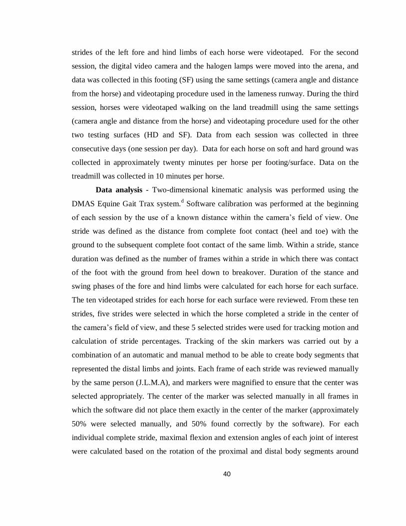
40
strides of the left fore and hind limbs of each horse were videotaped. For the second
session, the digital video camera and the halogen lamps were moved into the arena, and
data was collected in this footing (SF) using the same settings (camera angle and distance
from the horse) and videotaping procedure used in the lameness runway. During the third
session, horses were videotaped walking on the land treadmill using the same settings
(camera angle and distance from the horse) and videotaping procedure used for the other
two testing surfaces (HD and SF). Data from each session was collected in three
consecutive days (one session per day). Data for each horse on soft and hard ground was
collected in approximately twenty minutes per horse per footing/surface. Data on the
treadmill was collected in 10 minutes per horse.
Data analysis - Two-dimensional kinematic analysis was performed using the
DMAS Equine Gait Trax system.d Software calibration was performed at the beginning
of each session by the use of a known distance within the camera’s field of view. One
stride was defined as the distance from complete foot contact (heel and toe) with the
ground to the subsequent complete foot contact of the same limb. Within a stride, stance
duration was defined as the number of frames within a stride in which there was contact
of the foot with the ground from heel down to breakover. Duration of the stance and
swing phases of the fore and hind limbs were calculated for each horse for each surface.
The ten videotaped strides for each horse for each surface were reviewed. From these ten
strides, five strides were selected in which the horse completed a stride in the center of
the camera’s field of view, and these 5 selected strides were used for tracking motion and
calculation of stride percentages. Tracking of the skin markers was carried out by a
combination of an automatic and manual method to be able to create body segments that
represented the distal limbs and joints. Each frame of each stride was reviewed manually
by the same person (J.L.M.A), and markers were magnified to ensure that the center was
selected appropriately. The center of the marker was selected manually in all frames in
which the software did not place them exactly in the center of the marker (approximately
50% were selected manually, and 50% found correctly by the software). For each
individual complete stride, maximal flexion and extension angles of each joint of interest
were calculated based on the rotation of the proximal and distal body segments around

41
the palmar/plantar (carpus and fetclok joints) and dorsal (tarsus) aspects of the joints
using the inverse tangent function (Figure 5a and b). Range of motion (ROM) of each
joint, represented by the relative inter-segmental motion, was calculated for each stride of
each horse in each water level as maximal extension minus maximal flexion.
A post-hoc error calculation was performed for the camera angle by placing a
right angle in the areas of maximum flexion and extension of each joint (fetlock region
and carpus/tarsus region) in 10 different locations (5 at the fetlock level and 5 at the
carpus/tarsus level) within the camera’s field of view. The right angle had 2 cm diameter
markers on both branches and center of rotation, and was analyzed (using the same
software and settings as in the study) at each location. To determine the error based off of
the camera angle itself, additional measurements using the same conditions described
above were collected with the camera placed perpendicular to the different footings. A
total of 30 measurements were collected and the markers on the right angle were tracked.
The error was determined based off of the deviance from 90 degrees, and the average
error identified at each location at each footing was subsequently corrected in the data
(manually in each stride of each horse) before statistical analysis.
Statistics – Mean ± SD of maximal flexion and extension angles, joint ROM, and
the stance and swing percentages of each surface were calculated by first averaging 5
strides of each horse, and then combining the average of all horses at each walking
surface. Extreme studentized deviate tests were conducted on the 5 strides for each joint
for each horse under each footing to identify outliers, which were subsequently removed
from further analysis. Stance and swing temporal data was transformed to percentages.
Normality of the data was assessed using the Shapiro-Wilk normality test. Differences
between the 3 footing surfaces for each joint of interest were determined using a
repeated-measured ANOVA with a Tukey-Kramer’s multiple comparison test. Statistical
significance was set at p<0.05. All data were analyzed using a statistical software
packagee.

42
Figure 5. Photographs of a horse instrumented for data collection. Observe the locations
of the retroreflective skin markers on the fore (A) and hind (B) limbs used to determine
the body segments (blue lines) used to calculate the joint angles. Measured angles of each
joint are represented by a white line.
Results
The amount of error that was present based on camera angle of our 2-D kinematic
analysis system was < 2.2 degrees. There were no differences in error with the camera
positioned at a 30 degree angle versus being perpendicular to the footing surfaces. The
amount of error was different based on the location. The locations where maximal
extension occurred for the fetlocks measured angles between 87.8 to 89.9 degrees, and
for the carpus/tarsus angles between 88.3 and 90.2 degrees. The locations where flexion
occurred for the fetlocks measured angles between 89.3 and 91.8 degrees, and for the
carpus/tarsus angles between 88.8 and 91.1 degrees. The average error calculated for the
different locations was used to correct the peak flexion/extension data points (of each
stride of each horse in each footing surface) of the results presented below.

43
Kinematic analysis showed that the greatest maximal flexion for all joints was
attained when horses were walked on LT. In this surface, the carpus showed a significant
increase in flexion compared to the other two footing surfaces (HD and SF; Figure 6).
Additionally, the tarsus and hind fetlock demonstrated a significant increase in flexion
when horses walked on LT and SF compared to HD (Figure 6).
The greatest maximal extension of the most proximal joints (carpus and tarsus)
was achieved when horses were walked on HD, which was significantly greater when
compared to LT for both joints, and when compared to SF for the carpus (Figure 6).
Walking on SF also caused greater extension of the tarsus when compared to LT.
Conversely, the distal joints (both fetlocks) achieved the greatest maximal extension when
horses were walked on LT, which was significantly greater than HD and SF (Figure 6).
The greatest overall ROM of the carpus and both fetlocks was achieved when
horses were walked on LT, and the greatest ROM of the tarsus was achieved when
walking on SF (Figure 6). ROM was significantly (P<0.001) greater for all joints when
horses walked on LT compared to HD (Figure 6). Additionally, ROM for the tarsal joint
was significantly greater when horses walked on SF compared to HD. ROM of the fore
and hind fetlocks was also significantly greater when LT was compared to SF.
The percentage of the stride spent in stance phase decreased in the forelimb and
hind limb walking on SF compared to HD and LT, although only the hind limb showed
significant differences. In addition, LT showed a significantly greater percentage of the
stance phase of the hind limb when compared to HD (Table 5).

44
Figure 6. Maximal flexion (left-hand column) and extension (middle column) angles,
and ROM (right-hand column) of the carpus (1st row), tarsus (2
nd row), fore fetlock (3
rd
row), and hind fetlock (4th row) joints of healthy horses (n=9) walking on hard ground
(HD; white bar), soft ground (SF; gray bar), and on a land treadmill (LT; black bar).
Mean + SD are presented in the bar graphs. Significance denoted as: * P<0.05, **
P<0.01, and *** P<0.001.

45
Table 4. Mean ± SD of percentages of the stance and swing phases of the fore and hind
limbs of 9 clinically sound horses walking on hard ground (HD), soft ground (SF), and on
a land treadmill (LT).
Variable (%) HD SF LT
Stance forelimb 61.56 ± 0.7a 59.55 ± 1.4
a 60.17 ± 2.7
a
Swing forelimb 38.54 ± 1.2a 40.45 ± 1.4
a 39.83 ± 2.7
a
Stance hind limb 60.01 ± 1.0a 57.57 ± 1.2
b 60.80 ± 1.4
c
Swing hind limb 39.99 ± 1.0a 42.43 ± 1.2
a 39.79 ± 2.2
a
a–c Within a row, values with different superscript are significantly (P<0.05) different
Discussion
Results of this study found that the footing surface affects the flexion of the
carpus, tarsus and hind fetlock and the extension of all evaluated joints (i.e. carpus,
tarsus, fore and hind fetlocks) to a different extent when the horse is walking. In general,
the treadmill was the best surface at increasing joint flexion and overall ROM, as well as
extension of the fetlocks (fore and hind). However, walking on hard ground resulted in
the greatest extension of the carpal and tarsal joints. These findings confirmed our first
hypothesis, and are in agreement with other equine and human studies that have shown
an impact of walking surface on limb kinematics.83-86
Additionally, when soft and hard
grounds were compared, soft ground was superior at increasing flexion and overall ROM
of all joints except for the fore fetlock; hard ground was more effective at increasing joint
extension. Our second hypothesis was refuted because walking on soft ground did not
provide the greatest ROM for all evaluated joints.
Results of flexion and extension of the carpus, tarsus, fore and hind fetlocks of
horses walking on a rubber surface have been reported by other authors.59
In the present
study, we measured flexion/extension angles of the distal limb joints of horses walking
on asphalt, arena footing, and on a land treadmill, and our results are similar to those

46
reported on a rubberized surface using a similar method of data collection.59
In our study,
despite the differences found between surfaces, changes in flexion and extension of the
joints were less than 14 degrees. These increases, although subtle, may be enough to
promote ROM in the early rehabilitation stage following injury, surgery, or a prolonged
period of rest. However, other equine rehabilitation techniques such as passive
flexion/extension, walking over poles or cavalletis, walking in an underwater treadmill
(Chapter 2), or attachment of tactile stimulators and/or weights around the pastern of the
hind limbs may provide additional joint flexion (increases up to 30-45 degrees), which
may be more beneficial later on in the rehabilitation period.31,42
Currently, objective data
about rehabilitation techniques is lacking in horses, and to the author’s knowledge, the
amount of flexion/extension needed to help a clinical case is unknown at this time.
Results of this study showed that walking on the land treadmill causes the greatest
flexion and extension of both fetlock joints, suggesting that this surface may be the most
beneficial for promoting increased ROM of the fetlocks (Table 5). However, the greatest
flexion and the greatest extension of the carpal and tarsal joints did not happen on the
same surface (greatest flexion on LT, and greatest extension on HD), which suggests that
clinicians may need to alternate walking in two different surfaces in order to encourage
flexion and/or extension of the more proximal joints (Table 5). For instance, in horses
with reduced motion of the carpus, alternating exercise on the treadmill and asphalt may
be recommended because walking on the treadmill would encourage flexion, and walking
on asphalt would provide more extension of the joint (Table 5). Additionally, in
situations in which a treadmill is not available, alternating exercise in soft and hard
ground may also encourage joint ROM because in general soft ground would promote
more flexion, and hard ground would provide more extension. These are general
recommendations based on data from this study but it should be clarified that our
population was mainly Quarter Horses of similar sizes and fitness, and other breeds, sizes
and fitness levels may have different increases in flexion and extension on conventional
surfaces. Furthermore, horses that have been trained, especially to an advanced level, in
a specific discipline may have changes unique to that discipline due to the specific
outcomes of their training that were not able to be measured in this study.

47
Table 5. Footing surface at which the greatest flexion, extension, and overall range of
motion (ROM) of each evaluated joint was achieved.
Joint Flexion Extension ROM
Carpus Land Treadmill Hard Ground Land Treadmill
Tarsus Land Treadmill Hard Ground Soft Ground
Fore fetlock Land Treadmill Land Treadmill Land Treadmill
Hind fetlock Land Treadmill Land Treadmill Land Treadmill
Several studies have demonstrated the influence of walking surface on the stance
and swing phases of the equine stride.83,84,87,88
For instance, a study showed that at the
walk, stride length was shorter on a flat turf track than on a treadmill.83
Another study
demonstrated that at the trot, the stance phase duration of the front limbs was shorter on a
rubber surface than on a land treadmill.84
Similarly, a more recent study showed that
horses trotting on beach sand have a shorter stance phase than trotting on asphalt.88
In the
present study, we evaluated the percentages of the stance and swing phases on asphalt,
arena footing, and the treadmill at the walk, and found that the length of the stance phase
was shorter for horses walking on arena footing compared to asphalt, which agrees with
the findings at the trot of previous studies,84,88
and could be caused by an easier breakover
in the arena footing compared to asphalt. We found that the stance phase of the hind limb
was different for all evaluated footings, with the longest stance phase of the hind limb
occurring while walking on the treadmill. This finding contrasts with the results of a
similar study conducted at the trot, in which stance phase of the hind limb was shorter on
the treadmill than on asphalt or rubber.84
We did not find differences in the stance phase
of the forelimbs, which indicate that walking surface has a lesser effect in the phases of
the stride of the fore limbs.
Several limitations can be identified in this study, and most of them are due to the
fact that the methodology for data collection was chosen to mimic data collected in the

48
underwater treadmill study (Chapter 2). First, a 2-dimensional system was used to
perform the kinematic analysis since it was the only option to collect the data through the
windows of the underwater treadmill. This system (2-D) only reflects motion in the
sagittal plane and may not be as accurate as a three dimensional system for certain
kinematic parameters; however, a previous comparison of the two systems in horses at
the trot found no differences between the two dimensional and three dimensional systems
for angles of the joints evaluated in our study (i.e. carpus, hock, and fetlocks).45
Another
limitation of our study is the fact that the camera had to be angled 30 degrees down to be
able to compare the data with the underwater treadmill study (Chapter 2). This factor
likely reduced the sensitivity of our measurements because the majority of the joint
movement occurs in the sagittal plane and videos were captured in a skewed angle.
However, we attempted to minimize its effect within the system as much as we could.
The same person marked all horses, walked the horses in the hard and soft grounds and
run the treadmill, set up the video camera for each horse in each session, and manually
tracked every single frame (moved back and forth between contiguous frames) in an
attempt to ensure adequate central placement of the markers by the software. In addition,
the video camera was placed very close to the footing surfaces having a field of view of
3.5 meters, and multiple strides (n=5) for each horse at each footing surface were
collected in an attempt to minimize variability that would arise due to our data collection
system. We attempted to correct for our camera position error by placing a fixed angle
(90 degrees) at the different locations where the carpus, tarsus and fetlocks reached the
maximal flexion and extension in each surface. This analysis allowed for further error
correction in our data which should have further minimized variability from this source.
Based off of this analysis, we believe the error of our set up is less than 2.2 degrees for
any joint at any surface, and our corrected results are representative of the general motion
of the distal limbs. When comparing our corrected baseline results to those reported in
other studies59,60
where limb kinematics at the walk were evaluated over ground on sound
horses, our means and standard deviations are very similar (Table 1). Finally, although
speed of the over ground trials was controlled with timers, walking speed may have

49
influenced the results as walking speed has been shown to affect angular kinematics in
horses.89
In conclusion, walking surface influences the flexion and extension of the carpus,
tarsus, and fore and hind fetlock joints. This study provides objective data that can help
equine clinicians and physical therapists to decide which footing surface is best to walk
an individual orthopedic patient in the early rehabilitation period based on the location of
that patient’s injury.

50
Chapter 4
General conclusions and
future directions

51
Chapter 4. Conclusions and future directions.
Conclusions
This study showed that the underwater treadmill (UWTM) is a rehabilitation
modality that can be used to encourage range of motion of the carpi, hocks, and both
fetlocks (i.e. fore and hind) in the rehabilitation of equine patients. This finding is in
agreement with other human and canine studies that have suggested that the UWTM is a
useful technique to promote flexion and extension of the appendicular joints.
Results from this study showed that the UWTM can increase ROM of the carpus
by 25 degrees, ROM of the hock by 33 degrees, the ROM of the fore fetlock by 25
degrees, and the ROM of the hind fetlock by 46 degrees. Based on these data, the UWTM
seems to be the most effective rehabilitation modality to increase ROM of the distal limb
joints if we compare it to all the other available techniques for which scientific
information has been reported in horses.
Additionally, this study demonstrated that conventional footing surfaces such as
asphalt, arena, and the land treadmill affect ROM of the carpus, hock, and fetlocks, with
increases up to 14 degrees. These increases, although subtle, may be enough to promote
ROM in the early rehabilitation stage following injury, surgery, or a prolonged period of
rest. In general, the treadmill was the best conventional surface at increasing joint flexion
and overall ROM. Soft ground was also superior at increasing flexion and overall ROM
than hard ground. In contrast, hard ground was the most effective surface at increasing
joint extension of the carpus and hock. The extension of the fetlocks (fore and hind) was
maximal on the land treadmill.
When we compared the increases in flexion and extension of the joints in the
UWTM versus on the conventional footing surfaces, we observed substantial differences.
For instance, when comparing the two treadmills (UWTM and LT), the land treadmill
was superior to the UWTM baseline (<1cm of water) at increasing flexion, extension, and
overall ROM of all joints. This could be explained in part by the fact that the speed of the
LT belt (1.2 m/s) was slightly faster than the UWTM belt (0.9 m/s), and research has

52
shown that horses at higher speeds flex and extend their distal limbs more.89
Also, in
general, our study showed that the UWTM dramatically increases the flexion and overall
ROM of the distal limb joints at all water levels compared to over ground exercise.
However, increases in joint extension in the UWTM were similar to those observed in the
conventional surfaces, suggesting that both rehabilitation modalities are comparable for
this purpose.
The results of this study have been summarized in Table 6 to provide clinicians
and therapists with objective data that should be considered when designing a
rehabilitation program for an equine orthopedic patient.

53
Table 6. Maximal flexion and extension angles, and ROM of the carpus, tarsus, and fore and hind fetlocks joints of clinically
sound horses walking in the UWTM without water (B), and with water up to the hind fetlocks (F), tarsi (T), and stifles (S), and
walking on hard ground (cement, HD), soft ground (arena, SF), and land treadmill (LT). Values are expressed as degrees
(mean ± SD).
Joint Variable B F T S HD SF LT
Carpus
Maximal Flexion
Maximal Extension
ROM
121.6 ± 4.6
181.3 ± 4.4
59.5 ± 4.4
108.8 ± 4.9
181.1 ± 4.8
72.4 ± 6.3
101.8 ± 7.1
186.2 ± 3.1
84.2 ± 7.8
105.8 ± 5.5
180.4 ± 4.8
74.9 ± 8.1
118.3 ± 8.2
184.6 ± 1.6
65.6 ± 8.9
117.2 ± 6.1
183.2 ± 3.9
65.9 ± 6.1
113.6 ± 6.2
181.9 ± 2.5
68.0 ± 5.1
Tarsus
Maximal Flexion
Maximal Extension
ROM
130.9 ± 4.9
162.9 ± 2.6
32.6 ± 4.9
105.3 ± 4.9
164.8 ± 2.6
59.9 ±5.6
103.9 ± 6.3
163.9 ± 1.8
60.4 ± 7.1
101.3 ± 7.2
167.0 ± 3.2
65.8 ± 8.6
135.7 ± 6.4
171.3 ± 5.3
34.9 ± 4.2
130.3 ± 7.0
170.2 ± 4.6
40.1 ± 5.5
128.8 ± 6.5
168.8 ± 5.2
39.7 ± 3.4
Fore fetlock
Maximal Flexion Maximal Extension
ROM
165.9 ± 6.4 231.0 ± 10.7
65.0 ± 6.7
153.4 ± 7.7 243.6 ± 8.9
89.8 ± 6.8
141.1 ± 10.9
226.0 ± 8.0
86.8 ± 8.4
144.3 ± 5.4 231.2 ± 9.8
86.4 ± 9.1
166.2 ± 6.2 232.6 ± 9.3
66.0 ± 6.6
166.2 ± 7.9
230.9 ± 10.9
65.1 ± 8.2
165.0 ± 7.1 236.2 ± 11.2
70.7 ± 8.7
Hind
fetlock
Maximal Flexion Maximal Extension
ROM
157.2 ± 12.3 222.8 ± 7.9
65.7 ± 7.9
131.2 ± 7.4 242.8 ± 7.6
111.4 ± 5.1
110.1 ± 5.7 221.6 ± 6.9
111.6 ± 10.9
115.5 ± 8.2 221.1 ± 7.5
105.6 ± 8.1
155.8 ± 11.6 223.3 ±11.2
64.1 ± 12.5
151.3 ± 9.9 219.4 ± 10.2
68.6 ± 7.3
149.2 ± 13.7 227.3 ± 9.3
78.6 ± 9.2
53
54
Future directions
This study provides valuable information on how the water level affect the
flexion/extension angles, and range of motion (ROM) of the carpi, hocks, and fetlocks of
healthy horses walking in the underwater treadmill (UWTM). However, the effect of
water level on the other distal limb joints (i.e. pastern and coffin joints), and the proximal
joints (i.e. elbow, shoulder, stifle and hip) still needs to be determined. Unfortunately,
this information cannot be obtained in our current UWTM because it is not possible to
videotape the remaining joints through the small windows that the treadmill has on the
left side. Thus, this is a pending study that we would like to do in the future if a different
design of UWTM becomes available in the market, and any of our collaborators have
access to it.
Currently, objective data about rehabilitation techniques is lacking in horses, and
the amount of flexion/extension needed to help a clinical case is unknown. This study
provides data on how much the distal limb joints of sound horses can flex and extend
while walking on conventional surfaces and in the UWTM with different water levels.
So, now that we have a deeper understanding on how UWTM can be used to encourage
joint ROM in healthy horses, we would like to further evaluate the effect of the UWTM
in orthopedic patients, and find out what ROM is needed to clinically influence patients.
For this purpose, we would like to evaluate the effect of UWTM exercise in the
rehabilitation of horses that have had a limb immobilized (casted) for a long period of
time. This is a population of horses that often develops stiffness and loss of joint mobility
as a consequence of adaptive shortening of the soft tissues due to the immobilization. For
instance, a recent experimental study in which a metacarpophalangeal joint (i.e. fetlock)
was casted for 7 weeks showed that joint ROM was still restricted after 8 weeks of
treadmill exercise following cast removal.57
Therefore, we would like to perform a
similar study to assess the value of adding water treadmill exercise as part of the
rehabilitation of horses that have had a limb casted. In this future study, we would like to
rehabilitate horses with decreased ROM of the fetlock using the UWTM with different
water levels. This way, we should be able to identify which water level is the best to

55
rehabilitate a clinical patient with decreased fetlock motion and determine what ROM is
needed to rehabilitate this specific joint.
Besides these additional studies on the underwater treadmill, this project has
raised our curiosity in the kinematics of horses during swimming. This is another method
of hydrotherapy that has been used for training and conditioning of race horses for many
years.6,7
and is now gaining popularity as a rehabilitation method for some
musculoskeletal conditions. So far, there is no scientific information on the kinematics of
horses during swimming, although canine studies have showed that this modality
encourage the ROM of multiple joints.31
We would like to design a research project to
evaluate the kinematics of horses during swimming, and calculate the flexion and
extension angles of several joints. This information combined with the data of the
underwater treadmill could be used by equine clinicians and physical therapist to design
better and more specific rehabilitation programs of horses with orthopedic lesions.

56
Footnotes
a) Equine Aquapacer, Ferno Veterinary Systems, Inc., Wilmington, OH.
b) Lloyd Laboratories, Shenandoah, IA.
c) IDS Imaging Development Systems GmbH, Obersulm, Germany.
d) Motion Imaging Corporation, Simi Valley, CA.
e) SAS/STAT®, v9.2 SAS Inst. Inc., Cary, NC.
f) Polaris Wireless Timer; FarmTek Inc, Wylie, TX.
g) Säto, Knivsta, Sweden.
Bibliography
1. Kelly BT, Roskin LA, Kirkendall DT, et al. Shoulder muscle activation during aquatic
and dry land exercises in nonimpaired subjects. J Orthop Sports Phys Ther 2000;30:204-
210.
2. Konilan C. Aquatic therapy: making a wave in the treatment of low back injuries.
Orthop Nurs 1999;18:11-18.
3. Harasen G. Postoperative physical therapy in orthopedic patients. Can Vet J
2001;42:655.
4. Borgia LA, Valberg SJ, Essen-Gustavsson B. Differences in the metabolic properties
of gluteus medius and superficial digital flexor muscles and the effect of water treadmill
training in the horse. Equine Vet J Suppl 2010;38:665-670.
5. Voss B, Mohr E, Krzywanek H. Effects of aqua-treadmill exercise on selected blood
parameters and on heart-rate variability of horses. J Vet Med A Physiol Pathol Clin Med
2002;49:137-143.
6. Misumi K, Sakamoto H, Shimizu R. Changes in blood lactate and heart rate in
thoroughbred horses during swimming and running according to their stage of training.
Vet Rec 1994;135:226-228.
7. Misumi K, Sakamoto H, Shimizu R. The validity of swimming training for two-year-
old thoroughbreds. J Vet Med Sci 1994;56:217-222.
8. Haralson KM. Therapeutic pool programs. J Clin Management 1988;5:510-513.

57
9. Levine D, Rittenberry L, Millis DL. Aquatic therapy. In: Millis DL, Levine D, Taylor
RA, eds. Canine rehabilitation and physical therapy. St. Louis, MO: Saunders,
2004;264-276.
10. Edlich RF, Towler MA, Goitz RJ, et al. Bioengineering principles of hydrotherapy. J
Burn Care Rehabil 1987;8:580-584.
11. Geigle PR, Cheek WL, Gould ML, et al. Aquatic physical therapy for balance: the
interaction of somatosensory and hydrodynamic principles. J Aquatic Phys Ther
1997;5:4-10.
12. McClintock SA, Hutchins DR, Brownlow MA. Determination of weight reduction in
horses in flotation tanks. Equine Vet J 1987;19:70-71.
13. Levine D, Marcellin-Little DJ, Millis DL, et al. Effects of partial immersion in water
on vertical ground reaction forces and weight distribution in dogs. Am J Vet Res
2010;71:1413-1416.
14. Harrison RA, Bulstrode S. Percentage weight-bearing during partial immersion in
hydrotherapy pool. Physiother Pract 1987;3:60-63.
15. Thein JM, Brody LT. Aquatic-based rehabilitation and training for the elite athlete. J
Orthop Sports Phys Ther 1998;27:32-41.
16. Johns KM. Aquatic Therapy: therapeutic treatment for today's patient. Phys Ther
Prod 1993:24-25.
17. Babb RW, Simelson-Warr A. Manual techniques of the lower extremities in aquatic
physical therapy. J Aquatic Phys Ther 1996;14:7-15.
18. Thomas DP, Fregin GF, Gerber NH, et al. Cardiorespiratory adjustments to tethered-
swimming in the horse. Pflugers Arch 1980;385:65-70.
19. Cureton KJ. Physiologic responses to water exercise. In: Routi RG, Morris DM, eds.
Aquatic Rehabilitation. Philadelphia, PA: Lippincott, 1997;158-159.
20. Bromiley MW. Rehabilitation. In: Bromiley MW, ed. Equine Injury, Therapy and
Rehabilitation. 3rd ed. Ames, IA: Blackwell Publishing, 2007;130-159.
21. Nankervis KJ, Williams RJ. Heart rate responses during acclimation of horses to
water treadmill exercise. Equine Vet J Suppl 2006;36:110-112.
22. Lindner A, Waschle S, Sasse HL. Effect of exercise on a treadmill submerged in
water on biochemical and physiological variables of horses. Pferdeheilkunde 2010;26:6.

58
23. Monk M. Hydrotherapy. In: McGowan C, Goff L, Stubbs NC, eds. Animal
Physiotherapy: Assessment, Treatment and Rehabilitation of Animals. Oxford,UK:
Blackwell Publishing, 2007;187-198.
24. Hall J, Bisson D, O'Hare P. The physiology of immersion. Physiother Can
1990;79:517-521.
25. Valtonen A, Poyhonen T, Sipila S, et al. Effects of aquatic resistance training on
mobility limitation and lower-limb impairments after knee replacement. Arch Phys Med
Rehabil 2010;91:833-839.
26. Brady B, Redfern J, MacDougal G, et al. The addition of aquatic therapy to
rehabilitation following surgical rotator cuff repair: a feasibility study. Physiother Res Int
2008;13:153-161.
27. Tovin BJ, Wolf SL, Greenfield BH, et al. Comparison of the effects of exercise in
water and on land on the rehabilitation of patients with intra-articular anterior cruciate
ligament reconstructions. Phys Ther 1994;74:710-719.
28. Wang TJ, Lee SC, Liang SY, et al. Comparing the efficacy of aquatic exercises and
land-based exercises for patients with knee osteoarthritis. J Clin Nurs 2011;20:2609-
2622.
29. Batterham SI, Heywood S, Keating JL. Systematic review and meta-analysis
comparing land and aquatic exercise for people with hip or knee arthritis on function,
mobility and other health outcomes. BMC Musculoskelet Disord 2011;12:123.
30. Lim JY, Tchai E, Jang SN. Effectiveness of aquatic exercise for obese patients with
knee osteoarthritis: a randomized controlled trial. PM R 2010;2:723-731.
31. Marsolais GS, McLean S, Derrick T, et al. Kinematic analysis of the hind limb during
swimming and walking in healthy dogs and dogs with surgically corrected cranial
cruciate ligament rupture. J Am Vet Med Assoc 2003;222:739-743.
32. Marsolais GS, Dvorak G, Conzemius MG. Effects of postoperative rehabilitation on
limb function after cranial cruciate ligament repair in dogs. J Am Vet Med Assoc
2002;220:1325-1330.
33. Monk ML, Preston CA, McGowan CM. Effects of early intensive postoperative
physiotherapy on limb function after tibial plateau leveling osteotomy in dogs with
deficiency of the cranial cruciate ligament. Am J Vet Res 2006;67:529-536.
34. Jackson AM, Millis DL, Stevens M, et al. Joint kinematics during underwater
treadmill activity. In: Proc 2nd Int Symp Rehabil Phys Ther Vet Med 2002:191.

59
35. Kisner C, Colby LA. Range of motion. In: Kisner C, Colby LA, eds. Therapeutic
exercise: foundations and techniques. Philadelphia, PA: FA Davis, 2002;34-57.
36. Brody LT. Mobility impairment. In: Hall CM, Brody LT, eds. Therapeutic exercise:
moving toward function. Philadelphia, PA: Williams & Wilkins, 1999;97-104.
37. Millis DL, Lewelling A, Hamilton S. Range-of-motion and stretching exercises In:
Millis DL, Levine D, Taylor RA, eds. Canine rehabilitation and physical therapy. St.
Louis, MO: Saunders, 2004;228-243.
38. Doucet MY, Bertone AL, Hendrickson D, et al. Comparison of efficacy and safety of
paste formulations of firocoxib and phenylbutazone in horses with naturally occurring
osteoarthritis. J Am Vet Med Assoc 2008;232:91-97.
39. Schaer TP, Bramlage LR, Embertson RM, et al. Proximal interphalangeal arthrodesis
in 22 horses. Equine Vet J 2001;33:360-365.
40. Simmons EJ, Bertone AL, Weisbrode SE. Instability-induced osteoarthritis in the
metacarpophalangeal joint of horses. Am J Vet Res 1999;60:7-13.
41. Adair HS. Aquatic Therapy for Conditioning and Treatment of Tendon and Ligament
Injuries. In: Proc Am Assoc Equine Practnrs 2011;57:181-185.
42. Clayton HM, Lavagnino M, Kaiser LJ, et al. Evaluation of biomechanical effects of
four stimulation devices placed on the hind feet of trotting horses. Am J Vet Res
2011;72:1489-1495.
43. Clayton HM, Schamhardt HC. Measurement techniques for gait analysis. In: Back W,
Clayton HM, eds. Equine Locomotion. London, UK: W. B. Saunders, 2001;55-76.
44. Drevemo S, Dalin G, Fredricson I, et al. Equine locomotion; 1. The analysis of linear
and temporal stride characteristics of trotting standardbreds. Equine Vet J 1980;12:60-65.
45. Miró F, Santos R, Garrido-Castro JL, et al. 2D versus 3D in the kinematic analysis of
the horse at the trot. Vet Res Commun 2009;33:507-513.
46. Leach DH, Dyson S. Instant centres of rotation of equine limb joints and their
relationship to standard skin marker locations. Equine Vet J Suppl 1988;6:113-119.
47. Burn JF, Portus B, Brockington C. The effect of speed and gradient on
hyperextension of the equine carpus. Vet J 2006;171:169-171.
48. Schamhardt HC, van den Bogert AJ, Hartman W. Measurement techniques in animal
locomotion analysis. Acta Anat (Basel) 1993;146:123-129.

60
49. van Weeren PR, van den Bogert AJ, Barneveld A. Quantification of skin
displacement in the proximal parts of the limbs of the walking horse. Equine Vet J Suppl
1990;9:110-118.
50. van Weeren PR, van den Bogert AJ, Barneveld A. A quantitative analysis of skin
displacement in the trotting horse. Equine Vet J Suppl 1990;9:101-109.
51. Lanovaz JL, Clayton HM, Colborne GR, et al. Forelimb kinematics and net joint
moments during the swing phase of the trot. Equine Vet J Suppl 1999;30:235-239.
52. Clayton HM, Schamhardt HC, Willemen MA, et al. Kinematics and ground reaction
forces in horses with superficial digital flexor tendinitis. Am J Vet Res 2000;61:191-196.
53. van Weeren PR, van den Bogert AJ, Back W, et al. Kinematics of the standardbred
trotter measured at 6, 7, 8 and 9 m/s on a treadmill, before and after 5 months of prerace
training. Acta Anat (Basel) 1993;146:154-161.
54. Ratzlaff MH, Wilson PD, Hyde ML, et al. Relationship between locomotor forces,
hoof position and joint motion during the support phase of the stride of galloping horses.
Acta Anat (Basel) 1993;146:200-204.
55. Taylor BM, Tipton CM, Adrian M, et al. Action of certain joints in the legs of the
horse recorded electrogoniometrically. Am J Vet Res 1966;27:85-89.
56. Liljebrink Y, Bergh A. Goniometry: is it a reliable tool to monitor passive joint range
of motion in horses? Equine Vet J Suppl 2010;38:676-682.
57. Van Harreveld PD, Lillich JD, Kawcak CE, et al. Clinical evaluation of the effects of
immobilization followed by remobilization and exercise on the metacarpophalangeal
joint in horses. Am J Vet Res 2002;63:282-288.
58. Millis DL. Assessing and measuring outcomes. In: Millis DL, Levine D, Taylor RA,
eds. Canine rehabilitation and physical therapy. St. Louis, MO: Saunders, 2004;211-227.
59. Hodson E, Clayton HM, Lanovaz JL. The forelimb in walking horses: 1. Kinematics
and ground reaction forces. Equine Vet J 2000;32:287-294.
60. Hodson E, Clayton HM, Lanovaz JL. The hindlimb in walking horses: 1. Kinematics
and ground reaction forces. Equine Vet J 2001;33:38-43.
61. Clayton HM, Sha D, Stick J, et al. 3D kinematics of the equine metacarpophalangeal
joint at walk and trot. Vet Comp Orthop Traumatol 2007;20:86-91.
62. Ramon T, Prades M, Armengou L, et al. Effects of athletic taping of the fetlock on
distal limb mechanics. Equine Vet J 2004;36:764-768.

61
63. Ratzlaff MH, Grant BD, Adrian M, et al. Evaluation of equine locomotion using
electrogoniometry and cinematography: research and clinical applications. In: Proc Am
Assoc Equine Practnrs 1979;25:381.
64. Crevier-Denoix N, Robin D, Pourcelot P, et al. Ground reaction force and kinematic
analysis of limb loading on two different beach sand tracks in harness trotters. Equine Vet
J Suppl 2010;38:544-551.
65. Gandini G, Cizinauskas S, Lang J, et al. Fibrocartilaginous embolism in 75 dogs:
clinical findings and factors influencing the recovery rate. J Small Anim Pract
2003;44:76-80.
66. Lindner A, Waschle S, Sasse HH. Physiological and blood biochemical variables in
horses exercising on a treadmill submerged in water. J Anim Physiol Anim Nutr (Berl)
2012;96:563-569.
67. Geigle P, Cheek WL, Gould ML, et al. Aquatic physical therapy for balance: the
interaction of somatosensory and hydrodynamic principles. J Aquatic Phys Ther
1997;5:4-10.
68. Steiss JE. Canine Rehabilitation. In: Braund KG, ed. Clinical Neurology in Small
Animals - Localization, Diagnosis and Treatment. New York, NY: IVIS, 2003;1-53.
69. Yen CH, Chan WL, Wong JW, et al. Clinical results of early active mobilisation after
flexor tendon repair. Hand Surg 2008;13:45-50.
70. Scott R, Nankervis K, Stringer C, et al. The effect of water height on stride frequency,
stride length and heart rate during water treadmill exercise. Equine Vet J Suppl
2010;38:662-664.
71. Goff L, Stubbs NC. Equine treatment and rehabilitation. In: McGowan C, Goff L,
Stubbs NC, eds. Animal Physiotherapy: Assessment, treatment and rehabilitation of
animals. Oxford UK: Blackwell Publising, 2007;238-250.
72. Holler PJ, Brazda V, Dal-Bianco B, et al. Kinematic motion analysis of the joints of
the forelimbs and hind limbs of dogs during walking exercise regimens. Am J Vet Res
2010;71:734-740.
73. Clayton HM, White AD, Kaiser LJ, et al. Hindlimb response to tactile stimulation of
the pastern and coronet. Equine Vet J 2010;42:227-233.
74. Singleton WH, Clayton HM, Lanovaz JL, et al. Effects of shoeing on forelimb swing
phase kinetics of trotting horses. Vet Comp Orthop Traumatol 2003;16:16-20.
75. Wickler SJ, Hoyt DF, Clayton HM, et al. Energetic and kinematic consequences of
weighting the distal limb. Equine Vet J 2004;36:772-777.
62
76. Rumpler B, Riha A, Licka T, et al. Influence of shoes with different weights on the
motion of the limbs in Icelandic horses during toelt at different speeds. Equine Vet J
Suppl 2010;38:451-454.
77. Kato T, Onishi S, Kitagawa K. Kinematical analysis of underwater walking and
running. Sports Med Train and Rehabil 2001;10:165-182.
78. Wentink GH. Biokinetical analysis of the movements of the pelvic limb of the horse
and the role of the muscles in the walk and the trot. Anat Embryol (Berl) 1978;152:261-
272.
79. Giaquinto S, Ciotola E, Dall'Armi V, et al. Hydrotherapy after total knee arthroplasty.
A follow-up study. Arch Gerontol Geriatr 2010;51:59-63.
80. Jung T, Lee D, Charalambous C, et al. The influence of applying additional weight to
the affected leg on gait patterns during aquatic treadmill walking in people poststroke.
Arch Phys Med Rehabil 2010;91:129-136.
81. Barrey E. Inter-limb coordination. In: Back W, Clayton H, eds. Equine locomotion.
London, UK: W. B. Saunders, 2001;77-133.
82. Levine D, Millis DL, Marcellin-Little DJ. Introduction to veterinary physical
rehabilitation. Vet Clin North Am Small Anim Pract 2005;35:1247-1254.
83. Barrey E, Galloux P, Valette JP, et al. Stride characteristics of overground versus
treadmill locomotion in the saddle horse. Acta Anat (Basel) 1993;146:90-94.
84. Buchner HH, Savelberg HH, Schamhardt HC, et al. Kinematics of treadmill versus
overground locomotion in horses. Vet Q Suppl 1994;16 87-90.
85. Ralston HJ. Comparison of energy expenditure during treadmill walking and floor
walking. J Appl Physiol 1960;15:1156.
86. Elliott BC, Blanksby BA. A cinematographic analysis of overground and treadmill
running by males and females. Med Sci Sports 1976;8:84-87.
87. Fredricson I, Drevemo S, Dalin G, et al. Treadmill for equine locomotion analysis.
Equine Vet J 1983;15:111-115.
88. Chateau H, Holden L, Robin D, et al. Biomechanical analysis of hoof landing and
stride parameters in harness trotter horses running on different tracks of a sand beach
(from wet to dry) and on an asphalt road. Equine Vet J Suppl 2010;38:488-495.
89. Galisteo AM, Cano MR, Morales JL, et al. The influence of speed and height at the
withers on the kinematics of sound horses at the hand-led trot. Vet Res Commun
1998;22:415-423.


![KINEMATICS - new.excellencia.co.innew.excellencia.co.in/college/web/pdf/Kinematics-merged.pdf · KINEMATICS KINEMATICS WORKSHEET 1 1) Displacement is a _____ [ ] 1) Vector quantity](https://img.pdfslide.net/doc/110x75/5f356d4687229051801abace/kinematics-new-kinematics-kinematics-worksheet-1-1-displacement-is-a-.jpg)