Embed Size (px)
Citation preview
Effect of Transfer Factor on Lymphocyte
Function in Anergic Patients
CHARLEsH. KIKPATRICK, ROBERTR. RiCH, and TEimuL K. SMrrH
From the Laboratory of Clinical Investigation, National Institute of Allergyand Infectious Diseases, National Institutes of Health,Bethesda, Maryland 20014
A B S T R A C T Dialyzable transfer factor, obtained fromfrozen-thawed peripheral blood leukocytes from a singledonor, was given to five anergic patients with chronicmucocutaneous candidiasis. Studies of immunologicalresponses including delayed cutaneous hypersensitivity,in vitro antigen-induced thymidine incorporation, andproduction of macrophage migration inhibition factor(MIF) were conducted both before and after injectionof transfer factor.
Before transfer factor, none of the patients had de-layed skin responses to any of the natural antigens stud-ied. Their lymphocytes did not produce MIF after ex-posure to antigens in vitro and only one patient showedincreased thymnidine incorporation when his lymphocyteswere cultured with candida and streptokinase-streptodor-nase (SK-SD). After injection of transfer factor, fourpatients developed delayed skin responses to antigens towhich the donor was sensitive; no recipient reactedto an antigen to which the donor was nonreactive.Lymphocytes from recipients produced MIF when cul-tured with antigens that evoked positive delayed skintests. Only one patient developed antigen-induced lympho-cyte transformation and this response occurred only in-termittently. Attempts to sensitize three of the patientswith the contact allergen, chlorodinitrobenzene, both be-fore and after transfer factor, were unsuccessful.
The fifth patient, a 9-yr old boy with an immunologicprofile similar to the Nezelof syndrome, did not becomeskin test-reactive or develop positive responses to thein vitro tests.
These findings suggest that transfer factor acts on theimmunocompetent cells that respond to antigens withlymphokine production, but has little, if any, effect oncells that respond to antigens by blastogenesis. The fail-ure to sensitize the subjects with chlorodinitrobenzene
Received for publication 31 May 1972 and in revised form17 July 1972.
illustrates the specificity of the immunologic effects oftransfer factor, and implies that it does not functionthrough nonspecific, adjuvant-like mechanisms. Failureof transfer factor to produce positive skin tests or MIFproduction in a patient with Nezelof's syndrome may beevidence that lymphokine-producing cells are thymusderived.
INTRODUCTIONIn 1955, Lawrence (1) reported that disrupted leuko-cytes from skin test-reactive donors could transfer de-layed hypersensitivity to skin test-negative subjects.The skin test conversion was specific and long lived. Theactive component, transfer factor, was subsequentlyfound to be dialyzable and resistant to degradation bytrypsin, ribonuclease, or deoxyribonuclease (2). How-ever, its composition and mechanism of action have notbeen elucidated.
Recently, transfer factor has been employed in treat-ment of a variety of disorders with disturbed cellular im-munity. Patients with the Wiskott-Aldrich syndrome,leprosy, chronic candidiasis, and neoplastic disordershave received transfer factor, and while the results havebeen variable, some encouraging reports have emerged(2-7).
The purpose of this study was to investigate the effectof transfer factor on immunologic responses of anergicpatients with chronic mucocutaneous candidiasis. Cellu-lar immunity was characterized in vivo by intradermaltests and in vitro by lymphocyte transformation andmacrophage migration inhibition factor (MIF)' produc-tion before and after transfer factor. The clinical re-sponses of the recipients were also observed.
I Abbreviations used in this paper: CDNB, 1-chloro-2,4-dinitrobenzene; MIF, migration inhibition factor; PPD,purified protein derivative; SK-SD, streptokinase-strepto-doranse.
2948 The Journal of Clinical Investigation Volume 51 November 1972
Downloaded from http://www.jci.org on February 25, 2015. http://dx.doi.org/10.1172/JCI107119
METHODSSkin testing. Each subject was tested with a panel of
commercial antigens which included intermediate strengthpurified protein derivative (PPD), histoplasmin, mumpsskin test antigen, trichophytin (1000 pnu/ml),' Candidaalbicans extract (Dermatophytin 0, 1: 100),' and strepto-kinase-streptodornase (SK-SD)8 diluted to contain 40 U ofSK and 10 U of SD/ml. When delayed skin tests with thisconcentration of SK-SD were negative, the tests were re-peated using solutions containing 10 times more antigen.Additional antigenic preparations- of C. albicans were madeby sonication of organisms isolated from cases R. H. andJ. A. C. (8), and here the preparations used for skin test-ing contained 10 or 100 ,&g of protein/ml. For testing, 0.1ml doses of antigen were injected intradermally. Cutaneousresponses were read at 15 min and 5, 24, and 48 hr. Positivedelayed reactions produced 0.5 cm or more of induration.
Induction of contact allergy. If skin tests with the panelof naturally encountered antigens suggested anergy, anattempt was made to induce contact allergy with 1-chloro-2,4-dinitrobenzene (CDNB). To screen for pre-existing sen-,sitivity, the subjects were first tested with 100 ug of CDNBin acetone. If this test was negative, a 2000 Mug sensitizingdose was applied dropwise to the skin of the medial upperarm. The acetone was evaporated and the area was coveredwith a Telfa patch for 48 hr. This site was examined regu-larly for the appearance of a spontaneous flare compatiblewith induction of contact allergy. Challenge tests with 50and 100 Ag of CDNBwere conducted 2 wk later, with theantigens applied to the contralateral arm. The scoring ofresponses at 24, 48, and 72 hr was essentially the same asthat recently described by Catalona, Taylor, Rabson, andChretien (9), except that biopsies were not done to evalu-ate grossly negative areas. This schedule of sensitizationhas been shown by ourselves and others (9) to induce con-tact allergy in approximately 95% of control subjects.
Lymphocyte transformation. The general technique formeasurement of antigen-stimulated thymidine incorporationhas been described previously (10). Briefly, blood was col-lected into heparinized (20 U/ml) syringes and the lympho-cyte-rich leukocyte fraction was obtained by differentialcentrifugation and sedimentation. Each culture tube con-tained 2 X 10' lymphocytes in 2.0 ml of Eagle's minimumessential medium with penicillin, streptomycin, and either20% heat-inactivated fetal bovine serum or 20% freshautologous or homologous plasma. Antigen-stimulated andcontrol cultures were conducted in triplicate or quadrupli-cate. No patient was receiving drugs known to alter lym-phocyte-mediated immunologic responses.
Preliminary studies had shown that peak thymidine in-corporation by lymphocytes from skin test-positive subjectsoccurred on the 5th day of culture, and dose-response ex-periments indicated that optimal responses were obtained incultures containing 5 ug/ml of candida extract or 50/12.5U/ml of SK-SD. Depending upon the number of cells avail-able, cultures contained the optimal concentrations of anti-gens and other doses ranging from 0.01 to 100 times theoptimal doses.
Studies of thymidine incorporation by lymphocytes stimu-lated with purified phytohemagglutinin,' or concanavalin A'were harvested on the 3rd day of culture. Mixed leukocyte
2Hollister-Stier Laboratories, Downers Grove, Ill.'Varidase, Lederle Laboratories, Pearl River, N. Y.'Burroughs Wellcome Co., Research Triangle Park, N. C.'ICN Nutritional Biochemicals Div., Cleveland, Ohio.
reactions were done by culturing responding cells withmitomycin-blocked stimulating cells (11) from unrelated,phenotypically disparate donors6 and were harvested onthe 7th day.
Cells were labeled with thymidine-'H 4 hr before har-vesting. The cultures were terminated by collecting cells bycentrifugation, washing the cells with saline, and precipi-tating the protein with cold trichloracetic acid. The pelletwas washed with cold methanol, digested with sodiumhydroxide at 60'C, and dissolved in a toluene-base solventfor liquid scintillation counting.
Macrophage migration inhibition factor (MIF). Themethod of Rocklin, Meyers, and David (12) was employedto study MIF production by lymphocytes from the patientsas well as control subjects with either positive or negativeskin tests to candida or SK-SD. Lymphocyte-rich leuko-cyte suspensions were incubated in serum-free minimumessential medium that had been supplemented with glucose,pyruvate, and nonessential amino acids (13), either with orwithout antigens. Every 24 hr for 3 days the culture fluidswere harvested and replaced. Cell-free culture fluids werealso collected for control of inhibitory effects of the anti-gens alone. The supernatants were concentrated fivefold byvacuum dialysis and MIF activity was determined by the"indirect" assay using oil-induced peritoneal exudate cellsfrom guinea pigs. The fields of migration were proj ectedonto a screen and traced and the areas were measured witha planimeter. The per cent inhibition was calculated as sug-gested by Rocklin et al. (12) and the differences betweenmeans were analyzed with the two-tailed t test.
Preparation of transfer factor. The transfer factor wasprepared from cells from one healthy adult donor. Intra-dermal tests had produced the following diameters of in-duration at 24 hr: histoplasmin, 4.5 cm with subsequentslough; candida (10 /g/ml), 2.5 cm; SK-SD (40/10 U/ml),2.0 cm; and mumps, 3.0 cm. There were no responses toPPD or trichophytin. Serologic studies for the hepatitis-associated agent were negative.
A lymphocyte-rich leukocyte suspension was obtained bycontinuous flow centrifugation with an NCI-IBM cell sepa-rator (14). Transfer factor was prepared by the method ofLawrence (2). Cells were lysed by 10 cycles of freezing indry ice-alcohol and thawing at 370C. The lysate was thendigested with deoxyribonuclease7 in the presence of mag-nesium for 1 hr at 370C. This material was dialyzed against20 vol of sterile distilled water for 40 hr at 40C. Thedialyzable fraction was lyophilized, then reconstituted withdistilled water so that the material from 300 X 10. lympho-cytes was contained in 1 ml. The solution was passedthrough a Millipore filter, portioned into sterile vials, andstored at - 30°C. All steps used sterile glassware andpyrogen-free reagents, and the final product was sterile. Inthese studies, a "dose" of transfer factor was 2 ml andcontained the material from 600 X 106 lymphocytes. It wasgiven both subcutaneously and into multiple intradermalsites. Skin tests after injections of transfer factor wereapplied to the contralateral arm.
RESULTSClinical and immunologic findings before transfer
factor. The initial clinical and immunologic studies of
H-LA phenotypes were determined by a microcytotox-icity method in the laboratory of Dr. Paul Terasaki.
TWorthington Biochemical Corp., Freehold, N. J.
Transfer Factor in Anergic Patients 2949
Downloaded from http://www.jci.org on February 25, 2015. http://dx.doi.org/10.1172/JCI107119
F WM - AMPHOTERICIN B
0I0090mg
to°nI[
o 0o 0o 0
06(?) 05()
000
0.6(?)
0 30 0
0 00.9 1.0 1.2 17 0.9 1.10.5 1.2 1.9 1.0 0.9 0803 1.5 1.5 0.9 07 0.7
00
0.6()
0000
21 9 00
OControlMCandidaWVaridase
MAY JULY.^&e
AUG SEP DEC FEB JUN JUN AUG SEP OCT NOV4' 1970 +-1971
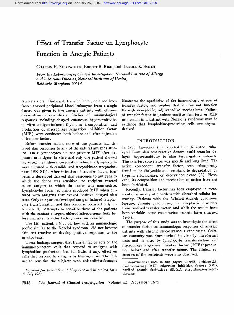
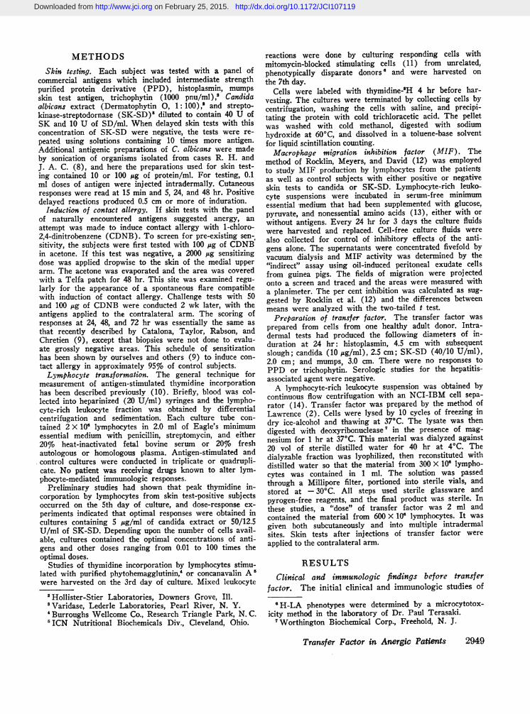
FIGURE 1 Serial studies of lymphocyte transformation, delayed cutaneous hypersensitivity,and MIF production in patient R. H. In all figures the lymphocyte transformation experi-ments were conducted in 20% auto'logous plasma. Candida-stimulated cultures contained 5 /Agprotein/ml of candida extract and Varidase-stimulated cultures contained 50 U of SK and12.5 U of SD/ml. The ratio of mean dpm in antigen-stimulated cultures to mean dpm inunstimulated cultures (stimulation ratio) is given at the top of each bar. Underlined values forMIF activity represent results in which antigen-stimulated fluids did not produce inhibitionthat was significantly greater than the control fluids.
three patients (R. H., J. A. C., and D. L.) have been de-scribed previously (15). R. H. is a 19-yr old white malewho has had extensive candidiasis of the skin, nails, andmucous membranes since age 9 months. Associated dis-orders included hypoparathyroidism recognized at age 7yr, and dysplasia of the dental enamel. Treatment withtopical medications had produced brief and incompleteremissions. The immunologic studies had revealed nonre-
activity to all antigens in the panel, including CDNB.His lymphocytes failed to produce macrophage migrationinhibition factor (MIF) when stimulated with candidaor SK-SD although both antigens stimulated thymidineincorporation (Fig. 1).
In 1969, the therapeutic potential of immunologic re-
constitution was evaluated (16). The patient's fatherhad reactive skin tests to candida, mumps, and SK-SDand was sensitized with CDNB. After transfusion of63 X 10° paternal lymphocytes, the patient's skin testsbecame positive and there was marked clearing of thecutaneous and mucous membrane lesions. Clinical relapseoccurred approximately 8 months later and was accom-
panied by loss of skin test reactivities. In 1970, treat-
ment with clotrimazole (17) produced essentially com-plete clearing of the oral and cutaneous lesions. Un-fortunately, the infections recurred while he was still re-ceiving the drug and increased dosages produced in-tolerable side effects (nausea, abdominal discomfort, andhypercalcemia), but no clinical benefit.
Because programs directed at correction of the im-munologic defect and against the microorganism had eachbeen temporarily beneficial, combined anti-fungal and im-munologic treatment was begun in June 1971. Arnpho-tericin B, 1090 mg, was given intravenously duringJune and July and injections of transfer factor werestarted in July 1971. There was prompt and completeclearing of all lesions except for the nails, which wereavulsed under general anesthesia. The only recurrenceshave been two episodes of candidiasis on the tonguewhich followed treatment with antibiotics. These werecleared with amphotericin.
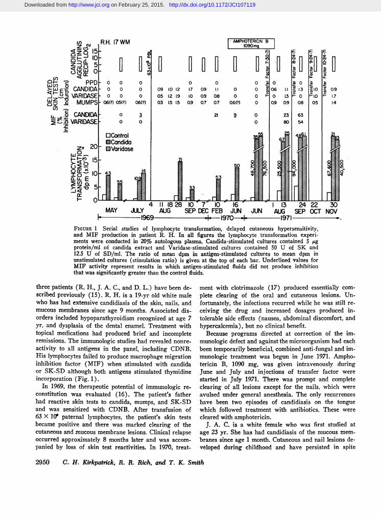
J. A. C. is a white female who was first studied atage 23 yr. She has had candidiasis of the mucous mem-branes since age 1 month. Cutaneous and nail lesions de-veloped during childhood and have persisted in spite
2950 C. H. Kirkpatrickj R. R. Rich, and T. K. Smith
R.H. 17
czI 00.91.71.4
k
Downloaded from http://www.jci.org on February 25, 2015. http://dx.doi.org/10.1172/JCI107119
JAC 23 WF
25'zI04
0000
1.7
0 0 0
0 0 0 0 0 00 0 E 07 05 % Q6 0.6 050 0 1.0 IA 12 12 12 0.90 0 0.6 08 1.0 1.0 0.7 0.5
0 14.7 I 16J 27 18.4 53 37 31
E
0
oo0 0
0.9 0o 0
9
0
01.4141.0
0 1 24.7 25.1 31.5 29
OControlECandidaWVaridase
MAY JUN DEC JAN FEB MAR JUN JUL AIUG SEP OCT DECIs-l1970 + 1971
FIGuRE 2 Serial studies of MIF production, lymphocyte transformation, and delayed skintests in patient J. A. C. The culture conditions are the same as Fig. 1. In; contrast to theother subjects of this study, this patient had positive lymphocyte transformation responsesafter the first injection of transfer factor.
of vigorous topical therapy. Symptoms of hypothyroidismappeared at age 21 yr, and the diagnosis was confirmed2 yr later. Her immunologic studies also revealedanergy to all of the natural antigens in the panel and anattempt at sensitization with CDNBwas unsuccessful.The results of serial skin testing and in vitro studieswith her peripheral blood lymphocytes are summarized inFig. 2. Her cells did not respond to antigenic stimulationwith increased thymidine incorporation or MIF pro-duction.
The first attempt at immunologically directed therapyof this patient was in January 1971, when she was giventransfer factor from 600 X 106 lymphocytes. Althoughthe skin tests to candida, SK-SD, and mumps, becamepositive, there was no clinical improvement. The com-bined program of anti-fungal therapy and immunologi-cal reconstitution was begun in June 1971 (Fig. 2).The dose of 880 mg of amphotericin B was given intra-venously over 10 wk and transfer factor was started 10August 1971. There was prompt clearing of the cu-taneous and mucous membrane lesions, and the nails wereavulsed after cultures from the nail folds ceased yieldingC. albicans. There has been no relapse.
R. V. was first seen at NIH at age 32 yr. At age 3yr she developed a crusting, pustular dermatitis of thescalp. At age 5 the disorder was diagnosed as candidiasis
and during the next 12 yr the lesions progressed to in-volve the skin of the face, extremities, and torso, as wellas the nails and mucous membranes. Diabetes mellituswas found at a routine examination at age 18 yr. At age24, severe frequency and dysuria prompted a urologicalevaluation. Cystoscopy disclosed a contracted bladder, aurethral stricture, and severe urethritis and cystitis.Pyelography showed right hydronephrosis and hydro-ureter. A compensated Coombs-positive hemolytic anemiaand episodic leukopenia have been present since 1965.In addition to continuous treatment with topical anti-fungal agents, she had received intravenous amphoteri-cin B in 1965, 1967, and 1970. Each course had producedmarked clinical improvement, but the mucous membraneand cutaneous lesions recurred within a few months.5-fluorocytosine had also been given, but without benefit.
Upon admission to NIH, the hematocrit was 32% andthere was 3.1% reticulocytes. The blood urea nitrogenand creatinine were 19 and 1.1 mg/100 ml, respectively,and the 24 hr creatinine clearance was 24 ml/min. Withthe exception of diabetes mellitus, there was no endo-crinopathy. The anti-nuclear factor was intermittentlypositive and titers to 1: 160, the anti-DNA antibody wasnegative, and the bentonite flocculation titer was 1:128.Six lupus erythematosus preparations were negative, andthe total serum complement was normal. Immunologic
Transfer Factor in Anergic Patients 2951
Downloaded from http://www.jci.org on February 25, 2015. http://dx.doi.org/10.1172/JCI107119
RV. 32 WF4
z4 El
-zfc PPD- 02 CANDIDA- 0
w z ~ JVARIDASE - 0e MUMPS- 0
Ad ; CANDIDA- 5.3.2 VARIDASE-
OControlEICandida
20 Varidase
w Q 15 -
X 10_
0 8M 26FEB
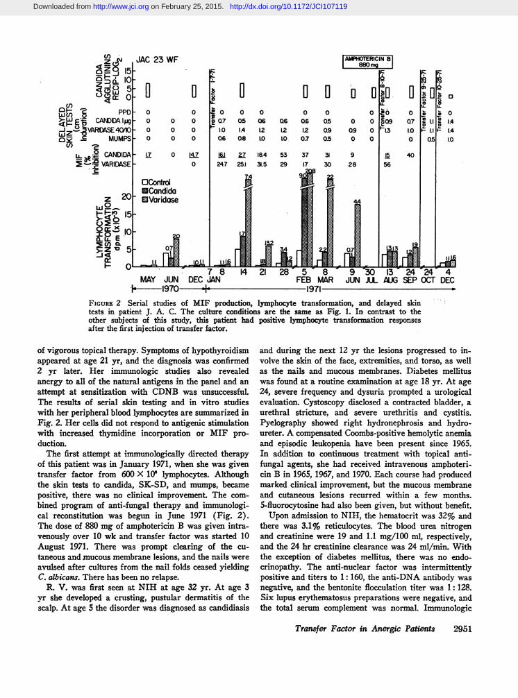
FIGUREz 3 Effect of transfer icyte responses of patient R. V.
U El E..O 0 0 0 b@0* 0
0 C 0.8 0 0 0 2.0a 0
0 A 0.8 0.8 0.5 0 P 0.60 0.3 0.3 0.5 0 0
1854
001.0Li
0.5
054
8 31 7" 2 9 '> 10 14 ' 24 27MAR APR JUL SEP OCT
1971
factor on delayed cutaneous hypersensitivity and in vitro lympho-
studies disclosed anergy to all the natural antigens in thepanel and two attempts at sensitization with CDNBdidnot induce contact allergy. When exposed to candida orSK-SD in short-term leukocyte cultures, her lymphocytesdid not respond with increased thymidine incorporationor MIF production (Fig. 3).
It was felt that the patient's renal status mitigatedagainst further systemic therapy with amphotericin B.A trial of transfer factor was begun in March 1971 (Fig.3). Although the cutaneous responses to candida andSK-SD became positive, there was only equivocal clear-ing of the cutaneous lesions. Transfer factor was discon-tinued in October 1971, when she had a respiratory in-fection requiring antibiotic therapy and a concommitantexacerbation of the hemolytic process.
D. L. is a 23-yr old male who has had mucocutaneouscandidiasis since age 6 months. The most extensive le-sions have involved the oral cavity, nails, and skin ofthe fingers and thorax. This patient had also had re-
peated staphylococcal infections including pyoderma, sub-cutaneous abscesses, and lung abscesses. Routine labora-tory studies and an extensive survey of his host-defensemechanisms were unremarkable except for energy to all
antigens of the test panel. Studies of lymphocyte re-sponses to antigens before administration of transferfactor revealed that his cells did not respond to antigenicexposure with thymidine incorporation or MIF produc-tion (Fig. 4).
R. P. was the product of a normal pregnancy but hisdevelopmental milestones were delayed. Mucocutaneouscandidiasis first appeared at age 2 yr and resisted therapywith topical nystatin and amphotericin B. Intravenousamphotericin at age 4 yr and clotrimazole at age 8 pro-duced remissions, but the lesions recurred shortly aftercessation of therapy. When admitted to the NIH at age9 yr the patient had extensive candidiasis of the oralcavity, scalp, skin, and nails. Infection with Herpeszoster was present over the right hemithorax.
Routine laboratory work disclosed mild anemia due toiron deficiency. The leukocyte count and differential werenormal; there was no lymphopenia. Endocrine glandfunction was normal. The stool did not contain trypsinactivity. The chest X-ray showed multiple cysts in theright middle lobe and an esophageal study disclosed astricture at the level of the carina.
Immunologic studies indicated a generalized defect of
2952 C. H. Kirkpatrick, R. R. Rich, and T. K. Smith
Downloaded from http://www.jci.org on February 25, 2015. http://dx.doi.org/10.1172/JCI107119
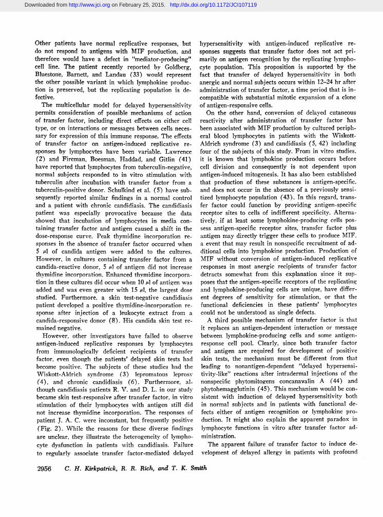
the cellular immune system that was more severe thanthe other patients. There were no responses to the anti-gens of the skin test panel, and no contact hypersensi-tivity developed after application of the sensitizing doseof CDNB. His lymphocytes did not respond to stimula-tion with antigens with increased thymidine incorpora-tion or MIF production. In contrast to the other sub-jects of this study, stimulation of his cells with phyto-hemagglutinin, concanavalin in A, or allogeneic cellsdid not result in increased thymidine incorporation (Ta-ble I). Humoral responses such as imxnunoglobulins, iso-hemagglutinins, and the acute and convalescent anti-varicella C-F antibody titers were normal. Except for thenormal lymphocyte count, the immunologic profile andlymph node histology of the patient were similar tothe Nezelof syndrome (18).
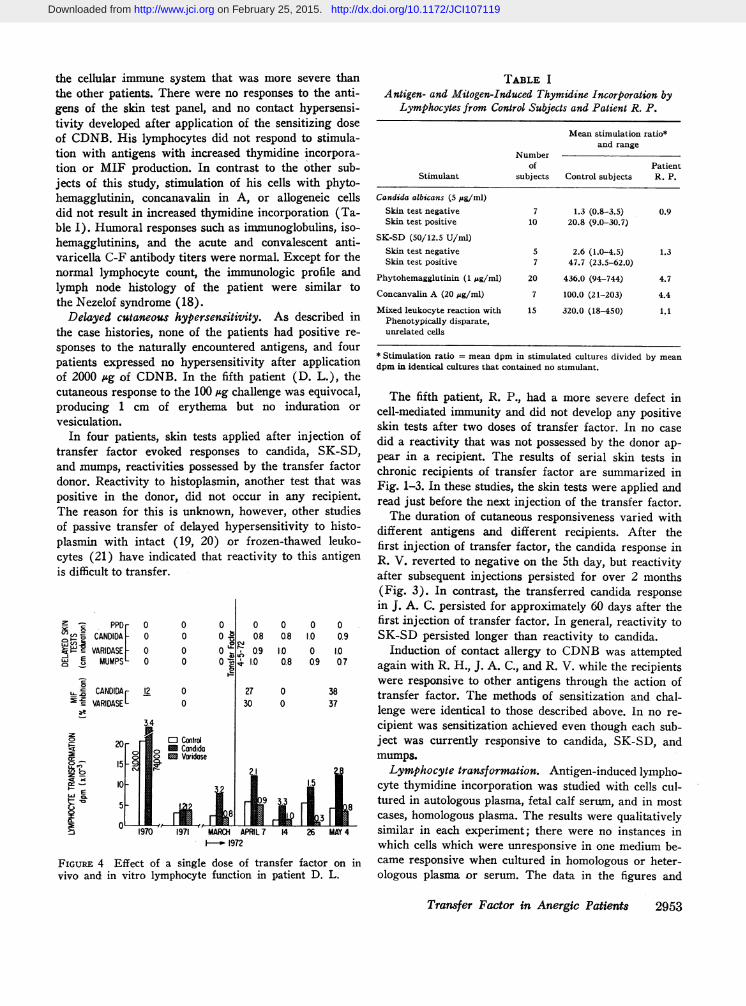
Delayed cutaneous hypersensitivity. As described inthe case histories, none of the patients had positive re-sponses to the naturally encountered antigens, and fourpatients expressed no hypersensitivity after applicationof 2000 Ag of CDNB. In the fifth patient (D. L.), thecutaneous response to the 100 Ag challenge was equivocal,producing 1 cm of erythema but no induration orvesiculation.
In four patients, skin tests applied after injection oftransfer factor evoked responses to candida, SK-SD,and mumps, reactivities possessed by the transfer factordonor. Reactivity to histoplasmin, another test that waspositive in the donor, did not occur in any recipient.The reason for this is unknown, however, other studiesof passive transfer of delayed hypersensitivity to histo-plasmin with intact (19, 20) or frozen-thawed leuko-cytes (21) have indicated that reactivity to this antigenis difficult to transfer.
x- PPD
OFB CANDIDAw tHVARIDASE
I MUMPSL0
j CANDIDA= VARIDASEL
ae
0000
12
0 0 00 0_O. 0.80 0. 09'-a
o 0 m 1.0I--
0 270 30
ControlCondidaV dricose
2.1
4i 8L1971
008
1.00.8
0 01.0 0.9
0 1.00.9 0.7
0 380 37
1.5
IQ3
>S-o mlo iS EI INVAd
MARCH APRIL 7 14 26 MAY4i- 1972
FIGuRE 4 Effect of a single dose of transfer factor on invivo and in vitro lymphocyte function in patient D. L.
TABLE I
Antigen- and Mitogen-Induced Thymidine Incorporation byLymphocytes from Control Subjects and Patient R. P.
Mean stimulation ratio*and range
Numberof Patient
Stimulant subjects Control subjects R. P.
Candida albicans (5 #g/ml)Skin test negative 7 1.3 (0.8-3.5) 0.9Skin test positive 10 20.8 (9.0-30.7)
SK-SD (50/12.5 U/ml)Skin test negative 5 2.6 (1.0-4.5) 1.3Skin test positive 7 47.7 (23.5-62.0)
Phytohemagglutinin (1 jsg/ml) 20 436.0 (94-744) 4.7
Concanvalin A (20 ,g/ml) 7 100.0 (21-203) 4.4
Mixed leukocyte reaction with 15 320.0 (18-450) 1.1Phenotypically disparate,unrelated cells
* Stimulation ratio = mean dpm in stimulated cultures divided by meandpm in identical cultures that contained no stimulant.
The fifth patient, R. P., had a more severe defect incell-mediated immunity and did not develop any positiveskin tests after two doses of transfer factor. In no casedid a reactivity that was not possessed by the donor ap-pear in a recipient. The results of serial skin tests inchronic recipients of transfer factor are summarized inFig. 1-3. In these studies, the skin tests were applied andread just before the next injection of the transfer factor.
The duration of cutaneous responsiveness varied withdifferent antigens and different recipients. After thefirst injection of transfer factor, the candida response inR. V. reverted to negative on the 5th day, but reactivityafter subsequent injections persisted for over 2 months(Fig. 3). In contrast, the transferred candida responsein J. A. C. persisted for approximately 60 days after thefirst injection of transfer factor. In general, reactivity toSK-SD persisted longer than reactivity to candida.
Induction of contact allergy to CDNBwas attemptedagain with R. H., J. A. C., and R. V. while the recipientswere responsive to other antigens through the action oftransfer factor. The methods of sensitization and chal-lenge were identical to those described above. In no re-cipient was sensitization achieved even though each sub-ject was currently responsive to candida, SK-SD, andmumps.
Lymphocyte transformation. Antigen-induced lympho-cyte thymidine incorporation was studied with cells cul-tured in autologous plasma, fetal calf serum, and in mostcases, homologous plasma. The results were qualitativelysimilar in each experiment; there were no instances inwhich cells which were unresponsive in one medium be-came responsive when cultured in homologous or heter-ologous plasma or serum. The data in the figures and
Transfer Factor in Anergic Patients 2953
3.4mC> 20 0t.i mm C> C>cr C) a Mc>,;;- 15 9U- C"i2 bcx c
cr - 10-I.- EUJ C.J.
'Df-> 5-9CL2 0>-, 1970 V
Downloaded from http://www.jci.org on February 25, 2015. http://dx.doi.org/10.1172/JCI107119
Table I are from experiments conducted with 20%autologous plasma.
Skin test-negative control subjects frequently showedsmall increases in thymidine incorporation with candidaor SK-SD. These were usually two- to threefold incre-ments, but rarely fourfold increases were observed. Incontrast, lymphocytes from skin test-positive controlsubjects responded to antigenic stimulation in vitro with9- to 60-fold increments of thymidine incorporation, andeven greater increments of thymidine incorporation oc-curred in mixed leukocyte cultures or mitogen stimulatedcultures.
Lymphocytes from four patients (J. A. C., R. V., R. P.,and D. L.) did not respond to in vitro antigenic stimu-lation by increased thymidine incorporation, but thelymphocyte transformation responses by R. H. werenormal. Administration of transfer factor did not convertthe thymidine incorporation responses by R. V. or D. L.even though the cutaneous responses were positive.With R. V., after the third injection of transfer factoron 24 September 1971 there was a marked increase inthe rate of thymidine incorporation in both unstimulatedand antigen-containing cultures. The reason for thischange in base line activity was not identified.
After transfer factor, the thymidine incorporation bylymphocytes from J. A. C. was variable. In four of sevenexperiments her cells responded vigorously to SK-SDwith stimulation ratios of 7.4-22 and one occasion, 5February 1971, was there a significant response to can-dida. The lymphocyte transformation responses by R. H.were positive before transfer factor and there were noconsistent changes in the magnitude of the responses orthe dose-response relationship after the injection oftransfer factor.
MIF production. Serial studies of antigen-inducedMIF production are summarized in Figs. 1-4. Under-lined numbers indicate that the inhibition of migrationin culture fluids from antigen-stimulated cells did notdiffer significantly (P > 0.05) from control fluids. Thestudies with R. V. were compromised by leukopenia andadequate numbers of cells were not always available forall studies.
A consistent change in lymphocyte function in recipi-ents of transfer factor was production of MIF, and inmost cases, MIF production correlated with delayed skintest reactivity. The converse was observed with patientR. P. in whom transfer factor did not cause conversionof skin tests or in vitro MIF production.
The results of the immunologic studies are sum-marized in Table II. Conversion of negative skin teststo positive occurred in four patients and in each casewas accompanied by MIF production by antigen-stimu-lated lymphocytes. In only one patient did the previously
negative lymphocyte transformation reaction become posi-tive and this was an inconstant finding.
DISCUSSIONDuring recent years a variety of metabolic and immuno-logic abnormalities have been recognized in patients withinfections due to C. albicans. Most frequently observedhave been associations between candidiasis and functionalabnormalities of the thymus-dependent or cellular im-mune system. Children with the DiGeorge or Nezelofsyndromes of congenital absence of the thymus frequentlyhave infections with C. albicans. In three instances,restoration of immune competence with thymus graftswas accompanied by clearing of candidiasis (22-24).Candidiasis has also been observed in subjects with thy-moma (25-27), and patients with chronic mucocutaneouscandidiasis often fail to express delayed cutaneous hyper-sensitivity to candida extracts (15, 28, 29).
From studies of cellular hypersensitivity in vivo ithas been possible to segregate patients with chronic mu-cocutaneous candidiasis into three categories: patientswith generalized defects in cell-mediated immunity suchas the subjects of this study, patients in whom the cellu-lar defects are limited to candida antigens, and patientsin whomno cellular immune abnormalities are recogniz-able (15). In general, patients in the first category havehad candidiasis since infancy or early childhood, sug-gesting a congenital defect in the cellular immune system.The age of onset in the other two groups is variable, andthe basis of the underlying abnormalities of cellular im-munity is poorly understood.
In vitro studies of lymphocytes from candidiasis pa-tients in whom cellular immune defects are present haveprovided additional evidence for defective cellular im-munity and have further illustrated the complexity ofthis syndrome. Although some patients possess. serumfactors which inhibit cellular immune functions (30, 31),most studies have established primary defects in lympho-cyte function. Cells from some candidiasis patients fail torespond to stimulation with antigens, especially candida,with either increased thymidine incorporation or MIFproduction (15). Other patients have a different ab-normality in which the replicative responses to antigensare normal, but MIF production is deficient (16, 32). Athird aberrant pattern in patients with candidiasis wasreported recently by Goldberg, Bluestone, Barnett, andLandau (33). Their patient had cutaneous anergy andnormal MIF production, but her lymphocytes did notincorporate thymidine upon stimulation with candida.
The concept of treatment of chronic or recurrent infec-tious diseases by restoration of host defects is not new.It has long been an essential part of management of hy-pogammaglobulinemia and during recent years has beenextended to disorders with isolated deficiencies of the
2954 C. H. Kirkpatrick, R. R. Rich, and T. K. Smith
Downloaded from http://www.jci.org on February 25, 2015. http://dx.doi.org/10.1172/JCI107119
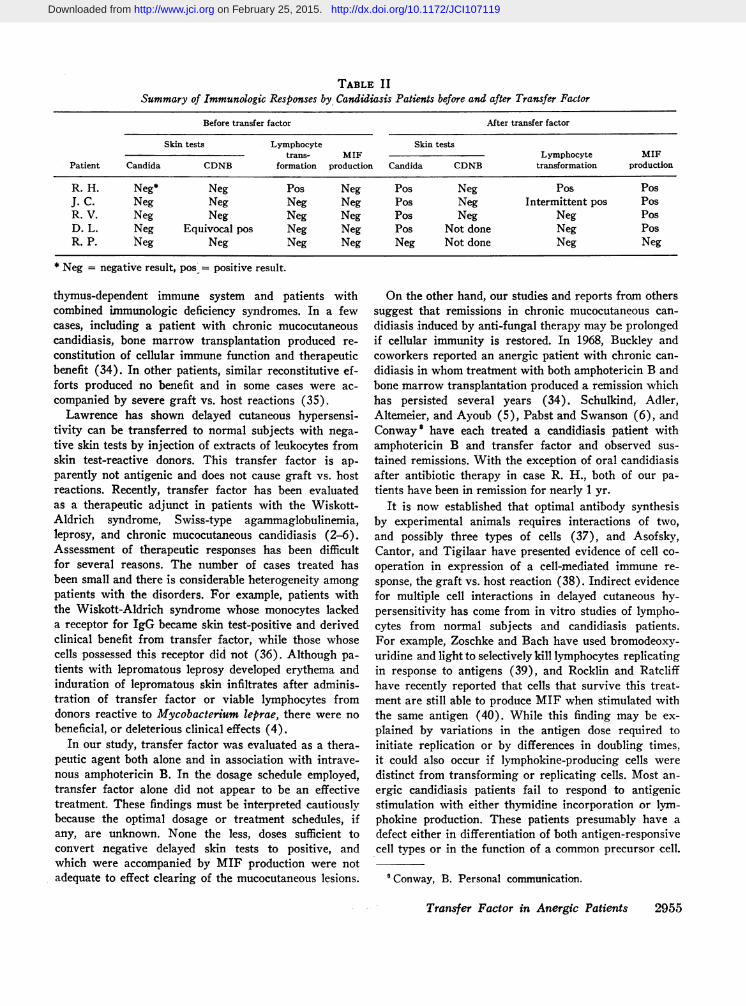
TABLE I ISummary of Immunologic Responses by Candidiasis Patients before and after Transfer Factor
Before transfer factor After transfer factor
Skin tests Lymphocyte Skin teststrans- MIF Lymphocyte MIF
Patient Candida CDNB formation production Candida CDNB transformation production
R. H. Neg* Neg Pos Neg Pos Neg Pos PosJ. C. Neg Neg Neg Neg Pos Neg Intermittent pos PosR. V. Neg Neg Neg Neg Pos Neg Neg PosD. L. Neg Equivocal pos Neg Neg Pos Not done Neg PosR. P. Neg Neg Neg Neg Neg Not done Neg Neg
* Neg = negative result, pos = positive result.
thymus-dependent immune system and patients withcombined immunologic deficiency syndromes. In a fewcases, including a patient with chronic mucocutaneouscandidiasis, bone marrow transplantation produced re-constitution of cellular immune function and therapeuticbenefit (34). In other patients, similar reconstitutive ef-forts produced no benefit and in some cases were ac-companied by severe graft vs. host reactions (35).
Lawrence has shown delayed cutaneous hypersensi-tivity can be transferred to normal subjects with nega-tive skin tests by injection of extracts of leukocytes fromskin test-reactive donors. This transfer factor is ap-parently not antigenic and does not cause graft vs. hostreactions. Recently, transfer factor has been evaluatedas a therapeutic adjunct in patients with the Wiskott-Aldrich syndrome, Swiss-type agammaglobulinemia,leprosy, and chronic mucocutaneous candidiasis (2-6).Assessment of therapeutic responses has been difficultfor several reasons. The number of cases treated hasbeen small and there is considerable heterogeneity amongpatients with the disorders. For example, patients withthe Wiskott-Aldrich syndrome whose monocytes lackeda receptor for IgG became skin test-positive and derivedclinical benefit from transfer factor, while those whosecells possessed this receptor did not (36). Although pa-tients with lepromatous leprosy developed erythema andinduration of lepromatous skin infiltrates after adminis-tration of transfer factor or viable lymphocytes fromdonors reactive to Mycobacterium leprae, there were nobeneficial, or deleterious clinical effects (4).
In our study, transfer factor was evaluated as a thera-peutic agent both alone and in association with intrave-nous amphotericin B. In the dosage schedule employed,transfer factor alone did not appear to be an effectivetreatment. These findings must be interpreted cautiouslybecause the optimal dosage or treatment schedules, ifany, are unknown. None the less, doses sufficient toconvert negative delayed skin tests to positive, andwhich were accompanied by MIF production were notadequate to effect clearing of the mucocutaneous lesions.
On the other hand, our studies and reports from otherssuggest that remissions in chronic mucocutaneous can-didiasis induced by anti-fungal therapy may be prolongedif cellular immunity is restored. In 1968, Buckley andcoworkers reported an anergic patient with chronic can-didiasis in whomtreatment with both amphotericin B andbone marrow transplantation produced a remission whichhas persisted several years (34). Schulkind, Adler,Altemeier, and Ayoub (5), Pabst and Swanson (6), andConway8 have each treated a candidiasis patient withamphotericin B and transfer factor and observed sus-tained remissions. With the exception of oral candidiasisafter antibiotic therapy in case R. H., both of our pa-tients have been in remission for nearly 1 yr.
It is now established that optimal antibody synthesisby experimental animals requires interactions of two,and possibly three types of cells (37), and Asofsky,Cantor, and Tigilaar have presented evidence of cell co-operation in expression of a cell-nediated immune re-sponse, the graft vs. host reaction (38). Indirect evidencefor multiple cell interactions in delayed cutaneous hy-persensitivity has come from in vitro studies of lympho-cytes from normal subjects and candidiasis patients.For example, Zoschke and Bach have used bromodeoxy-uridine and light to selectively kill lymphocytes replicatingin response to antigens (39), and Rocklin and Ratcliffhave recently reported that cells that survive this treat-ment are still able to produce MIF when stimulated withthe same antigen (40). While this finding may be ex-plained by variations in the antigen dose required toinitiate replication or by differences in doubling times,it could also occur if lymphokine-producing cells weredistinct from transforming or replicating cells. Most an-ergic candidiasis patients fail to respond to antigenicstimulation with either thymidine incorporation or lym-phokine production. These patients presumably have adefect either in differentiation of both antigen-responsivecell types or in the function of a common precursor cell.
Conway, B. Personal communication.
Transfer Factor in Anergic Patients 2955
Downloaded from http://www.jci.org on February 25, 2015. http://dx.doi.org/10.1172/JCI107119
Other patients have normal replicative responses, butdo not respond to antigens with MIF production, andtherefore would have a defect in "mediator-producing"cell line. The patient recently reported by Goldberg,Bluestone, Barnett, and Landau (33) would representthe other possible variant in which lymphokine produc-tion is preserved, but the replicating population is de-fective.
The multicellular model for delayed hypersensitivitypermits consideration of possible mechanisms of actionof transfer factor, including direct effects on either celltype, or on interactions or messages between cells neces-sary for expression of this immune response. The effectsof transfer factor on antigen-induced replicative re-sponses by lymphocytes have been variable. Lawrence(2) and Fireman, Boesman, Haddad, and Gitlin (41)have reported that lymphocytes from tuberculin-negative,normal subjects responded to in vitro stimulation withtuberculin after incubation with transfer factor from atuberculin-positive donor. Schulkind et al. (5) have sub-sequently reported similar findings in a normal controland a patient with chronic candidiasis. The candidiasispatient was especially provocative because the datashowed that incubation of lymphocytes in media con-taining transfer factor and antigen caused a shift in thedose-response curve. Peak thymidine incorporation re-sponses in the absence of transfer factor occurred when5 A1 of candida antigen were added to the cultures.However, in cultures containing transfer factor from acandida-reactive donor, 5 Al of antigen did not increasethymidine incorporation. Enhanced thymidine incorpora-tion in these cultures did occur when 10 Al of antigen wasadded and was even greater with 15 ,4, the largest dosestudied. Furthermore, a skin test-negative candidiasispatient developed a positive thymidine-incorporation re-sponse after injection of a leukocyte extract from acandida-responsive donor (8). His candida skin test re-mained negative.
However, other investigators have failed to observeantigen-induced replicative responses by lymphocytesfrom immunologically deficient recipients of transferfactor, even though the patients' delayed skin tests hadbecome positive. The subjects of these studies had theWiskott-Aldrich syndrome (3) lepromatous leprosy(4), and chronic candidiasis (6). Furthermore, al-though candidiasis patients R. V. and D. L. in our studybecame skin test-responsive after transfer factor, in vitrostimulation of their lymphocytes with antigen still didnot increase thymidine incorporation. The responses ofpatient J. A. C. were inconstant, but frequently positive(Fig. 2). While the reasons for these diverse findingsare unclear, they illustrate the heterogeneity of lympho-cyte dysfunction in patients with candidiasis. Failureto regularly associate transfer factor-mediated delayed
hypersensitivity with antigen-induced replicative re-sponses suggests that transfer factor does not act pri-marily on antigen recognition by the replicating lympho-cyte population. This proposition is supported by thefact that transfer of delayed hypersensitivity in bothanergic and normal subjects occurs within 12-24 hr afteradministration of transfer factor, a time period that is in-compatible with substantial mitotic expansion of a cloneof antigen-responsive cells.
On the other hand, conversion of delayed cutaneousreactivity after administration of transfer factor hasbeen associated with MIF production by cultured periph-eral blood lymphocytes in patients with the Wiskott-Aldrich syndrome (3) and candidiasis (5, 42) includingfour of the subjects of this study. From in vitro studies,it is known that lymphokine production occurs beforecell division and consequently is not dependent uponantigen-induced mitogenesis. It has also been establishedthat production of these substances is antigen-specific.and does not occur in the absence of a previously sensi-tized lymphocyte population (43). In this regard, trans-fer factor could function by providing antigen-specificreceptor sites to cells of indifferent specificity. Alterna-tively, if at least some lymphokine-producing cells pos-sess antigen-specific receptor sites, transfer factor plusantigen may directly trigger these cells to produce MIF,a event that may result in nonspecific recruitment of ad-ditional cells into lymphokine production. Production ofMIF without conversion of antigen-induced replicativeresponses in most anergic recipients of transfer factordetracts somewhat from this explanation since it sup-poses that the antigen-specific receptors of the replicatingand lymphokine-producing cells are unique, have differ-ent degrees of sensitivity for stimulation, or that thefunctional deficiencies in these patients' lymphocytescould not be understood as single defects.
A third possible mechanism of transfer factor is thatit replaces an antigen-dependent interaction or messagebetween lymphokine-producing cells and some antigen-response cell pool. Clearly, since both transfer factorand antigen are required for development of positiveskin tests, the mechanism must be different from thatleading to nonantigen-dependent "delayed hypersensi-tivity-like" reactions after intradermal injections of thenonspecific phytomitogens concanavalin A (44) andphytohemagglutinin (45). This mechanism would be con-sistent with induction of delayed hypersensitivity bothin normal subjects and in patients with functional de-fects either of antigen recognition or lymphokine pro-duction. It might also explain the apparent paradox inlymphocyte functions in vitro after transfer factor ad-ministration.
The apparent failure of transfer factor to induce de-velopment of delayed allergy in patients with profound
2956 C. H. Kirkpatrick, R. R. Rich, and T. K. Smith
Downloaded from http://www.jci.org on February 25, 2015. http://dx.doi.org/10.1172/JCI107119
defects in cellular immune functions such as R. P. in ourstudy is consistant with any of the proposed xnecha-nisms. One would predict that only certain defects couldbe abrogated by transfer factor and that more severe orfundamental deficiencies would be less amenable to resti-tution. It is apparent that further investigations of pa-tients with immunological deficiencies will be useful forunderstanding the biologic basis of transfer of delayedhypersensitivity by soluble agents.
ACKNOWLEDGMENTSThe authors wish to thank Drs. Sheldon Wolff and IraGreen for critically reviewing the manuscript.
REFERENCES1. Lawrence, H. S. 1955. The transfer in humans of de-
layed skin sensitivity to streptococcal M substance andto tuberculin with disrupted leucocytes. J. Clin. Invest.34: 219.
2. Lawrence, H. S. 1969. Transfer factor. Adv. Immunol.11: 195.
3. Levin, A. S., L. E. Spitler, D. P. Stites, and H. H.Fudenberg. 1970. Wiskott-Aldrich syndrome, a geneti-cally determined cellular immunologic deficiency: clini-cal and laboratory responses to therapy with transferfactor. Proc. Natl. Acad. Sci. U. S. A. 67: 821.
4. Bullock, W. E., J. Fields, and M Brandriss. 1971.Transfer factor therapy in lepromatous leprosy: anevaluation. J. Clin. Invest. 50: 16a. (Abstr.)
5. Schulkind, M. L., W. H. Adler, W. A. Altemeier, III,and E. M. Ayoub. 1972. Transfer factor in the treat-ment of a case of chronic mucocutaneous candidiasis.Cell. Immunol. 3: 606.
6. Pabst, H. F., and R. Swanson. 1972. Successful treat-ment of candidiasis with transfer factor. Br. Mled. J. 2:442.
7. Spitler, L. E., A. S. Levin, M. S. Blois, W. Epstein,H. H. Fudenberg, I. Hellstrom, and K. E. Hellstrom.1972. Lymphocyte responses to tumor-specific antigensin patients with malignant melanoma and results oftransfer factor therapy. J. Clin. Invest. 51: 92 (Abstr.)
8. Kirkpatrick, C. H., J. W. Chandler, and R. N.Schimke. 1970. Chronic mucocutaneous moniliasis withimpaired delayed hypersensitivity. Clin. Exp. Immunol.6: 375.
9. Catalona, W. J., P. T. Taylor, A. S. Rabson, and P. B.Chretien. 1972. A method for dinitrochlorobenzene con-tact sensitization. A clinicopathological study. N. Engl.J. Med. 286: 399.
10. Newberry, W. M., Jr., J. Chandler, Jr., T. D. Y. Chin,and C. H. Kirkpatrick. 1968. Immunology of the my-coses. I. Depressed lymphocyte transformation in chronichistoplasmosis. J. Immunol. 100: 436.
11. Bach, F. H., and N. K. Voynow. 1966. One-way stimu-lation in mixed leukocyte cultures. Science (Wash.D. C.). 153: 545.
12. Rocklin, R. E., 0. L. Meyers, and J. R. David. 1970.An in vitro assay for cellular hypersensitivity in man.J. Immunol. 104: 95.
13. Rich, R. R., C. H. Kirkpatrick, and T. K. Smith. 1972.Simultaneous suppression of responses to allogeneictissue in vitro and in vivo. Cell. Immunol. In press.
14. Buckner, D., R. G. Graw, Jr., R. J. Eisel, E. S. Hen-derson, and S. Perry. 1969. Leukapheresis by continuousflow centrifugation (CFC) in patients with chronicmyelocytic leukemia (CML). Blood J. Hematol. 33:353.
15. Kirkpatrick, C. H., R. R. Rich, and J. E. Bennett. 1971.Chronic mucocutaneous candidiasis: model-building incellular immunity. Ann. Intern. Med. 74: 955.
16. Kirkpatrick, C. H., R. R. Rich, R. G. Graw, Jr., T. K.Smith, I. D. Mickenberg, and G. N. Rogentine. 1971.Treatment of chronic mucocutaneous moniliasis by im-munologic reconstitution. Clin. Exp. Immunol. 9: 733.
17. Shadomy, S. 1971. In vitro antifungal activity of clo-trimazole (Bay b 5097). Infect. Immun. 4: 143.
18. Nezelof, C. 1968. Thymic dysplasia with normal im-munoglobulins and immunologic deficiency: pure alym-phocytosis. Birth Defects Orig. Artic. Ser. 4: 104.
19. Warwick, W. J., R. A. Good, and R. T. Smith. 1960.Failure of passive transfer of delayed hypersensitivityin the newborn human infant. J. Lab. Clin. Med. 56:139.
20. Kirkpatrick, C. H., W. E. Wilson, and D. W. Talmage.1964. Immunologic studies in human organ transplanta-tion. I. Observation and characterization of suppressedcutaneous reactivity in uremia. J. Exp. Med. 119: 727.
21. Jensen, K., R. A. Patnode, H. C. Townsley, and M. M.Cummings. 1962. Multiple passive transfer of the de-layed type of hypersensitivity in humans. Am. Rcv.Respir. Dis. 85: 373.
22. Cleveland, W. W., B. J. Fogel, W. T. Brown, and H.E. M. Kay. 1968. Foetal thymic transplant in a case ofDiGeorge's syndrome. Lancet. 2: 1211.
23. August, C. S., R. H. Levey, A. I. Berkel, and F. S.Rosen. 1970. Establishment of immunological compe-tence in a child with congenital thymic aplasia by agraft of fetal thymus. Lancet. 1: 1080.
24. Levy, R. L., S-W. Huang, M. L. Bach, F. H. Bach,R. Hong, A. J. Ammann, M. Bortin, and H. E. M. Kay.1971. Thymic transplantation in a case of chronic muco-cutaneous candidiasis. Lancet. 2: 898.
25. Schoch, E. P., Jr. 1971. Thymic conversion of Candidaalbicans from commensalism to pathogenism. Arch.Dermatol. 103: 311.
26. Montez, L. F., M. D. Cooper, L. G. Bradford, R. 0.Lauderdale, and C. D. Taylor. 1971. Prolonged oraltreatment of chronic mucocutaneous candidiasis withamphotericin B. Arch. Dermatol. 104: 45.
27. Maize, J. C., and P. J. Lynch. 1972. Chronic muco-cutaneous candidiasis of the adult. A report of a patientwith an associated thynoma. Arch. Dermatol. 105: 96.
28. Chilgren, R. A., P. G. Quie, H. J. Meuwissen, and R.Hong. 1967. Chronic mucocutaneous candidiasis, de-ficiency of delayed hypersensitivity, and selective localantibody defect. Lancet. 1: 688.
29. Louria, D. B., J. K. Smith, R. G. Brayton, and M.Buse. 1972. Anti-candida factors in serum and their in-hibitors. I. Clinical and laboratory observations. J. In-fect. Dis. 125: 102.
30. Canales, L., R. 0. Middlemas III, J. M. Louro, andM. A. South. 1969. Immunological observations inchronic mucocutaneous candidiasis. Lancet. 2: 567.
31. Paterson, P. Y., R. Semo, G. Blumenschein, and J.Swelstad. 1971. Mucocutaneous candidiasis, anergy anda plasma inhibitor of cellular immunity: reversal afteramphotericin B therapy. Clin. Exp. Immunol. 9: 595.
Transfer Factor in Anergic Patients 2957
Downloaded from http://www.jci.org on February 25, 2015. http://dx.doi.org/10.1172/JCI107119
32. Valdimarsson, H., L. Holt, H. R. C. Riches, and J. R.Hobbs. 1970. Lymphocyte abnormality in chronic muco-cutaneous candidiasis. Lancet. 1: 1259.
33. Goldberg, L. S., R. Bluestone, E. V. Barnett, and J. W.Landau. 1971. Studies on lymphocyte and monocytefunction in chronic mucocutaneous candidiasis. Clin.Exp. Imnimnol. 8: 37.
34. Buckley, R. H., Z. J. Lucas, B. G. Hattler, Jr., C. M.Zmijewski, and D. B. Amos. 1968. Defective cellularimmunity associated with chronic mucocutaneous monili-asis and recurrent staphylococcal botryomycosis: im-munological reconstitution by allogeneic bone marrow.Clin. Exp. Inimunol. 3: 153.
35. Meuwissen, H. J., G. Rodey, J. McArthur, H. Pabst,R. Gatti, R. Chilgren, R. Hong, D. Frommel, R. Coif-man, and R. A. Good. 1971. Bone marrow transplanta-tion. Therapeutic usefulness and complications. Amt. J.Med. 51: 513.
36. Spitler, L. E., A. S. Levin, and H. H. Fudenberg.1972. Prediction of results of transfer factor therapyin the Wiskott-Aldrich syndrome by monocyte IgGreceptors: a preliminary report. In Proceedings of theSixth Leukocyte Culture Conference. M. R. Schwarz,editor. Academic Press, Inc., New York. 795.
37. Claman, H. N., and E. A. Chaperon. 1969. Immuno-logical complementation between thymus and marrowcells-a model for the two-cell theory of immuno-competence. Transplant. Rev. 1: 92.
38. Asofsky, R., H. Cantor, and R. E. Tigelaar. 1971. Cellinteractions in the graft-versus-host response. In Prog-
ress in Immunology. B. Amos, editor. Academic Press,Inc., New York. 369.
39. Zoschke, D. C., and F. H. Bach. 1972. Lymphocytereactivity in vitro. VIII. Specificity of allogeneic cellrecognition in mixed leukocyte cultures. In Proceedingsof the Sixth Leukocyte Culture Conference. M. R.Schwarz, editor. Academic Press, Inc., New York.639.
40. Rocklin, R. E., and H. E. Ratcliffe. 1972. Antigen-induced production of migration inhibition factor (MIF)by non-dividing human lymphocytes. Fed. Proc. 31: 753.(Abstr.)
41. Fireman, P., M. Boesman, Z. H. Haddad, and D. Gitlin.1967. Passive transfer of tuberculin reactivity in vitro.Science (Wash. D. C.). 155: 337.
42. Rocklin, R. E., R. A. Chilgren, R. Hong, and J. R.David. 1970. Transfer of cellular hypersensitivity inchronic mucocutaneous candidiasis monitored in vivoand in vitro. Cell. Immunol. 1: 290.
43. Bloom, B. R. 1971. In vitro approaches to the mechanismof cell-mediated immune reactions. Adv. Immunol. 13:101.
44. Schwartz, H. J., P. J. Catanzaro, and M. P. Leon.1971. An analysis of the effects of skin reactive factorreleased from lymphoid cells by concanavalin A in vivo.Am. J. Pathol. 63: 443.
45. Bonforte, R. J., R. M. Blaese, M. Topilsky, L. E. Siltzbach, and P. R. Glade. 1971. Phytohemmaglutinir.(PHA) skin test: a measure of intact cell-mediatedimmunity. Pediatr. Res. 5: 378 a. (Abstr.)
2958 C. H. Kirkpatrick, R. R. Rich, and T. K. Smith
Downloaded from http://www.jci.org on February 25, 2015. http://dx.doi.org/10.1172/JCI107119