Embed Size (px)
Citation preview

Vol. 43, No. 1INFECTION AND IMMUNITY, Jan. 1984, p. 66-710019-9567/84/010066-06$02.00/0 -
Copyright ©3 1984, American Society for Microbiology
Effect of Type A Pasteurella multocida Fractions on BovinePolymorphonuclear Leukocyte Functions
HYOIK RYU,* MERLIN L. KAEBERLE, JAMES A. ROTH, AND RONALD W. GRIFFITHDepartment of Veterinary Microbiology and Preventive Medicine, College of Veterinary Medicine, Iowa State University,
Ames, Iowa 50011
Received 5 July 1983/Accepted 14 October 1983
The effect of various Pasteurella multocida fractions on bovine polymorphonuclear leukocyte (PMN)functions was examined in vitro by using two encapsulated strains, P-2383 and P-1062 (both are Cartercapsular type A and of bovine origin). The ability of PMNs to ingest Staphylococcus aiureus and iodinateprotein was significantly inhibited in the presence of live cells, heat-killed whole cells, or saline-extractedcapsules but not in the presence of the decapsulated heat-killed cells. None of the fractions of the twostrains inhibited nitroblue tetrazolium reduction by PMNs. The saline extract did not inhibit the binding ofiodine to protein by a reaction involving xanthine, xanthine oxidase, and horseradish peroxidase. The PMNinhibitory factor was further characterized as a heat-stable capsular material of greater than 300,000molecular weight.
Pasteurella multocida is a major etiological agent inbovine respiratory disease (3, 6), with capsular type Astrains being most frequently isolated in North America.Some encapsulated strains of P. multocida are known to behighly pathogenic in experimental animals, and the presenceof the mucoid capsule is important for virulence (3, 7). Thecapsule of type A strains is primarily composed of hyaluron-ic acid (4, 5), which serves as a framework for the attach-ment of polysaccharides, proteins, and lipids (3).The importance of pasteurellae in bovine respiratory dis-
ease has led to the development and use of various bacterialproducts for immunization. The efficacy and safety of mostof these products still remain in question (2, 13, 17, 19).There is evidence that the use of bacterins that are currentlyavailable may, in fact, be detrimental to the health of theanimal (2, 19).Recent studies on type A P. multocida capsular materials
indicated that a KSCN extract (12, 20) and a saline extract(16, 25) were protective against experimental challenge inmice, chickens, and turkeys. The capsule of a type A P.multocida has also been demonstrated to inhibit the phago-cytic activity of bovine neutrophils (18). Therefore, thecapsule may contain not only a protective antigen but also acomponent which interferes with phagocytic cell function. Itis not unusual for bacterial surface material to contain both aprotective antigen and a virulence factor (8, 9, 14).
Phagocytosis of invading microorganisms by polymorpho-nuclear leukocytes (PMNs) can be one of the major cellulardefense mechanisms in protecting animals from microbialinfection. Maheswaran and Thies (18) reported that anencapsulated type A P. multocida (NA77) inhibited thephagocytic activity of neutrophils (PMNS). When they mea-sured the uptake of [3H]thymidine-labeled bacteria byPMNs, only 3.8% of the encapsulated organisms wereingested. When the encapsulated bacteria were treated withbovine testicular hyaluronidase, however, 90% of the decap-sulated organisms were ingested. They concluded that thefactor which inhibited the phagocytic activity of PMNs wasprobably hyaluronic acid, which is a major component of thecapsule of type A P. multocida.
* Corresponding author.
The purpose of the present experiment was to furthercharacterize an inhibitory factor present in type A P. multo-cida capsule and determine its effects on specific aspects ofPMN function.
MATERIALS AND METHODSOrganisms. Two strains of P. multocida were used
throughout this study (P-2383 and P-1062). Strain P-2383 wasisolated from a case of bovine pneumonia presented to theVeterinary Diagnostic Laboratory, Iowa State University,Ames, Iowa. This isolate was a typical P. multocida strain;subsequent typing confirmed it to be Carter capsular type A.Strain P-1062, also a type A strain of bovine origin, is achallenge strain (IRP-198, National Veterinary ServicesLaboratory, U.S. Department of Agriculture, Ames, Iowa).Each strain was inoculated into the yolk sac of 6-day-oldembryonated chicken eggs. After incubation at 37°C for 18 h,the yolk material was aseptically removed and frozen inaliquots at -70°C. These aliquots were used as inoculum forpreparation of bacterial fractions used in this study.
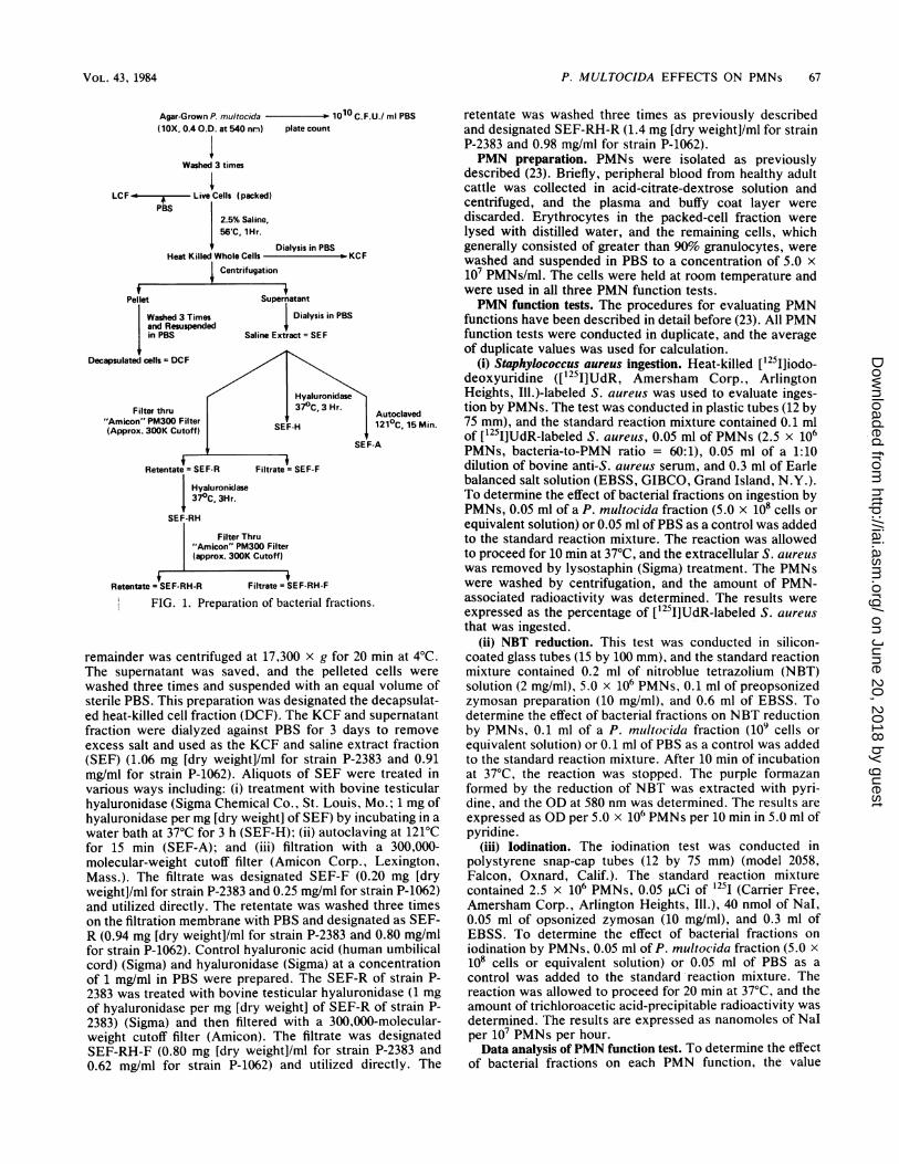
Bacterial fractions. The preparation of bacterial fractionsis illustrated in Fig. 1. Infected yolk material was thawed,inoculated on a 5% bovine blood agar plate and incubated at37°C for 24 h. One colony was transferred to 5 ml of brainheart infusion (BHI) broth (Difco Laboratories, Detroit,Mich.) containing 0.5% yeast extract (Difco) and 5% sterilebovine serum (BHISY) and incubated at 37°C for 4 h. Thisculture was then added to 100 ml of BHISY and incubated anadditional 4 h. Roux bottles containing dextrose-starch agar(Difco) were inoculated with 2.0 ml of BHISY culture andincubated at 37°C for 36 h. Cells were harvested by washingthe agar surface with sterile phosphate-buffered saline solu-tion (PBS, pH. 7.2), and cell concentration was adjusted sothat a 10-fold dilution had an optical density (OD) of 0.4 at540 nm (approximately 1010 cells per ml). The cells werewashed three times in PBS, and an aliquot was saved for thelive-cell fraction (LCF). The remaining cells were washedone more time in PBS. Cells packed by centrifugation weresuspended to the original volume in 2.5% (wt/vol) sodiumchloride solution and placed in a water bath at 56°C for 1 h.After this saline extraction, an aliquot was saved for thepreparation of a heat-killed whole-cell fraction (KCF). The
66
on June 20, 2018 by guesthttp://iai.asm
.org/D
ownloaded from
P. MULTOCIDA EFFECTS ON PMNs 67
Agar-Grown P. multocida # 1010 C.F.U./ ml PBS(lOX, 0.4 O.D. at 540 nrn) plate count
Washed 3 times
LCF < + Live Cells (packed)PBS
2.5% Saline,56'C, lHr.
Dialysis in PBSHeat Killed Whole Cells KCF
Centrifugation'
Supernatant
I Dialysis in PBS
Saline Extract = SEF
Icells= DCF /
/ ~~~~HyaluronidaseIter thru 37°C, 3 Hr. Autoclaved
"PM300 Filter
.300K Cutoff) SEF-H 121°C, 15 Min.
. ~~~~~~SEF-A
Retentate = SEF-R Filtrate = SEF-F
SEF
Hyaluronidlase370C, 3Hr.
:-RH
Filter Thru"Amicon" PM300 Filter(approx. 300K Cutoff)
Retentatc SEF-RH-R Filtrate = SEF-RH-F
i FIG. 1. Preparation of bacterial fractions.
remainder was centrifuged at 17,300 x g for 20 min at 4°C.The supernatant was saved, and the pelleted cells were
washed three times and suspended with an equal volume ofsterile PBS. This preparation was designated the decapsulat-ed heat-killed cell fraction (DCF). The KCF and supernatantfraction were dialyzed against PBS for 3 days to removeexcess salt and used as the KCF and saline extract fraction(SEF) (1.06 mg [dry weight]/ml for strain P-2383 and 0.91mg/ml for strain P-1062). Aliquots of SEF were treated invarious ways including: (i) treatment with bovine testicularhyaluronidase (Sigma Chemical Co., St. Louis, Mo.; 1 mg ofhyaluronidase per mg [dry weight] of SEF) by incubating in a
water bath at 37°C for 3 h (SEF-H); (ii) autoclaving at 121°Cfor 15 min (SEF-A); and (iii) filtration with a 300,000-molecular-weight cutoff filter (Amicon Corp., Lexington,Mass.). The filtrate was designated SEF-F (0.20 mg [dryweight]/ml for strain P-2383 and 0.25 mg/ml for strain P-1062)and utilized directly. The retentate was washed three timeson the filtration membrane with PBS and designated as SEF-R (0.94 mg [dry weight]/ml for strain P-2383 and 0.80 mg/mlfor strain P-1062). Control hyaluronic acid (human umbilicalcord) (Sigma) and hyaluronidase (Sigma) at a concentrationof 1 mg/ml in PBS were prepared. The SEF-R of strain P-2383 was treated with bovine testicular hyaluronidase (1 mgof hyaluronidase per mg [dry weight] of SEF-R of strain P-2383) (Sigma) and then filtered with a 300,000-molecular-weight cutoff filter (Amicon). The filtrate was designatedSEF-RH-F (0.80 mg [dry weight]/ml for strain P-2383 and0.62 mg/ml for strain P-1062) and utilized directly. The
retentate was washed three times as previously describedand designated SEF-RH-R (1.4 mg [dry weight]/ml for strainP-2383 and 0.98 mg/ml for strain P-1062).PMN preparation. PMNs were isolated as previously
described (23). Briefly, peripheral blood from healthy adultcattle was collected in acid-citrate-dextrose solution andcentrifuged, and the plasma and buffy coat layer werediscarded. Erythrocytes in the packed-cell fraction werelysed with distilled water, and the remaining cells, whichgenerally consisted of greater than 90% granulocytes, werewashed and suspended in PBS to a concentration of 5.0 x10⁷ PMNs/ml. The cells were held at room temperature andwere used in all three PMN function tests.PMN function tests. The procedures for evaluating PMN
functions have been described in detail before (23). All PMNfunction tests were conducted in duplicate, and the averageof duplicate values was used for calculation.
(i) Staphylococcus aureus ingestion. Heat-killed [t25I]iodo-deoxyuridine ([1251]UdR, Amersham Corp., ArlingtonHeights, Ill.)-labeled S. aureus was used to evaluate inges-tion by PMNs. The test was conducted in plastic tubes (12 by75 mm), and the standard reaction mixture contained 0.1 mlof [125 ]UdR-labeled S. aureus, 0.05 ml of PMNs (2.5 x 10⁶PMNs, bacteria-to-PMN ratio = 60:1), 0.05 ml of a 1:10dilution of bovine anti-S. aureus serum, and 0.3 ml of Earlebalanced salt solution (EBSS, GIBCO, Grand Island, N.Y.).To determine the effect of bacterial fractions on ingestion byPMNs, 0.05 ml of a P. multocida fraction (5.0 x 10⁸ cells orequivalent solution) or 0.05 ml of PBS as a control was addedto the standard reaction mixture. The reaction was allowedto proceed for 10 min at 37°C, and the extracellular S. aureuswas removed by lysostaphin (Sigma) treatment. The PMNswere washed by centrifugation, and the amount of PMN-associated radioactivity was determined. The results wereexpressed as the percentage of [1251]UdR-labeled S. aureusthat was ingested.
(ii) NBT reduction. This test was conducted in silicon-coated glass tubes (15 by 100 mm), and the standard reactionmixture contained 0.2 ml of nitroblue tetrazolium (NBT)solution (2 mg/ml), 5.0 x 10⁶ PMNs, 0.1 ml of preopsonizedzymosan preparation (10 mg/ml), and 0.6 ml of EBSS. Todetermine the effect of bacterial fractions on NBT reductionby PMNs, 0.1 ml of a P. multocida fraction (109 cells orequivalent solution) or 0.1 ml of PBS as a control was addedto the standard reaction mixture. After 10 min of incubationat 37°C, the reaction was stopped. The purple formazanformed by the reduction of NBT was extracted with pyri-dine, and the OD at 580 nm was determined. The results areexpressed as OD per 5.0 x 10⁶ PMNs per 10 min in 5.0 ml ofpyridine.
(iii) lodination. The iodination test was conducted inpolystyrene snap-cap tubes (12 by 75 mm) (model 2058,Falcon, Oxnard, Calif.). The standard reaction mixturecontained 2.5 x 10⁶ PMNs, 0.05 ,Ci of 1251 (Carrier Free,Amersham Corp., Arlington Heights, Ill.), 40 nmol of Nal,0.05 ml of opsonized zymosan (10 mg/ml), and 0.3 ml ofEBSS. To determine the effect of bacterial fractions oniodination by PMNs, 0.05 ml of P. multocida fraction (5.0 x10⁸ cells or equivalent solution) or 0.05 ml of PBS as acontrol was added to the standard reaction mixture. Thereaction was allowed to proceed for 20 min at 37°C, and theamount of trichloroacetic acid-precipitable radioactivity wasdetermined. The results are expressed as nanomoles of Nalper 107 PMNs per hour.Data analysis of PMN function test. To determine the effect
of bacterial fractions on each PMN function, the value
fPellet
Washed 3 Timesand Resuspendedin PBS
IDecapsulated
Fil"Amicon'(Approx.
VOL. 43, 1984
on June 20, 2018 by guesthttp://iai.asm
.org/D
ownloaded from

68 RYU ET AL.
obtained when a bacterial fraction was added to the PMNswas compared with the value obtained with control (PBS-treated) PMNs. An analysis-of-variance procedure was usedto determine significance of the differences in PMN function.For the graphic presentation of the data, all treatment valueswere expressed as a percentage of the control.
Chemical analysis of bacterial fractions. Total carbohy-drate was determined with a phenol-sulfuric acid procedure(11), using glucose as a standard. Hyaluronic acid wasdetermined by the reaction of hexuronic acid with carba-zole (Sigma) and H2SO4 (10), using D-glucuronic acid (Sig-ma) as a standard. Protein content was determined colori-metrically from the reaction of protein with Serva blue G dye(Serva fine Chemicals, Inc., Long Island, N.Y.), usingbovine serum albumin (Sigma) as a standard (21).
Xanthine-xanthine oxidase-horseradish peroxidase-mediat-ed iodination. A chemical iodination procedure was formu-lated by following the basic principle ofPMN iodination (15,22). Xanthine (Sigma) was used as a substrate, and xanthineoxidase (Sigma) served as the enzyme for the production ofsuperoxide anion. Horseradish peroxidase (Sigma) was usedto catalyze the iodination reaction. The standard mixturecontained 40 nmol of Nal, 0.05 p.Ci of 1251, 0.3 ml of EBSScontaining 0.1% bovine serum albumin, 0.5 mg of xanthine,0.5 U of horseradish peroxidase, and 0.05 ml of SEF or PBSas a control. The reaction was started by the addition of 0.02U of xanthine oxidase. The mixture was incubated andprocessed by the same procedures as that used for PMNiodination. A blank containing all components except xan-thine oxidase was run with each experiment. Results areexpressed in counts per minute.
RESULTSEffect of P. multocida fractions on S. aureus ingestion. To
determine the effect of P. multocida fractions on phagocyticactivity, PMNs were added to a standard suspension ofopsonized S. aureus in the presence or absence of P.multocida fractions. Control PMNs ingested 28.6 ± 2.3%(mean ± standard error) (n = 22) of the S. aiureus in the
100-_
00Strain P-2383
Strain P.106280
70
~60o 0
~5O40
30
20-
10
LCF KCF DCF SEF
FIG. 2. Effect of type A P. multocida fractions on S. aiureusingestion by bovine PMNs. Values represent mean percentages(±standard deviation) of the control value. Statistically significantdifferences from the control value are as indicated: **, P < 0.01; n =16 for the SEF and n = 6 for all other fractions.
110
100
90
Strain P-2383
Strain P-1062
80s-
701-
I so50
40
30
20
10 I I ISEF-A
T
ISEF-H SE F-R SEF-F
FIG. 3. Characterization of the inhibitory activity in the SEF onS. aureus ingestion by bovine PMNs. Values represent meanpercentages (±standard deviation) of the control value. Statisticallysignificant differences from the control value are as indicated: **,P < 0.01; n = 6 for all fractions.
reaction mixture. In the presence of LCF, KCF, and SEF ofthe two strains of P. multocida, S. aureus ingestion wasinhibited by 42 to 51% (Fig. 2). DCF had no effect on S.aureus ingestion by PMNs. To further characterize theinhibitory factor, SEF was treated with hyaluronidase, auto-claved, or filtered through a 300,000-molecular-weight cutofffilter. Neither autoclaving nor treatment with hyaluronidasedestroyed the inhibitory activity of SEF on S. aureusingestion by PMNs (Fig. 3). SEF-R did inhibit S. aureusingestion by PMNs by 41 to 60%, but SEF-F had no effect onthe PMN ability to ingest S. aureus. In a separate experi-ment to further characterize SEF-R of strain P-2383, thecontrol PMNs ingested 36.1 ± 1.4% (mean ± standard error)(n = 8) of the S. aureus in the reaction mixture. SEF-RH-Rinhibited S. aureus ingestion by PMNs by 59%, but SEF-RH-F had no effect on the PMNs ability to ingest S. aureus(see Fig. 7). S. aureus ingestion by PMNs was not inhibitedby either hyaluronic acid or hyaluronidase. To determinewhether the inhibitory substance was binding to the S.aureus or the PMNs, the S. aureus ingestion assay wasperformed by using S. aureus and PMNs which had beenseparately treated with strain P-2383 SEF-RH-R by incuba-tion for 20 min at 37°C in a shaking water bath and thenwashed three times with PBS. The results (Table 1) indicatethat the inhibitory substance was removed by washing anddid not bind to either the PMNs or the S. aureus.
Effect of P. multocida fractions on NBT reduction. To studythe effect of P. multocida fractions on oxidative metabolismof PMNs, the ability of PMNs to reduce NBT by theproduction of superoxide anion in the presence or absence ofthe bacterial fractions was determined. NBT reduction bycontrol PMNs was 0.47 ± 0.01 OD at 580 nm (mean ±standard error) (n = 15). None of the fractions of the twotype A strains significantly (P > 0.05) affected NBT reduc-tion by PMNs (Fig. 4).
Effect of P. multocida fractions on iodination. To study theeffect of P. minlto(ida fractions on the myeloperoxidase-H2O,-halide antibacterial system of the PMNs, the ability ofPMNs to iodinate protein in the presence or absence of the
INFECT. IMMUN.
T
on June 20, 2018 by guesthttp://iai.asm
.org/D
ownloaded from
P. MULTOCIDA EFFECTS ON PMNs 69
TABLE 1. Effect of preincubation of PMNs and S. aureius withstrain P-2383 SEF-RH-R on the ingestion of S. alureius by PMNs
% ingested S. aureius" in bacterialPretreatment (followed fraction added to reaction mixture:
by washing)None SEF-RH-R
None 29.8 ± 3.3 19.9 ± 1.8S. aureus preincubated 27.1 ± 2.1 17.6 ± 2.4
with SEF-RH-RPMNs preincubated 31.8 ± 4.4 21.2 ± 4.8
with SEF-RH-RBoth S. aureus and 29.8 ± 3.1 23.4 ± 2.4PMNs preincubatedwith SEF-RH-Ra Mean ± standard error (n = 6).
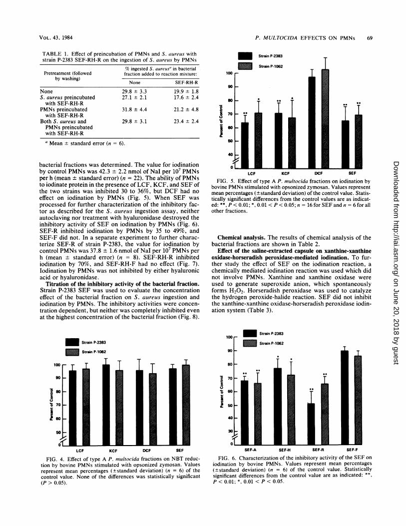
bacterial fractions was determined. The value for iodinationby control PMNs was 42.3 + 2.2 nmol of Nal per 107 PMNsper h (mean ± standard error) (n = 22). The ability of PMNsto iodinate protein in the presence of LCF, KCF, and SEF ofthe two strains was inhibited 30 to 36%, but DCF had noeffect on iodination by PMNs (Fig. 5). When SEF was
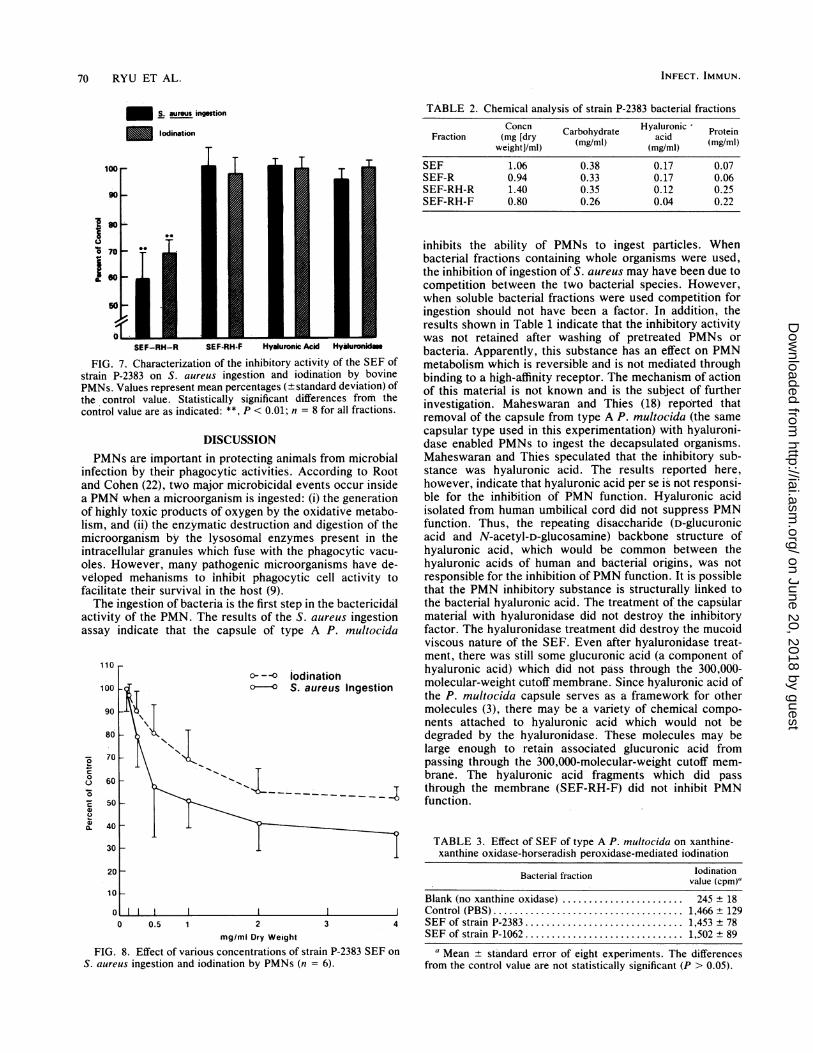
processed for further characterization of the inhibitory fac-tor as described for the S. aureus ingestion assay, neitherautoclaving nor treatment with hyaluronidase destroyed theinhibitory activity of SEF on iodination by PMNs (Fig. 6).SEF-R inhibited iodination by PMNs by 35 to 49%, andSEF-F did not. In a separate experiment to further charac-terize SEF-R of strain P-2383, the value for iodination bycontrol PMNs was 37.8 ± 1.6 nmol of Nal per 107 PMNs perh (mean ± standard error) (n = 8). SEF-RH-R inhibitediodination by 70%, and SEF-RH-F had no effect (Fig. 7).lodination by PMNs was not inhibited by either hyaluronicacid or hyaluronidase.
Titration of the inhibitory activity of the bacterial fraction.Strain P-2383 SEF was used to evaluate the concentrationeffect of the bacterial fraction on S. aureus ingestion andiodination by PMNs. The inhibitory activities were concen-
tration dependent, but neither was completely inhibited evenat the highest concentration of the bacterial fraction (Fig. 8).
Strain P-2383
Strain P-1062
100
90
80s-
70-
I60
LCF KCF DCF SEF
FIG. 4. Effect of type A P. multocida fractions on NBT reduc-tion by bovine PMNs stimulated with opsonized zymosan. Valuesrepresent mean percentages (+standard deviation) (n = 6) of thecontrol value. None of the differences was statistically significant(P > 0.05).
Strain P-2383
Strain P-1062
570
50
LCF KCF DCF SEF
FIG. 5. Effect of type A P. multocida fractions on iodination bybovine PMNs stimulated with opsonized zymosan. Values representmean percentages (± standard deviation) of the control value. Statis-tically significant differences from the control values are as indicat-ed: **, P< 0.01; *, 0.01 <P < 0.05; n = 16 for SEF and n = 6 for allother fractions.
Chemical analysis. The results of chemical analysis of thebacterial fractions are shown in Table 2.
Effect of the saline-extracted capsule on xanthine-xanthineoxidase-horseradish peroxidase-mediated iodination. To fur-ther study the effect of SEF on the iodination reaction, achemically mediated iodination reaction was used which didnot involve PMNs. Xanthine and xanthine oxidase wereused to generate superoxide anion, which spontaneouslyforms H202. Horseradish peroxidase was used to catalyzethe hydrogen peroxide-halide reaction. SEF did not inhibitthe xanthine-xanthine oxidase-horseradish peroxidase iodin-ation system (Table 3).
100Strain P-2383
100_Strain P-1062
90
80
~70-
060
I
SEF-A SEF-H SEF-R SEF-F
FIG. 6. Characterization of the inhibitory activity of the SEF oniodination by bovine PMNs. Values represent mean percentages(+standard deviation) (n = 6) of the control value. Statisticallysignificant differences from the control value are as indicated: **,P < 0.01; *, 0.01 < P < 0.05.
VOL. 43, 1984
on June 20, 2018 by guesthttp://iai.asm
.org/D
ownloaded from
70 RYU ET AL.
£S. aureus ingestion
Ism lodination
TABLE 2. Chemical analysis of strain P-2383 bacterial fractions
Concn Carbohydrate Hyaluronic ProteinFraction (mg [dry (mg/ml) acid(mgweight]/ml) (mg/ml)
SEF 1.06 0.38 0.17 0.07SEF-R 0.94 0.33 0.17 0.06SEF-RH-R 1.40 0.35 0.12 0.25SEF-RH-F 0.80 0.26 0.04 0.22
Is
SEF-RH-R SEF-RH-F Hyaluronic Acid Hyaluronidm
FIG. 7. Characterization of the inhibitory activity of the SEF ofstrain P-2383 on S. aureus ingestion and iodination by bovinePMNs. Values represent mean percentages (±standard deviation) ofthe control value. Statistically significant differences from thecontrol value are as indicated: **, P < 0.01; n = 8 for all fractions.
DISCUSSIONPMNs are important in protecting animals from microbial
infection by their phagocytic activities. According to Rootand Cohen (22), two major microbicidal events occur insidea PMN when a microorganism is ingested: (i) the generationof highly toxic products of oxygen by the oxidative metabo-lism, and (ii) the enzymatic destruction and digestion of themicroorganism by the lysosomal enzymes present in theintracellular granules which fuse with the phagocytic vacu-oles. However, many pathogenic microorganisms have de-veloped mehanisms to inhibit phagocytic cell activity tofacilitate their survival in the host (9).The ingestion of bacteria is the first step in the bactericidal
activity of the PMN. The results of the S. aureus ingestionassay indicate that the capsule of type A P. multocida
-6
0
cC.)
0.
0.
110
100
90
80
70
60
50
40
30
KTK,6
c -- iodinatiohO-O S. aureus Ingestion
T~~~
inhibits the ability of PMNs to ingest particles. Whenbacterial fractions containing whole organisms were used,the inhibition of ingestion of S. aureus may have been due tocompetition between the two bacterial species. However,when soluble bacterial fractions were used competition foringestion should not have been a factor. In addition, theresults shown in Table 1 indicate that the inhibitory activitywas not retained after washing of pretreated PMNs orbacteria. Apparently, this substance has an effect on PMNmetabolism which is reversible and is not mediated throughbinding to a high-affinity receptor. The mechanism of actionof this material is not known and is the subject of furtherinvestigation. Maheswaran and Thies (18) reported thatremoval of -the capsule from type A P. multocida (the samecapsular type used in this experimentation) with hyaluroni-dase enabled PMNs to ingest the decapsulated organisms.Maheswaran and Thies speculated that the inhibitory sub-stance was hyaluronic acid. The results reported here,however, indicate that hyaluronic acid per se is not responsi-ble for the inhibition of PMN function. Hyaluronic acidisolated from human umbilical cord did not suppress PMNfunction. Thus, the repeating disaccharide (D-glucuronicacid and N-acetyl-D-glucosamine) backbone structure ofhyaluronic acid, which would be common between thehyaluronic acids of human and bacterial origins, was notresponsible for the inhibition ofPMN function. It is possiblethat the PMN inhibitory substance is structurally linked tothe bacterial hyaluronic acid. The treatment of the capsularmaterial with hyaluronidase did not destroy the inhibitoryfactor. The hyaluronidase treatment did destroy the mucoidviscous nature of the SEF. Even after hyaluronidase treat-ment, there was still some glucuronic acid (a component ofhyaluronic acid) which did not pass through the 300,000-molecular-weight cutoff membrane. Since hyaluronic acid ofthe P. multocida capsule serves as a framework for othermolecules (3), there may be a variety of chemical compo-nents attached to hyaluronic acid which would not bedegraded by the hyaluronidase. These molecules may belarge enough to retain associated glucuronic acid frompassing through the 300,000-molecular-weight cutoff mem-brane. The hyaluronic acid fragments which did passthrough the membrane (SEF-RH-F) did not inhibit PMNfunction.
TABLE 3. Effect of SEF of type A P. multocida on xanthine-xanthine oxidase-horseradish peroxidase-mediated iodination
Bacterial fraction vodinationvalue (cpm)"
Blank (no xanthine oxidase) ........ ............... 245 ± 18J Control (PBS).............. 1,466 ± 1294 SEF of strain P-2383 .............. 1,453 ± 78
SEF of strain P-1062 .............. 1,502 ± 89
a Mean ± standard error of eight experiments. The differencesfrom the control value are not statistically significant (P > 0.05).
0 0.5 1 2 3mg/ml Dry Weight
FIG. 8. Effect of various concentrations of strain P-2383 SEFS. aureus ingestion and iodination by PMNs (n = 6).
INFECT. IMMUN.
on
on June 20, 2018 by guesthttp://iai.asm
.org/D
ownloaded from

P. MULTOCIDA EFFECTS ON PMNs 71
Oxidative metabolism of the PMN is an important aspectof its bactericidal activity (22). When a PMN receives theproper stimulus, an oxidase enzyme on the surface of theplasma membrane or phagosomal membrane will catalyzethe conversion of oxygen to superoxide anion. Superoxideanion spontaneously dismutates to hydrogen peroxide. NBTis directly reduced by the superoxide anion to an insolublepurple formazan (26). NBT reduction is therefore a measureof superoxide anion generation by the PMN. Since NBTreduction was not inhibited by whole bacteria or bacterialfractions (Fig. 4), type A P. mnultocida apparently does notinhibit the production of superoxide anion by PMNs.The iodination reaction is a measure of the ability of
PMNs to convert inorganic iodide to a trichloroacetic acid-precipitable (protein-bound) form and occurs inside thephagocytic vacuole via the action of hydrogen peroxide andmyeloperoxidase. This system has been found to exhibit amarked toxic activity toward bacteria, fungi, and viruses (1,24). The iodination reaction by PMNs is dependent upon thegeneration of hydrogen peroxide, degranulation to releasemyeloperoxidase, the presence of iodine, the unimpairedability of myeloperoxidase to catalyze the reaction, and thepresence of tyrosine to bind iodine. Hydrogen peroxide isformed spontaneously from superoxide anion. Since SEFdid not inhibit NBT reduction, superoxide anion generationby PMNs is apparently not affected by the inhibitory factor.Since the xanthine-xanthine oxidase-horseradish peroxi-dase-mediated iodination was not inhibited by SEF, the rateof formation of hydrogen peroxide from superoxide anionand the rate of hydrogen peroxide destruction were appar-ently not affected. In addition, the ability of the peroxidaseenzyme to catalyze the reaction was apparently not im-paired. It must be kept in mind that horseradish peroxidaseand myeloperoxidase are different enzymes. It is possiblethat the inhibitory factor could inhibit myeloperoxidasedirectly without inhibiting horseradish peroxidase, but thisdoes not seem likely.
This experimentation demonstrated that phagocytosis andprotein iodination by PMNs were inhibited in the presence ofwhole P. inultocida organisms and bacterial fractions. Sincethe removal of the capsule removed the inhibitory capability,the inhibitory activity apparently resides in the capsule orsurface structure of the bacterial cell. The inhibitory factor isa heat-stable, saline (2.5%, wt/vol)-extractable capsular ma-terial of greater than 300,000 molecular weight. The inhibi-tory activity cannot be attributed to hyaluronic acid, but itmay be structurally associated with it.
ACKNOWLEDGMENTSThis work was supported by the State of Iowa Livestock Health
Advisory Council grant 400-23-80 and by grant 416-23-03 from theUnited States Department of Agriculture. Science and EducationAdministration.
LITERATURE CITED
1. Belding, M. E., and S. J. Klebanoff. 1970. Peroxidase-mediatedvirucidal systems. Science 167:195-196.
2. Bennett, B. W. 1982. Efficacy of Pasteiurella bacterins foryearling feedlot cattle. Bovine Pract. 3:26-30.
3. Carter, G. R. 1967. Pasteiiellosis: Pasteiurella multocida andPasteiurella heinolxtica, p. 321-379. In C. A. Brandly and C.Cornelius (ed.). Advances in veterinary science, vol. 2. Aca-demic Press. Inc.. New York.
4. Carter, G. R., and E. Annau. 1953. Isolation of capsular poly-
saccharides from colonial variants for Pasteiurella inultocida.Am. J. Vet. Res. 14:475-478.
5. Carter, G. R., and S. W. Rundell. 1975. Identification of type Astrains of P. nultocidda using Staphylococcal hyaluronidase.Vet. Rec. 96:343.
6. Collier, J. R. 1968. Pasteiurellae in bovine respiratory disease.J. Am. Vet. Med. Assoc. 152:824-828.
7. Collins, F. M. 1977. Mechanisms of acquired resistance toPasteuirella inultocida infection: a review. Cornell Vet. 67:103-138.
8. Davis, B. D., R. Dulbecco, H. S. Ginsberg, and W. B. Wood, Jr.1973. Microbiology, 2nd ed., p. 707-726. Harper and Row,Hagerstown, Md.
9. Densen, P., and G. L. Mandell. 1980. Phagocyte strategy versusmicrobial tactics. Rev. Infect. Dis. 2:239-318.
10. Dische, Z. 1955. New color reactions for determination of sugarsin polysaccharides, p. 313-358. In D. Glick (ed.). Methods ofbiochemical analysis, vol. 2. Interscience publishers, Inc., NewYork.
11. Dubois, M., K. A. Gilles, J. K. Hamilton, P. A. Pevers, and R.Smith. 1956. Colorimetric method for determination of sugarsand related compound. Anal. Chem. 28:350-356.
12. Gaunt, G., R. Moffat, and T. K. S. Mukker. 1977. Fowl cholera:immunization of chickens with potassium thiocyanate (KSCN)extract of Pasteiurella mnultocida serotype 3. Avian Dis. 21:543-548.
13. Harris, W. F. 1973. Are the licensed bovine bacterins effective'?Should the formula be changed'? J. Am. Vet. Med. Assoc.163:841-844.
14. Insel, R., and P. Anderson. 1980. Hanemophilus influenzae typeb, p. 482-488. In N. R. Rose and H. Friedman (ed.), Manual ofclinical immunology, 2nd ed. American Society for Microbiolo-gy, Washington, D.C.
15. Klebanoff, S. J., and R. A. Clark. 1980. lodination by humanpolymorphonuclear leukocytes: a re-evaluation. J. Lab. Clin.Med. 89:675-686.
16. Kodama, H., M. Matsumoto, and L. M. Snow. 1981. Immunoge-nicity of capsular antigens of Pasteirdel/a mnultocida in turkeys.Am. J. Vet. Res. 42:1838-1841.
17. Larson, K. A., and K. R. Schell. 1969. Toxicity and antigenicityof shipping fever vaccines in calves. J. Am. Vet. Med. Assoc.155:495-499.
18. Maheswaran, S. K., and E. S. Thies. 1979. Influence of encapsu-lation on phagocytosis of Paisteiarellca inultocida by bovineneutrophils. Infect. Immun. 26:76-81.
19. Markham, R. J. F., and B. N. Wilkie. 1980. Interaction betweenPasteiurella hlaelnolvtica and bovine alveolar macrophages: cy-totoxic effect on macrophages and impaired phagocytosis. Am.J. Vet. Res. 41:18-22.
20. Mukkur, T. K. S. 1979. Immunogenicity of a chaotrophicallyextracted protective antigen(s) of Pasteuirella multocida type A(bovine origin) against experimental Pastelurellosis in mice. J.Gen. Microbiol. 113:37-43.
21. Read, S. M., and D. H. Northcote. 1981. Minimization of varia-tion in the response to different proteins of the Coomassie blueG dye assay for protein. Anal. Biochem. 116:53-64.
22. Root, K. R., and M. S. Cohen. 1981. The microbicidal mecha-nisms of human neutrophils and eosinophils. Rev. Infect. Dis.3:565-598.
23. Roth, J. A., and M. L. Kaeberle. 1981. Evaluation of bovinepolymorphonuclear leukocyte functions. Vet. Immunol. Im-munopathol. 2:157-174.
24. Simmons, S. R., and M. L. Karnovsky. 1973. lodination abilityof various leukocytes and their bactericidal activity. J. Exp.Med. 138:44-63.
25. Syuto, B., and M. Matsumoto. 1982. Purification of a protectiveantigen from a saline extract of Paisteiurella Inultocida. Infect.Immun. 37:1218-1226.
26. Yost, F. J., and I. Fridovich. 1974. Superoxide radicals andphagocytosis. Arch. Biochem. Biophys. 161:395-401.
VOL. 43, 1984
on June 20, 2018 by guesthttp://iai.asm
.org/D
ownloaded from

![Leukocyte Dysfunction in the Bovine Homologue of Chediak ...iai.asm.org/content/10/4/928.full.pdf · RENSHAWETAL. mined with [1-_4C]glucose and [6-"4C]glucose by a modification ofthe](https://img.pdfslide.net/doc/110x75/5b7cf5227f8b9ada6d8bb486/leukocyte-dysfunction-in-the-bovine-homologue-of-chediak-iaiasmorgcontent104928fullpdf.jpg)