Embed Size (px)
Citation preview

Effective 3D Object Detection and Regression Using Probabilistic SegmentationFeatures in CT Images
Le Lu Jinbo Bi Matthias Wolf Marcos SalganicoffCAD & Knowledge Solutions, Siemens Medical Solutions, Inc.
Malvern, PA 19355, [email protected]
Abstract
3D object detection and importance regression/rankingare at the core for semantically interpreting 3D medical im-ages of computer aided diagnosis (CAD). In this paper, wepropose effective image segmentation features and a novelmultiple instance regression method for solving the abovechallenges. We perform supervised learning based seg-mentation algorithm on numerous lesion candidates (as 3DVOIs: Volumes Of Interest in CT images) which can be trueor false. By assessing the statistical properties in the jointspace of segmentation output (e.g., a 3D class-specific prob-ability map or cloud), and original image appearance, 57descriptive features in six subgroups are derived. The newfeature set shows excellent performance on effectively clas-sifying ambiguous positive and negative VOIs, for our CADsystem of detecting colonic polyps using CT images. Theproposed regression model on our segmentation derivedfeatures behaves as a robust object (polyp) size/importanceestimator and ranking module with high reliability, which iscritical for automatic clinical reporting and cancer staging.Extensive evaluation is executed on a large clinical datasetof 770 CT scans from 12 medical sites for validation, withthe best state-of-the-art results.
1. Introduction
Robust, highly sensitive 3D object (e.g., cancer tumors)detection and its importance staging/ranking are the keycomputer vision components to develop a semantically use-ful tool for computer aided diagnosis (CAD). We propose astratified learning framework including (supervised) object-specific image segmentation, segmentation feature extrac-tion, robust object classification and importance regression(taking into account the segmentation ambiguity and uncer-tainty). Segmentation has been extensively explored for var-ious medical imaging purpose [27, 15, 29, 8, 22, 11, 24], butexplicit descriptive feature extraction and analysis on statis-tically characterizing segmentation outputs, for object clas-sification and robust staging of cancer (as estimate of objectkey attributes), has not been much studied. In this paper,
we focus on finding and analyzing polyps1, the precursorsof colon cancer, but the proposed method can be extendedto other medical imaging applications (e.g., lung nodule de-tection), or generic object segmentation and detection tasksin 3D range, LIDAR or spatial-temporal volumetric data.
Our algorithm use object-specific Figure-Ground seg-mentation results (i.e., probabilistic 3D map or clouds) asinput for separately encoding or wrapping the object-imageinformation for classification and analysis. Explicit or im-plicit figure-ground segmentation [18, 6, 10, 20, 22, 12]has shown of being capable to improve object recognitionand detection problems in both computer vision and medi-cal imaging. Particularly, the seminal work [10] develops aseamlessly interleaved segmentation and detection processto iteratively refine both segmentation and detection in aloop. [22] follows the similar “interleaved-looping” strat-egy and apply to lung nodule detection in 3D CT images.In our work, however, segmentation process and segmen-tation feature extraction, detection and regression steps areseparate, consecutive building blocks in a flow-sequence.
Giving a per-voxel soft foreground/background segmen-tation map (as each pixel with the object-class posteriorprobability [0, 1] assigned by the segmentor; close to 1means more foreground class) and the original image, wetry to classify if the segmented foreground is the objectto be detected, in the joint image and segmentation prob-ability space. As shown in Fig. 1, our algorithmic flowis a straightforward process, with no loops needed. Inother words, the proposed segmentation feature extraction,classification and regression techniques are not fully de-pendent on a specific segmentation algorithm tuned for aspecific object class (different from interleaved approaches[10, 20, 22]). The segmentation process and output prob-ability map must be sensitive to different object classes(e.g., colon polyp, lung nodule, emphysema, or other dis-eases/tumors) or sub-categories (e.g., sessile, pedunculated,
1Colon cancer is the second leading cause of cancer death in westerncountries, but it is one of the most preventable of cancers because doctorscan identify and remove its precursor known as a polyp. 3D ComputedTomography Colonography (CTC) has emerged as a powerful screeningtool for polyp detection and the field of computer aided detection (CAD)of colonic polyps in CTC is highly intensive [2].
1049

Figure 1. Flow-chart of the staged object/polyp segmentation, clas-sification/detection, and size/importance regression process.
flat polyps; and solid, partial solid and GGO nodules),while our method is not, but applicable, as a more generalsegmentation+appearance feature extraction and classifica-tion/regression process.
Polyp segmentation in CT images is defined as identify-ing and isolating polyp voxels from non-polyp contexts (air,soft-tissue, colonic wall and tagging material), using a 3Dmask in volumetric space, or a closed 1D curve boundaryon 3D colonic surface. It can be heuristic, non-probabilistic[15, 29, 8, 24], or data-driven learned and probabilistic[12]. In [12], a compositional polyp segmentation frame-work is proposed (locating possible polyp tips; finding in-side/outside polyp surface voxels; and optimizing polypboundaries), by supervisedly learning the medical experts’knowledge as the annotated polyp boundary curves in adatabase. It also generates per-voxel polyp class probabilityfor each segmented polyp voxel. Examples of the boundarybased segmentation and probability maps on polyps withdifferent shape morphologies, and under various surfacecontexts, are shown in Fig. 2. We choose and implement[12] as our object/polyp segmentor, due to its good segmen-tation accuracy, generality over polyp variations, and in-trinsic probability output which represents the segmentationuncertainty and ambiguity. The non-probabilistic anatomi-cal object segmentation approaches in medical imaging cangive heuristically defined, hard segmentation sizes for ana-lyzed lesions. We exploit 57 segmentation descriptive fea-tures in six groups of cues (in the joint probability and im-age space): polyp shapeness and dimension statistics, seg-mentation posterior probability statistics, Multi-resolutionsegmentation boundary smoothness, spatially-banded prob-ability and area statistics, 3D Ellipsoid based shape descrip-tors, and multiscale intensity histogram features. The pro-posed feature extraction approach from segments of proba-bility and image appearance is relevant with generic featuredesign [9, 19, 14] for other computer vision problems.
Our framework does not require perfectly accurateobject-level segmentation which can be very challenging.Since the ultimate goal is for object detection (i.e., detectinga cancer VOI and staging), not on improving segmentationaccuracy, we focus on deriving and fusing informative or
discriminative statistics as features, drawn from a noisy seg-mentation output. Though [12] is the state-of-the-art polypsegmentor, spurious polyp-class probability responses arestill visible in some polyp VOIs, in Fig. 2. Further-more, it performance on negative VOIs (non-polyp) is un-controlled because supervised segmentation classifiers cannot be trained from negative populations (There is no an-notation or definition on how to segment negative anatomicstructures, unless used for bootstrapping.). Thus image seg-mentation is primarily used as a probing process to find ob-servations on positive/negative populations, for object clas-sifier and regressor.
On the other hand, our classifier (MILRVM: multiple in-stance relevence vector machine) and regressor (section 3)can be trained to learn and handle the bottom-up segmen-tation/feature noise or bias, in a hierarchical learning sense.For example, not directly (semantically) relevant features(e.g., segmentation boundary smoothness) can be integratedto improve the estimation of object size (for staging) in re-gression, as they may be statistically correlated. In a sim-ilar spirit, [19, 14] use image regional features, or Gestaltgrouping features to classify edges as object or non-objectboundary classes in natural images.
Our contribution are three folds. First, we propose a setof probabilistic segmentation (PSM) features, serving as anintermediate-level object representation for object recogni-tion and attribute regression purposes2. Second, we exploita new probabilistic ridge regression model on object (polyp)size estimation which can handle the multiple instance set-ting, essential for CAD problems[4]; and a soft-gating clas-sification framework over MILRVM for polyp detection.Third, the validity of new feature set, regression model andgating classification architecture is demonstrated on a com-prehensive clinical dataset of 770 CT scans, collected from12 hospitals in US, Europe and Asia.
2. Features & AlgorithmIn this section, we first briefly review the polyp segmen-
tation algorithm [12], for self-contained content. Then thedetailed derivation of 57 probabilistic segmentation (PSM)features in six subgroups, ranging from statistics of di-mensions, shapeness, gloabl-local probability distributionsto boundary smoothness and multiscale histogram, is pre-sented.
2.1. Supervised Probabilistic Polyp SegmentationVOI Proposal: To generate polyp-like candidates
(VOIs) from a 3D CT scan, various heuristics on sym-metric, spherical or semi-spherical curvature pattern aggre-gation [15, 29, 8, 24] can be exploited on colon surface.Though many polyps are not spherical in a global objectshape sense, they may still have some spherical local sur-face patches as parts, to be identified by Candidate Gen-eration (CG) procedure. Then VOIs proposed by CG will
2Previous work mainly explore on directly observable, low-level inten-sity, texture and curvature features [15, 29, 8, 24, 22, 25, 27] in medicalimaging.
1050

(a) (b) (c) (d)Figure 2. Illustrative examples of polyp-class voxel labeling probability responses and contour boundary segmentation on 3D colonicsurface in CT images, using [12]. Polyps can have various shape morphologies and ambiguous surface/intensity contexts thus spuriouspolyp-class probability responses are still visible in all above segmentation maps. (a,b,c) are 3D colon volumetric renderings where surfacevoxels with higher polyp posterior probabilities are enhanced by higher intensities, and the purple and red-colored curves presents theradiologist annotated or computer segmented polyp boundary contours, respectively. The green or blue curves are the radial axes of polarcoordinates [12]. (d) is a 2D projection view and the orange curve is the current polar coordinates axis, with two blue dots marking thepolyp boundary by segmentation algorithm. (a) a 15.0mm irregular-shaped polyp on a colon fold; (b) a 9.1mm sessile polyp on colonwall; (c) a 7.6mm lobulated flat polyp on colon wall; (d) a 13.8mm pedunculated polyp on a colonic fold.
be inputed into the pipeline in Fig. 1. Our detection byclassification is also a rare-event cascade detection [26], asthe number of VOIs are normally two orders of magnitudemore than the true object (polyp). To achieve high sensi-tivity on detecting positives, CG cascade produces a largeportion of negatives (> 99%, triggered by a large variety ofcolonic anatomical structures resembling polyp-like shapes,e.g., haustral folds, Ileo-Cecal Valve), which makes the ob-ject classification task challenging. For Lung nodel CG, 3Dmultiscale Gaussian and DOG (Derivatives of Gaussian) fil-ters [22] can be used to pick round-like structures throughtemplate matching.
A hierarchical, three-staged supervised learning archi-tecture is employed for polyp segmentation [12] where onlytrue polyp VOIs are considered. First the polyp tip is es-timated by labeling and grouping the polyp-tip probabil-ity by scanning all surface voxels using a trained classifier.A surface polar coordinate system is then fitted centeringthe located tip as its origin and sampling the surroundingcolonic surface with a set of spatially-evenly distributed ra-dial curves/axes. Second the polyp boundary learning isperformed through two-layered stacked learning: scanninga small 7×7×7 cubic volumetric window centered at eachvoxel on each polar axis to give its polyp-class probabil-ity ℘,; followed by running a 1D curve parser on the se-quence of {℘} along each ith polar axis to determine thecutting boundary point Bi with bi-partitioning confidenceρi, to separate the interior portion of polyp and exterior por-tion of non-polyp region. Third the polyp boundary contour(1D surface curve) is formed by connecting the boundarypoints according to the axis proximity of polar coordinates.
The segmented polyp voxels are represented as a setof S{υij} = {υij} : (PC(υij) ≤ Bi). The re-maining polyp dimensioning process of finding three sizes(Diameteri, i = 1, 2, 3) considering S as a volumetricmass, can be referred from [12]. Note that [12] and ourimplementation are both trained on 200 ∼ 300 clean-preppolyps (easier for radiologists to annotate polyp segmen-
tation boundaries than tagging-prep polyps with possibleartifact-coatings). However, we apply the trained segmen-tor on both positive and negative VOIs (as false positivesfrom CG process) for detection purpose, in more challeng-ing but more clinically important and popular tagging-prepdatasets.
2.2. Probabilistic Segmentation (PSM) FeaturesOur probabilistic segmentation (PSM) features compute
various types of intermediate-level statistics, capturing thepolyp segmentation process uncertainty and confidence dis-tributions in 3D space. This “descriptor of segments ofprobability+appearance” aggregates on low level, per-voxelpolyp-class labeling probability map and the original objectimage appearance. There is no need to set hard thresholds,apart from previous work [15, 29, 8]. We expect that PSMfeatures can effectively discriminate true objects (polyps)out of various candidates in CG. The estimated polyp di-mensions (Diameteri, i = 1, 2, 3) [12], are denoted as(D1, D2, D3) for conciseness. Each classified polyp voxelof S{υij} = {υij} : (PC(υij) ≤ Bi) is represented as atuple of (℘ij , Iij , (xij , yij , zij)) where ℘ij is the polyp in-terior posterior probability, and Iij = I(xij , yij , zij) and(xij , yij , zij) are the intensity value of υij and its 3D volu-metric coordinates, respectively. We compute the followingsix subgroups of PSM features as follows.
Statistics of polyp dimensions:(9) This group of 9 sizerelated features include three diameters (D1, D2, D3) [12]and three other composed features: (D1 × D2 × D3) toapproximate the polyp volume; (D1 × D2) to describethe area of 2D polyp base on colon surface; D3/D1 toindicate the flatness or plateness of polyp as a simpleheight-length ratio; and three nonlinearly expanded fea-tures (D2/D1, D1/D2, D1/D3) to better fit into the linearclassifier3 MILRVM we will use. We can also incorporatethe shapeness descriptor for faint or light blob-like, light
3This is analogical to the nonlinear feature expansion/mapping in non-linear support vector machines.
1051

tubular-like and faint plate-like structures in medical im-ages [25, 22] or ballness, stickness and plateness features[5] in Space-Time shape action modeling, as functions of(D1, D2, D3). We leave this as future work.
Statistics of polyp probabilities {℘}:(3) This featuregroup computes three overall confidence of {℘ij} on thefinal segmented polyp surface S (negative VOIs may haveempty or non-empty S). ProbSum is the sum of polyp-class posterior probabilities
∑i,j ℘ij within segmentation
υij ∈ S{υij}, similar to the data-model fidelity energy termin conditional random field formulation; Area is the areaof the segmented polyp surface as count(S{υij}) in voxelcounts; and ProbAvg is the averaged polyp probability orconfidence ProbSum/count(S{υij}).
Multi-resolution polyp boundary smoothness:(5)Given the estimated polyp boundary points Bi, i =1, 2, ..., 120 with parsing confidence ρi in polar coordinates,we can measure the segmentation boundary smoothnessterm by
BS(inter) =
∑‖Bi − Bi+inter‖ × (ρi × ρi+inter)∑
(ρi × ρi+inter)(1)
where∑
i=1,2,...,120−inter and inter is a parameter whichcontrols how far the two boundary points are compared forsmoothness. Multi-resolution smoothness is implementedby varying inter = 2i (i = 0, 1, 2, 3, 4) exponentially. Truepolyps are expected to have smoother boundaries whereasfalse polyps may return irregularly-rugged boundaries. Thisis inspired by Gestalt Perception Law that a good object-level segmentation should have smooth boundaries in natu-ral images [19, 14].
Spatially banded probability and area statistics:(15)To capture the finer spatial granularity statistics, we dividethe segmented polyp surface intoK “circular belts”, by uni-formly shrinking the polyp boundary contour inwards fromoutskirt. It forms equal-width banded zones which share thesame geometric centroid as polyp tip and are proportional tothe segmented polyp boundary, in a similar spirit to differ-ent contour levels on topographical height map [28] of 3Dsurfaces, or log-polar spatial histogram in shape context [1].For implementation, we compute the band number as
BN(υij) = ceil(υij/(Bi/5)) (2)
where ceil(•) is a function to return the next bigger orequal integer of any given input, and contribute υij onlyto the band zone BN(υij) for features of probability sums{ProbSumk}, areas {Areak} and averaged probabilities{ProbAvgk}. Without loss of generality, we set K = 5with 0th band at the core and 4th close to boundary, grad-ually. Band histogram pooling can also be implemented bygeneral point-to-center geodesic distance binning, thus in-dependent of polar coordinates [12].
3D Ellipsoid Shape Descriptor:(9) For the 3D voxelmass of S{υij} per VOI, we first estimate its centroidand covariance matrix in volumetric coordinates using
(xij , yij , zij).
[x, y, z] =
∑i,j [xij , yij , zij ]× ℘ij∑
i,j ℘ij(3)
CoMat =
∑i,j(∆X)T (∆X)× ℘ij∑
i,j ℘ij(4)
where ∆X = ([xij , yij , zij ] − [x, y, z]) Then, SingularValue Decomposition (SVD) is employed to calculate threeEigen-values of CoMat: R1, R2, R3 that geometricallymaps to the three radii if fitting the mass of S as an ellip-soid. For comparison, D1, D2, D3 are computed directlyfrom S. The covariance matrix CoMat models the 3D vol-umetric spatial distribution of underlying polyp segmenta-tion, including and beneath the colonic surface. Unique toregular Ellipsoid fitting, ℘ij is used as a weight factor inEq. 3,4, to reflect per-voxel segmentation confidence or un-certainty. Assuming R1 ≥ R2 ≥ R3, six other features(R1×R2×R3, R1×R2, R1/R2, R2/R1, R1/R3, R3/R1)are also computed for feature expansion purpose.
Multiscale Intensity Histogram Features:(16) By us-ing the ℘ij-weighted covariance matrix CoMat, we searchall voxels {v} of volumetric coordinates [vx, vy, vz], withinthe√L Mahalanobis distance, originating from the ellip-
soid centroid [x, y, z], i.e.,
MHD(v) = (∆V )(CoMat)−1(∆V )T ≤ L = 8 (5)
where ∆V = [vx, vy, vz] − [x, y, z]. L is set as 2, 4, 6, 8,corresponding to fitted 3D object (polyp) ellipsoids withmultiple spatial scales, while keeping their radii aspect ra-tios. A domain-knowledge based CT intensity binning of[0, 350); [350, 950); [950, 1100); [1100, 212] is used to con-struct an intensity histogram IHk, k = 0, 1, 2, 3 of voxelsfor each ellipsoid, where [0, 350) stands for air, [350, 950)for soft tissue, [950, 1100) for fat and [1100, 212] for boneor tagging materials. Polyps are mostly composed of softtissues. A total of 16 features are calculated (4 bins by 4scales) to model the intensity patterns of the object and itsmultiscale contexts. Similar analogy is applicable to ana-lyzing other tumors in CT imaging. Deriving histograms inthe image Gradient domain, such as 3D HOG (Histogramof Oriented Gradients [3]) features, is another option, buthow to compactly parametrize 2D orientation histogramsand achieve 3D rotation-invariance for effective object de-tection [23] is non-trivial. This is left for future work.
3. Probabilistic Multiple Instance RegressionIn this section, we describe a robust regression model
on object (polyp) size estimation for cancer lesion stag-ing4. Many probabilistic segmentation features are statis-tically correlated in terms of dimensioning polyps, and they
4The lesion’s true size is the clinical gold-standard indicating the lesiondevelopment, but very hard to estimate reliably. Staging critically affectsthe follow-up therapy treatment plan.
1052

can be robustly fused through a statistical regression model,to estimate the polyp diameter more accurately than D1 in[12]. Our method is developed and extended from ridge re-gression [7], by adding a probabilistic formulation and han-dling multiple instance learning. Suppose we have a set ofsamples (xi, yi), i = 1, · · ·N , where xi is a d-dimensionalvector and yi is the size measured by radiologists. De-note X the feature matrix whose i-th row contains the fea-tures for the i-th data point, and y the label vector of Nlabels. The conventional linear ridge regression constructsa hyperplane-based functionwTx to approximate the outputy by minimizing the following loss function:
LRR(w) = ||y −Xw||2 + λ||w||2 (6)
where || · || denotes the 2-norm of a vector and λ > 0is the regularization parameter. Here the first term is thesquared loss of the output, the second term is the regular-ization term which penalizes the weight vector w with largenorm, and parameter λ balances off the two terms. By ze-roing the derivative of L with respect to w, it is not diffi-cult to see that ridge regression has a closed-form solutionw = (XTX + λI)−1XT y.
The regularization parameter λ is important for gettinga good weight vector w. It is mostly tuned via a cross-validation procedure though there are some other ways ofestimating λ in the ridge regression literature. Neverthe-less, we present a probabilistic interpretation for the methodand derive a principled way of adapting these parameters.Assume the output yi follows a Gaussian distribution withmean wTxi and variance σ2, i.e., yi ∼ N (wTxi, σ
2), andthe weight vector w satisfies a Gaussian prior distribution:w ∼ N (0, I). Then the negative log-posterior density of wis exactly the LRR(w) as defined in Eq. 6, with λ = σ2.One advantage of this interpretation is that one can opti-mize the regularization parameter λ = σ2 by maximizingthe marginal likelihood of the data, which is also called theevidence maximization [13] (or the type-II likelihood):
logP (y|σ2) = −N2 log 2π − 1
2 log |XXT + σ2I|− 1
2yT (XXT + σ2I)−1y
(7)Alternatively, one can also derive an Expectation-Maximization algorithm, taking w as the missing data andσ2 the model parameter. In this approach, we estimate theposterior distribution ofw in the E-step, which is a Gaussianwith
µw = (XTX + σ2I)−1XT y,Cw = σ2(XTX + σ2I)−1.
(8)
Then in the M-step we maximize the “complete” log-likelihood with respect to σ2, assuming the posterior of was given in the E-step. This leads to the following updatefor σ2:
σ2 =1
N
[||y −Xw||2 + tr(XCWXT )
](9)
The final algorithm iterates the E-step and M-step until con-vergence. The posterior mean of w can be used to make
predictions for test samples, and we can also report the vari-ances of these predictions (by considering the posterior co-variance of w).
In addition, the proposed probabilistic ridge regressionapproach makes use of the specific observation in polyp di-mensioning that a polyp may be represented by multiple CGcandidates. Hence all these candidates will be labeled withthe same size annotated by the radiologist. However, thecandidates may represent fundamentally different segmen-tations based on their different locations on the polyp. Itis desirable to use the candidate whose segmentation is theclosest to the actual layout of the polyp and hence its seg-mented size approximates the diameter of the polyp withthe best accuracy, to make the size prediction. Mathemati-cally, it can be translated into “finding the candidate whoseestimated size f(x) = wTx differs the least from y, i.e.,min |y − f(x)| ”. Hence, the objective of ridge regressionEq. 6 can be revised to the following function:
minw
∑j
mini∈Ij
(yj − wTxi)2 + λ||w||2 (10)
where Ij corresponds to the index set of candidates thatpoint to the same polyp j. We use an alternating algorithmto optimize this objective. It initiates all candidates as inridge regression to learn a weight vector w. Then at eachiteration, only the candidates xi which have the predictedsize wTxi the closest to yj are selected to construct the nextw, until algorithm reaches a fixed point. A general featureselection approach can also be devised for the multiple in-stance regression formulation when replacing ||w||2 by its1-norm counterpart ||w||1. Minimizing the 1-norm regular-ization condition leads to sparse w which relies only on fewfeatures.
4. Experimental ResultsWe report extensive experimental evaluations on
tasks of discriminative feature analysis, polyp classifica-tion/detection, and polyp size regression and ranking. Anew two-layer, hierarchical soft-gating tree classifier onpolyp detection is also discussed. Note that an unseen clin-ical database of 770 Tagged-prep CT scans collected frommultiple medical sites is employed, with 239 patient-leveland 416 volume-level polyps.No data volumes from thesame patient are used for both training and testing. Onlyclean-prep polyps are used for polyp segmentor training.
Polyp Detection by Classification: The Fisher Discrim-inant (FD) Score of any given feature f is defined as follows
J(f) =(f+ − f−)2
σ2(f+) + σ2(f−)(11)
where f+ and f− denote the mean; σ2(f+) and σ2(f−)present the covariance of feature f ’s distribution on posi-tive {f+} (e.g., object, polyp) and negative {f−} classes ofpopulations. In general, FD score describes the two-classseparatibility using a single feature f , or the discrimina-tive power of feature. The FD scores of PSM features with
1053

ranks are given in Table 4. The group of locally bandedfeatures is more discriminative than global-scale features,e.g., ProbSumk features rank higher than the original es-timated D2, D3, D1 polyp sizes [12] and ProbSum. Referto Fig. 6 for the kernel density plots of the highest rankedProbSum0 on positive and negative populations.
PSM features combined with MILRVM classifier [16]achieve better detection rates under the same false positive(FP) rate per volume5, than without using PSM features (butoriginal feature set in CAD), in both tagging-prep trainingand testing datasets. The improvement on training datasetis similar with the testing dataset and only testing perfor-mance is reported below (118 patient-level or 222 volume-level polyps), due to space limit. This demonstrates goodgenerality across different datasets by the combination ofclassifier and features. In testing, with PSM feature inte-grated, our CAD system improves the per-patient polyp de-tection sensitivity from 82.20% to 84.75%. Furthermore, itsignificantly improves the detection rates of two most diffi-cult polyp subcategories: from 76.92% to 82.05% for large,and from 67.35% to 71.43% for flat polyps, with ∼ 3.5 FPsper-volume. For details, refer to the FROC curves in Fig. 3.
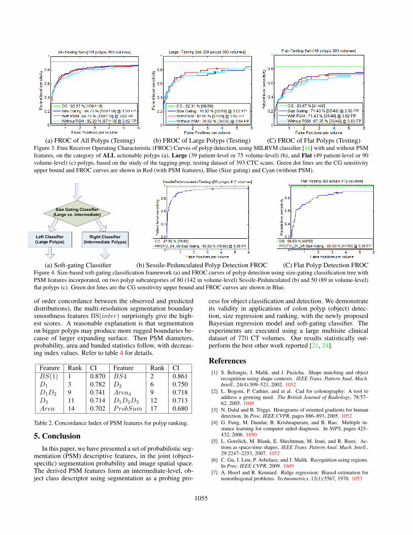
Soft-gating Classification: We next evaluate the PSMfeatures using a two-layer, hierarchical tree classifier ar-chitecture, called “soft-gating” framework in a Bayesian“divide-and-conquer” setting. We first train a basis clas-sifier (as MILRVM) using large (≥ 10mm) polyps ver-sus intermediate size (6 ∼ 9mm) polyps. In runtime, thisso-learned “size classifier” can assign weights Pb(large),Pb(interm) for each VOI input (including negatives)as its probabilities of being large or intermediate polypclass. Two weights are further normalized as Pb(large) +Pb(interm) = 1. Then all positive/negative trainingVOIs are passed into both the left and right branches ofthe soft-gating tree, with different weights of Pb(large),or Pb(interm) respectively. A modified MILRVM classi-fier is applied again on each leaf to learn PbL(prob) andPbR(prob), which can handle weighted training samples.Finally, the probability of being “Polyp” for any candidateVOI is obtained in a Bayesian manner
Pb(Polyp) = Pb(large)× PbL(polyp|large)+Pb(interm)× PbR(polyp|interm)
(12)
, different from hard cascading [26]. This binary tree clas-sification framework is illustrated in Figure 4 (a). Othergating strategies other than size gating (e.g., object shapemorphology gating) can also be exploited.
We further investigate the effectiveness of PSM fea-tures using within the size-gating tree classifier. FivePSM features (D1D2;Area;Area3;D1D2D3;D1) are se-lected with dominating weights for the size-gating branchMILRVM classifier for learning Pb(large), Pb(interm),out of total 10 selected features. PSM features also sig-nificantly contribute to compose the left PbL(prob) and
5We provide the patient-level sensitivity (i.e., if a polyp is detected ineither view, it is counted as a true positive) as gold-standard in colon CADand per-volume false positive rate where a single negative structure (e.g., astool) can appear as two FPs in both Prone and Supine views.
Feature Rank FDS Feature Rank FDSProbSum0 1 1.341 ProbSum3 2 1.314ProbSum2 3 1.299 ProbSum1 4 1.289D2 5 1.268 ProbAvg0 6 1.172ProbSum4 7 1.167 D3 9 1.132ProbAvg1 12 1.111 D1 14 1.102ProbAvg2 15 1.092 ProbSum 17 0.977
Table 1. The FD score comparison of PSM features for polyp de-tection. Core band features are more discriminative than outer,boundary band features.
right PbR(prob) leaf classifiers. MILRVM has an intrin-sic feature selection and weighting mechanism on buildingclassification margins/probabilities. Size based soft-gatingachieves the best performance for our CAD system (thanplain and shape-gating MILRVM ), on an enhanced testingdataset of 412 CT volumes. The detection FROC curvesof two subcategories of Sessile-Pedunculated (SP) and Flatpolyps are shown in Fig. 4 (b,c). We obtain the detectionrates of 95.0% and 88.0% for SP or Flat polyps respectively,under the same 3.36 FPs per-volume. The most competitiverecent work [21] reports the detection sensitivity of 90% forSP, and 75 ∼ 80% for flat polyps, with 4.5 FPs per-scan onaverage6. [24] achieves 95% or 85% sensitivity rates, at 5FPs per scan, on two 86 or 141 patient datasets, respectively.
Polyp Size Regression & Ranking: After classifica-tion, we first exploit the cross-correlation or Pearson’s cor-relation coefficient between any single PSM feature andthe annotated polyp size (in the range of [3, 30]mm) inthe database. The correlation value is ∈ [−1, 1], withhigher value meaning better approximation. Among all≥ 350 classification features (a superset of PSM), D1 re-turns the highest correlation value as 0.6843 which validatesgood generality of the clean-prep trained polyp segmenta-tion module for unseen data. For details, refer to Fig. 5(a) where multiple instances per-polyp are shown. Second,the correlation coefficient of the estimated polyp size usingour probabilistic regression model (section 3) and PSM fea-tures, improves to 0.8102 or 0.7174 for training or testingdataset respectively, as shown in Fig. 5 (b,c). The RM-SEs also decreases from 4.3915mm (a) to 3.3072mm (b)and 3.6426mm (c), compared with using D1 only. The fi-nal regressor is a linear combination of 16 weighted PSMfeatures including D1, ProbSum, boundary smoothnessBS(inter) and banded Areak, ProbSumk features, withautomatic feature selection. 185 volume-level polyps areused for training and 231 polyps for testing.
Finally, it is an important task for a useful CAD system toreturn detected objects/polyps in an order of their predictedimportances. In training, we study the statistical rank con-sistency measurement of two lists, ranked by a PSM featureor the annotated polyp size respectively. A lot of group-wisenonparametric comparison statistics can be applied. Basedon concordance index (CI) criterion [17] (as the probability
6Note that we do not have access to the dataset in [21], but generally as-sume that our dataset is comparable to [21], as they both are in the order ofhundreds of CT volumes and collected from a variety of clinical hispitals.
1054
(a) FROC of All Polyps (Testing) (b) FROC of Large Polyps (Testing) (C) FROC of Flat Polyps (Testing)Figure 3. Free Receiver Operating Characteristic (FROC) Curves of polyp detection, using MILRVM classifier [16] with and without PSMfeatures, on the category of ALL actionable polyps (a), Large (39 patient-level or 75 volume-level) (b), and Flat (49 patient-level or 90volume-level) (c) polyps, based on the study of the tagging-prep, testing dataset of 393 CTC scans. Green dot lines are the CG sensitivityupper bound and FROC curves are shown in Red (with PSM features), Blue (Size gating) and Cyan (without PSM).
(a) Soft-gating Classifier (b) Sessile-Pedunculated Polyp Detection FROC (C) Flat Polyp Detection FROCFigure 4. Size-based soft-gating classification framework (a) and FROC curves of polyp detection using size-gating classification tree withPSM features incorporated, on two polyp subcategories of 80 (142 in volume-level) Sessile-Pedunculated (b) and 50 (89 in volume-level)flat polyps (c). Green dot lines are the CG sensitivity upper bound and FROC curves are shown in Blue.
of order concordance between the observed and predicteddistributions), the multi-resolution segmentation boundarysmoothness features BS(inter) surprisingly give the high-est scores. A reasonable explanation is that segmentationon bigger polyps may produce more rugged boundaries be-cause of larger expanding surface. Then PSM diameters,probability, area and banded statistics follow, with decreas-ing index values. Refer to table 4 for details.
Feature Rank CI Feature Rank CIBS(1) 1 0.870 BS4 2 0.861D1 3 0.782 D2 6 0.750D1D2 9 0.741 Area4 9 0.718D3 11 0.714 D1D2D3 12 0.713Area 14 0.702 ProbSum 17 0.680
Table 2. Concordance Index of PSM features for polyp ranking.
5. ConclusionIn this paper, we have presented a set of probabilistic seg-
mentation (PSM) descriptive features, in the joint (object-specific) segmentation probability and image spatial space.The derived PSM features form an intermediate-level, ob-ject class descriptor using segmentation as a probing pro-
cess for object classification and detection. We demonstrateits validity in applications of colon polyp (object) detec-tion, size regression and ranking, with the newly proposedBayesian regression model and soft-gating classifier. Theexperiments are executed using a large multisite clinicaldataset of 770 CT volumes. Our results statistically out-perform the best other work reported [21, 24].
References[1] S. Belongie, J. Malik, and J. Puzicha. Shape matching and object
recognition using shape contexts. IEEE Trans. Pattern Anal. Mach.Intell., 24(4):509–521, 2002. 1052
[2] L. Bogoni, P. Cathier, and et al. Cad for colonography: A tool toaddress a growing need. The British Journal of Radiology, 78:57–62, 2005. 1049
[3] N. Dalal and B. Triggs. Histograms of oriented gradients for humandetection. In Proc. IEEE CVPR, pages 886–893, 2005. 1052
[4] G. Fung, M. Dundar, B. Krishnapuram, and B. Rao. Multiple in-stance learning for computer aided diagnosis. In NIPS, pages 425–432, 2006. 1050
[5] L. Gorelick, M. Blank, E. Shechtman, M. Irani, and R. Basri. Ac-tions as space-time shapes. IEEE Trans. Pattern Anal. Mach. Intell.,29:2247–2253, 2007. 1052
[6] C. Gu, J. Lim, P. Arbelaez, and J. Malik. Recognition using regions.In Proc. IEEE CVPR, 2009. 1049
[7] A. Hoerl and R. Kennard. Ridge regression: Biased estimation fornonorthogonal problems. Technometrics, 12(1):5567, 1970. 1053
1055

(a) Diameter1 correlation (b) PSM regression size correlation (Training) (c) PSM regression size correlation (Testing)Figure 5. Comparison of correlation between the polyp segmentation diameter D1 [12] and annotated polyp size in (a), versus correlationsusing the sizes from our Probabilistic regression model, in training (b) and testing datasets (c).
Figure 6. Local-scale banded feature SumProb0 has the highestFisher Discriminant (FD) Score of 1.3410, by evaluating a set of≥ 350 classification features including PSM features. As compar-ison, the second polyp dimension D2 [12] has the score of 1.2684(higher than D1;D3), measuring the whole object-scale segmen-tation. A subgroup of “local”, spatially banded features can out-perform the discriminativeness of “global” object size features.
[8] A. Jerebko, S. Lakare, P. Cathier, S. Periaswamy, and L. Bogoni.Symmetric curvature patterns for colonic polyp detection. In MIC-CAI, pages 169–176, 2006. 1049, 1050, 1051
[9] Y. Ke, X. Tang, and F. Jing. The design of high-level features forphoto quality assessment. In CVPR, 2006. 1050
[10] B. Leibe, A. Leonardis, and B. Schiele. Robust object detection withinterleaved categorization and segmentation. IJCV, 2008. 1049
[11] H. Ling, S. Zhou, Y. Zheng, and et al. Hierarchical, learning-basedautomatic liver segmentation. In Proc. IEEE CVPR, 2008. 1049
[12] L. Lu, A. Barbu, M. Wolf, J. Liang, L. Bogoni, M. Salganicoff, andD. Comaniciu. Accurate polyp segmentation for 3d ct colonographyusing multi-staged probabilistic binary learning and compositionalmodel. In IEEE CVPR, 2008. 1049, 1050, 1051, 1052, 1053, 1054,1056
[13] D. MacKay. Ensemble learning and evidence maximization. In NIPS,1995. 1053
[14] D. Martin, C. Fowlkes, and J. Malik. Learning to detect natural im-age boundaries using local brightness, color and texture cues. IEEETrans. PAMI, 26(5): 530-549, 2004. 1050, 1052
[15] D. Paik and et al. Surface normal overlap: a computer-aided detec-tion algorithm with application to colonic polyps and lung nodules inhelical ct. IEEE Trans. Med. Imag., page 23(6):661675, 2004. 1049,1050, 1051
[16] V. Raykar, B. Krishnapuram, J. Bi, M. Dundar, and R. Rao. Bayesianmultiple instance learning: automatic feature selection and inductivetransfer. In ICML, pages 808–815, 2008. 1054, 1055
[17] V. Raykar, H. Steck, B. Krishnapuram, C. Dehing-Oberije, andP. Lambin. On ranking in survival analysis: Bounds on the con-cordance index. In NIPS, pages 1209–1216, 2007. 1054
[18] X. Ren and C. Gu. Figure-ground segmentation improves handledobject recognition in egocentric video. In Proc. IEEE CVPR, pages886–893, 2010. 1049
[19] X. Ren and J. Malik. Learning a classification model for segmenta-tion. In Proc. IEEE ICCV, pages 10–17, 2003. 1050, 1052
[20] J. Shotton, J. Winn, C. Rother, and A. Criminisi. Textonboost for im-age understanding: Multi-class object recognition and segmentationby jointly modeling appearance, shape and context. Int. J. Comp.Vis., 2009. 1049
[21] G. Slabaugh, X. Yang, X. Ye, R. Boyes, and G. Beddoe. A robust andfast system for ctc computer-aided detection of colorectal lesions.Algorithms, 3(1):21–43, 2010. 1054, 1055
[22] Y. Tao, L. Lu, M. Dewan, and et al. Multi-level ground glass opacitydetection and segmentation in ct lung images. In Proc. MICCAI,pages 715–723, 2009. 1049, 1050, 1051, 1052
[23] Z. Tu, X. Zhou, L. Bogoni, A. Barbu, and D. Comaniciu. Probabilis-tic 3d polyp detection in ct images: The role of sample alignment. InCVPR, pages 1544–1551, 2006. 1052
[24] V. van Ravesteijn, C. van Wijk, F. Vos, R. Truyen, J. Peters, J. Stoker,and L. van Vliet. Computer aided detection of polyps in ct colonog-raphy using logistic regression. IEEE Trans. on Med. Imag., 2010.1049, 1050, 1054, 1055
[25] E. M. van Rikxoort, B. van Ginneken, M. Klik, and M. Prokop.Supervised enhancement filters: Application to fissure detection inchest ct scans. IEEE Trans. Med. Imag., 27:1–10, 2008. 1050, 1052
[26] J. Wu, J. Rehg, and M. Mullin. Learning a rare event detection cas-cade by direct feature selection. In NIPS, 2003. 1051, 1054
[27] Y. Xu, M. Sonka, and et al. Mdct-based 3-d texture classification ofemphysema and early smoking related lung pathologies. IEEE Trans.Med. Imag., 25:464–475, 2006. 1049, 1050
[28] J. Yao, J. Li, and R. Summers:. Employing topographical height mapin colonic polyp measurement and false positive reduction. PatternRecognition, 42(6):1029–1040, 2009. 1052
[29] J. Yao, M. Miller, M. Franaszek, and R. Summers. Colonic polypsegmentation in ct colongraphy-based on fuzzy clustering and de-formable models. IEEE Trans on Medical Imaging, 23(11):1344–1352, 2004. 1049, 1050, 1051
1056

![A MultiPath Network for Object Detection arXiv:1604 ... · 2015 detection and segmentation challenges. 1 Introduction Object classification [19,28,30] and object detection [10,27,29]](https://img.pdfslide.net/doc/110x75/6015fcbf097afe09266a899d/a-multipath-network-for-object-detection-arxiv1604-2015-detection-and-segmentation.jpg)


![DetNet: Design Backbone for Object Detection · 2018. 8. 28. · backbone DarkNet[29], and then simplifies detection as a regression problem. SSD [24] adopts reduced VGGNet[35] and](https://img.pdfslide.net/doc/110x75/60d0aa5e6a084a7bd35775df/detnet-design-backbone-for-object-detection-2018-8-28-backbone-darknet29.jpg)