Embed Size (px)
Citation preview

Effective Perinatal Care (EPC)
Facilitator Guide


Effective Perinatal Care (EPC)
Facilitator Guide

Facilitator’s Manual
Address requests about publications of the WHO Regional Office for Europe to:PublicationsWHO Regional Office for EuropeScherfigsvej 8DK-2100 Copenhagen Ø, Denmark
Alternatively, complete an online request form for documentation, health information, or forpermission to quote or translate, on the Regional Office web site(http://www.euro.who.int/pubrequest).
© World Health Organization 2010All rights reserved. The Regional Office for Europe of the World Health Organizationwelcomes requests for permission to reproduce or translate its publications, in part or in full.
The designations employed and the presentation of the material in this publication do notimply the expression of any opinion wh atsoever on the part of the World HealthOrganization concerning the legal status of any country, territory, city or area or of itsauthorities, or concerning the delimitation of its frontiers or boundaries. Dotted lines onmaps represent approximate border lines for which there may not yet be full agreement.
The mention of specific companies or of certain manufacturers’ products does not imply thatthey are endorsed or recommended by the World Health Organization in preference toothers of a similar nature that are not mentioned. Errors and omissions excepted, thenames of proprietary products are distinguished by initial capital letters.
All reasonable precautions have been taken by the World Health Organization to verify theinformation contained in this publication. However, the published material is beingdistributed without warranty of any kind, either express or implied. The responsibility for theinterpretation and use of the material lies with the reader. In no event shall the World HealthOrganization be liable for damages arising from its use. The views expressed by authors,editors, or expert groups do not necessarily represent the decisions or the stated policy ofthe World Health Organization.

Effective Perinatal Care (EPC)
Acknowledgments
This training package was developed by JSI/Ukraine and WHO Regional Office for Europe,Making Pregnancy Safer Programme with the financial support towards the preparation andproduction of this document provided by the Government of the Unites States of America andJSI.
Authors and contributors: Tengiz Asatiani, Nino Berdzuli, Atrem Chernov, GianpaoloChiaffoni, Dmytro Dobrianskyi, Pauline Glatleider, Gianfranco Gori, Daniela Iancu, TamaraIrkina, Dalia Jeckaite, Viacheslav Kabakov, Anna Karpushkina, Ivan Lejnev, KetevanNemsadze, Audrius Maciulevicius, Irina Matvienko, Natalia Podolchak, Liubov Pozdniakova,Igor Semenenko, Oleg Shvabski, Gelmius Siupsinskas, Elena Sofronova, Fabio Uxa.
Coordinators: Alberta Bacci, WHO, Regional Office for Europe, Helene Lefèvre -Cholay,Oleg Kuzmenko, JSI/Ukraine


i-1
Contents
PART I: COURSE PECULIARITIES AND TEACHING METHODS
Introduction ................................ ................................ ................................ ........................... i-4
How does this course differ from other training courses? ................................ ....................... i-5
Main principles of active learning ................................ ................................ .......................... i-6
Who is the facilitator/instructor? ................................ ................................ ............................. i-6
What is facilitator’s role? ................................ ................................ ................................ ........ i-7
How can this facilitator’s manual help you? ................................ ................................ ............ i-8
Methods for motivating participants ................................ ................................ ....................... i-9
Methods to assist colleague facilitators ................................ ................................ ............... i-12
Methods of facilitator’s work ................................ ................................ ................................ i-13
PART II: MODULE INSTRUCTIONS
Perinatal Care
Module 1C. Safe Motherhood and Effective Perinatal Care: Are Changes Necessary ? ..... 1C-1
Module 2C. Introduction to Evidence-Based Medicine ................................ ....................... 2C-1
Module 3C. Counselling Skills in Maternal and Neonatal Care ................................ ........... 3C-1
Module 4C. Assessment of Foetal Well-Being During Pregnancy and Labour .Assessment of Small for Gestational Age (SGA) Foetuses ................................ ................ 4C-1
Module 5C. Management of Normal Labour and Delivery ................................ .................. 5C-1
Module 6C. Initial Rapid Assessment of the Newborn and Principles of Neonatal Care ..... 6C-1
Module 7C. Breastfeeding ................................ ................................ ................................ . 7C-1
Module 8C. Postpartum Care of Mothers and Newborns ................................ ................... 8C-1
Module 9C. Neonatal Resuscitation ................................ ................................ ................... 9C-1
Module 10C. Integration of Prevention of Mother t o Child HIV Transmission into EffectivePerinatal Care................................ ................................ ................................ .................. 10C-1
Module 11C. Infections in Pregnancy, Childbirth and Postpartum ................................ .... 11C-1
Module 12C. Preterm Labour ................................ ................................ ........................... 12C-1
Module 13C. Sudden Infant Death Syndrome (SIDS) ................................ ...................... 13C-1
Module 14C. Postpartum Depression, Loss and Tragedies ................................ ............. 14C-1

Facilitator’s Manual
i-2
Module 15C. How to Improve Existing Practices: Strategy of C hanges............................ 15C-1
Midwifery
Module 1MO. Antenatal Care................................ ................................ .......................... 1MO-1
Module 2MO. The Use of the Partograph................................ ................................ ........ 2MO-1
Module 3MO. Hypertension in Pregnancy ................................ ................................ ....... 3MO-1
Module 4MO. Obstetric Haemorrhages ................................ ................................ ........... 4MO-1
Module 5MO. Prelabour Rupture of Membranes ................................ ............................. 5MO-1
Module 6MO. Labour Induction: Who? Why? How? ................................ ........................ 6MO-1
Module 7MO. The Unsatisfactory Progress of Labour . Intrapartum OxytocinAdministration ................................ ................................ ................................ ................. 7MO-1
Neonatology
Module 1N. Complete Examination of a Newborn ................................ ............................. 1N-1
Module 2N. Post-Resuscitation Neonatal Care ................................ ................................ .. 2N-1
Module 3N. Breathing Difficulty in the Newborn ................................ ............................... 3N-1
Module 4N. Neonatal Jaundice ................................ ................................ .......................... 4N-1
Module 5N. Neonatal Bacterial Infections ................................ ................................ .......... 5N-1
Module 6N. Care of a Newborn with Birth Defects/Congenital Malformations or BirthTrauma ................................ ................................ ................................ .............................. 6N-1
Module 7N. Low-Birth Weight Baby/Small Baby: Care and Feeding ................................ .. 7N-1
PART III: INSTRUCTIONS ON COURSE ORGANIZATION
Criteria for selecting facilitators……………………………………………………………… ..iii-3
Model of the training course’ agenda…………………………………………………………. iii-5
General modules
Module 1G – Effective perinatal care – Opening ceremony…………………………iii-23
Module 2G – “The Icebreaker”…………………………………………………………..iii -27

Effective Perinatal Care (EPC)
i-3
PART I. COURSE PECULIARITIES AND TEACHING METHODS

Facilitator’s Manual
i-4
INTRODUCTION
Thorough planning and strong administrative support are required before the beg inning andthroughout the training course on Effective Perinatal Care (EPC). This part of Facilitator’sManual describes course peculiarities and teaching methods to be used.
Clinical practice is a significant part of the Effective Perinatal Care (EPC) tr aining course. Thecourse is designed to provide the participants with an opportunity to practice their skills ofcare during normal pregnancies and newborn care, so that they can effectively use theseskills upon return to their medical establishments. In addition to the first theoretical week ofstudies the participants will be working in a maternity hospital and will be using the methodsof effective perinatal care in practice.
Therefore, the training course requires many administrative and organization al preparations.Selection of suitable location for course delivery (city or district, which has severalappropriate medical establishments) is critical during early planning stage.
Course Director together with Ministry of Health employees who participat e in courseplanning may schedule a series of training courses instead of one. Taking into account theeffort required for course development/preparation, duration of facilitator training , and theneed for a series of trainings to cover a sufficient number of medical personnel in order tojustify the resources often requires considering long -term training planning. It may bereasonable to create a group of facilitators which will conduct training delivery on a regularbasis (e.g. monthly). In such case long -term training goals may influence facilitators’selection.

Effective Perinatal Care (EPC)
i-5
HOW DOES THIS COURSE DIFFER FROM OTHER TRAINING COURSES?
Training material in this course is presented for different aspects of effective perinatal care.
Modules are designed to help the par ticipants get certain skills needed for labourmanagement and newborn care. Participants acquire such skills during presentations, byobserving demonstrations, participating in discussions, exercises or role plays.
After module practice participants pract ise acquired skills in real clinical environmentsupervised by facilitators who control the adequacy of their clinical care techniques.
Teaching without distinct division into teacher and students is the most acceptable methodwhile working with adults; this facilitates experience exchange which is important in anyeducational process. Indeed, this method is particularly appropriate for this course, sincemany of the participants already have abundant experience of extensive clinical and otherwork.
Such teaching approach helps the participants to study actively , relying on their experience.Besides, this allows creating equitable relationship between the participants and courseorganizers, as opposed to traditional “teacher -student” or “instructor-participant”communication methods. Course organizers should always keep in mind that qualificationsand experience of many of the participants may be superior or equal to theirs.
While working in pairs with other facilitators it is very important to come to an agreement onall issues before the course starts; agree upon own roles and duties and assignresponsibilities for each session. A technique, when facilitators exchange roles may prove tobe effective allowing the participants to hear a different voice and feel the difference inteaching styles.
There are cases when participants anticipate a more authoritative and didactic approach ,expecting a specialist or facilitator to tell them what they need to know, think or do. To avoidundermining own authority it would be wise to familiarize the participants with the teachingstyle used during the seminar at the very beginning. However, one can carry own point byquoting a Chinese proverb:
"I hear and I forget. I see and I remember. I do and I understand. "
Confucius (551 BC - 479 BC)
An emphasis should be made at the beginning that participants will have to considerthemselves what would be important and useful in their work. This equally concerns thedecisions they will have to make and measures to take upon their return to work after thecourse. It is important to understand in this process that you, as facilitators, are ordinarypeople creating an environment for educational and decision -making process. You should notpoint out anybody what to do; your role is to advise and provide support as well as freedomfor their own decision-making.
Course participants, even if they all are medical personnel from the same country, maybelong to different age and religious groups, occupy different job positions, etc. S uch varietyis encouraged, considering the interactivity and active learning method of EPC. However,personal characters of course participants may also imply differences of opinion,communication methods as well as approaches to decision -making which will certainly revealthemselves during the seminar. It will be a rather complicated task for the facilitator to forgetabout own approaches/preferences and appeal to the participants for mutual respect, andencourage experience exchange by adopting it from th eir colleagues.

Facilitator’s Manual
i-6
This course requires you to use a whole series of various methods and approaches rangingfrom direct participation in a form of presentations to conducting role plays and problem -solving exercises in small groups. Further in this manual we will reveal the teaching/learningmethods which should be used in this course.
MAIN PRINCIPLES OF ACTIVE LEARNING
To provide for free unstrained relationship between facilitator and participants, it is veryimportant to establish a series of main rules at the beginning of the program. These rulesinclude the following:
Always treat all the participants with respect irrespective of culture, age, or sexdifferences.
Secure and respect confidentiality to allow facilitators and participants discussdelicate issues (concerning sexual, reproductive, and psychological health, abuse ofharmful substances) without fear of possible negative consequences.
Resort to expert opinion of both facilitators and participants while seeking solutions fora complicated situation.
Commitment to critical appraisal of your activities and respect of criticism to make thefairness of your decisions evident.
From the very beginning determine how facilitators and invited specialists willcooperate and voice their opinions – both positive and negative – and how to keepeach other within the context of the programme.
Conclude to consent, that every time when a facilitator or a particular expert is makinga presentation, the second facilitator would do the timing and warn a pr esenter thatallocated time had elapsed. Some facilitators set alarm-clocks at the beginning of thesession – a method which enlivens the participants but only under the stipulation thatthe alarm sound would not be too acute!
All the above, combined with basic teaching skills, will contribute to creation of a productivetraining environment. Some facilitators prefer making a “training contract” at the verybeginning of the programme to ensure that facilitators and participants agree on the mainprinciples underlying educational process for adults.
WHO IS THE FACILITATOR/INSTRUCTOR?
Facilitator is a person who helps participants to master skills, presented in the trainingcourse. Facilitator spends most of the time with participants: individually or in small groups.You are a FACILITATOR and your mission is to deliver training in this course.
As a facilitator, you should perfectly know the teaching material. Your task is to present themodule, conduct demonstrations, answer the questions, discuss with the participants theiranswers to the exercises, lead role -play exercises and group discussions, organize andsupervise clinical practice at the hospital, and generally provide any assistance to participantsthat they may require for successful completion of the training course.

Effective Perinatal Care (EPC)
i-7
WHAT IS FACILITATOR’S ROLE?
As a facilitator you must fulfil the 3 following functions:
YOU TEACH:
- Ensure that every participant understands how to work with the trainingmaterials and knows own assignment in each module .
- Answer participants’ questions as they appear.
- Explain the information which may be unclear to the participants and helpthem grasp the goal of each exercise.
- Conduct group exercises, like group discussions, oral material revisionexercises, video and game exercises to verify if training goals have beenreached.
- Assess each participant’s work and give correct answers.
- Discuss with the participant how has (s)he arrived to the answers in order toreveal weak skills/understanding.
- Provide additional explanations or practice opportunities to improve skills andunderstanding.
- Help the participant understand how acquired skills can be applied in his/hermedical establishment.
- Explain what needs to be done during each clinical session .
- During clinical sessions display an example of correct clinical andcommunication skills.
- During clinical sessions give recommendations as required and provide forfeedback.
YOU MOTIVATE:
- Compliment the participant for correct answers, improvements or progress.
- Watch for any serious disturbances which may hinder training (e.g. loud noiseor poor lightning).
YOU FACILITATE:
- Plan beforehand and receive all the materials needed for each training day, sothat they are available in the classroom or for taking to out-patientsdepartment if necessary.
- Monitor each participant’s progress.
- How do you conduct these activities?

Facilitator’s Manual
i-8
- Be enthusiastic about topics covered in the training course and aboutparticipants’ work.
- Be responsive to questions and requests of each participant. En courage theparticipants to turn to you with questions or comments.
- Observe participants’ work and offer individual help if you notice that aparticipant is nervous, stares at one point, does not answer questions or doesnot turn pages – this indicates that a participant requires help/assistance.
- Encourage friendly, mutually beneficial relationship . Answer all questions in apositive way (e.g. “I see your point .” or “That’s a good question”). Listen toquestions and try explaining issues which are uncle ar to a participant in detail,rather than quickly giving a correct answer.
- Always allocate sufficient time to provide full answers to all questions of eachparticipant (i.e. so that both you and the participant are satisfied with theanswers).
Things to AVOID...
- During the time allocated for planned sessions one must not do any work onother projects or discuss issues which are not related to the training course.
- While in discussion with the participants avoid facial expressions or commentswhich may make them feel uncomfortable.
- Do not call out participants to answer questions in turns, like in a regular class,thus creating awkward silence if the participant does not know the answer.Instead, ask questions during individual feedback .
- Avoid turning the course into a performance. Enthusiasm (as well as keepingparticipants’ interest) is good, but training is of higher importance. Make surethat participants understand the material. Recondite topics may requirereduced pace of work and individual de tail.
- Do not show condescension. In other words, do not treat the participants askids – they are adults.
- Do not talk too much. Encourage participants to voice their opinions .
- Do not be shy, nervous or anxious when you speak. This Facilitator’s Manua lwill help you remember what you have to say. Use it!
HOW CAN THIS FACILITATOR’S MANUAL HELP YOU?
This Facilitator’s Manual will help you delivering modules of the training course .
This manual includes the following items for each module :
List of module procedures.
Procedure instructions which describe :

Effective Perinatal Care (EPC)
i-9
how to conduct presentations, demonstrations, role -play exercises and groupdiscussions
materials required for these activities ,
how to conduct exercises
answers (or possible answers) to most exe rcises.
To self-prepare for each module you should :
read module and do the exercises
read all the information about the module in this Facilitator’s Manual
plan module work in detail and determine which topics require special attention
collect all the necessary materials for module exercises and prepare fordemonstrations or role-play exercises
think over which sections may be complicated for the participants and whichquestions they might ask
plan ways of possible assistance while covering difficult secti ons and answers topossible questions
think over skills covered by the module and their application in medicalestablishments of the participants
put questions to the participants which may make them think about application ofacquired skills in their medical establishments.
METHODS FOR MOTIVATING PARTICIPANTS
Encourage communication
From the first day try to have an individual conversation with each of the participants.If you demonstrate your amicability and eagerness to help during this initialintercourse, it will help the participants (a) to overcome their shyness, (b) tounderstand that you want to communicate with them, (c) communicate with you in amore open and productive way throughout the whole training course.
Closely watch the work of each participant to see if they have any problems even ifthey do not ask for help/assistance. If you show your interest and focus attention oneach participant individually, they will work with higher enthusiasm. Besides, ifparticipants know that somebody is interested in their work, they will most likely askfor help/assistance when necessary.
Be available to participants for advice and assistance.

Facilitator’s Manual
i-10
Engage the participants into discussions
Frequently ask questions to check how participants understand the material andengage them into active comprehension of the covered material and their participationin the training course. Questions that start with words “what”, “why” and “how” requirean expanded/detailed answer. Avoid asking “yes” or ”no” questions .
After asking a question make a PAUSE. Give the participants time to think and showtheir interest answering it. A frequent mistake is to ask a question and answer ityourself. If no one can answer your question, rephrase it to break the silence, but donot do so all the time – silence may sometimes be productive .
Answer participants’ questions with a comment, gratitude , or an approving nod. Thiswill increase participants’ concernment in their own eyes and will result in higherinvolvement into the training process. If you consider that a participant does notunderstand some material, ask him/her to explain it in detail and then offer anotherparticipant to provide any additions or comments. When the participant feels thathis/her comment was derided o r ignored, (s)he may drop out from the discussion oravoid answering next time on his/her own initiative.
Answer participants’ questions eagerly and encourage them to ask questions whenthey appear, rather than leaving for later.
Do not feel obliged to answer all questions yourself. Depending on a situation youmay readdress the question to the participant or ask another participant to answer.Before answering a question you might need to discuss it with the Course Director oryour fellow facilitator. Be prepared to say “I don’t know, but I will try to find out”.
Address the participants by their name when you ask questions, thank or praise them.While discussing a previous comment, refer to its author by name .
Keep constant visual contact with the pa rticipants, so that they feel involved into work.Avoid looking at the same participants all the time. Visual contact during a couple ofseconds may be translated as a call out for an answer even by a shy participant.
Session should be purposeful and de livered at a right pace
Keep the right pace of your presentations :
Clearly deliver your ideas. Change intonation and elocution velocity .
Use examples from your own experience and ask participants to share theirs.
Record key ideas on a flip-chart as they appear (this is a good way of encouraginganswers. The participant will know that his/her comment was heard and (s)he will likethat it was recorded to be displayed to the whole group ).
Record the ideas on a flip-chart using participant’s words, if poss ible. If you need toshorten the idea, rephrase it and get participant’s approval before recording. Youmust make sure the participant feels you understand his/her idea and have recorded itcorrectly. When writing do not turn your back to the participants for a long time.
At the start of discussion record the main question on a flip-chart. This will help theparticipants to concentrate on the point. Approach the flip -chart and point to thequestion when necessary.

Effective Perinatal Care (EPC)
i-11
Rephrase and frequently summarize the ma terial to keep participants’ concentration.If necessary, ask participants to elaborate on some answers. Encourage participantsto ask the respondent to repeat or explain his/her answer.
Rephrase the initial question for the group to concentrate on the ma in point again. Ifyou feel that somebody opposes such call-back, make a pause first to draw group’sattention and then inform the participants that they have strayed from the main ideaand then rephrase the initial question .
Do not allow several participants to speak simultaneously. If this happens, stop theorators and arrange the order of giving the floor to someone (e.g. say “Lets listen toDr.Samoilov’s comments first and then to Dr.Sharikova followed by Dr.Lastochkin).People would usually stop inter rupting others if they know they will receive the floorsoon.
Thank the participants who made laconic and practical comments.
Try to involve quite participants. Ask the participant that did not take part in thediscussion to comment, or approach someone to let him/her understand that youexpect his/her involvement in the discussion.
Solve any problem
Some participants might talk too much. A couple of suggestions are given below onhow to work with such garrulous participants:
Do not call out such participant right after putting a question .
After the participant had spoken for some time, say “You have explained your point.Let’s listen what other participants may have to say about this”. Then rephrase thequestion and offer other participants to answer or call out someone at once, forinstance: “Dr.Samoilova, you raised your hand a few minutes ago.”
After a participant stops, quickly interrupt him/her and ask the others “What do othermembers of the group think about this?”
Record the main idea of the participant on a flip-chart. If (s)he keeps discussinghis/her idea, point to a flipchart and say “Thank you, we have already recorded thisidea”. Then ask the group to generate other ideas .
Do not put additional questions to a loquacious participant. If (s)he keeps answeringall questions addressed to a group, ask a particular participant or group of participantsto answer (e.g. ask “Does anybody from this part of the table have any other ideas? ”)
Try to identify the participants who have difficulties understanding or using thelanguage used in the training course. Speak slowly and clearly to make understandingeasy; encourage participants’ efforts to improve communication .
Discuss with the Course Director all the language problems which may prevent theparticipants from understanding the material or explanations. Perhaps a specialparticipant’s guide/help should be worked out.
Discuss such participants with your fellow facilitator or Course Director (CourseDirector can discuss training material with such participant individually) .

Facilitator’s Manual
i-12
Encourage participants’ efforts
As a facilitator you have your own style of communicating with participants. Still, youmay use some methods to encourage participants’ efforts :
avoid facial expressions or comments which m ay make the participants feeluncomfortable
while talking to a participant, sit or stoop to appear on the same level with theparticipant
provide comprehensive and deliberate answers to questions
encourage the participants’ to talk to you by giving them sufficient time
express your interest by saying “This is a good question/suggestion”.
Encourage participants who:
- make an effort
- ask to explain an unclear question
- correctly complete exercises
- participate in group discussions
- assist other participants (without distracting by lengthy argumentations and talksthat do not refer to the point ).
METHODS TO ASSIST COLLEAGUE FACILITATORS
Spend some time with your fellow facilitator during first assignments. Exchangeinformation about accumulated trainin g experience, strong and weak areas andpreferences of each other. Divide roles and responsibilities and decide how you willwork together.
Assist each other during presentations and group discussions. For instance, onefacilitator can conduct a group discussion, while the other would record main pointson a flipchart. The second facilitator could also control the discussion flow by checkingthe Facilitator’s Manual and adding missing points.
Daily revise training actions which will take place the next day (e.g. role-playexercises, demonstrations and material revision exercises) and decide who willprepare demonstrations, lead material revision exercises, play each role, collectmaterials, etc.
Work on each module together instead of accounting module responsibilities by turns.

Effective Perinatal Care (EPC)
i-13
METHODS OF FACILITATOR’S WORK
When the participants are working
Be available to participants for advice and assistance , show interest and eagerness tohelp.
Observe participants’ work and offer individual help if you notic e that a participant isnervous, stares at one point, does not answer questions or does not turn pages – thisindicates that a participant requires help/assistance.
Encourage participants to ask questions when they need your help/assistance.
If important issues or problems arise during individual communication with theparticipant, record them for later discussion in a group .
If a question arises and you feel that you will not be able to provide an appropriateanswer, resort to your fellow facilitator or Course Director for assistance as soon aspossible.
Revise the main theses of this Facilitator’s Manual to prepare for discussion of thenext exercise with the participants.
During group discussion
Schedule group discussion to a time, by which you a re sure the participants will finishtheir previous work. Wait until all participants are ready and then announce the time ofgroup discussion; this way the participants will not be in a hurry .
Before starting the discussion remind its goal and main issue s by using correspondingnotes in this manual.
Always start a group discussion by informing the participants its objective .
Oftentimes it is impossible to conclude to one correct answer during a discussion.Just control that participants’ answers conform to the essence of the question and thatall participants understand how these conclusions were drawn.
Try to involve as many participants into the discussion as possible. Record key pointson a flipchart as they appear. Limit your own participation to a minimum, but at thesame time continue asking questions to keep the discussion pace and its compliancewith the main topic.
Always summarize or ask a participant to summarize what was discussed in anexercise. Hand out answer-sheets to participants if they are available.
During role play exercise
Before starting a role-play exercise review its objective, roles, situation descriptionand its main points for further group discussion by using corresponding notes in thismanual.

Facilitator’s Manual
i-14
As participants approach you for instructions before the beginning of a role-playexercise:
- divide roles. First select sociable participants who are not shy, possibly by askingif there are any volunteers to participate in a role-play. If necessary, a participantmay serve as a model by acting his/her role in the first role -play.
- hand out all the necessary materials to role-play participants, e.g. phantom babyand medical preparations.
- hand out situation descriptions to role-play participants (each exercise usuallycontains “mother information”, which needs to be photocopied or cut out from thismanual).
- tell the participants that roles should be read aloud
- allow the participants time to prepare
- When everybody is ready, seat/place all the participants of the exercise . Seat orplace the “mother” and the “medical personnel member” separately from the groupso that everybody can see them.
- Start by presenting the participants and their roles, inform exercise objective ordescribe the situation. For instance, you can describe mother ’s or baby’s age,assessment results, and already received treatment.
- Stop the participants if they encountered insuperable difficulties or if theydigressed from the exercise objective .
- When the role-play exercise is over, thank the performers. Make sur e thatfeedback provided by the rest of the group is constructive. First, discuss what wasdone correctly, and then discuss what could be perfected.
- Try to involve as many participants as possible into the discussion after the role -play exercise. In many cases, questions provided in the module may serve as abackground for discussion.
- Ask the participants to summarize everything they have learnt from the role -playexercise.

ii-1
PART II. INSTRUCTIONS ON COURSE ORGANIZATION

Facilitator’s Manual
ii - 2

Effective Perinatal Care (EPC) Facilitator Guide
Module 1C
Safe Motherhood and Effective Perinatal Care: Are Changes Necessary?
Learning Objectives By the end of this module the participants will: • Become familiar with the Millennium Development Goals and the ‘Health for
All in the 21st Century’ Strategy • Learn about the main problems and directions of perinatal health care in the
European region • Know the main indicators of maternal and infant mortality in the European
region • Understand that addressing physiological, psychological, emotional, and social
needs of pregnant women and their families is the basic factor in assessing the quality of perinatal care
• Learn about main effective perinatal technologies which will be reviewed in detail at the end of this module
• Learn how international collaboration can improve perinatal care
Outline of the Module Classroom work – 45 minutes
Activity 1 – Presentation 45 min
Preparation for the Module • Ensure that all the participants have the Participant Guide • Ensure that all other facilitators know their responsibilities during this module.
Training Materials, Audio and Video Equipment Training Materials
• Participant Manual • Presentation 1С EPC ENG • Local Guidelines and State Orders related to safe pregnancy and perinatal
care.
1C - 1

Effective Perinatal Care (EPC)
Training Materials, Audio and Video Equipment Equipment
• LCD or slide projector • Flip-chart • Colour markers • Name badges.
Key Messages • Maternal and infant health has been a priority long before the 1990s. Program
implementation and activities are based on over a century of experience
• Millennium Development Goals (MDGs) aim to decrease maternal and infant mortality as well as reduce the spread of HIV/AIDS
• A healthy start in life is a priority for all countries and a Safe Motherhood
program is the best strategy to reach this goal
• Family satisfaction with healthcare is a reliable indicator of the quality of medical care and overall quality of the healthcare system
• Women worldwide define quality perinatal care in the same ways
• Effective technologies and tools are in place to prevent deaths and long-term disabilities related to delivery
• Technology is defined as a combination of activities that includes evidence-based methods, procedures, and equipment applied systematically to solve a specific problem. Appropriate technology is effective, safe, affordable, feasible, adjusted to local conditions and good for both patients and providers
• Perinatal care can be made better by improving client satisfaction, which involves increased family involvement as an instrument to encourage participation of the woman during pregnancy, delivery, post-partum period, and in newborn care
• Effective perinatal care should be evidence-based, which is a result of information obtained through modern clinical research.
Classroom Work
Activity 1 – Presentation (45 min) • Show Slide 1C-1. Share with the participants the main objectives of this
module which are presented at the beginning of this document.
1C - 2

Module 1C
• The last 20 years have been characterized by fast progress, scientific development and the implementation of different technologies in maternity care. However, at the same time there has been a growing feeling of dissatisfaction in many countries. The users of health care services are dissatisfied that society cannot introduce a more human approach in maternal health care. Ask the participants whether it is necessary to change the existing system of perinatal care. If yes, why? Suggest that the participants consider this question in detail.
• Show the participants Slide 1С-2. There are eight main Millennium
Development Goals (MDGs) formulated by WHO in The World Health Report. (2005). Although only three goals are directly connected with perinatal care, others are also connected in some regard to the health care system.
• Motherhood is a positive experience for the majority of women (Slide 1С-3)
meaning that the most women and their families experience feelings of happiness, pride and elevated emotions during pregnancy, delivery, and in the postpartum period.
• However, in the world today, there are a lot of mothers that die because of
pregnancy-related problems and even more women suffer from morbidity and disability (Slide 1С-4).
• Slide 1С-5 shows the map of the European Region where countries and their
different levels of maternal mortality rates (MMRs) are marked with different colours. Find and show the country where the training course is conducted. Assess the maternal mortality rate in this country.
• Slide 1С-6 shows that a gradual decrease in the MMR has been observed in
all countries of the European Region, although the pace of this decrease is very much dependent upon healthcare workers, including the participants of this course. Pay attention to the fact that the highest level of maternal mortality is found in Central Asia where the probability of dying from pregnancy- and delivery-related complications is 6-7 times higher than in Western Europe.
• Slide 1С-7 shows the main causes of maternal mortality. Slide 1С-8 shows
that the majority of these cases can be prevented with the use of simple, effective, and low-cost interventions. These interventions are already known among the international community of perinatal care specialists.
• Slide 1С-9 shows world child mortality rates. Explain to the participants that in
the 21st century more than ten million children die each year although the majority of these deaths can be avoided, as in the case of many maternal deaths.
• Although neonatal mortality has decreased in the last half of the 20th century,
the discrepancy between poor and rich countries continues to grow. Slide 1С-10 demonstrates rates of neonatal mortality in different countries of the European Region.
• Slide 1С-11 shows the causes of neonatal mortality in the world, which
include asphyxia and trauma, prematurity and infections.
1C - 3

Effective Perinatal Care (EPC)
• Show the participants Slide 1С-12. Explain that the majority of neonatal
deaths, similar to maternal deaths, are preventable with the help of known and available interventions.
• For the first time in history, WHO announced 2005 as the year of Mother and
Child. The slogan of the 2005 World Health Day was Every Mother and Child Counts (Slide 1С-13).
• The WHO Safe Motherhood Initiative was a global initiative started in 1987.
The goal was to decrease maternal mortality in half by 2000. The program goal was not achieved however international efforts were strengthened. Building on the lessons learned from the Safe Motherhood experience, the WHO Making Pregnancy Safer Initiative (MPS) was launched in 2000 and continues today. The MPS goal is for all mothers and their babies to have skilled care at every birth within a context of a continuum of care. The focus is the health sector. The main program strategy is to facilitate improvement of effective perinatal care in the European Region (Slide 1С-14).
• Than go to the Slide 1C-15. Tell participants that skilled care refers to the care
provided to a woman and her newborn during pregnancy, childbirth and immediately after birth by an accredited and competent health care provider who has at her/his disposal the necessary equipment and the support of a functioning health system, including transport and referral facilities for emergency obstetric care.
• Show Slide 1C-16. Present to participants the “components of skilled
attendant care”. Emphasize that core midwifery skills are essential for all providers. Many countries in Eastern Europe, Russia and the CARAK report very high levels of skilled attendants present for birth however core midwifery skills and the midwifery model of care are not provided.
• The main objectives of perinatal care improvement are shown on Slide 1С-17. • Show Slide 1C-18 – 1C-19. List MPS Fundamentals and Principles to
participants. Explain them that the basic fundamentals and principles of effective perinatal interventions were developed by a group of WHO experts from the European Region during PEPC/MPS Task Force meetings in Venice (1998), Verona (2003) and MPS Experts Meeting in Catania, Italy (2007). These fundamentals and principles subsequently received wide support, dissemination and implementation in all countries of the region.
• WHO Europe and WHO America jointly organized a conference in Fortaleza,
Brazil (April 1985) where more than 60 experts (obstetricians, paediatricians, health care leaders, economists, psychologists and sociologists) took part. The participants of this conference made several conclusions that are based on the fundamental principle of effective perinatal care: the central role of the woman in all decisions related to safe pregnancy and safe delivery. The main problems characteristic to the majority of countries in the European Region were identified at this conference. These problems are presented on Slides 1С-20 and 1С-21.
• Show Slide 1С-22. Note that the best model of health care is based on three
pillars: safety, evidence-based medicine, and the patient’s needs.
1C - 4
Module 1C
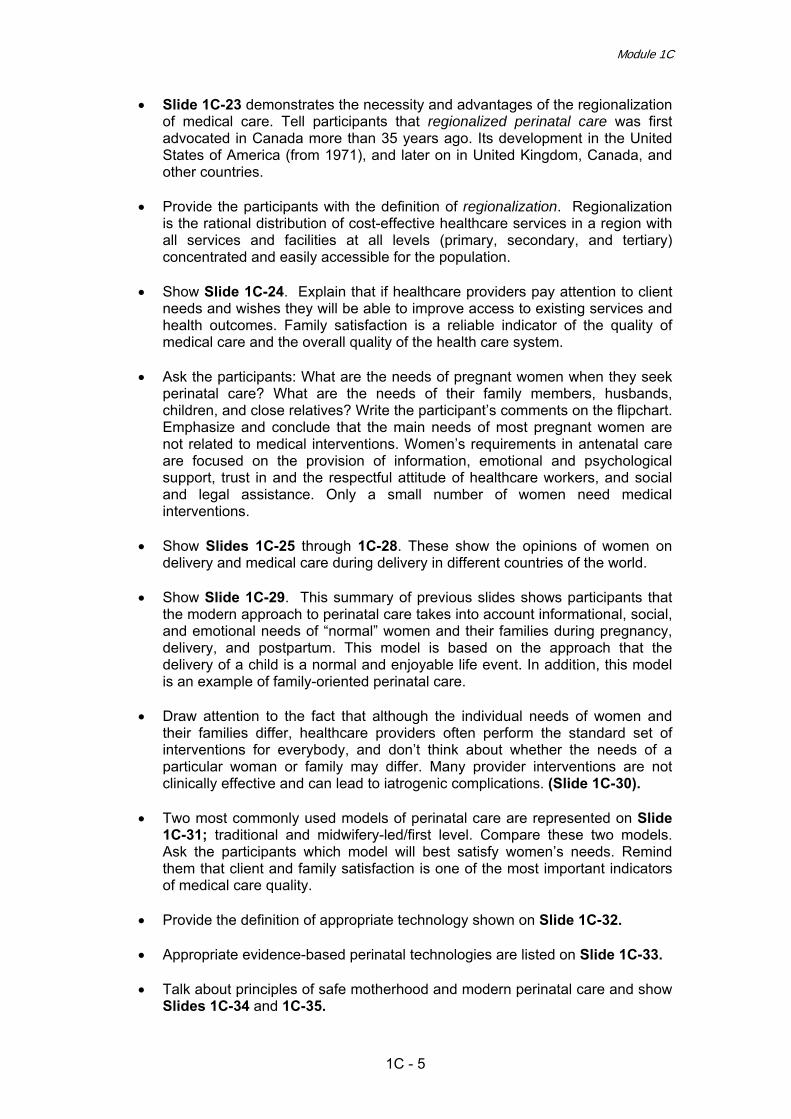
• Slide 1С-23 demonstrates the necessity and advantages of the regionalization of medical care. Tell participants that regionalized perinatal care was first advocated in Canada more than 35 years ago. Its development in the United States of America (from 1971), and later on in United Kingdom, Canada, and other countries.
• Provide the participants with the definition of regionalization. Regionalization
is the rational distribution of cost-effective healthcare services in a region with all services and facilities at all levels (primary, secondary, and tertiary) concentrated and easily accessible for the population.
• Show Slide 1С-24. Explain that if healthcare providers pay attention to client
needs and wishes they will be able to improve access to existing services and health outcomes. Family satisfaction is a reliable indicator of the quality of medical care and the overall quality of the health care system.
• Ask the participants: What are the needs of pregnant women when they seek
perinatal care? What are the needs of their family members, husbands, children, and close relatives? Write the participant’s comments on the flipchart. Emphasize and conclude that the main needs of most pregnant women are not related to medical interventions. Women’s requirements in antenatal care are focused on the provision of information, emotional and psychological support, trust in and the respectful attitude of healthcare workers, and social and legal assistance. Only a small number of women need medical interventions.
• Show Slides 1С-25 through 1С-28. These show the opinions of women on
delivery and medical care during delivery in different countries of the world. • Show Slide 1С-29. This summary of previous slides shows participants that
the modern approach to perinatal care takes into account informational, social, and emotional needs of “normal” women and their families during pregnancy, delivery, and postpartum. This model is based on the approach that the delivery of a child is a normal and enjoyable life event. In addition, this model is an example of family-oriented perinatal care.
• Draw attention to the fact that although the individual needs of women and
their families differ, healthcare providers often perform the standard set of interventions for everybody, and don’t think about whether the needs of a particular woman or family may differ. Many provider interventions are not clinically effective and can lead to iatrogenic complications. (Slide 1С-30).
• Two most commonly used models of perinatal care are represented on Slide
1С-31; traditional and midwifery-led/first level. Compare these two models. Ask the participants which model will best satisfy women’s needs. Remind them that client and family satisfaction is one of the most important indicators of medical care quality.
• Provide the definition of appropriate technology shown on Slide 1С-32. • Appropriate evidence-based perinatal technologies are listed on Slide 1С-33. • Talk about principles of safe motherhood and modern perinatal care and show
Slides 1С-34 and 1С-35.
1C - 5

Effective Perinatal Care (EPC)
• Show Slide 1С-36 and explain to the participants that using effective perinatal technologies means not only implementing effective interventions but also refusing unnecessary and sometimes harmful interventions.
• Ask the participants why known effective technologies are sometimes not used
because of limited resources (for example, self-dissolving synthetic threads, surfactants, prostaglandins, etc.), whereas resources are spent on ineffective and sometimes dangerous technologies. Show Slide 1С-37 with examples of ineffective technologies. Just list them without explaining in detail why they are ineffective. Tell the participants they you will explain later on during the training course.
• Slide 1С-38 shows the correlation between neonatal mortality and skilled
attendants. The presences of skilled attendants during childbirth and after produces a decrease in maternal and newborn mortality, saving the lives of women and their newborns. High-quality care is care provided by both medical doctors and midwives.
• Healthcare workers can prevent, avoid or solve many unpredictable and
dangerous problems that happen during delivery and thus reach a very low level of maternal and neonatal mortality. Slide 1С-39 lists targeted interventions for mothers and their children: an experienced healthcare provider for each delivery; identification of complications and referral to a hospital when needed; infection control; and exclusive breast feeding.
• While showing Slide 1С-40 read the quotes and explain that it is very
important to inform both healthcare workers and the population about appropriate perinatal technologies. Obstetrics cannot develop in isolation in each individual country. It is important to collaborate internationally and disseminate modern technologies demonstrated as a result of the implementation of various international perinatal projects.
• Show Slide 1С-41, which gives an example of a lengthy implementation of an
effective technology of lemon use for long-term sailors. Explain to participants that it is up to them how long they take to implement effective perinatal technologies. Also ask them how long women and their families will be waiting for the technologies to become routine medical practices.
• Summarize Slide 1С-42. Explain that much of what has been done has been
an excessive waste of resources and sometimes harmful. Emphasize the importance of medicine based on scientific evidence. More specifically, suggest that healthcare workers routinely ask themselves the slide’s four main questions to assess their interventions.
1C - 6

Module 1C
References
1. Antenatal Care. Training Course, Maternal Health Project. Russia. JSI/USAID, 2004.
2. Beverley Chalmers et al., Women's Experiences of Birth in St. Petersburg,
Russian Federation, Following a Maternal and Child Health Intervention Program, Birth, 1998, 25 (2), 107–116.
3. Beverley Chalmers, Viviana Mangiaterra, Richard Porter. WHO Principles
of Perinatal Care: The Essential Antenatal, Perinatal, and Postpartum Care Course. Birth 28 (3), (2001) 202–207.
4. Essential Antenatal, Perinatal and Postpartum Care. WHO EURO,
Copenhagen, 2002.
5. Health For All, database, WHO EURO, 2005.
6. Joint Interregional Conference on Appropriate Technology for Birth. Fortaleza, Brazil, 22 - 26 April . WHO EURO, PAHO, 1985.
7. Joy E Lawn, Simon Cousens, Jelka Zupan. Neonatal Survival 1: 4 million
neonatal deaths: When? Where? Why? Lancet, Vol. 365 March 5, 2005
8. Mark R. Anderson. A Short History of Scurvy. 2000.
9. Mother-Baby Package: Implementing safe motherhood in countries. Practical guide. WHO Geneva, 1996.
10. MPS making a difference in countries: strategic approach to improving
maternal and newborn survival and health. WHO, 2006
11. Murray W. Enkin et al. A guide to effective care in pregnancy and childbirth. Oxford University Press, 3rd Edition, 2000.
12. Oakley A. et al. Measuring the effectiveness of psychosocial interventions
in pregnancy. Intl J Technol Asses Health Care, 1992
13. Peter F. Schlenzka, "Safety of Alternative Approaches to Childbirth," Stanford University, March 1999
14. Starrs, A. The safe motherhood agenda: priorities for the next decade.
NewYork: Inter-Agency for safe motherhood, Family Care International, 1998.
15. Taylor, Diana and C. Dower. Creation of medical care system oriented to
the needs of women: experience, opinions, needs, 1997.
16. The world health report 2000 – Health systems: improving performance. Geneva, WHO, 2000.
17. The World Health Report. Make every mother and child count. WHO, 2005
1C - 7

Effective Perinatal Care (EPC)
18. Workshop on perinatal care: report on a WHO expert meeting, Venice, Italy, 16–18 April. WHO EURO, 1998.
1C - 8

Effective Perinatal Care (EPC) Facilitator Guide
Module 2C
Introduction to Evidence-Based Medicine
Learning Objectives By the end of this module the participants will:
• Have a better understanding of the principles of evidence-based medicine • Learn the methods and approaches of evidence-based medicine • Be able to critically assess the interventions they perform in their daily
practice in terms of evidenced-based medicine • Become familiar with available evidenced-based data and use.
Outline and Duration of Module Module duration - 90 min
Activity 1 – Introduction 5 min Activity 2 – Brainstorming 15 min Activity 3 – Presentation “Introduction to Evidence-Based Medicine” 40 min Activity 4 – Case study 30 min
Preparation to the Module • Ensure that all participants have a copy of the Participant’s Manual • Ensure that other facilitators know their respective duties when working on this
Module.
Materials and Audiovisual Equipment Materials
• Participant’s Manual (have enough copies for all participants) • Presentation 2С-EPC ENG • Local directives and orders related to safe motherhood and perinatal care
(if possible)
Equipment • Multimedia or overhead projector • Flipchart • Colour markers • Pens and pencils
2C - 1

Effective Perinatal Care (EPC)
Materials and Audiovisual Equipment • Name badges
Key Messages • About 6000 articles in Obstetrics and Gynaecology are published each year,
while only 15% of perinatal practices are based on clinical evidence • Much of what is practiced today is not based on evidence, but on clinical
experience of individual persons without consideration of patients’ needs • Many effective, life-saving treatment methods are many years behind
because evidence-based medicine principles are neglected. At the same time, other technologies are used long after their uselessness or even harmfulness have been proven by rigorous scientific trials
• Evidence based medicine deals with the issues immediately related to patient management: diagnosis, treatment and prognosis
• Evidence received from randomized clinical trials (RCT) are the most reliable and clinically valuable
• Practical recommendations based on the evidence received from RCT are considered the highest level of evidence – Level A
• Level B recommendations – recommendations of lower level of evidence – are based on cohort studies which have lower quality than RCT
• Recommendations based on the results of a descriptive trial without a control group (series of cases or a clinical case), expert opinion or consensus, or on the knowledge of pathophysiology belong to the lowest level of evidence (Levels C and D)
• The best approach for a practitioner is to use protocols, standards and algorithms based on evidence.
The Course Director should decide which activities of this module can be done during the week of training focusing on theory and which should be done and/or repeated during the practical, hands-on week of training.
2C - 2

Module 2C
Part I – Classroom work
Activity 1 – Introduction (5 min) • Show Slide 2С-1 and discuss with the participants the learning objectives and
the key issues to be reviewed in this Module.
Activity 2 – Brainstorming (15 min) • Show Slide 2С-2. The task of facilitator is to collect information from
participants: discuss each question with the participants and write their answers down on the flipchart. (You may want to ask a participant to record answers on the flipchart). Try to engage all participants in the discussion. Ensure that all answers of the participants are on the flipchart.
• By 1st mouse click will make the first question appear on the screen. A new
question will appear with every new click of the mouse. • Try to minimize comments/debate during this activity (other than generating
the answers). The purpose is to brainstorm answers, and discussion will take place later during this Module.
Activity 3 – Presentation “Introduction to Evidence-Based Medicine” (40 min) • Start the presentation showing Slides 2С-3 and 2С-4: quotes from Dave
Sackett, professor of clinical epidemiology and head of the Centre for Evidence Based Medicine in Oxford, the father of evidence based medicine and Peter Morgan, the Scientific Editor of the Canadian Medical Association.
• Show Slide 2С–5 and explain that very few medical practices were based on
rigorous, scientific studies. • Show Slide 2С-6 and discuss with participants the data on the number of
publications related to health. The number of medical journals has doubled since the 1970s and the amount of information grows daily. About 6000 articles on obstetrics and gynaecology are published each year. Simple math shows that doctors must read up to 20 articles per day to keep up with the new evidence in their field. Note that almost 95% of all medical journal articles, however, do not meet the minimum standards of quality.
• Ask the participants how many articles per day / week / month / year they
actively read. Go back to the answers recorded during Activity 2: Brainstorming and review where the evidence for decision-making should come from.
• Show Slides 2С-7 and 2С-8 and specify that there are 2 types of information:
basic information and information directly related to patient care.
2C - 3

Effective Perinatal Care (EPC)
• Basic information is relatively stable and relates to anatomy, physiology, pathogenesis and aetiology. This information can be obtained from textbooks, manuals and other common sources.
• Information directly related to patient care (Slide 2С-8) covers diagnosis,
treatment and prognosis. Emphasize that it is the issues of patient care that evidence-based medicine deals with.
• Show Slide 2С-9 and remind the participants that much of what is practiced
today is not based on good quality medical evidence. It is largely based on clinical experiences of individual people and does not necessarily consider patients’ needs. Discuss.
• Show Slide 2С-10 and discuss technologies which are not based on good
quality evidence: o Uncontrolled oxygen therapy:
- 1900 – Budin recommends the use of oxygen in preterm newborns with the cyanosis attacks
- 1923 – Bakwin notes that administration of oxygen not only reduces cyanosis, but also prevents its further episodes
- 1940 – Retrolental fibroplasia or retinopathy was described - 1942 – Wilson reports that breathing of 70% oxygen normalizes
breathing in preterm neonates - 1950 – Retrolental fibroplasia or retinopathy was recognized as the
main cause of infant blindness - 1951 – The connection between uncontrolled oxygen therapy and
infant blindness was suspected - 1953 – About 10,000 of cases of blindness due to retrolental
fibroplasia were registered worldwide and, finally, a RCT on liberal versus limited use of oxygen in newborns with birth weight less than 1,500 g was initiated by 8 American hospitals.
o Phocomelia (severe birth defects, especially of the upper limbs) due to
Thalidomide consumption by a pregnant woman. Thalidomide was a sedative given to pregnant women to combat symptoms associated with morning sickness.
• Show Slide 2С-11. Note that in many situations experience helps the doctor to
make the right decision. However, in some situations methods based only on experience are not supported by evidence and may be ineffective and even harmful.
• Show Slide 2С-12 and discuss examples of how clinical experience isn’t
always enough. • Show Slide 2С-13. Emphasize that clinical experience of any doctor is not
sufficient and that clinical practice quickly becomes out-of-date without evidence.
• Show Slide 2С-14. Many painful, unpleasant, humiliating technologies were
used for many years despite no proven effectiveness.
2C - 4

Module 2C
• Show Slide 2С-15. There are a number of common technologies or practices can be unpleasant for patients and have no proven effectiveness. They include:
o Prohibition / limitation of visits to mothers in hospital o Rakhmanov birthing bed o Restriction of food and liquid consumption in labour o Routine enema o Routine pubic shaving o Routine catheterization of the urinary bladder after birth o Use of ice pack o Routine vaginal examination o Antiseptic treatment of the vagina o Separation of mother and baby immediately after birth.
• Show Slide 2С-16. Explain that before introducing any treatment into clinical
practice it should be tested for effectiveness and safety in a quality clinical trial. Evidence received in RCTs is the most reliable and clinically valuable. To prove the effectiveness and safety of a treatment or preventive measure a clinical trial should be performed. In a clinical trial, one group of eligible study participants receives the new, experimental treatment while the other group (control group) receives the old, traditional treatment or a placebo. The new method is considered effective if it results in a lower statistically significant rate of undesirable outcomes (mortality, morbidity).
• Clinical trials can differ in reliability of results, depending on the design or
methodology of the study. Evidence obtained in RCTs is the most reliable and clinically valuable.
• Show Slides 2С-17 to 2С-19. Explain the design and the advantages of RCTs
using the example presented. • Show Slide 2С-17. Imagine that a scientist organizes a clinical trial of a new
drug to reduce arterial blood pressure. The scientist forms two groups of patients: one group (А) receives the new treatment; the other group (B) receives the traditional treatment. To determine the effectiveness of the new treatment, the scientist plans a study of the two groups for three years to compare mortality and severe morbidity between the two.
• Show Slide 2С-18. The scientist analyzes the data after 3 years and finds that
in Group A, mortality and morbidity (myocardial infarction and stroke rates) were half that of Group B. The scientist concludes that the new treatment is highly effective and shows the results to an expert in the field. The expert reviews the results of the trial and asks the scientist: Did the decrease in morbidity and mortality in Group A result from the new treatment or from the fact that the average patient age of Group A is significantly lower than that of Group B? Could the age difference affect the outcome?
• Show Slide 2С-19. The scientist performs another clinical trial of the new drug
to treat hypertension. This time the scientist defines two groups of patients and ensures that all patients are of the same age. Once again, one group receives the new treatment and another group receives the traditional treatment.
2C - 5

Effective Perinatal Care (EPC)
After three more years, morbidity and mortality, in Group A (new treatment) are half that of Group B (traditional treatment). However, the expert is still not sure whether it was the treatment that affected morbidity and mortality in Group A since the average body weight of Group A is significantly lower than that of Group B and weight is related to cardiovascular disease. Explain that to prove that it was the new treatment rather than a difference in age or weight (or some other factor that might independently affect the outcomes of interest), upon enrolment in the study the participants in both groups must be similar in all ways except the type of treatment received.
• Show Slide 2С-20. Ask (based on the previous example):
o What other factors might influence the outcomes? o Is it important to measure these factors which might influence the
outcome? o Is it important that the groups be identical? Why or why not? o How might this young scientist ensure that any differences in outcome
are attributable to the new drug? • Emphasize that adequate sample size and random assignment of patients
(randomization) to the intervention and control groups helps ensure validity of the results.
• Randomization is a procedure of randomly assigning patients to the
intervention and control groups. By doing this, one can assure that there are no significant differences between the two groups and ensure that changes in outcomes cannot be attributed to anything other than the treatment.
• There are a number of other characteristics of good quality clinical trials. For
example:
o the majority of patients involved must be followed for a sufficient period of time to reveal the outcomes (completeness of the trial)
o the patients must be analyzed in the groups where they were placed as a result of randomization (regardless if they received experimental treatment or not)
o the groups must be homogenous at the beginning of the trial (if they are not, randomization was probably not well done)
• Show Slide 2С-21. Explain that one of the characteristics of a good quality
clinical trial is that it is double blind. Describe briefly the methodology of this type of trial. Discuss with participants “placebo effect”, which may influence on results of trial.
• Show Slide 2С-22. Explain that a number of RCTs have been done related to
midwifery and obstetric care including:
o A review of studies on continuous support for women during labour and birth shows beneficial effects: more likely to have a vaginal birth, less likely to have intrapartum analgesia or to report dissatisfaction with their childbirth experience.
2C - 6

Module 2C
o Several benefits for upright position in second stage including shorter second stage, less assisted delivery, reduced episiotomy, reduced reports of pain, less abnormal fetal heart rate.
o The Bristol trial is one of many studies proving the effectiveness of active management of the 3rd stage of labour for reducing the risk of postpartum haemorrhage
o A study on eclampsia proved that magnesium sulphate is the most effective drug to treat fits of eclampsia. It ended the long argument between those supporting the use of magnesium sulphate and those who thought that Diazepam was the best drug for eclampsia
o The MAGPIE RCT involved over 10,000 women with pre-eclampsia. The trial proved that magnesium sulphate is also effective for severe pre-eclampsia
o Other RCTs have shown that corticosteroids significantly reduced perinatal mortality and morbidity in women with threatened preterm delivery.
• Show Slide 2С-23. Explain that there are also examples of RCTs that have
found that the proposed or tested treatment is not effective. Some of these procedures have been performed during labour for many years. Examples include:
o Hormone replacement therapy to reduce the risk of cardiovascular
disease in menopausal women o Low dose of aspirin to reduce the risk of pre-eclampsia o Most treatments for intrauterine growth retardation o Routine directed pushing o Routine electronic foetal monitoring during labour for low-risk
pregnancies o Routine or liberal use of episiotomy.
• Show Slide 2С-24. Explain that there are also examples of trials conducted on
neonatal care practices which shown as effectiveness as useless of the proposed treatments or prevention methods.
• Show Slide 2С-25. Explain that this table is the basis for evaluating the
reliability of findings from different study designs. It was developed based on the likelihood of errors and wrong conclusions.
o Level A (highest level of reliability): recommendations based on the
results of systematic reviews of RCT provide the most reliable evidence (Level 1a) while recommendations based on the results of a single RCT are a notch lower (Level 1b)
o Level B: recommendations based on the results of clinical trials but which are of lower quality than RCTs. This includes cohort studies (Level 2a and 2b) and case-control studies (Level 3a and 3b)
o Level C: recommendations based on the results of a series of cases and poor quality cohort and case-control studies (without control group)
o Level D: recommendations based on expert opinions without explicit critical appraisal or recommendations based on physiology.
2C - 7

Effective Perinatal Care (EPC)
• Show Slide 2C-26. Explain that systematic reviews and meta-analyses of RCT are of the highest level of reliability. Remind that a good systematic review or meta-analysis is a better guide to action than an individual article.
• Show Slide 2С-27. Explain that this graph includes data from a meta-analysis,
presenting the odds of dying after myocardial infarction among those who have had and have not had general clinical management of the condition (odds ratio). Explain following terms: Odds ratio, confidence interval. Explain that the dots on the graph indicate the odds of dying from a myocardial infarction.
• Point out how, in the graph presented, the number of cases involved in the
studies increases over time. When the number of participants is below 1-3 thousand, the CI is long or wide and sometimes crosses the line of 1. As the number of patients involved in the trial increases, the CI becomes shorter or narrower and the reliability of obtained results increases.
• Explain that conducting a RCT is a very challenging task. Patients need to give
informed consent to participate in a trial. RCTs can be expensive and can run the risk of being unethical – providing an experimental treatment to some and only giving a placebo to others. Therefore, evidence does also come from other types of studies such as cohort and case-control studies. However, the results of such studies are more subject to errors, thus their level of reliability is lower.
• Show Slides 2C-28 through 2C-30. Explain the design, advantages and
disadvantages of cohort studies (Slide 2С-28), case-control studies (Slide 2С-29) and clinical cases (Slide 2С-30).
• Optionally: You can go back to Slide 2С-27 once more and remind
participants that the best evidence (Level A) is based on the results of high quality trials – RCT and systematic reviews of RCT. Recommendations based on the expert opinion or knowledge of pathophysiology that is not tested through clinical trials are the least reliable and belong to Level D.
• Show Slide 2С-31 and define evidence-based medicine as the convergence of
scientific evidence, clinical experience, and the needs of the patient. Note that implementation of the recommendations of the highest quality RCTs ensures the provision of the most effective care, but also note that the needs of the patient must also play a role in the delivery of health care services.
• Show Slide 2С-32 regarding the history of evidence-based medicine. Explain
that it was a very young science who conducted the first randomized trial of the use of streptomycin for TB treatment. Explain that EBM started to develop at a wider scale in the 1970s when Cochrane Community was formed.
• Show Slide 2С-33. Explain that, in practice, implementing evidence-based
medicine consists of five steps and describe them briefly.
• Tell the participants that in this course of this module they will have to evaluate the existing practices answering the four questions presented on Slide 2С-34. It will be their own decision whether to continue using the old practices or opt for more rational implementation of the new technologies with proven effectiveness and safety.
2C - 8

Module 2C
• Show Slide 2C-35. Emphasize that a practicing midwife, doctor or nurse may
find it difficult to go through all five steps required for implementing EBM. Clinicians need special skills and adequate time to search and critically analyze available evidence It is much easier to use summaries of evidence-based medicine developed by specialists (Cochrane database, WHO Reproductive Health Library, “A guide to effective care in pregnancy and childbirth” by Murray W. Enkin) or clinical guidelines and protocols.
• Explain that many useful discoveries were not implemented immediately, using
the example of lemon juice to prevent scurvy (Slide 2С-36). • Show the example of the delayed implementation of an effective technology
using the modified Antman table (Slide 2С-37). • The Antman table visually illustrates the considerable delay in the appearance
of reliable evidence of treatment effectiveness and its introduction in curriculum for heath professionals and, consequently, in routine clinical practices. The table consists of two adjacent parts: the left part is the graph showing the growth of reliability of evidence on thrombolytic therapy of myocardial infarction. As the number of trials on this issue increases the confidence intervals become narrower.
• Slide 2С–38 and explain that this table presents a number of technologies of
proven effectiveness which have limited or rare implemented around the world. • Show Slide 2С-39 and explain that this table presents medical interventions or
technologies which have limited effectiveness (offering limited or no protection), but are still commonly implemented.
• Show Slide 2С-40. Explain that evidence-based health care is the
conscientious use of current best evidence in making decisions about the care of individual patients or the delivery of health services. Current best evidence is up-to-date information from relevant, valid research about the effects of different forms of health care, the potential for harm from exposure to particular agents, the accuracy of diagnostic tests, and the predictive power of prognostic factors.
• Show Slide 2С-41. Present “A guide to effective care in pregnancy and
childbirth” by Murray W. Enkin et al – one of the many manuals for providers (midwives, doctors, nurses) containing many evidence-based technologies (both effective and ineffective). Explain that this guide accommodates different obstetric technologies and that during this training you will use this guide to make decisions and select tactics.
• Show Slide 2С-42 and summarize once again the unique characteristics of
this guide. Stress that the information in this guide is constantly updated. but mention that because of the speed of EBM development, some recommendations provided in this guide are already out-of-date.
• Show Slide 2С-43 and specify that for the convenience of readers, the guide
contains synopses with different levels of evidence.
2C - 9

Effective Perinatal Care (EPC)
Activity 4 – Case study (30 min) • This Activity can be conducted in any time: as during 1st week as during 2nd
week. • Show Slides 2С-44 and 2С-45, presenting the case study. Explain that
through this activity questions on diagnostics, laboratory testing, treatment and prognosis will be formulated and sources of evidence will be identified.
• Slides 2С-46 – 2C-48 show how the question on the newborn’s diagnosis is
formulated and the ways to search for evidence. • Slides 2С-49 – 2C-53 review the questions on the newborn’s treatment. Slide
2С-51 asks about therapy in general and Slide 2С-53 compares classical and fibro optic therapy.
• Slides 2С-54 – 2C-56 reviews the questions on possible risks for the
newborn’s health. • Slides 2С-57 – 2C-60 present the conclusions to this case.
2C - 10

Module 2C
References
1. A Healthy Heritage. Collecting for the Future of Medical History. Proceedings of a conference held at the Wellcome Trust 25–26 February 1999 The Wellcome Trust. London, UK, 1999.
2. Antman EM et al. A comparison of results of meta-analyses of randomized control trials and recommendations of guidelines on same issue. JAMA 1992, 268, 240-8.
3. Appleby J, Walshe K and Ham C. Acting on the Evidence. Birmingham: NAHAT, Research Paper No. 17, 1995.
4. Basevi V, Lavender T. Routine perineal shaving on admission in labour. The Cochrane Database of Systematic Reviews, 2005, Issue 4.
5. Bricker L, Neilson JP. Routine Doppler ultrasound in pregnancy. The Cochrane Database of Systematic Reviews, 2000, Issue 2.
6. Campbell K. Intensive oxygen therapy as a possible cause of. retrolental fibroplasia: a clinical approach. Med J Australia, 1951, 2, 48–50.
7. Cook, DJ and Levy MM. Evidence-based medicine. A tool for enhancing critical care practice. Crit Care Clin, 1998, 14, 353-8.
8. Crosse V.M. & Evans P.J. Prevention of retrolental fibroplasia. Arch. Ophthal., July 1952, 48, 83-87.
9. Cuervo LG, Rodríguez MN, Delgado MB. Enemas during labour. The Cochrane Database of Systematic Reviews, 2007, Issue 1.
10. Duley L, Gülmezoglu AM, Henderson-Smart D. Magnesium sulphate and other anticonvulsants for women with pre-eclampsia. In: The Cochrane Library, Issue 2, 2003.
11. Evidence-based medicine. A new approach to teaching the practice of medicine. Evidence-Based Medicine Working Group. JAMA, 1992, Vol. 268, Issue 17, 2420-2425.
12. First Annual Nordic Workshop on how to critically appraise and use evidence in decisions about healthcare, National Institute of Public Health, Oslo, Norway, 1996.
13. Greenhalgh, T. How to read a paper: the basics of evidence based medicine, London: BMJ, 1997, p. 2.
14. Guyatt GH et al. Users' guides to the medical literature. IX. A method for grading health care recommendations. Evidence-Based Medicine Working Group, JAMA, 1995, 274, 1800-4.
15. Halliday HL, Sweet D. Endotracheal intubation at birth for preventing morbidity and mortality in vigorous, meconium-stained infants born at term. The Cochrane Database of Systematic Reviews, 2001, Issue1.
2C - 11
Effective Perinatal Care (EPC)
16. Hodnett, E. D. et al. Continuous support for women during childbirth. The Cochrane Database of Systematic Reviews, 2004, Issue 1.
17. Katherine Hartmann et al. Outcomes of Routine Episiotomy. A Systematic Review. JAMA. 2005, Vol. 293, No. 17, 2141-2148.
18. Kristine Culp. Archie Cochrane. Scottish Medical Journal, August 2002.
19. Mark R. Anderson. A Short History of Scurvy. 2000.
20. Muir Gray JA. Evidence-based healthcare: how to make health policy and management decisions. London: Churchill Livingstone, 1997.
21. Murray W. Enkin et all, A guide to effective care in pregnancy and childbirth. Oxford University Press, 3rd Edition, 2000.
22. Pattison N, McCowan L. Cardiotocography for antepartum fetal assessment. The Cochrane Database of Systematic Reviews, 2005, Issue 4.
23. Prendiville WJ et al. Active versus expectant management in the third stage of labour. The Cochrane Database of Systematic Reviews, 2000, Issue 3.
24. Richard Smith. Thoughts for new medical students at a new medical school. BMJ. 2003;327;1430-1433 doi:10.1136/bmj.327.7429.1430
25. Rosenberg W, Donald A. Evidence based medicine: an approach to clinical
problem solving. BMJ, 1995, 310, 1122-1126.
26. Sackett DL et al. Evidence based medicine: what it is and what it isn't. BMJ, 1996, 312, 71-72.
27. Saugstad OD et al. Resuscitation of asphyxiated newborn infants with room air or oxygen: an international controlled trial: the Resair 2 study. Pediatrics, 1998; 102.
28. Silverman WA. Retrolental fibroplasia: a modern parable. Monographs in Neonatology. New York: Grune & Stratton, 1980, Chapter 7.
29. SUNY Downstate Evidence Based Medicine Course. State University of New York, USA, 2002.
30. Zupan J et al. Topical umbilical cord care at birth. The Cochrane Database of Systematic Reviews, 2004, Issue 3.
2C - 12

Effective Perinatal Care (EPC) Facilitator’s Guide
Module 3C
Counselling Skills in Maternal and Neonatal Care
Learning Objectives By the end of this module the participants will be able to:
• Define counselling and its role in improving medical care • Identify the level of information the user possesses and the user’s
needs • Characterize effective counselling focusing on the needs of the woman
and her family • Demonstrate effective communication skills including: non-verbal,
open-ended questions, paraphrasing the user and using non-judgmental
• Using “listening and recognizing” skills
Outline and Duration of Module: Part I – Classroom work - 150 min
Activity 1 – Introduction 10 min Activity 2 – Group work 20 min Activity 3 – Interactive presentation 50 min Activity 4 – Demonstration of effective counselling 15 min Activity 5 – Role plays 50 min Activity 6 – Conclusions 5 min
Part II – Clinical Work Activity 7 – Counselling of pregnant and postpartum women _____
Preparation for the Module • Ensure that all participants have the Participant’s Manual • Ensure that facilitators know their responsibilities during this training module
3C - 1

Effective Perinatal Care (EPC)
Training Materials and Audio-visual Equipment
Training Materials
• Participant’s Manual • Presentation 3С - EPC ENG • Local instructions and guidelines regarding safe pregnancy and perinatal
care
Equipment • Video projector or slide projector • Flip chart • Colour markers • Badges
Key Messages
• Counselling is communicating with people in order to understand their feelings and provide them assistance in decision-making.
• Health care workers should possess good communication skills in order to
understand the needs of their users. • Users’ main complaints in regard to medical care providers are that
providers do not want to communicate with them, listen to them, understand their needs and use this information in giving effective care.
• Counselling provides users with information which helps them to make the
right choices in their future behaviours. • During counselling, the user has an opportunity to look at the situation
from a different point of view, to evaluate its impact differently, to change her attitudes, and to make informed decisions.
• Good counselling requires a sympathetic attitude (confidentiality,
compassion, care, taking the user’s point of view), knowledge (facts, information) and communication skills (listening, questions, answers to questions).
3C - 2

Module 3C
Part I – Classroom work
Activity 1 – Introduction (10 min) • Tell the participants that one of the most important quality indicators of medical
care provided to mothers and children is the ability of health care workers to communicate with the users, to listen to them, to understand their needs and to use users’ information in providing effective care.
• Health care workers do not always demonstrate good communication skills but
they are expected to possess an understanding attitude. Lack of understanding can be detrimental to the user’s health.
• Participants will start learning basic communication and counselling skills
during this course. • Note that the participants will have an opportunity to practice counselling skills
with users during this course.
Activity 2 – Group work (20 min) • Split the participants into four groups. • Give the participants a blank sheet of paper and ask them to draw a picture of
how counselling is done in their facilities. For example, in the case of breastfeeding counselling, the participants can draw a demonstration of a counselling session with a woman, her baby, and the counsellor. Each group will draw a picture according to the following scenarios.
o Group 1 – counselling of a pregnant woman in their facility. o Group 2 – counselling of a family during labour and birth. o Group 3 – counselling users about breastfeeding support. o Group 4 – counselling users about family planning.
o Ask one participant from each group to show their drawings and explain what
is captured there. o Tell the participants that you will return to their drawings after the presentation
and they will be able to discuss whether they correspond to the model presented in this module.
Activity 3 – Interactive presentation (50 min)
• Show Slide 3С-2 and tell the participants that effective communication is one of the most important components of the positive behaviour change process. It is important that the counsellor uses the most effective methods of communication.
• Go to Slide 3С-3 and discuss possible ways to share information.
C - 3

Effective Perinatal Care (EPC)
• Show Slides 3С-4 – 3С-5 with the guidelines for effective verbal
communication. Explain that it is necessary to do the following: o Listen actively/be attentive; o Show your interest; o Support the user’s feelings and maintain the spirit of the conversation; o Ask questions.
• Go to Slide 3С-6 with the guidelines for effective non-verbal communication
and discuss maintaining appropriate personal distance. Explain that there are many types of personal distance:
o Intimate distance (up to 45 cm) – is suitable for personal conversations between friends, parents and children;
o Professional distance (45 cm to 1.20 m) – distance appropriate for counselling;
o Social distance (1.2 m to 3.5 m) – is maintained during business meetings;
o Public distance (3.5 m and more). • Ask the participants, “What is counselling?” • Once the participants respond, show them Slide 3С-7 with the definition of
counselling. Work with the participants on focusing on their users’ feelings and how it affects future decision-making.
• Go to Slide 3С-8 and list possible types of counselling. Explain that family
counselling is a sub-type of individual counselling since the whole family participates in infant care and not solely the mother.
• Show Slide 3С-9, list counselling steps and explain that during counselling,
the user has an opportunity to look at his/her situation differently, to re-evaluate her/his attitudes, and to make informed decisions.
• Go to Slide 3С-10 and explain:
o Counselling includes the following: Assisting the user in telling her story. Assisting the user in considering different options and activities. Assisting the user in making decisions and developing action
plans.
o Counselling is a combination of: Attitude (confidentiality, compassion, care, understanding of
user’s point of view). Knowledge (facts and information). Skills (listening, checking the correctness of understanding,
asking questions, answering questions).
• Go to Slide 3С-11 and tell the participants that you will discuss the six main types of counselling.
• Go to Slide 3С-12 and explain that non-verbal communication is communication through posture, facial expression and body language – everything but speech. Give examples of good non-verbal communication:
o Keep your head straight
3C - 4

Module 3C
o Establish eye contact o Lean forward o Avoid physical barriers (for example, a table between you and the user) o Use touching when it is appropriate o Avoid defensive postures, for example, crossed arms
• Ask a trainee to write the first skill of counselling on the flipchart, “Use effective non-verbal communication”.
• Show Slide 3С-13 and highlight the following:
o A close-ended question is a question that can only be answered with ”yes” or ”no”. Such questions do not provide many opportunities for detailed responses.
o On the contrary, open-ended questions allow the respondents to give many different answers.
o Open-ended questions usually start with ”how”, ”what”, ”when”, ”where” and “why”.
o Open-ended questions are an important tool used in counselling. There are no assumptions regarding how a person will answer an open-ended question.
• Ask a trainee to write the second skill of counselling: ”Ask open-ended
questions” on the flipchart. • Go to Slide 3С-14 and ask the participants to convert close-ended questions
to open-ended ones: o Read closed-ended question, ”Are you concerned about the upcoming
labour and birth?” Possible open-ended question is ”What are your thoughts about the upcoming labour and birth?”
o Read the question, ”Was your first birth difficult?” A possible open-ended question is, “What kind of difficulties did you have during your first birth?”
o Read the question, ”Do you breastfeed your child?” A possible open-ended question is, “How do you feed your infant?”
o Read the close-ended question, ”Do you swaddle your child tightly?” A possible open-ended question is, ”How do you dress your child?”
o Read the close-ended question, ”Do you use family planning methods?” A possible open-ended question is, “What kind of pregnancy prevention methods do you use?”
o Read the close-ended question, ”Do you feel good?” A possible open-ended question is, “How do you feel?”
o Read the close-ended question, ”Does the father help you in child care?” A possible open-ended question is, “Tell me what kind of assistance your husband provides to you in child care?”
• Show Slide 3С-15 and explain that if you want the user to keep on speaking
you should show her that you are interested in what she is saying to you. You can use the following expressions for this purpose “Really?”, ”And what else?”, etc. These expressions stimulate the user to continue the conversation.
• Ask a trainee to write the third skill of counselling, ”Show your interest” on the
flipchart.
C - 5

Effective Perinatal Care (EPC)
• Show Slide 3С-16 and explain that paraphrasing is a repetition of another speaker’s words or a slight change of their words but with the same meaning. Paraphrasing demonstrates to the user that you have heard her and allows you to check whether you have understood her correctly.
• Ask a trainee to write down the fourth skill of counselling, ”Mirror the user’s
words by paraphrasing them” on the flipchart. • Go to Slide 3С-17 and ask the participants to paraphrase the sentences on
the slide: o Read the sentence, ”I am worried because my milk is a bluish tint.” A
possible option for paraphrasing is, “Are you worried about the colour of your milk?”
o Read the sentence, ”I haven’t felt my baby move for two days.” A possible option for paraphrasing is, “You haven’t felt your baby move for two days now?”
o Read the sentence, ”I feel much better this month.” A possible option for paraphrasing is, “Do you feel better than last month?”
o Read the sentence, ”I feel fat and unattractive.” A possible option for paraphrasing is, “Are you concerned about extra weight and unattractiveness of your appearance?”
o Read the sentence, ”My child wants to eat very often and I get very tired because of that.” Possible paraphrasing is, ”Are you tired because you feed the baby often?”
• Show Slide 3С-18 and explain that in order to show that you understand the
user’s feelings, you need to have the following skills: o Empathic reaction – feeling compassionate for another out of past
personal experience. It is important that the user feels the counsellor is interested in her problems, even if she has none.
o Perspective - putting yourself in the other person’s shoes or trying to understand something from the user’s perspective. For example, the user says, ”I have had a very painful labour.” The counsellor could say, “I can imagine that your labour was very painful “
o Sympathetic reaction – feeling compassion for another out of concern for that person. For instance, if the user’s baby is in critical condition, the counsellor could say “I understand how difficult this is for you – you must be worried about your child.”
• Ask a trainee to write down the fifth counselling skill, ”Show that you understand the user’s feelings”.
• Show Slide 3С-19 and review words that can be judgmental. Pay attention to
the fact that:
o Judgmental words can be more common in close-ended questions.
o Asking open–ended questions can help to avoid using judgemental words.
o Judgmental words can imply that the counsellor is evaluating the user’s
behaviour. Use of judgemental words may lead the user to feel
3C - 6

Module 3C
uncomfortable. The user may think that the counsellor does not care about her and/or her baby..
• Ask a trainee to add the sixth counselling skill, ”Avoid judgmental words” on the flipchart.
• Go to slide 3С-20 and ask the participants to identify which words in the
questions are judgmental. Then ask them to formulate similar questions without using such words:
o Read the question from the slide with judgmental words, ”What kind of problems do you face in breastfeeding?” A possible alternative without judgmental words is, ”How is your breastfeeding?”
o Read the next question, ”Do you drink a lot of liquid?” A possible alternative is, ”How much liquid do you drink?”
o Read the next question, “Do you have normal bowel movements?” A possible alternative is, ”How is your bowel movements?”
o Read the next question, ”Do you feel bad?” A possible alternative is, ”How do you feel?”
o Read the next question, ”Does your child receive enough milk?” A possible alternative is, ”How is your child eating?”
• Show Slide 3С-21 and review the main counselling skills discussed during
this presentation. • Ask a trainee to post the flipchart with the main counselling skills written on it
onto the wall. Tell the participants that you will refer to this list during future role playing and other activities.
• Go to Slide 3С-22 and review the qualities of an effective counsellor. • Ask the participants whether they have any questions. Answer their
questions.
Activity 4 – Demonstration of effective counselling (15 min) • This role play should be performed by the trainers. Tell the participants that
you will demonstrate effective counselling skills now. Ask them to watch carefully and note skills that are used as well as possible mistakes made by the counsellor during the session.
Role play scenario: “Counselling of a couple on a companion for labour and birth.” A midwife or doctor trained in effective perinatal technologies (hereinafter reffered to as “Consultant”) is conducting a counselling session in the women’s clinic. A young couple comes to this session. The midwife or doctor is sitting by a table in his room. The young couple enters the room. The wife and husband: “Good morning!” The consultant gets up and approaches them: “Good morning! Come in please. Have a seat” and invites them to sit. He takes a chair and sits near by.
C - 7

Effective Perinatal Care (EPC)
The consultant: “What has brought you here today?” The wife: “I have heard about the possibility of my husband being with me when I give birth to my baby. I am afraid about my upcoming birth.” The consultant: “Are you afraid about your upcoming birth? Please tell me what specifically you are afraid about.” The wife: “I am afraid about the pain and who will help me to manage it.” The consultant: “I understand your feelings.” (The doctor nods and touches the woman’s shoulder.) Your decision to labour and give birth with your husband is the right one. He will massage you, talk with you, and the labour will go faster and smoother.” The wife speaking with the husband: “Do you know how to give massage?” The husband: “No, I do not.” The consultant: “Do not worry, please, if you do not know how to give massage. We conduct courses on how to prepare future parents and one of the topics is preparation of the partner for labour and birth. You will be able to learn how to support your wife during labour. Do you have other questions?” The wife: “If we have more questions we will definitely come back to you.” The consultant: “I will be glad to see you again and answer you questions. Here is my contact telephone number.” The couple gets up and leaves. The consultant accompanies them.
• Discuss with the participants which skills were used and what could have been
done better?
Activity 5 – Role plays (50 min) • Tell the participants that now they will be able to practice their counselling
skills. • Divide the participants into four groups and give each group one scenario:
o Role play 1. Oksana is 22. This is her first pregnancy. She comes to the reception room with complaints of labour pains. Oksana is very afraid of the birth. In addition, she does not want her husband to be present at the birth. Assignment: Counsel the couple about the advantages of a companion during labour and birth.
o Role play 2. Natasha gave birth to a baby girl three days ago. She is
complaining about her lack of breast milk.
3C - 8

Module 3C
Assignment: Counsel her using the principles of effective counselling.
o Role play 3. Valentina gave birth to a healthy baby boy 15 days ago. She is at home now. Her husband Victor helped her during birth and the couple is very happy. They want to have another child in 2 years.
Assignment: Counsel the couple about family planning.
o Role play 4. Larissa is in labour now with her mother assisting her.
Assignment: Counsel them about management of the third stage of labour.
• Give the participants ten minutes to prepare, after which they will demonstrate
their play to the group. • Ask the other participants to observe and pay special attention to which
counselling skills have been used, which have not been used enough, and which have not been used at all.
• Discuss the plays with participants after. Ask the participants to focus on
usage of counselling skills, rather than on the medical content.
Activity 6 - Conclusions (5 min) • Ask the participants to re-form the groups from Activity 2. Give each group
their drawings and ask, “Do your drawings depict counselling oriented towards the user and her family’s needs? What can be changed in the drawings?”
• Discuss with the participants their opinions/suggestions. • At the end of the session remind the participants that they will have an
opportunity to develop their counselling skills and use them throughout this training course.
C - 9

Effective Perinatal Care (EPC)
Part II – Clinical work
Activity 7 –Counselling of pregnant and postpartum women Use every opportunity to practice effective counselling skills in performing clinical work.
3C - 10

Module 3C
References 1. Adrianne W. Kunkel, Brant R. Burleson. Assessing Explanations for Sex
Differences in Emotional Support. A Test of the Different Cultures and Skill Specialization Accounts. Human Communication Research, 1999, 25 (3), 307–340.
2. Breastfeeding counselling: Training course. WHO, UNICEF, 1993. 3. Cynthia H. Adams, Peter D. Jones. Interpersonal communication skills for
health professionals. 2-d edition. Glencoe/McGraw-Hill, 2000. 4. Northouse, L.L., Northouse, P.G. Health Communication: Strategies for Health
Professionals. 3rd edition. Stamford, 1998. 5. Page K. Pressly, Martin Heesacker. The Physical Environment and
Counselling: A Review of Theory and Research. Journal of Counselling & Development, 2001, Volume 79, Number 2, 148 – 160.
6. Rinehart, W., Rudy, S., Drennan, M. GATHER Guide to Counselling.
Population Reports, John Hopkins University School of Public Health, Population information program, Baltimore, Series J, 1998, Number 48.
7. Roger E. Axtell. Do's and Taboos of Humor Around the World: Stories and
Tips of Business and Life. 1999.
C - 11

Effective Perinatal Care (EPC)
Attachment 1 Role play 1 Oksana is 22. This is her first pregnancy. She comes to the reception room with complaints of labour pains. Oksana is very afraid of the birth. In addition, she does not want her husband to be present at the birth.
Assignment Counsel the couple about the advantages of a companion for labour and birth.
Role play 2 Natasha gave birth to a baby girl three days ago. She is complaining about her lack of breast milk. Assignment Counsel her using the principles of effective counselling.
3C - 12
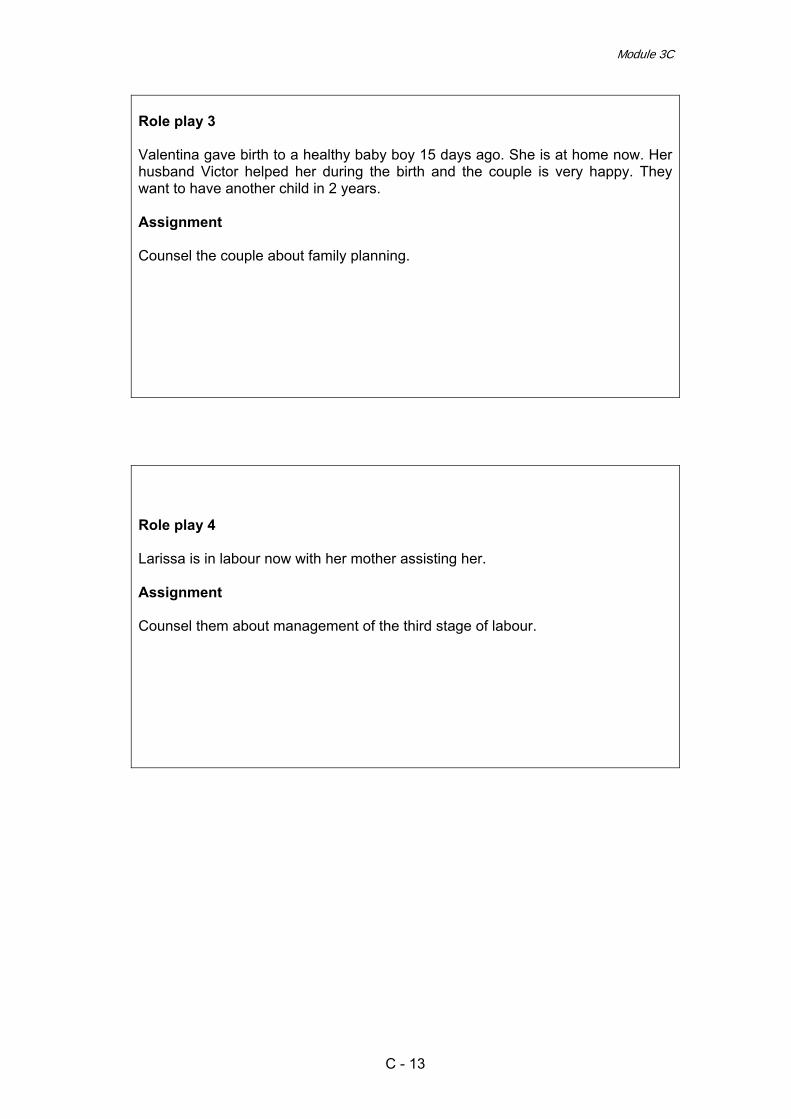
Module 3C
Role play 3 Valentina gave birth to a healthy baby boy 15 days ago. She is at home now. Her husband Victor helped her during the birth and the couple is very happy. They want to have another child in 2 years. Assignment Counsel the couple about family planning.
Role play 4 Larissa is in labour now with her mother assisting her. Assignment Counsel them about management of the third stage of labour.
C - 13

Effective Perinatal Care (EPC)
3C - 14

Effective Perinatal Care (EPC) Facilitator Guide
Module 4C Assessment of Foetal Well-Being during Pregnancy and Labour. Assessment of Small for Gestational Age (SGA) Foetuses
Learning objectives
At the end of the module participants will:
• Be able to define the following: o Intrauterine Growth Restriction (IUGR) o Small for Gestational Age Foetus (SGA) o Nonreassuring Foetal Status
• Understand that the conditions, “foetoplacental deficiency” and “foetal
hypoxia,” are pathophysiological and metabolic processes not subject to diagnostocs. However, they account for some of the main reasons for antenatal hospitalization and unnecessary interventions during pregnancy in Eastern European countries.
• Know the main risk factors for IUGR and conditions which need
dynamic antenatal examination. • Learn and be able to use and interpret correctly main antenatal
diagnostic tests improving perinatal outcomes. Understand clearly their low effectiveness in low risk pregnant women.
• Critically think over the current technologies used for improvement of
foetal well-being in utero and treatment of IUGR. Understand that most of them have low effectiveness and are unsafe.
Module outline and duration: Part I – Classroom work - 60 min
Activity 1 – Introduction 5 min Activity 2 – Work in small groups 10 min Activity 3 – Interactive presentation 40 min Activity 4 – Conclusions 5 min
Part II – Clinical work – 60 min Activity 5 – Revision of the delivery or pregnancy records 60 min
4C - 1

Effective Perinatal Care (EPC)
Preparation for the module
• Learn current practices on the subject in the region • Review new publications on the subject matter • Ensure that the participants from this region will understand the objectives
• Ensure that diagnostic procedures included in the module are used in this
region and that the participants can understand them.
• Ensure that all participants have the Participant Manual
• Ensure that all co-facilitators know their respective functions during work with this module
Materials and Audiovisual Equipment Materials
• Participant manual • Presentation 4C EPC ENG
Equipment • Video or overhead projector • Flip-chart • Colour markers
Key Messages
• Small-for-gestational age foetus (SGA) is defined as small weight or size for definite gestational age
• Intrauterine growth retardation (IUGR) is a sub-group of SGA (30-50%), in
which the foetus has not attained its growth potential. Characterized by high morbidity and mortality
• The conditions, “foetoplacental deficiency”, “foetal hypoxia” and
“asphyxia,” are some of the main reasons for antenatal hospitalization and unnecessary interventions. In many cases they are pathophysiological and metabolic processes and have no clinical significance.
• “Nonreassuring foetal status” is the only appropriate term for the
determination of foetal wellbeing disorders, including foetal heart rate disorders and changes in some biophysical tests, which can lead to neonatal asphyxia.
4C - 2

Module 4С
Key Messages
• To assess foetal wellbeing, biometric and biophysical diagnostic tests are used, but none of them has a prognostic value in women of low risk.
• The majority of perinatal interventions aimed at improving foetal growth
and prevention of foetal acidosis don’t show good results for perinatal outcomes.
• The only treatment for IUGR and “nonreassuring foetal status” is delivery
at the most optimal term and in most optimal way.
PART I – Classroom work
Activity 1 – Introduction (5 min) • Show Slide 4С-1 and explain to participants the learning objective of this
module. Say that such conditions as “foetoplacental deficiency”, “foetal hypoxia” and “asphyxia” are not diagnoses, but at the same time in many countries account for some of the main reasons for antenatal hospitalization.
• Stress that obstetricians and midwives are used to classifying abnormal foetal
conditions with various terms (foetal distress, foetal / perinatal anoxia / hypoxia, foetal growth restriction, foetal impairment, foetal acidemia, fetoplacental deficiency, etc.) on the basis of different tests, following different classifications and strictly relating such foetal “conditions” with neonatal outcomes.
• Tell participants that the purpose of this module is to clarify and revise
definitions and essential tests and therapies for normal and IUGR complicated pregnancies.
Activity 2 – Work in small groups (10min)
• Split the participants into four groups in such a way that every group includes participants of all categories (midwives, paediatric nurses, neonatologists and obstetricians).
• Note that the team approach, including ideas and participation of each group
member, is very important. • Ask the participants from each group to write their answers down on the flip-
chart and decide who will present the results of small group work to the other participants.
• Show Slide 4С-2, which presents the tasks for the small group work. The
number of the group matches the number of the task:
4C - 3

Effective Perinatal Care (EPC)
1. What questions do you ask when assessing foetal well-being? 2. Define the following conditions:
– Intrauterine Growth Retardation (IUGR) – Small for Gestational Age Foetus (SGA)
What are the predisposing factors for IUGR and what prevention methods are you using?
3. Which tests do you perform to diagnose IUGR and SGA? 4. How do you manage IUGR and SGA?
• Facilitator must explain that the reports of each group will be discussed and
then the slides for that topic will be presented.
Activity 3 – Interactive presentation (40 min) • At the beginning of your presentation ask the representative of Group 1 to
present the results of their group work. • Discuss with group members what the aim of each question is (i.e., what we
are expecting to receive for answers to each question). • After the discussion show Slide 4C-3 and ask the participants to summarize
the results of the group 1 discussion of the four questions listed on the slide. List these questions and ask participants if they agree with such questions or they want to add something. If participants have something to add (which on their opinion is very important) write their comments on the flip chart.
• Ask participants, do they have any questions? Answer their questions if they
have any. • Then ask the representative of Group 2 to present the results of their group
work. • Comment on the presentation of Group 2 using Slides 4C-4 tо 4C-10. • Show Slide 4C-4 and present to participants the aim of antenatal care (ANC)
including assessment of foetal well-being. Stress that the aim of ANC – of which assessment of foetal well-being is a component - is assisting women to remain healthy, finding and correcting adverse conditions when present, and aiding in the health of the unborn.
• Go to Slide 4C-5 and give the definition of SGA. Tell participants that SGA
refers to a foetus that has failed to achieve a specific anthropometric or estimated weight threshold by a specific gestational age. The commonly used threshold is the 10th percentile for abdominal circumference and estimated birth weight (as recommended by WHO).
• Note that SGA foetuses are a heterogeneous group comprised of foetuses
that have failed to achieve their growth potential (intra uterine growth restriction – IUGR or foetal growth retardation - FGR) and foetuses that are constitutionally small.
4C - 4

Module 4С
• Use the chart and notes to Slide 4С-6 to explain what the antenatal growth chart and directly 10th percentile are. Tell participants that fundal-symphyseal height measurement with a tape-line and marking its dynamics on an antenatal growth chart is a simple and inexpensive method of choice for ANC. This method allows the diagnosing of small or large foetus size for the corresponding gestational age however does not always indicate pathology.
• Slide 4С-7 is optional. If you decided not to use it in your presentation – hide it
before starting. • If you do decide to use it, use Slide 4С-7 to show the connection between
birth weight and perinatal mortality and morbidity. Note that not all babies with low birth weight have an increased risk of morbidity and mortality.
• Explain to the participants that there are two different definitions: small-for-
gestational age foetus (SGA) and intrauterine growth retardation (IUGR). The first condition in 50% is normal and babies will be healthy. The second condition is a sub-group of the first one, but the majority of those babies will be sick and have a high chance of dying (Slide 4С-8). The only problem is the following: when we diagnose “SGA” we don’t know if this is IUGR or the baby is small by constitution. To differentiate between the two, we need to use biophysical tests, which will give us information about foetal well-being.
• Go to Slide 4С-9 and make a conclusion on factors predisposing to IUGR. Tell
participants that there are several classifications of predisposing factors for IUGR, but the key ones are divided into 4 groups: maternal, placental, environmental, and hereditary or foetal.
• Emphasize the fact that there are many risk factors and they fall into 4 groups.
The most important risk factors which can lead to foetal well-being disorders and require careful antenatal care are:
o Pregnancy over 41 weeks o Preeclampsia / chronic hypertension o Diabetes o Multiple pregnancy o Infections in pregnancy o Decompensated chronic diseases o Premature Rupture of Membranes PROM
• Discuss with the group measures proposed by them for the prevention of
IUGR. Say that there are only 5 technologies, which could improve perinatal outcomes in the case of IUGR. List them using Slide 4C-10.
• Ask participants, do they have any questions? Answer their questions if they
have any. • Then ask a representative of Group 3 to present the results of their group
work. • Use Slides 4C-11 through 4C-26 for a short presentation on diagnostic tests. • Show Slide 4C-11 and list the routine tests for assessment of foetal well-
being.
4C - 5

Effective Perinatal Care (EPC)
• Tests used in ANC to assess foetal well-being: o Assessment of foetal activity o Auscultation of foetal heart beat o Ultrasounds (US)
• Go to Slide 4C-12 and tell participants that maternal recognition of decreased
foetal movement has long been used during antenatal care in an attempt to identify the jeopardized foetus and intervene to prevent death. Given the low prevalence of foetal compromise and an estimated specificity of 90% to 95%, the positive predictive value of the maternal perception of reduced foetal movements for foetal compromise is low, 2% to 7%. Following a reduction in foetal movements, women should be advised to contact their midwife or hospital for further assessment. The evidence does not support the routine use of formal foetal movement counting to prevent late foetal death.
• Show Slide 4C-13. Auscultation of the foetal heart may confirm that the foetus
is alive but is unlikely to have any predictive value and routine listening is therefore not recommended. However, when requested by the mother, auscultation of the foetal heart may provide reassurance.
• Go to Slide 4C-14 and say that routine ultrasound in early pregnancy (before
24 weeks) is effective in assessing gestational age, early detection of multiple pregnancies and early detection of clinically unsuspected foetal malformation at a time when termination of pregnancy is possible. Routine late pregnancy ultrasounds in low-risk women or unselected populations, does not benefit the mother or the baby.
• Show Slide 4C-15 and list the tests used in ANC to detect SGA. • Tests used in ANC to detect SGA
o Abdominal palpation o Fundal height o Ultrasound biometry o Biophysical tests to diagnose SGA/IUGR
• Go to Slide 4C-16 and tell participants that abdominal palpation has limited
diagnostic accuracy to predict an SGA foetus. • Show Slide 4C-17. Say that fundal height (FH) measurement has limited
diagnostic accuracy to predict an SGA neonate. Use of a customised fundal height chart improves accuracy to predict an SGA fetus.
• Stress that a series of measurements (fundal-symphyseal height
measurement chart) increases the sensitivity and specificity of this method. • Show Slide 4C-18 and ask the participants to answer the following questions:
o Is IUGR present in these cases? o In which case is the risk of IUGR higher?
• Remember that usually participants define IUGR in both cases and prognosis
is worse in the first one. Facilitator can count votes for different options. • Then show Slide 4C-19, where both cases are plotted on the antenatal growth
chart (the first one in blue, the second one in red). Note that it’s impossible to
4C - 6

Module 4С
define IURG without graphically plotting the values of fundal-symphyseal height measurements.
• Give special attention to the fact that growth dynamics are more important
than the values of fundal-symphyseal height according to the term. • Go to Slide 4C-20 and tell participants that abdominal circumference (AC) and
estimated fetal weight (EFW) are the most accurate diagnostic measurements to predict SGA. In high-risk women, AC at less than the tenth percentile has sensitivities of 72.9–94.5% and specificities of 50.6–83.8% in the prediction of foetuses with birth weight less than the tenth percentile. The respective figures for EFW have sensitivities of 33.3–89.2% and specificities of 53.7–90.9%.
• Use the notes to this slide to explain all points listed on the slide. • Show Slide 4C-21 and say that all biophysical tests, including amniotic fluid
volume (AFV), Doppler, cardiotocography and biophysical scoring, are poor at diagnosing a small or growth-restricted foetus. The diagnostic accuracies of AFV and uterine artery Doppler are given below as examples of limited accuracy of biophysical tests in diagnosing SGA/FGR.
• Stress that a distinction needs to be made between biometric tests (i.e. tests
to measure size) and biophysical tests (i.e. tests to assess fetal well-being). Biometric tests are designed to predict size and if performed longitudinally growth but not well-being. Biophysical tests, on the other hand, are not designed to predict size but fetal well-being.
• Show Slide 4C-22 which presents the list of the tests for assessment and
surveillance of suspected SGA. • Tests used in ANC for surveillance of suspected SGA
o Antenatal cardiotocography (non-stress test) o Biophysical profile of the foetus o Doppler velocimetry of umbilical artery
• Discuss each test and give the description of its effectiveness based on
scientific evidence (using Slides 4C-23 – 4C-25). • Say that all these tests have low prognostic value in pregnant women of low
risk and have no influence on perinatal outcomes. Performing these tests on in women of high risk has a positive effect. At the same time, interpretation of the test results, such as CTG and Doppler, need to be done by highly qualified and experienced medical staff only.
• Go to Slide 4C-23 and say that the use of cardiotocography (CTG)
antepartum to assess fetal condition is not associated with better perinatal outcome; in fact, a systematic review of randomised trials showed that there was a trend toward increased mortality in the group receiving CTG compared with those who did not. Computer systems for interpretation of CTG have better accuracy than clinical experts in predicting umbilical acidosis and depressed Apgar scores. However, further evaluation of this technology is required before clinical recommendations could be made regarding its widespread use.
4C - 7

Effective Perinatal Care (EPC)
• Show Slide 4C-24. Tell participants that conduction of BPF does not result in any improvements of the outcomes.
• Go to Slide 4C-25 and tell participants that a systematic review (meta-
analysis) has provided compelling evidence that the use of umbilical artery Doppler to monitor high-risk foetuses reduces perinatal morbidity and mortality. In addition, there was a significant reduction in the number of antenatal admissions and inductions of labour associated with Doppler use.
• Stress that if indicated (suspect of SGA) the use of Doppler velocimetry of the
umbilical artery is the primary surveillance tool. • Show Slide 4C-26 and say that in those cases when an anomaly scan and
umbilical artery Doppler are normal, the small foetus is likely to be a ‘normal small fetus’. Evidence suggests that outpatient management of such foetuses is safe. In addition, a randomised controlled trial of two regimens of foetal surveillance for SGA foetuses with normal umbilical artery Doppler found that twice-weekly compared with fortnightly monitoring resulted in earlier deliveries and more inductions of labour with no difference in neonatal morbidity. This suggests frequency of monitoring in SGA foetuses with normal Doppler need not generally be more than once every fortnight.
• Ask participants, do they have any questions? Answer their questions if they
have any. • Then ask the representative of Group 4 to present the results of their group
work. • Use Slides 4C-27 - 4C-35 for a short presentation on assessment of foetal
well-being during delivery and effective management of SGA and IUGR. • Show Slide 4C-27 and tell participants that in the case of severe IUGR and
severe foetal impairment, emergency delivery is the therapy of choice. In the case of IUGR in a compensated stage, periodic assessment of foetal well-being is necessary.
• The purpose of assessment is to timely identify the development of the
conditions threatening the life of the foetus. In such conditions an emergency pre-term delivery is the best choice to maintain the life of the foetus and prevent morbidity, rather than prolongation of pregnancy when being in utero becomes more and more dangerous.
• Go to Slide 4C-28 and present the purpose of intrapartum assessment of
foetal well-being. • Go to Slide 4C-29. List the indications for continuous electronic foetal heart
monitoring. Explain to participants which problems on behalf of mother and foetus can require the continuous electronic monitoring of foetal heart beat.
• Go to Slide 4C-30 and present participants the cardiotocograph (CTG)
classification of not reassuring categories of a foetal heart beat rate. Ask participants to look through the table in the notes to this slide. Discuss with them foetal heart-beat feature classifications.
4C - 8

Module 4С
• Then show Slide 4C-31 and say that in cases where the CTG falls into the suspicious category, conservative measures should be used.
• Note that in cases when the CTG falls into the pathological category,
conservative measures should be used and foetal blood should be sampled where appropriate/feasible. In situations where foetal blood sampling is not possible or appropriate delivery should be expedited.
• Show Slides 4C-32 – 4C-34. • Tell participants that the Committee on Obstetric Practice is concerned about
the continued use of the term “foetal distress” as an antepartum or intrapartum diagnosis, and also about the use of term “birth asphyxia” as a neonatal diagnosis.
• The Committee reaffirms that the term “foetal distress” is imprecise and non-
specific. The communication between clinicians caring for the woman and those caring for her neonate is best served by replacing the term “foetal distress” with “nonreassuring foetal status”, followed by a further description of findings (e.g., repetitive variable decelerations, foetal tachycardia or bradycardia, late decelerations, or low biophysical profile).
• The term has a low positive predictive value even in high-risk populations and
is often associated with an infant who is in good condition at birth as determined by the Apgar score or umbilical cord blood gas analysis or both.
• In the past the term “foetal distress” generally referred to an ill foetus, but the
term “nonreassuring foetal status” describes the clinician’s interpretation of data regarding foetal status (i.e. the clinician is not reassured by the findings). This acknowledges the imprecision inherent in the interpretation of the data. Therefore, the diagnosis of “nonreassuring foetal status” is consistent with the delivery of a vigorous neonate.
• Foetoplacental deficiency identified by an ultrasound examination is one of the
main justifications for hospitalizing women in departments of pregnancy pathology in post-Soviet countries.
• A disease named “foetoplacental deficiency” does not exist; this is a
pathological physiological process. “Newborn hypoxia” is not a clinical condition; it is a metabolic process (metabolic acidemia) that is confirmed by blood gas analysis. Intrauterine growth retardation is one of the key conditions indicating possible hypoxia and foetoplacental deficiency. IUGR is the most objective marker of decompensated foetoplacental deficiency.
• One definition of foetoplacental deficiency:
o A pathologic state when the ability of the placenta to maintain adequate metabolism between the mother and the foetus decreases, thus impairing the metabolic, trophic, endocrinal, transportation, barrier and gas exchange functions of the placenta and the foetus.
• Slide 4C-33. The essential criteria of the newborn’s response to asphyxia of
such a degree as to be likely to cause harm are: • Apgar score of 0 to 3 for ≥5 minutes; • Neonatal neurologic consequences (e.g. hypotonia,seizures,coma);
4C - 9

Effective Perinatal Care (EPC)
• Evidence of multi-organ system dysfunction in the immediate neonatal period;
• Umbilical cord arterial pH < 7.0; and • Umbilical cord arterial base deficit > 16 mmol/L.
• All of these conditions must be present to diagnose hypoxic acidemia. If they
are not all present, one cannot conclude that hypoxic acidemia existed or had the potential to cause neurologic deficits.
• Slide 4C-34. Tell participants that “intrapartum asphyxia” is an “a posteriori”
criteria. Obstetricians can only suspect a “not reassuring foetal status” on the basis of “a not reassuring or abnormal categories of FHR”. Stress that the most appropriate term for determination of foetal well-being disorders is “not reassuring foetal status”.
• Go to Slide 4C-35. Present participants the full standard observations of the
foetus that are recommended during delivery by the Scottish Ob Gyn College: 1. Women in active labour should receive continuous close support
from an appropriately trained professional. One-to-one nursing is recommended. (I-A)
2. Intermittent auscultation following an established protocol of
surveillance and response is the preferred method of foetal surveillance in healthy pregnancies during the active phase of labour. (I-A)
3. Labour induction requires close monitoring of uterine activity and
the foetal heart rate. (III-B)
4. If intermittent auscultation detects characteristics of abnormal foetal heart rate and the foetus is unresponsive to resuscitative measures, increased surveillance by continuous electronic foetal monitoring or foetal scalp sampling or delivery should be instituted. (I-A)
5. Continuous intrapartum electronic foetal monitoring is
recommended: a) For pregnancies where there is an increased risk of
perinatal death, cerebral palsy, or neonatal encephalopathy. (III-C)
b) When oxytocin is being used to augment labour. (1-A)
c) When oxytocin is being used to induce labour. (III-C)
6. With respect to continuous electronic foetal monitoring, all
professionals must be familiar with the paper speed used in each case to avoid misinterpretation. The correct time should be recorded on the electronic foetal monitoring record. (III-C)
7. Electronic foetal monitoring records should be inspected and
documented every 15 minutes in the active phase of labour and at least every 5 minutes in the second stage of labour. (III-C)
4C - 10

Module 4С
8. The timing of electronic fetal monitoring patterns should be determined in association with uterine contractions. Contraction frequency, duration, intensity, and resting tone should be assessed and documented. Abdominal palpation, a tocodynamometer, or an intrauterine pressure catheter may be used to facilitate the assessment. (III-C)
9. Practitioners should use standard terminology when describing
foetal heart rate characteristics of an electronic foetal monitoring record. (III-C)
10. Foetal scalp blood sampling is recommended when electronic
foetal monitoring patterns are uninterpretable or non-reassuring, such as sustained minimal or absent variability, uncorrectable late decelerations, increasing foetal tachycardia, and abnormal FHR characteristics on auscultation. (II-3B)
11. The limited knowledge available on the use of labour admission
tests warrants further research to establish the usefulness of this screening approach. (III-C)
• Answer any questions the participants have about this module.
Activity 5 - Conclusion (5 min) • Ask participants to share their conclusions about the subject matter. • Finish this part of the module by showing the conclusions listed on Slide 4C-
36.
4C - 11

Effective Perinatal Care (EPC)
PART II – Clinical Work
Activity 5 – Revision of delivery or pregnancy records (diagnostic, treatment, management) with diagnoses of: IUGR, “chronic foetal hypoxia”, “chronic foetoplacental deficiency” and/or “foetal distress” • During the clinical week discuss with the facility management the possibility of
working with women’s records and their anonymous analyses. • In the Department of Pregnancy Pathology and/or the Postpartum Department
select women’s records that have diagnoses of: IUGR, “chronic foetal hypoxia”, “chronic foetoplacental deficiency” and/or “foetal distress”
• Divide the participants in 2-3 groups and give them the selected records for
analysis. • Participants should analyze pregnancy and delivery records and evaluate the
effectiveness of diagnostic tests, discuss possible schemes of treatment and recommend further management of the case.
• After the above activity discuss all cases with the entire group of participants. • End the session by coming to a conclusion on each case and once again
listing effective diagnostic and treatment methods in the case of IUGR and/or a non-reassuring foetal status.
4C - 12

Module 4С
Selected references for the module 1. What is the effectiveness of antenatal care? WHO Regional Office for Europe -
Health Evidence Network report 2005 http://www.euro.who.int/Document/E87997.pdf 2. Royal College of Obstetricians and Gynaecologists The investigation and
management of the small for gestational age fetus December 2001 http://www.rcog.org.uk/resources/public/pdf/small_gest_age_fetus_no31.pdf 3. Vandenbosche RC, Kirchner JTR Intrauterine growth retardation Am Fam
Physician. 1998 Oct 15;58(6):1384-90 4. PERINATAL EDUCATION PROGRAMME ROUTINE USE OF THE
ANTENATAL CARD - SKILLS WORKSHOP 2 Geneva Foundation for Medical Education and Research http://www.gfmer.ch/PEP/pdf/SW-2-2005.pdf
5. Bernstein IM, Horbar JD, Badger GJ, Ohlsson A, Golan A Morbidity and
mortality among very-low-birth-weight neonates with intrauterine growth restriction. The Vermont Oxford Network Am J Obstet Gynecol. 2000 Jan;182(1 Pt 1):198-206
6. National Institute for Clinical Excellence Antenatal care Routine care for the
healthy pregnant woman (full version) Clinical Guideline# 6 October 2003 7. Parer JT, King T Fetal heart rate monitoring: is it salvageable? Am J Obstet
Gynecol. 2000 Apr;182(4):982-7 8. SOGC Clinical Practice Guidelines NO. 112 Fetal Health Surveillance In
Labour 2002. http://www.sogc.org/guidelines/public/112E-CPG1-March2002.pdf http://www.sogc.org/guidelines/public/112E-CPG2-April2002.pdf
9. Persson B, Stangenberg M Prediction of size of infants at birth by
measurement of symphysis fundus height BJOG 93: 206-211 1986 10. Neilson JP Symphysis-fundal height measurement in pregnancy (Cochrane
Review) In: The Reproductive Health Library, Issue 8, 2005 11. Pearce JM, Campbell S A comparison of symphysis-fundal height and
ultrasound as screening tests for light-for-gestational age infants BJOG 94: 100-104 1987
12. Gardosi, Jason & Francis, Andre Controlled trial of fundal height measurement
plotted on customised antenatal growth charts BJOG 106: 309-317 1999 13. Chang TC, Robson SC, Boys RJ, Spencer JA Prediction of the small for
gestational age infant: which ultrasonic measurement is best? Obstet Gynecol. 1992 80(6):1030-8.
14. Bricker L, Neilson JP Routine ultrasound in late pregnancy (after 24 weeks
gestation) (Cochrane Review) Cochrane Database Syst. Rev. 2006
4C - 13

Effective Perinatal Care (EPC)
15. Ferrazzi E, Bellotti M, Vegni C, Barbera A, Della Peruta S, Ferro B, et al Umbilical flow waveforms versus fetal biophysical profile in hypertensive pregnancies. Eur J Obstet Gynecol Reprod Biol 1989;33:
16. Harrington K, Kurdi W, Aquilina J, et al A prospective management study of
slow-release aspirin in the palliation of uteroplacental insufficiency predicted by uterine artery Doppler at 20 weeks . Ultrasound Obstet Gynecol 2000 15(1): 13-8
17. Hofmeyr GJ, Kulier R Piracetam for fetal distress in labour (Cochrane Review)
Cochrane Database Syst. Rev. 2006 18. RCOG Royal College of Obstetricians and Gynaecologists The Use of
Electronic Fetal Monitoring - The use and interpretation of cardiotocography in intrapartum fetal surveillance Evidence-based Clinical Guideline Number 8 - 2001 www.rcog.org.uk/resources/public/pdf/efm_guideline_final_2may2001.pdf www.rcog.org.uk/resources/public/pdf/efm_algorithm_final_1may2001.pdf
19. American College of Obstetricians and Gynecologists Committee on Obstetric
Practice ACOG Committee Opinion #303: Inappropriate use of the terms fetal distress and birth asphyxia Obstet Gynecol. 2004; 104(4):903
20. McIntire DD, Bloom SL, Casey BM, Leveno KJ. Birth weight in relation to
morbidity and mortality among newborn infants N Engl J Med. 1999 Apr 22;340(16):1234-8
21. Harper T & Garret L Fetal Growth Restriction. August 2005.
http://emedicine.com/MED/topic3247.htm
4C - 14

Module 4С
Attachment 1 Activity 2
Group 1 What questions do you ask when assessing foetal well-being?
Group 2 Define the following conditions:
o Intrauterine Growth Retardation (IUGR) o Small for Gestational Age Foetus (SGA)
What factors predispose one to IUGR and what methods of prevention are you using?
Group 3
Which tests do you perform to diagnose IUGR and SGA?
Group 4 How do you manage IUGR and SGA?
4C - 15

Effective Perinatal Care (EPC)
4C - 16

Effective perinatal care (EPC) Facilitator Guide
Module 5C
Management of Normal Labour and Birth
Learning objectives: By the end of the module, the participants will:
• Question any activity performed in labour without clear justification • Recognize the importance of providing comprehensive support to the
woman and family in childbirth • Be familiar with the WHO recommendations on labour and birth
management • Be able to offer non-pharmacological pain relief and different positions for
labouring women
Module structure and duration: Part I – Classroom work – 270 minutes
Activity 1 – Introduction 20 min Activity 2 – Interactive presentation 230 min Activity 3 – Conclusion 20 min
Part II – Clinical sessions Activity 4 – Practice with a manikin “Active Management of the Third Stage of Labour” Activity 5 – Discussion and preparation of the clinical week Activity 6 – Preparation of the individual birth rooms
60 min 60 min -----
Preparation to the Module • Review the current publications, evidence-based articles and
recommendations on normal labour and birth management • Review non-pharmacological methods of pain relief • Ensure that 10 copies of “A Guide to Effective Care in Pregnancy and
Childbirth” by M. Enkin are available • Ensure that all the facilitators know their functions in this module
5C - 1

Effective Perinatal Care (EPC)
Materials and Equipment Materials
• Participant’s manual • Reference book by M. Enkin “A Guide to Effective Care in Pregnancy and
Childbirth” (10 copies) • Power Point Presentation 5C EPC ENG • List of technologies printed out on separate sheets, to be used in group
work
Equipment • LCD-projector or slide projector • Flipchart • Markers
Key Messages
• Good quality of perinatal care means that physical complications (mortality and morbidity) avoided and psycho-emotional needs of the woman and the family are met.
• For the majority of women and families the latter may matter more, taking
into account the fact that the major part (85%) of all birth are normal and need only support and observation rather than medical interventions.
• There is no evidence of the effectiveness of many interventions /
technologies used to improve the outcome / prevent complications; despite the absence of the proof of effectiveness, many painful and humiliating forms of care are still used.
• The most effective birth interventions are simple, cost-effective and contain
minimal risk – such as partner involvement in delivery, skin-to-skin contact, ability of the woman to move freely in childbirth, vertical positions for birth, etc.
• Continuous support of the woman during labour and birth is one of the
most effective interventions, thus it must be fully encouraged by the staff. • There is no clear link between duration of the second stage of labour
and neonatal morbidity. If maternal and foetal status are stable, the length of the second stage, even if exceeding certain limits, is not an indication for interventions.
• Moreover, technologies / interventions used to shorten the duration of the
second stage (directed pushing, breath holding, early pushing, fundal pressure/ Kristeller maneuver) may affect the maternal and foetal status and lead to lacerations of the mother’s birth canal.
• The supine position is the least favourable position for birth, and the
mother should be informed about this. Vertical positions are more beneficial for delivery than horizontal. Woman should be encouraged to give birth in a vertical position.
5C - 2

Module 5C
5C - 3
• Guarding of the perineum has very few advantages to the “hands off”
tactics; the use of this procedure should not interfere with free adoption of the position.
• Restrictive episiotomy routine use of episiotomy is not encouraged. Restrictive use of episiotomy might have a number of advantages.
• According to the WHO definition, a safe delivery is the one that is clean and is carried out by someone who has the necessary skills, and with access to emergency care if necessary.
• Normal birth should be managed by a midwife; specialist’s skills are
necessary only if complications arise.
It is preferable if the session is conducted by a midwife assisted by an obstetrician-gynaecologist and a neonatologist.
Part I – Classroom work
Activity 1 – Introduction (20 min) • Show Slide 5C-1 and tell the participants that while working on the module,
you will discuss the principles of normal labour and birth management and assess their effectiveness based on medical evidence.
• Discuss the learning objectives with the participants.
• Go to Slide 5C-2 and tell the participant that lately women tend to have fewer
infants than before, and that the last several decades have brought along a change in the attitude toward perinatal care. In addition there have been many procedures that have improved perinatal care over the past decades.
• Show Slide 5C-3 “Women’s experiences of birth” and comment on it. Summarize that despite the use of many interventions and the decrease in the number of pregnancy and birth complications, many women are still unhappy with the quality of care and lack of a human touch. .
• Go to Slide 5C-4 and note that quality research showed the ineffectiveness of
many interventions routinely used in childbirth (the majority of which are painful and humiliating); moreover, some of them are harmful rather than beneficial. This is the reason why many women emerge from the maternity with the desire “not to have another child again” and “never go through this again”.
• Show Slides 5C-5 and 5C-6 listing the key factors of the women’s satisfaction
with birth, and factors which are much less important though health professionals award them unjustified significance.
• Go to Slide 5C-7 and tell the participants that over 20 years ago, the
Conference in Fortaleza, Brazil, made a number of recommendations about

Effective Perinatal Care (EPC)
appropriate practices during childbirth and proposed to discontinue those which effectiveness was not supported by evidences, and do not respect women's dignity and psychological needs. These recommendations are still valid and implemented by many health systems.
Activity 2 – Interactive presentation (230 min) • Ask two course participants to role play that they are pregnant women. Give
them the list of procedures / interventions prepared in advance, and ask them to answer the following question after reading through the list: “Which of the listed procedures would you like to be performed and which ones you would reject?” Remind the participants that they should try to answer these questions from the client’s view point, not from the point of view of the health professionals. Group assignment: Role play that you are a pregnant woman who is being admitted to the maternity, or members of her family. Select from the list the technologies / procedures which you would prefer to be performed at admission to the maternity or in delivery, and which ones you would refuse. Give the reasons for your acceptance of rejection of a procedure.
List of procedures: 1 Routine pubic shaving 2 Routine enema 3 Labour and birth in an individual room 4 Provided with the maximum of information and is involved in decision
making 5 Low risk birth is managed by a midwife 6 Companion presence at delivery 7 Restriction of food and liquid consumption during labour and birth 8 Ambulation and free choice of birth position 9 Non-pharmacological methods of pain relief 10 Routine cardiotocography in the first stage of labour 11 Supine position with the feet brought up to the stomach (lithotomic
position) in the second stage of labour 12 Arbitrary limitation of the duration of the second stage of labour 13 Routine directed pushing and breath holding in the second stage of labour 14 “Ironing-out” and massage of the perineum in the second stage of labour 15 Guarding of the perineum 16 Routine use of episiotomy 17 Ice pack put on the woman’s lower abdomen after the birth 18 Bladder catheterization after birth 19 Routine treatment of vagina with antiseptics after birth 20 Active management of the third stage of labour
• Split the rest of the participants into 10 small groups. Give one copy of M. Enkin’s “A Guide to Effective Care in Pregnancy and Childbirth” to each group as well as the group assignments and the list of procedures/interventions printed out on separate sheets of paper.
5C - 4

Module 5C
5C - 5
Group 1 Using the tabulated synopsis, find in the reference book by Enkin the information about the effectiveness of the following procedures:
1 Routine pubic shaving 2 Active management of the third stage of labour
Group 2 Using the tabulated synopsis, find in the reference book by Enkin the information about the effectiveness of the following procedures:
1 Routine enema 2 Routine treatment of vagina with antiseptics after birth
Group 3 Using the tabulated synopsis, find in the reference book by Enkin the information about the effectiveness of the following procedures:
1 Birth in an individual labour and birth room 2 Bladder catheterization
Group 4 Using the tabulated synopsis, find in the reference book by Enkin the information about the effectiveness of the following procedures:
1 The woman is provided with maximum information, involved in decision making
2 Ice pack put on the woman’s lower abdomen
Group 5 Using the tabulated synopsis, find in the reference book by Enkin the information about the effectiveness of the following procedures:
1 Low risk births are managed by midwives 2 Routine episiotomy
Group 6 Using the tabulated synopsis, find in the reference book by Enkin the information about the effectiveness of the following procedures:
1 Companion presence during labour and birth 2 Guarding of the perineum

Effective Perinatal Care (EPC)
Group 7 Using the tabulated synopsis, find in the reference book by Enkin the information about the effectiveness of the following procedures:
1 Restriction of food and liquid consumption in delivery 2 “Ironing out” and massage of the perineum in the second stage of labour
Group 8 Using the tabulated synopsis, find in the reference book by Enkin the information about the effectiveness of the following procedures:
1 Ambulation and free choice of position in the first stage of labour 2 Routine directed pushing and breath holding in the second stage of labour
Group 9 Using the tabulated synopsis, find in the reference book by Enkin the information about the effectiveness of the following procedures:
1 Alternative (non-pharmacological) methods of pain relief 2 Arbitrary limitation of the duration of the second stage of labour
Group 10 Using the tabulated synopsis, find in the reference book by Enkin the information about the effectiveness of the following procedures:
1 Routine cardiotocography in the first stage of labour 2 Supine position with the feet brought up to the stomach (lithotomic
position) in the second stage of labour
• Ask the participants to find data about the effectiveness of the procedures
listed on the separate sheets of paper which they received. The participants should look for such information in the synopsis during 20 minutes. Also ask the participants to read more about these procedures in the relevant chapters of the book.
• After the participants finish searching and reading the evidence presented in
the “Guide…” by M. Enkin, proceed as follows: • Ask the participants of the “client” group to present to the other trainees the
procedures/interventions listed on the sheet of paper they received, and comment on them. Would they like this procedure to be performed to them upon admission to the maternity / in labour?
• After the “clients” commented on each procedure / intervention, ask those
participants who were searching for the evidence of their effectiveness to present the search results.
5C - 6

Module 5C
5C - 7
• Provide additional information on each procedure / intervention using the slides:
• Routine pubic shaving – Slides 5C-8 and 5С-9 • Routine enema – Slides 5C-10 and 5С-11 • Birth in an individual labour and birth room – Slides 5C-12 to 5С-15.
• Show Slide 5C-16 and comment on the WHO definition of safe delivery. Show
Slide 5C-17 and draw the attention of the participants to the idea of “Clean delivery”.
• The woman is provided with maximum information and involved in decision-
making - Slides 5C-18 and 5С-19 • Low risk birth is managed by a midwife – Slides 5C-20 and 5С-21 • Companion presence during labour and birth – Slides 5C-22 and 5С-23. Tell
the participants that considering the benefit associated with this intervention, many professional associations recommend partners as the companions the for labour and birth. Thus a partner companion is actively encouraged as a key method of improving maternal and infant health.
• Restriction of food and liquid consumption in labour – Slide 5C-24 • Ambulation and free choice of position in the first stage of labour - Slides 5C-
25 to 5С-30 • Non-pharmacological methods of pain relief – Slides 5C-31 to 5С-33. The
degree of pain perceived by a woman in childbirth depends on her emotional state and cultural expectations. Her pain is less when she feels relaxed, unafraid, and reassured by the continuous, comforting support of her companion and/or birth attendant. Non-pharmacological methods of pain relief may help many women to cope with the pain without any risk of side effects or complications associated with analgesia and anesthesia. Note that non-pharmacological methods of pain relief are not as effective for decreasing pain as an epidural or other pharmacological methods. Show to the participants the different methods of non-pharmacological pain relief (5-7 minutes): counter pressure, massage, etc.
• Routine cardiotocography in the first stage of labour – Slides 5C-34 to 5С-35 • Supine position with the feet brought up to the stomach (lithotomic position) in
the second stage of labour – Slides 5C-36 to 5С-41. Stress that one of the key advantages of the vertical position is the decrease in the rate of abnormal foetal heartbeat patterns. Note that though an upright position of the woman may cause some inconvenience for the healthcare provider, women are very satisfied with this position.
• Arbitrary limitation of the duration of the second stage of labour – Slides 5C-
42 to 5С-44. Tell the participants that results of studies support the assumption that labour should not be terminated or augmented simply because an arbitrary period of time has elapsed in the second stage.

Effective Perinatal Care (EPC)
• Routine directed pushing and breath holding in the second stage of labour –
Slides 5C-45 and 5С-46. Stress that the only advantage of directed pushing is shortening of the second stage of labour, while the disadvantages are clinically much more significant. Emphasize that directed pushing in combination with breath holding may have adverse effects on the maternal and foetal status.
• Guarding of the perineum - Slides 5C-47 and 5С-48. Explain to the
participants the results of the randomized controlled trials evaluating the benefits of perineum care and emphasize that this technology must be applied to 33 women to prevent one case of pain. At the same time, there was only 20% increase in the number of episiotomies in the hands-poised group. Ask the participants which outcome matters more.
• Routine use of episiotomy – Slides 5C-49 and 5С-50 • Active management of the third stage of labour – Slides 5C-51 to 5С-57 • Intervention of unproven effectiveness – Slide 5C-58. The participants cannot
find the evidence for putting ice packs on woman’s lower abdomen to prevent post-partum haemorrhage, urinary bladder catheterization after each delivery, or routine treatment of vagina with antiseptics after delivery to prevent infection. Ask them if it is necessary to perform these inconvenient procedures with no proof of effectiveness for the mother and baby contact and if fact, are unnecessary interference with the mother and baby.
• Having presented the slides on each procedure/intervention, ask the
participants what they think about the effectiveness of this procedure / intervention. Discuss all the questions that the participants might have on each procedure / intervention.
• Upon completion of this activity, ask the participants if they have any questions
Discuss all issues that might arise. Activity 3 – Conclusion • Show Slides 5С-59 to 5С-62 listing all the essentials of management of all
stages of labour and summarize main points of the module. • Emphasize that during the clinical week you will come back to this module and
will work on the practical skills of active management of the third stage of labour on the mannequin.
• Finish working with this module by asking the participants if they have any
questions or issues to discuss.
5C - 8

Module 5C
5C - 9
Part II – Clinical work Activity 4 – Practice on a mannequin “Active management of the third stage of labour” (60 min) • Ask 2-3 participants (preferably, midwives) to describe the key steps of active
management of the third stage of labour and show them on a mannequin. • Then show Slides 5С-52 – 5С-54 and repeat the key points and steps of the
active management. • Show Slides 5С-55 - 5С-57 listing the benefits and disadvantages of the
active management. Ask the participants if everything is clear and answer any questions that they ask.
• Show all the steps of the active management on the mannequin. • Ask 2-3 other participants to perform all the steps on the mannequin again.
Ask the group if anyone would also like to try to do the same thing. Try to involve as many participants as you can in working on a mannequin.
• Tell the participants that one of the most essential steps of the active
management of the third stage of labour is informed consent of the patient. • Ask 2-3 participants to play roles in counselling a woman in the first stage of
labour or in the antenatal clinic, on different tactics of managing the third stage of labour.
• Recall the main steps of correct counselling. • Ask the participants to evaluate counselling both from the point of view of the
process of counselling itself, and information provided to the patient. • Split the participants into 2 groups. • Ask each group to draft a form to record the woman’s informed consent for
active management of the third stage of labour. • Give each group 5 minutes to present their draft of written informed consent
form. • Conduct panel discussion on what an appropriate consent form should
include. • Perform correct counselling on the active management of the third stage of
labour if the participants want it. • Tell the participants that during the clinical week they will have an opportunity
to counsel women on this issue and manage the third stage of labour actively if the woman gives her consent.

Effective Perinatal Care (EPC)
Activity 5 – Discussion and preparation of the clinical week (60 min) • Split the participants (obstetrician-gynaecologists and midwives) into two
groups. Try that each group has an equal number of midwives and doctors. • If participants are coming from different facilities, then try to split them so that
the participants from one facility are in different groups. You can explain that by splitting people from the same facility, the participants are exposed to colleagues with different experiences than their own.
• Tell the participants how the clinical practice sessions will be organized, their
goals and objectives. • The total number of hours worked by each group will be determined by the
scheduling option chosen for the clinical week (See the Director’s Guide) • Introduce the participants to the work plan for the clinical week. • Explain to the group that every morning both groups will, together with the
neonatologists, discuss the events of the previous day – both classroom and clinical work. The focal points of the discussions will be birth, postpartum care and newborn care.
• On the first day of the duty shift all the participants should be engaged in
preparing the individual labour and birth rooms (Activity 6). After the rooms are prepared, the group of neonatologists will assess their readiness.
• If the 12 hour shift option is selected: Tell the trainees that the labour and
births will be managed by the participants from the group working a 12 hour shift, and discuss the possibility of the delivery managing team working overtime if the delivery continues past 9 p.m.
• Each group should be split into teams (depending on the quantity and
composition), consisting of 1-2 midwives and 1-2 doctors. 1 midwife will be the leading midwife (the one managing the labour and birth), the other midwife – assisting with labour support. 1 doctor will be the physician consultant for the lead midwife. Together they will review the partograph, especially if the alert line crossed, and determine the appropriate actions or interventions. The other doctor will either observe the labour, or provide labour support if the woman wants it. The consultant midwife and consultant physician will provide clinical guidance and feedback to the participants.
• Remind the participants about the importance of privacy, confidentiality and
respect of the woman’s feelings and requests. • Tell the participants that, despite their desire to observe the labour and birth
only the participant midwife/physician team caring for the woman may be present in the birth room.
• The Course Director, in preparation for the clinical week, will have a discussion
with the maternity administration and clinical staff. Decisions will be made regarding appropriate women for care, record keeping, clinical responsibilities of the teams, communication and interaction with the working maternity staff,
5C - 10

Module 5C
5C - 11
how complications and interventions may be handled, transfer of care to the maternity staff, and true emergencies. Discuss with the participants the issue whether it will be the midwife or the doctor to perform the vaginal examination and to keep a record in the delivery file. The lead midwife will complete the partograph and use this tool for management decisions and consultation
• Emphasize that facilitators and trainees are “guests” here using their facility for
the training. Good communication and interactions with the working maternity staff will enhance the clinical experience.
• Ask the participants to bring a change of shoes and a set of working gowns for
the clinical week, if possible. Activity 6 – Preparation of individual labour and birth rooms • Split the participants into groups, depending on the situation in the maternity:
(1) the number of groups may correspond to the number of birth rooms which are planned to be re-organized (it is important that each group is accompanied by an obstetrician-gynaecologist or midwife facilitator); (2) the number of groups may be two, one working in the physiological department, the other one – in the observation department; (3) if the birth rooms are located on different floors and all of them are planned to be reorganized, then the number of groups should be corresponding. The groups must be diverse, each representing obstetricians-gynaecologists and midwives.
• Give each group the following task: to assess friendliness (home-like
atmosphere) and safety of the individual labour and birth rooms for the mother and the newborn (if there are any in the maternity) or to re-organize the delivery rooms into individual labour and birth rooms.
• Family-friendly birth room is a labour and birth room with a home-like
atmosphere: curtains, no Rakhmanov birthing bed, a normal bed for birth not facing the door; the bed should be approachable from all sides; the room must have chairs for companions, if possible, paintings on the walls, audio, flowers, etc.
• If necessary, re-discuss with the participants the negative aspects of the
lithotomy position in the second stage of labour. This technology is ineffective and harmful. Use open-ended questions to bring the participants to the conclusion that a Rakhmanov bed is not needed in the labour and birth room. The labour area should be organized so that the woman can be transported in minimum time to the operation theatre in an emergency situation.
• Safety of the birth room for the mother and the baby means the availability of
the equipment, supplies, and drugs for childbirth care as outlined in Attachment 2 (Table from IMPAC, L3).
• Groups should also assess:
o The presence of equipment to ensure free choice of labour and birth positions: ball, gymnastic wall, rubber carpet, chair that can be used for management of second stage.

Effective Perinatal Care (EPC)
o Places for hand washing: running water, liquid soap, paper towel, poster with the description of hand washing technique, antiseptic solution.
o The presence of posters: labour and birth positions, breastfeeding; booklets on diverse issues.
• After the assessment and discussion assist the participants to reequip the birth
rooms to make them safe for the mother and baby and be family friendly. • Ask the participants to assess the safety and friendliness of birth rooms re-
equipped by other groups of participants.
• Gather the participants and discuss the activity. Answer any possible questions.
5C - 12

Module 5C
5C - 13
References
1. Basevi V, Lavender T. Routine perineal shaving on admission in labour. The Cochrane Database of Systematic Reviews, 2007, Issue 1.
2. Beverley Chalmers et al., Women's Experiences of Birth in St. Petersburg, Russian Federation, Following a Maternal and Child Health Intervention Program, Birth, 1998, 25 (2), 107–116.
3. Bloom Steven L.; Casey Brian M.; Schaffer Joseph I.; Mcintire Donald D.; Leveno Kenneth J. Coached versus uncoached maternal pushing during the second stage of labour: a randomized controlled trial. Obstet Gynecol. 2002, 99(6), 1031-5.
4. Care in normal birth: A practical guide. Report of a technical working group. WHO, 1997.
5. Carolli G, Belizan J, Stamp G. Episiotomy for vaginal birth. Cochrane Database of Systematic Reviews, 2003, Issue 4.
6. Chalmers B, Levin A. Humane Perinatal Care. Tea publishers, Tallinn, Estonia, 2001.
7. Cohen WR. Influence of the duration of second stage labor on perinatal outcome and puerperal morbidity. Obstet Gynecol, 1977, 49(3), 266-9.
8. Consensus Conference on Appropriate Technology Following Birth. Trieste, Italy, 7-11 October. WHO EURO, 1986.
9. Cuervo LG, Rodríguez MN, Delgado MB. Enemas during labour. The Cochrane Database of Systematic Reviews, 2007, Issue 1.
10. Essential Antenatal, Perinatal and Postpartum Care. WHO EURO, Copenhagen, 2002.
11. Gupta JK, Hofmeyr GJ, Smyth R. Position in the second stage of labour for women without epidural anaesthesia. The Cochrane Database of Systematic Reviews, 2007, Issue 1.
12. Hodnett ED. Pain and women's satisfaction with the experience of childbirth. A systematic review. Am J Obstet Gynecol 2002 (suppl);186: S160-172.
13. Hodnett, E. D.; Gates, S.; Hofmeyr, G. J.; Sakala, C. Continuous support for women during childbirth. The Cochrane Database of Systematic Reviews, 2004, Issue 1.
14. Integrated Management of Pregnancy and Childbirth. Pregnancy, Childbirth, Postpartum and Newborn Care: A guide for essential practice. WHO, Geneva, 2003

Effective Perinatal Care (EPC)
15. International Confederation of Midwives (ICM) and International Federation of Gynaecology and Obstetrics (FIGO). Prevention and Treatment of Post-partum Haemorrhage: New Advances for Low Resource Settings. Joint Statement. 2006.
16. International Confederation of Midwives, International Federation of Gynaecology and Obstetrics. Joint statement management of the third stage of labour to prevent post-partum haemorrhage. The Hague: ICM, London, FIGO, 2003.
17. International Confederation of Midwives. Midwifery Competencies. April,
2002. www.internationalmidwives.org/index 18. Joint Interregional Conference on Appropriate Technology for Birth.
Fortaleza, Brazil, 22 - 26 April . WHO EURO, PAHO, 1985.
19. Kenneth C Johnson and Betty-Anne Daviss. Outcomes of planned home births with certified professional midwives: large prospective study in North America. BMJ, 2005, 330, 1416.
20. L. Impey, M. Reynolds, K. MacQuillan, S. Gates, J. Murphy, O. Sheil. Admission cardiotocography: a randomised controlled trial. The Lancet, 2003, Volume 361, Issue 9356, Pages 465-470.
21. Leah L. Albers, Kay D. Sedler; Edward J. Bedric, Dusty Teaf, Patricia Peralta. Midwifery Care Measures in the Second Stage of Labor and Reduction of Genital Tract Trauma at Birth: A Randomized Trial. Journal of Midwifery & Women's Health, 2005, 51(5), 365-372.
22. Mahan CS, McKay S. Routines: Preps and Enemas – Keep or Discard? Contemporary OB-GYN, November, 1983, 22, 241-248.
23. Marland H & A. M. Rafferty. “The midwife as health missionary: the reform of Dutch childbirth practices in the early twentieth century”. Midwives, Society and Childbirth, Debates and controversies in the modern period, Routledge, London, 1997, 153-179.
24. McCandlish R, Bowler U, van Asten H et al. A randomised controlled trial of care of the perineum during second stage of normal labour. Br J Obstet Gynaecol, 1998, 105(12), 1262-72.
25. Menticoglou SM et al. Perinatal outcome in relation to second-stage duration. Am J Obstet Gynecol. 1995, 173(3 Pt 1), 906-12.
26. Mother-Baby Package: Implementing safe motherhood in countries. Practical guide. WHO, Geneva, 1996.
27. Murray W. Enkin et al. A guide to effective care in pregnancy and childbirth. Oxford University Press, 3rd Edition, 2000.
28. Prendiville WJ et al. Active versus expectant management in the third stage of labour. The Cochrane Database of Systematic Reviews, Issue 3, 2000.
29. Royal College of Obstetricians and Gynaecologists (RCOG). Operative vaginal delivery. London (UK): RCOG Press, 2005, Guideline no. 26.
5C - 14

Module 5C
5C - 15
30. Royal College of Obstetricians and Gynaecologists. The use of electronic
foetal monitoring. Evidence-based Clinical Guideline. RCOG Press, 2001, Number 8.
31. Saunders NS et al. Neonatal and maternal morbidity in relation to the length of the second stage of labour. Br J Obstet Gynaecol. 1992, 99(5), 381-5.
32. Society of Obstetricians and Gynaecologists of Canada (SOGC). Guidelines for operative vaginal birth. Clinical practice guideline No 148, J Obstet Gynaecol Can, August 2004, 26, 747-53.
33. Thacker SB, Stroup D, Chang M. Continuous electronic heart rate monitoring for fetal assessment during labour. The Cochrane Database of Systematic Reviews, 2002, Issue 1.
34. Waldenström U et al. A Negative Birth Experience: Prevalence and Risk Factors in a National Sample. Birth, 2004, Volume 31, Issue 1, Page 17.
35. Waldenstrom U, Nilsson CA. Women's satisfaction with birth center care: a randomized controlled study. Birth, 1993, 20(1), 3-13.
36. Workshop on perinatal care: report on a WHO expert meeting, Venice, Italy, 16–18 April. WHO EURO, 1998.
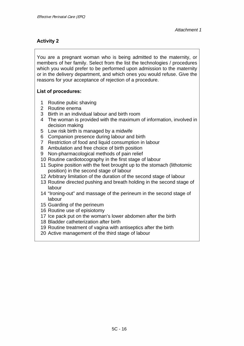
Effective Perinatal Care (EPC)
Attachment 1
Activity 2 You are a pregnant woman who is being admitted to the maternity, or members of her family. Select from the list the technologies / procedures which you would prefer to be performed upon admission to the maternity or in the delivery department, and which ones you would refuse. Give the reasons for your acceptance of rejection of a procedure. List of procedures:
1 Routine pubic shaving 2 Routine enema 3 Birth in an individual labour and birth room 4 The woman is provided with the maximum of information, involved in
decision making 5 Low risk birth is managed by a midwife 6 Companion presence during labour and birth 7 Restriction of food and liquid consumption in labour 8 Ambulation and free choice of birth position 9 Non-pharmacological methods of pain relief 10 Routine cardiotocography in the first stage of labour 11 Supine position with the feet brought up to the stomach (lithotomic
position) in the second stage of labour 12 Arbitrary limitation of the duration of the second stage of labour 13 Routine directed pushing and breath holding in the second stage of
labour 14 “Ironing-out” and massage of the perineum in the second stage of
labour 15 Guarding of the perineum 16 Routine use of episiotomy 17 Ice pack put on the woman’s lower abdomen after the birth 18 Bladder catheterization after birth 19 Routine treatment of vagina with antiseptics after the birth 20 Active management of the third stage of labour
5C - 16
Module 5C
5C - 17
Group 1 Using the tabulated synopsis, find in the reference book by Enkin the information about the effectiveness of the following procedures:
1 Routine pubic shaving 2 Active management of the third stage of labour
Group 2 Using the tabulated synopsis, find in the reference book by Enkin the information about the effectiveness of the following procedures:
1 Routine enema 2 Routine treatment of vagina with antiseptics after the birth
Group 3 Using the tabulated synopsis, find in the reference book by Enkin the information about the effectiveness of the following procedures:
1 Birth in an individual labour and birth room 2 Bladder catheterization
Group 4 Using the tabulated synopsis, find in the reference book by Enkin the information about the effectiveness of the following procedures:
1 The woman is provided with maximum of information for decision making
2 Ice pack is put on the woman’s lower abdomen
Group 5 Using the tabulated synopsis, find in the reference book by Enkin the information about the effectiveness of the following procedures:
1 Low risk births are managed by midwives 2 Routine episiotomy

Effective Perinatal Care (EPC)
5C - 18
Group 6 Using the tabulated synopsis, find in the reference book by Enkin the information about the effectiveness of the following procedures:
1 Companion presence during labour and birth 2 Guarding of the perineum
Group 7 Using the tabulated synopsis, find in the reference book by Enkin the information about the effectiveness of the following procedures:
1 Restriction of food and liquid consumption in labour 2 “Ironing out” and massage of the perineum in the second stage of
labour
Group 8 Using the tabulated synopsis, find in the reference book by Enkin the information about the effectiveness of the following procedures:
1 Ambulation and free choice of position in the first stage of labour 2 Routine directed pushing and breath holding in the second stage of
labour Group 9 Using the tabulated synopsis, find in the reference book by Enkin the information about the effectiveness of the following procedures:
1 Alternative (non-pharmacological) methods of pain relief 2 Arbitrary limitation of the duration of the second stage of labour
Group 10 Using the tabulated synopsis, find in the reference book by Enkin the information about the effectiveness of the following procedures:
1 Routine cardiotocography in the first stage of labour 2 Supine position with the feet brought up to the stomach (lithotomic
position) in the second stage of labour

Effective perinatal care (EPC) Facilitator Guide
5C - 19
Attachm
ent 2
EQU
IPMEN
T, SUPPLIES AN
D D
RU
GS FO
R C
HILD
BIRTH
CAR
E (IMP
AC
, L3
) W
arm and clean room
� Delivery bed: a bed that supports the wom
an in a semi-sitting or
lying in a lateral position, with removable stirrups (only for repairing
the perineum or instrum
ental delivery) � Clean bed linen � Curtains if m
ore than one bed � Clean surface (for alternative delivery position) � W
ork surface for resuscitation of newborn near delivery beds � Light source � Heat source � Room
thermom
eter Hand washing
� Clean water supply � Soap � Nail brush or stick � Clean towels
Waste
� Container for sharps disposal � Receptacle for soiled linens � Bucket for soiled pads and swabs � Bowl and plastic bag for placenta
Sterilization � Instrum
ent sterilizer � Jar for forceps
Miscellaneous
� Wall clock
� Torch with extra batteries and bulb � Log book � Records � Refrigerator
Equipment
� Blood pressure machine and stethoscope
� Body thermom
eter � Fetal stethoscope � Baby scale � Self inflating bag and m
ask - neonatal size � M
ucus extractor with suction tube Delivery instrum
ents (sterile) � Scissors � Needle holder � Artery forceps or clam
p � Dissecting forceps � Sponge forceps � Vaginal speculum
Supplies
� Gloves: -
utility -
sterile or highly disinfected -
long sterile for manual rem
oval of placenta -
Long plastic apron � Urinary catheter � Syringes and needles � IV tubing � Suture m
aterial for tear or episiotomy repair
� Antiseptic solution (iodophors or chlorhexidine) � Spirit (70%
alcohol) � Swabs � Bleach (chlorine-base com
pound) � Clean (plastic) sheet to place under m
other � Sanitary pads � Clean towels for drying and wrapping the baby � Cord ties (sterile)
� Blanket for the baby � Baby feeding cup � Impregnated bednet
Drugs � Oxytocin � Ergom
etrine � M
agnesium sulphate
� Calcium gluconate
� Diazepam
� Hydralazine � Am
picillin � Gentam
icin � M
etronidazole � Benzathine penicillin � Nevirapine or zidovudine � Lignocaine � Adrenaline � Ringer lactate � Norm
al saline 0.9%
� Water for injection
� Eye antimicrobial (1%
silver nitrate or 2.5%
povidone iodine) � Tetracycline 1%
eye ointment
� Vitamin A
� Izoniazid Vaccine
� BCG � OPV � Hepatitis B

Effective perinatal care (EPC) Facilitator Guide
5C - 20

Effective Perinatal Care (EPC) Facilitator guide
Module 6C Initial Rapid Assessment of the Newborn and Principles of Neonatal Care
Learning Objectives At the end of the module the participants will:
• Know main steps of newborn care at birth • Understand the major adaptations experienced by newborns at birth • Know how to prepare a safe and warm room for each birth • Know the list of needed equipment for newborn care • Know the universal precautions to prevent infection • Know how to implement the warm chain • Be able to assess the status of a newborn at birth and to take immediate
action • Know the main principles of newborn care during the first two hours of
life
Module Outline and Duration Part I – Classroom work - 120 min
Activity 1 – Introduction 5 min Activity 2 – Interactive presentation 75 min Activity 3 – Work in groups 20 min Activity 4 – Role play 10 min Activity 5 - Conclusion 10 min
Part II – Clinical work
Activity 6 – Assessing the preparation of a labour and birth room (warm, clean, light, family-oriented, safe, equipped)
90 min
Activity 7 - Assessing newborn care in the delivery room at birth ---------
6C - 1
Effective Perinatal Care (EPC)
Preparation for the Module • Review:
o Thermal Protection of the Newborn: A Practical Guide. WHO, 1997
o Pregnancy, childbirth, postnatal and newborn care: a guide for essential practice. IMPAC, WHO, Geneva, 2nd edition, 2006.
• Ensure that all participants have the Participant Manual.
• Ensure that all co-facilitators know their respective functions during
work with this module.
Materials and Audiovisual Equipment Materials
• Participant Manual • Thermal Protection of the Newborn: A Practical Guide. WHO, 1997 • Printed roles for the role play • Case studies for small group work
Equipment
• Video or overhead projector • Flipchart • Markers • Pens and pencils • Name badges • Doll • Baby cap and socks • Linen • Bulb for aspiration
Key Messages • Each birth must be carefully prepared for to allow the baby to adapt to life
outside the womb under warm, clean, safe, and friendly conditions. • The steps of newborn care are clearly defined which are implemented
almost simultaneously within the first few minutes after birth. • Assessment of the newborn should be performed in the first 30 seconds of
life in order to initiate action such as resuscitation if necessary.
6C - 2

Module 6C
Key Messages
• Aspiration of the newborn’s airways is necessary only if the baby has breathing difficulties or if there is blood- or meconium-stained fluid in her/his lungs.
• The warm chain needs to be fully implemented by a trained team.
• Neonatal hypothermia is an important issue leading to many complications.
It should be avoided through correct implementation of the warm chain. • Newborns need appropriate clothing. Swaddling should be discouraged. • The mother and baby should not be separated after birth. Immediate and
longstanding skin-to-skin contact, early breastfeeding and rooming-in should be encouraged.
• Delayed cord care clamping is beneficial for newborns and must
recommended as a component of active management of the third stage. Delayed cord clamping usually occurs by 3 minutes.,using dry cord management.
• Some procedures (weighing, measuring, and complete clinical
examination) should be delayed to respect the warm chain and mother/newborn bonding.
Note: It is important to conduct this module using 2 or 3 facilitators. The course director should decide which parts can be presented by a neonatologist, midwife or obstetrician.
PART I – Classroom work
Activity 1 – Introduction (5 min) • During the introduction discuss with participants the module objectives. • Ask the participants if they agree with the module objectives and if any of their
expectations are not covered by the list. If participants have any additional questions, list them on a flipchart.
Activity 2 – Interactive presentation (75 min) • Show Slide 6C-2 and discuss with participants information on how a baby adapts to
extra-uterine life: o At birth the baby leaves the warm and secure uterus for the cold world
outside. o The baby undergoes important changes, including initiating breathing,
maintaining his/her own body temperature (previously done by the mother),
6C - 3

Effective Perinatal Care (EPC)
feeding himself, fighting against germs, and interacting with the mother and the family.
• Show Slides 6C-3 and 6C-4 and briefly describe the basic steps of newborn care. • Underscore that some steps (2-5) must be done simultaneously, whereas some are
not mandatory for all newborns (e.g., step 5 cleaning of airways). • Emphasize that all steps are important for each newborn and are not dependent on
the baby’s gestational age or birth weight. • Show Slide 6C-5. Note the following points:
o Universal precautions need to be implemented during each delivery as every patient should be considered as possibly infected.
o Protection is for both the patient and the health staff. o Universal precautions must be implemented thoroughly, not partially (for
example use of non-sterile instruments or having unprotected eyes). • Ask the participants what is the most effective way to prevent cross-contamination/
infection in the maternity. After a short discussion, remind participants that hand washing is essential and is the most effective way to prevent cross-contamination and infections in the maternity.
• Show Slides 6C- 6 Preparation for Birth. If module 5С was given before this
module, information can be covered briefly. If not, facilitators must describe information on these slides in detail.
o Each birth should take place in a labour and birth room or operation theatre which meets the criteria listed in the slide.
o Individual rooms ensure privacy, confidentiality and comfort. o Clean delivery rooms help prevent infection. o Warm surroundings (not less than 25°C) help prevent hypothermia. o Good light facilitates effective resuscitation and more precise evaluation of
the baby’s status. • Ask participants who is the most qualified staff to support a birth.
o Correct answer: A qualified midwife is the key person in supporting a labour and birth.
• Show Slide 6C-7 and discuss the following points on equipment required for birth:
o Trained personnel able to initiate resuscitation with bag and mask need to be ready for each delivery and should have all equipment needed for neonatal resuscitation.
o Switched-on radiant heater, warm linens, cap and socks need to be prepared before every birth.
o Warm chain must be fully implemented to prevent newborn hypothermia. o Mother and baby’s safety is ensured through availability of equipment for
newborn resuscitation and urgent care for the mother in the delivery room. • Tell the participants that you will now role play a birth.
o Facilitator must prepare: • A doll • Linens • Bulb
6C - 4

Module 6C
• “Examination” table • Chair
o The following people participate in this role play:
• two facilitators: one playing the role of the mother, the other playing the role of the grandmother (or husband);
• a midwife (training participant); • an obstetrician (training participant); • If a neonatologist is present in all births in the facility where you
conduct the training, involve one from the trainee group in the role play.
o Note: it would be better if the midwife, obstetrician and neonatologist
selected come from the same facility.
Script of the role play:
• A baby’s birth is in progress. You are giving birth on your back and it is very painful. Your mother (or husband) is by your side. Your partner is nervous and asks to weigh the baby, clamp and disinfect the cord, etc.)
• Ask the participants (midwife, obstetrician and neonatologist) to demonstrate the
current baby care practices performed just after birth in the delivery room in their facility.
• Don’t make comments (and ask all other participants not to comment) during the
role play. Ask the observers to write their comments down. Explain that all comments will be reviewed at the end of the session.
o During the role play the “mother” does the following: • Insists on drying the baby only with sterile linen (to prevent infection). • After putting the doll on her chest, says to the midwife or
neonatologist that the baby is “blue,” breathing is not good, and insists on newborn examination.
• Insists on eye care just after birth. • Insists on weighing the newborn, because she needs to inform the
father of the baby’s weight. • Insists on feeding the baby, because now 15 minutes have passed
since birth and the baby may want to eat. • Insists on treatment of the cord with brilliant green to prevent
infection. • The “partner” supports the “mother” in all her requests during the role
play, but most of all insists on formula-feeding the baby (because the mother has only colostrum, not milk).
• After the role play, continue presentation without any comments and discussion
about the role play. • Show Slide 6C-8 and explain Drying the Baby and describe all steps in details. • Slide 6C-9. Remind the participants that a baby born in a delivery room at 23°C
experiences the same cold stress as a naked adult exposed to 0°C. During the birth, health care workers must have a thermometer in each room to: check the
6C - 5

Effective Perinatal Care (EPC)
baby’s temperature on the mother’s chest after 30 minutes and two hours post-delivery, recheck the temperature if the baby’s feet are cold (warmth of the baby’s feet assessed every 15 minutes), record the data received in special forms, and continuously analyze them.
• Slide 6C-10. Describe the basic approaches to diagnosing hypothermia and the
main consequences of hypothermia for the baby. o Normal temperature after birth is 36.5°C -37.5°C. o Lower than 36.5°С = hypothermia. o Hypothermia can be mild. Usually it can be treated by skin-to-skin contact in
a warm room. o Moderate and severe hypothermia are dangerous for the baby’s life. These
conditions need treatment by qualified personnel with oxygen and intravenous glucose.
• Show Slide 6C-11. This simplified diagram shows some consequences of
hypothermia in newborns. • Slide 6C-12. “Warm chain" may be a new concept for participants, who will be
unfamiliar with this topic. Therefore take your time on this slide. List of steps of the warm chain and emphasize the fact that these actions form a chain and that if a link is missing, the baby will be cold. The warm chain should be implemented by a team trained on the importance of hypothermia. Warm chain implementation is not easy. Thus the team needs to be trained and retrained.
• Slide 6C-13. The baby needs to be appropriately dressed in loose, warm clothes (in
order to keep air between the clothes and the baby’s body). Remind that the baby’s head needs to be covered because it is a big body surface and much heat is lost through radiation). Use of clothes from home is recommended, as the newborn becomes exposed to the mother’s/home’s flora and a link is created between the baby and mother.
• Slide 6C-14. Discuss the harm of tight swaddling.
• Slide 6C-15. Explain, that as soon as the infant is born, while drying him/her, the
health provider should immediately assess the infant’s well-being in order to identify if he/she needs special care or if he/she can be given immediately to the mother.
• Slide 6C-16. Lists the components of newborn assessment before and after the
birth. The majority of babies start to breathe and cry at birth without any assistance. If the baby is crying it means that he/she is breathing adequately and his/her heart rate is more then 100 beats per minute. Normal breathing is initiated within 30 seconds from birth. The rapid assessment must be done within 30 seconds
o Normal breathing rate is 30–60 breaths per minute, with no sign of respiratory distress such as chest indrawing, grunting or nasal flaring.
o The heart rate must be over 100 beats per min after the first minute from birth.
o If the baby is crying, he/she is breathing, so staff do not need to count heart rate.
o The baby’s face and chest must be pink, not grey or blue, after 1-3 minutes. o The pink colour of the baby’s face and chest is a good sign of adequate
breathing and circulation (arms and feet can be bluish). o It is important to be sure that the baby does not have central cyanosis (no
blue tongue or blue lips).
6C - 6
Module 6C
o In dark-skinned babies, assess the colour of the tongue, lips and mucous membranes.
o The baby should have firm muscle tone and should not be floppy. o It is necessary to assess the baby’s gestational age in order to make a
decision about additional care and creation of a warmer surrounding for deliveries (no less than 28°С).
o It is necessary to assess the amniotic fluid for meconium. At least two additional specialists trained on newborn resuscitation (in the case of preterm deliveries) and newborn intubations should remain in the delivery room.
• Slide 6C-17. Explain steps of Classification and Management after immediate
assessment. Describe four situations. • Slide 6C-18. Discuss with participants main principles of cleaning the airway and
precise that this step is not mandatory: unnecessary aspiration could create problems and delay the start of breathing. Draw participants’ attention to the fact that this manipulation needs to be done gently and according to the guidelines. A sterile catheter/bulb is needed for cleaning the airways. Use of the same catheter/bulb in different babies is unacceptable because of the difficulty sterilizating this equipment.
• Slide 6C-19. Discuss with participants main roles to organize Early Skin-to-Skin
Contact and after that pay special attention: o Skin-to-skin contact needs to be as natural as possible – no disinfectant, no
medical interference. Leave the mother to welcome the newborn. • Slide 6C-20. Skin-to-Skin Contact
o Photos demonstrating early skin-to-skin contact. Encourage the participants to explore all possibilities in organizing skin-to-skin contact of the healthy newborn with the mother and/or father.
• Slide 6C-21. Pay special attention for key statements:
o Delayed cord care clamping is beneficial for newborns and must be recommended as a component of active management of the third stage. Delayed cord clamping usually occurs by 3 minutes, using dry cord management.
o Do not cover the cord with a bandage. o Do not apply any substance to the cord, including antiseptic (alcohol).
• Slide 6C-22. Discuss with participants all steps of organization of Early
Breastfeeding: o Never force the baby to initiate breastfeeding before he/she is ready. o Start breastfeeding when the newborn shows signs of readiness to suck,
support the mother to find a comfortable and good position for breastfeeding.
o Help mother to attach the baby well if necessary. o Assess correct breast attachment. o Do not impose a time limit for breastfeeding; the baby will stop on his/her
own. • Slide 6C-23. Early Breastfeeding
o Show photo which depicts baby’s first breastfeeding with skin-to-skin contact. Note that baby is dressed correctly.
6C - 7

Effective Perinatal Care (EPC)
• Slide 6C-24. Describe procedures which may decrease neonatal morbidity. • Slide 6C-25. Discuss key points of Monitoring during the first two hours
o During the two hours the baby will spend in the birth room, he/she needs to be carefully monitored. The following must be reported in the baby’s file:
• breathing rate and breathing difficulties (chest indrawing, grunting or nasal flaring)
• skin colour • warmth of feet and body temperature • time of first breastfeeding, activity of sucking reflex • meconium and urine passing
o Conduct complete and detailed medical examination (before rooming-in). o Measure baby’s weight and length. o Bathing should be postponed for six to 24 hours, as it may cause
hypothermia. • Slide 6C-26. Discuss with participants step by step how to re-warm a newborn. • Slide 6C-27. Shows basic points of complete assessment of the newborn in the
delivery room two hours after birth and before rooming-in. • Slides 6C-28 and 6C-29 show basic principles of rooming-in.
Activity 3 – Group work (20 min) • Divide the participants into two multidisciplinary groups and give them the cards
with two case studies (Attachment 1). • Give each group a page of flipchart and marker. • Ask the participants to write the answers to the questions using the flip-chart. • Ensure that groups understand their tasks well.
Case Study 1
Sasha is a baby boy born after 39 weeks of gestation without any problems. He was breathing regularly, his heart rate was >120 beats/min, after one minute the baby was pink and very reactive. After birth, the midwife dried Sasha thoroughly with a warm linen and placed him skin-to-skin with his mother. The midwife covered the baby’s head with a hat. After 20 minutes the midwife weighed the baby, bandaged the cord, swaddled him tightly and placed him close to his mother in a crib. After 30 minutes the baby’s temperature was 36.2°C. Questions: • How would you define this infant?
6C - 8

Module 6C
• Was everything done in the right way?
Case Study 2 Sophia is born after 36 weeks of pregnancy in a delivery room at 23°C.
She cried immediately and was pink in 1 minute.
She was dried quickly by the midwife, and because she looked “small’’, the midwife weighed her naked immediately. Her weight was 2,300 g. The midwife was anxious and asked the neonatologist to check the baby’s medical status. Sophia stayed naked and covered with a piece of cotton under a radiant heater during 15 min. The examination was normal and after 20 min she was given back to her mother for skin-to-skin contact.
After 30 min her temperature was 36.2°C.
Questions: • How would you define this infant? • Was everything done in the right way?
• After the groups have finished working on the case studies, one representative from
each group presents the results of the group’s work. • Discuss the answers with the participants. • If needed, the facilitator can return to the presentation and review the slides to
confirm the right answers. Possible answers:
Case study 1:
o Sasha is a full term baby suffering from mild hypothermia. o Sasha was separated from his mother after only 20 minutes of skin-to-skin
contact and he was weighed too early. In addition, he was tightly swaddled and put in a crib.
o He needs to be put naked in skin-to-skin with the mother, with his head and feet covered.
o Mother and baby must be covered with a warm blanket in a warm room (the delivery room is 26°C).
o The warmth of Sasha’s feet need to be assessed every 15 minutes . If the feet are cold, the temperature should be taken at that moment. If the feet remain warm, the temperature needs to be assessed in one hour (> 36.5°C).
o The mother should be supported to initiate breastfeeding if the baby is ready or ask mother to express her milk and feed Sasha with mother’s milk.
6C - 9

Effective Perinatal Care (EPC)
Case study 2:
o Sophia is a preterm baby with no problem at birth. She suffers from mild hypothermia.
o The delivery room was cold, only 23°C. The drying was too quick; the weighing was premature as it was obvious that Sophia was not a “very small baby” needing special care.
o During the first 20 minutes of life, she was separated from her mother and deprived of skin-to-skin contact.
o Sophia needs to be put naked skin-to-skin with the mother, with her head and feet covered.
o Mother and baby need to be covered with a warm blanket, and a heating system needs to be brought to the room to reach 25°C.
o The warmth of Sophia’s feet need to be assessed every 15 minutes . If the feet are cold, the temperature should be taken at that moment. If the feet remain warm, the temperature needs to be assessed in one hour (> 36.5°C). If the temperature is not within normal range in 2 hours, Sophia’s needs further assessment and re-warming measures.
o The mother should be supported to initiate breastfeeding as soon the baby is ready or ask mother to express her milk and feed Sophia with mother’s milk..
Activity 4: Role plays (10 min)
Role play 1 o Needed materials:
o A doll o Linens o Bulb o “Examination” table o Chair
The following people participate in this role play:
o Two facilitators: one playing the role of the mother, the other the role of the grandmother (or husband). You can ask the same participants as during the first role play or ask other facilitators to participate.
o One midwife (training participant). o One obstetrician (training participant). o If the facility where you are conducting the training has neonatologists
participating in all births, involve a neonatologist from the trainee group. o Note: it would be good if the midwife, obstetrician and neonatologist were
the same as during the role play in Activity 2.
Script of the role play (the same role play as in Activity 2):
o A baby’s birth is in progress. You are giving on your back and it is very painful. Your mother (or husband) is by your side. Your partner is nervous and asks to weigh the baby, clamp and disinfect the cord, etc.
o Ask the participants (midwife, obstetrician and neonatologist) to demonstrate
the current baby care practices performed just after birth in the delivery room in their facility.
6C - 10

Module 6C
o Don’t make comments (and ask all other participants not to comment) during
the role play. Ask the observers to write their comments down. Explain that all comments will be reviewed at the end of the session.
o During the role play the “mother” does the following:
• Insists on drying the baby only with sterile linen (to prevent infection). • After putting the doll on her chest, says to the midwife or
neonatologist that the baby is “blue,” breathing is not good, and insists on newborn examination.
• Insists on eye care just after birth. • Insists on weighing the newborn, because she needs to inform the
father of the baby’s weight. • Insists on feeding the baby, because now 15 minutes have passed
since birth and the baby may want to eat. • Insists on treatment of the cord with brilliant green to prevent
infection. • The “partner” supports the “mother” in all her requests during the role
play, but most of all insists on formula-feeding the baby (because the mother has only colostrum, not milk).
• In addition to this role play you can conduct another one if you have free time. Role play 2 – Implementation of warm chain • This role play can be conducted at any time after the end of module 6C. • It is advisable to conduct this role play during the clinical week in two small groups
(obstetricians/midwifes and neonatologists/paediatric nurses).
o Needed materials: - A doll - Two pieces of cloth for drying baby - A baby hat or a piece of material to cover the baby’s head - A blanket to cover mother and baby
• Select participants to be mother and midwife (ask volunteers if possible) and
recommend to the rest of your group to observe carefully and to report what is done well and what needs to be improved.
o Give participants three minutes to prepare. o Stop the role play after 10 minutes even if not complete. o Conduct a discussion on the positive and negative aspects of players’
actions.
Script for “mother”:
o You just gave birth to your first baby (a big boy of 4,000 g) on your back with an episiotomy.
o The labour and birth was painful, you are tired, and you want to rest. o Your mother is with you.
Script for “midwife”:
o You are trained in warm chain implementation.
6C - 11

Effective Perinatal Care (EPC)
o You want to implement the warm chain perfectly.
Activity 5 – Conclusion (10 min) • After the role play ask the participants to compare the actions of the “medical
personnel” during the role play in Activity 2 and the first role play in Activity 4, paying special attention to the following points:
o Drying and putting the baby on the mother’s chest o Assessing the baby’s wellbeing o Putting on the cap and socks and covering the baby with a blanket together
with the mother o Clamping the cord o Counselling the mother (breastfeeding) o Attachment to the breast o Checking the baby’s feet for warmth and the body temperature at 30
minutes or if the feet are cold. o Eye care o Baby examination, weighing and cord care
• After the discussion comparing the role plays, ask the participants to make
conclusions on key points of newborn care in the delivery room (paying special attention to the following points):
o Preparation of warm premises o Preparation of needed equipment (linens, caps, socks, equipment for
resuscitation, heated table and radiant heater) o Drying of the baby and putting him/her on the mother’s chest; o Assessment of the baby’s wellbeing on mother’s chest: breathing,
malformations/birth trauma, prematurity/low weight; o Putting on the cap and socks and covering with common blanket o Cord clamping at the end of pulsation with physiologic management of the
placenta OR just prior to controlled cord traction with active management of the third stage
o Counselling the mother on breastfeeding (baby’s readiness, signs of correct attachment, frequency of feeding)
o Eye care (after the first attachment to the breast, but not later then one hour after birth)
o Checking the baby’s feet for warmth every 15 minutes and the body temperature thirty minutes after birth (digital thermometer, must be checked in axillaries)
o Actions on baby re-warming o Complete examination after two hours (until then maintain skin-to-skin
contact) o Twenty-four hour rooming-in o Warm chain steps
• Show Slide 6С-30 and make brief conclusions on the module. • Ask participants if they have any questions. Briefly answer any questions.
6C - 12

Module 6C
PART II – Clinical work
Activity 6 – Checking the preparation of a labour and birth room (90 min) • Divide participants into sub-groups with one facilitator per sub-group. • This activity can be conducted at any convenient time during the clinical week or
during the day of individual labour and birth room organization by obstetrical group participants.
Preparation of the group before the maternity visit: • Explain the task to the group: to check the labour and birth room readiness and
availability of everything needed for care. • Briefly explain the contents of Tables 1 and 2. Give each participant evaluation
forms (Tables 1 and 2), pen/pencils and flipchart paper. • After checking the birth rooms, discuss results with the participants.
6C - 13

Effective Perinatal Care (EPC)
Table 1. Check availability of the following equipment
Table with radiant heater Yes ( ) No ( )
Equipment for resuscitation (bag, masks in two sizes and T-piece)
Yes ( ) No ( )
Oxygen supply Yes ( ) No ( )
Suction apparatus Yes ( ) No ( )
Sterile suction catheters Yes ( ) No ( )
Disposable suction bulb Yes ( ) No ( )
Infant laryngoscope Yes ( ) No ( )
Sterile endotracheal tubes Yes ( ) No ( )
Sterile gloves Yes ( ) No ( )
Sterile materials (cotton, gauze napkin) Yes ( ) No ( )
Thermometer Yes ( ) No ( )
Linens/ towels for drying the infant Yes ( ) No ( )
Blanket to cover mother and baby Yes ( ) No ( )
Cord cutting/cord clamping set Yes ( ) No ( )
Cord clamp Yes ( ) No ( )
Oxytocin Yes ( ) No ( )
Adrenalin Yes ( ) No ( )
Vitamin К Yes ( ) No ( )
1% tetracycline eye ointment Yes ( ) No ( )
Syringes Yes ( ) No ( )
Infant weighing scale Yes ( ) No ( )
6C - 14

Module 6C
Table 2. Check the condition of the birth room and the functioning of the equipment/devices Is the birth room free of draught? Yes ( ) No ( ) Is there a good light in the birth room? Yes ( ) No ( ) Is there a clock with a second hand in the birth room? Yes ( ) No ( ) Is the birth room equipped to support a free labour and birth position?
Yes ( ) No ( )
Is the labour and birth room “family-oriented” and friendly? Yes ( ) No ( ) The temperature of the birth room is:
o 20-25°C Yes ( ) No ( ) o > 25°C Yes ( ) No ( ) o < 20°C Yes ( ) No ( )
Is the overhead heater of the table functioning? Yes ( ) No ( ) Is the bag:
o Self-expanding with a volume of about 250–750 ml? Yes ( ) No ( ) o Easy to take apart? Yes ( ) No ( ) o Easy for cleaning and sterilizing? Yes ( ) No ( ) o Without visual damage? Yes ( ) No ( )
Yes ( ) No ( ) Mask:
o Anatomical shape? Yes ( ) No ( ) o With soft border? Yes ( ) No ( ) o Without visual damage? Yes ( ) No ( ) o Two sizes? Yes ( ) No ( )
Yes ( ) No ( ) Is the suction apparatus functioning well? Yes ( ) No ( )
o Manual/foot-operated? Yes ( ) No ( ) o Electrically operated? Yes ( ) No ( )
Is the incubator/heated crib for newborn: Yes ( ) No ( )
o Clean? Yes ( ) No ( ) o With a temperature of about 35°C? Yes ( ) No ( ) o With a temperature < 30°C? Yes ( ) No ( ) o With a temperature > 38°C? Yes ( ) No ( )
Is the weighing scale functioning? Yes ( ) No ( )
6C - 15

Effective Perinatal Care (EPC)
Activity 7 – Assessing newborn care in the delivery room at birth • This part of the clinical work must be conducted when the group is attending
deliveries. • Each participant must receive Table 3 and complete it while attending births. • The facilitator’s role is to supervise what goes on during labour and birth (it is
recommended that the facilitator fills in the same form), to collect participants’ forms, and to conduct a group discussion the next day on what was done well and what was not good, as well as to discuss the assessment method and completing the forms.
• After the group discussion, facilitators should conduct a general discussion on
Activity 7’s results and create a plan for newborn care in the delivery room together with participants.
• Write down the results of the participants work in the following table. Name Comments by the facilitator 1
2
3
4
6C - 16

Module 6C
Table 3. Procedures performed at birth and immediately after birth Is a family member allowed to assist the women at the time of birth?
Yes ( ) No ( )
Does the staff wash their hands each time before assisting a birth? Yes ( ) No ( ) Does the staff use sterile gloves during the birth? Yes ( ) No ( ) Does the staff use only sterilized instruments and supplies? Yes ( ) No ( ) Are the newborn’s upper airways cleared at birth? If yes, specify:
o With a catheter by suction o With a bulb o Other _______________________
Yes ( ) Yes ( ) Yes ( ) Yes ( )
No ( ) No ( ) No ( ) No ( )
Is the baby dried after birth? If yes, specify:
o Immediately after birth o Within 30 minutes o With a dried warm towel/linen
Yes ( ) Yes ( ) Yes ( ) Yes ( )
No ( ) No ( ) No ( ) No ( )
Is the baby assessed at birth? If yes, what is the newborn assessed for:
o Breathing o Heart rate o Skin colour o Floppiness o Weight o Length o Head circumference o Congenital anomalies/birth trauma o Other ______________________
Yes ( ) ( ) ( ) ( ) ( ) ( ) ( ) ( ) ( ) ( )
No ( )
Is the cord cut: o Immediately after birth o At approximately 1 min after birth o After more than 1 min after birth
( ) ( ) ( )
Is the cord clamped with o A rubber band o A silk string o A disposable clamp
( ) ( ) ( )
Where is the healthy baby placed immediately after birth? o On the resuscitation table o On the mother’s chest (skin-to-skin) o Near the mother o Other ____________ ___________
( ) ( ) ( ) ( )
Is the newborn’s body temperature checked after birth? If yes, specify:
o 30 minutes after birth o Within 2 hours o After 2 hours o In all newborns? o Only in low birth weight and sick infants?
Yes ( ) ( ) ( ) ( ) Yes ( ) Yes ( )
No ( ) No ( ) No ( )
Is the baby washed (specify): o Immediately after birth o Within the first 2–6 hours o With warm water o With cold water o Other _____________________________________
Yes ( ) ( ) ( ) ( ) ( ) ( )
No ( )
6C - 17
Effective Perinatal Care (EPC)
When does the baby start breastfeeding? o When the baby shows signs of readiness o Within the first 30 minutes o Within the first 4 hours o After the first 4 hours o After the first 12 hours
( ) ( ) ( ) ( ) ( )
Which of the following prophylactic procedures are carried out at birth?
o Use of vitamin K o Prophylaxis of gonoblenorrhea
Yes ( ) Yes ( )
No ( ) No ( )
Is a complete examination of the baby carried out in the delivery room? If yes, specify:
o 2 hours after birth o Within 2 hours after birth o On a warm examination table
Yes ( ) ( ) ( ) ( )
No ( )
6C - 18

Module 6C
References
1. American Heart Association in collaboration with International Liaison Committee on Resuscitation. Guidelines 2005 for Cardiopulmonary Resuscitation and Emergency Cardiovascular Care. Part 13: Neonatal Resuscitation Guidelines. Circulation, 2005, 112: 188 -195.
2. Andersson GC. et al. Early Skin-to-Skin Contact for Mother and their Healthy
Newborn Infants. Review, Cochrane Library, issue 1, 2004. 3. Breastfeeding counselling: Training course. WHO, UNICEF, 1993. 4. Camila M Chaparro et al. Effect of Timing of Umbilical Cord Clamping on Iron
Status in Mexican Infants: A Randomised Controlled Trial. Lancet, June 2006, 367: 1997-2004.
5. European Resuscitation Council Guidelines for Resuscitation, 2005. Section 6:
Paediatric Life Support. 6. H Rabe, G Reynolds, J Diaz-Rossello. Early versus delayed umbilical cord
clamping in preterm infants. Cochrane Database of Systematic Reviews, 2004 Issue 1.
7. Jose´ M. Ceriani Cernadas et al. The Effect of Timing of Cord Clamping on
Neonatal Venous Hematocrit Values and Clinical Outcome at Term: A Randomized, Controlled Trial, Pediatrics, 2006, 117: 779-786.
8. Managing Newborn Problems: A Guide for Doctors, Nurses, and Midwives.
WHO, Geneva 2003. 9. Mother-baby package: Implementing safe motherhood in countries. Department
of Reproductive Health and Research, WHO, 1996. 10. Murray W. Enkin et al. A Guide to Effective Care in Pregnancy And Childbirth,
Oxford, Third edition, 2000. 11. Narendra Aladangady et al. Infants’ Blood Volume in a Controlled Trial of
Placental Transfusion at Preterm Delivery. Pediatrics. January 2006, 117: 93-98.
12. Pregnancy, Childbirth, Postpartum and Newborn Care: A Guide for Essential
Practice. WHO, Geneva, 2006. 13. Puckett RM, Offringa M. Prophylactic Vitamin K for Vitamin K Deficiency
Bleeding in Neonates (Cochrane Review). The Cochrane Library, 2005, Issue 3. 14. Thermal Protection of the Newborn: A Practical Guide. WHO, Geneva, 1997. 15. Vain, E. Szyld, L. Prudent, T. Wiswell, A. Aguilar, N. Vivas. Oropharyngeal and
Nasopharyngeal Suctioning of Meconium-Stained Neonates Before Delivery of their Shoulders: Multicentre, Randomised Controlled Trial. The Lancet, Volume 364, Issue 9434: 597-602.
6C - 19

Effective Perinatal Care (EPC)
16. WHO recommendations for the prevention of postpartum haemorrhage. WHO/MPS/07.06, 2007.
17. Wiswell TE, Gannon CM, Jacob J., et al. Delivery room management of the
apparently vigorous meconium-stained neonate. Results of the Multicenter, International Collaborative Trail. Paediatrics, 2000, Volume 105, Number1, 1-7.
‘
6C - 20

Module 6C
Attachment 1. Activity 3
Case Study 1 Sasha is a baby boy born after 39 weeks of gestation without any problems. He was breathing regularly, his heart rate was >120 beats/min, after one minute the baby was pink and very reactive. After birth, the midwife dried Sasha thoroughly with a warm linen and placed him skin-to-skin with his mother. The midwife covered the baby’s head with a hat. After 20 minutes the midwife weighed the baby, bandaged the cord, swaddled him tightly and placed him close to his mother in a crib. After 30 minutes the baby’s temperature was 36.2°C. Questions: • How would you define this infant? • Was everything done in the right way?
6C - 21

Effective Perinatal Care (EPC)
Case Study 2 Sophia is born after 36 weeks of pregnancy in a delivery room at 23°C. She cried immediately and was pink in 1 minute. She was dried quickly by the midwife, and because she looked “small’’, the midwife weighed her naked immediately. Her weight was 2,300 g. The midwife was anxious and asked the neonatologist to check the baby’s medical status. Sophia stayed naked and covered with a piece of cotton under a radiant heater during 15 min. The examination was normal and after 20 min she was given back to her mother for skin-to-skin contact. After 30 min her temperature was 36.2°C. Questions:
• How would you define this infant? • Was everything done in the right way?
6C - 22
Effective perinatal care (EPC) Facilitator Guide
Module 7C
Breastfeeding
Learning objectives By the end of the module the participants will:
• Understand the importance of breastfeeding • Understand the danger of artificial feeding • Know the main mechanisms of production and let-down • Know the main characteristics of breast milk • Understand the importance of “skin-to-skin” contact for initiation of effective
breastfeeding • Obtain skills to counsel women on first attachment to the breast • Know correct breastfeeding positions • Observe breastfeeding to provide supportive environment to the mother • Learn breastfeeding counselling skills • Learn to timely recognize difficulties related to breastfeeding and help
mothers to overcome them
Module outline and duration: Part I – Classroom work – 105 minutes
Activity 1 – Introduction 5 min Activity 2 – Beginning of interactive presentation 20 min Activity 3 – Massage demonstration on breastfeeding mother 10 min Activity 4 – Continuation of interactive presentation 15 min Activity 5 – Role play: first attachment to the breast 10 min Activity 6 – Continuation of interactive presentation 15 min Activity 7 – Role play: Counselling and helping mother to breastfeed 10 min Activity 8 – Continuation of interactive presentation 20 min
Part II – Class-room –120 minute
Activity 9 – Assessment of breastfeeding, breastfeeding counselling session
120 min
Preparation to the Module • Review of existing publications, evidence-based literature and health
strategies, recommended for breastfeeding. Make sure that all the participants have received the Module for participants.
• Make sure that all facilitators know their scopes of work during the training
7C - 1

Effective perinatal care (EPC)
Materials and audio-visual equipment Materials
• Module for participants • Local guidelines and state orders (policies) on breastfeeding • Presentation 7С EPC ENG
Equipment • Slide-projector or PowerPoint projector • Flipchart • Notebooks • Markers • Pens and pencils • Badges • Baby doll
Key messages • Exclusive breastfeeding (just breastfeeding) is recommended up to 6
months and breastfeeding (plus complementary foods) is recommended up to 2 years and beyond
• Breastfeeding protects the child from infections, allergies and chronic
diseases
• Exclusive breastfeeding improves the physical and psychological development of the child
• It is necessary to help the mother to initiate breastfeeding within one hour
after delivery
• It is recommended to breastfeed on demand 8-12 times a day on average, during the day and night
• Exclusive breastfeeding can protect women from unwanted pregnancy • Breast and nipple shapes do not hinder exclusive breastfeeding
• Every woman should be taught and counselled on how to breastfeed
Part I – Classroom work
Activity 1 – Introduction (5 min) • Show Slide 7С-1 and explain to the participants that while working on the Module
you will discuss:
7C - 2

Module 7С
o Advantages of exclusive breastfeeding and influence of breastfeeding on physical and psychological development of the child
o Advantages of breastfeeding for the woman and child
o Vital components of breast milk
o Mechanisms of breast milk production and let-down
o Possible breastfeeding difficulties and problems and the ways to overcome them
o Practical help and counselling for the mother on breastfeeding
Activity 2 – Beginning of interactive presentation (20 min)
• Then show Slide 7С-2 and ask the participants “How do you call these types of feeding?”
o 1 – Excusive breastfeeding
o 2 - Predominant breastfeeding.
o 3 – Artificial feeding
o 4 – Mixed feeding (which is not acceptable with availability of breast milk)
o 5 – Complementary (also called supplementary) feeding
• Then discuss terminology:
o Exclusive breastfeeding: baby doesn’t get any water or food except breast milk.
o Predominant breastfeeding: baby gets a little water or other liquid in addition to breastfeeding
o Artificial feeding: feeding baby with formula
o Mixed feeding: when the baby of the first six months of life receives some other food and or drinks in addition to breastmilk
o Weaning (supplementary feeding): introduction of semi-solid or solid food to the baby’s nutrition from 6 months of age
Pay attention to the fourth kind of feeding “mixed feeding” and underline that if the mother breastfeeds her child the use of formula is not acceptable.
• Show Slide 7С- 3 and ask the participants “What advantages of breastfeeding do they know?” Write the answers on the flipchart. Make sure that all the participants name the below-listed points:
o Complete nutrition, easy to digest
o Protects from infections
o Provides bonding and development
o Helps to avoid pregnancy
o Benefits the mother’s health
7C - 3

Effective perinatal care (EPC)
• Go to Slide 7С-4 and ask the participants to list disadvantages/risks of artificial feeding. Write the answers on flipchart. Then pass on to the information in the presentation and explain it:
o Risk of the child malnutrition and Vitamin A deficiency. Malnutrition is connected to the fact that the child is given certain portions of breast milk that do not suffice for his growth and development (that covers only the child’s energy needs)
o Formula does not contain Vitamin A which can lead to a deficiency
o Mothers can become pregnant immediately after delivery as very little prolactin is developed to suppress ovulation (prolactin level is very high among breastfeeding mothers)
o Risk of anaemia as well as breast and ovarian cancers is increased
• Slide 7С-5 discuss with the participants in brief about prolactin reflex and its function:
o When a child suckles the breast, impulses are produced that pass from the nipple to the brain. In response to this action, the hormone prolactin is secreted in the front part of the hypophysis, which then increases the production of milk.
o Draw participants’ attention to the fact that prolactin suppresses ovulation
o Draw participants’ attention to the fact that prolactin is produced abundantly at night.
o Then ask the participants: how to maintain the prolactin reflex? Then show the slide with brief summary on how to maintain prolactin reflex.
• Slide 7С-6 discuss briefly with the participants about the oxytocin reflex and its function. Draw participants’ attention to the fact that during suckling the uterus actively contracts. When a child suckles the breast, sensory impulses pass from the nipple to the brain. In response to these impulses, the hypophysis secretes the hormone oxytocin, which passes from the blood to the breast and causes the contraction of muscle cells around areola, leading to the “let down” of milk.
o Emphasize that stress, pain, anxiety can have a negative influence on oxytocin production. Ask the participants “What can a woman do to facilitate the oxytocin reflex?”
• Slide 7С-7 explains to the participants other things that help mothers breastfeed and how to avoid problems connected with insufficient production of oxytocin.
• After that conduct a role play.
Activity 3 – Role play - Massage of breastfeeding mother (10 min)
• Ask one of the participants to come to the front of the room and sit down on the chair. Ask another facilitator (or do it yourself) to show the technique of massaging
7C - 4

Module 7С
the mother’s back to stimulate the oxytocin reflex. Then ask the participants to sit behind each another and massage each others backs as demonstrated.
Activity 4 – Continuation of interactive presentation (15 min)
• Slide 7С-8 demonstrates the mechanism of suckling; specify that the child makes “pressing” movements with his tongue. This allows the milk from the milk sinuses to enter into the baby’s mouth, which is then swallowed. This is why it is important to properly attach him to the breast.
• Demonstrate Slide 7С-9 and ask the participants to list the vital components of
breast milk. Draw attention to the bifidus factor. Ask the participants whether it’s important to prescribe probiotics for the exclusively breastfed child. The gastrointestinal enzymes available in breastmilk help with the maturation of the intestinal system and with food digestion, therefore breastfeeding plays a critical role in caring for children with jaundice.
• Ask the participants whether the mother has to stop breastfeeding if she has
infections (e.g. viral infections). Ask them to explain answers. Then go to Slide 7С-10. Draw attention to the fact when a mother has an infection, her body produces antibodies to fight this infection. These antibodies are transferred to the baby through the breast milk, thereby protecting the baby.
• Discuss Slide 7C-11. Explain to the participants the differences between different
kinds of breastmilk. Colostrum contains more protein than later milk, foremilk contains more lactose, hindmilk contains more fat than foremilk. Focus on the significant differences between foremilk and hindmilk especially the need for hindmilk in caring for low-weight children.
Activity 5 – Role play: First attachment to the breast in the delivery room (10 min)
• One of the participants to play the role of a woman who just delivered. • Ask a participant (midwife, obstetrician/gynaecologist, neonatologist) to play the role
of a health care provider who helps/counsels the woman on first attachment to the breast. You may ask for help from those participants who routinely counsel women.
• Ask the group to silently write down their comments to be discussed after the role
play.
• Also ask the group to pay attention to the information delivering but not to focus on the counselling skills.
Task for participant playing the provider role: explain to the woman what the first attachment to the breast should look like.
Task for second participant: As the new mother, you have read a lot of literature during pregnancy and you know that breastfeeding is very useful for the mother and child and wish to breastfeed. In the women’s outpatient clinic you were told about breastfeeding. You’re worried about breastfeeding since you don’t have any experience in it. Use the baby-doll to show how you are breastfeeding. During the counselling role play counselling indicate that you
7C - 5

Effective perinatal care (EPC)
are worried that you will not be able to properly attach the baby to the breast and you have mastitis.
• Discuss as a group the notes that the other participants have written regarding the role play after showing Slide 7С-17. The training participants should compare the help and counselling given during the role-play using the information from Slides 7С-12 - 17. Focus on whether the counselling involved informing the mother of the main signs of the child’s readiness for the first breastfeeding and proper attachment.
Activity 6 – Continuation of interactive presentation (15 min)
• Slides 7С-12 - 7С-16 demonstrate the first attachment to the breast. While showing these slides, discuss with the participants how they would counsel a woman during regarding the first breastfeeding. Pay attention to the beginning of the first attachment.
• Slide 7С-12 – explain to the participants that success of breastfeeding is “skin-to-
skin” contact between the mother and baby immediately after birth.
• Slide 7С-13 – 7С-15 explains the signs of the child’s readiness for breastfeeding (first attachment to the breast). The baby opens his mouth widely and starts moving his head from one side to another; opens his mouth and pushes his tongue out. The baby can also suckle his fist and crawl towards the breast. The signs of readiness to breastfeed usually appear at least an hour (and sometimes longer) after birth depending upon the child’s state and the age of gestation. Pay attention that the healthcare provider SHOULD NOT attach the baby to the mother’s breast. Let the baby attach when the baby is ready.
• Slide 7С-16 the child’s attachment to the breast. Ask the participants to discuss the
signs of correct attachment to the breast. After the answers go to the next slide.
• Show Slide 7С-17 and summarize the main points of proper first attachment to the breast. Ask the participants to discuss the role-play: which information was given to the mother, which information should have been given and how should the providers counsel mothers on the initiation of breastfeeding.
• Show Slides 7С-18 – 7С-19 and using the pictures discuss with the participants:
whether the child is positioned correctly for breastfeeding. Ask the participants to describe the signs of proper attachment to the breast.
• Slide 7С-20. Summarize the participants’ answers using the slide.
• Show Slide 7С-21 and discuss with the participants the most comfortable positions
for breastfeeding. Note that there are no distinct recommendations as to certain positions that the mother has to use while breastfeeding. What is most important is that that the mother is relaxed and feels comfortable. Pay attention that all the signs of proper attachment to the breast are followed despite the mother’s position.
• Show Slide 7С- 22 and ask the participants “Why it is important to observe
breastfeeding?” and “How do you observe breastfeeding?” Expected answers: “quickly identifies any problems, able to correct the problem”. Slide 7С 22-23
7C - 6

Module 7С
demonstrate the list of main points to pay attention to while observing breastfeeding.
• Slide 7С-24 – shows main organizational points of observation for breastfeeding.
Activity 7 – Role play: Helping mother to breastfeed (10 min)
• Ask one of the facilitators to play the role of the breastfeeding mother.
• Ask one of the participants (midwife, obstetrician/gynaecologist, neonatologist) to play the role of a health care provider, who helps/counsels the woman on the first attachment to the breast in postpartum period. You may ask for help from those participants who routinely counsel women.
• Ask the group not to comment the actions of the role-play participants but rather write down their comments to be discussed after the role play.
• Also ask the group to pay attention to the information delivered rather than to counselling skills.
Task for facilitator: Act as if you are breastfeeding your child in an uncomfortable position: sitting on the chair, bent forward and swaying. The baby-doll (baby) is on your lap lying on the back with his head turned towards the breast, the baby’s legs are hanging. During the role play you insist that you are breast feeding your child correctly. You give the breast to the baby (the baby is not moved to the breast,)
Task for the participants: explain and help the woman to breastfeeding in the proper position. Ask the participants how breastfeeding counselling is done in their maternities wards.
Activity 8 – Continuation of interactive presentation and conclusion (20 min)
• Show Slide 7С- 25 and talk about main difficulties of breastfeeding.
• Slide 7С-26 – demonstrates reliable and possible signs of milk insufficiency. Discuss that real hypogalactia cases occur in less than 1% of women. Pay attention that this information should be discussed with the mother soon after delivery to reduce her worries and concerns.
• Discuss with the participants the role-play. Ask the participants to compare the help
and counselling skills in the role-play with the information in the presentation. Pay attention to the following: whether the health care provider observed the breastfeeding, which recommendations did the provider give to the mother and whether the provider helped the mother to choose a comfortable position for herself and the baby?
• Then show Slide 7С-27 and discuss the possibilities of breastfeeding for babies
with congenital abnormalities. You can show the “dancer’s hand” position, using the baby-doll.
Next discuss the conditions of the breasts which may cause difficulties in breastfeeding.
7C - 7

Effective perinatal care (EPC)
• On Slide 7С-28 you can see the list of breast conditions which can cause problems with breastfeeding.
• On Slide 7С-29 different breast shapes are shown. Help the participants to come to
the conclusion that a baby can be breastfed by any type of breast. Healthcare providers should teach and help the mother to choose the optimal breastfeeding position.
• Slide 7С-30 demonstrates the main points of helping the mother with flat and
inverted nipples. Remind the participants that preparation of the nipple before delivery is usually ineffective. Important – help the mother right after delivery and keep helping her during her stay in the maternity. The use of a suction device and milk pump is possible before breastfeeding.
• Slide 7С-31 shows the advantages of cup feeding if the baby is having difficulty
breastfeeding. Don’t discuss cup feeding in detail as it will be covered in the module “Caring for Low-weight Babies”.
• Slide 7С-32 demonstrates the list of factors that may hinder breastfeeding.
• Ask whether the participants have questions. If there are questions – discuss them
until the participants feel satisfied with the answer. Suggest that the participant answer the questions of their colleagues using the information they have learned from the presentation.
• Slides 7С-33 - 7С-34 shows and summarizes the information of the presentation:
women can learn to establish and maintain breastfeeding in a number of ways, but evidence suggests that the on-going support from an individual or healthcare provider who is knowledgeable about breastfeeding is most effective for a positive breastfeeding experience.
7C - 8

Module 7С
Part II – Clinical work
Activity 9 – Assessment of breastfeeding and breastfeeding counselling (120 min)
• Split the participants into two groups: 1 facilitator for each group. • In each sub-group identify 2 participants: one who will counsel a mother and the
other who will observe the counselling session.
• Ask the rest of the participants to not speak or comment during counselling. All information will be discussed afterwards.
• Remind the participants that before entering the room they should ask the mother’s
permission and introduce themselves.
• Give each participant who is watching the interaction a breastfeeding observation form.
• It’s possible to counsel more then one mother at a time.
• Check that most of the participants took part in this activity as counsellors.
7C - 9

Effective perinatal care (EPC) BREASTFEEDING OBSERVATION FORM Mother's name: __________________________________ Date: _________________ Baby's name: ____ _____________________ ________ Age of baby: __________ [Observations in brackets refer only to newborn, not to older babies]
Signs that breastfeeding is going well
Signs of possible difficulty
BODY POSITION Mother relaxed and comfortable Baby's body close, facing breast Baby's head and body straight Baby's chin touching breast [Baby's bottom supported]
Mother’s shoulders tense, leans over
baby Baby's body facing away from mother's Baby's neck twisted Baby's chin not touching breast [Only shoulder or head supported]
RESPONSES
Baby reaches for breast if hungry [Baby roots for breast] Baby explores breast with tongue Baby calm and alert at breast Baby stays attached to breast Signs of milk ejection,
[leaking, afterpains]
No response to breast [No rooting observed] Baby not interested in breast Baby restless or crying Baby slips off breast No signs of milk ejection
EMOTIONAL BONDING Secure, confident hold Face-to-face attention from mother Much touching by mother
Nervous or limp hold No mother/baby eye contact Little or no touching Shaking or poking baby
ANATOMY
Breasts soft after feeding Nipples stand out, protractile Skin appears healthy Breast looks round during feed
Breasts engorged Nipples flat or inverted Fissures or redness of skin Breast looks stretched or pulled
SUCKLING
Mouth wide open Lower lip turned outwards Tongue cupped around breast Cheeks round More areola above baby's mouth Slow deep sucks, bursts with pauses Can see or hear swallowing
Mouth not wide open, points forward Lower lip turned in Baby's tongue not seen Cheeks tense or pulled in More areola below baby's mouth Rapid sucks only Can hear smacking or clicking
TIME SPENT SUCKLING
Baby releases breast Baby suckled for ___ minutes
Mother takes baby off breast shortly after
starting
Notes: � Adapted with permission from "B-R-E-A-S-T-Feeding Observation Form" by H C Armstrong, Training Guide in Lactation
Management, New York, IBFAN and UNICEF 1992.
7C - 10

Effective Perinatal Care (EPC) Facilitator Guide
References
1. Breastfeeding counselling: Training course. WHO, UNICEF, 1993.
2. Essential newborn Care and Breastfeeding. WHO EURO, 2002
3. Feeding and nutrition of infants and young children. Guidelines for the WHO
European Region, with emphasis on the former Soviet countries. WHO Regional Publications, European Series, No. 87, 2003.
4. Integrated Management of Childhood Illness (IMCI), Training Course, WHO, 1997
5. Michael W. Woolridge. The anatomy of infant sucking. Midwifery. 1986 Dec; 2(4): 164-71.
6. Murray W. Enkin et al., A guide to effective care in pregnancy and childbirth. Oxford university press. Third edition. 2000
7. Quantifying the Benefits of Breastfeeding: A Summary of the Evidence. PAHO, Linkages, 2002.
8. The optimal duration of exclusive breastfeeding. A Systematic Review. WHO, 2001
7C - 11

Effective Perinatal Care (EPC)
7C - 12

Effective Perinatal Care (EPC) Facilitator Guide
8C - 1
Training Module 8C
Postpartum Care of Mothers and Newborns
Learning objectives By the end of this module the participants will:
• Understand the importance of effective postpartum care for mothers and newborns
• Gain effective and safe skills in mother and newborn postpartum care • Understand the advantages of breastfeeding for mothers and newborns • Understand the principles of correct breast attachment • Understand the methods of family planning available during the first 6
months of the postpartum period
Module Outline and Duration Part I – Classroom Work - 105 min
Activity 1 – Introduction 5 min Activity 2 – Small group work 20 min Activity 3 – Interactive presentation 35 min Activity 4 – Small group work 20 min Activity 5 – Role play 1 10 min Activity 6 – Role play 2 10 min Activity 7 – Conclusion 5 min
Part II – Clinical Work Activity 8 – Practical work: counseling women in postpartum contraception
---------
Preparation • Review existing evidence and WHO recommendations regarding postpartum
care of mothers and newborns • Ensure that all participants have the module for participants • Ensure that all facilitators understand their responsibilities for this training

Effective Perinatal Care (EPC)
8C - 2
Materials and equipment Materials
• Module for participants • Case studies
Equipment • LCD or overhead projector • Flipchart • Markers (several colors) • Name tags
Key messages
• Effective principles of mother and newborn postpartum care : o Close monitoring of a mother’s condition is essential for her future
health o Full-time rooming in o The importance of exclusive breastfeeding for mothers and
newborns o Effective prevention of infections
• Expensive and complicated technology is not required for effective post-
partum care. • The absence of care and psychological support are key causes of
frustration for new mothers. • A newborn is a human with rights and needs: love, warmth, breast milk,
cleanliness and safety.
• Lactational amenorrhea is a contraceptive method that is 98% effective if all conditions are met.
Part I – Classroom Work
Activity 1 –Introduction (5 min) • Show Slide 8C-1 and tell participants that women in the postpartum period
require practical assistance and emotional support. This kind of care depends on traditions and culture and can change with time. Every woman possesses an individual capacity for psychological adaptation to motherhood. Some women have conflicting reactions and feelings. Unfortunately, the importance of postpartum care is not widely recognized, but it is equally important as antenatal and intranatal care.

Module 8C
8C - 3
Activity 2 – Small group work (20 min) • The name of this activity is “Practicing postpartum care for mothers and
newborns in maternities” • This activity is designed to explore current practices of postpartum care for
mothers and newborns in the maternities participants are coming from. • Divide the participants into 2-3 groups (the quantity of groups should
correspond to the number of represented maternities). • Ask participants to list on a flipchart the practices of maternal and neonatal
postpartum care used in their maternities. • Ask participants to nominate one person from each group to present the
results of their group work. • The appropriateness of the practices presented should not be commented on
by the facilitator. • The facilitator’s objective is only to determine existing practices of postpartum
care of mothers and newborns. • Explain to the participants that the results of their group work will be discussed
in detail at the end of your presentation.
Activity 3 - Presentation (35 min) • During the facilitator’s presentation major attention should be paid to the
practices that differ greatly from accepted practices. Practices presented by participants on flipcharts during Activity 2 should be used.
• Show Slide 8С-2 and underline the importance of postpartum care as a
component of effective perinatal care. In the postpartum period emotional and physical bonds between the mother and newborn are forming. It is also a period when serious complications can develop. To avoid prolonged negative consequences, these complications should be discovered and addressed immediately.
• Ask participants about the system of monitoring a mother’s condition in the
early postpartum period (within 2 hours after delivery) that is accepted in their maternities. When the answers are received show Slides 8С-3 – 8С-4 and discuss with participants the basic principles of postpartum care.
• Show Slide 8С-5 and highlight the advantages of full time rooming-in. • Show Slide 8С-6 and tell the participants that postpartum exercises should be
started one day after delivery and continued for at least 3 months. These exercises should take 5 minutes twice a day.
• Show Slide 8С-7 and discuss with participants possible ways to prevent
maternal and neonatal postpartum infections.
Effective Perinatal Care (EPC)
8C - 4
• Slide 8С-8. Typical postpartum problems. A lack of assistance and psychological support are key reasons for a mother’s frustration. Two examples can be given:
o A preterm baby – the mother doesn’t have any information about the
infant’s condition and she is not allowed to take care of the infant. o Nipple cracks – there is no information about correct attachment, no
practical assistance, the child is restless and relatives are not allowed to visit.
• Turn to Slide 8С-9 and discuss with the participants major postpartum
complications and diseases. • Slide 8С-10. Note that specific evidence for the ineffectiveness of these
practices comes from multiple studies. • Slide 8С-11. Four needs of newborns. Display only the title of the slide at the
beginning. • Than ask the participants:
o What needs does a newborn have in your opinion? o Display one by one the four needs and interpret each of them. Note
that these recommendations of care are evidence-based.
o Ask the participants: What do you think are some advantages of breastfeeding? Then display Slide 8C-12. Give an example of the trial that was conducted by Widström et al (1990). It was shown that early initiation of breastfeeding within the first 30 minutes after birth has a positive influence on the relationship between the mother and newborn. Underline that breastfeeding is not only best for the infant’s growth and development; it is also important for establishing emotional bonds between the mother and newborn.
• Turn to Slides 8С-13 - 8С-14. List the 10 steps of successful breastfeeding
and ask the participants which step presents difficulties in their maternities. Emphasize that only the exact implementation of each step will enable the mother to establish successful breastfeeding.
• Show Slides 8С-15 - 8С-18 and explain the difficulties of breastfeeding. Note
that none of these difficulties is an indication to separate the mother and newborn. Discuss overcoming these difficulties and providing practical and psychological support for the mother.
• Slide 8С-19. Display the slide and ask participants: What techniques do you
know to prevent infection in newborns? Then display one by one the techniques to prevent infection in newborns.
• Turn to Slide 8С-20 and explain that the postpartum period is a splendid
opportunity for the family to find new ways (patterns) of communication. Husbands should visit the mother and newborn to establish new bonds.
• Show Slide 8С-21, note that several RCTs devoted to diverse methods of
postpartum training efficacy have been conducted. Despite the absence of a complete consensus in recommendations regarding what must be included in a curriculum for postpartum training, these trainings have a positive influence on parents’ behaviour and family health indices.

Module 8C
8C - 5
• Turn to Slide 8С-22 and explain the conditions in which the lactational
amenorrhea method is highly effective as a method of family planning. • Show Slide 8С-23 and tell participants which family planning methods can be
used in the postpartum period. Note that the choice of method depends on: o Method of feeding the baby o Safety, availability, and duration of the method o Effectiveness of method o Likelihood of side effects o Woman’s choice and the relationship between the couple o Concomitant diseases o Woman’s sexual behavior
• Turn to Slide 8С-24 and tell the participants about danger signs in the
postpartum period. Emphasize the importance of counseling women about this issue before discharge.
• Ask the participants how long mothers and newborns stay in their maternities
before discharge after a normal delivery. Ask the participants about the criteria for maternal and newborn discharge. Show Slides 8С-25 to 8С-28 and discuss the maternal and newborn criteria for discharge.
• Finish your presentation with these points: it is necessary to remember that
mothers are adults and responsible for their own behavior. Effective postpartum care includes monitoring a mother’s health and giving her precise and comprehensive information to allow her to make informed decisions.
Activity 4 –Small group work (20 min) • The name of this activity is: ”What changes in postpartum care can be made in
your maternity?”. • The aim of this activity is to detect practices that need to be changed in
maternities, and to ensure that participants learn the material well. • Divide the participants into 2-3 groups (similar to Activity 2). Ask the participants to refer to the list of practices created during Activity 2 and point out the practices of mother and newborn postpartum care that need to be changed or implemented. • Have participants give their answers. Correct or supplement the answers if
necessary.

Effective Perinatal Care (EPC)
8C - 6
Activity 5 – Role play 1 (10 min) • Ask two participants to assist you during the role play. • Distribute the assignment to the participants (Role Play 1) and give them 5
minutes to prepare and demonstrate the role play.
Role play 1 One participant plays the role of a health care worker, another plays the role of a woman. Role play statement: Irina, 22 years old, delivered at 39 weeks a girl with body mass 3.7 kg, length 54 cm. She breastfeeds and is planning to continue breastfeeding for 1.5 years. Irina will be discharged tomorrow. You need to counsel her in the lactational amenorrhea method. Irina asks the consultant:
- How long can she use the lactational amenorrhea method? - What is the method’s effectiveness?
It is important to include the following:
o The method is grounded in physiological infertility during breastfeeding – menstruation is absent and pregnancy is impossible.
o The lactational amenorrhea method can be used only for the first 6 months after delivery.
o The method is effective when there is exclusive breastfeeding and amenorrhea.
o The method is 98% effective (2-3 % chance of pregnancy during the first 6 months).
o The efficacy of the method decreases if fluid or additional feeding is applied.
• At the end of the demonstration discuss with the participants what was done
correctly by the “doctor” and what points must be considered when counseling women in this issue.
Activity 6 - Role play 2 (10 min) • Ask two participants to assist you during the role play. • Distribute the assignment to the participants (Role play 2) and give them 5
minutes to prepare and demonstrate the role play.
Role play 2
One participant plays the role of a health care worker, and another one plays the role of client.
Role play statement: Tatiana delivered yesterday a healthy boy with a body mass of 3,9 kilo, and had an episiotomy. You are visiting Tatiana the next day. Counsel her about:
1. Care for perineal sutures. 2. What can be used to decrease perineal pain.

Module 8C
8C - 7
It is important to include the following: - Shower daily - Clean the perineum - Frequently change sanitary pads - If there is perineal pain provide psychological support.
Paracetamol can be given orally for pain medication. If not effective, use another non-steroid analgesic (e.g. ibuprofen).
• After the demonstration discuss with participants what was done correctly by
the “doctor” and what points must be considered when counseling women on this issue.
Activity 7 - Conclusion (5 min) • When concluding underline that effective principles of care must be used for
postpartum care of mothers and newborns. • A universal scheme of postpartum period management that is suitable for all
women is unlikely to be developed. • Tell the participants that you will continue to work on this module during the
clinical week. • Complete this module by answering participants’ questions.

Effective Perinatal Care (EPC)
8C - 8
Part II – Clinical work
Activity 8 – Practical work: counseling women in postpartum contraception • Make sure that during the theoretical week all key points of mother and
newborn postpartum care were understood. • Ask the course director / head of the maternity to help you to choose 4-5
mothers in a postpartum department for counseling. The newborns should not have any problems, and should be 2-3 days old.
• Discuss with participants all methods of contraception (advantages and
disadvantages) in a classroom before they counsel in the postpartum department.
• Divide the participants into pairs. Tell them that they will go to a postpartum
department to counsel women in postpartum contraception. Each pair will communicate with one woman; one participant will counsel and the other will observe the counseling. The second participant should pay attention to the content of counseling and how well the elements of counseling were used. The second participant can also supplement if something was overlooked by the first participant.
• At the end assemble the participants in a classroom and discuss the activity.
Ask participants about difficulties they had when counseling. • Ask the participants who can counsel women in postpartum contraception and
on which day of the postpartum period counseling should be provided.

Module 8C
8C - 9
Literature
1. Breastfeeding counselling: Training course. WHO, UNICEF, 1993.
2. Care in normal birth: A practical guide. Report of a technical working group. WHO, 1997.
3. Christensson K, Cabrera T, Christensson E, Uvnas-Moberg K, Winberg J. Separation distress call in the human neonate in the absence of maternal body contact. 1: Acta Paediatr. 1995 May;84(5):468-73
4. Essential Antenatal, Perinatal and Postpartum Care. WHO EURO, Copenhagen, 2002.
5. Evidence for the ten steps to successful breastfeeding, WHO, 1998
6. Improving access to quality care in family planning: medical eligibility criteria for contraceptive use. Geneva: WHO, 1996.
7. Managing newborn problems: A guide for doctors, nurses and midwives. WHO, 2003
8. Mother-Baby Package: Implementing safe motherhood in countries. Practical guide. WHO Geneva, 1996.
9. Murray W. Enkin et al. A guide to effective care in pregnancy and childbirth. Oxford University Press, 3rd Edition, 2000.
10. Newborn health and survival. A call for action. WHO, USAID, 2006
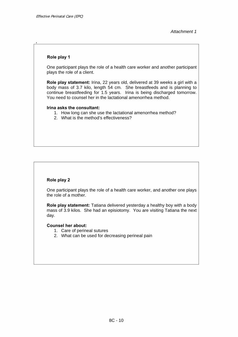
Effective Perinatal Care (EPC)
8C - 10
Attachment 1
. Role play 1
One participant plays the role of a health care worker and another participant plays the role of a client. Role play statement: Irina, 22 years old, delivered at 39 weeks a girl with a body mass of 3.7 kilo, length 54 cm. She breastfeeds and is planning to continue breastfeeding for 1.5 years. Irina is being discharged tomorrow. You need to counsel her in the lactational amenorrhea method. Irina asks the consultant:
1. How long can she use the lactational amenorrhea method? 2. What is the method’s effectiveness?
Role play 2
One participant plays the role of a health care worker, and another one plays the role of a mother.
Role play statement: Tatiana delivered yesterday a healthy boy with a body mass of 3.9 kilos. She had an episiotomy. You are visiting Tatiana the next day. Counsel her about:
1. Care of perineal sutures 2. What can be used for decreasing perineal pain

Effective Perinatal Care (EPC) Facilitator Guide
Module 9C Neonatal Resuscitation
Learning Objectives By the end of the module the participants will:
• Be ready for neonatal resuscitation in every birth, if needed • Ensure appropriate environment and equipment for every birth • Do the initial assessment of newborn status to decide when to start
resuscitation • Provide neonatal resuscitation through a continuous Assess-Classify-
Manage cycle • Understand the importance of the team approach for neonatal
resuscitation.
Module Outline and Duration Part I – Classroom work – 150 min
Activity 1 – Introduction 10 min Activity 2 – Interactive presentation 90 min Activity 3 – Conclusion 10 min Activity 4 – Small group work 40 min
Part 2 – Clinical practice
Activity 5 – Practical exercise on bag and mask assembling --------- Activity 6 – Practical exercise on neonatal resuscitation ---------
Preparation for the Module • Review of current publications, scientific evidence and healthcare strategies
focused on safe motherhood and effective neonatal care. • Ensure that all trainees have a copy of Participant Manual. • If possible, identify which common practices related to safe pregnancy and
effective neonatal care are used in the participants’ work settings. • Ensure that all facilitators know their roles and responsibilities regarding this
module.
9C - 1

Effective Perinatal Care (EPC)
Training Materials and Audiovisual Equipment Training Materials
• Participant Manual • Case studies for small group work (if needed) • Local guidelines and orders related to neonatal resuscitation (if possible)
Equipment
• LCD or slide projector • PowerPoint presentation 9С – EPC ENG • Flipchart • Markers • Pens or pencils • Name badges
Equipment for the practicum week
• Clock with second hand • 1 resuscitation mannequin and doll • 2 masks: size 1 and size 0 • 1 self inflating bag (250-400ml) • 1 suction device and tubing (mechanical or electrical or mouth-operated) • Cloth for folding and placing under shoulders • 2 towels/cloths for drying/warming
Key Messages
• Every newborn must be assessed at birth to decide when to start
resuscitation. • The appropriate environment and necessary resuscitation equipment must
be prepared for every birth in advance.
• Resuscitation equipment must be checked daily to ensure proper working condition.
• Each health worker attending deliveries must be skilled in neonatal
resuscitation and should be ready to perform resuscitation at every birth.
• A baby’s Apgar score at the end of the first minute does not determine whether or not neonatal resuscitation is needed.
• Effective neonatal resuscitation can potentially decrease neonatal mortality
and morbidity by about 40%.
9C - 2

Module 9C
PART I – Classroom work
Activity 1 – Introduction (10 min) • Show Slide 9C–1 and discuss the module objectives with the participants.
Explain that this module contains two parts: Part 1 – Classroom work which includes several activities, and Part 2 – Clinical practice done during the practicum week.
• Show Slide 9C–2 and ask the participants how many newborns require
resuscitation? Explain that “extensive resuscitation” includes intubation, chest compression, and drug administration, whereas “assistance to initiate breathing” can include tactile stimulation (drying), suction and ventilation with bag and mask.
• Show Slide 9C–3 and explain that all babies should be assessed for the need
for resuscitation. In the case when resuscitation is needed, a standard approach is to be applied.
• Show Slide 9C-4 and highlight that during this module, participants will study
neonatal resuscitation as an effective standard approach which can decrease neonatal mortality and morbidity by up to 40%.
Activity 2 – Interactive presentation (90 min) • Show Slide 9C-5 and discuss the main topics of effective neonatal
resuscitation which should be started without any delay. Highlight the need for any institution in which births occur to develop and administer local guidelines and training programmes based on current international practices, standards, skills and local clinical audits.
• Before showing the following slide, ask the participants, “Is the need for
neonatal resuscitation predictable?” Discuss briefly and then show slide 9C-6. Highlight that some neonates (as described on this slide), require neonatal resuscitation more often than others. Some clinical signs presenting during labour may also indicate a higher need for neonatal resuscitation.
• Slide 9C-7. Explain that up to half of newborns who require resuscitation have
no identifiable risk factors before birth. Thus the need for resuscitation must be anticipated at every birth and every birth attendant must be skilled in delivering neonatal resuscitation. Equipment for resuscitation and trained staff must be present in the delivery room or operation theatre not only when a high risk delivery is expected, but at every birth.
• Slide 9C-8 describes requirements for every delivery: a warm and draught-
free delivery room; warm, dry towels to help prevent neonatal hypothermia; a flat work surface; good light to facilitate assessing the newborn; a sterile kit to cut the cord (scalpel or scissors); and sterile gloves. Emphasize the importance of appropriate hand washing of the staff.
• Slide 9C-9. Discuss with participants neonatal resuscitation equipment which
must be prepared for every birth and checked daily. Every institution in which
9C - 3

Effective Perinatal Care (EPC)
births occur should select a staff member(s) who is responsible for preparing and checking resuscitation equipment and materials.
• Show Slides 9C-10 and 9C-11 and explain that the decision to start and
continue neonatal resuscitation is based on simultaneous assessment of the baby’s breathing, heartbeat, muscle tone and skin colour. These assessments are performed at birth and re-evaluated after every 30 seconds to decide whether to progress to the next step. Stress that the baby’s breathing is the key sign.
• Before showing Slide 9C-12, initiate a short discussion:
o What is an Apgar score? o What does it assess? o When the newborn’s Apgar score is determined?
• After this discussion, show Slides 9C-12 and 9C-13. Emphasize that the
results of scoring depend on many factors (for example, the mother’s use of anaesthetics and analgesics, neonatal infection, congenital anomaly, or prematurity) and that accurate scoring requires good training. Review how to determine a baby’s Apgar score with participants.
• Before showing Slide 9C-14, initiate a short discussion, “Can we use the
Apgar score at the end of the first minute of life as an indicator to start neonatal resuscitation?”
• After discussing, show Slide 9C-14 and explain that a baby’s need for
neonatal resuscitation is identified immediately at birth, whereas the first Apgar score is determined at the end of the first minute after birth. Therefore, by the time the Apgar score is determined, it is too late to start neonatal resuscitation.
• Slide 9C-15 shows the complete scheme of four categories of assessment
and action, ordered in strict sequence. Respiration, heart rate and colour are assessed simultaneously every 30 seconds, after which the baby is re-evaluated each time and the health care provider decides whether or not to progress to the next step in the sequence.
• Slide 9C-16 shows the initial assessment process which allows the provider to
determine the newborn’s status at birth and decide whether or not the baby requires resuscitation. If the newborn cries or breathes well (30-60 breaths per minute and does not grunt and gasp), becomes pink and has good muscle tone, he/she does not need resuscitation and should not be separated from the mother. The baby can be dried, placed directly on the mother's chest, and covered with a dry cloth to maintain body temperature. The provider should continue to monitor the baby’s breathing, activity, and colour.
• Slide 9C-17. If the baby is not breathing, or is grunting or gasping, or is
hypotonic, the provider should consider the need for neonatal resuscitation. In this case, the provider should do the steps listed on the slide and continue to assess the baby on a flat surface under a radiant heater.
• Slide 9C-18 shows the position the baby should be in order to clean his/her
airways. The baby should lie on a flat surface. Describe proper suctioning technique – first from the mouth, then from the nose, with suctioning and drying not taking longer than 30 seconds. Initiate a short discussion here by
9C - 4

Module 9C
asking, “Do we aspirate with a catheter or with a bulb?” A bulb is safer for the newborn and less expensive than a catheter. The bulb must not be reusable. Note: Ask another facilitator to demonstrate positioning the baby correctly, suctioning, and drying using a mannequin.
• Slide 9C-19 is hidden. The facilitator should decide whether or not it is
necessary to show this slide to participants. If not, the facilitator can describe this information during the discussion of Slide 9C-18.
• Slide 9C-20 describes steps to follow in the case of presence of meconium-
stained waters. Explain to the participants that routine aspiration of meconium before delivery of baby’s shoulders, during birth, or during resuscitation can cause severe aspiration pneumonia. One obstetric technique that was used in the past to decrease aspiration is intrapartum suctioning (suctioning meconium from the infant's airway after delivery of the head but before delivery of the shoulders). However, current recommendations no longer advise routine intrapartum oropharyngeal and nasopharyngeal suctioning for infants because there is a lack of evidence that intrapartum suctioning is effective for decreasing the risk of aspiration syndromeю
• It is important to immediately assess the baby at birth for breathing, heart rate
and muscle tone. If the newborn is breathing well (30-60 breaths per minute, no gasping, no grunting), his/her heart rate is over 100 beats per minute, and he/she has good muscle tone, provide essential care and close monitoring.
• If the newborn has breathing difficulties (no breathing or is gasping or
grunting), has a heart rate less than 100 beats per minute, or is hypotonic, do not dry the baby, immediately separate the baby and put him/her on a flat surface, clean the trachea through the endotracheal tube, and then dry him/her.
• Slide 9C-21. Explain that during resuscitation, endotracheal intubation can be
considered if a staff member skilled in endotracheal intubation is present. • Slide 9C-22. Explain that after the immediate assessment at birth and
administration of initial steps, further resuscitative efforts should be guided by simultaneous assessment of the baby’s breathing, heart rate, and colour. In general, after initial respiratory efforts, newborn infants should be able to establish adequate regular breathing that is sufficient to improve colour and maintain a heart rate of more than 100 beats per minute. In these cases, the baby should be put on the mother’s chest. Health workers should continue to provide essential care and monitoring.
• If the newborn has breathing difficulties, has a heart rate less than 100 beats
per minute, is cyanotic or pale, or is hypotonic, ventilation with a bag and mask should be started. In this case, health workers should call for help because this newborn may require intubation, chest compression, and/or drug administration.
• Slides 9C-23 and 9C-24. These slides describe ventilation techniques using a
bag and mask. Stress that the bag must only be squeezed using the fingers. Point out that if the maternity has no bag and mask equipment, health workers must deliver ventilation using a mouth-to-mouth or mouth-to-nose technique.
9C - 5

Effective Perinatal Care (EPC)
• Slide 9C-25 shows steps to perform if no chest wall movements are detected during ventilation. Shortly describe most probable reasons for a lack of chest wall movement.
• Note: Give a correct short ventilation demonstration (not more than 10
minutes) using a bag, mask, and mannequin.
• Describe briefly the key parts of a self-inflating bag (patient’s outlet, oxygen inlet, air inlet with oxygen reservoir attached, and safety valve). Show the participants how to check the working condition of a Ambu bag (safety valve, no leakage):
o Hold the patient’s exhale with your hand and squeeze the bag – you must see the safety valve and hear the sound of its opening.
o Hold the patient’s exhale and the safety valve with your hand and squeeze the bag – you will not be able to do it if the bag does not leak; you will feel resistance.
• Show the participants how to perform artificial lung ventilation with a bag and
mask using a resuscitation mannequin: o Note the time; o Position the newborn correctly (a cloth roll under the shoulders, head
extension position); o Attach the mask to the newborn’s face and seal it; o Squeeze the bag with your fingers gently; o Check for chest wall movements; o If there are no chest wall movements, act appropriately (refer to Slide
9C – 25); o Ventilate at a rate of 40-60 squeezes per minute.
• Slide 9C-26 describes REASSESSMENT of the newborn’s breathing and
heart rate after 30 seconds of ventilation with the bag and mask (now 1 minute after birth) where the baby’s breathing has improved. Remind the participants that health workers should assess the baby’s condition for the first time during the initial assessment, then again after the initial steps of resuscitation and ventilation with bag and mask has been performed.
• Slide 9C-27. If the baby’s condition improves after ventilation with the bag and
mask and has a good respiratory rate and heart beat rate, he/she should be given to his/her mother. The baby’s breathing condition and temperature should be carefully monitored during this time.
• Slide 9C-28 describes REASSESSMENT of the newborn’s breathing and
heart rate after 30 seconds of ventilation with the bag and mask (now 1 minute after birth) where the baby’s breathing has not improved. In the described cases, the baby will require continued resuscitation.
• Slides 9C-29 through 9C-32 show chest compression rules (slide 9C-29) and
techniques (slides 9C-30 to 9C-32). (Note: Slides 9C-31 and 9C-32 are hidden. The facilitator should decide whether to show them or not). Highlight the importance of achieving approximately 120 events per minute (30 ventilations and 90 chest compressions per minute). Emphasize that the quality of the compressions and breaths are more important than the rate. Remind the participants that lung ventilation and chest compression cannot be performed by one person alone; participation of at least two trained staff is
9C - 6

Module 9C
necessary. Highlight the fact that at this stage of resuscitation, the possibility of intubation should be considered. Therefore, a person with intubation skills should be present.
• Note: Demonstrate two neonatal resuscitation techniques on the mannequin. • Slides 9C-33 and 9C-34 describe reassessment of the baby’s breathing and
heart rate 1 minute and 30 seconds after birth, after ventilation with a bag and mask and chest compressions have been completed. In the case where the baby’s heart rate is less than 60 beats per minute, give 30 seconds of chest compression and ventilation with 100% of oxygen, followed by 10-30 mcg/kg of adrenalin
• Slide 9C-34. In addition to adrenaline, the facilitator can describe other drugs
that can be considered: o Volume expansion: Consider volume expansion when blood loss is
suspected or the infant appears to be in shock (pale skin, poor perfusion, weak pulse) and has not responded adequately to resuscitative measures (ventilation, chest compression, adrenaline). Normal saline rather than albumin is the solution of choice for volume expansion in the delivery room. The recommended dose is 10 mL/kg, which may need to be repeated. When resuscitating premature infants, care should be taken to avoid giving volume expanders too rapidly, because rapid infusions of large volumes have been associated with intraventricular haemorrhage.
o Bicarbonate: Please note that Sodium bicarbonate is controversial.
Beveridge & Wilkinson (2005) states the following in Background: “The most recent international consensus guidelines for neonatal resuscitation state that “there is insufficient data to recommend routine use of sodium bicarbonate in resuscitation of the newly born. Its use is discouraged during brief cardiopulmonary resuscitation and if it is used during prolonged arrests unresponsive to other therapy, it should be given only after establishment of adequate ventilation and circulation.
o The Resuscitation Council (UK) recommends administration of sodium bicarbonate when there is no effective cardiac output, or virtually none, prior to a second dose of adrenaline
o Naloxone: Administration of naloxone is not recommended as part of initial
resuscitative efforts in the delivery room for newborns with respiratory depression. If administration of naloxone is considered, heart rate and colour must first be restored by supporting ventilation. The preferred route is IV or intramuscular. Given the lack of clinical data in newborns, endotracheal administration of naloxone is not recommended. The recommended dose is 0.1 mg/kg, but no studies have examined the efficacy of this dose in newborns. In 1 case report, naloxone given to an infant born to an opioid-addicted mother was associated with seizures. Therefore, naloxone should be avoided in infants whose mothers are suspected of having had long-term exposure to opioids.
• Slide 9C-35. Preparation for transfer of the baby and providing care during
transfer: o Ensure that the baby’s condition is stable before transfer.
9C - 7

Effective Perinatal Care (EPC)
o Have a health care provider (with experience in establishing and maintaining intravenous lines, resuscitating the baby and giving drugs) accompany the baby (if possible).
o Gather essential equipment, supplies, drugs and fluid. o Treat low blood glucose, if possible:
- If the baby is able to feed, breastfeed the baby or use alternative feeding method;
- If the baby is not able to feed, establish the IV line (if possible) and give fluid.
o Keep the baby warm (using an incubator, skin-to-skin contact, or a warm blanket).
• Slide 9C-36. Ask the participants, “When should you consider stopping
neonatal resuscitation efforts?” This is a difficult decision both for health workers and for parents, as there is no clear information about the maximum length of time resuscitation efforts should be continued.
• Slide 9C–37. This diagram shows the main causes of 4 million neonatal
deaths worldwide in 2000. Complications of neonatal asphyxia are the third most common cause of neonatal mortality in the world, but these deaths can be reduced by providing effective neonatal resuscitation.
• Slide 9C-38 shows the definition of newborn asphyxia. Explain that asphyxia
is a disease, not a symptom. An asphyxia diagnosis must be supported by laboratory test results: acidosis after birth; Apgar score of 0-3 points for more than 5 minutes; neurological sequelae in 72 hours; and polyorgan dysfunction. Asphyxia is diagnosed if all four signs are present. As with the Apgar score, neonatal asphyxia should not be used as an indicator to start neonatal resuscitation.
Activity 3 – Conclusion (10 min) • Show Slide 9C-39 for conclusions. • Ask the participants if they have any questions. Answer questions.
Activity 4 – Small group work (40 min) This activity can be conducted at any time, as long as module 4N has been completed. • Divide participants into four groups. Ensure that each group includes
physicians and nurses. Give flip-chart paper and markers to each group. • Ask participants to carefully read the case study and questions in the
Participant’s Manual at the end of this module. • Explain that group 1 will answer question 1, group 2 question 2, etc. • Ensure that participants understand the task.
9C - 8

Module 9C
• Give each group 10 minutes to answer their question. Answers should be written on the flip-chart and presented to the big group by one subgroup member.
Case study: Bogdan is born after 40 weeks of gestation. Vacuum-extraction was performed. The amniotic fluid was clear. Immediately after birth the midwife put Bogdan on his mother’s chest, he was gasping. The midwife cut the cord and moved Bogdan to the table, turned the radiant heater on, dried the boy with towels, and conducted tactile stimulation along his back. Then the midwife aspirated from the nose and the mouth and assessed Bogdan (end of the first min). Bogdan was breathing irregularly at a rate of only 20 breaths per minute. When his nose and mouth were aspirated he grimaced. His heart rate was 90 beats per minute. His extremities were cyanotic and he was hypotonic. The midwife called for help. The doctor came within three minutes with a bag and a mask, re-assessed Bogdan and started ventilation. Questions.
1. Was everything done correctly in Bodgan’s case? What could have been done differently?
2. Which resuscitation equipment must be prepared for every birth?
• When should be the equipment have been prepared? • How was the preparation of equipment in Bogdan’s case?
3. Should Bogdan be assessed by an Apgar Score at 1 minute? Please explain?
4. What should be done for Bogdan in the following 5 minutes?
Possible answers: • Question 1: Was everything done correctly in Bodgan’s case? What
could have been done differently? No, in this case Bogdan was not assessed for breathing at birth. He was separated from his mother and put on a cold surface. The midwife turned on the radiant heater after Bogdan’s birth. Suction was performed in the wrong order (before positioning the baby correctly). Bogdan’s breathing; heart rate and colour were assessed too late (at the end of first minute).
• Things that could have been done: prepared environment before delivery (turn
on radiant heater, prepare warm towel); assessed breathing at birth; separated from mother and positioned correctly; suctioned first from mouth, then from nose; assessed for breathing, heart rate and colour after initial steps. All skilled attendants must be able to perform neonatal resuscitation. In case need the midwife should call for help and start immediately bag and mask ventilation. It is strongly recommended that the midwife or the skilled attendant for birth must be certified in neonatal resuscitation.
9C - 9

Effective Perinatal Care (EPC)
• Question 2: Which resuscitation equipment must be prepared for every birth?
o Bag and masks in two sizes; mucus extractor, bulb, or catheter;
laryngoscope with appropriate blade; intubation tube; normal saline; adrenaline; syringes prepared and checked for working condition before birth.
- When should be the equipment have been prepared??
o Resuscitation equipment should have been prepared before the baby’s birth.
- How was the preparation of equipment in Bogdan’s case?
o In Bogdan’s case, the resuscitation equipment was not prepared in
advance and was not placed in the birth room. Thus ventilation with a bag and mask started too late.
• Question 3: Should Bogdan be assessed by an Apgar Score at 1 minute? Please explain?
o Bogdan must be assessed by Apgar score at the end of 1st minute but not
for decision to start neonatal resuscitation.
o Apgar score at 1 minute – 5 points: heartbeat = 1 point; breathing = 1 point; skin colour = 1 point; muscle tone = 1 point; reflex response = 1 point.
• Question 4: What should be done for Bogdan in the following 5 minutes?
o Continue ventilating with bag and mask for 30 seconds. Reassess his
breathing, heart rate and colour.
9C - 10

Module 9C
PART II – Clinical practice • During the clinical week, hands-on sessions on neonatal resuscitation
(Activities 5 and 6 below) must be conducted. • All the participants can be split into 2 groups.
Activities 5 – Practical exercise on bag and mask assembly
• The goal of this session is to give the participants practice in bag and mask assembly.
• Show the participants how to assemble and dissemble an Ambu bag
correctly (attach and detach the mask to/from the bag). • Stress that it is necessary to attach the oxygen reservoir to the self-inflating
bag to ensure 90-100% oxygen supply to the newborn. • Explain to the participants how to select the correct mask size for neonatal
resuscitation: size 0 for a preterm newborn, size 1 for a term newborn. • Remind participants that it is preferable to use masks with soft edges for
newborn resuscitation. • Show participants how to assemble and disassemble the bag and masks. • During the course, each participant must practice preparing and using the
bag and mask at least three times.
Activity 6 – Practical exercise on neonatal resuscitation • The goal of this session is to practice neonatal resuscitation procedures. • This activity is conducted in the joint group of obstetricians-gynaecologists,
neonatologists, midwives and paediatric nurses. All participants must practice neonatal resuscitation (drying the mannequin, suction, ventilation with bag and mask, chest compression) at least three times.
• Before each exercise, the bag and mask must be dissembled.
• Show participants all of the neonatal resuscitation steps: position the baby
correctly; suction first from mouth then from nose; dry the baby; ventilate with bag and mask (attach the mask correctly, seal the mask, squeeze the bag with fingers gently with a rate of around 40 squeezes per minute, ventilate for 30 seconds); and perform chest compression (ask a co-facilitator to help you and show participants two chest compression techniques: with thumbs and with fingers).
• When ventilating, suggest to participants an easy way to count:
o SQUEEZE – count aloud “one hundred and one”, SQUEEZE – “one hundred and two”, SQUEEZE – “one hundred and three”, etc.,
9C - 11

Effective Perinatal Care (EPC)
until you reach ”one hundred and forty”. You can then start again or continue with SQUEEZE – “two hundred and one”, SQUEEZE, etc.
• Use Table 1 to evaluate participants’ neonatal resuscitation skills during
Activity 6. Check off each action if it was done correctly.
• Use the case studies below for more practice. Scenario 1 Describe the case step by step and select participants to perform appropriate resuscitation measures required after each step. Ask the rest of the group to observe and be ready to discuss this case after it is finished. Task 1: A “full term newborn” is gasping at birth. Your actions? Possible actions: cut the cord; position the mannequin correctly and put a cloth roll under its shoulders to extend the neck slightly; suction from mouth, then from nose; and describe assessing the baby’s breathing, heart rate and colour. Task 2: After performing these actions, the newborn is gasping, is cyanotic and has a heart rate of 120 beats per minute. Your actions? Possible actions: start bag and mask ventilation under positive pressure for 30 seconds. After that, assess breathing, heart rate and colour. Task 3: After this the newborn starts breathing well, the heart rate is 120 beats per minute, and the newborn becomes pink. Your actions? Possible actions: stop bag and mask ventilating; assess the newborn by Apgar scale; place baby on the mother’s chest for skin-to-skin contact.
Scenario 2 Describe the case step by step and select participants to perform appropriate resuscitation measures required after each step. Ask the rest of the group to observe and be ready to comment when this case is finished. Task 1: A “full term newborn” is gasping breathing at birth. Your actions? Possible actions: cut the cord; position the mannequin correctly and put a cloth roll under its shoulders to extend the neck slightly; suction from mouth, then from nose; and describe assessing the baby’s breathing, heart rate and colour.
Task 2: After this the newborn is gasping, is cyanotic and has a heart rate of 90 beats per minute. Your actions? Possible actions: start bag and mask ventilation under positive pressure for 30 seconds. After that, assess breathing, heart rate and colour.
Task 3: The newborn is gasping, is cyanotic and his heart rate is 50 beats per minute. Your actions? Possible actions: start chest compression and continue bag and mask ventilation. At the same time, quickly assess the baby’s Apgar score. After 30 seconds, re-evaluate the baby’s breathing, heart rate and colour.
9C - 12

Module 9C
Table 1. Evaluation of participant neonatal resuscitation skills
Participant Actions
1st 2nd 3rd 4th 5th 6th 7th 8th 1. Assess breathing, muscle tone at birth 2. Position the newborn correctly: with a roll of
cloth under the shoulders to slightly extend the neck
3. Aspirate from the newborn’s mouth and nose 4. Dry the newborn 5. Assess breathing, heart rate, colour 6. Position the newborn correctly again 7. Select an appropriate mask (size 0 for preterm
babies and size 1 for term babies)
8. Attach the mask to the newborn’s face so that it covers the chin, mouth and nose
9. Seal the mask to the newborn’s face 10. Clearly note the time ventilation begins and ask
the assistant to record it
11. Squeeze the bag with the fingers 12. Ventilate at a rate of 40-60 squeezes per
minute
13. Evaluate chest wall movements during ventilation
14. Take correct measures if no chest wall movements:
o Reposition the newborn’s head o Take off and reseal the mask o Squeeze the bag with full hand
15. Assess the newborn’s breathing and heart rate after 30 seconds of bag and mask ventilation
16. Start chest compression if the heartbeat is below 60 beats per minute, continue ventilating with bag and mask
17. Assess the newborn’s heartbeat and breathing rate after 30 seconds of chest compression and bag and mask ventilation
Conclusions:
9C - 13
Effective Perinatal Care (EPC)
References
1. American Heart Association, American Academy of Pediatrics 2005 American Heart Association (AHA) Guidelines for Cardiopulmonary Resuscitation (CPR) and Emergency Cardiovascular Care (ECC) of Pediatric and Neonatal Patients: Neonatal Resuscitation Guidelines; Pediatrics, May 2006; 117: e1029 - e1038.
2. Basic Newborn resuscitation: a practical guide. WHO, Geneva, 1997.
3. Chalmers I, Enkin M.W, Keirse MJNC, eds. Effective care in pregnancy and childbirth. Oxford, Oxford University Press, 1989.
4. European Resuscitation Council Guidelines for Resuscitation, 2005. Section 6: Pediatric life support.
5. Gary L Darmstadt, Zulfigar A Bhutta, Simon Cousens et al “Evidence-based, cost-effective interventions: how many newborn babies can we save?”. Lancet, March 2005:19-30
6. Joy E Lawn, Simon Cousens, Jelka Zupan et al “4 million neonatal death: When? Where? Why?”. Lancet, March 2005: 9-18 (2005 Mar 5-11;365(9462):891-900)
7. Palme-Kilander C, Tunell R, Chiwei Y. Pulmonary gas exchange immediately after birth in spontaneously breathing infants. Arch Dis Child, 1993,68:6-10
8. Peliowski A, Finner N.N. Birth asphyxia in the term infant. In: Sinclair JC, Bracken MB (eds.) Effective care of the newborn infant. Oxford, Oxford University Press, 1992, 249-273.
9. Saugstad OD, Rootwelt T, Aalen O. Resuscitation of asphyxiated newborn infants with room air or oxygen: an international controlled trial. Pediatrics, 1998
10. Thermal Protection of the newborn: a practical guide. Geneva, World Health organization, 1997.
11. Tyson JE. Immediate care of the newborn infant. In: Sinclair JC, Bracken MB (eds.) Effective care of the newborn infants. Oxford University Press, 1992.
12. Vain NE, Prudent LM, Wiswell T, et al. Oropharyngeal and nasopharyngeal suctioning of meconium stained neonates before delivery of their shoulders: multicenter, randomised trial. Lancet. 2004;364:597–602
13. European Resuscitation Council Guidelines for, Resuscitation 2005, Section 9.
Principles of training in resuscitation, Peter J. F. Baskett, Jerry P. Nolan, Anthony Handley, Jasmeet Soar, Dominique Biarent, Sam Richmond, Resuscitation (2005) 67S1, S181—S189
14. American Heart Association, American Academy of Pediatrics, and Neonatal
Patients: Neonatal Resuscitation Guidelines and Emergency Cardiovascular Care (ECC) of Pediatric 2005 American Heart Association (AHA) Guidelines
9C - 14

Module 9C
for Cardiopulmonary Resuscitation (CPR),, http://www.pediatrics.org/cgi/content/full/117/5/e1029
15. Cochrane review relevant to the module: Endotracheal intubation at birth for
preventing morbidity and mortality in vigorous, meconium-stained infants born at term
16. Cochrane review relevant to the module: Epinephrine for the resuscitation of
apparently stillborn or extremely bradycardic newborn infants 17. Cochrane review relevant to the module: Laryngeal mask airway versus bag-
mask ventilation or endotracheal intubation for neonatal resuscitation 18. Cochrane review relevant to the module: Sodium bicarbonate infusion during
resuscitation of infants at birth
9C - 15

Effective Perinatal Care (EPC)
9C - 16

10C - 1
Module 10 C
Integration of Prevention of Mother to Child HIV Transmission into Effective Perinatal Care
Learning objectives By the end of the module, participants should:
• Be informed on the magnitude of MTCT in the European region • Be informed about the risk of Mother to Child Transmission of HIV • Understand that PMTCT needs to be fully integrated in routine antenatal,
intra partum and postpartum care • Understand that HIV-positive mothers and babies need to be treated with
respect and confidentiality • Be familiar with prophylaxis ARV treatment to prevent MTCT,
counselling and specific care for HIV-positive women in antenatal period
• Be familiar with major obstetric scenarios for HIV-positive women at admission to maternity
• Be knowledgeable about specific care for the immediate postpartum period for HIV-positive mother and newborn preventing MTCT
• Be conversant with infant feeding patterns in cases of HIV-positive mother for preventing MTCT
• Be trained in basic counselling skills
Module outline and length
Part I – Classroom work (110 min) Activity 1 – Introduction 5 min Activity 2 – Interactive presentation 1st part 15 min Activity 3 – Group discussion 10 min Activity 4 – Interactive presentation 2nd part 15 min Activity 5 – Interactive presentation 3rd part 60 min Activity 6 – Conclusion 5 min
Part II – Clinical work
Activity 7 – Checking potential stigmatization in maternity 60 min Activity 8 – Role play: Infant feeding counselling for HIV-positive mother
30 min
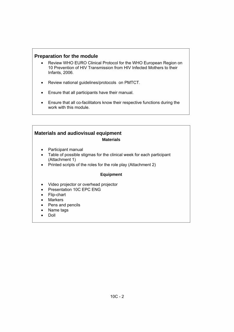
10C - 2
Preparation for the module • Review WHO EURO Clinical Protocol for the WHO European Region on
10 Prevention of HIV Transmission from HIV Infected Mothers to their Infants, 2006.
• Review national guidelines/protocols on PMTCT. • Ensure that all participants have their manual. • Ensure that all co-facilitators know their respective functions during the
work with this module.
Materials and audiovisual equipment Materials
• Participant manual • Table of possible stigmas for the clinical week for each participant
(Attachment 1) • Printed scripts of the roles for the role play (Attachment 2)
Equipment • Video projector or overhead projector • Presentation 10С EPC ENG • Flip-chart • Markers • Pens and pencils • Name tags • Doll

10C - 3
Key Module Messages
• Eastern Europe has one of the fastest growing HIV epidemics in the world. • The prevention of MTCT needs to be fully integrated into routine care in
antenatal and maternity site, thereby preventing the creation of a “PMTCT vertical program."
• HIV voluntary testing and counselling should be routinely offered, as it is
an entry point to preventing MTCT (antenatal care and maternity). • The risk of MTCT could be reduced from > 40% to < 2% by receiving
appropriate care during the antenatal, labour and postpartum periods. • ARV prophylaxis for mother during pregnancy, labour and postpartum
period and for newborn is the main strategy to prevent MTCT.
• Elective caesarean section is recommended to reduce MTCT for 50%.
• Safe delivery practices should be implemented to reduce newborn exposure to mothers’ blood and secretions.
• Breastfeeding replacement should be discussed: if available, affordable, sustainable and safe as up to 15% of newborns could be infected through breastfeeding.
• HIV status in infants born to HIV-positive mothers can be established by 6
weeks of age by using PCR test. PCR diagnosis is used in children younger 18 month of age because ELISA test can be false positive due to presence of maternal antibodies before the age of 18 month.
• Infants born to HIV+ mothers should receive prophylaxis against
Pneumocistic carnii. If the negative status is proven the prophylaxis is stopped.
Organizational part
• It is important that a minimum of two facilitators conduct this module. The Course Director must decide which part should be presented by a neonatologist, midwife and/or obstetrician.

10C - 4
PART I – Classroom work
Activity 1 – Introduction (5 min)
• Show slide 10C-1. The prevention of MTCT needs to be fully integrated into routine care in antenatal and maternity site, thereby preventing the creation of a “PMTCT vertical program."
• It is also important to engage more HIV-positive women to seek medical care;
with integration the effectiveness of PMTCT program would likely increase.
• Present and discuss the learning objective of this module with participants.
• Then a facilitator will share the following main points: o The risk of MTCT is significant in the European region o The risk of a newborn being infected without any intervention is
approximately 40% and could be reduced to 2% if appropriate interventions are implemented.
a. The facilitator should emphasize the necessity of having PMTCT fully integrated
into routine antenatal, intra partum and postpartum care in order to avoid stigmatization and creating a new vertical program.
b. The facilitator should emphasize the necessity of treating HIV-positive mothers
and babies with respect and confidentiality therefore each medical staff need to have basic counselling skills.
Activity 2 – Interactive presentation 1st part (15 min)
• Show slide 10C-2. Explain to participants: o The highest prevalence of HIV/AIDS is in Sub-Saharan Africa followed by
East and South Asia. o Presented data are estimates. Note information may be underreported.
• Stress that Eastern Europe has one of the fastest-growing HIV epidemics in the
world. • Show slide 10C-3. These graphs show the increase of HIV prevalence (number
of cases per 10,000 people) among pregnant women in the Russian Federation, Ukraine, Belarus and Georgia – Georgian data is not available before 2004.
• Show slide 10C-4. Explain to participants that the comprehensive strategy to
reduce HIV infection in mothers and infants includes four complementary components. Only the third one - the Prevention of Mother to Child Transmission - will be explored during the module.

10C - 5
Interactive activity 2-3 min: Before showing the following slide, please ask participants: Will all babies born to an HIV-positive mother be infected with HIV? Lead a short discussion (under 2 or 3 minutes).
• Show slide 10C-5. Present to participants the following information: Preble and Piwoz (2002) used MTCT data from scientific studies to show the risk of MTCT in a hypothetical group of 100 HIV positive mothers if no interventions are done during pregnancy, labour and if the mother is breastfeeding for 2 years.
• Explain to participants that on the slide they can see the results of the analysis of
this hypothetical group showing that 63 infants out of 100 born to HIV-positive mothers are not infected despite the fact the mothers were not treated at all and breastfed their children until they were two years age. The point is that not all children born of HIV-positive mothers will be HIV-positive themselves.
• Show slide 10C-6. Present to participants the interventions which should be
done to prevent mother-to-child HIV transmission. Tell them that in the absence of any intervention the risk of such transmission is 15–30% in non-breastfeeding populations. Breastfeeding by an infected mother increases the risk by 5–20% to a total of 20–45%. The risk of MTCT can be reduced to under 2% by interventions that include antiretroviral (ARV) prophylaxis given to women during pregnancy and labour and to the infant in the first weeks of life, obstetrical interventions including elective caesarean delivery (prior to the onset of labour and rupture of membranes), and complete avoidance of breastfeeding. With these interventions, new HIV infections in children are becoming increasingly rare in many parts of the world, particularly in high-income countries.
• Note that in many resource-constrained settings, elective caesarean delivery is
seldom feasible and it is often neither acceptable nor safe for mothers to refrain from breastfeeding. In these settings, the efforts to prevent HIV infection in infants initially focused on reducing MTCT around the time of labour and delivery, which accounts for one to two thirds of overall transmission, depending on whether the mother breastfeeds.
Interactive activity (3 min): Please ask one participant: Which interventions, ideally, should be implemented to reduce MTCT to 2%? 1. ARV prophylaxis or ART during pregnancy and labour; 2. Elective caesarean section; and/or 3. Safe breastfeeding replacement.

10C - 6
• Show slide 10C-7. Tell the participants that goals for the European region to eliminate HIV infection in infants by 2010 could be achieved if the four prongs of Strategy on Elimination of HIV in Infants in Europe are implemented
• Show slide 10C-8. Explain to participants that due to stigma and discriminationI
women are often afraid to seek care, even to prevent MTCT. Therefore, in each clinic and in each maternity, all staff needs to respect the rules of confidentiality and help in the fight against discrimination and stigmatization targeting HIV-positive women and their children. Discrimination against PLWHA or people thought to be infected is a violation of human rights.
Activity 3 – Group discussion (10 min) • Show slide 10C-9. HIV-positive mothers and babies need to be treated as other
mothers and babies, with love, respect and benefiting from evidence-based perinatal care.
• Ask one participant to read out loud a letter written by an HIV-positive woman to
maternity staff. • Then ask all participants: Why was this woman so thankful to the maternity staff? • Write their answers on the flip-chart. Discuss answers.
Activity 4 - Interactive presentation 2nd part (15 min) PMTCT in antenatal period • Show slides 10C-10 and 10C-11. The first intervention during the antenatal period
is to provide appropriate information and offer HIV testing for each woman receiving antenatal care. Tell participants that HIV counselling and testing during antenatal clinic visits is a key PMTCT strategy. Partner involvement needs to be supported.
• Show slides 10C-12 and 10C-13. The second PMTCT intervention in antenatal
care is to ensure special care and support to each HIV-positive pregnant woman. o Tell participants that all HIV-positive women should make decisions about her
pregnancy by herself/with her partner. o The role of health workers needs to be strictly limited to providing clear and
detailed information to women about MTCT risks as well as information about preventative procedures.
o HIV-positive pregnant women need to receive appropriate care and additional information and counselling about MTCT prevention
o Staging of HIV disease; ARV prophylaxis or therapy o Counselling on safest mode for birth o Counselling on safest infant feeding

10C - 7
• Medical care and support to HIV-positive pregnant women needs to be multidisciplinary. Most HIV-positive women discover their HIV-positive status in antenatal clinics, and this can be extremely stressful and difficult for women. Peer support via HIV-positive social workers is essential, and should be provided to HIV-positive women as soon as possible.
• After confirming a woman's HIV-positive status, the woman should be gently
referred to a special department/centre where she should have a complete medical assessment for her condition.
• Show slides 10C-14 and 10C-15. The third intervention in the antenatal period is
the prescription of antiretroviral drugs to HIV-positive pregnant women. • Based on the women's health status and the availability of ARV medications,
pregnant women could receive different ARV regimens during pregnancy. • The decision of ART regimen during pregnancy (MTCT prophylaxis regimen or
treatment regimen) should be based on clinical stage of HIV disease and in combination with immunological criteria.
• ARV treatment regimens for those women who are on need of ART for their own
health is also effective to prevent MTCT. ARV prophylaxis is also given to mothers during labour and postpartum period and for their infants during first weeks of life.
• The most commonly recommended prophylaxis ART regimen (for women who do
not need ARV treatment for their own health) during pregnancy is Zidovudine (AZT or ZDV) from 24-28 weeks of pregnancy or as soon as possible thereafter.
• ZDV could also be used if Highly Active Antiretroviral Treatment (HAART) is not
available. • HAART, a combination of 3 drugs, is the core intervention for the treatment of
advanced HIV disease or AIDS. HAART is also the most effective prophylaxis for MTCT.
• The most common drug combinations used as HAART
o Zidovudine/Lamivudine/Nevirapine
• Ask participants to open the Attachment 3 at the end of this module and tell them that here they can find more detailed information on stages and recommendations for treatment of HIV+ pregnant women, but you will not do it now because of the limited time. Propose them to read it carefully at home.
Interactive activity 5 min: Please ask the group or one participant: Which ARV regimens should be prescribed to an HIV+ asymptomatic pregnant woman, without immunosuppression (count of CD4 > 350 cells/mm3) and if Viral load is less then 10 000 copies/ml and patient is ZDV naive?

10C - 8
The correct answer is: Zidovudine (AZT or ZDV) started from 24-28 weeks of pregnancy or as soon as possible thereafter. Activity 5 - Interactive presentation 3rd part (60 min) PMTCT in maternity (intrapartum and post partum) • Show slide 10C-16. Inform the participants that the following slides will describe
PMTCT interventions in maternity. The first PMTCT intervention at admission to the maternity is the assessment of the maternal HIV status, if HIV status is unknown pre-test counseling and rapid HIV tests should be done.
• Show slide 10C-17. This slide repeats the main message to participants, which is
that all maternity staff need to respect the rules of confidentiality everywhere providing care for women and their children: o Do not isolate HIV-positive women into special department or room; o Do not deliver HIV-positive woman in a special delivery room or special operating
theater; o Do not mark HIV-positive patients’ files so they are easily recognized in the
maternity; o Never disclose HIV-positive status; o Provide to HIV-positive women and their children the same level of care as HIV-
negative women; o Implement rooming-in and free visiting of relatives and friends for all women
including HIV-positive mothers • Main reasons preventing HIV-positive women from seeking medical care is their fear
of their HIV-positive status and the potential stigmatization and discrimination towards them and their children.
• Medical care providers should respect the confidentiality and privacy of each woman,
including HIV-positive mothers and their babies, and do their best to fight against stigma and discrimination in their maternity.
• Show slide 10C-18. Explain to the participants that the HIV status of women
admitted to the maternity could be assessed from the antenatal records if existing. If the HIV status is not known prior to admission conduct rapid HIV pre-test counselling and if the woman agrees to be tested, conduct a rapid HIV test in the maternity. If the woman refuses to be tested provide her with safe delivery and appropriate counselling on safe baby feeding.
• Show slide 10-C-19. Tell participants that the second intervention is the choice of
delivery method is very important as it could reduce the MTCT risk • Show slide 10-C-20. Tell that whenever a woman is admitted to the maternity an
assessment of labour needs to be conducted immediately. • If this woman is known as HIV-positive
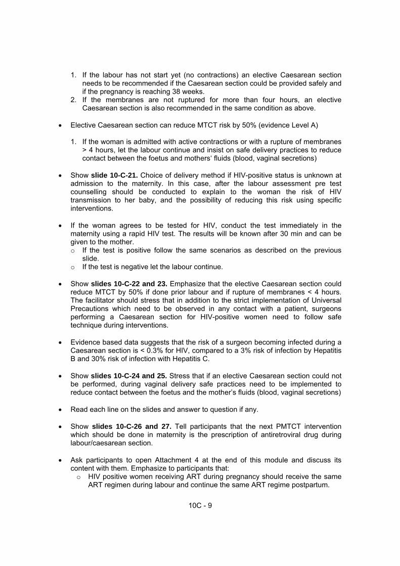
10C - 9
1. If the labour has not start yet (no contractions) an elective Caesarean section needs to be recommended if the Caesarean section could be provided safely and if the pregnancy is reaching 38 weeks.
2. If the membranes are not ruptured for more than four hours, an elective Caesarean section is also recommended in the same condition as above.
• Elective Caesarean section can reduce MTCT risk by 50% (evidence Level A)
1. If the woman is admitted with active contractions or with a rupture of membranes > 4 hours, let the labour continue and insist on safe delivery practices to reduce contact between the foetus and mothers‘ fluids (blood, vaginal secretions)
• Show slide 10-C-21. Choice of delivery method if HIV-positive status is unknown at
admission to the maternity. In this case, after the labour assessment pre test counselling should be conducted to explain to the woman the risk of HIV transmission to her baby, and the possibility of reducing this risk using specific interventions.
• If the woman agrees to be tested for HIV, conduct the test immediately in the
maternity using a rapid HIV test. The results will be known after 30 min and can be given to the mother. o If the test is positive follow the same scenarios as described on the previous
slide. o If the test is negative let the labour continue.
• Show slides 10-C-22 and 23. Emphasize that the elective Caesarean section could
reduce MTCT by 50% if done prior labour and if rupture of membranes < 4 hours. The facilitator should stress that in addition to the strict implementation of Universal Precautions which need to be observed in any contact with a patient, surgeons performing a Caesarean section for HIV-positive women need to follow safe technique during interventions.
• Evidence based data suggests that the risk of a surgeon becoming infected during a
Caesarean section is < 0.3% for HIV, compared to a 3% risk of infection by Hepatitis B and 30% risk of infection with Hepatitis C.
• Show slides 10-C-24 and 25. Stress that if an elective Caesarean section could not
be performed, during vaginal delivery safe practices need to be implemented to reduce contact between the foetus and the mother’s fluids (blood, vaginal secretions)
• Read each line on the slides and answer to question if any. • Show slides 10-C-26 and 27. Tell participants that the next PMTCT intervention
which should be done in maternity is the prescription of antiretroviral drug during labour/caesarean section.
• Ask participants to open Attachment 4 at the end of this module and discuss its
content with them. Emphasize to participants that: o HIV positive women receiving ART during pregnancy should receive the same
ART regimen during labour and continue the same ART regime postpartum.

10C - 10
o If pre-labour caesarean section (PLCS) woman should be prescribed intravenous Zidovudine is available, start IV infusion four hours before PLCS (2mg/kg for first hour and 1mg/kg/hour until the cord is clamped).
• Tell participants that if pregnant woman on ARV prophylaxis or not on ARV
prophylaxis during pregnancy: o At the onset of labour they should prescribe:
- AZT 600mg (two tablets of 300mg) once* (Note that the woman may have already taken this dose at home – ask her and record the results. If she has taken this dose at home, administer NVP and 3TC) PLUS
- NVP 200mg as a single dose* (not necessary to give this if the woman was on AZT for > 4 weeks) PLUS
- 3TC 150mg and continue 3TC 150mg every 12 hours until delivery o After childbirth give
- AZT 300mg and 3TC 150mg twice a day for 7 days
• Show slide 10-C-28 and 29. Tell participants that HIV is present in breast milk of HIV-positive mothers. The risk of MTCT through breastfeeding is about 15 %. The risk becomes higher as the breastfeeding period continues longer.
• Show slide 10C-30. Describe to participants that this study shows the levels of
MTCT depend on the feeding patterns during a 15 - month period: exclusive breastfeeding, mixed feeding and breastfeeding replacement. Stress that the greatest risk of MTCT occurs in the case of mixed feeding.
• Show slide 10C-31. Emphasize that breastfeeding replacement should be discussed
with the mother and the family and recommended only if available, feasible, affordable, safe and sustainable. Otherwise exclusive breastfeeding is recommended during the first months of life and should be discontinued as soon as conditions for the replacement feeding are met.
• Table: Quantity of needed milk from birth until six month (ml/kg/day).
Age Quantity of milk ml /kg/day 1st day 60 ml 2nd day 80 ml
3d day 100 ml
4th day 120 ml
5th day 140 ml Example : Birth weight 3kg 3,000х140= 420 ml/24 hours 70 ml if 6 feeding a day or 55 ml if 8 feeding per day use a cup to feed the baby
6th day 160 ml
7-14th day 160-200 ml
15-31st day-6months 200 ml

10C - 11
• Main rules of milk formula preparation:
o Strictly follow basic hygiene (hand washing, safe water, clean containers) o Strictly follow ratio water and formula powder (don’t overdilute the preparation) o Keep milk formula in proper place
• Show slide 10C-32. Tell that the mother has the right to choose which feeding
choice she prefers for her child. The role of the health worker is limited to providing the mother with evidence based information about the risks and benefits of all feeding infant options. When the mother has selected her feeding option, the health worker should train her on the selected feeding technique.
• Show Slide 10C-33 with the steps on counselling of HIV+ mother on infant feeding.
This algorithm of counselling HIV-positive women on infant feeding options helps health workers to conduct effective counselling.
• It is important to demonstrate proper techniques such as infant preparation, cup feeding, and expression of breast milk. It is also very important to observe the mother when she is practising and to give her some feedback, praising her for the good practices and correcting her if she needs assistance.
• This will allow medical workers to be sure that the mother understands the recommendation and will be able to safely feed her baby.
• Show slide 10-C-34 and 35. Tell participants that all babies born to HIV-positive
mothers need similar care as other newborns. They need to be skin-to-skin with the mother if possible. The cord needs to be cut carefully to prevent blood splashing.
• ARV newborn prophylaxis prevents virus replication as the baby could be exposed to
HIV virus during labour through the mother’s fluids (blood, vaginal secretions). This is a preventive post exposure prophylaxis.
• The facilitator should stress that ARV prophylaxis for the newborn depends on: the
mother’s ART regimen or ARV prophylaxis during pregnancy and labour. • Ask participants to open Attachment 5 at the end of this module and tell them that all
infants born to HIV-infected women need to receive a course of ARV drugs as post-exposure prophylaxis. The ARV regimen for newborns will depend upon if and for how long the mother received either ART or ARV prophylaxis during pregnancy. Present shortly the content of the Attachment 5 to the participants and note that if the capacity to deliver combination ARV prophylaxis (NVP plus AZT) does not exist, give single dose NVP to mother and newborn.
• Show slide 10-C-36. Stress that the postpartum period in maternity is an ideal time
for providing counselling on important issues such as infant feeding, infant care and growth monitoring, infant HIV testing and on the prophylaxis of Pneumocistic carinii pneumonia. Infant HIV testing depends on the test availability, but needs to be done as soon as possible. This will also prevent healthy babies from being considered ‘HIV-positive babies’ for an unnecessary period of time.

10C - 12
• Ask participants to open Attachment 6 at the end of this module and discuss with them how to interpret correctly the results of HIV antibody test.
• Show slide 10-C-37 and 38. Emphasize that post partum care for HIV-positive
mothers need to be similar to HIV-negative mothers. Nevertheless PLWHA are more vulnerable to bacterial infections because of their depressed immune systems. Special attention needs to be given to post Caesarean section antibiotic treatment of possible infections.
• Any specific treatment received previously by the HIV-positive mother needs to be continue, except HAART if it was prescribed for PMTCT, which needs to be stopped after birth.
• All efforts should be made to help HIV-positive mothers who are addicted to drugs. • Show slide 10-C-39, 40 and 41. Tell participants that it is extremely important to use
the postpartum period as an opportunity to counsel HIV-positive mothers and her family on family planning. One of the PMTCT strategic interventions is the prevention of unwanted pregnancies for HIV-positive women.
• Partner presence is essential. Therefore, partners need to be actively and pleasantly
invited to attend family planning counselling sessions together with the mother. • Facilitator needs to stress that condom use has a dual effect in preventing
pregnancies and STIs. • Care for HIV-positive women needs to take a multidisciplinary approach. A reliable
referral system should be established including HIV/AIDS specialists, NGOs, harm reduction programs and narcological clinics including substitution therapy.
Activity 6: Conclusion 5 min • Emphasize to participants that PMTCT interventions should be integrated into daily
routine care in antenatal care and in maternities. Stress that basic care for HIV-positive women and babies should be identical to those provided to HIV-negative patients.
• Remind participants that summary of stages and recommendations for treatment,
prophylaxis and infant testing are in the Attachments 3 - 6 of this module in their participant guides (same Attachments are at the end of this module in the facilitator’s guide).

10C - 13
PART II – Clinical work These activities will be conducted during the second week and the precise plan needs to be developed together with the course director. Activity 7: Assessing possible stigmatisation in the maternity department (60 min) This activity should be conducted at any convenient time during the clinical week • Divide participants on sub-groups: 1 facilitator per 1 each sub-group (ideally not
more than 10 participants per sub group) Prepare the groups before the visit to maternity: • Explain the task to the group: check for signs of stigmatization and discrimination in
different maternity departments: at admission, in delivery room, in operation theatre, in postpartum department or in nursery if existing.
• Each sub-group should go to a different department.
o Duration of the assessment (20 min) o Preparation of presentation on flip-chart (10 min in class room) o Presentation of one representative from each sub-group (3-5 min)
• Time for discussion and summarization (10 min) List of potential signs of HIV and AIDS-related stigma and discrimination that may be found in a maternity Stigmatization during hospitalization of HIV-positive mothers/babies in maternity may include:
1. HIV specialized (referral) maternity or ward for HIV-positive women 2. HIV specialized delivery room, operation theatre 3. Special entrance in maternity for HIV-positive mother and visitors 4. Isolated wards for HIV-positive newborns 5. Specific HIV-positive separation for mother/baby 6. Visits limitation for HIV-positive women during in maternity 7. Limited ambulation of HIV-positive mothers in maternity (limitation of the use
of common toilets, dining room, meeting rooms and corridors) 8. Refusal of HIV-positive woman’s companion during labour/Caesarean section 9. Labelling of patient’s files
Stigmatization during provision of medical care for HIV-positive mothers/babies in maternity may include:
1. Absence of HIV rapid test in admission 2. Mandatory HIV testing for the baby despite mother refusal

10C - 14
3. Absence or poor quality of pre and post test counselling in admission 4. Refusal or low (<60%) level of elective Caesarean section for HIV-positive
women 5. No “skin-to-skin” contact for HIV-positive mothers/companions and babies 6. Special no rooming-in policy for HIV-positive mother/babies 7. Poor quality of infant feeding replacement (inaccuracy in quantity and quality) 8. Refusal of newborn immunization (BCG) 9. Frequent out stocks of ARV drugs for mother and baby 10. Absence or poor quality of specific postpartum family planning counselling for
HIV-positive mother. Stigmatization by staff (attitude) may include:
1. Recommendation to HIV-positive mother to terminate pregnancy and/or get sterilized
2. Public disclosure of HIV status, violation of confidentiality 3. Intended avoidance of examination, care and communication with HIV-
positive mothers 4. Assimilation by the staff of all HIV-positive women to marginalized social
groups (drugs users or prostitutes ) 5. Use of special insulting names, words or gestures to designate HIV-positive
mother /baby; 6. Inadequate use of protective gear, demonstrative precaution and disinfection 7. Special and demonstrative infection precautions used for HIV-positive
women/baby in order to protect the staff 8. Special disinfection after HIV-positive woman stay.
Activity 8: Role play: Infant feeding counselling to HIV-positive mother (30 min) • Select two participants to be a "mother" and a "health worker" (ask for volunteers if
possible) and recommend the rest of your group observe the role play carefully. They should not interrupt the actors. At the end, everyone will have the opportunity to report on what occurred and what could be improved.
• Give 3 minutes to the actors to read their roles and to prepare. • Recommend the actors perform in the middle of the room where everyone can see
and hear them. • Inform the actor that they have only 10 minutes to perform. • The facilitator should interrupt the performance after 10 minutes regardless of
completion (may want to provide an 8 minute warning). • The facilitator should congratulate the actors and lead a discussion on positive and
negative aspects of their role behaviour by writing comments on a flip-chart. (It is recommended the flip chart be separated by a vertical line into positive and negative sides (10 min))

10C - 15
• Summarise the discussion, focus on the positive (7 min)
Script for the “mother”: The name of mother is Natalia, she is 24 years old, and she discovered that she is HIV-positive when she was 12 weeks pregnant. She decided to keep her baby and received ART since 28 weeks. Natalia is married and her husband was in maternity during operation. Baby Sergey was born by planned Caesarean section performed this morning. Natalia feels good after the operation but she worries about Sergey’s future. Natalia is not sure about how she will feed her Sergey. Feeding replacement will be financially difficult for the family. During counselling she is keeping Sergey close to her. Script for “medical worker”: Medical worker has to conduct counselling on infant feeding for HIV-positive mother. Facilitator The facilitator records on the flip chart positive and negatives recommendations and actions. Then he/she will ask participants to open their module and to look to slide 10C- 33: Steps on Counselling HIV-positive Mothers on Infant Feeding . The different comments can be reanalyzed reviewing these steps. The facilitator will conclude stressing the importance of organised and well planned counselling to support the mother in decision making for appropriate infant feeding

10C - 16
References
1. “AIDS Epidemic Update: December 2006”. UNAIDS/WHO, Geneva, 2006. 2. 10 Prevention of HIV Transmission from HIV Infected Mothers to their Infants.
Clinical Protocol for the WHO European Region. World Health Organization, Regional Office for Europe, Copenhagen, 2006.
3. A Nardone, J Alix, I Devaux, A Downs, G Likatavicius. HIV in Eastern Europe.
EuroHIV, Institut de veille sanitaire, France, 2006. 4. Antiretroviral drugs for treating and preventing HIV infection in infants: towards
universal access - recommendations for a public health approach, WHO, Geneva, 2006
5. Antiretroviral drugs for treating pregnant women and preventing HIV infection in
infants: towards universal access. Recommendations for a public health approach. WHO, Geneva, 2006
6. C. Bartholomew, M. Bartholomew, A. Jones. HIV transmission from surrogate
breastfeeding. Lancet, 2005 Nov, 26, 366, 1902. 7. Coutsoudis A. et al. “Influence of infant-feeding patterns on early mother-to-child
transmission of HIV-1 in Durban, South Africa: a prospective cohort study.” The Lancet, 1999, 354, 471-476.
8. Coutsoudis A. et al. “Method of feeding and transmission of HIV-1 from mos to
children by 15 months of age: prospective cohort study from Durban, South Africa.” AIDS, 2001, 15, 379-387.
9. Declaration of Commitment on HIV/AIDS. “Global Crisis – Global Action”, United
Nations General Assembly, Special Session on HIV/AIDS, 25-27 June 2001, New York
10. Elizabeth A. Preble, Ellen G. Piwoz. Prevention of mother-to-child transmission of
HIV in Asia: practical guidance for programs. The LINKAGES Project, June 2002, p. 8.
11. Guidance on Global Scale-up of the Prevention of Mother to Child Transmission of
HIV. Towards universal access for women, infants and young children and eliemiating HIV and AIDS among children. UNAIDS/WHO; UNICEF; IATT, Geneva, 2007
12. Guidance on Provider-initiated HIV testing and counselling in health facilities.
WHO/UNAIDS, 2007 13. Haider, R., et al. “Effects of community-based peer counsellors on exclusive
breastfeeding practices in Dhaka, Bangladesh: a randomised, controlled trial.” The Lancet, 2000, 356, 1643-1647.

10C - 17
14. HIV/AIDS Treatment and Care. Clinical Protocols for the WHO European Region. Copenhagen, 2007.
15. HIV/AIDS Treatment and Care. WHO protocols for CIS Countries. Version 1. WHO
EURO, Copenhagen, 2004. 16. IMCI chart booklet for high HIV settings
http://www.who.int/hiv/capacity/modules/en/index.html 17. Integrated management of childhood illness complementary course on HIV/AIDS;
Module 2. WHO; UNICEF, Geneva, 2006. 18. Prevention of Mother-to-Child Transmission of HIV Infection. Generic Training
Package. WHO/CDC, Geneva, 2004. 19. Royal College of Obstetricians and Gynecologists (RCOG). MANAGEMENT OF HIV
IN PREGNANCY. RCOG Press, April 2004, Guideline No. 39. 20. The European Mode of Delivery Collaboration. Elective caesarean section versus
vaginal delivery in preventing vertical HIV transmission: a randomised clinical trial. Lancet, 1999, 353, 1035-1037.
21. The International Perinatal HIV Group. The mode of delivery and the risk of vertical
transmission of human immunodeficiency virus type 1: a meta-analysis of 15 prospective cohort studies. N Engl J Med, 1999, 340, 977–87.

Effective Perinatal Care (EPC) Facilitator guide
10C - 18
Attachment 1 List of potential signs of HIV and AIDS-related stigma and discrimination that may be found in a maternity. Stigmatization during hospitalization of HIV-positive mothers/babies in maternity may include:
1. HIV specialized (referral) maternity or ward for HIV-positive women 2. HIV specialized delivery room, operation theatre 3. Special entrance in maternity for HIV-positive mother and visitors 4. Isolated wards for HIV-positive newborns 5. Specific HIV-positive separation for mother/baby 6. Visits limitation for HIV-positive women during in maternity 7. Limited ambulation of HIV-positive mothers in maternity (limitation of
the use of common toilets, dining room, meeting rooms and corridors) 8. Refusal of HIV-positive woman’s companion during labour 9. Labelling of patient’s files
Stigmatization during provision of medical care for HIV-positive mothers/babies in maternity may include:
1. Absence of HIV rapid test in admission 2. Mandatory HIV testing for the baby despite mother refusal 3. Absence or poor quality of pre and post test counselling in admission 4. Refusal or low (<60%) level of elective Caesarean section for HIV-
positive women 5. No “skin-to-skin” contact for HIV-positive mothers/companions and
babies 6. Special no rooming-in policy for HIV-positive mother/babies 7. Poor quality of infant feeding replacement (inaccuracy in quantity and
quality) 8. Refusal of newborn immunization (BCG) 9. Frequent out stocks of ARV drugs for mother and baby 10. Absence or poor quality of specific postpartum family planning
counselling for HIV-positive mother. Stigmatization by staff (attitude) may include:
1. Recommendation to HIV-positive mother to terminate pregnancy and/or get sterilized
2. Public disclosure of HIV status, violation of confidentiality 3. Intended avoidance of examination, care and communication with
HIV-positive mothers 4. Assimilation by the staff of all HIV-positive women to marginalized
social groups (drugs users or prostitutes ) 5. Use of special insulting names, words or gestures to designate HIV-
positive mother /baby; 6. Inadequate use of protective gear, demonstrative precaution and
disinfection 7. Special and demonstrative infection precautions used for HIV-positive
women/baby in order to protect the staff 8. Special disinfection after HIV-positive woman stay

Module 10C
10C - 19
Attachment 2 Scripts of the roles for the role-plays: “Mother”: The name of mother is Natalia, she is 24 years old, and she discovered that she is HIV-positive when she was 12 weeks pregnant.She decided to keep her baby and received ART since 28 weeks. Natalia is married and her husband was in maternity during operation. Baby Sergey was born by planned Caesarean section performed this morning. Natalia feels good after the operation but she worries about Sergey’s future. Natalia is not sure about how she will feed her Sergey. Feeding replacement will be financially difficult for the family. During counselling she is keeping Sergey close to her. “Medical worker”: Medical worker has to conduct counselling on infant feeding for HIV-positive mother.

Effective Perinatal Care (EPC)
10C - 20
Attachment 3
SUMMARY RECOMMENDATIONS
WHO Clinical
Stage 1 Asymptomatic
WHO Clinical
Stage 2
Mild Disease
WHO Clinical
Stage 3
Advanced Disease
WHO Clinical
Stage 4
Severe Disease (AIDS)
Symptoms Treat common and opportunistic infections according to Acute Care guideline module and/or guidelines in this module. Follow the Treatment Plan from district clinic
No symptoms or only: ♦ Persistent
generalized lymphadenopathy
♦ Weight loss 5-10%*
♦ Sores or cracks around lips (angular cheilitis)
♦ Itching rash (seborrhoea or prurigo)
♦ Herpes zoster ♦ Recurrent
upper respiratory infections such as sinusitis or otitis
♦ Recurrent mouth ulcers
♦ Weight loss > 10%* ♦ Oral thrush (or
hairy leukoplakia) ♦ More than 1 month:
− Diarrhoea or − Unexplained fever
♦ Severe bacterial infections (pneumonia, muscle infection, etc)
♦ Pulmonary TB ♦ TB
lymphadenopathy ♦ Acute necrotizing
ulcerative gingivitis/periodontitis
* Conditions marked
with an asterisk require a clinical diagnosis –
this can be from records of a previous
hospitalization. Muscle infection,
Pneumocystis or any other severe pneumonia, toxoplasma, cryptococcal
meningitis, and Extrapulmonary TB, etc are all infections
which should be referred for hospital
diagnosis and treatment.
♦ HIV wasting syndrome
♦ Oesophageal thrush
♦ More than 1 month: − Herpes
simplex ulcerations
♦ Recurrent severe pneumonia within 6 months
♦ Lymphoma* ♦ Kaposi
sarcoma ♦ Invasive
cervical cancer*
♦ CMV retinitis* ♦ Pneumocystis
pneumonia ♦ Extrapulmonary
TB* ♦ Toxoplasma* ♦ Cryptococcal
meningitis* ♦ Visceral
leishmaniasis* ♦ HIV
encephalopathy
(Significant neurological impairment interfering with independent functioning and not due to other cause will often improve on ARV treatment)

Module 10C
10C - 21
Prophylaxis ♦ Cotrimoxazole prophylaxis
♦ INH prophylaxis
♦ Cotrimoxazole prophylaxis
♦ INH prophylaxis ♦ Other prophylaxis
in Treatment Plan
♦ Cotrimoxazole prophylaxis
♦ INH prophylaxis
♦ Other prophylaxis in Treatment Plan
ARV therapy
♦ Only if CD4 < 200
♦ Consider ART if CD4 between 200-350
♦ Only if CD4 < 200 or TLC < 1200/mm3
♦ Consider ART if CD4 between 200-350
♦ Give ART: − If CD4 not
available − If CD4 < 200 − If pregnant or
pulmonary TB or severe bacterial infection and CD4 < 350
♦ In other patients, consider ART if CD4 200-350 and initiate ART before CD4 count drops below 200.
♦ All in Stage 4 are medically eligible. Treat.
♦ Evaluate for ART (8.1)
♦ Prepare for adherence (8.9)
WHO Adolescent and Adult HIV Clinical Staging
CD4 count: Pregnant women who test HIV-positive need to have their CD4 count checked rapidly, if possible on the same day as receiving their test result. If the number of CD4 tests which can be done by the laboratory are limited, priority should be given to pregnant women in WHO clinical stage 1 and 2, in order to decide whether to start ART or provide ARV prophylaxis. Pregnant women in stage 4 are automatically eligible for ART, irrespective of their CD4 count result. If CD4 is not available, all women in stage 3 or 4 are medically eligible for ART. The absence of CD4 count should not delay ART for pregnant women in stage 3 or 4.
Some pregnant women may delay getting a CD4 count or HIV care and treatment because there is delay in obtaining the result, or the woman is overwhelmed by the implications of a positive HIV test and might need more time to consider ART. It is important to counsel all pregnant women with a positive test result and educate health workers (including laboratory personnel) on the advantages and the urgency of PMTCT interventions.
Haemoglobin: In patients with pre-existing anaemia, AZT has been shown to worsen the condition. Therefore, if the proposed ARV regimen for an HIV-positive pregnant woman contains AZT, it is important to check her haemoglobin level before initiation of AZT and again at 4, 8 and 12 weeks after initiation for monitoring purposes. Haemoglobin testing should always be available at the point of care.
If the woman’s haemoglobin is less than 7 g/dl before initiation, do not start AZT – instead treat the anaemia. Similarly, if the haemoglobin level falls to below 7 g/dl once the woman is started on AZT, AZT should be stopped and treat the anaemia. If the woman is receiving ART that includes AZT, replace AZT by d4T. If she is on AZT prophylaxis, stop the AZT. In both cases, treat the anaemia and reassess the woman.

Effective Perinatal Care (EPC)
10C - 22
STAGES AND RECOMMENDATIONS FOR INITIATING ANTIRETROVIRAL TREATMENT IN PREGNANT WOMEN BASED ON CLINICAL STAGE AND AVAILABILITY OF IMMUNOLOGICAL MARKERS
WHO Clinical Stage
CD4 testing not available CD4 testing not available
1 Do not treat (Level A-III recommendations)
2 Do not treat (Level B-III recommendations)
Treat if CD4 cell count <200 cells/mm3
(Level A-III recommendations)
3 Treat (Level A-III recommendations)
Treat if CD4 cell count <350 cells/mm3
(Level A-III recommendations)
4 Treat (Level A-III recommendations)
Treat irrespectively of CD4 cell count (Level A-III recommendations)
* - Women have lower CD4 cell count during pregnancy compared to postpartum, partly due to pregnancy-related haemodilution. The impact of this on using the CD4 350 threshold in pregnant women, especially in those in clinical stage 1 or 2, is not known.
Antiretroviral drugs for treating pregnant women and preventing HIV infection in infants: towards universal access.
Recommendations for a public health approach. WHO, Geneva, 2006, p.17

Module 10C
10C - 23
RECOMMENDATIONS FIRST-LINE ARV REGIMENS FOR TREATING PREGNANT WOMEN AND PROPHYLACTIC REGIMEN FOR INFANTS Mother
Antepartum Intrapartum Postpartum
AZT + 3TC + NVP twice dailya
AZT + 3TC + NVP twice daily AZT + 3TC + NVP twice daily
Infant
AZT x 7 daysb
a – When NVP therapy is started, NVP should be given in half of the daily dose once a
day for 14 days (e.g. 200 mg once daily), with escalation to standard twice the daily dose (e.g. 200 mg twice daily) after 14 days if there are no side-effects.
b – If the mother receives less than four weeks of ART during pregnancy, then four weeks, instead of one week, of infant AZT is recommended.
Antiretroviral drugs for treating pregnant women and
preventing HIV infection in infants: towards universal access. Recommendations for a public health approach.
WHO, Geneva, 2006, p.22
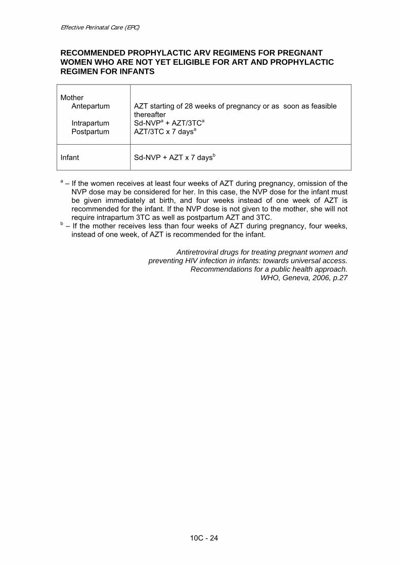
Effective Perinatal Care (EPC)
10C - 24
RECOMMENDED PROPHYLACTIC ARV REGIMENS FOR PREGNANT WOMEN WHO ARE NOT YET ELIGIBLE FOR ART AND PROPHYLACTIC REGIMEN FOR INFANTS Mother
Antepartum Intrapartum Postpartum
AZT starting of 28 weeks of pregnancy or as soon as feasible thereafter Sd-NVPa + AZT/3TCa AZT/3TC x 7 daysa
Infant
Sd-NVP + AZT x 7 daysb
a – If the women receives at least four weeks of AZT during pregnancy, omission of the
NVP dose may be considered for her. In this case, the NVP dose for the infant must be given immediately at birth, and four weeks instead of one week of AZT is recommended for the infant. If the NVP dose is not given to the mother, she will not require intrapartum 3TC as well as postpartum AZT and 3TC.
b – If the mother receives less than four weeks of AZT during pregnancy, four weeks, instead of one week, of AZT is recommended for the infant.
Antiretroviral drugs for treating pregnant women and
preventing HIV infection in infants: towards universal access. Recommendations for a public health approach.
WHO, Geneva, 2006, p.27
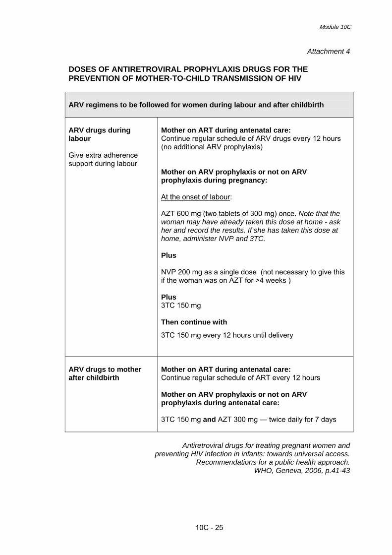
Module 10C
10C - 25
Attachment 4
DOSES OF ANTIRETROVIRAL PROPHYLAXIS DRUGS FOR THE PREVENTION OF MOTHER-TO-CHILD TRANSMISSION OF HIV ARV regimens to be followed for women during labour and after childbirth ARV drugs during labour Give extra adherence support during labour
Mother on ART during antenatal care: Continue regular schedule of ARV drugs every 12 hours (no additional ARV prophylaxis) Mother on ARV prophylaxis or not on ARV prophylaxis during pregnancy: At the onset of labour: AZT 600 mg (two tablets of 300 mg) once. Note that the woman may have already taken this dose at home - ask her and record the results. If she has taken this dose at home, administer NVP and 3TC. Plus NVP 200 mg as a single dose (not necessary to give this if the woman was on AZT for >4 weeks ) Plus 3TC 150 mg Then continue with
3TC 150 mg every 12 hours until delivery
ARV drugs to mother after childbirth
Mother on ART during antenatal care: Continue regular schedule of ART every 12 hours Mother on ARV prophylaxis or not on ARV prophylaxis during antenatal care: 3TC 150 mg and AZT 300 mg — twice daily for 7 days
Antiretroviral drugs for treating pregnant women and
preventing HIV infection in infants: towards universal access. Recommendations for a public health approach.
WHO, Geneva, 2006, p.41-43

Effective Perinatal Care (EPC)
10C - 26
Attachment 5 ARV PROPHYLAXIS REGIMENS FOR INFANTS All infants born to HIV-infected women need to receive a course of ARV drugs as post-exposure prophylaxis. The ARV regimen for newborns will depend upon if and for how long the mother received either ART or ARV prophylaxis during pregnancy. Newborn: give ARV prophylaxis as soon after birth as possible. Duration depends on if and how long the mother took AZT prophylaxis or ART Mother’s antenatal ARV regimen
Newborn ARV prophylaxis regimen
> 4 weeks AZT prophylaxis Single dose NVP plus AZT for 1 week
< 4 weeks AZT prophylaxis or no ARV prophylaxis or ART
Single dose NVP plus AZT for 4 weeks
> 4 weeks ART
AZT for 1 week
< 4 weeks ART
AZT for 4 weeks
Dosages for ARV prophylaxis for the newborn AZT 4 mg/kg twice daily NVP 2mg/kg as soon as possible after the birth
ARV drugs formulation for the newborn AZT 10 mg/ml NVP 10 mg/ml
Note: If the capacity to deliver combination ARV prophylaxis (NVP plus AZT) does not exist, give single dose NVP to mother and newborn.
Antiretroviral drugs for treating pregnant women and preventing HIV infection in infants: towards universal access.
Recommendations for a public health approach. WHO, Geneva, 2006, p.40-43

Effective Perinatal Care (EPC) Facilitator guide
10C - 27
Attachment 6
HIV TESTING FOR THE HIV EXPOSED CHILD Encourage HIV testing for: • All children born to an HIV positive mother • All sick children with symptomatic suspected HIV infection For children >18 months, a positive HIV antibody test result means the child is infected For HIV exposed children <18 months of age: • If PCR or other virological test is available, test from 6 weeks of age
A positive result means the child is infected A negative result means the child is not infected, but could become infected if they
are still breastfeeding • If PCR or other virological test is not available, use HIV antibody test
A positive result is consistent with the fact that the child has been exposed for HIV, but does not tell us if the child is definitely infected
A negative test usually means the child is not infected
If PCR or other virological test is not available, use HIV antibody test: • If child becomes sick, test immediately • If child remains well, test at 9-12 months • If child > 12 months has not been tested, recommend HIV antibody test
Interpreting the HIV antibody test results in a child <18 months of age Test result HIV antibody test is positive HIV antibody test is
negative Not breastfeeding or not breastfed in last 6 weeks
HIV exposed and/or HIV infected Manage as if they could be infected. Repeat test at 18 months
HIV negative Child is not HIV infected
Breastfeeding HIV exposed and/or HIV infected Manage as they could be infected. Repeat test at 18 months or once breastfeeding has been discontinued for more than 6 weeks
Child can still be infected by breastfeeding. Repeat test once breastfeeding has been discontinued for more than 6 weeks
Integrated management of childhood illness complementary course on HIV/AIDS. WHO; UNICEF, Geneva, 2006; Module 2, p. 17

Effective Perinatal Care (EPC) Facilitator guide
10C - 28

Effective Perinatal Care (EPC) Facilitator Guide
11C - 1
Module 11C Infections in Pregnancy, Childbirth and Postpartum
Learning Objectives At the end of the module participants will:
• Have a basic knowledge on the main infections affecting pregnancy and their possible adverse consequences for the mother and foetus
• Know how to manage these infections to prevent complications • Question the current practices of infections prevention in pregnancy and
recognize the harm of an inappropriate management • Know the definition of Nosocomial Infection and effective ways of
prevention • Understand the importance of hand washing and be able to do it correctly
Module Outline and Duration
Part I - Classroom work –195 min Activity 1 – Introduction 20 min Activity 2 – Interactive presentation 120 min Activity 3 – Conclusions 5 min Activity 4 – Interactive presentation Activity 5 – Conclusions
45 min 5 min
Part II – Practical work Activity 6 – Practical work: hand washing ---------
Preparation for the Module
• The facilitator should review current evidence and public health strategies
regarding infections during pregnancy and infection control. • Write the following statements on different flipchart sheets: A. Infections which affect the pregnancy/foetus B. Infections which not do affect the pregnancy/foetus • For Activity 1: prepare pieces of paper with the names of infections written on
them (Attachment 1) or write the following names of infections on the separate

Effective Perinatal Care (EPC)
11C - 2
Preparation for the Module
sheets of paper: o Urinary tract infections (asymptomatic bacteriuria) o Syphilis o Gonorrhoea o Chlamydiosis o Bacterial vaginosis o Group B streptococcus o Listeriosis o Tuberculosis o Hepatitis B o Genital Herpes o Cytomegalovirus Infection o Rubella o Toxoplasmosis o Malaria o Trichomoniasis o Vaginal Candidiasis
• For Activity 2: prepare 16 sheets of paper with the name of infections written
on them and questions for discussion (Attachment 2). • Invite representatives from the Sanitary-Epidemiological Department to this
session (if possible). Materials and Audiovisual Equipment
Materials • Participant Manual • 16 pieces of paper with names of infections for each participant (Attachment
1) • Murray W. Enkin et al “A guide to effective care in pregnancy and childbirth”
3-rd edition, 2000 (preferably 16 books) • 16 sheets of paper with names of infections (Attachment 2) • Two statements about infections written on flipchart pages:
o “Infections which affect pregnancy” o “Infections which do not affect pregnancy”
Equipment
• Video projector or projector overhead • Flipchart • Felt pens • Pens and pencils • Liquid soap • Paper towels • Alcohol antiseptic in individual container

Module 11C
11C - 3
Key Messages • Many infections adversely affect pregnancy or can be transmitted to the
foetus/newborn. Many others have no negative effect on pregnancy or perinatal outcomes.
• The adverse effects of some infections can be reduced by implementation
of relatively simple and effective preventive measures. • Nevertheless, in many countries there is widespread use of ineffective or
even harmful practices regarding infection management. Evidence exist that inappropriate diagnosis and infection management during pregnancy causes considerable waste of limited healthcare resources.
• Patients can experience embarrassment, stress and dissatisfaction when
infections are inappropriately managed. • Factors which limit the ability to prevent adverse effects of infections
include:
o Lack of reliable diagnostic tests and/or resources to conduct them; o Limited ability to confirm infection in the foetus; o Absence of effective treatment options for many infections; o Limited impact on pregnancy outcome despite effective treatment
options for some infections; and o Lack of resources to implement effective but expensive preventive
strategies.
• With very few exceptions, there is no need to isolate a mother with an infection from her baby or from other women/families.
• Hand washing is an effective way to prevent nosocomial infection
transmission. • Nosocomial infection prevention is a key priority for maintaining quality
services in the hospital. • Continuous training of personnel regarding infection control is necessary. • Effective practices for preventing nosocomial infections include: rooming-in,
breastfeeding, unrestricted family visits, rational therapy and prophylaxis with antibiotics.
• Routine use of ultraviolet radiation, disinfectants and wearing of medical
robes and caps are ineffective for preventing nosocomial infections.

Effective Perinatal Care (EPC)
11C - 4
PART I – Classroom work (195 min) Activity 1 – Introduction (20 min) • Show Slide 11C-1-1 and explain that this module is divided on 2 parts. During the
Part I of this module, the participants will discuss strategies for preventing and managing infections during pregnancy, childbirth and postpartum period. Tell to participants that not all infections will be discussed during the module. Remind participants that HIV infection and pMTCT issues were covered in the module 10C. During the Part II you will discuss the issues of nosocomial infections and infection control in general.
• Go to Slide 11C-1-2 and tell participants that most infections in pregnancy are not
worrying, and it is important that those giving care to the pregnant woman do not impose unnecessary restrictions on the pregnancy, or unnecessarily waste resources. Of course, some infections can be disastrous for mother, baby or both, but they are very much in the minority.
• In any health care system there is an imperative need not to waste resources,
particularly where such resources are limited. Analysis of health care practices in the European region has demonstrated in many countries widespread practices in the field of infections in pregnancy that are either ineffective or, worse still, likely to be harmful. This module will therefore concentrate heavily on interventions that appear at present to be effective, and will attempt to identify practices that are inappropriate.
• Infections in pregnancy can be subdivided into those which affect pregnancy, and
those which do not affect pregnancy. This grouping is important, not least because those in the second category should not be managed in any different way from in the non-pregnant state. There is of course a third category: infections that are influenced by pregnancy. However this category is in effect a sub-category of the above two categories, as will become clear.
• Ask participants, which general routes of infection they know? Listen carefully all
their opinions. • Then go to Slide 11C-1-3 and present to participants the general routes and
possible time (periods) of infection for the women/pregnant women/mother and for the foetus/newborn.
• Post 2 flipchart sheets with statements about the different types of infections and
their impact on pregnancy (see “Preparation for the Module” section above) on the walls in different places so that they are visible to all participants.
• Divide participants for 16 groups and give to each group one sheet of paper with
the name of an infection written on it (Attachment 1) and ask them to stand next to the flipchart statement that they think corresponds to their infection.
• Ask participants from each group to hold up their papers with the names of the
infections so that everybody can see them. One facilitator should write the names of the infections on the corresponding sheet of flipchart (according to participants’ placement). The same infection can be written on multiple different flipcharts, according to participants’ ideas.

Module 11C
11C - 5
• Ask participants to look at the infections that have been added to the flipchart
pages. Thank them and proceed to the next activity.
Activity 2 – Interactive presentation (120 min) • Tell participants that now you are starting to work with the Part I of this module and
will discuss infections in pregnancy, childbirth and postpartum. • Divide participants into 16 groups and give each group a sheet of paper with
names of infections and questions on it (Attachment 2). Depending on the number of participants, each group may have 2 or 3 people in it.
• Each sheet of paper with infection names and questions should include:
o Urinary tract infections (asymptomatic bacteriuria) o Syphilis o Gonorrhoea o Chlamydiosis o Bacterial vaginosis o Group B streptococcus o Listeriosis o Tuberculosis o Hepatitis B o Genital Herpes o Cytomegalovirus Infection o Rubella o Toxoplasmosis o Malaria o Trichomoniasis o Vaginal Candidiasis
• Ask participants to prepare a presentation using the Murray W. Enkin et al “A guide
to effective care in pregnancy and childbirth” 3-rd edition, 2000. Give them 10 minutes to answer the following questions:
o What is the incidence of this infection in general as well as in your geographic region (if data exist), and how does this infection affect pregnancy and the foetus?
o Is a reliable and inexpensive diagnostic test available for this infection? o Is there an efficient method of treatment? o Are there any effective strategies for preventing/decreasing the adverse
influences of this infection on pregnancy or for preventing transmission to the foetus/newborn? Are these strategies feasible/affordable locally?
o Is it necessary to isolate a mother with this infection from other people or to separate her from her baby?
• Explain that answers need to be written on flipchart sheet. One to three sentences
per question is sufficient. • When the task is completed, show Slide 11C-1-4 with the list of infections which
can affect pregnancy. Tell participants that now you will not compare this list with those developed by participants - you will do it at the end of the work with this part of the module.

Effective Perinatal Care (EPC)
11C - 6
• Post completed flipcharts in a visible place and ask a representative from each
group to present the results of their group work. • After each group’s presentation, the facilitator can show the corresponding
PowerPoint slide (Slides 11C-1-5 to 11C-71). You need to focus on important aspects of each infection and discuss them in more detail. Discuss with participants the rationale behind and need for screening for each infection. Put the letter S near those infections which require screening.
Present/discuss the following infections in the order listed and stress the following aspects for each infection: • Slides 11С-1-5 to 11С-1-8 Urinary Tract Infections o Explain that urinary tract infections have three main manifestations:
asymptomatic bacteriuria, acute cystitis and pyelonephritis. o Define the criteria for an “acute cystitis” diagnosis: presence of symptoms such
as dysuria, incontinence and frequent urination in febrile patients with no evidence of systemic illness.
o Define the criteria for a “pyelonephritis” diagnosis: presence of bacteriuria
accompanied by systemic symptoms/signs such as fever, nausea, vomiting and flank pain; very often symptoms of lower urinary tract infection. Acute pyelonephritis during pregnancy is a serious systemic disease that can progress to maternal sepsis, preterm labour and preterm delivery.
o Define the criteria for an “asymptomatic bacteriuria” diagnosis: finding of more
than 105 colony-forming units per ml of midstream specimen of urine. Asymptomatic bacteriuria is common, with a prevalence of 3-8% during pregnancy.
o Tell participants that there is a large number of drugs, and combination of them,
available to treat urinary tract infections: a. Analysis of 9 studies recruiting a total of 997 pregnant women showed that in
most of the comparisons there were no significant differences between the treatments under study with regard to cure rates, recurrent infection, incidence of preterm delivery, admission to neonatal intensive care unit, need for change of antibiotic and incidence of prolonged pyrexia. Only when cefuroxime and cephradine were compared, there were better cure rates (29/49 versus 41/52) and fewer recurrences (20/49 versus 11/52) in the cefuroxime group, but the sample size is insufficient to ensure that differences found in the effect of the drugs were real.
b. Make a conclusion: Although antibiotic treatment is effective for the cure of urinary tract infections, there are insufficient data to recommend any specific treatment regimen for symptomatic urinary tract infections during pregnancy. All the antibiotics studied were shown to be very effective in decreasing the incidence of the different outcomes. Complications were very rare.
o Slide 11С-1-8 Management of Asymptomatic Bacteriuria during Pregnancy:
a. Explain that asymptomatic bacteriuria is associated with an increased risk of pyelonephritis, preterm delivery and low birth-weight infants.
b. Screening and treatment of asymptomatic bacteriuria in pregnant women significantly decreases incidence of pyelonephritis during pregnancy, as well

Module 11C
11C - 7
as incidence of preterm delivery. c. Screening for bacteriuria is justified by the relatively high prevalence of
asymptomatic bacteriuria among pregnant women; the significant consequences of bacteriuria on women and their pregnancies; and the effectiveness of bacteriuria treatment.
d. However, despite the proven effectiveness of asymptomatic bacteriuria screening and treatment strategies, these measures are not implemented in many countries.
• Slides 11C-1-9 to 11C-1-13 Syphilis o Tell participants that foetal infection occurs with high frequency in untreated early
infections of pregnant women and with lower frequency later in pregnancy. It frequently causes abortion or stillbirth and may cause infant death due to preterm delivery of low birth weight infants or from generalized systemic disease.
o Stress that syphilis is an infection for which there is an effective screening and
treatment strategy for reducing adverse effects on pregnancy/foetus: a. Reliable diagnostic test exists and is not expensive; b. Effective treatment method exists and is affordable; c. Diagnosis and treatment reduces infection’s adverse affects on the foetus. d. Women with an allergy to penicillin should be desensitized or referred to a
higher level of care. e. Partners must be treated. Thus syphilis screening is recommended in most countries
o If the mother tested positive for syphilis and was treated adequately (2.4 million
units of penicillin) and the treatment started at least 30 days before birth, NO treatment is necessary (WHO, 2003; CDC, 2006). However, some specialists would treat with benzathine penicillin G 50,000 units/kg as a single IM injection, particularly if follow-up was uncertain. (CDC, 2006)
o If the mother was not treated for syphilis, she was treated inadequately, or her
treatment status is unknown or uncertain AND the baby has no signs of syphilis: Give the baby:
a. Benzathine penicillin G 50,000 units/kg/dose IM in a single dose (CDC,2006; WHO, 2007)
OR b. Procaine benzylpenicillin 50 000 units/kg/dose IM a single dose for
10 days (CDC, 2006). OR
c. Aqueous crystalline penicillin G, 100,000-150 000 units/kg/dose, administered as 50,000 units/kg/dose IV every 12 hours during the first 7 days of the life, and every 8 hours thereafter for a total of 10 days (CDC, 2006)
o Follow up in four weeks to examine the baby for growth and signs of congenital
syphilis.
o Clinical evidence of early congenital syphilis is similar to that of secondary syphilis in adults. The rash has a higher probability of being atypical and can be vesicular or bullous instead of the characteristic reddish brown macular rash.
o For early congenital syphilis:
a. Aqueous crystalline penicillin G, 50 000 units/kg/dose, given IV or IM

Effective Perinatal Care (EPC)
11C - 8
every 12 hours during the first 7 days of the life, and every 8 hours thereafter for a total of 10 days
OR b. Procaine penicillin G 50,000 units/kg/dose IM in a single daily dose
for 10 days. (CDC, 2006; WHO, 2007) o For late congenital syphilis, if the CSF is normal and there is no neurologic
involvement, children can be treated as for latent syphilis. If the CSF is abnormal, treatment for neuro syphilis is required: Aqueous crystalline penicillin G 200 000-300,000 units/kg/day IV administered as 50,000 units/kg every 4-6 hours for 10 days (CDC, 2006).
o Tell them that the best approach to preventing syphilis is to avoid exposure. At
this first level of prevention, the likelihood of being exposed to syphilis can be reduced by: a. Decreasing the number of sex partners; b. Using condoms correctly and consistently.
• Slides 11С-1-14 to 11С-1-17 Gonorrhoea o Tell participants that gonorrhoea in pregnancy is associated with several adverse
outcomes, including chorioamnionitis, preterm rupture of membranes, and preterm delivery. Perinatal transmission to infants can cause severe conjunctivitis resulting in blindness.
o Discuss reliability of diagnostic tests: the “gold standard” is culture; but its rather
complex and expensive so can not be widely used. a. Most studies identify taking a culture as the best way to screen for
gonorrhoea. The specificity of a culture specimen is 100%. A culture specimen has high specificity for differentiating Neisseria gonorrhoea from other organisms; however, the sensitivity of cultures varies widely, ranging from 61.8% to 92.6%. Bacterioscopy has a low sensibility and specificity. Currently there are no studies that have directly examined the harms of screening for or treatment of gonorrhoea infection. Potential harms of screening may include high costs and false-positive test results that may lead to stress, stigma, and the need for further testing. Even using a test with a specificity of 99% in a population at high risk for gonorrhoea with a prevalence of 0.5%, 2/3 of positive screening tests would be expected to be false-positive results.
o In reality, bacterioscopy (which has very low sensitivity) is used for screening. o Stress that only in a region with a high prevalence of gonorrhoea, it can be
argued that screening for gonorrhoea might be effective. o Some countries with low gonorrhoea prevalence do not implement routine use of
ointment for prevention of ophthalmia neonatorum (also known as neonatal conjunctivitis).
o Ask participants, “Should we at least offer mothers an informed choice regarding
presumptive treatment of neonatal conjunctivitis?”. o The best approach to preventing gonorrhoea is the same to all STIs.

Module 11C
11C - 9
• Slides 11С-1-18 to 11C-1-21 Chlamydiosis o Chlamydia infection during pregnancy is associated with higher rates of preterm
birth and intrauterine growth retardation, and leads to neonatal conjunctivitis and pneumonia in 30–40% of cases. Chlamydia may coexist with other genital infections and may facilitate transmission of HIV infection.
o Ask participants which diagnostic tests are used locally for Chlamydia? Listen
carefully all participants’ answers. o Explain which tests are recommended and how much they cost:
a. Currently, no simple, inexpensive laboratory tests exist for diagnosing chlamydia trachomatis.
b. Rapid tests include: direct fluorescent antibody staining (50% to 90% sensitive); enzyme-linked immunoassays (75-80% sensitivity and 85-100% specificity); and RNA-DNA hybridization (70-85% sensitivity).
c. Serology is not useful in diagnosing acute chlamydia infection. d. Pregnant women should not be offered routine screening for asymptomatic
chlamydia because there is insufficient evidence supporting the effectiveness and cost-effectiveness of screening.
o Note that in many populations screening programs are not cost-effective.
Diagnostic tests are expensive and cannot be afforded in most countries. o Discuss the treatment options and their effectiveness. Tell participants that
uncomplicated genital chlamydia infection in pregnancy should be treated with: a. Erythromycin 500 mg four times/day for 7 days, or b. Amoxicillin 500 mg three times/day for 7 days c. Another option includes Azithromycin.
o Discuss with participants the newborn management in case of chlamidial
conjunctivitis and pneumonia. o The best approach to preventing chlamydiosis is the same to all STIs. • Slides 11С-1-22 to 11С-24 Bacterial Vaginosis o Explain participants that bacterial vaginosis (BV) is a microbial disease
characterized by a change in the bacterial flora of the vagina: decreasing of Lactobacillus species and high concentrations of anaerobic bacteria, Gardnerella, Mobiluncus and Mycoplasma hominis.
o BV is associated with many adverse effects on pregnancy and can be easily
diagnosed and treated, but screening and treatment of asymptomatic infection does not reduce the incidence of pregnancy complications, especially of premature delivery. There is reliable evidence that screening and treatment of asymptomatic bacterial vaginosis without high risk factors does not improve delivery outcomes (including preterm delivery). a. 15 Randomized trials comparing antibiotic treatment with placebo or no
treatment, or comparing two or more antibiotic regimens in pregnant women with bacterial vaginosis or intermediate vaginal flora have been analyzed. Antibiotic therapy was effective at eradicating bacterial vaginosis during pregnancy. Treatment did not reduce the risk of preterm birth before 37 weeks (Peto OR 0.91, 95% CI 0.78 to 1.06; 15 trials, 5888 women), or the

Effective Perinatal Care (EPC)
11C - 10
risk of preterm prelabour rupture of membranes (PPROM) (Peto OR 0.88, 95% CI 0.61 to 1.28; four trials, 2579 women).
b. However, treatment before 20 weeks' gestation may reduce the risk of preterm birth less than 37 weeks (Peto OR 0.63, 95% CI 0.48 to 0.84; five trials, 2387 women).
c. In women with a previous PTB, treatment did not affect the risk of subsequent PTB (Peto OR 0.83, 95% CI 0.59 to 1.17, five trials of 622); however, it may decrease the risk of PPROM (Peto OR 0.14, 95% CI 0.05 to 0.38) and low birth weight (Peto OR 0.31, 95% CI 0.13 to 0.75)(two trials, 114 women).
d. In women with abnormal vaginal flora (intermediate flora or bacterial vaginosis) treatment may reduce the risk of PTB before 37 weeks (Peto OR 0.51, 95% CI 0.32 to 0.81; two trials, 894 women).
e. Clindamycin did not reduce the risk of PTB before 37 weeks (Peto OR 0.80, 95% CI 0.60 to 1.05; six trials, 2406 women).
o Antibiotic treatment decreased preterm prelabour rupture of membranes and low
birth weight significantly only in the subgroup of women who had a previous preterm birth.
o Stress that there is no significant correlation between BV and neonatal infection.
In absence of clinical sings of infection in newborn the last do not need any specific evaluation or treatment.
• Slides 11С-1-25 to 11С-1-30 Group B Streptococcus o Explain that group B streptococcus (GBS) is a common bacterium that normally
lives in the intestines, vagina, and rectal areas. GBS colonization is not a sexually transmitted disease. Approximately 10-30% of pregnant women will be colonized with GBS.
o Explain that group B streptococcus (GBS) is an important cause of neonatal
sepsis and death but that newborns can be protected against infection if antibiotics are given to the mother intrapartum according to the regimens described in Slide 11C-1-27. a. Even if all women colonized with GBS receive antibiotic therapy in pregnancy,
the effectiveness will be insignificant because colonization is transient and returns in many cases prior to delivery. Therefore, there is no sense in treating asymptomatic women during pregnancy; it is recommended that treatment be given intrapartum (including in the case of preterm rupture of membranes).
b. It is recommended by Royal College of Obstetric and Gynaecology (RCOG) that intravenous penicillin 3 g be given as soon as possible after the onset of labour and 1.5 g four-hourly until delivery.
c. Clindamycin 900 mg should be given intravenously every eight hours to those allergic to penicillin. It should be noted that these doses are based on tradition rather than good evidence. Broad-spectrum antibiotics such as ampicillin should be avoided if possible, as concerns have been raised regarding increased rates of neonatal Gram negative sepsis. To optimise the efficacy of antibiotic prophylaxis, the first dose should be given at least two hours prior to delivery.
o The results of the trials show that the minimal exposure to antibiotic should
exceed 4 hours, that is, to reduce colonization level; over 4 hours must pass from antibiotic administration until the birth of the baby. In this case, the rate of

Module 11C
11C - 11
neonatal colonization with Group B Streptococcus reduces to <1%. Therefore, the exact time of intrapartum antibiotic administration must be carefully recorded (Slide 11C-1-28).
o Providers in different countries use different preventive strategies against group B
streptococcus infection. Discuss with participants the different strategies: a. The first approach – universal screening of women in late pregnancy and
intrapartum antibiotic treatment of all infected women. This approach prevents 65-80% of all cases of neonatal sepsis with GBS.
b. The second approach - antibiotics are given during labour to women with high risk factors: preterm delivery; fever in labour; prolonged rupture of membranes >18 hours; previous baby with GBS disease; and GBS bacteriuria in the current pregnancy. Using this approach, to prevent one neonatal death it is necessary to treat 5,580 women with high risk factors. To prevent one case of sepsis, it is necessary to treat 625 women. Sixty-five percent of all cases will be prevented.
c. The third approach - screen all women (culture) and offer intrapartum antibiotic prevention to GBS carriers who also have clinical risk factors. This approach has been suggested by the Canadian Task Force on Preventative Health Care, who estimate that 3.4% of women will require intrapartum antibiotic prophylaxis and that the incidence of early GBS sepsis is reduced by 51%.
o There is no agreement on which is the best strategy since each differs in its cost
and effectiveness. o Ask, “Is it rational to implement preventive strategies in your area and what
approach would be most suitable?” o Conclude by explaining that many NIS countries cannot afford the cost of GBS
screening. In addition, in most NIS countries intrapartum antibiotic treatment in the presence of high risk factors is not practiced either.
o Discuss with participants the management of a newborn born to a mother with
GBS. Tell that GBS colonization in woman should not affect breastfeeding in any way, as well as should not be an indication for mother and baby separation.
• Slides 11С-1-31 to 11C-1-34 Listeriosis o Tell participants that listeriosis, a serious infection caused by eating food
contaminated with the bacterium Listeria monocytogenes. The disease affects primarily pregnant women, newborns, and adults with weakened immune systems.
o Emphasize that pregnant women are about 20 times more likely than other
healthy adults to get listeriosis. About one-third of listeriosis cases happen during pregnancy.
o Stress that maternal listeriosis in the second or third trimester results in a
mortality of 40-50% for the foetus. o Tell participants that there is no routine screening test for susceptibility to
listeriosis during pregnancy. During pregnancy, a blood test is the most reliable way to find out if presented symptoms are due to listeriosis.

Effective Perinatal Care (EPC)
11C - 12
o Discuss with participants the possible regimens for listeria treatment. o Tell participants that babies with listeriosis should receive the same antibiotics as
adults, although a combination of antibiotics is often used until physicians are certain of the diagnosis.
o Discuss with participants the general recommendations on prevention of listeria
infection: a. Thoroughly cook raw food from animal sources, such as beef, pork, or
poultry. b. Wash raw vegetables thoroughly before eating. c. Keep uncooked meats separate from vegetables and from cooked foods and
ready-to-eat foods. d. Avoid unpasteurized (raw) milk or foods made from unpasteurized milk. e. Wash hands, knives, and cutting boards after handling uncooked foods. f. Consume perishable and ready-to-eat foods as soon as possible.
o Also tell participants that in case when infection occurs during pregnancy, antibiotics given promptly to the pregnant woman can often prevent infection of the foetus or newborn.
• Slides 11С-1-35 to 11C-1-39 Tuberculosis (TB) o Stress that tuberculosis is a mycobacterial disease that is important as a major
cause and death in many parts of the world. The recent increase in numbers of cases of tuberculosis in the European region indicates that this is a significant public health issue once more in the region.
o Tell participants that all pregnant women who are at high risk of TB should be
screened with a Mantoux skin test with purified protein derivative (PPD) when they begin receiving prenatal care. Discuss with participants the risk factors for TB.
o Emphasize that management of a newborn whose mother is suspected of having
TB is based on individual considerations. Whenever possible, separation of the mother and the neonate should be minimized.
o Discuss with participants the general recommendations on preventative
measures: a. BCG vaccination for all newborns b. To avoid contacts with potentially infected people.
• Slide 11С-1-40 to 11C-1-43 Hepatitis B o Explain that foetuses can be infected in utero, only if the mother has contacted
the infection during pregnancy. In patients with acute hepatitis B vertical transmission occurs in up to 10% of neonates when infection occurs in the first trimester and in 80 - 90% of neonates when acute infection occurs in the third trimester
o Identification of the chronic carrier state in pregnant women is very important
because babies born to these mothers are at significant risk of becoming infected at birth. Congenital infection results in a chronic carrier state more frequently than

Module 11C
11C - 13
being infected as an adult, therefore all babies from these mothers should receive hepatitis B immunoglobulin and vaccine.
o Ask if immunoglobulin is locally available? If no, ask what the purpose is of
screening for hepatitis B if specific immunoglobulin is not available? Discuss this issue with participants.
o This presents an example of screening without practical benefit and waste of
screening resources since the aim of screening is to prevent mother to child transmission through the use of specific immunoglobulin after delivery (babies from these mothers should receive hepatitis B immunoglobulin and hepatitis B vaccine just after delivery), where local recourses allow doing this. a. 29 randomised clinical trials were analyzed. Compared with placebo or no
intervention, vaccination reduced the occurrence of hepatitis B (relative risk 0.28, 95% confidence interval 0.20 to 0.40; four trials). No significant difference in hepatitis B occurrence was found between recombinant vaccine and plasma derived vaccine (1.00, 0.71 to 1.42; four trials) and between high dose versus low dose vaccine (plasma derived vaccine 0.97, 0.55 to 1.68, three trials; recombinant vaccine 0.78, 0.31 to 1.94, one trial). Compared with placebo or no intervention, hepatitis B immunoglobulin or the combination of plasma derived vaccine and hepatitis B immunoglobulin reduced hepatitis B occurrence (immunoglobulin 0.50, 0.41 to 0.60, one trial; vaccine and immunoglobulin 0.08, 0.03 to 0.17, three trials). Compared with vaccine alone, vaccine plus hepatitis B immunoglobulin reduced hepatitis B occurrence (0.54, 0.41 to 0.73; 10 trials).
o Most NIS countries continue to screen for hepatitis B despite a lack of access to
immunoglobulin. • Slides 11С-1-44 to 11С-1-49 Genital Herpes o Tell participants that incidence of this infection varies in different countries:
a. In the United States the annually reported incidence of neonatal herpes infection is 11 to 33 cases per 100 000 live births.
b. In the UK the annually reported incidence of neonatal herpes infection is 1.65 per 100 000 live births.
o Transmission from an infected mother to her foetus cause severe disease with
high neonatal mortality and morbidity o It is difficult to interpret serology to distinguish first episode of infection from
recurrent one At the same time, more than 2/3 of all cases of neonatal herpes occur in children born to infected pregnant women who have no lesions and no HSV history.
o Risk of herpes simplex virus (HSV) transmission from an infected mother to her
foetus is highest when the mother’s primary infection occurs in the days prior to labour (which are rare). In this case, delivery by caesarean section is recommended.
o Tell participants that Aciclovir has been used extensively in pregnancy and the
Aciclovir Pregnancy Registry was established in 1984 to collect data on prenatal exposure to the drug. Data from 1207 pregnancies reported prospectively to the Aciclovir Pregnancy Registry between 1984 and 1998 did not demonstrate any

Effective Perinatal Care (EPC)
11C - 14
increase in the number of birth defects, nor any discernible pattern of defects, and this registry has since been disbanded.
o Slide 11C-1-47. Example of two different preventive practices for reducing
neonatal infection in case of recurrent episode of genital herpes in USA and UK: a. UK practice - In the case of recurrent genital herpes, the risk of mother to
child transmission is extremely small and should be set against the risks to the mother of caesarean section [B]. Put participants attention to those fact that a cost-benefit analysis has suggested that, if all women with an episode of recurrent genital herpes at the onset of labour were to undergo caesarean section, 1583 (range 632–6340) caesarean sections would be performed to prevent one case of herpes-related mortality or morbidity, at a cost of 2.5 million USD per case averted.
b. USA practice - Caesarean delivery should be performed on women with recurrent HSV infection who have active genital lesions or prodromal symptoms at delivery [C].
o Draw the attention to the participants to the different levels of recommendations
([B] in UK and [C] in USA). o Remind the participants that the prevalence of neonatal herpes in the USA is
higher than in UK so this could be one of the reasons to justify the policy of Caesarean delivery for women with recurrent HSV infection, aimed to reduce the level of neonatal herpes and its consequences.
o Stress the participants that in case of maternal HVS infection the infant may be
breastfeed and the mother should be counselled in good hand-washing technique.
• Slides 11С-1-50 to 11С-1-55 Cytomegalovirus (CMV) Infection o Incidence of this viral infection is quite high in the general population (among
women and men), and the consequences of infection to a foetus can be dramatic (Slide 11С-1-51). However, the possibilities for preventing adverse outcomes in the foetus are limited, even in developed countries: a. The most important risk to the foetus is attributed to primary infection of the
mother during pregnancy. During reactivation, the risk to the foetus is almost negligible
b. One third of foetuses are infected via transmission from a mother whose primary infection occurs during pregnancy, but only 10-15% of infected foetuses develop severe disease later. Tell participants that each year in the United States, about 1 in 750 children are born with or develop disabilities as a result of CMV infection.
c. It is very difficult to catch and confirm primary infection, there is no effective method of treatment of CMV infection, and it is very difficult to diagnose foetal infection and damage (Slide 11С-1-52).
o Emphasize that the great majority of primary HCMV infections in the
immunocompetent host are clinically silent. In addition, less than 5% of pregnant women with primary infection are reported to be symptomatic, and an even smaller percentage suffers from a mononucleosis syndrome. Thus, a primary HCMV infection can not generally be diagnosed on clinical grounds alone. The most definitive diagnosis of primary HCMV infection in a pregnant woman is by detection of seroconversion, i.e., the appearance of HCMV-specific IgG antibody

Module 11C
11C - 15
during pregnancy in a previously sero negative woman. When this result cannot be achieved, detection of IgM antibody during pregnancy, as well as during follow-up (whenever possible), can be used to determine clinically significant primary HCMV infection. Further testing by the IgG avidity test may be of great help in both confirming and clarifying the clinical significance of IgM antibody. When, at the end of the diagnostic algorithm, a primary HCMV infection is either diagnosed or suspected, prenatal diagnosis should be offered to a pregnant woman to verify whether the infection has been transmitted to the foetus. Ultrasound imaging may demonstrate migrational disorders with microcephaly and polymicrogyria, diminished white matter, delayed myelination, cerebellar hypoplasia, and cerebral calcifications.
o Draw participant attention to the fact that the challenging issue is that knowing the
serological status of a woman can not help in prevention. This is particularly so since the presence of antibodies cannot reliably identify infectivity. Indeed the concern is that women who are found to be sero positive might receive interventions of no proven benefit. No study has ever investigated how many pregnancies are terminated in early stages (<12 weeks of gestation) on the basis of a positive IgM result and inadequate or misleading information ("your baby will be mentally retarded").
o Stress that the “gold standard” for CMV diagnosis in newborn is urine and saliva
culture. Most urine specimens from infants with congenital CMV are positive within 48-72 hours. Given that saliva can be collected with less difficultly and expense, it may eventually replace the current use of urine screening.
No antiviral agent is yet approved for treatment of congenital CMV.. o Tell participants that the decision to breastfeed a very low birth weight infant
needs to consider the potential benefits of human milk versus the risk of CMV transmission. (AAP, 2005)
o The benefits of breastfeeding a preterm infant versus the risk of transferring CMV
in the human milk remain controversial. “…the benefits of breastfeeding greatly outweigh the minimal risk, if any, of infections transmitted to term infants. Caution is warranted, however, in low-birth-weight premature infants, who are at increased risk of CMV disease. Interventions to screen breast milk, or attempt to render breast milk non-infectious through treatments such as freezing, may be warranted in high-risk premature infants” Schleiss, MR (2006)
o Tell participants that “…a more recent study that used more sensitive tests for
quantitative detection of CMV in breastmilk has shown that late viral RNA and viral infectivity are preserved even after freezing at –20 C for up to 10 days. Pasteurization removes CMV infectivity and should be carried out with donated milk. For a mother known to be infected with CMV, freeze storage of her own milk does not seem to be a perfect solution, but the rate of CMV transmission is likely to be lowered; the observed infections were asymptomatic (WHO, 2006, p17).
o Affected infants may excrete the virus for months to years and are often a
concern to personnel caring for them. These newborns need to be isolated from they other one and room in with their mothers.

Effective Perinatal Care (EPC)
11C - 16
• Slides 11С-1-56 to 11С-1-59 Rubella (German Measles) o Tell participants that rubella (also known as German measles) is usually a non-
severe infection in children and adults and can pass asymptomatically. Immunity is stable and is a base for further immunization.
o Emphasize that rubella is an example of a teratogenic infection which is
associated with significant foetal damage in cases where the first symptoms appeared in women during the first 16 weeks of pregnancy. Despite the risk to the foetus, there is no risk of damage to the mother.
o Tell that transplacental rubella transmission from a mother to her foetus occurs in
over 65% of cases where the mother’s primary infection occurs during the first pregnancy trimester. The risk to the foetus of maternal infection decreases with increasing duration of pregnancy. In a prospective study, infants whose had confirmed rubella at successive stages of pregnancy were followed for two years. No defects attributed to rubella were found in children infected after 16 weeks of gestation, while infants infected before the 11-th week had significant cardiac disease and deafness.
o Stress that the diagnosis of rubella in pregnant woman who has been exposed to,
or develops, rubella-like infection, is often difficult. The laboratory must be provided with a detailed history of, as routine screening tests are inadequate and additional testing to detect IgM antibody is required. False-negative results can occur if the specimen is drawn to soon after exposure. The pattern of antibody response to acute infection and re-infection will vary according to the test method used, and expert consultation may be required for interpretation of data.
o Tell participants that there is no treatment for the disease. o Women thought to have developed rubella infection should not be in contact with
other pregnant (or potentially pregnant) women. Where possible they certainly should not (except for labour) be admitted to hospital when they are acutely effected (where they might infect others) but once the infection is clinically over they are no longer potentially infectious.
o Immunization of the woman successfully prevents rubella transmission from
mother to child. o Immunization programs against rubella have led to significant decreases in
diseases prevalence. • Slides 11С-1-60 to 11С-1-64 Toxoplasmosis o Tell participants that toxoplasmosis is caused by a protozoan parasite. The risk of
congenital disease is lowest (10-25%) when maternal infection occurs during the first trimester and highest (60-90%) when maternal infection occurs during the third trimester. However, congenital disease is more severe when infection is acquired in the first trimester. The overall risk of congenital infection from acute (first episode) T. gondii infection during pregnancy ranges from approximately 20 to 50%.
o Emphasize that toxoplasmosis usually is diagnosed on the basis of antibody

Module 11C
11C - 17
detection. The presence of elevated levels of specific IgG antibodies does not distinguish between recent infection and infection acquired in the past. Detection of ¬specific IgM antibodies has been used as an aid in determining the time of infection: a negative IgM test result with a positive IgG result usually indicates infection at least six months previously. However, the interpretation of ¬specific IgM positive results is complicated by the persistence of IgM antibodies up to 18 months after infection and by false-positive reactions in commercial tests. That is why, IgM-positive test results should be confirmed by a Toxoplasmosis reference laboratory.
o Tell that unfortunately, evidence does not suggest that the treatment improves
pregnancy outcomes (Slide 11С-1-62). a. Two systematic reviews in women who seroconvert during pregnancy found
insufficient evidence on the effects of current treatment compared with no treatment on mother or baby. Five studies of the first review (none of them randomized) found that treating mothers significantly reduced foetal infection rates versus no treatment (P<0.01), the other four found no significant reduction in foetal infection. The second review had stricter inclusion criteria. The research studied a series of women treated with spiramycin or spiramycin plus pyremethamyne–sulpha and found no evidence of a difference in outcomes (foetal infection, neonatal diseases).
o Discuss with participants the possible ways of toxoplasma infection prevention. • Slides 11С-1-65 to 11С-1-69 Malaria The current malaria trends o Tell participants that the perception that countries of the WHO European Region
are free from malaria has changed rapidly over the past decades. Since the early 1980s and throughout the decades to follow, the number of countries affected by malaria has increased from three to ten. At the beginning of the 1990s, the residual reservoir of malaria infection, aggravated by political and socio-economic situations, mass population migration, extensive development projects, and almost discontinued activities on malaria prevention and control constituted conditions favourable for malaria transmission. As a result, large-scale epidemics have broken out in Central Asia and Trans-Caucasian countries since 1995. Azerbaijan, Tajikistan and Turkey suffered explosive and extensive epidemics, while Armenia, Turkmenistan and Kyrgyzstan faced outbreaks on a smaller scale1.
Malaria in pregnancy o Emphasize that pregnant women with symptomatic acute malaria are a high-risk
group, and must receive effective antimalarials. Malaria in pregnancy is associated with low birth weight, increased anaemia and, in low-transmission areas, an increased risk of severe malaria. In high-transmission settings, despite the adverse effects on fetal growth, malaria is usually asymptomatic in pregnancy. There is insufficient information on the safety and efficacy of most antimalarials in pregnancy, particularly for exposure in the first trimester, and so treatment recommendations are different to those for non-pregnant adults.
1 http://www.euro.who.int/malaria (11.03.2008)
Effective Perinatal Care (EPC)
11C - 18
Organogenesis occurs mainly in the first trimester and this is therefore the time of greatest concern for potential teratogenicity, although nervous system development continues throughout pregnancy2.
Parasitological Diagnosis o Stress that the the two methods in use for parasitological diagnosis are light
microscopy and rapid diagnostic tests (RDTs). Light microscopy has the advantage of low cost and high sensitivity and specificity when used by well-trained staff. RDTs for detection of parasite antigen are generally more expensive, but the prices of some of these products have recently decreased to an extent that makes their deployment cost-effective in some settings.
Treatment o Tell participants that the antimalarials considered safe in the first trimester of
pregnancy are quinine, chloroquine, proguanil, pyrimethamine and sulfadoxine–pyrimethamine. Of these, quinine remains the most effective and can be used in all trimesters of pregnancy including the first trimester. In reality women often do not declare their pregnancies in the first trimester and so, early pregnancies will often be exposed inadvertently to the available firstline treatment. Inadvertent exposure to antimalarials is not an indication for termination of the pregnancy. There is increasing experience with artemisinin derivatives in the second and third trimesters (over 1000 documented pregnancies). There have been no adverse effects on the mother or fetus. The current assessment of benefits compared with potential risks suggests that the artemisinin derivatives should be used to treat uncomplicated falciparum malaria in the second and third trimesters of pregnancy, but should not be used in the first trimester until more information becomes available.
o Note that lactating woman should receive standard antimalarial treatment
(including ACTs) except for tetracylines and dapsone, which should be withheld during lactation.3
Congenital malaria o Tell that congenital malaria is rare. It usually presents in the first 7 days of life. It
may resemble neonatal sepsis, with fever, anaemia, and splenomegaly occurring in the most neonates and hyperbilirubinemia and hepatomegaly in less than half. No evidence indicates that malaria is transmitted through breast milk. The greatest risk to the infant is exposure to the anoppheline mosquito infected with malaria.
o Discuss with participants the possible ways of malaria prevention both in mother
and newborn. • Slide 11С-70 and 11C-1-71 Trichomoniasis o Tell participants that trichomoniasis is a good example when screening and
treatment of infection is not only ineffective, but seems to increase number of adverse outcomes.
2 WHO, guidelines for the treatment of Malaria, 2006, WHO/HTM/MAL/2006/1108 3 WHO, guidelines for the treatment of Malaria, 2006, WHO/HTM/MAL/2006/1108

Module 11C
11C - 19
o The findings indicate that metronidazole is an effective treatment for symptomatic
and asymptomatic trichomoniasis in pregnant women, but may be associated with increased risk of preterm delivery
o There is evidence that screening and treatment of asymptomatic trichomonosis
(without clinical sings) increases incidence of preterm delivery and premature rupture of membranes. a. In the US trial women with asymptomatic trichomoniasis between 16 to 23
weeks were treated with metronidazole on two occasions at least two weeks apart. The trial was stopped before reaching its target recruitment because metronidazole was not effective in reducing preterm birth and there was a likelihood of harm.
b. Another trials show that treatment with metronidazole is associated with increased low birth weight (relative risk, 2.49; 95% CI, 1.12-5.50), preterm birth rate (relative risk, 1.28; 95% CI, 0.81-2.02) and 2-year mortality rate (relative risk, 1.58; 95% CI, 0.99-2.52), compared with children of 112 women with Trichomonas who were not treated.
Tell participants that now you will discuss Vaginal Candidiasis which does not affect pregnancy and the foetus. This infection should be treated only in cases where women are troubled by symptoms.
• Slide 11С-1-72 Vaginal Candidiasis o Tell participants that screening for vaginal candidiasis is not recommended. o Emphasize that infection can be treated with oral fluconazole or topical
clotrimazole, miconazole, butaconazole, terconazole, tioconazole or nystatin, but infection is frequently recurrent, drugs are not cheap, and the most important that foetal effects of oral treatment are unknown.
o Stress that women do not need to be hospitalized or isolated from other women. • Once presentation is finished, ask participants to review flipcharts from Activity 1
and determine if all infections were correctly grouped and which of them require screening programs.
• Conclude by asking if participants have any questions or additional points to
make. Activity 3 – Conclusions (5 min) • Show Slide 11С-1-73 and review the basic requirements of an effective
screening program. These criteria were first developed by Wilson and Jugner for the World Health Organization (Wilson & Jugner, 1968). For each infection of concern, it is necessary to have information on its prevalence in the population, the risk of its transmission during pregnancy, and its consequences for pregnancy and the newborn. It is also necessary to have screening tests with high sensitivity and specificity. In other words, screening tests should not result in false diagnoses. False diagnoses can result in stress for the patient and can lead to unnecessary intervention. The consequences of false-positive and false-negative

Effective Perinatal Care (EPC)
11C - 20
diagnosis should not be underestimated. Interventions and treatment aimed at decreasing unwanted consequences of maternal infection should be clear, proven, effective and accessible.
• Slide 11С-1-74. Review the necessity of infection screening and treatment during
pregnancy, but only under appropriate circumstances. Give examples of some countries’ approaches to infection screening during pregnancy.
• Make a short conclusion: Each country chooses a screening program for
pregnant women based on: need (prevalence of infection in population); significance of impact on the foetus (infections, congenital anomalies, prematurity); safeness of screening for women and foetus; cost; and potential for treatment or prevention (accessibility and effects of drugs/vaccines). These criteria help to determine whether a screening program could be routinely implemented on a state or regional level.
• Go to the next activity. Activity 4 – Interactive presentation (45 min) • Show Slide 11C-2-1 and remind the participants that the next part of the module
(Part II) is focused on infection control in the maternity participants. • Ask participants, “Are the pathogenic organisms that we discussed during the
previous session the only ones that can cause infection in mothers or newborns?” After a short discussion, emphasize that: o Any bacteria (staphylococci, enterococci, Escherichia coli, etc.) or virus can
cause infection and disease. o These pathogens can be introduced to the hospital environment by hospital
patients or can circulate in hospitals as “nosocomial flora”.
• Show Slides 11С-2-2 and 11C-2-3, which present the definition of nosocomial infection and official WHO data of its frequency.
• Tell the participants that despite progress in public health and hospital care,
infections continue to develop in hospitalized patients, and may also affect hospital staff. Many factors promote infection among hospitalized patients: decreased immunity among patients; the increasing variety of medical procedures and invasive techniques creating potential routes of infection; and the transmission of drug-resistant bacteria among crowded hospital populations, where poor infection control practices may facilitate transmission.
• Emphasize that the most frequent nosocomial infections are infections of surgical
wounds, urinary tract infections and lower respiratory tract infections. The WHO study, and others, have also shown that the highest prevalence of nosocomial infections occurs in intensive care units and in acute surgical and orthopaedic wards. Infection rates are higher among patients with increased susceptibility because of old age, underlying disease, or chemotherapy.
• Show Slides 11С-2-4 and 11C-1-5 and discuss with the participants possible
ways in which that bacteria which cause nosocomial infections can be acquired.
Module 11C
11C - 21
• Emphasise that people are at the centre of epidemiological process (Slide 11C-1-5): o As main reservoir and source of micro organisms o As main transmitter, notably during treatment o As receptor for micro organisms, thus becoming a new reservoir
• Show Slides 11С-2-6 and 11C-2-7 and tell the participants that prevention of
nosocomial infections requires an integrated, monitored, programme. Discuss the key components of this programme. Highlight the importance of continuing staff education and enhancing staff patient care practices.
• Show Slide 11С-2-8 and underline the importance of hand hygiene in minimizing
of hospital infections. Remind the participants that pathogenic organisms (bacteria) can be transmitted to patients via the hands of medical staff and/or other patients. Therefore appropriate hand washing is one of the key ways to prevent nosocomial infections.
• Tell the participants that proper hand washing techniques will be practised during
the clinical week. • Show Slide 11С-2-9. Discuss common reasons why personnel do not wash their
hands: o Hand washing takes a lot of time. o Lack of soap (54%) and towels (65%). o Idea that one careful hand washing is enough for an entire day. o Use of gloves to replace hand washing (25% of interviewed, 50% of them
were physicians). o Belief that hand washing is not necessary if the baby is receiving antibiotics.
• Show Slides 11С-2-10 and 11С-2-11 and explain that each hospital should enable good hand washing practices by providing training for all staff as well as liquid soap, disposable towels and water.
• Increased education, individual reinforcement technique, appropriate rewarding,
administrative sanction, enhanced self-participation, active involvement of a larger number of organizational leaders, enhanced perception of health threat, self-efficacy, and perceived social pressure can improve Health Care Workers’ (HCWs’) adherence with hand hygiene.
• Show Slide 11С-2-12 which explains proper hand washing technique and
discuss the following: o Do not use non-disposable towels for drying hands after washing. o All staff visiting the delivery department or newborn department (physicians,
nurses, visitors, consultants, laboratory assistant) should wash their hands. o Hands should be washed before and after contact with every patient and/or
item which has had contact with a patient. o Hands should be washed for not less than 30-60 seconds and should be
followed by rinsing under running water. o Hands should be dried with a disposable towel after washing. o Hands should be washed before and after use of gloves; after any
manipulation gloves must be removed. Never use the same pair of gloves in treating different patients, even when they are washed using disinfectant.

Effective Perinatal Care (EPC)
11C - 22
• Slide 11С-2-13 shows areas on the hands which usually are missed in hand washing.
• Slide 11С-2-14 presents proper steps in hand washing technique. Discuss the
steps described point by point. Recommend that participants place similar posters near all hand washing stations in their hospitals.
• Show Slides 11С-2-15 to 11C-2-17 and discussed the procedures of hand
decontamination. Procedures will vary with the patient risk assessment (from minimal to high).
• Slide 11С-2-18 demonstrates indications for proper glove use. • Tell participants that before the emergence of the acquired immunodeficiency
syndrome (AIDS) epidemic, gloves were worn primarily by personnel caring for patients colonized or infected with certain pathogens or by personnel exposed to patients with a high risk of hepatitis B. Since 1987, a dramatic increase in glove use has occurred in an effort to prevent transmission of HIV and other blood borne pathogens from patients to HCWs. The Occupational Safety and Health Administration (OSHA) mandate that gloves be worn during all patient-care activities that may involve exposure to blood or body fluids that may be contaminated with blood. The effectiveness of gloves in preventing contamination of HCWs’ hands has been confirmed in several clinical studies. One study found that HCWs who wore gloves during patient contact contaminated their hands with an average of only 3 CFUs per minute of patient care, compared with 16 CFUs per minute for those not wearing gloves.
• Emphasize that:
o Staff should wear sterile gloves for surgery, care for immunocompromised patients, invasive procedures which enter body cavities.
o Non-sterile gloves should be worn for all patient contacts where hands are likely to be contaminated, or for any mucous membrane contact.
o Staff should wear non-sterile gloves to care for patients with communicable disease transmitted by contact, to perform bronchoscopies or similar examinations.
• Slide 11С-2-19 presents the rational antibiotic prophylaxis in pregnant women:
indications and choice of agents. Indications:
o Prolonged membrane rupture > 18 hours at 37 and more weeks of pregnancy: prophylaxis with antibiotics should be started from the moment of membrane rupture (or from the moment of admission to hospital regardless of the time after membrane rupture).
o Start treatment with penicillin (≥2 doses of penicillin or ampicillin (1 g/dose) for 4 injections).
o If the woman is allergic to penicillin, macrolides are recommended (500 mg erythromycin every 8 hours; 900 mg clindamycin every 8 hours).
o Preterm rupture of membranes at < 37 weeks of gestation: macrolides are recommended (500 mg erythromycin every 8 hours; 900 mg clindamycin every 8 hours).
o Treatment with erythromycin is associated with a lower complication rate than other antibiotics (e.g., augmentin and necrotizing enterocolitis.
o Prophylaxis recommendations for chorioamnionitis (given presence of clinical

Module 11C
11C - 23
sings in any term of pregnancy) are as follows: - Cephalosporin of 3rd generation + metronidasol - Gentamycin + metronidasol - Ampicillin + gentamycin
o Peri-operation prophylaxis with antibiotics: - Negative effects of antibiotics on the baby can be decreased by injecting
antibiotics just after cord cutting. - It is recommended to use one dose of intravenous ampicillin (500 mg) or
gentamycin just after cord cutting. o Regarding caesarean section, administration of three or five doses of
antibiotics for infection prophylaxis does not offer any advantages over administering one dose.
o If group B streptococci is detected in vaginal discharge: - Administer intravenous benzyl penicillin (2.4 g) at onset of labour, followed
by 1.2 g every 4 hours until the baby is delivered.
• It is important to consider local epidemiological/microbiological data when prescribing or changing antibiotics. Antibiotic regimens can be changed according to identified microbial sensitivity. Duration of antibiotic therapy depends on the patient’s clinical form of infection.
• Ask the participants to brainstorm ineffective infection prevention practices and
write them on a flipchart. • Then show Slides 11С-2-20, where common yet ineffective infection prevention
practices are listed. • Stress that there is no need to perform disinfectant fogging in patient care areas.
Recommend participants to use in their healthcare facilities a one-step process and registered hospital detergent/disinfectant designed for general housekeeping purposes in patient-care areas where 1) uncertainty exists as to the nature of the soil on the surfaces (e.g., blood or body fluid contamination versus routine dust or dirt); or 2) uncertainty exists regarding the presence of multi-drug resistant organisms on such surfaces.
• Emphasize that walls, blinds, and window curtains in patient care areas should be
cleaned when they are visibly dusty or soiled. • Slide 11С-2-21. Discuss why restriction of relatives’ visits is ineffective at
preventing nosocomial infection. • Slide 11С-2-22. Explain that there is no evidence to support the efficacy of
wearing of medical masks and caps at preventing infection. There is no evidence from this systematic review and meta-analysis to demonstrate that over gowns are effective in limiting death, infection or bacterial colonisation in infants admitted to newborn nurseries. Explain that clean clothes, washed hair (collected in a tidy hair-do), and clean shoes are sufficient.
• Slide 11С-2-23. As a supplemental air-cleaning measure Ultraviolet Germicidal
Irradiation (UVGI) is effective in reducing the transmission of airborne bacterial and viral infections in hospitals (room or corridor), military housing, and classrooms, but it has only a minimal inactivating effect on fungal spores.
• Air velocity, air mixing, relative humidity, UVGI intensity, and lamp position all

Effective Perinatal Care (EPC)
11C - 24
affect the efficacy of UVGI systems. • The use of fans or systems to generate air movement may increase the
effectiveness of UVGI if airborne microorganisms are exposed to the light energy for a sufficient length of time. The optimal relationship between ventilation and UVGI is not known.
• Regular maintenance of UVGI systems is crucial and usually consists of keeping the
bulbs free of dust and replacing old bulbs as necessary. UVGI tubes should be changed and cleaned according to the instructions of the manufacturer or when irradiance measurements indicate that output is reduced below effective levels.
• In settings that use UVGI systems, HCWs should be trained in its correct use. • Slide 11С-2-24 Use of Disinfectants. Explain that cleaning examination tables
using disinfectants can negatively affect hospital staff, mothers and babies. Some disinfectants give off harmful fumes that can damage people’s mucous membranes and cause other acute conditions.
Activity 5 – Conclusions (5 min) • Review final conclusions using Slides 11С-2-25 and 11С-2-26, summarizing the
key information discussed in this module. • Ask the participants if they have any questions. Answer all participants’
questions. (Discussion of some questions can be postponed for the clinical week.)

Module 11C
11C - 25
Part II – Practical work Activity 6 – Practical work: hand washing • It is important that all participants practice proper hand washing technique.
1. Prepare liquid soap, paper towels, a container for used towels and alcohol antiseptic.
2. Split the participants into groups with one facilitator per group. 3. Review proper hand washing technique with participants (Slides 11С-2-12 to
11С-2-14). 4. Ask the participants to wash their hands according to the algorithm. 5. Ask the other participants to observe their colleagues hand washing
technique. 6. After each demonstration, ask a participant from each group to describe what
she/he thought was done correctly and what was done incorrectly. 7. Summarize the groups’ discussion and conclusions.
• Use Table 1 below to assess the participants’ hand washing technique. If the
action is performed fully and correctly, mark the “+” column. If the action is done incompletely, inappropriately or in the wrong sequence, mark the “-“ column.

Effective Perinatal Care (EPC)
11C - 26
Table 1. Assessment of hand washing practice
1st participant 2nd participant 3rd participant 1 Run the water
+ - + - + -
2 Squeeze out soap onto hands
+ - + - + -
3 Soap hands by rubbing them against each other
+ - + - + -
4 Rub right palm over left dorsum
+ - + - + -
5 Rub left palm over right dorsum
+ - + - + -
6 Palm to palm, fingers interlaced
+ - + - + -
7 Back of fingers to opposing palms, fingers interlocked
+ - + - + -
8 Rotational rubbing of right thumb clasped in left palm
+ - + - + -
9 Rotational rubbing of left thumb clasped in right palm
+ - + - + -
10 Rotational rubbing backwards and forwards with clasped fingers on the right hand of the left hand
+ - + - + -
11 Rotational rubbing backwards and forwards with clasped fingers on the left hand of the right hand
+ - + - + -
12 Wash hands under running water
+ - + - + -
13 Carefully dry hands with a paper towel
+ - + - + -
14 Turn water tap off with used paper towel
+ - + - + -
15 Discard used towel
+ - + - + -
Module 11C
11C - 27
References 1. ACOG educational bulletin. Viral hepatitis in pregnancy. Number 248, July 1998.
American College of Obstetricians and Gynecologists. Int J Gynaecol Obstet. 1998; 63:195-202.
2. Alicia J. Mangram, MD; Teresa C. Horan, MPH, CIC et al. GUIDELINE FOR
PREVENTION OF SURGICAL SITE INFECTION The Hospital Infection Control Practices Advisory Committee. Infection control and hospital epidemiology1999, Vol. 20, #4:247-278.
3. American Academy of Paediatrics. Breastfeeding and the Use of Human Milk.
Pediatrics; 115;496-506, 2005. 4. American College of Obstetricians and Gynaecologists. Management of herpes in
pregnancy. ACOG practice bulletin, No. 8, 2004. 5. American College of Obstetricians and Gynecologists (ACOG). Perinatal viral and
parasitic infections. Washington (DC): ACOG practice bulletin; no.20, 2000/09/13. 6. Antenatal care: routine care for the healthy pregnant woman. Clinical Guideline.
National Collaborating Centre for Women’s and Children’s Health. RCOG Press, October 2003.
7. Association for Genitourinary Medicine (AGUM), Medical Society for the Study of
Venereal Disease (MSSVD). 2002 national guideline on the management of the viral hepatitis A, B, and C. London, 2002.
8. Bader TJ, Macones GA, Asch DA. Prenatal screening for toxoplasmosis. Obstet
Gynecol 1997; 90:457–464. 9. Centers for Disease Control and Prevention. Group B strep prevention; general
public, frequently asked questions, CDC, 2008. 10. Centers for Disease Control and Prevention. Prevention of perinatal group B
streptococcal disease: a public health perspective. MMWR Morbidity and Mortal Weekly Rep 1996; 45(RR-7):1-24.
11. Centers for Disease Control and Prevention. Prevention of perinatal group B
streptococcal disease: a public health perspective. MMWR Morbidity and Mortal Weekly Rep 2002; 51(RR-11):1-24.
12. Chuanfang Lee et al., Effect of hepatitis B immunisation in newborn infants of
mothers positive for hepatitis B surface antigen: systematic review and meta-analysis BMJ 2006; 332:328-336.
13. Cytomegalovirus (CMV). National Centers for Infectious Diseases Copntrol (CDC),
Division of Viral and Rickettsial Diseases, 2006. 14. Delzell J, Lefevre M. Urinary Tract Infections during Pregnancy. Am Fam Physician
2000; 61:713-21. 15. Dunn D, Wallon M, Peyron F, Petersen E, Peckham C, Gilbert R. Mother-to-child
transmission of toxoplasmosis: risk estimates for clinical counselling. Lancet 1999;

Effective Perinatal Care (EPC)
11C - 28
353:1829-33. 16. Edmond, K and Rajiv, B. Optimal feeding of low-birth-weight infants: technical
review. World Health Organization, 2006 17. Eva Jungmann. Genital herpes. Clin Evid 2004; 12:1–3. 18. Forna F, Gülmezoglu A M. Interventions for treating trichomoniasis in women.
Cochrane Database of Systematic Reviews 2003, Issue 2. 19. Gratacos E, Torres PJ, Vila J, Alonso PL, Cararach V. Screening and treatment of
asymptomatic bacteriuria in pregnancy prevent pyelonephritis. J Infect Dis 1994; 169:1390-2.
20. Guideline for Hand Hygiene in Health-Care Settings Recommendations of the
Healthcare Infection Control Practices Advisory Committee and the HICPAC/SHEA/APIC/IDSA Hand Hygiene Task Force October 25, 2002 / Vol. 51 / No. RR-16 Centers for Disease Control and Prevention.
21. Guidelines for Environmental Infection Control in Health-Care Facilities
Recommendations of CDC and the Healthcare Infection Control Practices Advisory Committee (HICPAC) U.S. Department of Health and Human Services Centers for Disease Control and Prevention (CDC) Atlanta, 2003.
22. Guidelines for Preventing the Transmission of Mycobacterium tuberculosis in
Health-Care Settings, 2005 CDC, December 30, 2005, Vol. 54, No. RR-17. 23. Guidelines for the Prevention of Intravascular Catheter-Related Infections, CDC,
August 9, 2002 / Vol. 51 / No. RR-10. 24. Gülmezoglu AM. Interventions for trichomoniasis in pregnancy. Cochrane
Database of Systematic Reviews 2002, Issue 3. 25. Gwendolyn L Gilbert. The Infections in pregnant women. Medical Journal of
Australia 4 March 2002 176 5: 229-236. 26. Herpes infections in pregnant women and neonates.A CME publication of the
American Herpes Foundation. The Herpes Monitor, Vol. 4, No. 1, March 2004. 27. Hopkins L and Smaill F Antibiotic prophylaxis regimens and drugs for cesarean
section, Cochrane Database of Systematic Reviews, 2006, Issue 4. 28. Jadsada Thinkhamrop. Interventions for treating bacterial vaginosis in pregnancy:
RHL commentary (last revised: 13 March 2006). The WHO Reproductive Health Library, No 9, Update Software Ltd, Oxford, 2006.
29. Jones J., Lopez A., Wilson M. Congenital Toxoplasmosis. Am Fam Physician 2003;
67:2131-8, 2145-6. 30. Kenyon S. et al. Antibiotics for preterm premature rupture of membranes
(Cochrane Review), Cochrane Library, Issue 3, 2002. 31. Kigozi GG et al. Treatment of Trichomonas in pregnancy and adverse outcomes of
pregnancy: a subanalysis of a randomized trial in Rakai, Uganda. Am J Obstet Gynecol. 2003 Nov; 189(5):1398-400.
Module 11C
11C - 29
32. King J, Flenady V. Prophylactic antibiotics for inhibiting preterm labour with intact
membranes (Cochrane Review). The Cochrane Database of Systematic Reviews 2002, Issue 4.
33. Klebanoff M et al. Failure of metronidazole to prevent preterm delivery among
pregnant women with asymptomatic Trichomonas vaginalis infection. N Engl J Med. 2001 Aug 16; 345(7):487-93.
34. Larson E: Skin hygiene and infection prevention. Clin Infect Dis 29, 1999. 35. M Riduan Joesoef and George Schmid. Bacterial Vaginosis. Clin Evid 2005; 13:1–
3. 36. Management of genital herpes in pregnancy. Royal College of Obstetricians and
Gynaecologists. Clinical Guideline No. 30, March 2002. 37. Maria Grazia Revello and Giuseppe Gerna. Diagnosis and Management of Human
Cytomegalovirus Infection in the Mother, Fetus, and Newborn Infant Clinical Microbiology Reviews, October 2002, p. 680-715, Vol. 15, No. 4.
38. McDonald HM, Brocklehurst P, Gordon A. Antibiotics for treating bacterial vaginosis
in pregnancy. Cochrane Database of Systematic Reviews 2007, Issue 1. 39. Meis PJ, Goldenberg RL et al. The preterm prediction study: significance of vaginal
infections. National Institute of Child Health and Human Development Maternal-Fetal Medicine Units Network Am J Obstet Gynecol. 1995 Oct; 173(4):1231-5.
40. Murray W. Enkin et al, A guide to effective care in pregnancy and childbirth, 3-rd
ed, 2000. 41. National Center for Infectious Diseases, Division of Viral and Rickettsial Diseases,
2006. www.cdc.gov/cmv 42. National Collaborating Centre for Women's and Children's Health. Antenatal care:
routine care for the healthy pregnant woman. London: RCOG Press; 2003. 43. Organization of teratology information services (OTIS), Cytomegalovirus and
Pregnancy, December 2001. 44. Piero Olliaro. Congenital toxoplasmosis. Clin Evid 2004; 12:1058–1061. 45. Pittet D. Improving compliance with hand hygiene in hospitals. Infect Control Hosp
Epidemiol 2000; 21:381–6. 46. Preventing congenital toxoplasmosis. Centers for Disease Control and Prevention.
MMWR Morbidity and Mortal Wkly Rep 2000; 49(RR-2):57-75. 47. Prevention of early onset neonatal group B streptococcal disease. Royal College of
Obstetricians Gynecologists. Guideline N 36, November 2003. 48. Prevention of group B streptococcal infection in newborns: recommendation
statement from the Canadian Task Force on Preventive Health Care. CMAJ 2002 Apr 2; 166(7):928-30.
Effective Perinatal Care (EPC)
11C - 30
49. Prevention of hospital-acquired infections. A PRACTICAL GUIDE, 2nd edition, WHO, 2002.
50. Rotter ML, Arguments for alcoholic hand disinfection. J Hosp Infect. 2001 Aug; 48
Suppl A: S4-8. 51. Schleiss, MR. Role of breast milk in acquisition of cytomegalovirus infection:
recent advances. Current Opinions Pediatrics, Feb; 18(1):48-52, 2005. 52. Scottish Intercollegiate Guidelines Network. Management of genital Chlamydia
trachomatis infection. A national clinical guideline. Edinburgh (Scotland): Scottish Intercollegiate Guidelines Network (SIGN); 2000 Mar. 26 p. (SIGN publication; no. 42).
53. Screening for Gonorrhea: Recommendation Statement: U.S. Preventive Services
Task Force. Fam Med. 2005; 3(3):263-267. 54. Sexually transmitted diseases treatment guidelines 2002. Centers for Disease
Control and Prevention. MMWR Recomm Rep. 2002; 51 (RR-6):1-78. 55. Sexually Transmitted Diseases Treatment Guidelines 2006. Centers for Disease
Control and Prevention. MMWR August 4, 2006/Vol. 55/No. RR-11 56. Smaill F Antibiotics for asymptomatic bacteriuria in pregnancy Cochrane Database
of Systematic Reviews 2006 Issue 4. 57. Stagno, S., and R. J. Whitley. Herpesvirus infection of pregnancy. N. Engl. J. Med.
1985. 313:1270-1274. 58. Stephanie Schrag et al, Centers for Disease Control and Prevention (CDC),
Morbidity and Mortality Weekly Report (MMWR), Prevention of Perinatal Group B Streptococcal Disease, Revised Guidel;ines from CDC, August 16, 2002, Vol. 51, No. RR-11.
59. U.S. Preventive Services Task Force (USPSTF). Screening for asymptomatic
bacteriuria: recommendation statement. Rockville (MD): Agency for Healthcare Research and Quality (AHRQ); 2004 Feb. 5 p.
60. UK’s Royal College of Nursing, 2006.These materials are available on the Web-
site: www.rcn.org.uk/resources/mrsa/healthcarestaff/mrsa/handhygiene.php 61. Vazquez JC, Villar J. Treatments for symptomatic urinary tract infections during
pregnancy. Cochrane Database of Systematic Reviews 2003, Issue 4. 62. Villar J, Widmer M, Lydon-Rochelle MT, Gülmezoglu AM, Roganti A. Duration of
treatment for asymptomatic bacteriuria during pregnancy. Cochrane Database of Systematic Reviews 2000, Issue 2.
63. Villar J, Widmer M, Lydon-Rochelle MT, Gülmezoglu AM, Roganti A. Duration of
treatment for asymptomatic bacteriuria during pregnancy. Cochrane Database of Systematic Reviews 2000, Issue 2.
64. Webster J, Pritchard M.A. Gowning by attendants and visitors in newborn nurseries
for prevention of neonatal morbidity and mortality. Cochrane Database Syst Rev, 2006; (4).

Module 11C
11C - 31
65. WHO Euro, Essential Antenatal, Perinatal and Postpartum Care, Copenhagen, 2002.
66. The global elimination of congenital syphilis: rationale and strategy for action.
Annex 2a Standard for the preventive treatment of mother-to-child transmission of syphilis. World Health Organization, Geneva, 2007
67. WHO The global elimination of congenital syphilis: rationale and strategy for action.
Annex 2b Standard for the preventive treatment and care of congenital syphilis in the newborn. WHO, Geneva, 2007
68. WHO, guidelines for the treatment of Malaria, 2006,
WHO/HTM/MAL/2006/1108 69. WHO, Pregnancy, Childbirth, Postpartum and Newborn Care: A guide for essential
practice. Geneva, 2006.

Effective Perinatal Care (EPC)
11C - 32
Attachment 1 Activity 1
Syphilis
Gonorrhoea
Chlamydiosis
Bacterial vaginosis
Group B streptococcus
Listeriosis
Tuberculosis
Hepatitis B
Genital Herpes
Cytomegalovirus
Rubella
Toxoplasmosis
Malaria
Trichomoniasis
Vaginal Candidiasis
Urinary tract infections (asymptomatic bacteriuria)

Module 11C
11C - 33
Attachment 2 Activity 2
Syphilis
Questions: 1. What is the incidence of this infection in general as well as in your
geographic region (if data exist), and how does this infection affect pregnancy and the foetus?
2. Is a reliable and inexpensive diagnostic test available for this infection? 3. Is there an efficient method of treatment? 4. Are there any effective strategies for preventing/decreasing the adverse
influences of this infection on pregnancy or for preventing transmission to the foetus/newborn? Are these strategies feasible/affordable locally?
5. Is it necessary to isolate a mother with this infection from other people or
to separate her from her baby?
Urinary tract infections (asymptomatic bacteriuria)
Questions:
1. What is the incidence of this infection in general as well as in your geographic region (if data exist), and how does this infection affect pregnancy and the foetus?
2. Is a reliable and inexpensive diagnostic test available for this infection? 3. Is there an efficient method of treatment? 4. Are there any effective strategies for preventing/decreasing the adverse
influences of this infection on pregnancy or for preventing transmission to the foetus/newborn? Are these strategies feasible/affordable locally?
5. Is it necessary to isolate a mother with this infection from other people or to
separate her from her baby?

Effective Perinatal Care (EPC)
11C - 34
Gonorrhoea
Questions: 1. What is the incidence of this infection in general as well as in your
geographic region (if data exist), and how does this infection affect pregnancy and the foetus?
2. Is a reliable and inexpensive diagnostic test available for this infection?
3. Is there an efficient method of treatment?
4. Are there any effective strategies for preventing/decreasing the adverse influences of this infection on pregnancy or for preventing transmission to the foetus/newborn? Are these strategies feasible/affordable locally?
5. Is it necessary to isolate a mother with this infection from other people or to separate her from her baby?
Chlamydiosis
Questions: 1. What is the incidence of this infection in general as well as in your geographic
region (if data exist), and how does this infection affect pregnancy and the foetus?
2. Is a reliable and inexpensive diagnostic test available for this infection? 3. Is there an efficient method of treatment? 4. Are there any effective strategies for preventing/decreasing the adverse
influences of this infection on pregnancy or for preventing transmission to the foetus/newborn? Are these strategies feasible/affordable locally?
5. Is it necessary to isolate a mother with this infection from other people or to
separate her from her baby?

Module 11C
11C - 35
Bacterial vaginosis
Questions: 1. What is the incidence of this infection in general as well as in your
geographic region (if data exist), and how does this infection affect pregnancy and the foetus?
2. Is a reliable and inexpensive diagnostic test available for this infection? 3. Is there an efficient method of treatment? 4. Are there any effective strategies for preventing/decreasing the adverse
influences of this infection on pregnancy or for preventing transmission to the foetus/newborn? Are these strategies feasible/affordable locally?
5. Is it necessary to isolate a mother with this infection from other people or to
separate her from her baby?
Group B streptococcus
Questions: 1. What is the incidence of maternal colonization and neonatal infection in
general as well as in your geographic region (if data exist), and how does this affect pregnancy and the foetus?
2. Is a reliable and inexpensive diagnostic test available for this maternal
colonization, GBS bacturia in pregnancy and GBS neonatal infection? 3. Is there an efficient method of treatment in pregnancy, labour and for
neonatal infection? 4. Are there any effective strategies for preventing/decreasing the adverse
influences of this infection on pregnancy or for preventing transmission to the foetus/newborn? Are these strategies feasible/affordable locally?
5. Is it necessary to isolate a mother colonized with GBS from other people or
to separate her from her baby?

Effective Perinatal Care (EPC)
11C - 36
Listeriosis
Questions: 1. What is the incidence of this infection in general as well as in your
geographic region (if data exist), and how does this infection affect pregnancy and the foetus?
2. Is a reliable and inexpensive diagnostic test available for this infection? 3. Is there an efficient method of treatment? 4. Are there any effective strategies for preventing/decreasing the adverse
influences of this infection on pregnancy or for preventing transmission to the foetus/newborn? Are these strategies feasible/affordable locally?
5. Is it necessary to isolate a mother with this infection from other people or to
separate her from her baby?
Tuberculosis
Questions: 1. What is the incidence of this infection in general as well as in your
geographic region (if data exist), and how does this infection affect pregnancy and the foetus?
2. Is a reliable and inexpensive diagnostic test available for this infection? 3. Is there an efficient method of treatment? 4. Are there any effective strategies for preventing/decreasing the adverse
influences of this infection on pregnancy or for preventing transmission to the foetus/newborn? Are these strategies feasible/affordable locally?
5. Is it necessary to isolate a mother with this infection from other people or to
separate her from her baby?

Module 11C
11C - 37
Hepatitis B
Questions: 1. What is the incidence of this infection in general as well as in your geographic
region (if data exist), and how does this infection affect pregnancy and the foetus?
2. Is a reliable and inexpensive diagnostic test available for this infection? 3. Is there an efficient method of treatment? 4. Are there any effective strategies for preventing/decreasing the adverse
influences of this infection on pregnancy or for preventing transmission to the foetus/newborn? Are these strategies feasible/affordable locally?
5. Is it necessary to isolate a mother with this infection from other people or to
separate her from her baby?
Genital Herpes
Questions: 1. What is the incidence of this infection in general as well as in your geographic
region (if data exist), and how does this infection affect pregnancy and the foetus?
2. Is a reliable and inexpensive diagnostic test available for this infection? 3. Is there an efficient method of treatment? 4. Are there any effective strategies for preventing/decreasing the adverse
influences of this infection on pregnancy or for preventing transmission to the foetus/newborn? Are these strategies feasible/affordable locally?
5. Is it necessary to isolate a mother with this infection from other people or to
separate her from her baby?

Effective Perinatal Care (EPC)
11C - 38
Cytomegalovirus
Questions: 1. What is the incidence of this infection in general as well as in your geographic
region (if data exist), and how does this infection affect pregnancy and the foetus?
2. Is a reliable and inexpensive diagnostic test available for this infection? 3. Is there an efficient method of treatment? 4. Are there any effective strategies for preventing/decreasing the adverse
influences of this infection on pregnancy or for preventing transmission to the foetus/newborn? Are these strategies feasible/affordable locally?
5. Is it necessary to isolate a mother with this infection from other people or to
separate her from her baby?
Rubella
Questions: 1. What is the incidence of this infection in general as well as in your geographic
region (if data exist), and how does this infection affect pregnancy and the foetus?
2. Is a reliable and inexpensive diagnostic test available for this infection? 3. Is there an efficient method of treatment? 4. Are there any effective strategies for preventing/decreasing the adverse
influences of this infection on pregnancy or for preventing transmission to the foetus/newborn? Are these strategies feasible/affordable locally?
5. Is it necessary to isolate a mother with this infection from other people or to
separate her from her baby?

Module 11C
11C - 39
Toxoplasmosis
Questions: 1. What is the incidence of this infection in general as well as in your geographic
region (if data exist), and how does this infection affect pregnancy and the foetus?
2. Is a reliable and inexpensive diagnostic test available for this infection? 3. Is there an efficient method of treatment? 4. Are there any effective strategies for preventing/decreasing the adverse
influences of this infection on pregnancy or for preventing transmission to the foetus/newborn? Are these strategies feasible/affordable locally?
5. Is it necessary to isolate a mother with this infection from other people or to
separate her from her baby?
Malaria
Questions: 1. What is the incidence of this infection in general as well as in your geographic
region (if data exist), and how does this infection affect pregnancy and the foetus?
2. Is a reliable and inexpensive diagnostic test available for this infection? 3. Is there an efficient method of treatment? 4. Are there any effective strategies for preventing/decreasing the adverse
influences of this infection on pregnancy or for preventing transmission to the foetus/newborn? Are these strategies feasible/affordable locally?
5. Is it necessary to isolate a mother with this infection from other people or to
separate her from her baby?

Effective Perinatal Care (EPC)
11C - 40
Trichomoniasis
Questions: 1. What is the incidence of this infection in general as well as in your geographic
region (if data exist), and how does this infection affect pregnancy and the foetus?
2. Is a reliable and inexpensive diagnostic test available for this infection? 3. Is there an efficient method of treatment? 4. Are there any effective strategies for preventing/decreasing the adverse
influences of this infection on pregnancy or for preventing transmission to the foetus/newborn? Are these strategies feasible/affordable locally?
5. Is it necessary to isolate a mother with this infection from other people or to
separate her from her baby?
Vaginal Candidiasis
Questions: 1. What is the incidence of this infection in general as well as in your geographic
region (if data exist), and how does this infection affect pregnancy and the foetus?
2. Is a reliable and inexpensive diagnostic test available for this infection? 3. Is there an efficient method of treatment? 4. Are there any effective strategies for preventing/decreasing the adverse
influences of this infection on pregnancy or for preventing transmission to the foetus/newborn? Are these strategies feasible/affordable locally?
5. Is it necessary to isolate a mother with this infection from other people or to
separate her from her baby?

Effective Perinatal Care (EPC) Facilitator guide
Module 12С
Preterm Labour Learning objectives: At the end of the module participants will:
• Learn and be able to use obstetric technologies correctly, which could
improve perinatal outcomes in preterm newborns • Understand problems with early diagnostic methods and preventative
methods for preterm labour
• Critically assess the use of tocolytics in cases of threatened preterm labour
• Understand the recommended use of corticosteroids
• Understand the basic principles of managing low birth weight newborns
Module outline and duration: Classroom work - 105 min
Activity 1 – Introduction 10 min Activity 2 – Work in small groups Activity 3 – Interactive presentation by obstetrician-gynaecologist with discussion of small group work results
10 min 50 min
Activity 4 – Interactive presentation by a neonatologist 30 min Activity 5 – Conclusion 5 min Preparation
• Review the existing publications, evidence-based materials, and public health strategies recommended to prevent negative outcomes of preterm labour.
• If possible, find and understand current data on rates and
outcomes of preterm labour, common practices of managing preterm labour and premature newborn care.
• Ensure that all participants have the participant manual.
• Ensure that all co-facilitators know their respective functions during
work with this module.
12C - 1

Effective Perinatal Care (EPC)
Materials and Audiovisual Equipment Materials
• Participant manual • Power Point presentation 12C EPC ENG
Equipment • LCD or overhead projector • Flipchart • Markers • Pens and pencils • Name badges
Key Module Messages
• Other than regionalized medical care, the basic obstetric technologies used to decrease perinatal mortality are use of corticosteroids for high risk women and intranatal prophylactic antibiotics.
• Although advantages of antenatal corticosteroids therapy can vary
depending on gestational age, in general the use of such therapy significantly decreases the risk of RDS, intraventricular haemorrhages, and neonatal mortality. If such therapy is used correctly it has no short-term or long-term negative effects.
• It’s needed to use correct regiment of chosen corticosteroid use to reach
the maximal effectiveness. Betamethasone has higher effectiveness and safety versus dexamethasone
• Existing data suggests that repeated courses of antenatal corticosteroids
therapy are ineffective. Repeated courses of such therapy during the antenatal period may have negative consequences for the fetus, such as decreased birth weight.
• The use of corticosteroids is both clinically sound and cost effective, due
to its low price. Corticosteroids therapy may be included into the general package of medical services and in official guidelines on obstetrical care.
• Despite the fact that risk factors of preterm labour are well known,
currently there are no effective strategies for preterm labour prevention. • The use of tocolytics prolongs pregnancy, but there is no evidence that
this leads to decreasing perinatal morbidity and mortality. • Tocolytics should be used only cases when additional time allows for the
possibility of using evidence based technologies, such as transportation to the perinatal center or corticosteroids treatment.
• Magnesia Sulfate is not recommended for tocolysis. It is shown that its
use can increase perinatal mortality.
12C - 2

Module 12C
Key Module Messages • The main principles of managing low weight babies includes: 1) effective
resuscitation in time; 2) special thermal protection; 3) prophylaxis/ treatment of hypoglycemia; 4) adequate feeding, breastfeeding, “kangaroo” care method and rooming-in; 5) special hygiene, correct cord care; 6) timely and effective breathing support, if needed; 7) timely transportation to a higher level of medical care, if needed.
Classroom work
Activity 1 – Introduction (10 min) • Show Slide 12С-1 and explain that during this module participants will learn
basic organizational, obstetrical and neonatal technologies to improve perinatal outcomes for preterm labour.
• Discuss learning objectives:
o Learn and correctly use obstetric technologies, to improve perinatal outcomes in preterm infants.
o Understand ineffectiveness of early diagnostic methods and preventative methods for preterm labour
o Critically assess the use of tocolytics in cases of threatened preterm labour.
o Understand the recommended regiments of corticosteroids use and their significance for improving perinatal outcomes.
• Tell participants that the module consists of two presentations: a presentation
from a obstetrician-gynaecologist on basic organizational and obstetric technologies; and a presentation by a neonatologist on clinical aspects of glucocorticoids use and on the basic principles of caring for low weight babies. There will also be group work.
• Give information from Slide 12С-2. Ask participants about the rate of preterm
labour in their region and how it has changed over the years. How do they think their indicators compare to indicators in developed countries? (You may add that in Amsterdam the frequency of preterm labour is 22%)
• Show and comment on Slides 12С-3 and 12С-4. Ask participants what
percentage of babies with birth weights between 1000-1500 g survived in their facilities (regions). Ask participants, how, in their opinion, developed countries decreased mortality of premature babies.
• Ask participants which technologies, on their opinion, led to a decrease of
neonatal mortality in developed countries? Show Slide 12С-5 and list the basic technologies, implementation of which led to significant decreases in perinatal mortality and morbidity in cases of preterm deliveries. Ask them which technologies they use in their practice.
12C - 3

Effective Perinatal Care (EPC)
• Show the table of early neonatal mortality based on birth weight and level of
medical facility (Slide 12С-6). Show Slide 12С-7 to illustrate the advantages of corticosteroids. Tell participants that the use of corticosteroids will be discussed in depth at the end of the module.
• Describe positive aspects of antibiotic use in labour (Slide 12С-8). Note that
only the above methods improve perinatal outcomes or prevent preterm labour. All the other methods have very low or no effectiveness.
Activity 2 – Small groups work (10 min) • Divide participants into 4 groups. Give each group a sheet of flip chart paper
and a marker. Ask them discuss and write answers to the following questions in 10 minutes:
o Group 1: what do they use in their practice for the prevention of
preterm labour? o Group 2: what do they use in their practice for the prognosis of
preterm labour? In other words, how do they assess the possibility of a preterm labour for high-risk women?
o Group 3: what interventions do they use for the treatment of preterm
labour? o Group 4: what technologies for managing preterm labour do they use
in their facility or region? • When participants finish writing their answers, proceed to Activity 3. Activity 3 – Interactive presentation from obstetrician-gynaecologist and discussion of small group work (50 min) • Ask the representative of Group 1 to present the results of his/her small group
work. • Show Slide 12С-9, where prophylactic and treatment methods aimed to
decrease the frequency and consequences of preterm labour are listed. • Note that risk factors of preterm labour are well known (Slide 12С-10). Ask
participants what can be done to prevent preterm labour if these risk factors are present.
• List the measures which were collected in defining “enforced antenatal care”
(Slide 12С-11) and proof of their ineffectiveness (Slide 12С-12). Note that during the antenatal period pregnant women need to receive complete information about the signs of preterm labour to take timely action.
• Ask the participants the aim of all treatment and prophylactic measures: the
prevention of preterm labour (pregnancy prolongation) or decreasing mortality
12C - 4

Module 12C
and morbidity? Discuss this issue. Comment on clinical trails which studied the effectiveness of cervical cerclage (Slide 12С-13), and give participants an opportunity to draw conclusions about its effectiveness by themselves. Note ineffectiveness of the screening and treatment of bacterial vaginosis (Slide 12С-14).
• Ask the representative of Group 2 to present the results of his/her small group
work. • Tell participants that at the present time no effective methods exist to prevent
preterm labour. There were many attempts to diagnose this condition. Note ineffectiveness of identifying the maturity of the cervix (Slide 12С-15) and irrationality of many expensive diagnostic tests (defining the cervix length by a transvaginal ultrasound examination; level of fibronectin in vaginal discharge (Slide 12С-16)), because of the limited ability to prevent or treat preterm labour.
• Ask the representatives from Groups 3 and 4 to present the results of their
small group work. Note the most interesting proposals. Discuss briefly a possibility to differentiate threatened preterm labour and physiological changes during normal labour. Tell participants that possible “effect” from the treatment of threatened preterm labour could be explained by the fact that there was not threatened preterm labour.
• Use one example – use of antibiotics for treatment of threatened preterm
labour in case of intact membranes and in case of PPROM (Slides 12С-17-18). Though it’s shown that infection plays an important role in pathogenesis of preterm labour the use of antibiotics does not decrease and even increases perinatal mortality.
• Note that the basic treatment in case of threatened preterm labour is the use
of tocolytics for pregnancy prolongation and decreasing morbidity and mortality. Ask participants what, in their opinion, the effectiveness of the use of tocolytics is for reaching this goal.
• Make a comment on the results of systematic review which studied the
effectiveness of tocolytics in cases of threatened preterm labour (Slides 12С-19 and 12С-21). Draw their attention to insignificant differences between the number of prolonged pregnancies in the group with tocolytics treatment and in the group with placebo and no treatment, despite the statistically significant difference (Slide 12С-20). Note the absence of influence on perinatal mortality and severe morbidity (Slide 12С-21) and the fact that the aim of treatment is not to prolong pregnancy, but to decrease morbidity and mortality.
• Give a recommendation from the Royal College of Obstetrics and
Gynaecologists on use of tocolytics (Slide 12С-22). • Ask participants: Is early treatment harmful? As an answer to this question
show and comment Slide 12С-23 on choosing of tocolytic. Note the increase in mortality in women treated with Magnesium Sulphate. Ask participants to define how many women must be treated to get one additional case of perinatal death. Absolute risk is 6.5-3.8=2.7%; NNT – 100/2.7=37.
12C - 5

Effective Perinatal Care (EPC)
• Show comparing of calcium channel blockers and Atosiban effectiveness and side effects versus other tocolytics (Slides 12С-24 – 12С-25), as well as recommendations of the Royal College of Obstetrics and Gynaecologists on necessity of supportive treatment with tocolytics when tocolytics use was effective (slide 12С-26).
• Note absence of the effectiveness of some technologies that are used in cases
of preterm labour (slide 12С-27). • State conclusions on the effectiveness of obstetrical technologies’ using
Slides 12С-28 - 12С-29. • Introduce and give the floor to the neonatologist.
Activity 4 – Interactive presentation of neonatologist (30 min) • Before starting this presentation, ask participants:
o To raise their hands if they personally or if the medical staff of their departments prescribe corticosteroids in the antenatal period.
o Ask the participants the following questions and write their answers on the flipchart:
What indications do they use for treatment with steroids?
What kind of corticosteroids do they use?
What regiments and dosages do they use?
Do they use repeated courses of corticosteroids?
Do they involve neonatologists in the decision-making process regarding the use of steroids during pregnancy?
• Comments on participants’ answers will be made during presentation. • Start presentation showing Slides 12С-30 and 12С-31. Note that recognition
of effectiveness of antenatal corticosteroids use became one of the most important things in obstetrics. Briefly note that RDS is a leading cause of preterm infant mortality.
• Note that despite strong evidence available since 1987, obstetricians all over
the world are very slow to support this therapy. Put their attention to the number of trials and patients.
• During the presentation of Slides 12С-32 - 12С-33 it is important to explain
why it is necessary to use corticosteroids in 24 – 28 weeks of gestation, despite the fact that they are effective for decreasing of the risk of RDS. In these terms use of corticosteroids versus placebo reliably decreases the risk of neonatal mortality, intraventricular haemorrhages and necrotizing enterocolitis.
• Slide 12С-34 – Long-term consequences for newborns in cases of use of
corticosteroids. Meta-analysis conducted by Crowley (2003) showed a
12C - 6

Module 12C
tendency to decrease the rate of neurological disorders in babies previously treated with corticosteroids.
• The following slides describe the use and effectiveness of steroids. Show Slides 12С-35 and 12С-36, where basic clinical indications for use of steroids are listed.
o Note the possibility of steroids use if the risk of preterm labour is high at 22-24 weeks of gestation. List other factors which need to be taken into consideration when you decide to use steroids, for example, gestational age (availability of NICU and the rate of VLWB survival in the region).
o Note importance of at least 24-hour intervals between the use of steroids and labour (significantly decreasing of the level of neonatal mortality, RDS, intraventricular haemorrhages).
• Comment on two special conditions in women: preeclampsia and diabetes. Note that preeclampsia does not stimulate the development of foetus’ lungs as was previously believed. Also note that preterm babies born to such mothers are at high risk of complications of prematurity, including severe RDS despite the late gestational age.
• Go to Slide 12С-37 and show evidence of steroid use in women with PPROM.
Note that here are many disagreements. There is strong evidence that a single course of antenatal steroids versus multiple courses improves neonatal outcomes, and does not increase the risk of infections for mothers and babies. It is important to use antibiotics in parallel with steroids. Make a brief conclusion on this part of the presentation.
• Show Slides 12С-38 - 12С-40 and note the importance of using selected
steroids only (there is evidence of effectiveness for betamethasone and dexamethasone only), correct regiment and dosage (24 mg for 2 days), as well as the method of use (IM only).
• Discuss evidence of preferences for betamethasone’ use. It must be noted that
in some European countries, obstetricians prefer to use 24-hour courses of treatment with steroids. Explain to participants that this regiment has no benefits and can lead to negative consequences, due to the high concentration of steroids in the mother’s blood.
• Note that glucocorticoids are strong modulators and can increase the risk of
long-term negative consequences. Increasing the dosage or term of use can lead to so called ‘non-genomic’ effects of corticosteroids and increased power of dexamethasone can cause unwanted consequences instead of therapeutic advantages.
• Discuss with participants the answers they gave at the beginning of this part of the presentation.
• Go to Slide 12С-41, which shows another conflict area. Explain why repeated
courses became a world wide practice. Draw participants’ attention to the fact that modern evidence shows that repeated courses of steroid use are unsafe.
• Briefly list the potentially negative consequences of repeated courses of
corticosteroids for mother and infant showing Slide 12С-42.
12C - 7

Effective Perinatal Care (EPC)
• Show Slide 12С-43, where modern evidence of ineffective and unsafe use of repeated courses of corticosteroids is listed. Repeated use of corticosteroids during pregnancy can decrease severity of neonatal lung disease, but the data on advantages and risks are insufficient to recommend repeated courses to women with high risk of preterm labour for prophylaxis of neonatal lung disease. To study this issue, additional trials need to be conducted.
• Slides 12С-44 to 12С-45 – danger of routine postnatal use of corticosteroids
in newborns. • Show Slide 12С-46. Discuss the problems shown on the slide. Note that not
all low weight babies are sick and need intensive therapy. Ask obstetricians and midwives to answer the following questions and write their answers on the flipchart:
o Which newborns do we consider “low weight babies”? o Which actions (interventions) after the low weight babies’ birth need to
be done to maximize survival and minimize morbidity?
• Go to Slides 12С-47 – 12С-49 and discuss basic points of LWBs’ care in the delivery room. Note that effective resuscitation and hypothermia prevention are of most concern, because these interventions need special training and additional efforts on the part of the personnel. It is important to observe (monitor) the low weight baby during first hours of his/her life to identify RDS and organize appropriate transportation of the baby from the delivery room.
• Explain to participants that more detailed information on care for low birth
weight babies will be given in the presentation to the group of neonatologists.
Activity 5 – Conclusion (5 min)
• Finish your presentation by showing Slides 12С-50 to 12С-51. Summarize the main ideas of the module. To reach maximum effectiveness and safety it is important to use correct dosages for chosen corticosteroids.
• Note a very important point – the majority of effective neonatal technologies
that improve perinatal outcomes in preterm babies are very expensive. At the same time, obstetric technologies have higher effectiveness, are cost-effective, and in combination with basic low-price technologies of neonatal care and some organizational measures could significantly improve the results of treatment of low birth weight babies.
• Ask the participants if they have any questions regarding this topic. If yes,
answer the questions.
12C - 8
Module 12C
References 1. Ahn MO, Cha KY, Phelan JP. The low birth weight infant: is there a preferred
route of delivery? Clin Perinatol 1992; 19:411-423
2. Althuisius SM, Dekker GA, van Geijn HP, Bekedam D, Kuik D, van Geijn H. Cervical incompetence prevention randomized cerclage trial (CIPRACT): study design and preliminary results. Am J Obstet Gynecol 2001;183:823-9
3. Andrew H Shennan Recent developments in obstetrics. BMJ 2003;327:604–8
4. Buekens P, Alexander S, Boutsen M, Blondel B, Kaminski M, Reid M. Randomised controlled trial of routine cervical examinations in pregnancy. Lancet 1994;344:841-4
5. Byrne D., Morrison J. Preterm birth. Clin Evid 2003;10:1700-1715
6. Carey JC, Klebanoff MA, Hauth JC, Hillier SL, Thom EA, Ernest JM, et al. Metronidazole to prevent preterm delivery in pregnant women with asymptomatic bacterial vaginosis. National Institute of Child Health and Human Development Network of Maternal-Fetal Medicine Units. N Engl J Med 2000;342:534-40
7. Chalmers I, Enkin M, Keirse MJNC, eds. Effective care in pregnancy and childbirth. Oxford, Oxford University Press, 1989.
8. Collaborative Group on Preterm Birth Prevention. Multicenter randomized, controlled trial of a preterm birth prevention program. Am J Obstet Gynecol 1993;169:352-366
9. Crowley P. Prophylactic corticosteroids for preterm birth (Cochrane Review). In: The Cochrane Library, Issue 4, 2003. Chichester, UK: John Wiley & Sons, Ltd. WHO RHL,7,2004
10. Crowther CA, Moore V. Magnesium for preventing preterm birth after threatened preterm labour. Cochrane Database Syst Rev 2001;(1)
11. Edwin Chandraharan, Sabaratnam Arulkumaran. Recent advances in management of preterm labor. J Obstet Gynecol India Vol. 55, No. 2 : March/April 2005 Pg 118- 124
12. Goldenberg RL, Rouse DJ. Prevention of premature birth. N Engl J Med 1998;339:313-320
13. Grant A, Penn ZJ, Steer PJ. Elective or selective caesarean delivery of the small baby? A systematic review of the controlled trials. Br J Obstet Gynaecol 1996;103:1197–1200
14. Guinn DA, Atkinson MW, Sullivan L et al. Single versus weekly courses of antenatal corticosteroids for women at risk of preterm delivery: A randomised controlled trial. JAMA 2001;286:1581-7.
12C - 9

Effective Perinatal Care (EPC)
15. Gyetvai K, Hannah ME, Hodnett ED, Ohlsson A. Tocolytics for preterm labor: a systematic review. Obstet Gynecol 1999;94:869–77.
16. Hassan SS, Romero R, et al. Patients with an ultrasonographic cervical length < or = 15 mm have nearly a 50% risk of early spontaneous preterm delivery. Am J Obstet Gynecol 2000;182:1458-67
17. Information and Statistics Division. Scottish stillbirth and infant death report 1996. Edinburgh: NHS in Scotland , 1997.
18. Institute for Clinical Systems Improvement (ICSI). Preterm birth prevention. Bloomington (MN): Institute for Clinical Systems Improvement (ICSI); 2002 Aug. 59 p.
19. Janet Tucker. Epidemiology of preterm birth. BMJ 2004;329:675-678
20. Kenyon S, Boulvain M. Antibiotics for preterm premature rupture of membranes. In: The Cochrane Library, Issue 2, 2002. Oxford: Update Software
21. Kenyon SL, Taylor DJ, Tarnow-Mordi W, for the ORACLE Collaborative Group. Broad-spectrum antibiotics for preterm, prelabour rupture of fetal membranes: the ORACLE I randomised trial. Lancet 2001;357:979-88
22. King J, Flenady V. Antibiotics for preterm labour with intact membranes (Cochrane Review). In: The Cochrane Library, Issue 3 2002. Oxford: Update Software.
23. King J., Flenady V. et al. Calcium channel blockers for inhibiting preterm labour. Cochrane Database Syst. Rev. 2002; (3)
24. King JF, Flenady VJ, Papatsonis DNM, et al. Calcium channel blockers for inhibiting preterm labour. In: The Cochrane Library, Issue 1, 2003.
25. Leitich H, Kaider A. Fetal fibronectin – how useful is it in the prediction of preterm birth? BJOG 2003;10 suppl 20:66-70
26. Macones GA, Berlin M, Berlin JA. Efficacy of oral beta-agonist maintenance therapy in preterm labor: a meta-analysis. Obstet Gynecol 1995;85:313–7.
27. Martin JA, Hamilton BE, Ventura SJ, et al. Births: Final data for 2001. National Vital Statistics Report; 2002;51(2).
28. McDonald H, Brocklehurst P, Parsons J. Antibiotics for treating bacterial vaginosis in pregnancy. The Cochrane Database of Systematic Reviews 2005, Issue 1. Art. No.: CD000262
29. MRC/RCOG Working party on cervical cerclage. Final report of the Medical Research Council/Royal College of Obstetricians and Gynaecologists multicentre randomised trial of cervical cerclage. Br J Obstet Gynaecol 1993;100:516–523.
30. National Center for Health Statistics. Goldenberg. Management of Preterm Labor. Obstet Gynecol 2002.
12C - 10

Module 12C
31. Prevention of yearly onset neonatal group B streptococcal disease. Royal College of Obstetricians and Gynecologists. Guideline No. 36, November 2003
32. Romero R, Sibai BM, Sanchez-Ramos L, et al. An oxytocin receptor antagonist (atosiban) in the treatment of preterm labor: a randomized, double-blind, placebo-controlled trial with tocolytic rescue. Am J Obstet Gynecol 2000;182;1173–1183.
33. Rust OA, Atlas RO, Reed J, et al. Revisiting the short cervix detected by transvaginal ultrasound in the second trimester: Why cerclage therapy may not help. Am J Obstet Gynecol 2001;185:1098–1105.
34. Sanchez-Ramos L, Kaunitz AM, Gaudier FL, Delke I. Efficacy of maintenance therapy after acute tocolysis: a meta-analysis. Am J Obstet Gynecol 1999;181:484–90
35. Tocolitic drugs for women in preterm labor. Royal College of Obstetrics and Gynaecologists. Clinical Guideline No. 1 (B), October 2002
36. Worldwide Atosiban versus Beta-agonists Study Group. Effectiveness and safety of the oxytocin antagonist atosiban versus beta-adrenergic agonists in the treatment of preterm labour. BJOG 2001;108:133–42
12C - 11

Effective Perinatal Care (EPC)
12C - 12

Effective Perinatal Care (EPC) Facilitator Guide
Module 13С
Sudden Infant Death Syndrome (SIDS)
Learning Objectives At the end of the module participants will:
• Know what Sudden Infant Death Syndrome (SIDS) is • Be aware of the risk factors for SIDS • Apply risk-reduction practices for SIDS (“Back to sleep”)
Module Outline and Duration Classroom work- 45 min
Activity 1 – Brainstorming exercise 15 min Activity 2 - Presentation 25 min Activity 3 – Conclusion 5 min
Preparation for the Module • Review current publications and evidence-based materials on SIDS
prevention. • Make sure that all participants have the Participant Manual. • Make sure that the facilitator and co-facilitator know their roles and
responsibilities in teaching this module.
13C - 1

Effective Perinatal Care (EPC)
Materials and Audiovisual Equipment Materials
• Participant Manual
Equipment • Video projector or projector overhead • Flipchart • Markers • Name badges
Key Messages
• SIDS, also called “crib death,” is the most common cause of death for infants aged 1 month to 1 year in developed countries.
• Researchers have not been able to determine the exact cause of SIDS yet.
• Implementation of SIDS risk-reduction recommendations reduces SIDS
incidence. • SIDS risk-reduction education is not intended to create fear in families, but to
inform parents of what they can do to reduce the risk of SIDS.
• Implementation of SIDS-reduction recommendations both by health care providers and families effectively reduces SIDS risk.
• Sleeping on the back is the safest sleeping position and reduces SIDS
incidence by 50%.
• The second most important recommendation is to avoid smoking during pregnancy and to keep baby in a smoke-free environment.
• The third most important recommendation is to avoid overheating the baby.
• It is also recommended that babies sleep on firm mattresses. The baby’s face
should stay uncovered while sleeping.
13C - 2

Module 13С
Classroom work – 45 min
Activity 1 – Brainstorming exercise (15 min)
• Show the participants Slide 13C-1 and explain module objectives.
• Ask the participants to answer the following questions and write down (or ask your co-facilitator to write down) all answers on a flipchart. This brainstorming exercise will help you to evaluate participants’ knowledge about SIDS and recommendations they would provide to families.
o What is SIDS? o What do you recommend to parents to prevent SIDS?
• At the end of the presentation it is necessary to go back to the answers on the flipchart and to discuss them with the participants.
Activity 2 – Interactive presentation (25 min) • Start your presentation by showing Slide 13C-2 which includes the definition
of SIDS diagnosis. Emphasize that SIDS can be diagnosed only when the three criteria are met. Point out that in 60% of SIDS cases, a mild infection was present, but that upon autopsy, the seriousness of these infections was found insufficient to be the cause of death. Underscore the fact that SIDS can not be diagnosed without a complete autopsy and examination of the death scene.
• Show Slide 13C-3 which shows mortality data of infants from birth to 1 year
old in New Zealand. Point out that the causes of mortality in this age group are similar for Europe, USA and Canada. Point out that the SIDS takes third place in this structure.
• Show Slide 13C-4 which lists facts about SIDS. Point out that despite major
decreases in the incidence of SIDS over the past decade in developed countries, SIDS is the still the most common cause of death for babies aged 1 months to 1 year. Exact reasons of death in SIDS are still not known, though there are many theories. Even so, evidence-based, effective simple recommendations exist which can reduce SIDS incidence by more than 50%.
• Show Slide 13C-5 which shows distribution of all SIDS deaths in the province
of Alberta Canada by age group in 1998. SIDS incidence during the baby’s first month of life is rare. Incidence increases to a peak between two and three months of age, and then decreases after three months.
• Show Slide 13C-6 which lists SIDS incidence in different countries (per
thousand).These data reflect differences in SIDS diagnostic criteria in different countries in 2000. In some countries mandatory autopsy are not requested (for example Italy). As a result SIDS diagnoses often rest on clinical symptoms alone and thus rates can vary significantly by country.
• Slide 13C-7 SIDS Major Risk Factors. Explain that several studies showed a
50% reduction in SIDS incidence after national campaigns were conducted which provided advice on avoiding SIDS risk factors, including prone sleeping position, tobacco smoke and overheating.
13C - 3

Effective Perinatal Care (EPC)
• Show Slide 13C-8 and Slide 13C-9. In 2000, on the basis of new evidence,
the American Academy of Pediatrics (AAP) advised that placing infants on their backs while sleeping confers the lowest SIDS risk and is the preferred sleeping position. SIDS risk is especially high for infants sleeping on their sides or on their bellies. The chance that an infant will turn onto the belly from a side position is higher then from a prone position.
• Slide13С-10. This slide show the incidence of SIDS in England and Wales fell
by two thirds between 1989 and 1993. The decline in death followed the “back to sleep” health education campaign (started in 1991), which advised parents to place babies on their back to sleep/to avoid overheating and smoky environments/ and to contact a doctor if their baby was unwell.
• Slide 13С-11. This figure shows SIDS rate versus prone sleeping rate in the
United States, 1983 to 1995. In 1992, the SIDS rate in the U.S.A. was 1.2 deaths per 1000 live births. In 2001 the rate had halved to 0.56 deaths per 1,000 live births and remained the same in 2002. Much of this decline has been attributed to the 1992 recommendations of the AAP that healthy infants should be placed on their backs to sleep.
• Show Slide 13C-12. Another SIDS risk factor is prenatal smoking. Smoking during
pregnancy has been found to be a major risk factor in almost every SIDS-related epidemiologic study and thus women are recommended not to smoke while pregnant. Smoking in an infant’s environment after birth also raises SIDS risk.
• Show Slide 13C-13. The next risk factor is overheating of the baby. The child
must be dressed light for sleep to prevent overheating. You may recommend that parents dress a child like they dress. Explain that the child does not need many blankets or clothes. Explain that if a child perspires, his/her hair is wet with sweat, or he/she has heat rash, then maybe he/she is overheated.
• Slide 13C-14 Other SIDS Risk Factors
o Recommend to parents that the baby’s sleeping surface should be firm - ideally a firm crib mattress covered by a sheet. Soft materials (e.g., pillows, sheepskins) should not be placed under a sleeping infant.
o Loose blankets and sheets can also be hazardous. If blankets are used, they should be tucked in around the mattress to avoid having the infant's face covered by bedding.
o One strategy is to allow the infant's feet to touch the foot of the crib, with blankets tucked in around the mattress and reaching only to the baby’s chest level.
o Another strategy is to use warm pajamas or infant sleep sacks instead of bedding over the infant to avoid bedding covering the baby’s head.
o Several case series of accidental suffocation or death from undetermined cause suggest that bed sharing is hazardous. The risk of SIDS seems to be particularly high when there are multiple bed sharers and also may be increased when the bed share has consumed alcohol or is tired. Also the risk of SIDS is higher when the bed sharing occurs with young infant.
• Slide 13С-15 Preterm Birth and SIDS. SIDS risk is influenced by an infant’s
weight and gestational age. When the risk of SIDS was less than 1 per 1,000 for term babies, the incidence increased sharply with declining birth weightin
13C - 4

Module 13С
preterm infants: 3,8/1000 for 2000 to 2500g, 6,4/1000 for 1500 to 2000 g, and to 7,5/1000 for less than 1000 g birth weight babies.
• Slide 13C-16 Near miss for SIDS. Explain to the participants that for many years
apnoea was thought to be predecessor of SIDS and was interpreted as possible near miss for SIDS episode or “life-threatening event”. Due to these home apnea monitors were thought to be effective strategy for preventing SIDS.
• SIDS and Apparent Life-Threatening Event. After interpreting data from
Collaborative Infant Home monitoring Study group AAP recommended that home monitoring should not be considered as a strategy to prevent SIDS, because the great majority of infant dying of SIDS are apparently healthy infants. And home monitoring did not show significant impact on the overall SIDS rate. But in some cases home monitoring is still recommended such as one or more episodes of severe apparent life-threatening event (ALTE), siblings of SIDS victims, preterm infants with abnormal apnea and bradycardia, infants on respiratory support.
• Show Slide 13C-17. Possible protective factors against SIDS include
breastfeeding, having the baby and parents sleep in the same room, and giving babies pacifiers.
o Breastfeeding: Infant sleep studies show that breastfed infants wake up more easily than their formula-fed counterparts. This may be one reason why breastfeeding is protective against SIDS
o Research conducted in New Zealand shows that SIDS occurs three times less often in breastfed infants than in formula-fed infants.
o SIDS risk when breastfeeding was lacking was even higher than SIDS risk when mothers smoked.
o Research conducted by the American Children’s Health and Human Development Institute discovered that SIDS babies were breastfed less and for shorter periods than non SIDS babies.
o However, while some researchers have demonstrated breastfeeding protective effects, other researchers have not found such an effect.
o Room-sharing: Infants should sleep separately from parents but
nearby. Having an infant sleep in the same room with the mother reduces SIDS risk.
o Pacifiers: According to a November 2005 recommendation from the
AAP, pacifier use during sleep is associated with reduced SIDS risk. Investigators calculated that one SIDS death could be prevented for every 2,733 infants who use a pacifier when sleeping. Keep in mind that there is an approximate 1.2- to 2-fold increased risk of otitis media associated with pacifier use, but that the incidence of otitis media is generally lower in the first year of life (especially in the first 6 months), when the risk of SIDS is at its highest.
• Show Slides 13C-18. During the 1990’s, SIDS prevention campaigns were
conducted in many countries. The campaigns resulted in reductions in post-neonatal death rates by 50%.
• Slide 13С-19. This slide shows examples of information, communication and
education (IEC) materials used in SIDS prevention programs. These types of materials could be used in interpersonal counselling sessions between
13C - 5

Effective Perinatal Care (EPC)
providers and parents. A example of how a counselling session might be conducted follows: o Begin counselling by asking about baby’s sleep position. o Tell the parents to lay their baby on his/her back to sleep because this
reduces SIDS risk. o Explain that though some children can sleep safely in a prone or side
position, this creates a SIDS risk. o If the parents have questions about SIDS and sleep positions, give them a
brochure about SIDS. o Do not agree with parents’ arguments to let their baby sleep on his/her
belly “sometimes” because he/she is “cold”, “is teething”, or “prefers sleeping on his/her belly”, etc.
• Slide 13С-20.This slide shows the recommendation to parents: infants should
be placed to sleep on their backs. It is the safer position to sleep .For infants with medical conditions such as symptomatic gastroesophageal reflux, sleeping position recommendations must take into account the potential risk of sleeping on their back, especially for infants with swallowing dysfunction or unilateral vocal cord paralysis, who have an increased risk of aspiration. However, at the present time no scientific evidence shows that vomiting and aspiration pose a threat to healthy infants. Countries that have switched predominantly from the prone to the supine sleeping position for infants have shown no increase in reports of choking on vomitus or of aspiration pneumonia.
• Slide 13С-21. This slide shows the recommendation to parents: use a firm
sleep surface. Soft materials or objects such as pillows, quilts, comforters, or sheepskins should not be placed under a sleeping infant. The recommended sleeping surface is a firm crib mattress, covered by a sheet, If bumper pads are used in cribs, they should be thin, firm, well secured, and not “pillow-like.” In addition, loose bedding such as blankets and sheets may be hazardous.
• Slide 13С-22. This slide shows recommendation to parents: avoid overheating
the baby and smoking near the baby.. • Slide 13С-23. Steps to prevent SIDS. Steps 1-3 have been proven effective.
Steps 4-6 are complementary.
Activity 3 – Conclusion (5 min) • After your presentation, return to the flipchart where you wrote participants’
definition of SIDS and SIDS prevention recommendations in Activity 1. Ask the participants to identify the correct answers for these two questions.
• Ask participants to answer the following question and discuss their answers to
ensure that they have understood all of the recommendations on reducing the risk of SIDS:
o How can you help to prevent SIDS in your health setting? • Show Slide 13С-23 and review steps for SIDS prevention with participants. • After you complete the module, ask participants if they have any questions or if
there are any points that are unclear.
13C - 6

Module 13С
References
1. Annual review Office of the Chief Medical Examiner of province Alberta,
Canada. Sudden Infant death syndrome. 1998.
2. Collins CT, Ryan P, Crowther CA, McPhee AJ, Paterson S, Hiller JE. Effects of bottles, cups, and dummies on breastfeeding on preterm infants: a randomized controlled trial. BMJ
3. David Creery, Angelo Mikrogianakis. Sudden infant death syndrome. Clinical Evidence, 2006, 15, 1-2.
4. Fleming, P J. 1994. Proceedings of the Fourth Annual SIDS Alliance National Conference, Orlando, FL, November 9-12.
5. Global Strategy Task Force Report. Fifth meeting Auckland, New Zealand. 2002. International statistics.
6. Hauck FR, Omojokun OO, Siadaty MS. Do pacifiers reduce the risk of sudden infant death syndrome? A meta-analysis. Pediatrics. 2005;116:716.
7. Jeffery HE, Henderson-Smart DJ, Hill DA. 1996 Competency-based learning in neonatology. Med Educ 30: 440-4.
8. John Caroll, Ellen Siska. SIDS: Counselling Parents to reduce the risk. American Family physician. 1998.
9. New Zealand health information service. Principle causes of infant mortality.2000.
10. Ponsonby AL, Dwyer T, Gibbons LE, et al. Thermal environment and sudden infant death syndrome: case-control study. BMJ 1992; 304:277–282.
11. Ronald Ariagno, Majid Mirmiran. Back to sleep for preterm infants to reduce the risk of SIDS. 2003.
12. Ronald L. Ariagno, Majid Mirmiran, Back to sleep for preterm infants to reduce the risk of SIDS. NeoReviews Vol.4 No.11 November 2003.
13. Ronald Ariagno, Near –Miss for Sudden infant death syndromeInfants: a
clinical problem.
14. Sudden infant death syndrome: after the “back to sleep” campaign. BMJ. 1996, 313:180-181.
15. Steinschneider A: Prolonged sleep apnea and the sudden infant death
syndrome: Clinical and laboratory observations. Pediatrics. 1972; 16. The Changing Concept of Sudden Infant Death Syndrome: Diagnostic Coding
Shifts, Controversies Regarding the Sleeping Environment, and New Variables to Consider in Reducing Risk. Policy statement AAP. November 2005.
13C - 7

Effective Perinatal Care (EPC)
17. Willinger M. Defining the sudden infant death syndrome: deliberations of an expert panel convened by the National Institute of Child Health and Human development, 1991.
13C - 8

Effective Perinatal Care (EPC) Facilitator Guide
Module 14С Postpartum Depression, Loss and Tragedies
Learning Objectives By the end of this module, participants will be able to:
• Describe the symptoms and treatment methods of postpartum depression. • Provide support to women with postpartum depression • Identify early and late psychological reactions of parents to the birth of a
seriously ill child, a child born with a birth defect or dead, or the death of a newborn child.
• Help parents of a seriously ill child or child with a birth defect to be strong enough to provide the appropriate for the child.
• Provide timely support to parents whose baby died, helping them cope with the grief.
Module Outline and Duration Classroom work – 60 minutes
Activity 1 – Introduction 5 min Activity 2 – Group work 15 min Activity 3 – Presentation 35 min Activity 4 – Conclusion 5 min
Preparation for the Module • Review current publications, materials on evidence-based medicine and public
health strategies related to coping with death of a newborn, birth of an ill child, and postpartum depression.
• Make sure all the participants are provided with materials. • If possible, become acquainted with common practices in participants’ health
facilities related to coping with death of a newborn, birth of an ill child, and postpartum depression.
• Make sure that the other facilitators know their respective roles during
implementation of this module
14C - 1

Effective Perinatal Care (EPC)
Materials and Audiovisual Equipment Materials
• Participant manual • Power Point Presentation 14С - EPC ENG • Local guidelines and regulations relevant to safe motherhood and perinatal
care
Equipment • Video projector or projector overhead • Flipchart • Markers
Key Messages
Postpartum depression • Postpartum depression develops in 10-15% of all women. • Most women and their family members do not realize that this condition
requires support and treatment. • In order to overcome postpartum depression successfully a woman needs
the support of her friends, relatives, healthcare providers and social workers.
• Health care providers should identify postpartum depression and provide
individual support and treatment in a timely manner.
Tragedy and loss • All parents hope that they will have a healthy child. If their hope does not
come true (e.g. the child is born seriously ill or with a birth defect or the child is born dead or dies shortly after birth), it can come as a shock to parents. They not only feel the loss but also lose hope of ever having a healthy baby and start to doubt health care providers.
• Continuous medical support, performed by health professionals plays a
very important role during this period of loss.
• In order to discuss plans for caring for a seriously ill child or a child with a birth defect, it is very important for health care providers to be in close contact with the parents as soon as possible.
• It is also important for parents to bond with their child, regardless of illness
or birth defect. This is made possible with the support of health care providers.
• Health care providers should support parents and share information about
14C - 2

Module 14С
Key Messages the causes of the illness, birth defect, or death to help prevent a severe psychological reaction of the parents.
• The participation of both parents in all discussions will help them to
improve their own relationship and to prevent misunderstandings and misinterpretations of the information provided.
Classroom work
Activity 1 – Introduction (5 min) • Show Slide 14C-1 and discuss with participants the objectives of the module.
By the end of this module, participants will be able to: o Describe the symptoms and management of postpartum depression. o Provide support to women with postpartum depression o Identify early and late psychological reactions of parents to serious
illnesses, birth defects or the death of a newborn. o Help parents of a child who is seriously ill or has a birth defect to be
strong enough to provide the appropriate for the baby. o Provide timely support to parents whose baby died, helping them cope
with the grief. • Show Slide 14C-2 and discuss with participants how all parents hope that they
will have a healthy child. If their hope does not come true (e.g. the child is born seriously ill, with a birth defect, or dead or dies shortly after birth), it comes as a shock to the parents. They not only feel the loss but also lose hope of ever having a healthy baby and start to doubt health care providers.
• Show Slide 14C-3 and call participants’ attention to the fact that the
postpartum period is a time of conflicting emotions – new mothers experience great excitement at the same time as they must deal with stressful situations. Mothers may also experience of sense of uncertainty, fear, and disappointment. They tend to feel drained both emotionally and physically. However, it is essential for parents to bond with or form a relationship with their child. This is important whether the child is healthy or sick. This must be done through close contact between the parents and child and with the support of health care providers.
• Show Slide 14C-4 and discuss the prevalence of postpartum depression and
puerperal psychosis. Many scientists and clinicians have investigated this postpartum condition and agree that it is critical issue from the clinician’s point of view. Postpartum depression has greater clinical significance than temporary melancholia and is more widespread than postpartum psychosis. Symptoms of postpartum depression are difficult to describe or diagnose. Symptoms can manifest in first weeks or months postpartum and can exist for more than a year.
14C - 3

Effective Perinatal Care (EPC)
Activity 2 – Group Work (15 min) • Divide participants into three groups. • Provide each group with questions and ask them to discuss possible answers
to those questions. Give them approx. 5-7 minutes for this task. • Questions to the small groups:
o Group 1 – What is postpartum depression? How can we help to identify it?
o Group 2 – How do you see the role of a medical worker in providing
help to women with postpartum depression? Who else can help her? o Group 3 – What steps are taken in your medical facility to help women
with postpartum depression? • After the specified time and/or when all groups have finished answering their
questions, ask each group to have a representative share with the larger group what they had discussed.
Activity 3 – Presentation (35 min) • Start your presentation with Slide 14C-5 and discuss with participants the
definitions. • Show Slide 14C-6 and discuss with participants the factors that may lead to
postpartum depression. The following are types of factors which influence the development of postpartum depression:
o Psychological and Social: expectations of the role and responsibilities of a woman in terms of how she cares for her child. A mother may feel that she doesn’t meet her own expectations or those of others.
o Family: relationships between a woman and her husband and other family members can make the situation easier or more difficult.
o Biological: hormonal changes after birth.
• Show Slide 14C-7 and discuss key practices for management and prevention
of postpartum depression and psychosis during antenatal period. • Slide 14C-8 focuses on key tasks during postnatal period. Emphasize the fact
that the continuous psychological support from medical workers plays a vital role for parents during this period. It is, for this reason, important for medical workers to develop a close relationship with parents as soon as possible – to discuss coping strategies for postpartum depression and/or plans for care of sick children or children with birth defects.
• Slide 14C-9 shows key clinical signs of postpartum depression. • Show Slide 14C-10 and explain how medical workers can assess and
diagnose PPD in a timely manner in order to provide the appropriate support and treatment.
• The following questions are helpful in identifying women who may have PPD:
o How have you been feeling lately?
14C - 4

Module 14С
o What is your level of energy, compared to normal? Have you felt especially fatigued?
o How have you been sleeping? o Are you able to enjoy activities which are usually pleasant for you? o How is your ability to concentrate (for example, on a newspaper article
or on your favourite TV show)? • Slide 14C-11 outlines the main principles of management of postpartum
depression and psychosis.
• Show Slide 14C-12. It describes three key elements of psychosocial management of postnatal depression.
• Show Slides 14C-13 to 14C-14 and introduce the concept of counselling for
woman with postnatal depression. • Show Slide 14C-15 gives an overview of family focused interventions, which
could be potentially effective for management of postnatal depression.
• Go over to Slide 14C-16 asking participants to suggest possible reactions of parents whose baby is born with a serious illness or birth defect. Explain how a parent’s reaction to having a baby born with a serious illness or birth defect can evolve:
o During the first phase, parents typically feel grief from the loss of the healthy child expected. The first phase is one of shock, panic (“I cannot look after this child.”) and denial (“This is not my child.”) Parents may also feel guilt or anger.
o Parents then begin to accept and cope with the reality of situation. They may also feel anxiety about caring for the child. It is at this point that medical workers should make contact with the parents to discuss the plan future care of the child.
o Some parents, however, cannot adapt and remain in continual grief. o Explain that although parents may experience grief and it may be
difficult for them to accept (and even look at) the condition of the child, a mother should not be separated from her child. To the contrary, with the support of health care providers, close contact between the mother, family, and child is essential for forming bonds between parents and the child. Feeding the baby can help the mother understand and feel her unique importance for the baby.
o Some parents may withdraw emotionally and physically from the child (“anticipatory mourning”) even before doctors lose hope that the child will survive. This might be in reaction to something health care providers said – a grave diagnosis or even an accidental remark concerning the prognosis. Explain that it is important that health care providers are cautious about way they say.
• Show Slide 14C-17 and describe what assistance can health care providers
provide to a family if the child was born with a serious illness or birth defect: o Allow and encourage parents to have a contact with the baby o Allow mother to express milk if she wants. o Be prepared to answer parents’ questions o Be prepared to repeat information or respond to the same question
many times o Be prepared to accept outbursts of anger and distress directed at the
health care providers o Give parents a photograph of the ill baby.
14C - 5

Effective Perinatal Care (EPC)
• Show Slide 14C-18 and explain that, of course, parents always expect and hope to have a healthy child. If their expectations do not come true (e.g. the baby is born dead or dies shortly after birth) parents feel a great sense of shock and loss. Grief after the death of a newborn is no different from the grief of a loss of other loved one. In addition to the grief and shock, parents may feel a sense of despair, doubting that they will ever be able to have a healthy baby. They may also begin to doubt doctors and medical practice. They may feel angry and blame the medical workers. This is particularly so when the death is unexpected and the feeling intensifies with lack of “scientific explanations”. By providing support and full information about the cause of the tragedy, health care providers can help prevent a more serious or pathological reaction.
• Show Slide 14C-19. Discuss possible reactions and stages of grief (grief,
shock, panic, denial, guilt and anger, bargaining). Explain that parents may also: o Search for an explanation or reason for the death o Experience the “empty hands” syndrome – the mother can hallucinate
or hear things such as the baby crying o Feel negative feelings toward other children o Distress and surprise at lactation o Sense of self inferiority in being a mother o Some evidence show that fathers come to consciousness after grief
faster than mothers do. This may result in relationship problems (sexual or communication) between the husband and wife.
• Show Slides 14С-20 to 14С-22 with information about how to help parents
whose child dies. Explain: o The parents and other family members of a child born seriously ill, with
a birth defect or dead need help from a specialist. o Health care providers can do a lot to help this family cope with grief. o It is important to provide parents with an opportunity to have contact
with the dead child – especially at the end when the baby is free of any medical equipment.
o Allow mother to express milk if she wants. o Allow parents to keep a few things to remember the baby. o It is very important to let parents have time to talk about their tragedy
and have a chance to listen to other people who have experienced the same type of tragedy. Social support helps with psychological rehabilitation and the return to a normal life. Therefore, it is important to identify an appropriate place to care for mothers who lost their babies and to provide counselling.
o Provide full postpartum care for the mothers who lost their babies. • Show Slide 14C-23 and draw attention to the fact that the medical institution
also can help parents whose baby dies with death registration and funeral services. It may be a good idea to develop a booklet for parents on the procedures and resources for registering the death and arranging the funeral.
• Show Slide 14C-24 and remind participants that healthcare providers have to
provide overall psychological assistance and support to parents whose baby dies by:
o Organizing help while mother is in in-patient department o Providing the important information to the Women’s Clinic (or the
department where the woman is cared for) o Organizing good counselling o Contacting and involving a professional psychologist in care and
treatment of the mother
• Discuss with participants why this kind of assistance and support is important.
14C - 6

Module 14С
• Show Slide 14С-25 and explain that sometimes families whose child dies or is
born dead may decide to file a complaint or law suit. To prevent unjustified complaints and legal action healthcare providers are recommended to follow “the 10 don’ts.” NEVER:
o Blame the woman or family o Try to prove your innocence o Try to avoid questions o Give information you are not sure about o Lie to the woman or family o Postpone giving help you can provide immediately o Refer for answers to someone else o Avoid relatives of the mother o Give discrepant or contradictory information o Refuse requests of the woman or family
• Discuss with participants these rules. Ask participants if they have any
questions or additions. Listen to participants and give answers to questions that arise.
Activity 6 – Conclusion (5 min) • Show Slide 14C – 26 Finish the work wit the module by emphasizing the
following main points: o 10-15% of all women experience postpartum depression. Most women
and family members don’t understand that women with postpartum depression need care and treatment for this condition.
o To deal with postpartum depression women need help from all of those surrounding her including relatives, friends, health care providers and social workers.
o Health care providers need to diagnose the postpartum depression opportunely in order to provide the necessary individual support and treatment.
o Providing full information about the cause(s) of illness or death is important for preventing a pathological reaction.
o The provision of continuous psychological support by health care providers to parents of children born with a serious illness, deformity or death is equally important.
o Joint participation both parents in all discussions can help them to strengthen their relationship and prevent misunderstandings and disconnect.
14C - 7

Effective Perinatal Care (EPC)
References
1. A course in antenatal care. Facilitator guide. John Snow, Inc. / Russia WIN Project. 2004.
2. Appleby L, Warner R, Whitton A, Faragher B. A controlled study of fluoxetine and cognitive-behavioural counselling in the treatment of postnatal depression. BMJ 1997;314:932-6.
3. Beck CT. A meta-analysis of predictors of postpartum depression. Nurs Res 1996;45:297-303.
4. C. Neill Epperson. Postpartum Major Depression: Detection and Treatment. American Family Physician, 15 April, 1999.
5. Department of Health. Treatment choice in psychological therapies and counselling: evidence based clinical practice guideline. The Department: London; 2001.
6. Enkin, M., M.J.N.C. Keirse, J. Neilson, C. Crowther, L. Duley, E. Hodnett, and J. Hofmeyr.. A Guide to effective care in pregnancy and childbirth. New York: Oxford University Press. 2000
7. Evans J, Heron J, Francomb H, Oke S, Golding J. Cohort study of depressed mood during pregnancy and after childbirth. BMJ 2001;323:257-60.
8. Field T, Grizzle N, Scafidi F, Abrams SM, Richardson S. Massage therapy for infants of depressed mothers. Infant Behav Dev 1996;19:107-12
9. Field T. Maternal depression: effects on infants and early interventions. Prev Med 1998;27:200-3.
10. Forman DN, Videbech P, Hedegaard M, D, Salvig JD, Secher NJ. Postpartum depression: identification of women at risk. Br J Obstet Gynaecol 2000;107:1210-7.
11. Holden JM, Sagovsky R, Cox JL. Counselling in a general practice setting: a controlled study of health visitor intervention in treatment of postnatal depression. BMJ 1989;298:223-6.
12. Kendell RE, Chalmers JC, Platz C. Epidemiology of puerperal psychoses. Br J Psychiatry 1987;150:662-73.
13. Lawrie TA, Herxheimer A, Dalton K. Oestrogens and progestogens for preventing and treating postnatal depression (Cochrane Review). In: The Cochrane Library, Issue 1, 2001. Oxford: Update Software.
14. Malphurs JE, Field T, Larraine C, Pickens J, Pelaez-Nogueras M, Yando R, et al. Altering withdrawn and intrusive interaction behaviors of depressed mothers. Infant Mental Health J 1996;17:152-60.
15. Misri S, Kostaras X, Fox D, Kostaras D. Impact of partner support in the treatment of postpartum depression. Can J Psychiatry 2000;45:554-8.
14C - 8
Module 14С
16. Murray CJ, Lopez AD. Evidence-based health policy lessons from the Global Burden of Disease Study. Science 1996;274:740-3.
17. Neill Epperson. Postpartum Major Depression: Detection and Treatment. American Family Physician, 15 April, 1999.
18. O’Hara MW, Swain AM. Rates and risk of postnatal depression – a meta-analysis. Int Rev Psychiatry 1996;8:37-54.
19. Onzawa K, Glover V, Adams D, Modi N, Kumar RC. Infant massage improves mother-infant interaction for mothers with postnatal depression. J Affect Disord 2001;63:201-7.
20. Postnatal Depression and Puerperal Psychosis, A national clinical guideline. Scottish Intercollegiate Guidelines Network, June 2002.
21. WHO essential antenatal, perinatal and postpartum care, 2002. 22. WHO postpartum care of the mother and newborn: a practical guide, 1998.
23. Wickberg B, Hwang CP. Counselling of postnatal depression: controlled study
on a population based Swedish sample. J Affect Disord 1996;39:209-16.
24. Wilson LM, Reid AJ, Midmer DK, Biringer A, Carroll JC, Stewart DE. Antenatal psychosocial risk factors associated with adverse postnatal family outcomes. Can Med Assoc J 1996;154:785-99.
25. Wisner KL, Gelenberg AJ, Leonard H, Zarin D, Frank E. Pharmacologic treatment of depression during pregnancy. JAMA 1999;282:1264-9.
14C - 9

Effective Perinatal Care (EPC)
Attachment 1 Activity 2 Group 1 Questions to discuss:
What is postpartum depression?
How can we identify it?
Group 2 Questions to discuss: How do you see medical worker role to provide help to women
with postpartum depression?
How else can we help her?
Group 3 Questions to discuss:
What steps are taken in your medical facility to provide necessary help to women with postpartum depression?
14C - 10

Effective perinatal care (EPC) Facilitator’s guide
Module 15C How to Improve Existing Practices?
Learning objectives: By the end of the session the participants will:
• Be able to define quality medical care • Understand basic principles and component of quality medical care • Be able to organize work in a healthcare setting on improvement of
medical care quality • Understand the importance of implementing evidence-based practices • Understand the main stages in developing clinical guidelines/protocols • Be able to think critically about and use clinical guidelines/ protocols • Understand the necessity and main components of a clinical audit • Be able to organize and implement a clinical audit.
Module outline and duration: Classroom work - 315 min
Activity 1 – Presentation 60 min Activity 2 – General discussion “Is it necessary to change the exist-ing system of perinatal care for women and children in maternities?”
60 min
Activity 3 – Participants develop plan of action 90 min Activity 4 – Presentation of developed plans of action 105 min
Preparation to the module • Review up-to-date publications, evidence-based materials and strategies of
public health with recommendations for improvement of existing technologies • Provide all the participants with modules for participants • Make sure that all facilitators know their respective functions during the work
on the module.
Materials and audio-visual equipment Materials
• Module for participants • Local guidelines and state orders
Equipment
15C - 1

Effective perinatal care (EPC)
Materials and audio-visual equipment • Multimedia or overhead projector • Flip-chart • Markers • Pens and pencils • Name badges
Key Points
• Quality of medical care – is the application of medical science and tech-nology with utmost benefit to man’s health without increased risk
• Continuous Quality Improvement – continuous work with the aim to
evaluate and improve medical care quality
• Clinical guidelines – statements that are based on clinical methodology and directed to improve quality of medical care quality in different clinical situations
• In principal, guidelines can be developed for each topic and medical inter-
vention. However, the development process requires resources. Addition-ally, one has to select the areas where the new guidelines are most needed
• Clinical guidelines should be developed by multidisciplinary groups of
specialists in collaboration with healthcare providers (HCP) whom the in-terventions will affect. This group would include doctors, patients and other medical staff
• Clinical audit – systematic and critical analysis of medical care quality
• A clinical audit is a continuous process where HCP systematically change
their practices according to the results of the analysis
• The aim of the clinical audit is the improvement of medical care with analysis of health care provider’s practices and identification of which practices should and need to be improved.
• A clinical audit is a process which identifies priorities for quality improve-
ment and develops a plan of action.
15C - 2

Module 15C
Classroom work
Activity 1 – Presentation (60 min.) • Sow Slide C15-1 and explain to the participants that during their work over the
module modern strategies of quality improvement of existing practices will be discussed.
• Ask the participants to define the quality of medical care. Listen to their opin-
ions and show Slide C15-2 with quotation of Dr. Avedis Donabedian (1980). Also give participants other definitions including the WHO definition that quality is the "... appropriate running of all activities (according to the standards) which are safe, economically acceptable in the present society and influence mortality, disability and inappropriate nutrition." WHO (2000).
• Tell the participants that there is much simpler definition of quality “Do it per-
fectly the first time, next time do it better.” (URC, 2000). In a healthcare setting the task is to hire HCP who know how to do things correctly and create sys-tems that enable and encourage correct practices.
• Go to Slide С15-3 and tell the participants that problems of quality care assur-
ance are reflected in a wide variation in the use of health care services be-tween countries, regions and even various health care facilities, underuse of some services which results in lack of necessary care; as a result of overuse of other services people are exposed to unnecessary interventions, and mis-use of services that leads to disability or even death, including an unaccept-able level of errors.
• Show Slide С15–4 and present the concept of continuous improvement of
medical care and tell participants that continuous quality improvement (CQI) is an integral part of quality management. Continuous quality improvement is de-fined as “the process of quality improvement is directed towards understanding complicated processes and achieving certain goals a high quality final prod-uct."
• Explain that there are several tasks related to quality improvement, namely:
o Services should be acceptable to patients, providers and society o Services should match the needs o Treatment should be effective and reach desirable results o Treatment should be economical o Means should be equally distributed among those who really need
them.
• Go to Slide 15С-5. Give the next definitions: o Standard of care –official definition of an action or practice should be
followed by health care providers in the same (similar) clinical case o Clinical practice guidelines have been defined as "systematically de-
veloped statements to assist practitioner and patient decisions about appropriate health care for specific clinical circumstances." They are designed to help practitioners assimilate, evaluate and implement the ever-increasing amount of evidence and opinion on best current prac-tice
15C - 3

Effective perinatal care (EPC)
o Regulation (enactment, order) – gives legal authority to recommenda-tions or guidelines.
• Explain the difference between Standard and Clinical practice guidelines (pro-
tocols): o Give an example of Standard – “Every newborn after birth must be kept
warm” o Give an example of Clinical practice guidelines (protocols) - “Warm
Chain” which represents the ways of keeping baby warm and hypo-thermia prevention.
• Show Slide 15С-6. Present the definition of clinical guidelines (protocols). Ex-
plain that the aim of clinical guidelines (protocols) is improvement of medical care quality. Correctly developed guidelines (protocols) reduce expenses on hospitals.
• Explain that guidelines on different topics should be developed. Guidelines can
describe the specifics of managing different medical states such as haemor-rhage and pre-eclampsia or managing medical procedures such as hysterec-tomy.
• Explain the main advantages and disadvantages of clinical proto-
cols/guidelines: Advantages for patients:
o Improve the quality of care o Reduce morbidity and mortality o Standardize treatment o Inform patients about treatment (through brochures, leaflets, video,
magazines) o Can serve as a mechanism of payment for services rendered o Shape public opinion
Disadvantages:
o Can be developed with mistakes o Frequently treatment and diagnostic options are not evidence- based o Protocols developers may feel responsible o Opinion of leading specialists can prevail o Sometimes the needs of the patients are not prioritized.
• Show Slide 15С-7. Explain to the participants the structure of proto-
cols/guidelines. Encourages the participants to use the structure and develop a protocol for “active management of the third stage of labour”. Correct an-swers are listed in the comments to the slide.
• Go to Slide 15С-8. Tell the participants that there is often a gap between the
development of guidelines and their implementation into practice. Implement-ing guidelines is not simple or straightforward. List the steps need to be taken for successful implementation and maintenance of Clinical Guideline.
o Identifying barriers to implementation Internal to the guideline itself External barriers relating to the clinical environment and particu-
lar local circumstances: Structural factors (e.g. financial disin-centives); Organisational factors (e.g. inappropriate skill mix, lack of facilities or equipment); Peer group (e.g. local standards
15C - 4

Module 15C
of care not in line with desired practice); Individual factors (e.g. knowledge attitudes, skills); professional-patient interaction (e.g. problems with information processing).
o Implementation initiatives o Distribution o Monitoring implementation
• Explain that protocol’s implementation: o Requires time, enthusiasm and resources o Accompanies continuous training o Requires an audit and regular update.
• Show Slide 15С-9. Explain to the participants that there are several definitions
of clinical audit. Site the following definitions of audit: o “Clinical audit is a quality improvement process that seeks to improve
the patient care and outcomes through systematic review of care against explicit criteria and the implementation of change. Aspects of the structures, processes and outcomes of care are selected and sys-tematically evaluated against explicit criteria. Where indicated, changes are implemented at an individual team, or service level and further monitoring is used to confirm improvement in healthcare delivery”
• Clinical audit is not research. Research is about obtaining new knowledge;
about finding out what is best practice. Clinical audit is about quality; about finding out if best practice is being followed
• Tell the participants’ that recently audits have become an integral part of the
health system, and a medical and clinical audit is accepted as a normal prac-tice in many Western healthcare settings
• Explain that an audit is a systematic, formal means of assessment and im-
provement in the quality of healthcare. An audit is the evaluation of clinical practices with the purpose to determine if they are appropriate and successful.
• Go to Slide 15С-10. Explain the “audit spiral.” Audits can be considered to
have five principle stages, which together form the “audit spiral”. o Topic identification o Standards identification o Data collection to confirm the necessity of standards implementation o Implementation of changes to improve medical care (if needed) o Data collection for secondary identification of the practices needing im-
provement. • Program of uninterrupted quality improvement:
o Set standards which are to be achieved o Identify means for the audit, the assessment of work and the final indi-
cators of appropriate practices o Determine areas for improvement o Identify areas where the system currently works well.
• Show Slide 15С-11. Explain the different types of audits in detail.
Basic clinical audit
15C - 5

Effective perinatal care (EPC)
o Provides several indicators; usually registered in clinical records or computerized information centres. This alone is not an audit but is of-ten the first stage of the process and can be useful to identify what will be audited.
Collection of documentation
o Collects required records to be reviewed independently by colleagues. This method is best used to study how records are filed.
Prospective audit
o The availability of a control list for a patient to be sure that all proce-dures have been implemented and that additional protocols are avail-able in the case of abnormal results. Such an audit will be time con-suming and has minimal effectiveness.
Thematic audit
o Auditing a specific topic that is based on concerns at local level. Addi-tional data collection may be required.
Monitoring of unwanted outcomes
o This is a special form of thematic audit which reviews all undesirable outcomes such as maternal mortality, perinatal mortality or neonatal morbidity.
• Go to Slide 15С-12. Explain that an audit can be conducted at different levels.
o At local level: audit of variations within a department, facility or pro-fessional group
o At national or regional level: audit of regional tendencies or varia-tions within a country
o At international level: audit of differences among countries. • Show Slide 15С-13. Explain the main audit principles/steps.
o Choose an audit topic/question o Form an audit team o Set audit objectives and standards o Consider ethics o Plan and carry out data collection o Analyze your data o Write a report o Implement changes and re-audit.
• Tell the participants that use of available data needs to be used: information,
necessary for audit, will depend upon the audit topic. In all possible cases rou-tine data should be used; additional data should be collected when needed. Countries are different in terms of scales and kinds of data collection systems.
• Go to Slide 15С-14. Show the participants the data collection process. Ex-
plain, that data collection in criterion-based audit is generally undertaken to de-termine the proportion of cases where care is in accordance with the criteria.
• Data collection includes:
o Identification of data items are needed in order to answer the audit question
o Identification of sources of data
15C - 6
Module 15C
o Nomination of responsible team o Data management o Data analysis.
• Show Slides 15С-15 – 15С-16. Summarize the first part of the presentation. Draw participants’ attention to the following:
o Improving the quality of care and reducing medical errors are priority areas for any health care system
o A central goal of health care quality improvement is to maintain what is good about the existing health care system while focusing on the areas that need improvement
o The purpose of the clinical audit is to improve quality of care for pa-tients by analyzing current interventions and identifying what interven-tions need to be added or improved
o A clinical audit is the process by which priorities are identified; plans of action are developed; and assessments and implementations of results are carried out.
• Ask whether the participants have questions on the above-stated material. An-
swer the participants’ questions. Activity 2 – General discussion: “Is it necessary to change the existing system of perinatal care for women?” (60 min) • Show Slide 15С-17 and explain that the second part of this presentation will
consider the questions of strategy, specifically how to convey information to their colleagues and implement a new healthcare model.
• Go to Slide 15С-18. Explain that we have practiced behaviours. We do things
automatically and follow the same patterns of work from year to year. Thus changing our behaviour will be difficult. Implementation of perinatal technolo-gies requires changes in routine.
• Show Slide 15С-19. Ask the participants to recall a case when they had un-
derwent or experienced serious changes. Ask them: o What was your first impression about the change? o What difficulties did you face at the different stages? o What helped you adopt the changes? o How difficult was it was to implement the changes?
• Emphasize the fact that changes in professional work are difficult. Ask one of
the participants to clench his hands tightly then to unclench and clench again but with different fingers’ position. If an index finger of the right hand was on the top place the index finger of the left hand on the top. Ask the participant if it was difficult to do. Emphasize the fact that it’s difficult to change habits. Addi-tional examples, such as putting on your jacket, can be used.
• Go to Slide 15С-20. Ask the participant why do people resist new changes?
Write all the options on the flipchart. Then, using the slide, show them the list of notions that reinforce resistance. Probably most of what participants have named can also strengthen resistance. Discuss with the participants each rea-son why people refuse to change their stereotypes.
15C - 7

Effective perinatal care (EPC)
Reasons: o Expense o Work volume o Danger o Conflict of interest o Fear to fail o Fear to become disorganized.
• Ask the participants which strategies they propose in order to incorporate the
new behaviour. • Show Slide 15С-21. Present the strategies which can help to decrease resis-
tance to change: o Support of senior management at different levels o Interest of the participants o Joint development of unanimous decision o Feedback o Open to new opinions.
• Ask: In your opinion which personal qualities are required for change? Possi-ble answers could include: flexibility, persistence, socially concerned, empathy to people, ability to get other interested and motivated to change.
• Focus on the fact that quick changes might be dangerous. It’s important not to
rush. • Introduce and discuss with participants every step of Lewin’s Change The-
ory.
Lewin’s model “Unfreezing”: At first systems are disorganized. As a result people start to:
o Feel that they need to put things in order; organize work in a new mode o Become morally prepared to change.
At this stage it can be helpful to organize a study tour to visit a new model that is well implemented; or a new experimental model can be proposed and tried. It is important to avoid extreme innovations to prevent aggressive resistance. “Moving to a new level”: Conditions that are favourable for change are cre-ated.
o These conditions are flexible and encourage education and innovation. At this stage it is helpful to provide encouragement and/or incentives for a suc-cessful transition to new practices. “Refreezing”: When the main system is stable it is time to reinforce it:
o Positive feedback can facilitate the implementation of innovations into routine practices. A continuous process of formal assessment will cre-ate true understanding of the usefulness of these changes as well as identify areas needing improvement.
15C - 8

Module 15C
• Ask the participant whether they have questions. Listen to their opinions and answer their question.
• Go to Slide 15С-22. That even though it is difficult and dangerous to introduce
changes and even though the changes can influence attitudes among HCP it’s easier to leave things as they are and the changes are necessary to improve quality. Ask the participant whether they agree with the statement. Discuss the participants’ opinions.
• Tell the participants that in order to implement changes they will have to ana-
lyze their starting conditions. • Ask the participants within 5 minutes to write down their opinions on the follow-
ing: o What will most effectively facilitate the implementation of improved
perinatal technologies? o What will interfere the most?
• Ask the participants to make brief notes. Let some of the participants share
their answers. • Show Slide 15С-23 and distribute printed schemes of situation analysis.
KKnnoowwlleeddggee SSkkiillllss AAttttiittuuddeess RReessoouurrcceess HHCCPP IInnssttiittuuttiioonnss CCoonnssuummeerrss OOtthheerrss • Explain that this scheme will allow the participants to think over knowledge,
skills, viewpoints and that changes are needed in their behaviour. This analy-sis will help them identify starting points and to focus of the areas which re-quire bigger efforts.
• All aspects should be considered in light of:
o Medical workers — midwives, obstetricians and gynaecologists, neona-tologists and other medical specialists
o Institutions — administration, resources, internal rules and regulations; o Consumers — women and their families o External conditions — laws, instructions, rules, regulations of Oblast
health authorities, and public opinion. • Go point by point in the table and make sure that the participants understand
everything. • Ask the participants to split in to groups with other representatives of their facil-
ity and complete the tables. If there a larger group from one maternity ask them to form sub-groups of 3-4 people. Allow 35 min for their work.
• Bring the participants together. Let each maternity (or group) name one of the
most important factors in support to implementation of new perinatal technolo-gies (e.g., ”to the best of their knowledge HCP promote breastfeeding”) and one of the most important obstacles (e.g., “doctors do not entrust midwives to do it”). Mark the similarities and differences in answers of groups’ representa-
15C - 9

Effective perinatal care (EPC)
tives. Select one or more obstacles (as time allows) and ask the participant to brainstorm the strategies to overcome them.
Activity 3 – Participants make-up plan of action (90 min) • Tell the participants that having now analyzed the situation in their materni-
ties they will have to make-up a plan of concrete actions to implement at effective perinatal technologies in their maternities with regard to identified advantages and obstacles.
• Show Slide 15С-24 “Action Plan” and again ask the participants to split into
groups as to their maternities (if all the participant are from the same ma-ternity let them split in small groups). Participants should discuss and de-velop plans of action paying attention to the following points:
o Identify responsible people for each task and deadline o List resources and necessary support to implement each task,
marking what is available and what is needed. • Distribute the template of Action Plan among participants:
• Ask the participants to write down all the ideas on flipchart paper.
Activity 4 – Presentation of developed plans of action (105 min.)
• Bring the participants together after they have finished their work on the plans of action.
• Ask each group to present developed plans paying attention to the main
tasks as well as resources and needed support.
• Discuss with representatives of each group how to access needed re-sources.
• Praise the participants for their work. Ask them if they have questions,
comments, additional statements. Answer the participants’ questions.
• Summarize the work on the module and remind the participants that they will have to work hard to convince their colleagues who have not partici-pated in the training that the changes are vital and will improve mother and baby care and increase overall maternal satisfaction.
TTaasskk RReessppoonnssiibbllee ppeerrssoonn TTiimmeeffrraammeess NNeeeeddeedd rreessoouurrcceess aanndd ssuuppppoorrtt
WWhhaatt wwiillll yyoouu ddoo aatt oonnccee??
WWhhaatt ccaann bbee ddoonnee llaatteerr??
WWhhoo ccaann hheellpp??
WWhhoo oorr wwhhaatt ccaann hhiinn--ddeerr??
15C - 10
Module 15C
References
1. A guideline developers' handbook. Scottish Intercollegiate Guidelines Net-work (SIGN), 2004.
2. A Practical Handbook for Clinical Audit. Clinical Governance Support Team. National Health Service, March 2005.
3. Curtis P. McLaughlin, Arnold D. Kaluzny. Continuous Quality Improvement in Health Care. Theory, Implementations and Applications. Third Edition, 2006.
4. Donabedian A. Exploration in quality assessment and monitoring. 5. Field MJ, Lohr KN (editors). Institute of Medicine Committee to Advise the
Public Health Service on Clinical Practice Guidelines. Clinical practice guidelines: directions for a new program. Washington DC: National Acad-emy Press; 1990
6. How to do Clinical Audit – a brief guide. United Bristol Healthcare Trust
(UBHT) Clinical Audit Central Office, 2005. 7. Improving Health Care Quality. Fact sheet. Agency for Healthcare Re-
search and Quality. September 2002. 8. Joseph M. Juran, A. Blanton Godfrey. Juran's Quality Handbook. Fifth edi-
tion, McGraw-Hill, 1999. 9. Kathryn Fitch, Steven J. Bernstein, Mary S. Aguilar et al. RAND Mono-
graph Report: The RAND/UCLA Appropriateness Method User's Manual. Association of American University Presses, 2001.
10. Lewin, K. Field theory in social sciences: selected theoretical pa-
pers. D. Cartwright, (Ed.). New York: Harper & Row; 1951 www.esd.ornl.gov/programs/SERDP/EcoModels/metapop.html 11. Michael A. Counte, Steven Meurer. Issues in the assessment of continu-
ous quality improvement implementation in health care organizations In-ternational Journal for Quality in Health Care, 2001, 13:197-207.
12. Principles for Best Practice in Clinical Audit. National Health Service, Na-
tional Institute for Clinical Excellence (NICE), Commission for Health Im-provement, Royal College of Nursing, University of Leicester., Radcliffe Medical Press, 2002.
13. Smith R. Audit and research. BMJ 1992, 305:905–6. 14. Understanding Audit. Royal College of Obstetricians and Gynecologists
(RCOG), Clinical Governance Advice No. 5, October 2003 15. Vahe A, Kazandjan. The effectiveness of CQI in health care: stories from a
global perspective. Milwaukee, Wisconsin, ASQC Quality Press, 1997.
15C - 11

Effective perinatal care (EPC)
Vol.1. The definition of quality and approaches to its assessment, 1980; Vol.2. The criteria and standards of quality, 1982; Vol.3. The methods and findings of quality assessment and monitoring: an illustrated analysis. Ann Arbor: Health Administration Press, 1985.
16. What is Clinical Audit? Clinical Audit Guide. United Bristol Healthcare Trust
(UBHT) Clinical Audit Central Office, 2005. 17. Woolf SH, Grol R, Hutchinson A, Eccles M, Grimshaw J. Clinical guide-
lines: potential benefits, limitations, and harms of clinical guidelines. BMJ 1999;318: 527-30.
18. World Health Organisation. The world health report 2000 – Health sys-
tems: improving performance. Geneva, WHO, 2000.
15C - 12

Module 15C
Attachment 1
Activity 2
KKnnoowwlleeddggee SSkkiillllss AAttttiittuuddeess RReessoouurrcceess HHeeaalltthhccaarree pprroo--vviiddeerrss
IInnssttiittuuttiioonnss
CCoonnssuummeerrss
PPeeooppllee aarroouunndd
15C - 13

Effective perinatal care (EPC)
Attachment 2 Activity 3
TTaasskk RReessppoonnssiibbllee ppeerrssoonn TTiimmeeffrraammeess NNeeeeddeedd rreessoouurrcceess aanndd ssuuppppoorrtt
WWhhaatt ttoo ddoo aatt oonnccee??
WWhhaatt ccaann bbee ddoonnee llaatteerr?? WWhhoo ccaann hheellpp??
WWhhoo oorr wwhhaatt hhiinnddeerrss??
15C - 14

Effective Perinatal Care (EPC) Facilitator Guide
Module 1MO
Antenatal Care
Learning objectives At the end of this module participants will:
• Understand the importance of antenatal care • Understand the purpose of antenatal care • Know the main principles of antenatal care, based on best evidence • Be able to explain the importance of visits to a medical establishment
before pregnancy and in early antenatal period • Know which tests to conduct during antenatal care • Describe problem-oriented approach as an alternative to traditional
routine risk assessment and explain its advantages • Be able to prioritize care in critical situations with involvement of a
woman and/or her family in decision-making
Module Outline and Duration: Part I – Classroom Activities - 75 minutes
Activity 1 – Introduction 5 min Activity 2 – Brainstorming 10 min Activity 3 – Interactive Presentation 55 min Activity 4 – Summary 5 min
Part II – Clinical Work – 105 minutes Activity 5 – Practice: consulting pregnant women on the topic: Birth Planning
105 min
Preparations of the Module • Ensure that all participants have a copy of a Participant’s Manual • Ensure that other facilitators know their responsibilities during the
implementation of this module • Before the clinical part of the module discuss Activity 5 with the management
of Pregnancy Pathologies Department
1MO - 1

Effective Perinatal Care (EPC)
Materials and Audio-visual Equipment Materials
• Participant’s Manual • Presentation 1MO EPC ENG
Equipment • Video-projector or OHP • Lecture notes • Colour markers • Name tags • Blank transparencies
Key Messages
• The main objective of antenatal care is support and informing future parents, as well as ability to resolve problems that emerge during pregnancy.
• Pregnancy is not an illness. Over 85% of pregnancies require no medical
interventions. • It is important for a woman to receive care before pregnancy and as early
as possible in the antenatal period. • Care should be not risk-oriented but problem-oriented. • Women that receive antenatal care need to have continuous and reliable
information and understanding of their social needs and requirements. • The key factor is not the number of antenatal visits, but performing
procedures of proven effectiveness, which will increase the woman’s satisfaction with the provided care. Any intervention into the natural pregnancy process flow must be justified to be beneficial rather than harmful.
• Employment of traditional systems for risk assessment often leads to
unjustified specialized care, while women with severe complications oftentimes lack such care.
• WHO does not recommend traditional risk-assessment systems. Instead
vigilant approach for all pregnant women is being advocated. This means that women are to not to be initially regarded as “high risk”.
• Rather all women should be considered as having normal pregnancies
until there is clear evidence to the contrary.
1MO - 2

Module 1MO
Part I – Classroom Activities
Activity 1 – Introduction (5 min) • Display Slide 1MO-1 and discuss with participants the objectives of this
module. • Go to Slide 1MO-2 and tell the participants that antenatal care programmes,
that are currently in use initially appeared from models developed in Europe during the first decades of the 20th century. Programmes’ core remained unchanged until now; however new, mostly diagnostic, medical technologies contribute new components to the antenatal care practice, yet, these innovations are often introduced without sufficient scientific evidence.
• Giving women more self-control opportunities during pregnancy fully coincides
with their desires and needs. • Women themselves emphasize the importance of continuous care by the
same personnel, being listened to, and being provided with reliable information.
Move to Slide 1MO-3 with the aim of antenatal care, emphasize that the main goal of antenatal care is to assist the woman to remain healthy and thus aid the health of the unborn baby. This is accomplished through education, psychological and emotional support while providing vigilant care e.g. preventive interventions, screening throughout pregnancy and appropriate referral. Highlight the three important opportunities during antenatal care that should not be missed: promotion of healthy lifestyles, creating a birth plan, and preparation for childbirth and parenting.
Activity 2 – Brainstorming (10 min) • Activity objective: learn what antenatal care practices exist in medical
establishments represented by training participants. • Ask the participants to describe their existing in-patient practice in terms of:
o Organization of antenatal visits and their quantity o Routine antenatal screening
• Record everything reported by the participants in lecture notes. • Ask the participants what is an optimal number of visits to an antenatal clinic in
the case of a normal pregnancy and why? • Discuss the reported options with the participants. • Ask the participants which routine examinations should be conducted during
pregnancy according to their opinion and experience, and why? • Discuss the reasons for listing particular examinations with the participants.
1MO - 3

Effective Perinatal Care (EPC)
Activity 3 - Interactive Presentation (55 min) • Ask the participants their opinion on expectations of women that request
antenatal care. • After participants voice their opinions display Slide 1MO-4 and emphasize that
medical personnel should keep in mind that pregnant women may have other priorities besides antenatal care, e.g. allocating resources and time for already existing children.
• Emphasize that the role of medical personnel is:
o to support psychological adaptation to pregnancy, delivery, breast-feeding and parenthood;
o to supervise pregnancy in order to secure health and wellbeing of mother and her foetus;
o to examine all women in order to detect signs of obstetric complications; o to provide women with important information regarding their health: healthy
nutrition, giving up smoking, HIV prevention, family planning, abuse prevention.
• Display Slide 1MO-5 and tell the participants that studies have shown that
there is redundancy of visits for women with normal pregnancy flow. • Turn to Slide 1MO-6 and ask the participants to refer to Attachment 2 at the
end of “Participant’s Manual” and look at the “New WHO Antenatal Care Model: Table of Main Items”, which describes the new antenatal care organizational model developed by WHO. The model shows examinations and other activities that have to be included into each of the four visits that are required for women with normal progress of pregnancy. This model is just one of the many approved options. Discuss the proposed model of antenatal care with the participants. Participants may disagree with some of the model’s elements; emphasize that this is just one of the possible options.
• Display Slide 1MO-7 and explain that different countries, depending on their
level of development and available resources, choose an acceptable system of antenatal care (preferably taking women’s requests into account).
• Move to Slide 1MO-8 and emphasize the importance to conform to the
following principles: o Any intervention into the natural pregnancy and delivery process must be
justified to be of benefit, rather than harm. o Any method, restricting mother’s independence, her freedom of choice,
and access to her baby, requires absolute proof that such practice is of benefit, rather than harm.
• Display Slide 1MO-9 and tell the participants that receiving care during the
early antenatal stage is very important to a woman. This fact requires special attention and the key point is for a woman to request medical pregnancy supervision as soon as possible. The importance of counselling before pregnancy is evident. Many deviations can be detected during antenatal supervision and prophylactic measures taken before conception may help avoiding a number of such deviations. For instance, it is difficult to influence on
1MO - 4

Module 1MO
frequency of preterm delivery during pregnancy, but it is much easier to do before pregnancy.
• Draw attention of participants to the importance of routine folic acid
prescription before pregnancy and during the first pregnancy trimester to prevent defects of neuraxis development.
• Turn to Slide 1MO-10 and ask the participants to locate Attachment 3 in the
“Participant’s Manual” with the description of routine procedures for antenatal care recommended by WHO. Discuss efficiency and advantages of the examination methods listed in the slide.
• Explain that besides routine examinations during antenatal care the following
general principles should be respected: o Establish good contact; encourage woman to tell about how she feels and
what bothers her. o Pay attention to general appearance of a woman, indicators of stress,
anxiety, nervousness, any disturbances, as well as not getting enough sleep.
• Move to slide 1MO-11 and explain why it is not recommended to conduct the
procedures listed in the slide. • Display Slide 1MO-12 and discuss in detail the efficiency of fundal-symphisial
height measurement during pregnancy and its representation on an antenatal growth chart (Slide 1MO-13). Ask the participants are their medical establishments use antenatal growth chart to follow fundus of uterus elevation changes? Draw participants’ attention to the fact that fundal-symphisial height measurement with a tape-measure and charting its dynamics on a antenatal growth chart is a simple and inexpensive method-of-choice for antenatal care. This method diagnoses small or large foetus size for the corresponding gestational age, however it does not always indicate pathology. Draw attention to antenatal growth chart’ completion and foetus development assessment techniques.
• Display Slide 1MO-14 and discuss in detail the efficiency of utlrasonography
during pregnancy. Note, however, that the only thing being worse than the absence of an ultrasonic investigation is conducting a poor ultrasonic investigation. There is no evidence that an ultrasonic investigation has any benefit after Week 24 and therefore it is not recommended [А].
• Go to Slide 1MO-15 and give the participants a definition of obstetric risk.
Explain the need to determine risk during pregnancy in obstetrics (by displaying Slide 1MO-16). Point out the requirements for vigilant, attentive and supportive attitude towards all women regardless of whether they belong to the risk-group or not.
• Display Slide 1MO-17 and discuss the difficulties that may arise during risk
identification. • Ask the participants if they are satisfied with risk assessment by scoring?
Does this method predict which women will suffer complications and which won’t? Why?
1MO - 5

Effective Perinatal Care (EPC)
Display Slide 1MO-18 with Case Study 1: Anna, 22 years Came to antenatal clinic with amenorrhea History
o Second pregnancy - gestational age 10 weeks o 1st delivery by caesarean section due to pelvis and head disproportion
(foetus weight: 4.500 kg) o No postoperative complications, the child’s growth is normal o Chronic pyelonephritis, severe myopia
• Ask the participants’ opinion on the level of risk in the current situation and
which factors would they use to attribute this woman to a particular risk-group? Also ask if a scoring system is used, what would this woman’s score be?
• Listen to participants’ opinions and discuss correctness and effectiveness of
scored risk assessment for the current situation. • Inform the participants that risk level is low in the current situation, since none
of the anamnesis facts (chronic pyelonephritis, myopia) as well as none of the previous pregnancy issues (foetus weight: 4.500 kg, Caesarean section, pelvis and head disproportion) carry any risk for the current pregnancy as of Week 10.
• Display Slide 1MO-19 with Case Study 2:
Anna, 22 years Came to antenatal clinic with amenorrhea History
o Second pregnancy - gestational age 38 weeks o 1st delivery by caesarean section due to pelvis and head
disproportion (foetus weight: 4.500 kg) o No postoperative complications, the child’s growth is normal o Chronic pyelonephritis, severe myopia
• Ask the participants’ opinion on the level of risk in the current situation (same
woman, 38th week of pregnancy) and which factors would they use to attribute this woman to a particular risk-group? Also ask if a scoring system is used, what would this woman’s score be?
• Listen to participants’ opinions and discuss correctness and effectiveness of
scored risk assessment for the current situation. • Tell the participants, that in this instance risk is increased, though neither
chronic pyelonephritis, nor myopia have any significance, but before delivery this woman will automatically be attributed to a high-risk group.
• Display Slide 1MO-20 with Case Study 3:
Victoria, 36 years Came to antenatal clinic with amenorrhea
1MO - 6

Module 1MO
History
o Fourth pregnancy – gestational age 14 weeks o 1st delivery: preterm birth at 34 weeks of gestation,
birth weight – 2,300 g. Child is alive. o 2 miscarriages during week 10 – 12 o Pyelonephritis at the age of 16, ovary surgery (ovarian apoplexy),
severe myopia o Haemoglobin - 99 g/l
• Ask the participants’ opinion on the level of risk in the current situation and
which factors would they use to attribute this woman to a particular risk-group? Also ask if a scoring system is used, what would this woman’s score be?
• Listen to participants’ opinions and discuss correctness and effectiveness of
scored risk assessment for the current situation. • Inform the participants that the only risk in the current situation is miscarriage/
premature birth. Miscarriage/premature birth risk is very high and this woman requires vigilant consulting and attention. All other factors mentioned in woman’s history have no risk for the current pregnancy.
• Display Slide 1MO-21 with Case Study 4:
Irina, 26 years Came to antenatal clinic with amenorrhea History
o Second pregnancy – gestational age 14 weeks o 1st delivery: preterm delivery at 34 weeks,
birth weight – 2,300 g. Infant is alive. o Haemoglobin - 109 g/l
• Ask the participants’ opinion on the level of risk in the current situation and
which factors would they use to attribute this woman to a particular risk-group? Also ask if a scoring system is used, what would this woman’s score be?
• Listen to participants’ opinions and discuss correctness and effectiveness of
scored risk assessment for the current situation. • Inform the participants that the only risk in the current situation is miscarriage/
premature birth. Miscarriage/premature birth risk is low in the current situation. • Discuss in the general group whether they agree with conclusions. • Display Slide 1MO-22 and explain to the participants one more time that some
establishments may use quantitative approach, for instance, the traditional risk-factor scoring method. In this method different “risk factors” concerning woman’s health and anamnesis, receive certain scores; based on total score, woman is attributed to a high, moderate, or low risk group.
• Move to Slide 1MO-23 and emphasize that use of traditional risk assessment
system often leads to unjustified specialized care, while women with severe complications oftentimes lack such care.
1MO - 7
Effective Perinatal Care (EPC)
• Display Slide 1MO-24 and inform the participants that WHO does not
recommend traditional scoring systems for risk assessment; however, any pregnancy cannot be defined as risk-free. Therefore it is very important to watch closely for warning signs of serious and/or life-threatening complications. Detecting serious complications and referring to the appropriate level of care is one of the primary goals of antenatal care. However applying a medical model to all pregnant women is disadvantageous. An approach of vigilance for all pregnant women is being advocated.
• Turn to Slide 1MO-25 and introduce the new proposed approach to the
participants by explaining its point: vigilant approach is recommended to all pregnant women. This does not mean that all women should belong to a risk group; conversely, they should all be attributed to a group with normal pregnancy process unless there appear explicit indicators showing evidence of complications.
• Display Slide 1MO-26 to illustrate consequences of traditional (score-based)
system of risk assessment. • Mention the following facts to the participants: “…only between 10 and 30% of
the women who are allocated to the high-risk groups actually experience the adverse outcome for which the scoring system declares them to be at risk. Between 20 and 50% of mothers who deliver preterm or low-birthweight infants have low risk scores.” (Murray W. Enkin et al., 2000, p.51)
• Assistance should not be risk-oriented, but rather address resolution of a
specific problem. • Once some complication is diagnosed, it should be carefully considered and
questioned as to the extent and severity of the risk it poses for the woman and her baby. Some complications may result in temporary risk and later resolve themselves (e.g., transient rises in blood pressure). Just what the specific complication means for mother and baby should be defined accurately rather than be classified in some all encompassing risk category.
• Classification as at risk should not automatically result in a routine treatment
protocol for all women (e.g., immediate hospitalization). Such treatment protocols should be determined on the basis of the particular risk factor which the woman has, and the care she is offered should be tailored to her specific needs.
Activity 4 - Summary (5 min) • Ask the participants if they have any questions; answer these questions. • Turn to Slide 1MO-27 and say that over 85% of all pregnancies do not require
interference of specialized doctors, therefore simple monitoring, psychological and emotional support should be considered a norm. Make recommendations while displaying Slide 1MO-28.
• At the end of the module, tell the participants that a vigilant approach to all
women is necessary and treatment protocols should be developed individually.
1MO - 8

Module 1MO
The main objectives of antenatal care are to support and inform future parents, as well as, to resolve problems that appear during pregnancy.
• Ask participants to open their “Participant guides” and find the Attachment 8 at
the end of this module. Tell them that in this Attachment they can find the basic information on education for parenthood. Note, that you will not discuss this issue during this training course and this information is for their self-reading after the training course.
Part II – Clinical Work (105 min) Activity 5 – Practice: «Consulting Pregnant Women on the Topic: Birth Planning » (105 min) • Before starting the activity explain the goal of your visit to the management of
the Pregnancy Pathologies Department and arrange for groups of participants to come and consult female patients of the department. Ask the medical personnel to assist in selecting women for this consulting.
• Ask the participants to locate “Birth Plan” (Attachment 6 to “Participant’s
Manual”). • Give the participants 5 minutes to review the “Birth Plan” and then discuss the
necessity and importance of completing the document: (1) the woman is involved in decision-making of what is going to happen to her during birth; where and how she will give birth; who will be present there; (2) the medical staff member, who conducts birth planning together with a woman, receives additional information about her, her wishes, preferences and priorities, which will make further communication with the woman during delivery easier.
• Divide the participants into groups of 2 people. Explain that each pair will have
to fill in birth plans with the female patients of Pregnancy Pathologies Department during 35-40 minutes.
• After completion of birth plans, ask each group to present their results. Ask
each group what difficulties they encountered. • After all presentations ask the participants’ opinion if this form is convenient.
Can the participants use this form in their maternity hospitals? • Ask the participants if they have any questions on birth planning; answer the
questions. • Finalize the module by emphasizing the convenience, efficiency and
affordability of this technology.
1MO - 9
Effective Perinatal Care (EPC)
References
1. Belizán J et al. Diagnosis of intrauterine growth retardation by a simple clinical method: measurement of uterine height. American Journal of Obstetrics and Gynecology, 1978, 131, 643–646.
2. Essential Antenatal, Perinatal and Postpartum Care. WHO, Copenhagen, 2002.
3. Gardosi, Jason & Francis, Andre. Controlled trial of fundal height measurement plotted on customised antenatal growth charts. BJOG 106: 309-317, 1999.
4. Murray W. Enkin et al. A guide to effective care in pregnancy and childbirth. Third edition, Oxford University Press, 2000.
5. Royal College of Obstetricians and Gynaecologists The investigation and management of the small for gestational age fetus. November 2002.
6. Villar J, Bergsjo J. WHO Antenatal Care Randomized Trial: Manual for the Implementation of the New Model. Geneva, World Health Organization (WHO), 2002.
1MO - 10

Effective Perinatal Care (EPC) Facilitator Guide
Module 2МО
The Use of the Partograph
Learning objectives At the end of this module, the participants will:
• Know the history and the background of the partograph • Understand the effectiveness of the partograph for improving perinatal outcomes • Know how a partograph is used and how to complete one • Be able to interpret the partograph and use it to make decisions in managing labour
Module structure and duration: Part I – Classroom work – 90 minutes
Activity 1 – Introduction 5 min Activity 2 – Presentation 50 min Activity 3 – Small group work 30 min Activity 4 – Conclusion 5 min
Part II – Clinical Practice Activity 5 – Practical use of a partograph ------
Module preparation • Review the existing evidence and WHO recommendation regarding the
partograph • Ensure that all participants have a copy of a Participant’s Manual • Ensure that the other facilitators know their duties in teaching this module
Materials and Audiovisual equipment Materials
• Participant’s Manual • Presentation 2MO EPC ENG • Partograph on sheet of paper (for each participant) • Laminated partograph of A3 format – 3
Equipment
2MO - 1

Effective Perinatal Care (EPC)
Materials and Audiovisual equipment • Multimedia or overhead projector • Flip-Chart • Colour markers • Name badges • Pencils (for each participant)
Key messages
• In the latent phase, the cervix dilates from 0 to 3 cm. The latent phase should not last longer than 8 hours.
• When labour moves from the latent to the active phase, the point of
cervical dilatation on the partograph should be moved from the area of the latent phase to the Alert line in the active phase.
• The Alert line corresponds to dilatation rate of 1 cm per hour
• In the active phase, the cervix dilates from 3 to 10 cm, with normal
dilatation of no less than 1 cm / hour.
• If the woman is admitted to the maternity in the active stage of labour, cervical dilatation should be plotted on the Alert line.
• If the labour progresses successfully, cervical dilatation should not move
to the right of the Alert line. • When cervical dilatation moves to the right of the Alert line, it means that
the dilatation rate is less than 1 cm per hour. In this case, obstetric interventions may be required (amniotomy, oxytocin).
• The Action line lies within 4 hours of the Alert line. If the cervical dilatation
chart reaches this line, the reasons for slow progress of labour should be identified and appropriate measures taken.
Part I – Classroom work
Activity 1 – Introduction (5 min) • Show Slide 2МО-2 and define the partograph. • Show Slides 2МО-3 – 2МО-6 and discuss the history of a partograph.
2MO - 2

Module 2МО
• Show Slide 2МО-3 and explain that Friedman’s curve was the first attempt to identify the limits of normal dilatation rate and establish a standard for normal labour, define pathologies in labour and determine obstetric interventions.
• Show Slide 2МО-4 and discuss the first Philpott’s partograph. Explain in
detail, the purpose of designing Alert and Action lines. This information is key to understanding the role of a partograph in the decision-making process. Interpretation which will be presented later. Highlight that the 1 cm per hour dilatation rate presented on the partograph as the Alert line is the slowest normal dilatation rate both for primaparae and for rmultiparae worldwide.
• Show Slide 2МО-5 and explain that by 1988 many different partographs had
been developed. Give each participant a copy of a partograph printed on a sheet of paper, and say that in 1988 a working group of WHO experts analyzed and synthesized the best points of the existing partographs and developed the WHO partograph within the Safe Motherhood initiative.
• Show the participants Slides 2МО-6 and 2МО-7 and show the benefits of a
partograph confirmed by a multicenter trial in a number of settings in South-East Asia. The number of women involved was 35,484.
• The WHO proposes using the partograph as a tool to improve labour
management and reducing maternal morbidity and mortality, as well as foetal morbidity and mortality.
Activity 2 – Presentation (40 min) • Show Slide 2MO-8 and 2MO-9 and explain how to use a partograph. • The main principles for use are as follows: • The partograph is used to manage mainly the first stage of labour the
partograph is designed to be used mainly for monitoring the first stage of labour.
• However, even after full cervical dilatation is reached, you should continue to
record vital information related to the mother and the fetus, such as foetal heart rate, uterine contractions, maternal pulse, and blood pressure. Listen and record the foetal heart rate at least every 5 minutes during the second stage of labour.
• Filling in the partograph starts when:
o Two or more uterine contraction in 10 minutes, each lasting 20 seconds or more in the latent phase
o One or more uterine contraction in 10 minutes, each lasting 20 seconds or more in the active phase
o No complications requiring emergency care and / or delivery • The partograph should be filled during labour, not after labour. • The partograph should be kept in the labour room during the labour
2MO - 3

Effective Perinatal Care (EPC)
• The partograph should be completed and interpreted by trained personnel (midwife or obstetrician)
• The partograph should be stopped if:
o The first stage of labour is over o There are complications requiring emergency delivery
Emphasize that the use of the partograph should not be limited to women in low risk groups. Since the partograph is an effective tool to monitor the progress of labour, and maternal and foetal status, it should be used for women in high risk groups. The partograph may be used for managing breech presentations, deliveries with a uterine scar and multiple births; however, its effectiveness for improving birth outcomes in these situations is not supported by evidence. The statement that a partograph should be used only for normal labour is incorrect because: • “Normal labour” is a conclusion made after labour, while a partograph is used
mainly during the first stage of labour. • The objective of a partograph is not only to detect abnormal labour but to
monitor the effectiveness of interventions (e.g., unsatisfactory progress of labour and oxytocin stimulation).
• Show Slide 2MO-10 and explain which patient information is recorded on the
partograph: first and last name, obstetric data (number of current pregnancy, number of births in anamnesis), the # of birth cases in the mother’s history file, date of hospitalization, time of hospitalization, hours since membrane rupture at the moment the partograph was started. All this general information is recorded in the upper part of the partograph.
• Show Slide 2MO-11 and explain that in the “Time” line the time of admission
to the maternity department is recorded. Below, the real time of the day from the moment of admission is written.
• Explain in detail the structure of the “cervical dilatation” chart:
o On the left, there is a vertical line of numbers from 0 to 10. Each number / cell refer to 1 cm dilatation. The X-axis represents 24 cells, each referring to 1 hour.
o The Latent phase section takes 8 hours (the latent phase should not last longer than 8 hours) and up to 3 cm (when dilatation reaches 3 cm the woman is said to enter the active phase). The section is bordered with thick lines.
o The Active phase section starts with 3 cm dilatation up to 10 cm in column and from the 9th hour on the right end of the partograph in line.
o In the Active line section the Action line is marked. It is a straight line from 3 to 10 cm. Remind participants that the Alert line is the 10th percentile of dilatation and corresponds to 1 cm per hour dilatation rate.
o The Action line is parallel to the Alert line, 4 hours to the right.
2MO - 4
Module 2МО
• Show Slide 2MO-12, an example of plotting cervical dilatation in the latent phase on the partograph and explain the information plotted on the partograph.
• Cervical dilatation is assessed every 4 hours (if no indications for more
frequent examinations) and is plotted with an X. The X-symbols are connected with a line.
• Tell the participants that there are two options for moving to the active phase.
Show Slide 2MO-13 and explain that the first mode is when cervix gradually (in 8 hours) dilates reaching 3 cm. In the active phase a vaginal examination is performed every 4 hours and plotted on the partograph with an X. The symbols are connected with a straight line.
• Show Slide 2MO-14 and explain the other option in details when cervical
dilatation reaches 3 cm or more in less than 8 hours. In this case, at first cervical dilatation is plotted according to the time line. The X symbol will be located on the thick line corresponding to 3 cm, or above it.
o As labour goes into the active phase, plotting must be TRANSFERED to
the Active phase area. To achieve this, X is plotted directly on the Alert line in the place which corresponds to the dilatation. The value of time is also moved and plotted to the left of the vertical line where the moved X is located. The X symbols located in line are connected with a broken line.
o Note that the all values plotted on the partograph are moved, including: foetal head descent, contractions, foetal heart rate (FHR), amniotic fluid, maternal status.
o Further time count and plotting is done from the TRANSFERED time. • Go to Slide 2MO-15 and explain that the 4 hours distance between Alert and
Action lines was randomly selected, but it proved to be most appropriate to assess the situation.
• Remind them that when the cervical dilatation chart crosses the Alert line, the
risk of neonatal resuscitation is 4 times higher, and when cervical dilatation chart reaches and crosses the Action line, the risk of intranatal stillbirth is 10 times higher.
• Show slides 2MO-16 - 2МО-19 and explain how to interpret the partograph
depending on the location of the dilatation chart regarding the Alert and Action lines. Make sure to tell participants which actions should be taken and why.
• Slide 2MO-16. If the cervical dilatation chart is located to the left to the Alert
line, it means that the dilatation rate is over 1 cm per hour. The progress is normal, no intervention is needed, but monitoring is required.
• Slide 2MO-17. If the cervical dilatation chart is located on the Alert line, it
means that the dilatation rate is 1 cm per hour. The progress is normal, no intervention is needed, but monitoring is required.
• Slide 2MO-18. If the cervical dilatation chart is located on the right of the Alert
line, it means that the dilatation rate is less than 1 cm per hour. The progress is abnormal, amniotomy is required. If one hour after amniotomy active labour
2MO - 5
Effective Perinatal Care (EPC)
does not start (3-4 contractions during 10 minutes, each lasting over 40 seconds), oxytocin stimulation is necessary.
• If the facility has no opportunity to provide operative care in delivery
(Caesarean section, vacuum-extraction, and forceps) due to lack of equipment and/or trained staff, the woman must be referred to higher level of care when the Alert line is crossed.
Slide 2MO-19. Reaching and crossing the Action line requires the following measures:
o Full clinical assessment of maternal and foetal status and the obstetric situation
o Delivery by caesarean section in case of foetal distress or obstructive labour
o If no contraindications – start IV infusion of oxytocin o Vaginal examination in 3 hours, then every 2 hours o If dilatation rate of 1 cm per hour between any two examinations is not
reached – caesarean delivery. • Move to Slide 2MO-20 and explain a RCT (n = 3000 women) comparing the
effectiveness of different partograph Action lines (2 hours vs. 4 hours). Primary outcomes were rate of caesarean delivery and maternal satisfaction. It was noted that the two-hour interval between the Alert and Action lines has no advantage over the 4 hour interval. Moreover, the 2 hour action line increases interventions without improving maternal and neonatal outcome.
• Move to Slide 2MO-21 and explain how to assess foetal head descent with
abdominal examination. o For convenience, the width of the five fingers is a guide to assessing
the head above the rim. A head which is mobile above the rim will accommodate the full width of five fingers.
o As the head descends, the portion of the head remaining above the rim, will be represented by fewer fingers (4/5th, 3/5th, etc.). It is generally accepted that the head is engaged when the portion above the rim is represented by two fingers’ width or less.
o Descent of the head should always be assessed by abdominal examination immediately before doing a vaginal examination so that you will know where to expect to feel the head during the vaginal examination.
• Show Slide 2MO-22 and explain to the participants the rules of plotting foetal
head descent. o Foetal head descent is plotted with a “О” on the chart. o To plot foetal head descent on the partograph, use the “Foetal head
descent” scale marked from 5 to 0. Remember that measuring descent of the baby’s head helps the health professional follow the progress of labour.
• Show Slide 2MO-23 an example of plotting cervical dilatation and foetal head
descent when the woman is admitted in the latent phase. • Show Slide 2MO-24 an example of plotting cervical dilatation and foetal head
descent when the woman is admitted in the active phase.
2MO - 6

Module 2МО
• Show Slide 2MO-25 and explain to the participants the rules of plotting the intensity and length of uterine contractions.
o Contraction frequency is plotted on the time axis. o The partograph records:
Below the time line and on the left hand side is written “contractions per 10 minutes”. Squares are numbered from 1–5. Each square vertically represents one contraction so that if two contractions are felt in ten minutes, two squares will be shaded. Each cell in line represents 30 minutes
o Three types of shading are used on a partograph: dots, diagonal lines and solid colours.
• Oxytocin: Oxytocin administration is recorded in a corresponding cell in MU/L
and drops/min. The drop frequency is counted and recorded every 30 minutes. • Go to Slide 2MO-26 show an example of recording contraction intensity on the
partograph and explain how the information is recorded. • Show Slide 2MO-27 and explain the principles of recording foetal heart rate
o Each cell on the Y-axis in the Foetal heart rate section represents the value; each cell on the X-axis represents 30 minutes.
o Foetal heart rate is registered and plotted on the partograph every 30
minutes. The plotting is done in dots which are connected forming a chart.
o Note that the mentioned frequency of FHR assessment (every 30
minutes) is the minimum rate that WHO recommends. More frequent auscultation, e.g., every 15 minutes (Ukraine) may be adopted nationally or in a facility, if resources allow (the midwife-patient ratio, the daily number of births). Less frequent auscultation than 30 minutes will not allow staff to detect abnormal foetal status in a timely way.
o A rate of >160 beats/min (tachycardia) and < 120 beats may indicate
fetal distress.
o If an abnormal heart rate is heard, listen every 15 minutes for at least 1 minute immediately after contraction. If the heart rate remains abnormal over 3 observations, action should be taken unless delivery is very close. A heart beat of 100 or lower indicates very severe distress and action should be taken at once1. In facilities with no other way to confirm foetal distress (cardiotocography, blood sampling (рН) of the presenting part, etc.), these findings support the decision for urgent delivery.
o Remind participants that today a number of sources give different
information regarding the “normal” FHR range. The possible options are: 120-160 (WHO), 110-150 (FIGO), 110-160 (RCOG),
o Also, highlight the WHO’s recommended auscultation technique:
• Auscultation is performed when the woman lies on the side
1 Safe Motherhood, Preventing Prolonged Labour: a practical guide, The Partograph, Part II, use’s manual, WHO1994, WHO/FHE/MSM/93.9
2MO - 7

Effective Perinatal Care (EPC)
• Auscultation begins at the end of the contraction peak • Duration of the auscultation – not less than 60 seconds
• Amniotic fluid: The integrity of membranes and the colour of amniotic fluid
are checked during every vaginal examination and recorded on the partograph (show Slide 2MO-28)
• The colour of amniotic fluid should be recorded: clear (“С”), blood (“B”) or
meconium-stained (“М”). If the membranes are intact – “I”. • Moulding of the foetal skull bones: Moulding is an important finding
because it indicates how well the pelvis will accommodate the foetal head. Moulding assessment is performed at every vaginal examination and recorded on the partograph (show Slide 2MO-29).
• Recorded as follows: bones are separated and the sutures can be felt easily
(0); bones are just touching each other (+); bones are overlapping (++); bones are seriously overlapping (+++).
• Show Slide 2MO-30 and explain how to record the maternal status in labour.
o Drugs and IV solutions: The injections administered are marked in empty fields.
o Pressure, pulse, temperature: Regularly registered in special cells of the partograph. Pulse (plotted with dots on the partograph) should be taken every half an hour, arterial pressure (reported by a line between systolic and diastolic pressure values) and temperature – every 4 hours (or more often, if necessary).
o Urine: The amount of urine is registered at every urination. Woman should be encouraged to pass urine every 2-4 hours. The protein and acetone should be tested, if indicated.
o All the observations about the mother’s condition are written at the bottom of the partograph at the time line.
• Ask one of the participants to answer questions (using the information on the
slide): o What was the mother’s pulse, blood pressure and temperature at the
first measurement? o How would you assess the general condition of the mother?
• Show Slide 2MO-31 and conclude by saying that the partograph is a simple,
clear, easy-to-use, cost-effective means of monitoring labour and there is strong evidence of real effectiveness even with limited equipment.
• Ask the participants if they have any questions. Answer the questions.
2MO - 8

Module 2МО
Activity 3 – Small group work (30 min) • Split the trainees into three groups. • The goal of the small group work is filling in partographs and presenting them
afterwards. • Distribute the case studies. Ask participants to plot the information presented
in the case on the partograph AND add the necessary information showing a normal FHR every 30 minutes, contractions per 10 minutes every 30 minutes and a normal maternal pulse every 30 minutes. They will present the completed partograph to the entire group.
• Ask the participants of the entire group to evaluate whether the partograph
was filled in correctly.
Case study 1 Iryna К. A history of 4 pregnancies and 3 births. Birth record file # 1620, admitted on 16.04 at 22:00 22:00
Fetal heart rate - 136 beats / min Amniotic fluid – intact Head moulding – no Cervical dilatation – 3 cm Head descent - 4/5 Contractions in 10 min – 3 contractions of less than 40 seconds Blood pressure – 120/70 Pulse – 70 beats /min Temperature – 36.4 ºC Urine – 90 ml
Fetal heart rate 22:30 - 140 beats / min, 23:00 - 150 beats / min, 23:30 - 140 beats / min, 00:00 - 130 beats / min, 00:30 - 140 beats / min, 01:00 - 146 beats / min, 01:30 - 132 beats / min, Contractions per 10 minutes 22:30 - 3 contractions of less than 40 seconds, 23:00 - 3 contractions of more than 40 seconds, 23:30 - 3 contractions of more than 40 seconds, 00:00 - 3 contractions of more than 40 seconds, 00:30 - 4 contractions of more than 40 seconds, 01:00 - 4 contractions of more than 40 seconds 01:30 - 4 contractions of more than 40 seconds
2MO - 9

Effective Perinatal Care (EPC)
Pulse 22:30 - 80 beats / min, 23:00 - 80 beats / min, 23:30 - 90 beats / min, 00:00 - 90 beats / min, 00:30 - 90 beats / min, 01:00 - 80 beats / min, 01:30 - 80 beats / min, 02:00
Fetal heart rate - 140 beats / min Amniotic fluid – clear (membrane rupture during the examination) Head moulding – the bones slightly overlap Cervical dilatation – 8 cm Head descent - 2/5 Contractions in 10 min – 4 contractions of more than 40 seconds Blood pressure – 125/80 Pulse – 82 beats /min Temperature – 36.7 ºC Urine – 140 ml
Delivery – 02:50
A live boy Weight – 3600 Length – 54 cm Apgar – 9/10 points
Case study 2
Olga M. Third pregnancy, second birth. Birth record file # 300, admitted on 31.03 at 09:00 09:00
Fetal heart rate – 132 beats /min Amniotic fluid – intact Head moulding – no Cervical dilatation – 2 cm Head descent – 5/5 Contractions in 10 min – 2 contractions each lasts 25 seconds Blood pressure – 140/80 Pulse – 80 beats /min Temperature – 36.4 ºC Urine – 120 ml
Fetal heart rate 09:30 - 140 beats / min, 10:00 - 150 beats / min, 10:30 - 130 beats / min, 11:00 - 130 beats / min, 11:30 - 140 beats / min, 12:00 - 150 beats / min, 12:30 - 150 beats / min,
2MO - 10

Module 2МО
Contractions per 10 minutes 09:30 - 2 contractions each lasts 25 seconds, 10:00 - 3 contractions each lasts 25 seconds, 10:30 - 3 contractions each lasts 30 seconds, 11:00 - 3 contractions each lasts 30 seconds, 11:30 - 3 contractions each lasts 35 seconds, 12:00 - 3 contractions each lasts 35 seconds 12:30 - 3 contractions each lasts 35 seconds Pulse 09:30 - 80 beats / min, 10:00 - 80 beats / min, 10:30 - 90 beats / min, 11:00 - 90 beats / min, 11:30 - 90 beats / min, 12:00 - 80 beats / min, 12:30 - 80 beats / min, 13:00
Fetal heart rate – 140 beats /min Amniotic fluid – clear, membrane rupture at 12.30 Head moulding – no Cervical dilatation – 5 cm Head descent – 4/5 Contractions in 10 min – 3 contractions each lasts 35 seconds Blood pressure – 135/80 Pulse – 82 beats /min Temperature – 36.4 ºC Urine – 150 ml
Fetal heart rate 13:30 - 140 beats / min, 14:00 - 150 beats / min, 14:30 - 130 beats / min, 15:00 - 130 beats / min, 15:30 - 140 beats / min, Contractions per 10 minutes 13:30 - 3 contractions each lasts 35 seconds, 14:00 - 3 contractions each lasts 40 seconds, 14:30 - 3 contractions each lasts 50 seconds, 15:00 - 4 contractions each lasts 55 seconds, 15:30 - 4 contractions each lasts 55 seconds, Pulse 13:30 - 80 beats / min, 14:00 - 80 beats / min, 14:30 - 90 beats / min, 15:00 - 90 beats / min, 15:30 - 90 beats / min, 16:00
Fetal heart rate – 146 beats /min
2MO - 11

Effective Perinatal Care (EPC)
Amniotic fluid – clear Head moulding – the bones slightly adjust Cervical dilatation – 10 cm Head descent – 1/5 Contractions in 10 min – 4 contractions each lasts 55 seconds Blood pressure – 140/80 Pulse – 88 beats /min
Delivery – 16:20
A live boy Weight – 3800 Length – 54 cm Apgar – 8/9 points
2MO - 12

Module 2МО
Case study 3 Valentyna S. First pregnancy, no history of births. Birth record file # 411, admitted on 25.01 at 04:00 04:00
Fetal heart rate – 132 beats /min Amniotic fluid – clear, membrane rupture at 2.30 Head moulding – no Cervical dilatation – 1 cm Head descent - 4/5 Contractions in 10 min – 3 contractions each lasts 30 seconds Blood pressure – 110/60 Pulse –70 beats /min Temperature – 36.2 ºC Urine – 80 ml
Fetal heart rate 04:30 - 140 beats / min, 05:00 - 150 beats / min, 05:30 - 150 beats / min, 06:00 - 140 beats / min, 06:30 - 140 beats / min, 07:00 - 130 beats / min, 07:30 - 150 beats / min, Contractions per 10 minutes 04:30 - 3 contractions each lasts 30 seconds, 05:00 - 3 contractions each lasts 25 seconds, 05:30 - 3 contractions each lasts 30 seconds, 06:00 - 3 contractions each lasts 30 seconds, 06:30 - 3 contractions each lasts 40 seconds, 07:00 - 3 contractions each lasts 40 seconds 07:30 - 3 contractions each lasts 40 seconds Pulse 04:30 - 80 beats / min, 05:00 - 80 beats / min, 05:30 - 90 beats / min, 06:00 - 90 beats / min, 06:30 - 90 beats / min, 07:00 - 80 beats / min, 07:30 - 80 beats / min, 08:00
Fetal heart rate – 136 beats /min Amniotic fluid – clear Head moulding – no Cervical dilatation – 4 cm Head descent - 4/5 Contractions in 10 min – 3 contractions each lasts 45 seconds Blood pressure – 115/65 Pulse – 74 beats /min
2MO - 13

Effective Perinatal Care (EPC)
Temperature – 36.6 ºC Urine – 150 ml
Fetal heart rate 08:30 - 140 beats / min, 09:00 - 150 beats / min, 09:30 - 150 beats / min, 10:00 - 140 beats / min, 10:30 - 140 beats / min, 11:00 - 130 beats / min, 11:30 - 150 beats / min, Contractions per 10 minutes 08:30 - 3 contractions each lasts 45 seconds, 09:00 - 3 contractions each lasts 45 seconds, 09:30 - 3 contractions each lasts 50 seconds, 10:00 - 3 contractions each lasts 50 seconds, 10:30 - 4 contractions each lasts 50 seconds, 11:00 - 4 contractions each lasts 50 seconds 11:30 - 4 contractions each lasts 50 seconds Pulse 08:30 - 80 beats / min, 09:00 - 80 beats / min, 09:30 - 90 beats / min, 10:00 - 90 beats / min, 10:30 - 90 beats / min, 11:00 - 80 beats / min, 11:30 - 80 beats / min, 12:00
Fetal heart rate – 150 beats /min Amniotic fluid – clear Head moulding – no Cervical dilatation – 9 cm Head descent - 2/5 Contractions in 10 min – 4 contractions each lasts 50 seconds Blood pressure – 120/80 Pulse – 90 beats /min Temperature – 36.5 ºC Urine – 110 ml
Delivery – 13:10
A live girl Weight – 3100 Length – 50 cm Apgar – 8/9 points
• Discuss each presentation with the whole group.
2MO - 14
Module 2МО
Activity 4 – Conclusion (5 min) • It is good to allow the participants to formulate their own conclusions about the
module. They need to answer the following questions:
o What is more informative and easy to read – a partograph or birth file records?
o How does a partograph make it easier to monitor the progress of labour?
o Does the partograph help to make logical obstetric decisions? o Is this technology expensive?
To conclude, draw the participants’ attention to the fact that a partograph is the simplest and one of the most effective tools ever developed for managing deliveries. A graphic representation is very helpful.
Part II – Clinical practice
Activity 5 – Practical use of partograph • Split the participants into 3 groups. • The objective of the small group work is to review clinical cases with the use of
a partograph. • Distribute copies of a partograph printed on a sheet of paper or on a
transparent sheet, to each group, or use a laminated A2 size partograph, and the cases. Ask the participants to plot the case information AND add the necessary information showing a normal FHR every 30 minutes, contractions per 10 minutes every 30 minutes and a normal maternal pulse every 30 minutes on the partograph. The group participants should discuss whether the management of the case was correct, and present the completed partograph and their comments regarding case management to the rest of the group.
• After each presentation, ask the participants to evaluate whether the
partograph was completed correctly and discuss the comments regarding the management of the case.
2MO - 15

Effective Perinatal Care (EPC)
Case study 4
Galina V. Second pregnancy, a history of 1 birth. Delivery record file # 802, admitted on 04.05 at 18:00 18:00
Fetal heart rate – 140 beats /min Amniotic fluid – intact Head moulding - no Cervical dilatation – 4 cm Head descent – 4/5 Contractions in 10 min – 3 contractions of 35 sec. Blood pressure – 110/65 Pulse - 68 beats /min Temperature – 36.7 ºC Urine – 120 ml
Fetal heart rate 18:30 - 140 beats / min, 19:00 - 150 beats / min, 19:30 - 150 beats / min, 20:00 - 140 beats / min, 20:30 - 140 beats / min, 21:00 - 130 beats / min, 21:30 - 150 beats / min, Contractions per 10 minutes 18:30 - 3 contractions each lasts 35 seconds, 19:00 - 3 contractions each lasts 35 seconds, 19:30 - 3 contractions each lasts 35 seconds, 20:00 - 3 contractions each lasts 40 seconds, 20:30 - 3 contractions each lasts 40 seconds, 21:00 - 3 contractions each lasts 40 seconds 21:30 - 3 contractions each lasts 35 seconds Pulse 18:30 - 80 beats / min, 19:00 - 80 beats / min, 19:30 - 90 beats / min, 20:00 - 90 beats / min, 20:30 - 90 beats / min, 21:00 - 80 beats / min, 21:30 - 80 beats / min, 22:00
Fetal heart rate – 132 beats /min Amniotic fluid – intact Head moulding – no Cervical dilatation – 7 cm Head descent – 3/5 Contractions in 10 min – 3 contractions of 35 sec. Blood pressure – 115/70 Pulse - 74 beats /min Temperature – 36.8 ºC Urine – 80 ml
2MO - 16

Module 2МО
Fetal heart rate 18:30 - 148 beats / min, 19:00 - 130 beats / min, 19:30 - 130 beats / min, 20:00 - 140 beats / min, 20:30 - 140 beats / min, 21:00 - 130 beats / min, 21:30 - 150 beats / min, Contractions per 10 minutes 18:30 - 3 contractions each lasts 35 seconds, 19:00 - 3 contractions each lasts 35 seconds, 19:30 - 3 contractions each lasts 35 seconds, 20:00 - 3 contractions each lasts 40 seconds, 20:30 - 3 contractions each lasts 40 seconds, 21:00 - 3 contractions each lasts 40 seconds 21:30 - 2 contractions each lasts 40 seconds Pulse 18:30 - 80 beats / min, 19:00 - 80 beats / min, 19:30 - 90 beats / min, 20:00 - 90 beats / min, 20:30 - 90 beats / min, 21:00 - 80 beats / min, 21:30 - 80 beats / min,
02:00
Fetal heart rate – 130 beats /min Amniotic fluid – spontaneous rupture. clear Head moulding – no Cervical dilatation – 8 cm Head descent – 3/5 Contractions in 10 min – 2 contractions of 35-40 sec. Blood pressure – 120/80 Pulse – 92 beats /min Urine – 70 ml Oxytocin infusion started, 10 U/L; 2 drops = 1milliunits per minute
Delivery – 03:45
A live boy Weight – 3200 Length – 55 cm Apgar – 6/7 points
• Possible answer for Case 4 • In this case, the progress of labour is unsatisfactory: contractions last less
than 40 seconds; dilatation progress from 18.00 to 22.00 was less than 1 cm per hour. Why was the progress slow? Consider the contraction pattern, the position of the fetus, the psychological state of the woman, the size of the fetus, the size of the pelvis. Options at 22:00 might have been upright position with partner, positions to facilitate rotation if the fetus is in a posterior or transverse position, oral fluids, shower, listening to the woman’s perceptions of labour. These activities may facilitate the labour
2MO - 17

Effective Perinatal Care (EPC)
progress. Another option at 22:00 is an amniotomy. The risks and benefits of these options should be discussed with the woman. If an amniotomy is performed and if contractions did not increase in one hour (3-4 contractions in 10 minutes, each lasting more than 40 seconds), oxytocin infusion should have been started AT 1 MILIUNITS PER MINUTE. At examination at 02:00 the Action Line was reached (high risk of neonatal resuscitation and intranatal death), and oxytocin infusion was obviously started too late. This example shows how a partograph can detect an unsatisfactory labour early and guide the staff to take the appropriate action in time.
Case study 5
Iryna P. Second pregnancy, a history of 1 vaginal birth (boy, 3600 g). Birth record file # 510, admitted on 02.06 at 06:00 06:00
Fetal heart rate – 136 beats / min Amniotic fluid – clear (membrane rupture at 04:00) Head moulding - no Cervical dilatation – 4 cm Head descent – 4/5 Contractions in 10 min – 3 contractions 40 seconds Blood pressure – 115/75 Pulse – 70 beats /min Temperature – 36.7 ºC Urine – 120 ml
Fetal heart rate 06:30 - 138 beats / min, 07:00 - 150 beats / min, 07:30 - 150 beats / min, 08:00 - 140 beats / min, 08:30 - 140 beats / min, 09:00 - 130 beats / min, 09:30 - 140 beats / min, Contractions per 10 minutes 06:30 - 3 contractions each lasts 40 seconds, 07:00 - 3 contractions each lasts 40 seconds, 07:30 - 3 contractions each lasts 40 seconds, 08:00 - 3 contractions each lasts 45 seconds, 08:30 - 3 contractions each lasts 45 seconds, 09:00 - 4 contractions each lasts 45 seconds 09:30 - 4 contractions each lasts 45 seconds Pulse 06:30 - 80 beats / min, 07:00 - 80 beats / min, 07:30 - 90 beats / min, 08:00 - 90 beats / min, 08:30 - 90 beats / min, 09:00 - 80 beats / min, 09:30 - 80 beats / min,
2MO - 18

Module 2МО
10:00
Fetal heart rate – 140 beats / min Amniotic fluid – clear Head moulding – the bones slightly adjoin Cervical dilatation – 8 cm Head descent – 3/5 Contractions in 10 min – 4 contractions of 50 – 55 seconds Blood pressure – 115/80 Pulse – 84 beats/min Temperature – 36.6 ºC Urine – 100 ml
Fetal heart rate 10:30 - 130 beats / min, 11:00 - 140 beats / min, 11:30 - 140 beats / min, Contractions per 10 minutes 10:30 - 3 contractions each lasts 45 seconds, 11:00 - 4 contractions each lasts 55 seconds, 11:30 - 4 contractions each lasts 65 seconds, Pulse 10:30 - 80 beats / min, 11:00 - 90 beats / min, 11:30 - 100 beats / min, 12:00
Fetal heart rate – 130 beats/min Amniotic fluid – meconium-stained Head moulding – the bones sseverely overlap Cervical dilatation – 8 cm Head descent – 3/5 Contractions in 10 min – 4 contractions of 55 – 65 seconds, painful Blood pressure – 140/90 Pulse – 100 beats /min Temperature – 36.7 ºC Urine – no spontaneous urination, catheterization failed
Caesarean delivery – 12:15 Alive girl Weight – 4200 Length – 52 cm Apgar – 7/8 points
• Possible answer for Case 5
o Why was the progress slow? Consider the contraction pattern, the position of the foetus, the psychological state of the woman, the size of the foetus, the size of the pelvis.
o The partograph obviously shows the signs of cephalopelvic disproportion at 12:00: no head descent with 3rd degree moulding and active labour. C-section was performed due to cephalopelvic disproportion (CPD), a large baby was delivered.
2MO - 19

Effective Perinatal Care (EPC)
Case study 6 Elena P., Third pregnancy, no history of birth. Birth record file # 612, admitted on 02.02 at 07:00
07:00 Fetal heart rate - 140 beats/min Amniotic fluid – intact Head moulding – no Cervical dilatation – 1 cm, cervical length 2 cm (50% effacement) Head descent – 5/5 Contractions in 10 min - 2 contractions of 20 to 25 seconds Blood pressure – 130/80 Pulse – 72 beats/min Temperature – 36.8 ºC Urine – 70 ml
Fetal heart rate 07:30 - 138 beats / min, 08:00 - 150 beats / min, 08:30 - 150 beats / min, 09:00 - 140 beats / min, 00:30 - 140 beats / min, 10:00 - 130 beats / min, 10:30 - 140 beats / min, Contractions per 10 minutes 07:30 - 2 contractions each lasts 25 seconds, 08:00 - 2 contractions each lasts 25 seconds, 08:30 - 2 contractions each lasts 25seconds, 09:00 - 2 contractions each lasts 30 seconds, 00:30 - 2 contractions each lasts 30 seconds, 10:00 - 2 contractions each lasts 30 seconds 10:30 - 2 contractions each lasts 30 seconds Pulse 07:30 - 80 beats / min, 08:00 - 80 beats / min, 08:30 - 70 beats / min, 09:00 - 70 beats / min, 09:30 - 70 beats / min, 10:00 - 80 beats / min, 10:30 - 70 beats / min, 11:00
Fetal heart rate - 136 beats /min Amniotic fluid – intact Head moulding – no Cervical dilatation – 2 cm, cervical length 1 cm (75% effacement) Head descent – 4/5 Contractions in 10 min - 2 contractions of 25 to 30 seconds Blood pressure – 130/80 Pulse – 74 beats/min Temperature – 36.7 ºC Urine – 110 ml
2MO - 20

Module 2МО
Fetal heart rate 11:30 - 138 beats / min, 12:00 - 150 beats / min, 12:30 - 150 beats / min, 13:00 - 140 beats / min, 13:30 - 140 beats / min, 14:00 - 130 beats / min, 14:30 - 140 beats / min, Contractions per 10 minutes 11:30 - 2 contractions each lasts 25 seconds, 12:00 - 2 contractions each lasts 25 seconds, 12:30 - 2 contractions each lasts 30seconds, 13:00 - 2 contractions each lasts 35 seconds, 13:30 - 2 contractions each lasts 35 seconds, 14:00 - 2 contractions each lasts 25 seconds 14:30 - 2 contractions each lasts 30 seconds Pulse 11:30 - 80 beats / min, 12:00 - 70 beats / min, 12:30 - 80 beats / min, 13:00 - 80 beats / min, 13:30 - 70 beats / min, 14:00 - 80 beats / min, 14:30 - 70 beats / min, 15:00
Fetal heart rate - 130 beats/min Amniotic fluid – intact Head moulding – no Cervical dilatation – 2 – 2.5 cm, cervical length 0.5 cm (>75% effacement) Head descent – 4/5 Contractions in 10 min - 2 contractions of 25 to 30 seconds Blood pressure – 130/80 Pulse – 74 beats/min Temperature – 36.6 ºC Urine – 120 ml
• Possible answer for Case 6
o In this case, the latent phase of labour is prolonged. The dynamic structural changes of the cervix are evidence contradicting the diagnosis of false labour pains. However there is more than one way to manage the care for this woman.
o Ask the participants how they manage such women in their maternities? Why was this woman admitted at 07:00? What was the indication? Unless this woman or her fetus is exhibiting signs of complications, she should be sent home with instructions of when to return to the hospital. Misdiagnosing false labour or prolonged latent phase leads to unnecessary induction or augmentation, which may fail. This may lead to unnecessary caesarean section and amnionitis.
o Remind them of tactics recommended by the WHO to manage a prolonged latent phase,:
2MO - 21

Effective Perinatal Care (EPC)
- If the latent phase lasts 8 hours and more, with no signs of labour advancing, the woman should be examined again for cervical dilatation
- If no structural changes (effacement and dilatation) and no
foetal distress is identified, the diagnosis should be revised. It is likely labour has not started.
- If structural changes are present (effacement or dilatation),
discuss the risks and benefits of AROM and oxytocin augmentation with the woman. If the woman agrees with intervention, rupture the membranes with amniotic hook or Kocher’s tweezers and note the colour of the fluid and listen to the FHR. Record the information on the partograph. If the contraction pattern is not increased in one hour, stimulate the labour with oxytocin OR begin oxytocin immediately after AROM. Discuss the options with the woman.
• Reassess the woman every 4 hours • If the woman has not entered the active phase after 8
hours of oxytocin infusion, a caesarean section is indicated
• Discuss the importance of correctly using and interpreting the
partograph During the clinical week, you will be able to: • Use the partograph when labour is managed by the group on duty. • Analyse the partograph at morning staff meetings (filling-in, assessment of the
progress of labour) • Use birth record files from the archive of this maternity to review clinical cases
and audit the actions of the staff (prolonged latent phase, labour induction and augmentation)
2MO - 22

Module 2МО
References
1. Dujardin B, De Schampheleire I, Sene H, Ndiaye F. Value of the alert and action lines on the partogram. Lancet. 1992 May 30;339(8805):1336-8.
2. Essential Antenatal, Perinatal and Postpartum Care. WHO, Copenhagen,
2002.
3. Friedman EA: The graphic analysis of labor. Amer J Obstet Gynecol, 1954; 68: 1568-1571.
4. John Studd. Partograms and Nomograms of Cervical Dilatation in
Management of Primigravid Labour British Medical Journal, 1973, 4, 451-455.
5. Lavender T, Alfirevic Z, Walkinshaw S. Effect of Different Partogram Action
Lines on Birth Outcomes: A Randomized Controlled Trial. Obstetrics & Gynecology 2006;108:295-302.
6. Managing Complications in Pregnancy and Childbirth. A guide for
midwives and doctors, WHO, 2002.
7. Philpott RH, Castle WM: Cervicographs in the management of labour in primigravidae.I. The alert line for detecting abnormal labour. J Obstet Gynaecol Br Commonw, 1972; 79: 592-598.
8. Philpott RH, Castle WM: Cervicographs in the management of labour in
primigravidae.II. The action line and treatment of abnormal labour. J Obstet Gynaecol Br Commomw, 1972; 79: 599-602.
9. Preventing Prolonged Labour: a practical guide. The Partograph, WHO,
Geneva, 1994.
10. World Health Organization Partograph in management of labour. WHO maternal health and safe motherhood program, Lancet 1994; 343:1399-404.
2MO - 23

Effective Perinatal Care (EPC)
2MO - 24

Effective Perinatal Care (EPC) Facilitator Guide
Module 3MO
Hypertension in Pregnancy
Learning objectives • To learn international criteria of diagnosis and classification of
hypertensive disorders in pregnancy • To be able to use evidence based management of hypertensive
disorders in pregnancy
• To be aware of the danger of over-diagnosis and over-treatment of hypertensive disorders in pregnant women
• To acknowledge risks associated with intubation and fluid overload in
patients with severe pre-eclampsia
Module outline and length Part I – Classroom work - 90 min
Activity 1 – Introduction 5 min Activity 2 – Small group work 20 min Activity 3 – Interactive presentation 40 min Activity 4 – Discussion of cases 20 min Activity 5 – Conclusions 5 min
Part II – Clinical work – 60 min Activity 6 – Management of severe pre-eclampsia and postoperative period in women with severe pre-eclampsia/eclampsia
60 min
Preparation for the module • Review current publications and evidence based materials regarding
hypertensive disorders during pregnancy • Ensure that all participants have Participant Manual • Ensure that case studies with clear descriptions are prepared.
3MO - 1

Effective Perinatal Care (EPC)
Materials and Audiovisual Equipment Materials
• Participant Manual • Case studies for small group work
Equipment
• Video projector or projector overhead • Flipchart • Felt pens • Pens and pencils
Key Messages • Pre-eclampsia cannot be prevented and there is no effective treatment
method of for this condition. • The only effective treatment method for pre-eclampsia is delivery.
• Management of severe pre-eclampsia and impending eclampsia includes
control of blood pressure, the prevention of convulsions and delivery.
• Currently magnesium sulphate is the drug of choice for treatment of eclampsia; it is also effective and should be used in case of severe pre-eclampsia.
• Prolongation of pregnancy in case of severe pre-eclampsia is acceptable
only in selected cases for the interest of the preterm foetus.
• Over-diagnosis and over-treatment of pre-eclampsia can be dangerous for mother and foetus, therefore:
o Strict criteria should be used for diagnosis. Hypertension and
proteinuria are very important signs of pre-eclampsia; oedema is not a useful criteria to diagnose pre-eclampsia; strict rules of blood pressure measurement should be followed.
o Use of diuretics, a very pronounced decrease of blood pressure,
excessive intravenous infusion or sedation may precipitate serious complications in mother and foetus and should be avoided.
• Eclampsia / severe pre-eclampsia are not themselves indications for
immediate caesarean section. It is recommended to stabilize the patient and only after that should delivery be expedited.
• The main causes of death in pre-eclampsia are intracerebral
haemorrhages caused mainly by inadequate control of blood pressure, and pulmonary complications, a consequence of excessive infusions after delivery and complications of tracheal intubation / extubation.
3MO - 2

Module 3MO
Key Messages • Strict control of blood pressure, prevention of seizures by use of
magnesium sulphate and limitation of fluid therapy are the cornerstones of management aimed to prevent postpartum complications of severe pre-eclampsia.
Part I – Classroom work
Activity 1 – Introduction (5 min) • Show Slide 3MO-1 and explain that while working on this module, the
participants will discuss modern strategies of hypertensive disorders management in pregnancy.
• Explain that this module consists of two parts: Part 1 is Classroom work, which
includes small group work (case studies), interactive presentation and re-discussion of case studies. Part II will be conducted during the clinical week.
• Present learning objectives to participants:
o To learn international diagnostic and classification criteria for hypertensive disorders in pregnancy
o To be able to use evidence based management of hypertensive disorders in pregnancy
o To realize the danger of over-diagnosis and over-treatment of hypertensive disorders in pregnant women
o To acknowledge the risks associated with intubation and fluid overload in patients with severe pre-eclampsia
• Use Slides 3MO-2 and 3MO-3 to present the importance of the problem and
the impact of hypertensive disorders on pregnancy outcomes.
Activity 2 – Small group work (20 min) • Divide participants into 4 groups. Give one case study printed on a sheet of
paper to each group.
Case study 1 Tatiana, a 30 year old woman, is pregnant for the first time. The gestational age of pregnancy is 36 weeks. Her blood pressure is 130/85 mm Hg. At the first visit her blood pressure is 100/70 mm Hg. She has oedema of the legs; her weight gain during this pregnancy is 18 kg. Urine proteins constitute 0.15 g/l. Symphysis - fundus height is 34 cm. Case study 2
3MO - 3
Effective Perinatal Care (EPC)
Rita is 22 years old. The gestational age of her pregnancy is 36 weeks. She has no complaints. Moderate oedema of legs is observed. Blood pressure is 150/100 mm Hg. Urine protein constitute – 0.1 g/l. Rita feels the foetal movements well. Symphysis - fundus distance is 34 cm. Case study 3 Svetlana is 31 years old. The gestational age of her pregnancy is 31 weeks. She has no complaints. Blood pressure is 150/100 mm Hg. Urine protein constitutes 0.5 g/l. Symphysis - fundus distance is 29 cm. Case study 4 Alena is 20 years old. The gestational age of her pregnancy is 34 weeks. Alena complains about headache, nausea, epigastria pain. Her blood pressure is 180/110 mm Hg. Urine protein constitutes 1.0 g/l. She notes that the foetus moves slowly. • Ask the participants from each group to discuss their case study during 10
minutes and answer the following questions: o What is the diagnosis in this woman? Justify your answer. o Which tests should be performed to confirm the diagnosis? o How should this case be managed?
• Ask participants to write their conclusions on the flipchart. Then one participant
from each group should present the results of the small group work to the other participants. Ask the participants to justify briefly the proposed actions.
• Do not make comments on participants’ presentations. Ask several questions
to participants for clarification, if needed. Explain that after the presentation you will come back to the cases and discuss them in detail.
Activity 3 – Interactive presentation (40 min) • Start your presentation by showing the Slide 3MO-2 with the list of
hypertensive disorders in pregnancy which will be discussed in detail during this presentation. Tell participants that WHO lists the following as hypertensive disorders of pregnancy (International Classification of Diseases, 10th Edition, 1990):
o Hypertension during pregnancy may develop as a result of the pregnancy or follow pre-existing hypertension (either essential or secondary). Hypertension arising for the first time after 20 weeks gestation may be an isolated finding, i.e., gestational hypertension, or part of a multisystem disorder, i.e. pre-eclampsia.
• Go to Slide 3MO-3 and present diagnostic criteria of hypertension and severe
hypertension. Tell participants that as of today, there is less agreement about the degree of moderate hypertension.
• Show Slide 3MO-4 and explain to participants that hypertension alone has
little risk for the mother or the foetus. Stress that antihypertensive treatment has only one aim: to prevent the development of severe hypertensive disease
3MO - 4

Module 3MO
and should be administered as dictated by the blood pressure. If needed, such patients should receive outpatient treatment.
• Also stress that level of blood pressure is influenced by many factors and
should be measured in a manner that reduces the likelihood of mistake. It is important to standardize methods of blood pressure assessment with the woman in the appropriate position.
• Explain to participants that the measurement of blood pressure in pregnancy
and pre-eclampsia should include the following: o Instrument: mercury/aneroid sphygmomanometer or validated
automated device o Cuff size: it is imperative that the appropriate cuff size is used; it is
better to use one that is too big rather than one that is too small o Setting: relaxed, quiet environment, preferably after rest o Position: lying at a 45-degree angle or sitting (cuff at heart level) o Arm: left or right (higher value if difference is greater than 10 mmHg) o Korotkov sounds: first (systolic) and fifth (diastolic); if diastolic is
persistently less than 40 mmHg use muffling or fourth sound and make a note.
• Display Slides 3MO-5 – 3MO-6. When you show Slide 3MO-5, initially you will
show only the title of the slide. Ask participants to tell you: what in their opinion is pregnancy-induced hypertension? Listen carefully to all their opinions and afterwards (press the button to continue the slide presentation) show them the definition of pregnancy-induced hypertension Recommended by the Council of the Australasian Society for the Study of Hypertension in Pregnancy in 2000. Go to Slide 3MO-6 and stress that this condition only, has little risk for the mother or the foetus. Emphasise that in those cases when proteinuria develops in addition to hypertension in pregnancy, the risk for both mother and foetus are substantially increased. Admission to hospital is then considered necessary. Hospitalization, bed-rest and use of diuretics are not proved and thus not recommended.
• Go to Slide 3MO-7 and explain that antihypertensive therapy of the mild or
moderate pregnancy-induced hypertension prevents the development of severe hypertension only. There is no clear evidence that antihypertensive treatment with any of the drugs available may defer or prevent the occurrence of proteinuric pre-eclampsia or of associated problems, such as foetal growth restriction and neonatal morbidity. Nor is there good evidence about the safety of such treatments, in particular with respect to child development.
• Show Slide 3MO-8 and explain that pre-eclampsia is one of the most
dangerous complications of the pregnancy and present the data associated with pre-eclampsia.
• Showing Slide 3MO-9 give the definitions of pre-eclampsia and severe pre-
eclampsia recommended by the Royal College of Obstetricians and Gynaecologists. Emphasize that severe pre-eclampsia is a life threatening stage of the disease and should be considered an obstetric emergency. In rare cases, signs and symptoms of severe pre-eclampsia may develop in the absence of very high blood pressure or proteinuria, especially when women are treated with antihypertensive drugs.
3MO - 5

Effective Perinatal Care (EPC)
• Go to Slide 3MO-10 and explain that for a diagnosis of pre-eclampsia the strict criteria for diagnosis should be used. Classification of severity is primarily based on the level of blood pressure and the presence of proteinuria. Present the criteria of proteinuria (Slide 3MO-11). Emphasize that the usual proteinuria screening test is visual dipstick assessment. A two plus dipstick measurement can be taken as evidence of proteinuria but ideally a more accurate test (either a spot protein creatinine ratio or ideally a 24-hour urine collection) is required to confirm this. Present Slide 3MO-12 and stress that oedema is no longer the diagnostic criteria. Increasing oedema by itself, is not a sign that should determine management
• Explain that for years health care workers tried to use different approaches:
1. Preventing development of pre-eclampsia in the general population of pregnant women or in high risk groups
2. Detecting the condition at an early stage 3. Treating the disease at an early stage to prevent progression to
advanced stage and the development of complications • Ask the participants how effective the mentioned approaches are. Stress that
one of the most important goals of these measures should be decreasing perinatal mortality and morbidity.
• Show Slide 3MO-13 which presents evidence of effectiveness of antiplatelet
drugs and calcium supplement for prevention of pre-eclampsia in high risk groups. Stress that aspirin has a moderate effect in prevention of pre-eclampsia and is recommended only to pregnant women with high risk factors such as chronic hypertension or antiphospholipid syndrome. Calcium supplementation is recommended for prevention of pre-eclampsia only in women at high risk of hypertension in pregnancy and those women with low dietary calcium.
• Show Slide 3MO-14 and explain that the listed methods are ineffective for pre-
eclampsia prevention. • Discuss with the participants how to monitor women with severe pre-eclampsia
(showing Slide 3MO-15) and emphasize that the blood pressure should be checked every 15 minutes until the woman is stabilized, and then every 30 minutes in the initial phase of assessment. The blood pressure should be checked every 4 hours if a conservative management plan is in place and the woman is stable and asymptomatic. Then go to Slide 3MO-16 and present how to assess the foetus. Stress that in the acute setting, an initial assessment with cardiotocography should be undertaken. This gives information about fetal wellbeing at that time but does not give any predictive information. Women in labour with severe pre-eclampsia should have continuous electronic fetal monitoring. If conservative management is planned, then further assessment of the fetus with ultrasound measurements of fetal size, umbilical artery Doppler and amniotic fluid volume should be undertaken. Serial assessment will allow timing of delivery to be optimised. The value of Doppler in other fetal blood vessels has yet to be clarified.
• Show Slide 3MO-17 presenting the main symptoms of women with severe
pre-eclampsia. Stress that the management of severe pre-eclampsia is based on careful assessment, stabilization, continued monitoring and delivery at the optimal time for the mother and her baby.
3MO - 6
Module 3MO
• Emphasize that senior obstetric and anaesthesia staff and experienced
midwives should be involved. Controlling blood pressure, although not treating the cause of pre-eclampsia, may reduce the severity of complications of severe pre-eclampsia.
• Note that prolongation of pregnancy in severe pre-eclampsia may increase the
incidence of maternal complications. • Show Slide 3MO-18 (initially show only the title of the slide). Ask participants:
when in their opinion, should the antihypertensive treatment be started? Listen carefully all their opinions and afterwards (press the button continuing the slide presentation) show them, one-by-one, three main indications for starting antihypertensive therapy. After the last one, a fourth bullet point will appear (in red) with the very important statement that if blood pressure is below 160/100 mmHg, there is no immediate need for antihypertensive therapy. Discuss all these statements with participants in detail and ensure that they understand every statement correctly and agree with all of them.
• Show Slides 3MO-19 - 3MO-21 which present information on drugs
recommended for antihypertensive therapy. When showing Slide 3MO-19 please note that Hydralazine and IV Labetalol are not available in most NIS countries, whereas the interaction of Nifedipine and magnesium sulphate may produce serious complications. So, other potential antihypertensive drugs such as sodium nitroprusside (or Isoket) should be available and used in refractory cases.
• Agents for long term control of blood pressure must be effective and safe for
the foetus. Methyldopa is preferred by many physicians as first-line therapy (Slide 3MO-20).
• Showing Slide 3MO-21 emphasize that atenolol is associated with an increase
in fetal growth restriction. ACE inhibitors and ARBs would appear to be contraindicated because of unacceptable fetal adverse effects (foetal growth retardation, oligohydramnion, neonatal renal failure, and neonatal death). Diuretics are relatively contraindicated for hypertension and should be reserved for pulmonary oedema.
• Go to Slide 3MO-22 and present the effectiveness of Magnesium sulphate in
the prevention of seizures. Emphasize that magnesium sulphate should be considered for women with pre-eclampsia for whom there is concern about the risk of eclampsia. This is usually in the context of severe pre-eclampsia once a delivery decision has been made and in the immediate postpartum period. In women with less severe disease the decision is less clear and will depend on individual case assessment.
• Stress that more women need to be treated with magnesium sulphate when
pre-eclampsia is not severe to prevent one seizure when compared with severe pre-eclampsia.When conservative management of a woman with severe hypertension and a premature foetus is made, it would be reasonable not to treat until the decision to deliver has been made.
• Showing Slide 3MO-23 say that the RCT “MAGPIE” is the biggest trial
(conducted in 33 countries and included 10,141 women) to study the
3MO - 7
Effective Perinatal Care (EPC)
preventive use of magnesium sulphate in case of pre-eclampsia and showed a 58% decrease in the risk of eclampsia, as well as other benefits associated with magnesium sulphate, such as decreasing the risk of maternal mortality and placental abruption.
• Slides 3MO-24 and 3MO-25 give information about two regimens of
magnesium therapy and control of this treatment. • Show Slide 3MO-26 and stress that eclampsia is a relatively rare but serious
complication of pregnancy. For example, in the UK, around 5 out of 10,000 pregnant woman suffer from eclampsia. In eclampsia, the case fatality rate has been reported as 1.8%, and a further 35% of women experience a major complication.
• Display Slide 3MO-27 and explain that magnesium sulphate is the therapy of
choice to control seizures. A loading dose of 4 g should be given by infusion pump over 5–10 minutes, followed by a further infusion of 1 g/hour maintained for 24 hours after the last seizure. (Evidence level A).
• Stress that in cases of eclampsia never leave a woman alone. • Go to Slide 3MO-28 and indicate that recurrent seizures should be treated
with either a further bolus of 2 g magnesium sulphate or an increase in the infusion rate to 1.5 g or 2.0 g/hour. (Evidence level A).
• Note, that if the woman enters the coma stage, ensure she is on her left side
with head slightly extended to maintain airway permeability. • Show Slide 3MO-29 and explain that fluid restriction is advisable to reduce the
risk of fluid overload in the intrapartum and postpartum periods. In usual circumstances, total fluids should be limited to 80 ml/hour or 1 ml/kg/hour. Intravascular volume expansion carries a serious risk of volume overload, which may lead to pulmonary and perhaps cerebral oedema in preeclamptic women in whom colloid osmotic pressure is usually low. Plasma volume expansion may be particularly dangerous after birth, when venous volume tends to rise. It should not be applied without careful monitoring. Also, the choice of agent may have a major impact on outcome. Recently it has become clear that for critically ill people plasma expansion with colloid is associated with a higher mortality than either not using any plasma expander or expansion with crystalloid.
• Stress that the only definitive treatment of pre-eclampsia is delivery at optimal
time for mother and foetus by showing Slide 3MO-30 (initially only the slide title will be shown). Press the button continuing the slide presentation and display the first of two statements and explain that the delivery should be well planned, done on the best day, performed in the best place, by the best route and with the best support team. A few hours’ delay in delivery may be helpful if it allows the neonatal unit to be more organized or allows transfer of a mother to a place where a cot is available. This assumes the mother is stable before delivery and prior to transfer. Ask the participants: if termination of pregnancy is a definitive treatment of pre-eclampsia why continue the pregnancy? Listen carefully all their opinions and afterwards (press the button to continue the slide presentation) and show them the second statement on the slide. Explain
3MO - 8

Module 3MO
that the only reason to prolong pregnancy in cases of pre-eclampsia is to increase chances of foetal survival.
• Go to Slide 3MO-31 and explain that if the gestation is less than 34 weeks and
the pregnancy can be prolonged in excess of 24 hours, steroids help to reduce foetal respiratory mortality. There is probable benefit from steroid therapy even if delivery is less than 24 hours after administration. Prolonging the pregnancy at very early gestations may improve the outcome for the premature infant but can only be considered if the mother remains stable. Stress that there is no sense to prolong pregnancy in case of foetus distress since the foetus may die in utero.
• Show Slide 3MO-32 with indications for delivery in pre-eclampsia. Emphasize
that delivery at optimal time for mother and foetus is the only effective treatment for pre-eclampsia. If the gestation is greater than 34 weeks, delivery after stabilisation is recommended. Delivery usually has advantages for both mother and baby unless the baby is very premature.
• Stress that if the complications of pre-eclampsia are threatening the life of the
mother there is no choice, even if immediate delivery means chances of the baby’s survival are low.
• The mode of delivery should be determined after considering the presentation
of the foetus and the foetal condition, together with the likelihood of success of induction of labour after assessment of the cervix.
• Explain to participants the main principles of management of the preeclamptic
women after delivery showing Slide 3MO-33. Note that Clinicians should be aware of the risk of late seizures and ensure that women have a careful review before discharge from hospital. Anti-hypertensive medication should be continued after delivery as dictated by the blood pressure. It may be necessary to maintain treatment for up to 3 months, although most women can have treatment stopped before this. Women with persisting hypertension and proteinuria at 6 weeks may have renal disease and should be considered for further investigation. Clinicians should also be aware that up to 44% of eclampsia occurs postpartum, especially at term, so women with signs or symptoms compatible with pre-eclampsia should be carefully assessed.
• Summarize using Slides 3MO-34 and 3MO-35. • Ask the participants if they have any questions. Answer their questions.
Activity 4 – Discussion of cases (20 min) • Explain that the aim of this activity is to re-define diagnosis and management
of each case discussed during small group work (Activity 2) according to contemporaneous recommendations.
• Take the cases written on the flipchart during Activity 2 one by one, and ask
the participants from each small group to make appropriate changes in management.
• Ensure that participants come to the conclusions presented below. Make the
necessary comments.
3MO - 9

Effective Perinatal Care (EPC)
Case study 1
This is an uncomplicated pregnancy Absolute figures of blood pressure are considered criteria of
hypertension, not relative increase (in 73% of pregnancies diastolic blood pressure increase more than by 15 mm Hg)
Weight gain is not a diagnostic criteria of pre-eclampsia Proteinuria is considered a protein loss of more than 0,3 g/day
Case study 2
This is gestational hypertension (if the same blood pressure level or more than 140/90 will be observed after 4 hours)
Oedema is not a diagnostic criteria of pre-eclampsia Subsequent management consists of careful monitoring of mother
and foetal status No treatment is necessary in this case.
Case study 3
Non-severe pre-eclampsia (hypertension + proteinuria) Admission to hospital may be required (for careful monitoring, not
treatment) If progression to severe pre-eclampsia is suspected, which could
required preterm delivery, corticosteroids should be use for RDS prophylaxis
Methods of foetal monitoring might be discussed
Case study 4 Severe pre-eclampsia Management consists of antihypertensive treatment, prevention of
convulsions (eclampsia) with magnesium sulphate and delivery. Vaginal delivery may be considered, under careful and continuous
monitoring of mother and foetus Caesarean section would be an option in case of unsuccessful
induction, inadequate progress of labour, or threatening condition of mother or foetus
• This recommendation can provoke questions in the participants. Explain
aspects of management of severe pre-eclampsia / eclampsia, appropriate modes of delivery and specificity of postoperative period management will be discussed during the clinical week.
• Ask the participants if they have any questions. Answer their questions.
Activity 5 – Conclusions (5 min) • Split the participants in pairs. Explain that each pair should find one difference,
which they consider relevant to their practice, between current, local management of pre-eclampsia and recommendations presented during this module.
• Write these differences on the flipchart.
3MO - 10

Module 3MO
• Ask the participants if they have any questions or additional points to make on the presented subject. Give answers to possible participants’ questions or comments.
PART II – Clinical work
Activity 6 – Management of severe pre-eclampsia and postoperative period in women with severe pre-eclampsia/ eclampsia (60 min)
• Show Slide 3МО-36 and explain that during this part of the module the general principles of severe pre-eclampsia treatment, safest modes of delivery and anaesthesia, postoperative period management in women with severe pre-eclampsia / eclampsia will be discussed.
• Ask the participants to list the main causes of death among women with pre-
eclampsia and eclampsia. Do they have any statistical data on maternal mortality in their regions?
• Show Slide 3МО-37: the main causes of maternal death from pre-eclampsia
in Great Britain (1988-1990). Draw participants’ attention to the fact that the main causes of maternal death are cerebral complications and pulmonary disorders. Ask them which factors lead to development of these complications (inadequate blood pressure control and replacement of too mach fluids volume) and when the risk of these complications is highest (in caesarean section with intubational narcosis).
• Show Slide 3МО-38 and discuss the dangers and difficulties, which can arise
during caesarean section with general anaesthesia. Stress the sharp increase in blood pressure and pulmonary vessel pressure at the moment of intubation and extubation / aspiration using the graph on Slide 3МО-39. Note the pressure effect in case of regional anaesthesia use (Slide 3МО-40).
• Discuss with the participants the following aspect: what is safer for a woman
with severe pre-eclampsia: immediate Caesarean section under general anaesthesia when her blood pressure is very high, or delivery (vaginal or caesarean section) after stabilization of her condition (decreasing of BP, convulsions control, hypoxia correction)?
• Show Slides 3МО-41 and 3МО-42 and note that immediate Caesarean
section is not only contraindicated, but even dangerous, if the condition of the patient is not stable or just after the eclampsia seizure.
• Slide 3МО-43 gives information of the main complications, which can develop
in women with severe pre-eclampsia / eclampsia. Note the very high risk of eclampsia, and the fact that in majority of cases, pulmonary oedema develops after delivery. List the basic mechanisms, which cause pulmonary oedema just after delivery / caesarean section.
3MO - 11

Effective Perinatal Care (EPC)
• One of the main factors increasing the risk of pulmonary oedema just after delivery / Caesarean section is iatrogenic fluid overload in case of so called “infusion therapy” in pre-eclampsia (Slide 3МО-44). That is why the volume of infusion in women with pre-eclampsia should be strictly limited: the volume must not exceed 80 ml per hour in case of adequately replaced volume of blood loss during the Caesarean section.
• Make conclusions on postoperative period management in women with severe
pre-eclampsia using Slide 3МО-45: it is necessary to continue the magnesium sulphate treatment to decrease the risk of eclampsia, maintain strict control of blood pressure and limit fluid intake (because of the risk of pulmonary oedema). These recommendations are also valid for women, who delivered vaginally.
3MO - 12

Module 3MO
References 1. Aali BS, Nejad SS. Nifedipine or hydralazine as a first-line agent to control
hypertension in severe pre-eclampsia. Acta Obstet Gynecol Scand, 2002, 81, 25–30.
2. Abalos E, Duley L, Steyn DW, Henderson-Smart DJ. Antihypertensive drug
therapy for mild to moderate hypertension during pregnancy. The Cochrane Database of Systematic Reviews, 2001, Issue 2.
3. American College of Obstetricians and Gynecologists (ACOG). Diagnosis and
management of pre-eclampsia and eclampsia. Washington (DC): American College of Obstetricians and Gynecologists (ACOG), Jan 2002.
4. Andrew H Shennan. Recent developments in obstetrics. Clinical review.
BMJ, 2003, 327, 604-608. 5. Coppage KH, Polzin WJ. Severe pre-eclampsia and delivery outcomes: is
immediate caesarean delivery beneficial? Am J Obstet Gynecol, 2002,186, 921-3.
6. Douglas K A, Redman CWG. Eclampsia in the United Kingdom. Br Med J,
1994, 309, 1395-1400. 7. Duley L, Henderson-Smart D. Magnesium sulphate versus diazepam for
eclampsia. In: The Cochrane Library, Issue 3, 2003. 8. Duley L, Henderson-Smart D. Magnesium sulphate versus phenytoin for
eclampsia. In: The Cochrane Library, Issue 3, 2003. 9. Duley L, Henderson-Smart DJ, Knight M, King JF. Antiplatelet agents for
preventing pre-eclampsia and its complications. The Cochrane Database of Systematic Reviews, 2003, Issue 4.
10. Duley L, Henderson-Smart DJ. Drugs for treatment of very high blood pressure
during pregnancy. In: The Cochrane Library, 2002, Issue 4. 11. Duley L, Williams J, Henderson-Smart DJ. Plasma volume expansion for
treatment of pre-eclampsia. The Cochrane Database of Systematic Reviews, 1999.
12. Duley L. Do women with pre-eclampsia, and their babies, benefit from
magnesium sulphate? The Magpie trial: a randomised placebo controlled trial. Lancet, 2002, 359, 1877-90.
13. Essential Antenatal, Perinatal and Postpartum Care. WHO Euro, Copenhagen,
2002. 14. Fiona Milne et al. The pre-eclampsia community guideline (PRECOG): how to
screen for and detect onset of pre-eclampsia in the community. BMJ, 2005, 330, 576-580.
3MO - 13

Effective Perinatal Care (EPC)
15. Friedman E; Neff R. Pregnancy Hypertension. Massachusetts: PSG Publishing Company Inc., 1977.
16. Hood D. “Pre-eclampsia” in book: “Practical Obstetric Anesthesia”. W.B.
Sounders Company, 1997, p. 211-35. 17. Hypertensive Disorders in Pregnancy’ in Why Mothers Die. Report on
Confidential Enquiries into Maternal Deaths in the United Kingdom 1994-1996. London: RCOG Press, 1999, Chapter 3, pp 36-46.
18. International Classification of Diseases (ICD), 10th Edition, 1990. 19. IMPC/Pregnancy, Childbirth, Postpartum and Newborn Care: A guide for
essential practice, WHO, Geneva, 2006 20. Knight M, Duley L, Henderson-Smart DJ, King JF. Antiplatelet agents for
preventing and treating pre-eclampsia. The Cochrane Database of Systematic Reviews, 2000, Issue 2.
21. Management of severe pre-eclampsia and eclampsia. Clinical Resource
Efficiency Support Team (CREST). August 2001. 22. Murray W. Enkin et al. A guide to effective care in pregnancy and childbirth.
Oxford University Press, 3rd edition, 2000. 23. Pritchard, J. A., Cunningham, F'. G. and Pritchard, S. A.: The Parkland
Memorial Hospital protocol for treatment of eclampsia: Evaluation of 245 cases. Amer. J. Obstet. Gynaecol., 148: 951, 1984
24. Report of the National High Blood Pressure Education Program Working
Group on High Blood Pressure in Pregnancy. Am J Obstet Gynecol, 2000, 183(1), S1-22.
25. Report on confidential enquiries into maternal deaths in the United Kingdom
1988-1990. Department of Health, London: HMSO, 1994. 26. Royal College of Obstetrician and Gynaecologists.THE MANAGEMENT OF
SEVERE PRE-ECLAMPSIA/ECLAMPSIA. Guideline No. 10(A), RCOG Press, March 2006.
27. The detection, investigation and management of hypertension in pregnancy:
executive summary. Recommendations from the Council of the Australasian Society for the Study of Hypertension in Pregnancy. ASSHP Consensus Statement, Executive Summary, Final edited version, 7/05/00, 1/55.
28. Villar J et al. Nutritional interventions during pregnancy for the prevention or
treatment of maternal morbidity and preterm delivery: an overview of randomised controlled trials. J Nutr, 2003, 133, 1S–20S.
29. Von Dadelszen P et al. Fall in mean arterial pressure and fetal growth
restriction in pregnancy hypertension: a meta-analysis. Lancet, 2000 Jan, 355, 87–92.
30. Which anticonvulsant for women with eclampsia? Evidence from the
Collaborative Eclampsia Trial. Lancet, 1995, 345(8963): 1455–63.
3MO - 14

Module 3MO
31. Zuspan FP. Treatment of severe preeclampsia and eclampsia. Clin Ob-stet
Gynecol 1966;9:954-972
3MO - 15

Effective Perinatal Care (EPC)
Attachment 1
Activity 2 Group 1 Tatiana a 30 year old woman and pregnant for the first time. The gestational age of her pregnancy is 36 weeks. Her blood pressure is 130/85 mm Hg. At the first visit her blood pressure was 100/70 mm Hg. She has oedema in her legs; weight gain during this pregnancy is 18 kg. Urine proteins constitute – 0.15 g/l. Symphysis - fundus distance is 34 cm. Questions for discussion: 1. What is the diagnosis in this woman? Justify your answer. 2. Which tests should be done to confirm the diagnosis? 3. What is the best way to manage this case?
Group 2 Rita is 22 year old. The gestational age of her pregnancy is 36 weeks. She has no complaints. Moderate oedema of the legs is observed. Blood pressure is 150/100 mm Hg. Urine protein constitute – 0.1 g/l. Rita feels the foetal movements well. Symphysis - fundus distance is 34 cm. Questions for discussion: 1. What is the diagnosis in this woman? Justify your answer. 2. Which tests should be done to confirm the diagnosis? 3. What is the best way to manage this case?
3MO - 16
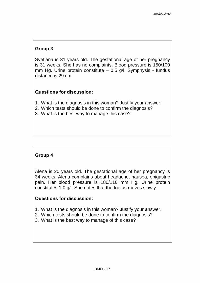
Module 3MO
Group 3 Svetlana is 31 years old. The gestational age of her pregnancy is 31 weeks. She has no complaints. Blood pressure is 150/100 mm Hg. Urine protein constitute – 0.5 g/l. Symphysis - fundus distance is 29 cm. Questions for discussion: 1. What is the diagnosis in this woman? Justify your answer. 2. Which tests should be done to confirm the diagnosis? 3. What is the best way to manage this case?
Group 4 Alena is 20 years old. The gestational age of her pregnancy is 34 weeks. Alena complains about headache, nausea, epigastric pain. Her blood pressure is 180/110 mm Hg. Urine protein constitutes 1.0 g/l. She notes that the foetus moves slowly. Questions for discussion: 1. What is the diagnosis in this woman? Justify your answer. 2. Which tests should be done to confirm the diagnosis? 3. What is the best way to manage of this case?
3MO - 17

Effective Perinatal Care (EPC)
3MO - 18

Effective Perinatal Care (EPC) Facilitator Guide
Module 4МО
Obstetrical Haemorrhages
Learning objectives
At the end of the module participants will: • Understand the importance of active management of the third stage of
labour in preventing postpartum haemorrhage • Understand the steps to identify and initiate the early management of
postpartum haemorrhage • Be able to make decisions about comprehensive measures to stop
bleeding and resuscitate the patient • Understand that the basis of care for women with PPH is the timely and
adequate replacement of blood loss the circulatory system Be able to critically consider the surgical methods of treatment for postpartum haemorrhage
• Understand the importance of appropriate local protocol for managing obstetrical haemorrhage and for conducting trainings on regular basis
Module outline and duration: Total duration – 120 min
Activity 1 – Introduction 15 min Activity 2 – Small group work 15 min Activity 3 – Presentations of group work results and discussions 75 min Activity 4 – Conclusions 15 min
Preparation for the module • Review current publications and evidence materials referred to in the
management of postpartum haemorrhage • Ensure that all participants have a Participant Manual
Materials and Audiovisual Equipment Materials
• Participant Manual • Power Point presentation 4MO EPC ENG • Integrated Management of Pregnancy and Childbirth. Pregnancy,
childbirth, postpartum and newborn care: A guide for essential practice. WHO, Geneva, 2006
4MO - 1

Effective Perinatal Care (EPC)
Materials and Audiovisual Equipment • Managing Complications in Pregnancy and Childbirth: A Guide for
Midwives and Doctors. Geneva: WHO (2000). • A Textbook of Postpartum Hemorrhage. Sapiens Publishing, Duncow, UK,
2006.
Equipment • Video projector or projector overhead • Flipchart • Markers • Pens and pencils
Key Messages
• Active management of the third stage of labour is effective in the prevention of postpartum haemorrhage.
• Early recognition and initiation of measures to stop bleeding are the
first-line in managing PPH.
• Each health facility should have a local clinical protocol for the prevention and treatment of PPH. This protocol should be based on national evidence-based guidelines.
• Oxytocin in doses up to 40 IU, as well as ergometrine and
prostaglandins are effective in many refractory cases of uterine atony.
• External and internal bimanual compression of the uterus and compression of the aorta are the methods recommended for temporary control of atonic bleeding.
• Initial measures to stop bleeding and resuscitate the patient are
effective in most cases of PPH. A midwife has the necessary skills to use these measures.
• Hysterectomy is not the only method for the complete arrest of
bleeding. There are many other simpler, less traumatic but effective alternatives: direct intramyometrial injection of prostaglandin, bilateral ligation of uterine arteries, bilateral ligation of internal iliac arteries and compression uterine sutures.
• Uterine atony is a rare indication for hysterectomy. Removal of the
uterus is indicated mostly in placenta increta, uterine rupture, haematoma of broad ligament and other cervical-uterine trauma.
• A total hysterectomy is not always necessary. Subtotal hysterectomy
is the operation of choice in most instances of PPH requiring hysterectomy.
4MO - 2

Module 4MO
Key Messages • There is no evidence that colloids for replacing blood loss are better
than crystalloids in reducing maternal mortality.
• Blood transfusion is vital in the most severe cases of PPH. At the same time, inappropriate or over-use may lead to many complications. There should be very strict indications for red-cells and fresh-frozen plasma transfusion
Activity 1 – Introduction (15 min) • Show Slide 4MO-1 and explain that while working with this module the
participants will discuss evidence based methods of treatment for postpartum haemorrhage.
• Discuss the magnitude of the problem using Slide 4MO-2. • Underline the main difficulties of PPH management (Slide 4MO-3) and stress
that underestimation of blood loss is one of the most important. • Initiate a discussion with the participants by asking; what is the most relevant
definition of PPH for clinical practice? (Slide 4MO-4). Haemodynamic response to haemorrhage is very individual, thus, in every blood loss suspected to be pathological, medical staff should proceed with the initial steps of resuscitation and bleeding control.
• Explain that the correct calculation of the total amount of blood loss is rather
difficult but crucial. Show Slide 4MO-5 which presents the WHO definition of postpartum haemorrhage.
• Show Slides 4MO-6 and 4MO-7 and highlight the importance of postpartum
haemorrhage prevention by implementation of the routine use of an active management of the third stage of labour
• Discuss (briefly) consequences of severe blood loss and stress the importance
of volume replacement therapy and the timely arrest of bleeding.
Activity 2 – Small group work (15 min) • Divide participants into 5 groups: two groups of midwives, two groups of
obstetricians and one mixed, consisting of midwives and obstetricians. In the case of a limited number of participants, the groups can be divided into 4 groups and combine the first two tasks.
• Give them clear flipcharts sheets and markers. • Show Slide 4MO-8 with the tasks. • Ask the first group of midwives to list the initial steps of managing early
postpartum haemorrhage.
4MO - 3

Effective Perinatal Care (EPC)
• Ask the second group of midwives to present a complete list of procedures
related to the arrest of bleeding before laparotomy, a procedure midwives can do.
• Ask the participants from the third group to present indications and surgical
methods for the complete arrest of bleeding. • Ask the fourth group to list the main rules and principles of volume
replacement therapy and blood transfusion. • Give the fifth group (mixed) the task of distributing roles among group
members in a case of severe PPH management. Participants of this group should imagine themselves on duty in maternity and describe the responsibilities of staff members caring for a woman with PPH.
• Explain that all ideas should be listed on a flipchart and two participants from
each group will present the results of small group work.
Activity 3 – Presentations of groups work results and discussions (75 min) • After the presentation done by the first group, show Slide 4MO-9. Stress that
after recognition of PPH, each health professional should call for assistance and simultaneously initiate the following: interventions aimed to resuscitate, monitoring the patient, finding the source of bleeding and stopping the bleeding.
• Note that timely action is very important. Show Slide 4MO-10. Explain that
early recognition is a key point in timely initiation of care. Stress that it is more logical to initiate resuscitation and careful monitoring, and to be clinically vigilant in 10 cases of non-severe bleeding than it is to be late in one case of severe PPH (>1000 ml or with signs of shock).
• Show Slide 4MO-11, and list the components of the initial assessment and
care which should be provided immediately after detecting postpartum bleeding.
• Show Slide 4MO-12, reminding participants what should be done to initiate
resuscitation. • Show Slide 4MO-13 which are the main causes of PPH. Stress that the most
common causes of PPH are uterine atony and genital tract lacerations. The more rare causes of PPH, such as placenta previa and accreta, uterine rupture and coagulation abnormalities, are more difficult to manage and are associated with much higher mortality.
• Slide 4MO-14 lists measures to stop bleeding in case the placenta is not
delivered. • Slide 4MO-15, when placenta is delivered but not complete.
4MO - 4
Module 4MO
• Slide 4MO-16 when the placenta is delivered and complete (uterine atony and genital tract trauma). Stress that oxytocin in doses of up to 40 IU may be used in cases of severe uterine atony.
• Ask the participants from the first group to prioritize actions in case of
recognition of early PPH. • Ask the participants to make a calculation of the total amount of time
necessary to perform initial steps to treat PPH. If procedures are done in correct order it takes not more than 5-7 minutes. Stress that most cases of PPH respond to these very simple methods of treatment.
• Ask midwives from the second group to present the list of procedures they are
able to do in case of postpartum haemorrhage. This list should include: intravenous access, uterine massage, speculum examination and suture of lacerations, bladder catheterization, pulse and blood pressure measurement, manual removal of placenta, manual control of uterine cavity, bimanual compression of the uterus and use of uterotonics.
• Discuss with participants the management of uterine atony (Slide 4MO-17)
and uterine atony unresponsive to oxytocin (Slide 4MO-18) by using methylergometrine and prostaglandins.
• Note the high success rate of ergot alkaloids (Slide 4MO-19) in haemorrhage
cases refractory to oxytocin and prostaglandins (Slides 4MO-20) • Draw the attention of participants to the disadvantages (Slide 4MO-21) and
advantages (Slide 4MO-22) of Misoprostol for the treatment of postpartum haemorrhage due to uterine atony. Stress that to date, Misoprostol has proven effective in postpartum haemorrhage prevention but not in treatment. However Misoprostol could be useful in controlling postpartum bleeding from uterine atony but further research is needed to prove it.
• Show Slide 4MO-23 and discuss what should be done if these measures are
unsuccessful and bleeding continues. Stress the necessity of informing the surgery team in a timely manner, and ordering blood components, as well as clearly recording fluid and blood transfusions.
• Show Slide 4MO-24, and stress that in case of continued bleeding and
ineffective of measures it is necessary to be ready for surgical treatment while continuing infusion therapy. Ask the participants which methods for the temporary arrest of bleeding they know.
• Explain the technique of bimanual compression of the uterus (Slide 4MO-25)
and initiate a discussion of theoretical advantages and disadvantages of bimanual compression versus internal massage of the uterus. Write them on the flipchart.
• Show another method to temporarily arrest bleeding with Slide 4MO-26,
(compression of the aorta) and stress that other methods to temporarily arrest bleeding did not prove effective, wasted time and should not be used.
4MO - 5

Effective Perinatal Care (EPC)
• Drive the discussion to the conclusion that the midwife is the appropriate professional to deal with PPH in case of emergency until operative measures are undertaken.
• Show Slide 4MO-27 and note that trauma is one of the leading causes of
PPH. Unnecessarily hysterectomies are frequently performed because the birth canal is often not examined for trauma.
• Ask the participants from group three to present indications and methods for
the complete arrest of bleeding. • Show Slide 4MO-28 and explain that surgical haemostasis should be initiated
as soon as possible if conservative measures are ineffective. Stress that late recourse to surgical haemostasis is one of the leading factors contributing to poor results in cases of obstetric haemorrhage
• Show Slide 4MO-29 and present alternatives to hysterectomy in case of
severe atonic PPH. Note that, except for ligation of internal iliac arteries, these procedures are simpler and safer than hysterectomy, are not so traumatic and preserve future fertility.
• Show evidence of effectiveness in Slides 4MO-30 and explain technique in
Slides 4MO-31 of compressive sutures. Tell the participants that the technique of the B-lynch suture operation will be discussed during the clinical week.
• Slides 4MO-32 and 4MO-33 demonstrate the evidence of the ligature of
uterine vessels. • Show Slide 4MO-34 and ask the participants about hysterectomy incidences
in their facility/region and compare them with the presented data. • Note the change in indications of hysterectomy: from uterine atony as the most
frequent in the 70s to anomalies of placental insertion in late 80s. Ask participants to explain why these changes happened. Possible explanations: at the end of the 80-s, and in the beginning of the 90s, evidence of high doses of oxytocin, prostaglandins and ligation of uterine vessels effectiveness for treatment of PPH were reported.
• Discuss in what cases total hysterectomy should be performed and when
leaving the cervix in place would be a better option (Slide 4MO-35 and notes). Explain the following:
o It is believed that if cervix is not removed during hysterectomy, it may
serve as a source of bleeding in case of coagulation disturbances and may lead to maternal death. In reality, in the case of disseminated intravascular coagulation syndrome (DIC), the cervix is just one of the many sources of bleeding (abdominal wall wound, another ligatures, and injection sites are the other sources).
o Increased rate of maternal deaths associated with subtotal
hysterectomy happens because this operation is performed on the most severely ill patients, who have already experienced a large loss of blood volume or who are severely shocked. There was no time for a total hysterectomy.
4MO - 6

Module 4MO
• Show Slide 4MO-36 which presents an excerpt from the Report on confidential enquiries into maternal deaths in the United Kingdom 2000-2002 and makes conclusion about surgical treatment (Slide 4MO-37). Knowing that there are many effective alternatives of hysterectomy (prostaglandins, uterine artery ligation, compression sutures), should make it easier for doctors to decide on surgical treatment (laparotomy) that is vital in the most severe cases of PPH.
• After group four presents, show the general principles of infusion therapy
(Slide 4MO-38) and the results of studies that evaluated whether colloid solutions are better than crystalloids in resuscitating severe patients (Slides 4MO-39 and 4MO-40). Ask the participants to speak from their experience about the adverse effects of dextrans, albumin and pentastarches.
• Discuss indications and adverse effects of red-cell transfusion. Show Slide
4MO-41 and note that the only purpose of red-cell transfusion is to restore the oxygen-carrying capacity of the blood. Mention that red-cell transfusion may adversely affect microcirculation (lots of microscopic clots) or blood coagulation (haemolysed erythrocytes, increase of enzymes activity, etc) in an already compromised patient. Risks of transfusion-transmissible infections, incompatibility and other transfusion complications should be considered.
• Show Slide 4MO-42 and note increased maternal mortality in cases when
transfusion occurred with women who had high haematocrits. At the same time mortality increased in the absence of transfusion in women with critically low haematocrits.
• Slide 4MO-43 presents indications and requirements for fresh-frozen plasma
transfusion. Stress that plasma causes the same complications as red-cell transfusion and should not be used for volume replacement therapy, or in the treatment of hypoproteinemia when a coagulation factor deficiency is not confirmed. The fresh-frozen plasma transfusion is effective only if infused in very large volumes and very quickly.
• Ask the participants from the fifth group to focus on the roles of staff during
management of severe PPH, when each needs to know precisely his/her responsibilities. Ask them to list essential supplies and equipment necessary in these cases and to calculate how many persons are needed for effective care in cases of severe PPH.
• After presentation by the fifth group show Slide 4MO-44 and make
conclusions on the importance of organizational aspects of severe PPH management: the availability of comprehensive protocols with clear definitions of responsibility for of each staff member, as well as conducting trainings on regular basis.
Activity 4 – Conclusions (15 min) • Show participants Slides 4MO-45 and 4MO-46 which summarize the key
information which participants received during their work with this module. • Conclude working with this module by asking if participants have any
questions or additional points they want to make. Answer all the participants’ questions.
4MO - 7

Effective Perinatal Care (EPC)
References 1. A Textbook of Postpartum Hemorrhage. Sapiens Publishing, Duncow, UK,
2006.
2. Alderson P, Bunn F, Lefebvre C, Li Wan Po A, Li L, Roberts I, Schierhout G. Human albumin solution for resuscitation and volume expansion in critically ill patients. The Cochrane Database of Systematic Reviews 2004, Issue 4.
3. American College of Obstetricians and Gynecologists: ACOG educational
bulletin. Postpartum hemorrhage. Number 243, January 1998 (replaces No. 143, July 1990). American College of Obstetricians and Gynecologists. Int J Gynaecol Obstet 1998 Apr; 61(1): 79-86.
4. Averting maternal death and disability: Preventing postpartum hemorrhage
in low-resource settings. International Journal of Gynecology and Obstetrics. 2002;77:267–275.
5. Blanchard K, Clark S, et al. Misoprostol for Women's Health: a
Review.Obstetrics& Gynecol, 2002, 99,316-332.
6. B-Lynch C, Coker A, Lawal AH, et al. The B-Lynch surgical technique for the control of massive postpartum haemorrhage: an alternative to hysterectomy? Five cases reported. Br J Obstet Gynaecol, 1997, 104,372-375.
7. Chamberlain GVP. The clinical aspects of massive haemorrhage. In: Patel
N, ed. Maternal mortality: the way forward. London: RCOG, 1992, 54-62.
8. Cunningham F.G., Gant N.F., Leveno K.J., eds. et al. Williams obstetrics. 21st ed. New York, NY: McGraw-Hill, 2001. 407- 687.
9. El-Hamamy E, B-Lynch C. A worldwide review of the uses of the uterine
compression suture techniques as alternative to hysterectomy in the management of severe post-partum haemorrhage. J Obstet Gynaecol. 2005, 25(2),143-149.
10. Harrison K et al. “Childbearing, Health and Social Priorities. A Survey of
22,774 Consecutive Births in Zaria, Northern Nigeria”. British Journal of Obstetrics and Gynaecology, 1985, 5, 61–71.
11. Hayman RG et al, Uterine compression sutures: surgical management of
postpartum haemorrhage. Obstet Gynecol., 2002, 99,502-506.
12. Managing Complications in Pregnancy and Childbirth: A Guide for Midwives and Doctors. Geneva: WHO, 2000.
13. McCormick ML, Sanghvi HCG, Kinzie B, McIntosh N. Averting maternal
death and disability: Preventing postpartum hemorrhage in low-resource settings. International Journal of Gynecology and Obstetrics. 2002;77:267–275.
4MO - 8

Module 4MO
14. Mother-Baby Package: Implementing safe motherhood in countries. WHO, 1995.
15. Mousa HA, Alfirevic Z. Treatment for primary postpartum haemorrhage
(Cochrane Review). Cochrane Database of Systematic Reviews 2007, Issue 1.
16. Pregnancy, Childbirth, Postpartum and Newborn Care: A guide for
essential practice. WHO, Geneva, 2006.
17. Prendiville WJ, Elbourne D, McDonald S. Active versus expectant management in the third stage of labour. Cochrane Database of Systematic Reviews. 2007, Issue 1.
18. Prevention and Treatment of Post-partum Haemorrhage: New Advances
for Low Resource Settings. Joint Statement. International Confederation of Midwives (ICM) International Federation of Gynaecology and Obstetrics (FIGO), 2006.
19. Radek Bukowski, Gary D.V. Hankins, Managing postpartum hemorrhage.
Contemporary OB/GYN, Sep 1, 2001.
20. Roberts I, Alderson P, Bunn F, Chinnock P, Ker K, Schierhout G. Colloids versus crystalloids for fluid resuscitation in critically ill patients. The Cochrane Database of Systematic Reviews 2004, Issue 4.
21. Schuurmans N, MacKinnon C et al. Prevention and management of
postpartum haemorrhage. SOGC Clinical Practice Guidelines, № 88, April 2000.
22. Why Mothers Die 2000-2002 – Report on confidential enquiries into
maternal deaths in the United Kingdom. Confidential Enquiry into Maternal Deaths. RCOG Press, Nov 2004.
4MO - 9

Effective Perinatal Care (EPC)
Attachment 1 Activity 2 Group 1 (Midwives)
List the initial steps of early PPH treatment
Group 2 (Midwives) List all procedures for arresting bleeding before a laparotomy, a
procedure that can be done by a midwife
Group 3 (Obstetricians) List the indications and surgical methods for the complete arrest
of bleeding
Group 4 (Obstetricians)
List the basic rules and principles of fluids replacement
Group 5 (Midwives and Obstetricians)
Give a role to each group member to describe the actions of each participant in a case of severe PPH
4MO - 10

Effective Perinatal Care (EPC) Facilitator guide
Module 5MO Prelabour Rupture of Membranes
Module objectives At the end of the module participants should:
• Know the recommendations regarding the diagnosis, investigation and management of women with prelabour rupture of membranes
• Understand the role of prophylactic antibiotics, steroids and tocolytic agents, and the appropriate time to deliver women with prelabour rupture of membranes
• Know the techniques to improve perinatal outcomes in women with prelabour rupture of membranes
• Have a clear understanding of the advantages and disadvantages of active and expectant management of prelabour rupture of the membranes at or near term.
Module structure and duration: General duration – 105 min
Activity 1 – Introduction 5 min Activity 2 – Small group work 25 min Activity 3 – “Brain storming” 15 min Activity 4 – Presentation 40 min Activity 5 – Conclusion 20 min
Module preparation • Review the current publications on prelabour rupture of membranes • Ensure that all participants have a copy of the Participant’s Manual • Ensure that all facilitators know their respective duties for working with this
module
5MO - 1

Effective Perinatal Care (EPC)
Materials and audiovisual equipment Materials
• Participant’s Manual • Murray W. Enkin at al. “A Guide to Effective Care in Pregnancy and
Childbirth”, 2000 • Case studies for small group work
Equipment
• Multimedia or overhead projector • Flipchart • Markers • Pens and pencils
Key messages Preterm prelabour rupture of membranes: • Expectant management is recommended if gestational age less than 34
weeks and no contraindications for pregnancy prolongation. • Digital cervical examination should not be performed in patients who are not
in labour and in whom immediate induction of labour is not planned. Speculum examination is preferred.
• Antibiotics prolong the latency period and improve perinatal outcome and
should be administered according to the published protocol. • Antenatal corticosteroids should be administer if gestational age 24 to 34
weeks. • Short-term tocolysis may be considered to facilitate maternal transportation as
well as the administration of antenatal corticosteroids and antibiotics. • Long-term tocolysis is not indicated. Prelabour rupture of membranes at or near term: • Labour may be induced at the time of presentation or patients may be
observed for up to 24 to 72 hours for the onset of spontaneous labour. • Digital cervical examination should not be performed in patients who are not
in labour and in whom immediate induction of labour is not planned. Speculum examination is preferred.
• Antimicrobial prophylaxis for GBS should be given only after the onset of
labour. • Antimicrobial prophylaxis for non-GBS infection can be restricted to those who
develop clinical indications for antibiotic treatment.
5MO - 2

Module 5MO
Key messages • Immediate induction with oxytocin associated with fewer rates of
chorioamnionitis than both vaginal prostaglandins and expectant management.
Classroom work (105 min)
Activity 1 - Introduction (5 min) • Show Slide 5MO-1 and explain that during the module they will discuss the
evidence-based tactics of PROM management. • Explain that in this module you will discuss the clinical issues regarding
rational PROM management, the evidence and recommendations regarding management of preterm prelabour rupture of membranes and prelabour rupture of membranes at and near term.
Activity 2 – Small group work (25 min) • Split the participants into 3 groups. • Give each group a case study printed on a separate sheet of paper, and
ensure that the groups understand their tasks. Give each group a sheet of flip-chart paper and a marker.
• Tell participants that they have 10 minutes to discuss the case and write their
answers on the flipchart paper.
Group 1 – Case study 1
o Olga was admitted to the maternity with preterm prelabour rupture of membranes which happened 2 hours earlier. This is her 1st pregnancy, gestational term is 30 weeks. The pregnancy was not complicated. The woman is healthy. No labour.
Which investigations are necessary in this case? Develop a plan of further management for this patient
Group 2 – Case study 2
o Valentina was admitted to the maternity with PROM which happened
2 hours earlier. This is her 1st pregnancy, gestational term is 40 weeks. The pregnancy was not complicated. The woman is healthy. No labour.
Which investigations are necessary in this case? Develop a plan of further management for this patient
5MO - 3
Effective Perinatal Care (EPC)
If the group selects active management, ask them to describe the tactics they would use if in 10 hour contractions are rare and weak and cervical dilatation is 3 cm.
Group 3 – Case study 3
o Tatiana was admitted to the maternity with PROM which happened 48 hours earlier. This is her 1st pregnancy, gestational term is 38 weeks. The pregnancy was not complicated. The woman is healthy. Labour was induced by oxytocin.
o In 5 hours the signs of choreoamnionitis and foetal distress have appeared. Cervical dilatation is 4 cm.
What tactics are recommended in this case? • Ask a representative of each group to present the results of group work to the
other trainees. During the presentation ask the participants to explain the goal of their investigations and management tactics. Do not comment on the participants’ presentations.
• Tell the participants that at the end of the module you will return to the results
of their group work written on the flipchart and will re-discuss them.
Activity 3 – “Brain storming” (15 min) • Based on the results of group work, ask the participants to think about
questions about PROM which they would like answered at the end of the module.
• Give them one example of a question they can develop (e.g. from Case study
1: Which tactic is the most logical in preterm PROM – expectant or active?) • Write all participants’ questions on a sheet of flipchart paper. Ensure that all
the questions are there. • Place this flipchart sheet on a visible place.
Activity 4 – Presentation (40 min) • Start the presentation showing Slide 5MO-2 and give two definitions of
prelabour rapture of membranes: 1) preterm prelabour rupture of membranes (PPROM) which occurred at 24 – 37 weeks of gestation and 2) prelabour rupture of membranes at or near term (PROM) which occurred at 37 full weeks of gestation or after.
• Go to Slide 5MO-3 and say that despite the fact that PPROM complicates only
2% of pregnancies, it is associated with many serious complications including neonatal death. The three causes of neonatal death associated with PPROM are prematurity, sepsis and pulmonary hypoplasia. Women with intrauterine infection deliver earlier than non-infected women and infants born with sepsis have a mortality rate four times higher than those without sepsis. In addition, there are maternal risks associated with chorioamnionitis.
5MO - 4

Module 5MO
• Stress that there is evidence demonstrating an association between infection ascending from the lower genital tract and PPROM. In women with PPROM about one-third of pregnancies have positive amniotic fluid cultures and studies have shown that bacteria have the ability to cross intact membranes.
• Show Slide 5МО-4 and discuss how to make a diagnosis of PROM.
Emphasize that the best diagnosis is made by a history suggestive of spontaneous rupture of membranes (SROM), followed by a sterile speculum examination demonstrating pooling of fluid in the posterior vaginal fornix; in this case a Nitrazine test is not necessary.
• Tell participants that a series of tests have been used to confirm membrane
rupture; the most widely used has been the Nitrazine test, which detects pH change, and has a sensitivity of 90% and a false positive rate of 17%. More recently, other tests have been evaluated in the diagnosis of ruptured membranes and foetal fibronectin and raised insulin-like growth factor binding protein-1 in cervical/vaginal secretions have reported sensitivities of 94% and 75% and specificities of 97%, respectively.
• Show Slide 5MO-5 and present the possible tactics for the management of
women with PPROM. • Than show Slide 5MO-6 and discuss when digital vaginal examination should
be performed. Emphasize that digital vaginal examination is best avoided unless there is a strong suspicion that the woman may be in labour. This is because micro-organisms may be transported from the fingers to the vagina and into the cervix, leading to intrauterine infection, prostaglandin release and preterm labour.
• Tell participants that one retrospective study reported that the latency interval
between SROM and delivery in those who had a digital vaginal examination was significantly shorter than if only a sterile speculum examination was performed.
• Show Slides 5MO-7 and 5MO-8. Discuss antenatal tests (monitoring) which
should be conducted in case of PPROM and their effectiveness (Slide 5MO-7). Tell participants that the criteria for the diagnosis of clinical chorioamnionitis include maternal pyrexia, tachycardia, leucocytosis, uterine tenderness, offensive vaginal discharge and foetal tachycardia. During inpatient observation, the woman should be regularly examined for such signs of intrauterine infection and an abnormal parameter or a combination of them may indicate intrauterine infection. The frequency of maternal temperature, pulse and foetal heart rate auscultation should be 4 - 8 hours. Then show Slide 5MO-8 and stress that biophysical profile scoring or Doppler velocimetry should not be considered as first-line surveillance or as a diagnostic test for foetal infection. Also tell participants that routine amniocentesis is not recommended for women with PPROM. Despite the fact that amniocentesis has the potential to detect sub-clinical infection before the onset of maternal signs of chorioamnionitis, and before the onset of foetal sepsis, allowing appropriate intervention such as administration of antibiotics in infected cases and/or delivery, depending on the gestation, and expectant management for women with negative amniotic fluid cultures, the role of amniocentesis in improving outcome remains to be determined.
5MO - 5

Effective Perinatal Care (EPC)
• Show Slide 5MO-9. Note that the use of antibiotics in PPROM statistically reduces the rate of serious complications. Tell participants that there was a variation in the choice of antibiotics used and the duration of therapy in the studies examined in one meta-analysis. As a result of this meta-analysis there are two proved recommendations:
o Erythromycin (250 mg orally every 6 hours) should be given for 10 days following the diagnosis of PPROM. [A]
o Co-amoxiclav is not recommended for women with PPROM because of concerns about necrotising enterocolitis. [A]
o Erythromycin 250 mg orally three times a day for 7 days PLUS Amoxicillin 500mg orally three times per day for seven days
• Show Slide 5MO-10 and explain that indications for antenatal corticosteroid
therapy include women with PPROM between 24 and 34 weeks of gestation. Emphasize that this technology is associated with statistically significant reduction in rates of:
o Respiratory distress syndrome o Intraventricular haemorrhage o Necrotising enterocolitis
• Go to Slide 5MO-11. Stress that only woman with PPROM and uterine activity
who require intrauterine transfer or antenatal corticosteroids should be considered for tocolysis. Prophylactic tocolysis in women with PPROM without uterine activity is not recommended, because tocolysis after PPROM did not increase the interval between membrane rupture and delivery or reduce neonatal morbidity.
• Show Slide 5MO-12 and say that in the absence of clear evidence that
tocolysis improves neonatal outcome following PPROM, it is reasonable not to use it. It is possible that tocolysis could have adverse effects, such as delaying delivery from an infected environment, since there is an association between intrauterine infection, prostaglandin and cytokine release and delivery. However, the benefits of antenatal steroids apply equally to women with PPROM and, in some clinical circumstances; the risk–benefit ratio may lead to consideration of tocolysis for this purpose. Similarly it would seem wise to consider tocolysis for transfer of women, depending on individual circumstances. Stress that aggressive tocolysis after PPROM did not increase latency or decrease neonatal morbidity compared with either limited tocolysis or no tocolysis at all.
• Using Slide 5MO-13 present information that transvaginal amnioinfusion in
labour is not recommended for women with preterm rupture of membranes. Also explain that transabdominal amnioinfusion is not recommended as a method of preventing pulmonary hypoplasia in very preterm PROM. Stress that at the present time, there is insufficient evidence to recommend this treatment outside randomised trials.
• Show Slide 5MO-14 and tell participants that women should be considered for
outpatient monitoring of PPROM only after rigorous individual selection by a consultant obstetrician.
• Stress that outpatient monitoring should be considered only after a period of
48–72 hours of inpatient observation. Women should be advised of the signs and symptoms of chorioamnionitis and under what circumstances they should
5MO - 6

Module 5MO
seek the advice of a specialist. Women being monitored at home for PPROM should take their temperature twice daily or should be advised of the symptoms associated with infection. There should be clearly described local arrangements for the frequency of outpatient visits and what should happen at these visits.
• Go to Slide 5МО-15. Remind participants that many studies have
demonstrated benefits in conservative management for gestations of less than 34 weeks, whereas the management of pregnancies complicated by PPROM between 34 and 37 weeks of gestation continues to be a contentious issue.
• Emphasize that delivery should be considered at 34 weeks of gestation. • Where expectant management is considered beyond 34 weeks of gestation,
women should be counselled about the increased risk of chorioamnionitis and its consequences versus the decreased risk of serious respiratory problems in the neonate, admission for neonatal intensive care and caesarean section.
• Show Slide 5MO-16 and explain that PROM complicates 6-19% of
pregnancies and is associated with many serious complications. The risks of PROM at term relate to maternal and neonatal infection, prolapsed cord and foetal distress resulting in operative delivery or low five-minute Apgar score. Foetal distress may be caused by any of the complications listed.
• Show Slide 5MO-17. It presents the evidence about management of PROM at
and near term: o With term, prelabour rupture of membranes (PROM), labour may be
induced at the time of presentation or patients may be observed for up to 24 to 72 hours for the onset of spontaneous labour.
o Women with prelabour rupture of the membranes at term (over 37 weeks) should be offered a choice of immediate induction of labour or expectant management.
o Expectant management of women with prelabour rupture of the membranes at term should not exceed 96 hours following membrane rupture.
• Show next Slide 5MO-18 with the epidemiological data on time interval from
term PROM to spontaneous labour and how it demonstrates that most women go into spontaneous labour within 24 hours of rupturing their membranes.
o 86% of women will labour within 12–23 hours o 91% will labour within 24–47 hours o 94% will labour within 48–95 hours. o 6% of women will not be in spontaneous labour within 96 hours of
PROM. • As the time between the rupture of the membranes and the onset of labour
increases, so may the risks of maternal and foetal infection. Induction of labour may reduce these risks
• Make conclusions regarding digital vaginal examinations in PROM, using
Slides 5MO-19 and 5MO-20. The analysis of different tactics of PROM at term management showed that digital cervical examination is the key reason for the post-partum endometritis. Early induction is associated with a higher risk of endomertitis than expectant management, as vaginal examination is not
5MO - 7

Effective Perinatal Care (EPC)
performed before labour in expectant management. In the one trial, in expectant management 28% of neonates (5 out of 18) born to mothers who had a vaginal examination at admission, but none of 78 babies were infected in the group whose mothers did not have a digital cervical examination before labour. Emphasize that there is a direct connection between the number of vaginal examination and the risk of choreoamnionitis. The risk of such a complication increases 5 times when 8 or more vaginal examinations are performed.
• Slide 5MO-21 contains information about the necessity of antibiotics
administration in PROM for Group B streptococcus prevention. Emphasize that antibiotic prophylaxis for GBS should be given only after the onset of labour
• Stress that antenatal treatment with penicillin is not recommended. Antenatal
prophylaxis with oral penicillin does not reduce the likelihood of GBS colonisation at the time of delivery and so is not indicated in this situation.
• Also explain that antibiotic prophylaxis for GBS is unnecessary for women with
preterm rupture of membranes unless they are in established labour. Antibiotic administration specifically for GBS colonisation is not necessary prior to labour. If these women are known to be colonised with GBS, antibiotic prophylaxis should be considered, especially if labour occurs prior to 37 weeks.
• Go to Slide 5MO-22. Tell participants that it is not clear if antibiotics should be
given routinely for the prevention of chorioamnionitis, postpartum endometritis, and neonatal infections that are caused by organisms other than Group B streptococcus. Further, well designed randomised controlled trials are needed to assess the effects of a routine use of maternal antibiotics for women with prelabour rupture of the membranes at or near term.
• Slides 5MO-23 and 5MO-24 contain information on different recommended
regimens of antibiotics administration in PROM. Discuss all listed regimens with participants.
• Show Slide 5MO-25 and discuss possible modes for the active management
of PROM. Stress that PROM at and near term can be managed by immediate oxytocin induction or by vaginal (or endocervical) prostaglandin E2, gel, suppositories or tablets. Emphasize that vaginal prostaglandins resulted in more chorioamnionitis than immediate oxytocin but less chorioamnionitis than conservative management. Immediate oxytocin induction resulted in fewer cases of chorioamnionitis and endometritis.
• Go to Slide 5MO-26 and discuss the advantages and disadvantages of active
versus conservative management in PROM. Stress that conservative management may result in more maternal infections than immediate induction with oxytocin or prostaglandins. Planned management (with methods such as oxytocin or prostaglandin) reduces the risk of some maternal infectious morbidity without increasing caesarean sections and operative vaginal births. Fewer infants went to neonatal intensive care under planned management although no differences were seen in neonatal infection rates. Since planned and expectant management may not be very different, women need to have appropriate information to make informed choices. Emphasize that women view induction of labour more positively than expectant management. Also tell
5MO - 8

Module 5MO
participants that the difference in cost between induction with oxytocin and the other management options was statistically significant. Induction with oxytocin was found to be less costly when compared to the other treatment alternatives.
• Show Slides 5MO-27 and 5МО-28 and discuss the key recommendations of
preterm PROM management. • Than go to Slides 5MO-29 and 5МО-30 and discuss the key
recommendations on management of PROM at and near term.
Activity 5 – Conclusion (20 min) • Draw the participants’ attention to those questions which they listed on the flip-
chart during Activity 3. • Ask participants to answer each of them briefly. Ensure that all questions are
answered. Answer the questions which are not covered by the presentation. • Answer additional questions that come up. • Than ask the participants to split into the same groups in which they worked
during Activity 2. Give them the flipchart sheets with their answers to the case study questions.
• Ask participants if they have any changes to their initial opinions regarding the
tactics of PROM management in the case studies. If the participants want to change any of the tactics they suggested at the beginning of the module, discuss why they want to make the changes. After that make the appropriate changes.
• Ask the participants if they have any questions on this topic. Answer these
questions.
5MO - 9

Effective Perinatal Care (EPC)
References
1. American College of Obstetricians and Gynecologists (ACOG). Premature rupture of membranes. Washington (DC): American College of Obstetricians and Gynecologists (ACOG), 1998 Jun., 10 p. (ACOG practice bulletin; no. 1). Current as of December 2004.
2. Amiram Gafni, Ron Goeree, Terri L. Myhr et al. Induction of labour
versus expectant management for prelabour rupture of the membranes at term: an economic evaluation. Canadian Medical Association Journal DEC. 1, 1997; 157:11.
3. Amy Marowitz, CNM; Heather Hunter, SNM. Management of Ruptured
Membranes at Term. Journal of Midwifery & Women's Health, 2004.
4. Blackwell SC, Berry SM. Role of amniocentesis for the diagnosis of subclinical intra–amniotic infection in preterm premature rupture of the membranes. Curr Opin Obstet Gynecol, 1999, 11, 541–47.
5. Carlan SJ et al. Preterm premature rupture of membranes: a randomised
study of home versus hospital management. Obstet Gynecol, 1993, 81, 61–4.
6. Centers for Disease Control and Prevention. Prevention of Perinatal Group
B Streptococcal Disease. Morbidity and Mortality Weekly Report (MMWR). Revised Guidelines from CDC, 2002, 51.
7. Crowley P. Prophylactic corticosteroids for preterm birth. The Cochrane
Database of Systematic Reviews, Issue 3, 2005.
8. Dare MR, Middleton P, Crowther CA, Flenady VJ, Varatharaju B. Planned early birth versus expectant management (waiting) for prelabour rupture of membranes at term (37 weeks or more). Cochrane Database of Systematic Reviews 2006, Issue 1.
9. De Santis M et al. Transaabdominal amnioinfusion treatment of severe
oligohydramnios in preterm premature rupture of membranes at less than 26 gestational weeks. Fetal Diagn Ther, 2003, 18, 412–17.
10. Flenady V, King J. Antibiotics for prelabour rupture of membranes at or
near term. The Cochrane Database of Systematic Reviews. 2007, Issue 1.
11. Gaucherand P et al. Comparative study of three amniotic fluid markers in premature of membranes: fetal fibronectin, alpha-fetoprotein, diaminooxydase. Acta Obstet Gynecol Scand, 1995, 74, 118–21.
12. Hannah ME, Ohlsson A, Farine D, Hewson SA, Hodnett ED, Myhr TL,
Wang EE, Weston JA, Willan AR. Induction of labour compared with expectant management for prelabour rupture of the membranes at term. TERMPROM Study Group. N Engl J Med. 1996 Apr 18;334(16):1005-10.
13. How HY et al. Preterm premature rupture of membranes: aggressive
tocolysis versus expectant management. J Matern Fetal Med, 1998, 8–12.
5MO - 10

Module 5MO
14. Jazayeri A et al. Tocolysis does not improve neonatal outcome in patients with preterm rupture of membranes. Am J Perinatol, 2003, 20, 189–93.
15. Kenyon S. et al. Antibiotics for preterm premature rupture of membranes
(Cochrane Review), Cochrane Library, Issue 3, 2002.
16. Lewis DF et al. Effects of digital vaginal examination on latency period in preterm premature rupture of membranes. Am J Obstet Gynecol, 1992, 80, 630–4.
17. Managing Complications in Pregnancy and Childbirth. A guide for
midwives and doctors. WHO, 2005.
18. Maxwell GL. Preterm premature rupture of membranes. Obstet Gynecol Surv, 1993, 48, 576–83.
19. Merenstein GB, Weisman LE. Premature rupture of the membranes:
neonatal consequences. Semin Perinatol, 1996, 20, 375–80.
20. Mozurekewich E., Wolf F. Premature rupture of membranes at term: A meta-analysis of three management strategies. Obstet Gynecol 1997;89:1035-43.
21. Murray W. Enkin et al, A guide to effective care in pregnancy and
childbirth. Oxford University Press, 3-rd edition, 2000.
22. MV Wagner, et al. A comparison of early and delayed induction of labour with spontaneous rupture of membranes at term. Obstetrics & Gynaecology. 1989;74:93-97.
23. Royal College of Obstetricians and Gynaecologists (RCOG). Preterm
Prelabour Rupture of Membranes. Guideline No. 44, RCOG Press, November 2006.
24. Royal College of Obstetricians and Gynaecologists (RCOG).
PREVENTION OF EARLY ONSET NEONATAL GROUP B STREPTOCOCCAL DISEASE. Guideline No. 36. November 2003.
25. Rutanen EM, Pekonen F, Karkkainen T. Measurement of insulin-like
growth factor binding protein-1 in cervical/vaginal secretions: comparison with the ROM-check Membrane Immunoassay in the diagnosis of ruptured fetal membranes. Clin Chim Acta, 1993, 214, 73–81.
26. Seaward PG et al International Multicentre Term Prelabor Rupture of
Membranes Study: evaluation of predictors of clinical chorioamnionitis and postpartum fever in patients with prelabor rupture of membranes at term. Am J Obstet Gynecol. 1997 Nov;177(5):1024-9.
27. Tranquilli AL et al. Transabdominal amnioinfusion in preterm premature
rupture of membranes: a randomised controlled trial. BJOG, 2005, 112, 759–63.
5MO - 11

Effective Perinatal Care (EPC)
Attachment 1 Activity 2 Group 1 Case study Olga was admitted to the maternity with preterm prelabour rupture of membranes which happened 2 hours earlier. This is her 1st pregnancy; gestational term is 30 weeks. The pregnancy was not complicated. The woman is healthy. No labour. Questions for discussion
1. Which investigations are necessary in this case? 2. Develop a plan of further management for this patient
Group 2 Case study Valentina was admitted to the maternity with PROM which happened 2 hours earlier. This is her 1st pregnancy; gestational term is 40 weeks. The pregnancy was not complicated. The woman is healthy. No labour. Questions for discussion
1. Which investigations are necessary in this case? 2. Develop a plan of further management for this patient
5MO - 12
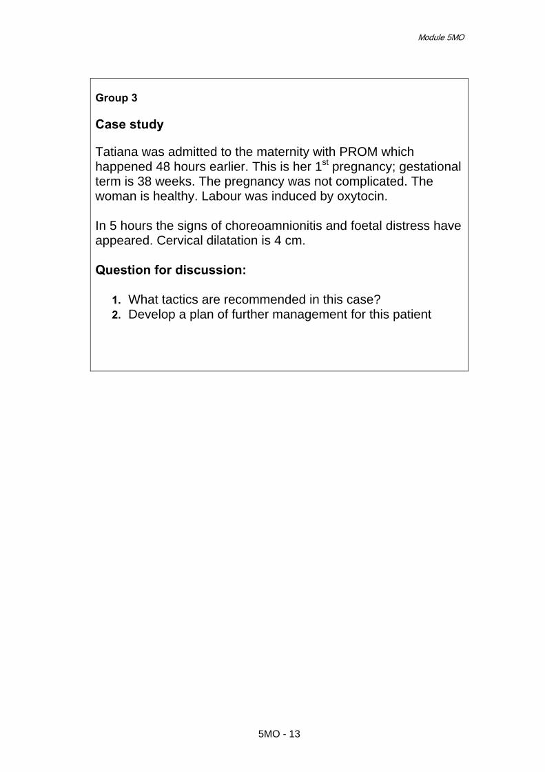
Module 5MO
Group 3 Case study Tatiana was admitted to the maternity with PROM which happened 48 hours earlier. This is her 1st pregnancy; gestational term is 38 weeks. The pregnancy was not complicated. The woman is healthy. Labour was induced by oxytocin. In 5 hours the signs of choreoamnionitis and foetal distress have appeared. Cervical dilatation is 4 cm. Question for discussion:
1. What tactics are recommended in this case? 2. Develop a plan of further management for this patient
5MO - 13

Effective Perinatal Care (EPC)
5MO - 14

Effective perinatal care (EPC) Facilitator guide
Module 6МО Labour Induction: Who? Why? How?
Learning objectives At the end of the module the participants should:
• Understand that inducing labour represents a complex series of interventions to artificially induce contractions before they begin spontaneously.
• Understand that labour should only be induced when vaginal delivery is
possible, because inducing labour can prolong delivery.
• Understand the potential complications of induced labour.
• Be aware of the main indications for labour induction. • Understand that the main methods of labour induction depend on the
state of the cervix.
• Be aware that there are special cases where inducing labour requires the utmost caution because of the possible effects of induction on mother and foetus.
Module outline and length: Part I – Classroom work – 60 minutes
Activity 1 – Introduction 5 min Activity 2 – Work in small groups 15 min Activity 3 – Interactive presentation 35 min Activity 4 – Conclusion 5 min
Preparation for the module
• Get acquainted with the practices in other countries and regions that relate to this topic.
• Analyze the use of these practices in the present health care setting.
• Make sure that the tasks for the participants are understood where
they are being taught.
• Make sure that all the participants have Participant Manual
6MO - 1

Effective Perinatal Care (EPC)
Preparation for the module
• Ensure that the other trainers know the scope of work they are responsible for during the training.
Materials and Audiovisual Equipment
Materials
• Participant Manual • Murray W. Enkin et al. A Guide to Effective Care in Pregnancy and
Childbirth • Integrated Management of Pregnancy And Childbirth: Managing
Complications in Pregnancy and Childbirth: A guide for midwives and doctors. WHO, 2005,
• A set of tasks for the participants
Equipment • PowerPoint or slide-projector • Flipchart • Markers in different colours
Key Messages • Inducing labour shouldn’t be undertaken lightly. It can lead to various
complications for mother and child. There should be strict reasons for both mother and foetus.
• Key questions for inducing labour: Why? (indications) When? (conditions) How? (method – medication) • “The decision to induce labour before the spontaneous commencement of
delivery is one of the most radical means of intervention in the natural process pregnancy and child-birth.”
• Delivery should be induced in case where the risks of prolonging pregnancy
are higher than the risks related to induction. In reality, there are not many indications for labour induction. False-positive results of diagnostic tests should be considered.
• The most important conditions for successful labour induction include
documented confirmation that the reasons and risks were discussed with the
6MO - 2

Module 6МО
Key Messages patient; repeated assessment of gestational age, biological preparedness of the cervix and foetal heart rate before inducing; and the availability of effective methods for preparation of the cervix for induction.
• An ultrasound to confirm gestational age should be offered before 20 weeks
of gestation, as this reduces the need to induce labour for perceived prolonged pregnancy.
• Women with uncomplicated pregnancies should be offered labour induction
beyond 41 weeks. • After 41 weeks, women who decline labour induction should be offered
increased antenatal monitoring that consists of twice weekly CTG and ultrasound estimation of amniotic fluid volume.
• Prior to a formally induced labour, women should be offered sweeping of the
membranes. • When membrane sweeping is proposed, discussions should inform the
woman that it: o is not associated with an increase in maternal or neonatal infection o is associated with increased levels of discomfort during the examination
and with bleeding. • When an induced labour is started with prostaglandins, intravaginal PGE2
should be used in preference to intracervical preparations, as they are equally effective, and administration of vaginal PGE2 is less invasive.
• Wherever induced labour occurs, facilities should be available for continuous
uterine and foetal heart rate (FHR) monitoring. • Following an induced labour with vaginal prostaglandins (PGE2), foetal
wellbeing should be established once contractions are detected or reported. • Where oxytocin is being used for induction or augmentation of labour,
continuous electronic foetal monitoring should be used. • In the presence of abnormal FHR patterns and uterine hypercontractility (not
secondary to oxytocin infusion), tocolysis should be considered. A suggested regimen is subcutaneous terbutaline 0.25 milligrams.
• In cases of suspected or confirmed acute foetal compromise, delivery should
be accomplished as soon as possible, taking account of the severity of the FHR abnormality and relevant maternal factors. The accepted standard is delivery within 30 minutes.
6MO - 3

Effective Perinatal Care (EPC)
Part I – Classroom work (60 min)
Activity 1 – Introduction (5 min) • Show Slide 6МО-1 and discuss the objectives of this module. • Go to Slide 6МО-2 and explain what labour induction is. Tell participants that
induced labours were used widely in the 1950s when oxytocin was synthesized. The frequency of induced labours varies in different health care settings and regions and it is increasing. The understanding of delivery mechanisms and successful induced labours has been increasing in recent years.
• Many factors affect how a cervix ripens. The mediators in this process are
prostaglandins E2 (PGE2) and F2alpha (PGF2alpha). Their exogenous application stimulates ripening the cervix. Endogenous and exogenous oxytocin is the main stimulator of uterine contractions. It also stimulates the production of PGE2 and PGF2alpha.
• Stress the fact that inducing labour interferes with the natural process of
pregnancy and can lead to a number of complications. It should only be administered in situations where the risk of continuing the pregnancy is higher than the risk related to induction.
Activity 2 – Small group work (15 min) • Split the participants into 4 groups and give each group a piece of flipchart
paper and one marker. • Stress that the team-work is very important and that the opinions of every
participant is needed to solve the tasks. • Show Slide 6МО-3 and give each group a topic for discussion and ask each
group to write their answers on the flipchart papers within 5 minutes. Let the group decide who will present the results of the group.
• Tasks for the small-group work:
Group 1: Indications for labour induction Group 2: Contraindications and conditions for labour induction Group 3: Methods of labour induction Group 4: Possible complications during labour induction
• Explain to the participants that after the presentation of each small-group a
trainer will present information on the topic and then there will be time for discussion and questions.
Activity 3 – Interactive presentation (35 min)
• Ask the participants of the Group 1 to present the results of their group work.
6MO - 4

Module 6МО
• After the presentation, show Slide 6МО-4 and read the quotation from the Murray Enkin book about the fact that the decision to induce labour before spontaneous commencement of delivery is one of the most radical ways of interfering in the natural process of pregnancy and birth. That’s why labour induction should be administered only in situations when the risk of allowing the pregnancy to continue is higher than the risk of induction.
• Go to Slide 6MO-5 and present the list of indications for labour induction. The
most frequent reasons for inducing labour include a pregnancy term of more than 41 weeks and premature rapture of membranes. Point out that issues of pregnancy-induced hypertension, preeclampsia, eclampsia, premature rapture of membranes, and chorioamnionitis will be discussed in separate training modules.
• Show Slide 6MO-6 and explain that an ultrasound to confirm gestation should
be offered before 20 weeks of gestation, as this reduces the need for induction for perceived prolonged pregnancy. Stress that women with uncomplicated pregnancies should be offered labour induction beyond 41 weeks. Emphasize that from 42 weeks, women who decline an induced labour should be offered increased antenatal monitoring consisting of a twice weekly CTG and ultrasound estimation of maximum amniotic pool depth.
• Go to Slide 6MO-7 and describe the reasons why women request induced
labours as well as the economic considerations of a policy of routinely offering ‘elective’ induction for psychological or social reasons. Emphasize that there is insufficient evidence to recommend the elective induction of labour for maternal request.
• Showing Slide 6MO-8, discuss each reason listed on the slide, and the
benefits and disadvantages of labour induction for each of them. Tell them the following:
o Women who have pregnancies complicated by diabetes should be offered labour induction prior to their estimated delivery date. Induced labour in term pregnancies in women with diabetes is associated with a reduced risk of macrosomia. Routine induction does not appear to increase the risk of caesarean section or neonatal morbidity, cases of which were rare.
o Currently, the evidence is inconclusive that a policy of labour induction for suspected foetal macrosomia in women who are not diabetic can reduce maternal or neonatal morbidity.
o The perinatal mortality rate in twin pregnancies is increased in comparison with singleton pregnancies at term.
o No conclusions can be drawn from the available trial evidence relating to the merits of an active policy of induced labours in uncomplicated multifoetal pregnancies.
o Labour induction is not contraindicated in women with a history of a previous caesarean section but careful consideration of the women’s clinical condition should be taken before induction is started.
o In history of previous caesarean section, women should be informed of the two- to three-fold increased risk of uterine rupture and around 1.5 fold increased risk of caesarean section in induced and/or augmented labours compared with spontaneous labours.
o In history of previous caesarean section, women should be informed that there is a higher risk of uterine rupture with induction of labour with prostaglandins.
6MO - 5

Effective Perinatal Care (EPC)
o There is an increased risk associated with planned vaginal breech delivery. The risks associated with induced labour with a breech presentation cannot be quantified from the available trial literature.
o When undertaking to induce labour in women with recognised risk factors (including suspected foetal growth compromise), the clinical discussion regarding the timing and method of induction should be undertaken at a consultant level. The induction process should not occur on an antenatal ward.
o No study could be located that considered induced labour specifically in babies with suspected foetal growth compromise. There is insufficient data to comment on the risks of inducing labour in women with babies with known growth restriction.
• Make the conclusion that adequately designed RCTs reporting relevant clinical
outcomes in specific clinical groups are needed to evaluate the risks and benefits of labour induction for women whose pregnancies are complicated by or associated with above-mentioned conditions.
• Ask participants if they have any questions about the indications for labour
induction. Answer all possible questions. • Ask the participants of group 2 to present contraindications and conditions
needed for inducing labour. • After the presentation show Slide 6МО-9 and explain that the process of
labour induction should only be considered when vaginal delivery is possible. Contraindications for inducing labour include, but are not limited to, the situations shown on the slide.
• Go to Slide 6МО-10 and discuss the prerequisites to labour induction. Stress
that before beginning induction it is necessary to discuss the possible complications with the mother. Emphasize that inducing labour should only follow informed consent by the woman. For consent to be fully informed it should include the reasons for inducing, the choice of method to be used and the potential risks and consequences for accepting or refusing an offer to induce labour.
• Ask participants if they have any questions about contraindications and
reasons for labour induction. Answer all possible questions. • Ask the participants of group 3 to present methods of labour induction. • After the presentation show Slide 6МО-11 and explain that there are different
methods of labour induction, but most often the method depends on the condition of the cervix. Point out that only 4 methods for inducing labour are currently used, and each depends on the readiness of the cervix.
• Go to Slide 6МО-12 and explain that the simplest procedure is to sweep the
membranes with a gloved finger lubricated with antiseptic cream and inserted gently into the cervical canal. If performed by an experienced doctor or midwife, this need not be uncomfortable. After 40 weeks' gestation, this procedure can halve the subsequent need for further induction techniques, but at 38-40 weeks it does not significantly increase the number of women who go into labour within 7 days.
6MO - 6

Module 6МО
• Stress that when membrane sweeping is proposed, discussions inform the
woman that membrane sweeping: o Is not associated with an increase in maternal or neonatal infection o Is associated with increased levels of discomfort during the examination
and with bleeding. • Emphasize that prior to formal induction, women should be offered sweeping
of the membranes. • Show Slide 6МО-13 and present methods of cervical ripening. Explain that a
ripe cervix is defined as one with a modified Bishop’s score of greater than eight. Stress that it is recommended that the physician or certified midwife use the Bishop Score as part of the assessment process.
• Discuss the assessment of cervical readiness using the Bishop scale. Bishop score Cervical dilation
1. Cervical dilation < 1 cm: 0 point 2. Cervical dilation 1-2 cm: 1 point 3. Cervical dilation 3-4 cm: 2 points 4. Cervical dilation > 5 cm: 3 points
Cervix length (effacement) 1. Cervical length > 4 cm (0% effacement): 0 point 2. Cervical length 2-4 cm (0 to 50% effacement): 1 point 3. Cervical length 1-2 cm (50 to 75% effacement): 2 points 4. Cervical length < 1 cm (>75% effacement): 3 points
Consistency of cervix 1. Hard consistency of cervix: 0 points 2. Medium consistency of cervix: 1 point 3. Soft consistency of cervix: 2 points
Cervix position 1. Cervix is in posterior position: 0 point 2. Cervix is in mid-position: 1 point 3. Cervix is in anterior position: 2 points
Position of presenting part 1. Position of presenting part with regard to inter-spinal line -3 cm: 0 point 2. Position of presenting part with regard to inter-spinal line -1 cm: 1 point 3. Position of presenting part with regard to inter-spinal line +1 cm: 2 points 4. Position of presenting part with regard to inter-spinal line +2 cm: 3 points
Modifiers
• Add 1 point to the scale if: i. Preeclampsia ii. For each previous vaginal delivery
• Deduct 1 point if: i. Pregnancy is being planned ii. Woman who never delivered iii. Premature or prolonged rupture of membranes
Explanations Indications for cervical ripening with the use of prostaglandins:
6MO - 7

Effective Perinatal Care (EPC)
• Bishop score <5 • Wholeness of membranes • Irregular contractions
Indications for labour induction with the use of oxytocin: • Bishop score >= 5 • Rupture of membranes
• Present Slide 6МО-14. Discuss the possible regimens of intracervical and
intravaginal prostaglandins administration. Explain that there are no randomized clinical trials comparing different timing of the use of oxytocin after prostaglandin gel. The manufacturer of intravaginal dinoprostone suggests a minimum of 12 hours, while the manufacturer of intracervical dinoprostone suggests a minimum of six hours. Therefore, it is not recommended to administer oxytocin earlier than 6 hours after the last dose of any prostaglandins.
• Show Slide 6МО-15 and explain that when labour is inducing with
prostaglandins, intravaginal PGE2 should be used instead of intracervical preparations, as they are equally effective and administration of vaginal PGE2 is less invasive.
• Go to Slide 6МО-16 and show that the use of estrogens for cervical ripening is
suggested on the theoretical grounds that these agents might ripen the cervix without concomitant effects on uterine contractility. Data from controlled trials with a variety of estrogenic preparations failed to show any beneficial effects. A meta-analysis of five trials has concluded that the use of oxytocin to ripen the cervix is not effective. The use of porcine relaxin to soften the cervix and shorten labour had a brief vogue of popularity in the 1950s. Placebo-controlled trials failed to show any benefits. There is no research that shows the effectiveness of the intravenous use of prostaglandins for ripening the cervix.
• Present Slide 6МО-17 and explain that amniotomy (artificial rupture of
membranes) is a very simple procedure, which can be performed without the assistance of other health care personnel if membranes are accessible. Thus, pharmacological interference can be avoided. Direct their attention to the fact that amniotomy alone is not effective among 50% of women with a favourable state of cervix. In this case oxytocin is necessary.
• Show Slide 6МО-18 and stress that a number of undesirable consequences
have been attributed to artificial rupture of membranes. These include: pain and discomfort, intra-uterine infections (occasionally leading to septicaemia), early decelerations in the foetal heart rate, umbilical cord prolapse and bleeding, either from foetal vessels in the membranes, from the cervix, or from the placental site. Serious complications, fortunately, are rare. The view that amniotomy predisposes to foetal heart-rate decelerations is largely based on potential cord compression due to diminished amniotic fluid volume but there is no evidence that this risk is important enough to be a main determinant in choosing a method for inducing labour.
• Go to Slide 6МО-19 and emphasize that oxytocin can be administered to
induce labour by controlled intravenous infusion in standard dilution only. It is forbidden to use oxytocin orally.
6MO - 8

Module 6МО
• Slides 6MO-20 and 6MO-21 present method of oxytocin administration. Tell participants that for the conversion to the equivalent to drops per minute (20 drops = 1 ml) they can use the following scheme:
Upon dilution of 10 IU of Oxytocin in 500 ml of Normal Saline: 1mU = 3 ml/hour = 60 drops/60 minutes = 1 drop/minute. Upon dilution of 5 IU of Oxytocin in 500 ml of Normal Saline 1mU = 6ml/hour = 120 drops/60 minutes = 2 drops/minute Upon dilution of 30 IU of Oxytocin in 500 ml of Normal Saline 1mU = 1ml/hour = 20 drops/60 minutes = 0.33 drops/minutes Upon dilution of 5 IU of Oxytocin in 1000 ml of Normal Saline 1mU = 12ml/hour = 240 drops/60 minutes = 4 drops/minute
• Ask the participants to find the Attachment 1 in their “Participant guides” at the
end of this module – “Oxytocin infusion rates and different concentrations”. Tell them that in this attachment they can find different options for different concentrations of oxytocin solution with infusion rates in drops per minute and volume infused per hour. Tell them that these tables can facilitate calculations and can be used in their future work.
• Tell participants that different sources recommend different maximum dose of
oxytocin use for labour induction (i.e. RCOG – 32 mU/min; WHO – 60 mU/min). Emphasize that healthcare providers should strictly follow the local protocols in identifying the maximum dose of oxytocin for labour induction.
• The oxytocin use should be documented on the WHO partograph throughout
the labour. • Show Slide 6МО-22 and point out that Misoprostol is a very effective method
to ripen the cervix and induce labour. Its advantages are simplicity of use, low cost and high effectiveness. However, the use of Misoprostol is associated also with a higher risk of negative effects and the optimal dose regime has not been studied and reported. Also, its use for labour induction is not approved by many countries. For example, the Royal College of Obstetricians and Gynaecologists recommends that until the best dose regime is determined, Misoprostol’s use should be confined to clinical trials.
• Present Slide 6МО-23 with the chart of Misoprostol use. Also discuss the
issue that there is not enough evidence for optimal regimens and safety. Direct participants’ attention to the result of research on low-dose Misoprostol use (which they have in their “Participants Guide” in notes to this slide) and tell them that lower doses lead to lower levels of uterus hyperstimulation.. Point out that 25 mcg Misoprostol pills are included in the WHO Essential Drugs list, however Misoprostol can only be used in countries where it is registered and allowed for use in obstetric services.
• Go to Slide 6МО-24 and discuss the necessity of foetal wellbeing assessment
when inducing labour. Tell participants that wherever labour is induced, facilities should be available for continuous uterine and foetal heart rate (FHR) monitoring. Stress that following an induction with vaginal prostaglandins (PGE2), the woman should be advised to lie down for at least 30 minutes,
6MO - 9

Effective Perinatal Care (EPC)
foetal wellbeing should be established once contractions are detected or reported. Where oxytocin is being used for induction or augmentation of labour, continuous electronic foetal monitoring should be used.
• Ask participants if they have any questions about the methods of inducing
labour. Answer all possible questions. • Ask the participants of group 4 to present possible complications of labour
induction. • After the presentation show Slide 6МО-25. Discuss with participants each
possible complication of inducing labour in detail. • Ask the participants to define tachysystole, hyper-tonus and hyperactivity.
Listen carefully to their answers. Tell them that uterine hypercontractility without FHR changes included uterine tachysystole (more than five contractions per ten minutes for at least 20 minutes) and uterine hypersystole/ hypertonus (a contraction lasting at least two minutes). Uterine hyperstimulation with FHR changes denoted uterine hyperstimulation syndrome (tachysystole or hypersystole with FHR changes such as persistent decelerations, tachycardia or decreased short term variability).
• Go to Slide 6MO-26 and stress that in cases of uterine hypercontractility with
a suspicious or pathological cardiotocograph (CTG) secondary to oxytocin infusions, the oxytocin infusion should be decreased or discontinued. In the presence of abnormal FHR patterns and uterine hypercontractility (not secondary to oxytocin infusion), tocolysis should be considered. A suggested regimen is subcutaneous terbutaline 0.25 milligrams. In cases of suspected or confirmed acute foetal compromise, delivery should be accomplished as soon as possible, taking into account the severity of the FHR abnormality and relevant maternal factors. The accepted standard is that delivery should happen within 30 minutes. If prostaglandin only has been used, removal of the remainder of the agent may help to alleviate the uterine hypercontractility. However, irrigation of the cervix or vagina is not beneficial.
Activity 4 - Conclusion (5 min) • Show Slide 6МО- 27 and summarize. Stress once again that the decision to
induce labour should not be quick or easy. There should be distinct reasons, conditions and a choice of medications available depending on the readiness of the cervix for induction. Labour should be induced only in cases where the risk of continuing the pregnancy is higher than the risk of induction.
• Ask the participants if they have any questions related to this topic. Answer the
questions.
6MO - 10

Module 6МО
References 1. Alfirevic Z. Oral misoprostol for induction of labour. The Cochrane Library,
Issue 4, 2004. 2. American College of Obstetricians and Gynecologists (ACOG). Induction of
labor. Washington (DC): American College of Obstetricians and Gynecologists (ACOG), 1999 Nov, No. 10.
3. B Bueno et al. The labor induction: integrated clinical and sonographic
variables that predict the outcome. Journal of Perinatology, 2007, 27, 4–8. 4. Bishop EH. Pelvic scoring for elective induction. Obstet Gynecol, 1964, 24,
267. 5. Boulvain M, Stan C, Irion O. Membrane sweeping for induction of labour. The
Cochrane Database of Systematic Reviews 2005, Issue 1. 6. Bricker L, Luckas M. Amniotomy alone for induction of labour. The Cochrane
Database of Systematic Reviews 2000, Issue 4. 7. Crane JMG,Young DC. Meta-analysis of low-dose versus high-dose oxytocin
for labour induction. J Society Obstet Gynaecol Can, 1998, 20, 1215-23. 8. Crowley P. Interventions for preventing or improving the outcome of delivery at
or beyond term. Cochrane Database of Systematic Reviews, 2005, Issue 2. 9. Geoffrey Chamberlain, Luke Zander. ABC of labour care: Induction. BMJ,
1999, 318, 995-998. 10. Hilder L, Costeloe K, Thilaganathan B. Prolonged pregnancy: evaluating
gestation-specific risks of fetal and infant mortality. Br J Obstet Gynaecol, 1998, 105, 169-173.
11. Hofmeyr GJ, Gülmezoglu AM. Vaginal misoprostol for cervical ripening and
induction of labour. The Cochrane Library, 2004, Issue 4. 12. Luckas M, Bricker L. Intravenous prostaglandin for induction of labour. The
Cochrane Database of Systematic Reviews 2000, Issue 3. 13. Managing Complications in Pregnancy and Childbirth: A guide for midwives
and doctors. WHO, 2005. 14. Merrill DC, Zlatnik FJ. Randomized, double-masked comparison of oxytocin
dosage in induction and augmentation of labour. Obstet Gynecol, 1999, 94, 455-63.
15. Murray W. Enkin et al. A guide to effective care in pregnancy and childbirth.
Oxford University Press, 3rd Edition, 2000. 16. Royal College of Obstetricians and Gynaecologists (RCOG). Induction of
labour. Evidence-based Clinical Guideline Number 9. RCOG Press, 2001.
6MO - 11

Effective Perinatal Care (EPC)
17. Society of Obstetricians and Gynaecologists of Canada (SOGC). Induction of Labour at Term. Clinical Practice Guideline. JOGC, August 2001, No. 107.
6MO - 12

Effective Perinatal Care (EPC) Facilitator Guide
Module 7МО
The Unsatisfactory Progress of Labour Intrapartum Oxytocin Administration
Learning objectives At the end of this module the participants will:
• Understand the importance of recognizing the when labour is not progressing satisfactorily.
• Know the methods of labour augmentation • Know how to augment labour with oxytocin and understand the
dangers related to this procedure • Know when to stop augmentation
Module structure and duration: Classroom work – 60 minutes
Activity 1 – Introduction 5 min Activity 2 – Small group work 15 min Activity 3 – Interactive presentation 25 min Activity 4 – Small group work 10 min Activity 5 – Conclusion 5 min
Module preparation • Review all the existing evidence and the WHO recommendations concerning
how to manage a labour that is not progressing satisfactorily. • Ensure that all participants have a copy of the Participant’s Manual. • Ensure that other facilitators know their duties when teaching this Module.
Materials and audiovisual equipment Materials
• Participant’s Manual • Murray W. Enkin et al. A guide to effective care in pregnancy and
childbirth, 3-rd edition, 2000
7MO - 1

Effective Perinatal Care (EPC)
Materials and audiovisual equipment • Managing Complications in Pregnancy and Childbirth. A guide for
midwives and doctors, WHO, 2002 • Managing Complications in Pregnancy and Childbirth. A guide for
midwives and doctors, WHO, 2005 • A set of case studies (for each participant)
Equipment • Video projector or projector overhead • Presentation 7MO - EPC ENG • Flipchart • Markers • Pens and pencils • Name badges
Key messages
• It is very important to quickly identify any changes in the progress of labour that can have adverse effects on the mother and baby. In such cases, it is urgent that caregivers intervene in the progress of labour.
• The WHO Partograph is an effective tool to help caregivers recognize
early that labour is not progressing well. It helps them make the appropriate decisions.
• Create a welcoming and friendly atmosphere in the maternity, have a
companion present at birth if the mother wishes, provide food and fluid for her to reduce the rate of prolonged labour.
• Early amniotomy should not be used routinely.
• Amniotomy should be reserved for women whose labour is progressing
abnormally.
• Oxytocin should be used with caution, followed by a close monitoring of the progress of labour and the condition of the mother and baby.
PART I – CLASSROOM WORK – 60 MIN
Activity 1 – Introduction (5 min) • Start the session by presenting Slide 7МО-1 and direct their attention to the
fact that an unsatisfactory progress of labour is the most frequent reason for caesarean section, forceps delivery or vacuum-extraction in developed counties, those issues, as well as the use of pain relief medication, all worsen
7MO - 2

Module 7МО
foetal well-being, increase the risk of postpartum haemorrhage leading to haemotransfusion, and increase the rate of infectious complications.
• Show slide 7МО-2 and highlight the signs that indicate an unsatisfactory
progress of labour. Explain that the Partograph is an effective tool that helps caregivers to recognize early that labour is not progressing well. It helps them make the appropriate decisions.
Activity 2 – Small group work (15 min) • The objective of this activity is to find what maternal and neonatal care
practices exist in the participants’ healthcare settings. • Split the participants into 3 groups and offer each group a case scenario. Case study 1
• This is Irena’s 1st pregnancy. The uterus started to contract 4 hours earlier
and she is at 39 weeks gestation. She was admitted to the birth room at 10 am with:
o Cervical dilatation was 2 cm, effacement – 40%, intermediate consistency
o Intact membranes o The head was 5/5 above the pelvic brim o Uterus was contracting periodically – 2-3 contractions per 10 min, each
lasting 15-20 seconds • A vaginal examination after 4 hours showed
o The same cervical dilatation, consistency and effacement o Membranes are still intact o Foetal head descent of 4/5 o Uterus was contracting periodically – 2-3 contractions per 10 min, each
lasting 20-25 seconds Case study 2
• This is Elena’s 1st pregnancy and she is at 40 weeks gestation. • She arrived at 2 p.m. with:
o Regular uterine contractions: 2 per 10 minutes, each lasting 25 seconds
o Cervical dilatation of 2 cm, o Intact membranes o Head 5/5 above the pelvic brim
• Next vaginal examination after 4 hours showed: o 5 cm cervical dilatation o Intact membranes o Foetal head descent of 4/5 o Uterine contractions: 3 per 10 minutes each lasting 35 seconds.
• The next vaginal examination at 10 p.m. showed: o 6 cm cervical dilatation o Intact membranes o Head descent of 4/5 o Uterine contractions: 3 per 10 minutes, each lasting 40 seconds
7MO - 3

Effective Perinatal Care (EPC)
Case Study 3 • This is Maria’s 1st pregnancy and she is at 41 weeks gestation. Labour started
at 3 a.m. and membranes were intact. When she was examined in the birth room at 6 a.m.:
o 2 cm of cervical dilatation o Intact membranes o No moulding of foetal skull bones o The head was 5/5 above the pelvic brim, o Uterine contractions were regular: 2 per 10 min, each lasting 25-30
seconds. • Another vaginal examination at 10 a.m. showed:
o 4 cm of cervical dilatation o Intact membranes o No moulding o Foetal head descent of 4/5, o Uterine contractions were regular: 3 per 10 min, each lasting 35 - 40
seconds. Maria moves actively during her labour. • A vaginal examination at 2 p.m. detected:
o 7 cm of cervical dilatation, o Membranes were ruptured with amniotic hook, clear amniotic fluid o First degree moulding o Head descent of 3/5 and o Regular uterine contractions: 3 per 10 min, each lasting 35-40
seconds. Maria moves actively during her labour. A progress of uterine contraction was observed 30 minutes after amniotomy - uterine contractions became 4 per 10 min, each lasting 50-55 seconds
• The next examination 4 hours later showed: o Head descent of 3/5 o 8 cm cervical dilatation o Moulding of third degree o Uterine contractions are regular: 4 per 10 min, each lasting 50-55
seconds o Clear amniotic fluid
• Ask the participants to plot the case on the Partograph and diagnose and develop a plan of action explaining it.
• Then explain you will return to the case studies at the end of the session to
solve them with knowledge and information they learn in the module.
Activity 3 – Interactive presentation (30 min) • Show Slide 7МО-3 which presents the WHO classification of the
unsatisfactory progress of labour. • Show Slide 7MO-4 and explain that it is very important to correctly
differentiate between “false labour” and the “beginning of labour.” Tell them that the “beginning of labour” is always a retrospective diagnoses.
7MO - 4

Module 7МО
• Ask participants to recall the signs of the start of labour discussed in the
Module “Partograph”. • Go to Slide 7MO-5 and explain that misdiagnosing false labour or a prolonged
latent phase, leads to unnecessary induction or augmentation, which may fail. This may lead to unnecessary caesarean sections and amnionitis. The diagnosis of prolonged latent phase is also made retrospectively.
• Show Slide 7MO-6 and explain that cervical dilatation to the right of the Alert
line on the Partograph indicates a prolonged active phase. • Explain that term “prolonged active phase” includes such conditions as
cephalopelvic disproportion, obstruction, malposition or malpresentation and inadequate uterine activity. In order to tell these conditions apart, uterine contractions should be assessed.
• Go to Slide 7MO-7 and talk about the main signs of cephalo-pelvic
disproportion and the key points of management. • Show Slides 7MO-8 and 7MO-9 and talk about the main signs of obstruction
and the key points of management. • Go to Slide 7MO-10 and explain that inadequate uterine activity is the most
common reason that labour does not progress well. Present the main signs of inadequate uterine activity and the key points of management.
• Ask participants about the methods they use in everyday practice to manage
inadequate uterine activity. Go to Slide 7MO-11 and point out that there are several means of prophylaxis to manage inadequate uterine activity with proven effectiveness.
• The women allocated to labour and give birth in home-like birth settings used,
on average, less pain medication during labour, were slightly less likely to have their labour augmented with oxytocin and had a slightly greater chance of satisfaction with their birth experience.
• Studies show that a supportive companion and ambulation during labour result
in shorter labours and less frequent use of oxytocin. The support offered to women can include the continuous presence of a companion she knows or someone she doesn’t but this depends on the mother’s wishes, or the provision of hands-on comfort and verbal encouragement from the staff.
• The results of several studies suggest that the supine position can adversely
affect both the condition of the foetus and the progression of labour, because it interferes with the uterine blood supply by making uterine contractions less efficient. Allowing the mother to change her position frequently is an important way to avoid the adverse effects lying down during labour.
• Early amniotomy (artificial rupture of the amniotic membranes beyond 5 cm of
cervical dilatation) has been advocated to prevent problems with the progress of labour in women in spontaneous labour. Go to Slide 7MO-12 and discuss the advantages and disadvantages of early amniotomy, emphasizing that early
7MO - 5

Effective Perinatal Care (EPC)
amniotomy should not be routinely recommended as a way to deal with inadequate uterine activity.
• Show Slide 7MO-13 and explain that membrane rupture, whether
spontaneous or artificial, often sets off the chain of events resulting in the development of uterine contractions (if the woman is not in labour) or augmentation of contractions (if she is already in labour).
• Show Slide 7MO-14 and present the key points of performing an amniotomy
and monitoring the condition of mother and baby • Show Slides 7MO-15 and 7MO-16 and say that oxytocin to augment labour is
an effective but quite a dangerous intervention in the process of labour that requires a relevant protocol agreed to by everyone involved and developed by the maternity for this situation.
• Discuss in detail how oxytocin to augment labour will be administered and how
oxytocin dosages will be calculated by using Slides 7MO-17 and 7MO-18. • Tell participants that assessing the effectiveness of augmenting labour is very
important. Show Slides 7MO-19 and 7MO-20 and discuss all the points, both effective and ineffective, about labour stimulation.
• Show Slide 7МО-21 and 7МО-22 and discuss the possible complications of
oxytocin infusion and how to manage them. • Summarize the presentation with Slide 7MO-23 and discuss every bullet point
thoroughly. • Ask the participants if they have any questions. Answer all their questions.
Activity 4 – Small group work (10 min) • Suggest discussing the clinical cases distributed to the participants during the
small group session (Activity 2). Afterwards, each group will report if their approach to this problem changed after the presentation.
• Correct answer for Case 1
This case presents “false labour”. It confirms by the absence of cervical changes. Discussion points: Ask how this case would be managed according local clinical guideline? Correct management of this woman may include “medicated sleep” or discharge home or admission to the pathology department according to the local clinical guideline. .
• Correct answer for Case 2
In this case, the initially normal progress of labour became slow due to inadequate uterine activity. During the first four hours of observation the labour moved to the active phase, but the rate of cervical dilatation during the next four hours was less then 1cm/hour. To augment the labour, amniotomy should be performed. Continue monitoring labour for one hour and encourage the
7MO - 6

Module 7МО
woman to move. Reassess the contractions in one hour, and, if still weak, augment with oxytocin.
• Correct answer for Case 3 In this case, discuss the assessment done at 7cm, the interventions recommended and rationale. Explain that cervical dilatation to the right of the Alert line on the Partograph indicates a prolonged active phase, that includes such conditions as cephalopelvic disproportion, obstruction, malposition or malpresentation and inadequate uterine activity. In order to tell these conditions apart, uterine contractions should be assessed. Tell the participants that duration of contractions at 2 a.m. (each lasting 35-40 seconds) was inadequate for active labour, so artificial rupture of membranes at was indicated. Since the progress of uterine contractions was observed 30 minutes after amniotomy no augmentation with oxytocin was needed. Indicate that Maria was moving actively during her labour. After the range of interventions are discussed, state the results of the next vaginal exam. The next examination 4 hours later showed head descent of 3/5, 8 cm cervical dilatation and moulding of third degree, uterine contractions are regular: 4 per 10 min, each lasting 50-55 seconds. What is the diagnosis now? What is the appropriate action now? Given the slow rate of cervical dilatation in presence of active labour, no foetal head descent, and moulding of third degree are the signs of cephalo-pelvic disproportion. Deliver by cesarean section.
• Propose discussing each case with the whole group.
Activity 5 - Conclusion (5 min) • Labour abnormalities can be revealed early by using the WHO Partograph • Creating a welcoming, friendly atmosphere in the maternity, having a
companion present at birth and providing food and fluid reduce the rate of prolonged labour.
• Early amniotomy should not be used routinely. • Amniotomy should be reserved for women who are not progressing well in
labour. • Oxytocin should be used with caution and the progress of labour should be
closely monitored to assess the condition of mother and baby.
7MO - 7

Effective Perinatal Care (EPC)
References
1. Crane JMG,Young DC. Meta-analysis of low-dose versus high-dose oxytocin for labour induction. J Society Obstet Gynaecol Can, 1998, 20, 1215-23.
2. Effect of Early Amniotomy on the Risk of Dystocia, The New England
Journal of Medicine, 1993; 328:1145-1149
3. Fraser WD, Turcot L, Krauss I, Brisson-Carrol G. Amniotomy for shortening spontaneous labour. Cochrane Database Syst Rev. 2000, Issue 2
4. G Brisson-Carroll, W Fraser, G Breart, I Krauss, and J Thornton. The effect
of routine early amniotomy on spontaneous labor: a meta-analysis. Obstetrics & Gynecology 1996;87:891-896
5. Li N, Wang Y, Zhou H. Effects of routine early amniotomy on labor and
health status of foetus and neonate: a meta-analysis. Zhonghua Fu Chan Ke Za Zhi. 2006 Jan;41(1):16-9.
6. Managing Complications in Pregnancy and Childbirth. A guide for midwives and doctors, WHO, 2002
7. Merrill DC, Zlatnik FJ. Randomized, double-masked comparison of
oxytocin dosage in induction and augmentation of labour. Obstet Gynecol, 1999, 94, 455-63.
8. Murray W. Enkin et al. A guide to effective care in pregnancy and
childbirth, 3-rd edition, 2000
9. Preventing prolonged labour: a practical guide. The Partograph. WHO, 1994
10. Royal College of Obstetricians and Gynaecologists (RCOG). Induction of
labour. Evidence-based Clinical Guideline Number 9. RCOG Press, 2001.
11. Saju Joy, Deborah Lyon. Diagnosis of Abnormal Labor. eMedicine, August 6, 2005 /http://www.emedicine.com/
12. Society of Obstetricians and Gynaecologists of Canada (SOGC). Induction
of Labour at Term. Clinical Practice Guideline. JOGC, August 2001, No. 107.
13. The Use of Electronic Fetal Monitoring. The use and interpretation of
cardiotocography in intrapartum fetal surveillance Evidence-based Clinical Guideline Number 8. Royal College of Obstetricians and Gynaecologists, 2001
14. William D. Fraser, Sylvie Marcoux, Jean-Marie Moutquin, Andree Christen.
Effect of Early Amniotomy on the Risk of Dystocia, The New England Journal of Medicine, 1993; 328:1145-1149
7MO - 8
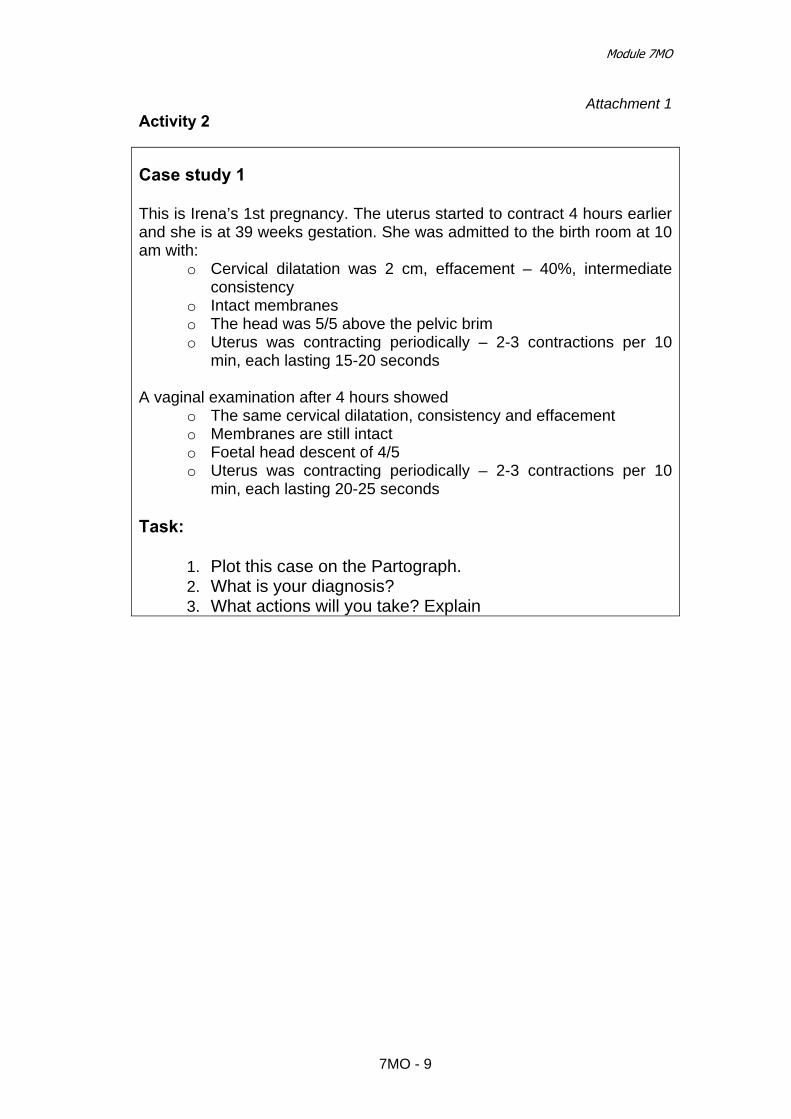
Module 7МО
Attachment 1 Activity 2 Case study 1 This is Irena’s 1st pregnancy. The uterus started to contract 4 hours earlier and she is at 39 weeks gestation. She was admitted to the birth room at 10 am with:
o Cervical dilatation was 2 cm, effacement – 40%, intermediate consistency
o Intact membranes o The head was 5/5 above the pelvic brim o Uterus was contracting periodically – 2-3 contractions per 10
min, each lasting 15-20 seconds A vaginal examination after 4 hours showed
o The same cervical dilatation, consistency and effacement o Membranes are still intact o Foetal head descent of 4/5 o Uterus was contracting periodically – 2-3 contractions per 10
min, each lasting 20-25 seconds Task:
1. Plot this case on the Partograph. 2. What is your diagnosis? 3. What actions will you take? Explain
7MO - 9

Effective Perinatal Care (EPC)
Case study 2 She arrived at 2 p.m. with:
o Regular uterine contractions: 2 per 10 minutes, each lasting 25 seconds
o Cervical dilatation of 2 cm, o Intact membranes o Head 5/5 above the pelvic brim
Next vaginal examination after 4 hours showed:
o 5 cm cervical dilatation o Intact membranes o Foetal head descent of 4/5 o Uterine contractions: 3 per 10 minutes each lasting 35 seconds.
The next vaginal examination at 10 p.m. showed:
o 6 cm cervical dilatation o Intact membranes o Head descent of 4/5 o Uterine contractions: 3 per 10 minutes, each lasting 40 seconds
Task:
1. Plot this case on the Partograph. 2. What is your diagnosis? 3. What actions will you take? Explain
7MO - 10

Module 7МО
Case study 3 This is Maria’s 1st pregnancy and she is at 41 weeks gestation. Labour started at 3 a.m. and membranes were intact.
When she was examined in the birth room at 6 a.m.: o 2 cm of cervical dilatation o Intact membranes o No moulding of foetal skull bones o The head was 5/5 above the pelvic brim, o Uterine contractions were regular: 2 per 10 min, each lasting 25-30
seconds. Another vaginal examination at 10 a.m. showed:
o 4 cm of cervical dilatation o Intact membranes o No moulding o Foetal head descent of 4/5, o Uterine contractions were regular: 3 per 10 min, each lasting 35 - 40
seconds. Maria moves actively during her labour. A vaginal examination at 2 p.m. detected:
o 7 cm of cervical dilatation, o Membranes were ruptured with amniotic hook, clear amniotic fluid o First degree moulding o Head descent of 3/5 and o Regular uterine contractions: 3 per 10 min, each lasting 35-40
seconds. Maria moves actively during her labour. A progress of uterine contractions was observed 30 minutes after amniotomy - uterine contractions became 4 per 10 min, each lasting 50-55 seconds
The next examination 4 hours later showed:
o Head descent of 3/5 o 8 cm cervical dilatation o Moulding of third degree o Uterine contractions are regular: 4 per 10 min, each lasting 50-55
seconds o Clear amniotic fluid
Task:
1. Plot this case on the Partograph. 2. What is your diagnosis? 3. What actions will you take? Explain
7MO - 11

Effective Perinatal Care (EPC)
7MO - 12

Effective perinatal care (EPC) Facilitator’s Guide
1N - 1
Module 1N
Complete Examination of a Newborn
Training objectives: Upon completion of the module the participants will be able to perform a complete neonatal examination from “Head to Toes “ in order to:
• Quickly identify quickly any danger signs and organize the appropriate referral after pre-referral treatment
• Assess the normal adaptations of a newborn after birth • Identify conditions requiring special care or follow-up observation. • Identify any birth defect or birth trauma; • Monitor growth • Counsel the mother
Content and duration of the module: Part I – Classroom work – 170 min
Activity 1 – Introduction 10 min Activity 2 – Group discussion exercise 10 min Activity 3 – Interactive presentation 45 min
Part II – Clinical work Activity 4 – Case studies 30 min Activity 5 – Newborn examination 75 min
Preparation for the module
• To read Pregnancy, Childbirth, Post Partum Care Mother and Newborn Care: a guide for essential practice - WHO 2006
• To provide all participants with the Participant’s Manual
• To ensure that other facilitators know their respective functions while
working with this module

Effective perinatal care (EPC)
1N - 2
Materials and equipment Materials
• Participant’s Manual • Case studies for small group work • Roles for role play
Equipment • Video projector or slide projector • Flip chart • Colour markers • Pens and pencils • Badges • Doll • Stethoscope
Key messages
• The complete newborn examination is extremely important as it shows whether the newborn is ready for life outside of the womb, and if he/she is facing any medical problem.
• The complete newborn examination is the tool that identifies danger signs
that threaten the life of the newborn. • The examination should be thorough, systematic and complete from “head
to toes “.
• It is critical to know normal newborn behaviour in order to recognise abnormality and correctly prescribe further tests and/or treatment.
• A routine newborn examination is performed at a time convenient for the
newborn, the parents and the health worker. • The routine newborn examination needs to be a pleasant experience for
baby, parent and examiner. • The environment influences the baby’s behaviour during the assessment. • The complete newborn examination is one of the best opportunities to
involve parents in theirs baby’s health and to establish a dialogue between the medical staff and family.
• It is necessary to ensure confidentiality during the examination especially
when discussing delicate issues • It is necessary to discuss finding in detail and take the time necessary to
discuss the results of the examination with the mother/family

Module 1N
1N - 3
PART I – CLASSROOM WORK
Activity 1 – Introduction (10 min) • Slides 1N-1. Explain that this module consists of two parts. Part 1, the
theoretical part includes several activities and part 2, clinical part. The clinical part will be conducted during the second week.
• Highlight the following points during the introduction Let them know that upon
completion of this module they will be able to perform a complete neonatal examination from “Head to Toes“ in order:
o Quickly identify any danger signs and organize the appropriate referral after pre-referral treatment
o Assess the normal adaptations of a newborn after birth o Identify conditions requiring special care or follow-up observation. o Identify any birth defect or birth trauma; o Monitor growth o Counsel the mother
o The results of the examination should be discussed in detail with
mother/family.
• Explain to participants that the majority of newborns will be found strictly normal and will need to stay with their mother.
• Only in cases with danger signs or where there are significant birth defects the
newborn needs to be referred to a neonatal or a special paediatric unit. • Slide 1N-2. List the key objectives of the examination.
The purpose of the physical exam is to find out if the baby is healthy or if the newborn faces any health problems or adaptation issues.
Activity 2 – Group discussion exercise (10 min) • Ask the group of participants to answer two questions before displaying slides
further on: o When should the complete newborn clinical examination be
performed? o How and where should the complete newborn examination be
performed? • Write down (or ask your co-facilitator to do it) all the answers on the flip chart.
Ensure that all the answers are listed • Don’t comment on the answers. Inform the participants that they will review the
answers after they have seen the following slides.

Effective perinatal care (EPC)
1N - 4
Activity 3- Interactive presentation (45 min) • Restart the presentation with Slide 1N-3 after the group discussion exercise.
• Slide 1N-3. Present the slide and compare with the results of the group
discussion. Note the differences and discuss the following points: It is important to pay a special attention to the following: The routine newborn examination should be conducted at a convenient time convenient for baby, parents and healthcare worker.
• If the newborn is not facing acute problem after birth, and is breathing
well it is extremely important to delay t the complete assessment for 2 hours so e mother and baby can have skin to skin contact without interference.
• If a danger sign is found during the initial assessment it is necessary to perform the complete examination immediately.
• Explain to the participants that this comprehensive assessment will guide the
health staff in counselling mother and family. The same complete newborn examination will be use during the first months to assess the growth and development of the infant. In cases of child abuse or neglect the full examination will be an important tool for identifying these cases.
• Slide 1N-4 Steps of Newborn Examination:
o Assess, o Classify (normal signs, signs to be monitor, danger signs), o Treat if necessary o Counsel the family.
Insist that it is important that the results of newborn assessment be carefully recorded in the infant’s file so it can be compared later with other assessments.
• Slide 1N-5. Present the slide and compare with the results of the group
discussion. Note the differences and make this points: - The importance of having parents presents during the assessment. - The newborn needs to be kept warm, - Do the complete examination, - Communicate and discuss all results with the family
• Slide 1N-6 Insist on the importance of hand washing. Remind participants that they should wash their hands before and after assessing the newborn.
• Slide 1N-7. Shows WHO’s danger signs. Explain to the participants that the
danger signs for newborn are often non specific, such as not feeding well, fast breathing, difficult breathing with severe chest indrawing when breathing in and grunting when breathing out, convulsions, hypothermia, fever, reduced few movement, and jaundice appearing anywhere on the first day or palmar or sole jaundice at any age .
• Slide 1N-.8. Danger Signs are a threat to the infant’s life. If a danger sign is
founded it is necessary to rapidly complete the assessment, give immediate

Module 1N
1N - 5
pre-referral treatment, prepare the baby for safe transport (keeping him warm, preventing low blood glucose through breastfeeding or IV glucose infusion ), and to refer to a higher level of care as soon as possible.
• Slides 1N-9 - 1N--10. The Main Characteristics of a Newborn at birth. All
medical health workers in charge of newborns should clearly know and understand the physiologic characteristics of a normal newborn in order to recognise any difference or pathology.
• Slide 1N-11. Explain that the health status of a newborn is directly connected
with pregnancy and delivery. o Check the mother’s records (if available). o In case of delivery at home, refer all questions to the mother. o It is extremely important to ask the mother if she has any concern
about the baby’s health or behaviour. Mother is the best observer of the baby and she has to be part of the team in charge of observing the baby surveillance from the first medical assessment.
o It is also important to observe the mother’s attitude toward her baby. Is she emphatic, interested or is she distant, and not interested in the newborn examination?
• Slide 1N-12. Respiratory system. Explain that the newborn needs to be calm during this assessment. If possible
count the respiratory rate while she/he is sleeping. • Slide 1N-13. Cardiovascular System.
At birth, a cardiac murmur is not always the sign of heart disease, and the absence of a cardiac murmur doesn’t give the assure normality. Further evaluation is required if a murmur persists beyond several weeks or if a murmur is associated with a severe condition.
• Slide 1N-14. The normal resting posture of a term newborn is in flexion and
the preterm baby rests in extension. Babies born in breech presentation may have fully flexed hips and knees, and the feet may be near the mouth; alternatively, the legs and feet may be to the side of the baby.
• Slide 1N-15 Shows the normal resting postures of preterm and full term
babies. • Slide 1N-16. Explain that some skin conditions are not pathological and don’t
need any treatment. • Slide 1N-17 Shows the newborn cranium. It is necessary to check the cranial
sutures, the anterior and posterior fontanels (their size and consistency) and the shape of the cranium to assess for birth trauma or presentation moulding.
• Slide 1N-18. Drawings of unilateral cephalohaematoma and moulding.
Cephalohaematoma is the most frequent birth trauma related to difficult labour, forceps or vacuum extraction. On the skull it is a fluctuant swelling limited to the sutures. No treatment is needed. The cephalohaematoma usually reabsorbs in 2 weeks to 3 months.

Effective perinatal care (EPC)
1N - 6
Before showing the slide 1N-19 ask participants what needs to be assessed on the newborn face. Quickly note the suggestion on the flip chart.
• Slide 1N-19 Compare with participants suggestions and answers any
questions
Eyes: - Check cornea for cloudiness (sign of congenital cataracts). - Check conjunctiva for erythema, exudate, orbital oedema,
subconjunctival haemorrhage, jaundice of sclera. - Check for pupil size, shape, equality and reactivity to light (PERRL:
pupil, equal, round, reactive to light). - Check for red reflex: (use +10 dioptre lens, hold ophthalmoscope 15–
20 cm from the eye). Normally the newborn’s eye transmits a red colour. Black dots may be a sign of cataracts and a whitish colour may suggest retinoblastoma.
Mouth:
- Check for defects such as cleft lip and palate. - Check for white patches - oral thrush, treated with oral nystatin. - Check that the tongue is normal size. Macroglossia indicates
hypothyroidism. • Slide 1N-20 Insist on checking for abdominal distension, masses, obvious
malformations such as omphalocele and gastroschisis. Umbilical cord:
- Count the vessels (one big vein and two arteries). - Check for effective clamping/ - Check for pus discharge and for skin redness around umbilicus.
• Slide 1N-21. The genitalia should be carefully assessed for any malformation
or sexual ambiguity. Often the scrotum is large due to hydrocele. The transillumination of the sac usually confirms the presence of hydrocele and the absence of hernia or testicular abnormalities. No treatment.
• If the anus is not perforated the newborn needs to be urgently referred to a
specialised department.
• Slide 1N-22 Assessment the newborn’s back. Carefully examine the newborn’s backbone for spinal defects.
• Slide 1N-23 Examine limbs and extremities for possible birth defect or birth
trauma. Some fractures need to be immobilised for few days (humerus, femur). Explain to the mother that these fractures will heal spontaneously without any residual deformities. The normal healing process includes the formation of a bone callus which disappears in 2-3 weeks.
• Slide 1N-24 Emphasize that there are that 2 manoeuvres for assessing hip
stability in the newborn; the Ortolani and Barlow Tests. If a one of them is found to be positive, the infant should be referred to a specialist. The Ortolani and Barlow tests are performed on a newborn lying in a supine position.

Module 1N
1N - 7
Ortolani maneuver is positive if it reduces a dislocated hip. The examiner’s index and middle fingers are on the great trochanter and the thumb is on the inner thigh. The hip is flexed to 90° but not more, and the leg is held in neutral rotation. The hip is gently abducted while lifting the leg anteriorly. While performing this manoeuvre, if a “clunk” is felt, that shows that a dislocated femoral head reduces into the acetabulum. This is a positive Ortolani sign.
Barlow test detects the unstable hip dislocating from the acetabulum.
The hips are flexed to 90°. The leg is then gently adducted while posteriorly directed pressure is placed on the knee. A palpable clunk or sensation of movement is felt as the femoral head exits the acetabulum posteriorly. This is a positive Barlow sign.
• Slide 1N-25. Newborn’s Neurobehavioral Status. This assessment will give
information about the central nervous system. • Functions of the cranial nerves 1 through 12
o I Smelling o II Response to light, vision o III Extrinsic ocular movements, response of the pupil to
light, eyelid elevation o IV Extrinsic ocular movements o V Facial sensibility, sucking, biting o VI Extrinsic ocular movements o VII Facial motility, taste o VIII Hearing, vestibular responses o IX & X Sucking, swallowing, vocalization, taste, gag reflex o XI Head and neck movements o XII Movements of the tongue
• Some reflexes exist only for specific time periods.
Some of the reflexes observed in newborns: o Rooting Reflex: present at birth and disappears at 4 months. The baby turns
the head and opens the mouth to follow the direction of mouth stimulation. This helps the baby find the breast and initiate breastfeeding.
o Sucking Reflex: begins about the 32nd week of pregnancy, and is fully developed by 36 weeks. Preterm babies may have weak or immature sucking ability.
o Palmar Grasp Reflex: present at birth, disappears at 2–3 months. If the palm is stimulate the baby closes the fingers and grasps what is in the palm.
o Moro Reflex: present at birth, disappears at 4–5 months. Often called the startle reflex because it usually observed when the baby is surprised by a loud sound or movement. The baby throws back the head, extends the arms and legs, cries and then pulls the arms and legs back in.
o Stepping Reflex: present at birth, disappears at 2 months. When the back of the foot is touched by an obstacle, the infant steps by lifting the foot and places it on the obstacle.
• Slide 1N-26 Explain to participants that when the full clinical examination is
completed the newborn could be weighted ,measured and his/her temperature needs to be checked again to be sure that the baby is not getting cold during the assessment. The size of the head, the weight, and the

Effective perinatal care (EPC)
1N - 8
length should be reported in the baby’s files as well as the results of the complete assessment. These measurements should be entered on national growth charts to initiate growth monitoring.
• Slide 1N-27 Explain that in case of early breastfeeding in the birth room and
rooming with mother 24h /24 the loss of weight is minimal. • It is important to teach the mother how to recognise if her baby is getting
enough milk. Assess breastfeeding:
- If the newborn is not gaining enough weight - If the mother complains about breastfeeding or - If the newborn is not passing enough urine or stools.
Ask the mother to put the baby to her breast and observe how the newborn is attached and the baby’s position. If the newborn is not correctly attached help the mother to find a comfortable breastfeeding position and help her to attach the newborn properly.
• Slides 1N-28- 1N-29 Counselling the mother and the family on appropriate
care for their newborn is an important part of the assessment process. It is important to teach parent to keep the baby warm, to promote exclusive breastfeeding and to plan further medical assessment and immunization with the family
• The important part of the counselling is telling the mother when to seek care
immediately. • Slide 1N-30. Special Focus during specific Newborn visits. - Newborn examination on the first day of life
o Difficultly maintaining body temperature o Initiate breastfeeding, support correct attachment and position. o Do not give any other feeding or liquid to the newborn. o Make sure the cord is well tied and does not bleed.
Check for jaundice on any location the first day. A jaundice which appears on the first day of life is always severe and requests an immediate treatment by phototherapy.
- Newborn examination on the 2nd,and 3rd day
o The mother and newborn are still adjusting to breastfeeding. o Breast engorgement can happen making breastfeeding difficult o Newborns that have been infected during birth can show symptoms of
infection o Weight loss of 5-7% is normal in the first days life but should not
exceed 10% of the birth weight .
- Newborn examination at 7 days o Breastfeeding and weight. The mother and newborn are still developing
breastfeeding. o The newborn should start gaining weight and return to her/his birth
weight by the 14th day after birth. o Infections. Newborns that have been infected during birth can show
signs of infection by the time of this visit.

Module 1N
1N - 9
o Examine the newborn for jaundice. o Immunization. Do the first vaccinations according to national
guidelines.
Activity 4 – Case studies. Group work (30 min) This activity can be conducted at any time during the first or second week after this module is completed. • Divide the participants into 2 groups and give them the case studies. • Give each group a sheet of flip chart paper and markers. • Ask the participants to write down their answers on the flip chart paper. • Ask each group to assign a representative who will present the results of the
group work. • Duration is 10 min. Case Study 1 Svetlana 3,200g was born in your maternity 12 hours ago. She spontaneously started breathing and her respiratory rate was 50 per minute. The mother tried to breastfeed, but Svetlana had difficulty sucking. The neonatologist assessed the baby immediately and found:
– The baby was pink with acrocyanosis – Respiratory rate was 56 breathings per minute – Axillary temperature was 35,4°C – No grunting, no chest indrawing – HBR 120 per minutes – The baby had a cephalohaematoma on the left side of the
head – The rest of the assessment was normal
Questions
– What problems do these babies have?
– How will you solve these problems?
– What treatment will you use and how will you follow up?
– What advices will you give to the mother? Possible answers:
1. Svetlana is a full-term baby suffering from mild hypothermia, she presents a cephalohaematoma.
2. It is necessary to start warming Svetlana immediately: provide skin to
skin contact with her mother, cover her with an extra blanket and monitor her temperature every 15-30 minutes until the temperature reaches no

Effective perinatal care (EPC)
1N - 10
less than 36.5oС. Ensure adequate feeding: if she is not ready to attach to the breast use expressed milk from a cup or with a syringe. For the cephalohaematoma no treatment is needed .Reassure the mother and check after one hour if the baby is able to breastfeed.
3. Svetlana does not need a special treatment but she needs monitoring for hypothermia and difficult feeding.
4. Teach the mother how to control the temperature and how to to feed Svetlana.
Case Study 2 Ivan 2,800 g was born 3 days ago. He was separated from this mother because of a caesarean section but a nurse brings him to his mother each 3 hours during the day to his mother to breastfeed. During the night he receives infant formula. On the 3rd day rooming-in was organized and the newborn was breastfed 8 times day and night and had no feeding problem. On the 5th day the neonatologist assessed him before discharge and found
– Weight was 2,500 g – Respiratory rate was 48 breaths per minute. No grunting, no chest
in drawing – Heartbeat rate was 110 per minute – Ivan’s belly was jaundiced but arms and legs were pink – The skin was red around the umbilicus – Axillary temperature was 36,8°C – The rest of the assessment was normal
Questions
– What problems do these babies have?
– How will you solve these problems?
– What treatment will you use and how will you follow up?
– What advices will you give to the mother?
Possible answers:
1. Ivan is a full-term baby who lost more than 10 % of his birth weight; he is also suffering from a local umbilical infection. His jaundice is a “physiologic since on day 5 arms and legs are pink.
2. Assess breastfeeding and counsel the mother. 3. Treat according to WHO recommendations. Local umbilical infection
treatment done 3 or 4 times daily • Wash hands with clean water and soap; wear clean gloves if
available • Gently wash off pus and crusts using boiled and cooled water
and soap OR an acceptable antiseptic (4% chlorhexidine gluconate); dry the area with a clean cloth or clean gauze sponges
• Paint the umbilicus and area around it with 0.5% gentian violet.

Module 1N
1N - 11
• Continue this treatment until there is no pus coming from the umbilicus.
• If there is no improvement in 2 days or the redness and swelling extend more than 1 cm beyond the umbilicus, treat for severe infection.
4. Delay discharge at least for 3 days :
o Advice the mother to breastfeed at least 8 times per 24 h o Check for correct position and attachment for
breastfeeding o Weight Ivan daily for 3 days o Cord care, if it is not improvement after 2 days of local
treatment according to WHO recommendations. Evaluate for severe infection and treat with antibiotics. It is necessary to teach the mother about caring for the cord with the antiseptic solution
• A representative from each group presents the results of the group work
Discuss the answers with the participants. • The trainer can go back to the presentation and show the slides supporting the
answers if it is needed.

Effective perinatal care (EPC)
1N - 12
PART II – Practical work
Activity 5 – Newborn examination in the postpartum unit (75 min) • The course director will have arranged ahead of time with the head of the
facility for the second week of clinical practice for the participants. • One of the facilitators should be responsible to select newborns for complete
assessment. 3 newborns need to be selected for the complete examination, one newborn for the demonstration and 2 for each group.
• The facilitators and the unit head should find appropriate mothers with
newborns. Inform the mothers) about the clinical training and ask for their authorization in advance.
А. PREPARATION OF FACILITATORS • The facilitator should receive the following information before training:
o Selected newborns and mother for this activity o The participants should have access to the medical records of mothers
and newborns
• A Facilitator will do a demonstration of a complete newborn examination for the entire group and the other facilitator will list on the flip chart the proper order of tasks: 1. Select mother and baby, introduce yourself and ask authorization from the
mother and explain to her what you will do. 2. Be sure that the room is warm. 3. Wash you hands 4. Ask the mother:
• Newborn’s name, date of birth, birth weight and quick history of pregnancy and birth
• How is the baby feeding? • Does she have concern about her baby’s health?
5. Conduct newborn examination according to the presentation o Respiratory system, count respiratory rate with participants; o cardiovascular system o Newborn’s posture and movements o Skin and colour o Cranium o Face, eyes , mouth o Abdomen o Genitals and anus o Backbone examination o Examination of extremities and limbs, with Ortolani and Barlow
Tests o Measurements
6. Counsel the mother

Module 1N
1N - 13
• Write on the flip chart the list of skills that are necessary for counselling
(or in the Participant’s Module):
COUNSELLING SKILLS (Information from module 3С) It is necessary to use and practice these during ALL interactions with mothers and health care staff:
o Use of open and close questions, listening skills o Praise o Simple words while providing information and counselling o Use of non-verbal means of communication (gestures)
• Prepare the forms monitoring the participants: check list of the trainer (Attachment 2)
B. PREPARATION OF THE GROUP • Prepare the group before visiting the unit:
o Remind them that the goal of the practical work is to conduct a complete newborn examination and to use good counselling skills.
o Make sure all group members understand what they are supposed to do and in which order
o Ask them to carefully read the order of newborn examination (on the flip chart list). The participants can write it down in their notes if necessary
o Ask the participants to carefully read (recall) the main counselling skills
• Divide the participants into 2 groups (1 facilitator for 1 group) • One of the participants will be selected to conduct the complete examination
while the other participants will observe without intervening and take notes on what was done properly and what was not done properly.
• Each participant should have:
o Pen/pencil and note/paper.
• Remind the participants that they should wash their hands before and after a newborn examination
• Fill out the form Attachment 2 during examination • Monitor participants’ actions and make sure that the newborn does not get
cold • The newborn examination should be quick • If one of the participants needs to repeat the task or lack some knowledge
spend more time with him/her • Notice what tasks caused difficulties for the participants or what task were not
completed.

Effective perinatal care (EPC)
1N - 14
• Write down all the questions that have to be discussed during the summary of clinical practice
C. CONDUCTING EXAMINATION • Do not forget to wash hands before and after the newborn exam. • Prepare the room and equipment for examination: warm room (250С); radiant
heater if needed. • Greet, congratulate and praise the mother • Gather the information from the mother: date of birth; weight at birth and on
the day of examination; newborn’s name • Ask the mother: what problems did she have during pregnancy and at birth.
What problems did the newborn have (feeding, breathing difficulties, skin)? • Conduct the examination quickly. • Explain the examination results to the mother and give her advice D. DISCUSSION: • Discuss the results after the examination in a place where the mother cannot
hear you, for example, in the corridor or in the classroom. • Discuss what has been done correctly during the examination. • Discuss what had been done incorrectly. • Discuss the type of care the newborn needs (tests/treatment). • Discuss what advice should be given to the mother.

Module 1N
1N - 15
References
1. Clinical Practice Guideline: Early Detection of Developmental Dysphasia of the Hip.AAP.2000.
2. Committee on Quality Improvement, Subcommittee on Developmental
Dysphasia of the Hip.
3. David .E Hertz. Care of the newborn. A handbook for primary care. 2005. 4. Diana Beck, et al. Care of the Newborn. Reference Manual. Saving
newborn lives.2004. 5. Essential Newborn Care and Breastfeeding. Training Modules. WHO
EURO, Copenhagen, 2002.
6. First Nations and Inuit Health Branch Paediatric Clinical Practice Guidelines for Nurses in Primary Care.Canada.2001
7. IMPAC/Pregnancy, Childbirth, Postpartum and Newborn care: A guide for
essential practice. WHO, Geneva, 2006
8. Managing newborn problems. A guide for doctors, nurses, and midwives. WHO, 2003.
9. Routine Examination of the Newborn. NHS Quality Improvement. Best
Practice Statement. Scotland 2004.
10. Volpe JJ, neurology of the newborn, 4th edition, Philadelphia, WB Sauders, 2001.

Effective perinatal care (EPC)
1N - 16
Attachment 1
Activity 4 Case Study 1 Svetlana 3,200g was born in your maternity 12 hours ago. She spontaneously started breathing and her respiratory rate was 50 per minute. The mother tried to breastfeed, but Svetlana had difficulty sucking. The neonatologist assessed the baby immediately and found:
– The baby was pink with acrocyanosis – Respiratory rate was 56 breathings per minute – Axillary temperature was 35,4°C – No grunting, no chest indrawing – HBR 120 per minutes – The baby had a cephalohaematoma on the left side
of the head – The rest of the assessment was normal
Questions
– What problems do these babies have? – How will you solve these problems? – What treatment will you use and how will you follow up? – What advices will you give to the mother?

Module 1N
1N - 17
Case Study 2 Ivan 2,800 g was born 3 days ago. He was separated from this mother because of a caesarean section but a nurse brings him to his mother each 3 hours during the day to his mother to breastfeed. During the night he receives infant formula. On the 3rd day rooming-in was organized and the newborn was breastfed 8 times day and night and had no feeding problem. On the 5th day the neonatologist assessed him before discharge and found
– Weight was 2,500 g – Respiratory rate was 48 breaths per minute. No
grunting, no chest in drawing – Heartbeat rate was 110 per minute – Ivan’s belly was jaundiced but arms and legs were
pink – The skin was red around the umbilicus – Axillary temperature was 36,8°C – The rest of the assessment was normal
Questions
– What problems do these babies have? – How will you solve these problems? – What treatment will you use and how will you follow up? – What advices will you give to the mother?

Effective perinatal care (EPC)
1N - 18
Attachment 2
Activity 5
Check list for the trainer # of participant 1 2 3 4 Participant’s initials
Primary newborn’s examination 1. Conducts the examination successfully following all the
rules
2. Breathing evaluation • Count of breathing movements • Grunting when breath out • Examination for chest indrawing when breathing in
3. List the danger signs
4. Describe the appropriate treatment, counselling and future care
Counselling skills
1. Uses:
Open questions to receive clear information Praise Does not judge
2. Provides the information correctly and in a simple way
without scientific terminology
Assessment of breastfeeding (if the Module 7C was conducted)
1. Assesses at least one breastfeeding session
2. Monitors the overall process of breastfeeding. Correctly classifies the ability of the newborn to suck the breast properly.
3. Lists the main indicators: Correct breast attachment Correct breast position

Effective Perinatal Care (EPC) Facilitator Guide
2N - 1
Module 2N Post-Resuscitation Neonatal Care
Module Objectives By the end the Module the participants will be able to :
• Assess an infant after neonatal resuscitation • Decide if after resuscitation the baby can stay with his/her mother or should
be transfer to a special neonatal or paediatric department • Know how to care for a newborn after neonatal resuscitation • Know how to monitor the infant’s condition after neonatal resuscitation • Know the main modes of treatment in the neonatal care unit • Know when the infant is ready for discharge.
Module agenda and duration: Part I – Classroom work - 110 min
Activity 1 – Introduction 10 min Activity 2 – Brain storming 10 min Activity 3 – Interactive presentation 55 min Activity 4 – Work in small groups 35 min
Part II – Clinical work – 140 min Activity 5 – Practical work: assessment of 1 or 2 newborns after neonatal resuscitation
140 min
Preparation for the Module • Review publications based on the evidence-based data and national
standards on newborn care after neonatal resuscitation • To provide all the participants with the Participant Manuel
• Ensure all the trainers know their responsibilities during work on this
Module
Materials and equipment Materials
• Participant Manual

Effective Perinatal Care (EPC)
2N - 2
Materials and equipment Equipment
• Video projector or projector overhead • Presentation 2N-EPC ENG • Flipchart • Markers • Pens and pencils • Name badges
Key information • After neonatal resuscitation all newborns should be assessed thoroughly in
order to make appropriate decisions for care • After neonatal resuscitation every newborn must be kept warm and receive
adequate calories • After neonatal resuscitation every newborn needs a thorough monitoring • The mother and child should not be separated if the newborn is not
transferred to a special department • All needed treatments should be provided in a timely manner • Mother /family must be there to observe and care for newborn even after
neonatal resuscitation.
PART I – Classroom work – 110 min
Activity 1 – Introduction (10 min) • Slide 2N-1 Explain the difficult issues involved in caring for newborn after
resuscitation. Explain also that there are 2 parts of this Module. Part 1 is the class work and part 2 is the clinical work during the second/practical week.
• Ask one participant to find in his/her Participant Manual the notes for
Slide 2N-1 and to read the list of learning objectives for this module. Discuss the main objectives of this Module with the participants. Ask the participants if they have any questions or suggestions the learning objectives. If there are questions or suggestions record them on the flip-chart.
• Before showing Slide 2N-2 conduct a group discussion.

Module 2N
2N - 3
Activity 2 – Brain storming (10 min) • Ask the participants to list the main principles of newborn care after neonatal
resuscitation that are practiced in their hospitals: o How do they manage a newborn after neonatal resuscitation in the
birth room? o How they treat a newborn after neonatal resuscitation.
• The second trainer or any participant should record the answers on the flipchart if possible.
• Explain that you will discuss these questions and suggestions during the
Module training and at the end of the training.
Activity 3 – Interactive presentation (55 min) • Restart the presentation with Slide 2N-2. Explain to participants that after
resuscitation newborns may have several problems; therefore the first goal is to prevent these problems. Thus, it is important to carefully assess the situation and to take immediate actions when problems occur.
• Slide 2N-3 shows the practical aspects of newborn assessment after
resuscitation. Insist on the fact that a good infant evaluation is necessary for all newborn not only those who are resuscitated.
• Before showing the slide 2N-4 the facilitator will draw a ”traffic light scheme” on the flipchart with yellow, green and red markers. 1. ‘Infant after resuscitation’ – yellow box (attention). 2. If newborns have a heart beat rate > 100 per min, breathing rate 30-60 per
min, no severe chest in drawing, no grunting on expiration and becoming pink: Go to the Green box: Room-in with mother with active monitoring.
3. If newborns received extensive resuscitation (intubation, chest compression, drug administration), or have breathing difficulties or severe conditions: Go to the Red Box: Neonatal care unit.

Effective Perinatal Care (EPC)
2N - 4
• Show slide 2N-4: if after neonatal resuscitation the newborn is in good clinical
condition, he /she has to be placed in skin to skin on the mother chest and to be carefully monitored during the first 2 hours.
• Slides 2N-5. In the birth room a close monitoring of temperature and vital
signs needs to be strictly followed. • Reassure the mother and support her to start as she starts breastfeeding to
the baby is feeding well. • Slide 2N-6. Train the mother to carefully monitor her baby. The mother should
pay special attention to keep her baby warm and effective breastfeeding. These newborns are healthy and need to receive essential newborn care like all newborns.
• Slide 2N-7. There are basic indicators that should be monitored in the post
partum unit. The recommended frequency of assessment depends on the infant’s age and condition.
• Slide 2N-8. 2nd case: After neonatal resuscitation newborns in poor clinical
conditions and/or those who experienced advanced resuscitation should be transferred to a neonatal unit where close monitoring and care can be provided.
Heart rate > 100 Breathing rate
30-60 Pink
Breathing difficulties Intubation
Chest compression Medications
Rooming-in Neonatal unit
Specialized unit Discharge
Active monitoring
Counseling
Infant after resuscitation
Condition is worsening

Module 2N
2N - 5
• Slides 2N-9 and 2N-10 show the main principles of management in the birth room and in the neonatal unit.
• Show Slide 2N-11 and Ask participants how they prepare an admission to the
neonatal unit, and discuss how to create a “nest” for newborns using anything available. Emphasize that these newborns need silence, a minimum of interventions (care and laboratory tests), the presence of the mother and family members and the implementation of main infant care principles.
• Slides 2N-12 and 2N-13. Discuss the main principles of management (care
and monitoring) in the neonatal unit. Emphasize that all indicators should be monitored precisely and recorded on the special form.
• Ask participants who is responsible for monitoring and who usually
record results on the monitoring format in their facilities? • Ask the participants to bring the monitoring form they use in their
hospital so they can be discussed during the second clinical week.
• Slide 2N-14. Discuss the issues of newborn feeding in the nursery/neonatal care unit: when it must be started, how it must be organized (help the mother to express the breast milk, involve the mother into the feeding process), and how to decrease IV fluid and to increase normal feeding simultaneously.
• Especially insist on the need to insure adequate caloric intake which is critical
for this category of newborns. • Slides 2N-15 to 2N-18. Demonstrate the different components of the routine
treatment given to newborns admitted to the nursery/neonatal care unit after neonatal resuscitation. Insist especially on preventing hypothermia, or keeping the baby warm.
• Show Slide 2N-19 and explain how to manage convulsions. Remind them that
the causes for convulsions can be hypoglycemia, asphyxia, metabolic and electrolytic disorders and hypothermia. Thus the initial management of convulsions needs to start immediately (define the level of blood glucose, give Phenobarbital) after that search for the cause of convulsion for specific management.
• Ask the participants what additional medical therapy they use in their
practice for post resuscitated newborns. Encourage them to discuss the use of frozen plasma, of sulphate of magnesium, of mannitol and of cortico-steroids (what are the indications, the frequency, and the dose of these drugs).
• Write this information on the flip chart.
• Slide 2N-20. Make final conclusions on using these medicines. • Slide 2N-21. Discuss the criteria for discharging a baby resuscitated at birth.
Emphasize that the discharge criteria are the same as those for other newborns.
• Slide 2N-22. Counseling and training mothers/families is extremely important.
Insist on good communication skills.

Effective Perinatal Care (EPC)
2N - 6
• Remind participants that training for mothers/families starts at infant’s birth.
Mother/family should know everything about the infant’s health and participate actively in infant care.
• Show slide 2N-23 and summarize the Module. Ask the participants whether
they have any questions and answer their questions. • Return to the results of the 1st and 2nd activities and discuss notes according to
information in Module 2N.
Activity 4 – Work in small groups (35 min) • This activity could be conducted any time when this module is taught during
the first or second week. • Divide the participants into 3 groups and make sure that there are
neonatologists and pediatric nurses in each group. • Give each group a list of paper from the flipchart and markers. • Give one question to each group and ask them to read the case study carefully
in the Participant Manual. Make sure that the participants understand the questions and the case study.
• Ask participants to work 10 minutes and after that to present the groups’
conclusions. Limit presentations to 5 minutes. • Trainers together with participants decide on a conclusion at the end of each
group presentation. Case Study Maria, a full term newborn, is Galina’s first baby. It was a normal pregnancy. At birth she was cyanotic with irregular breathing. The neonatologist resuscitated her with bag and mask. Immediately after resuscitation she assessed Maria’s condition: the baby was active; her breathing rate was 40 per min; her heart rate was 140 beats per min; no severe chest indrawing no grunting; skin and mucous were pink. Apgar score was 6-7 points. Maria was weighed and measured; her weight was 3, 400 g. After Maria was resuscitated she was transferred to the neonatal unit for monitoring. Immediately after admission blood test results showed: red cells: 5.4 х 1012; Hb: 210 g/L; white cells: 24 х 109; Ht: 50%.

Module 2N
2N - 7
The neonatologist decided to feed Maria 5 ml of 5% glucose orally. Two hours after birth Maria’s temperature was 36.60С. As Maria was in good condition the neonatologist allowed Maria to room in with her mother. The next morning (16 hours after birth ) Maria’s temperature was 36.40С; she was breathing irregularly and her respiratory rate 46 per min; heart rate 130 beats per min; and she was little hypotonic . Maria’s mother said that Maria did not eat at night because she refused the breast and in the morning only sucked for 10 min 2 times and then refused to feed. The doctor weights Maria. Her weight was 3.250g and she decides to put the baby in an incubator and checks the temperature after 1 hour: the temperature is 36.60С. The doctor recommends checking the temperature again in the evening. Due to the 150 g weight loss in a few hours, the neonatologist prescribed baby formula on 10 ml per each feeding 6 times a day. 1st Question:
What are the basic principles of management of resuscitated newborns in the birth room? Explain your answer and compare with what was done in Maria’s case.
2nd Question:
What are the basic principles of management of resuscitated newborns in the postpartum department? Explain your answer and compare with what was done in Maria’s case.
3rd Question:
When can Maria be discharged from the hospital? Explain your answer. Correct answers: 1st Question What are the basic principles of management of resuscitated newborns in the birth room? Explain your answer and compare with what was done in Maria’s case. • Assess the general condition of a newborn after neonatal resuscitation. If a
baby does not have any problems (breathing rate is 30-60 per min, heat beat rate more than 100 per min, no severe chest indrawing and grunting, baby is active and pink) put baby on the mother’s chest, take his/her temperature not later than 30 min after birth; monitor breathing, skin color and body temperature by feeling the baby’s feet every 15 min during the 1st hour and every 30 min during the 2nd hour. Recheck the baby’s temperature at 2 hours and at any time the feet feel cold. Start breastfeeding within 1 hour after birth, when the baby shows signs of readiness.
• Comparison with the case study:
o In the case study the neonatologist has not correctly managed Maria’s case

Effective Perinatal Care (EPC)
2N - 8
o He measured and weighed her immediately after resuscitation and transferred Maria to the neonatal unit despite her good condition after resuscitation
o He didn’t control the temperature in time (the first body temperature was taken after 2 hours)
o The mother did not receive any information on Maria’s condition o The first breastfeeding was late, 4 hours later and the baby received
glucose before breastfeeding. Correct answer: 2nd Question: What are the basic principles of management of resuscitated newborns in the postpartum department? Explain your answer and compare with what was done in Maria’s case. • Basic principles of care:
o To transfer a baby to room in with /his her mother o To help the mother to breastfeed her baby on demand (If breastfeeding
is difficult train the mother to express her milk and to use alternative feeding (cup, spoon)
o Monitor a baby’s condition regularly to assess the breathing , the temperature , the feeding and the sucking reflex
o Check frequency of feeding and duration o Organize essential newborn care o The newborn does not need any interventions if there are no precise
reasons.
• Comparison with the case study: o Maria was fed the first time with 5% glucose, then the doctor
prescribed baby formula immediately without assessing breastfeeding o Maria wasn’t monitored thoroughly o The -warming and temperature control was not well organized. Maria
was warmed in an incubator instead using skin to skin technology o When Maria was diagnosed with hypothermia the first temperature
assessment was done 1 hour after birth and the next temperature assessment was recommended only in evening.
Correct answer : 3rd Question: When can Maria be discharged from the hospital? Explain your answer. • Maria can be discharged when:
o Her temperature is stable within 36.8-37.20С o She is feeding well and her mother trained in alternative methods if
necessary o Maria is in good clinical condition: no breathing difficulties, no signs of
disease o Her mother is trained to take care of Maria at home: she knows how to
keep the baby warm, breastfeeding on demand, putting the baby to sleep on his back, no smoking in the baby room, knowing how to recognize danger signs and to seek care immediately.

Module 2N
2N - 9
PART II – Clinical Work – 140 min
Activity 5 – Practical work (140 min) • Make sure the participants understand the key points of newborn care after
neonatal resuscitation during the theoretical week. • Ask the Course Director and/or the Facility Head to select 3 infants who have
received resuscitation to conduct clinical assessment: 2 infants in the rooming-in with their mothers; 3rd is in the nursery/intensive care unit.
• Meet the mothers and obtain their agreement for the newborn examination. • Before examination of selected newborns check the baby’s history and collect
all the following information: o Gestational age, birth weight o Characteristics of pregnancy and birth o How was each of these babies resuscitated: with bag and mask only or
with more advanced resuscitation? o After resuscitation what kind of management was used? o What were the results of the different clinical examinations since the
baby’s birth (baby’s general condition, urine, neurological status)? o What were the results of different lab tests? o What monitoring was done for these babies? o How these babies were fed? o What treatment was provided?
• All this information should be communicated to participants verbally before
examination or if you have the chance to print and give out to each participant. • Then one facilitator will demonstrate in front of the entire group the best
technique for assessing the baby in neonatal unit, following the steps listed in Table 1 (plan for 30 minutes for clinical demonstration and 30 minutes for discussion).

Effective Perinatal Care (EPC)
2N - 10
Table 1. Steps recommended for assessing a resuscitated newborn
Step Comments 1 Wash hands Soap, towel, water
2
Ensure (check) all necessary conditions for assessment
- Warm room (no less than 25°С) - Warm surface (table) - Good light - Source of radiant heat - Warm blanket/cloths for baby
3
Greet the mother and introduce yourself if the mother is present
- Greet the mother - Introduce yourself - Congratulate the mother on her newborn and ask her
permission to assess her baby in front of participants
4
Collect general information Ask the mother : - How she feels today - Name of the baby and gender - Date of baby’s birth, birth weight and weight on assessment day if available
5
Collect health information Ask the mother : - Were pregnancy and delivery normal? - Did the baby face problems during the first days of life
(ask her to be precise)? - Did the baby convulse? - Does she know if her baby received any treatment? - How is the baby fed: breastfeeding or using alternative
feeding? - Describe how the baby is feeding: e.g. well or with
difficulty - How many times per day is she breastfeeding?
6
Check monitoring procedures (if any form is available, ask to see it )
Check for the following indicators and frequency of monitoring: - Temperature - Breathing (Breathing rate, Apnea, Severe chest indrawing,
Grunting when breathing out, Oxygen saturation (if possible))
- Heartbeat rate - Skin colour of skin and colour of mucous membranes - Alertness and muscle tone - Urine and stool output - Feeding: kind, frequency, quantity - Weight
7 Recording the information on note pad
8 Clinical examination: Assess the baby’s physical status
- Look before undressing the baby - Count respiratory rate during 1 minute - Listen for grunting when breathing out
- Undress the baby gently and check for presence/absence of danger symptoms
- Look for skin color (cyanosis/jaundice/pallor) - Look for chest indrawing and grunting - Look for infant‘s movements: Does the infant move only when stimulated?
Does the infant not move even when stimulated? - Count the heart beat rate
- Assess color and quantity of urine
- Ask the mother what she observed or check on the observation form.
- Finalize the clinical examination - Dress the baby
9 Assess the feeding and baby’s weight gain
If the baby is breastfed, kindly ask the mother if she would agree to breastfeed her baby in front of the participants, assessing
- If the attachment to the breast is correct

Module 2N
2N - 11
- If the baby’s position is correct - Does the baby suck effectively
Observe carefully, take notes and ask participants to take notes on all this issues because you will ask them to answer questions later. If the baby is fed by the alternative method (cup, or through gavage or syringe):
- What quantity in ml is given at each fed? - What is the quantity received during the last 24 hours ? - How often is the baby fed? - Who is feeding the baby?
10 Finalization of assessment
- Thank the mother and ask her if she would like to ask any question
- Leave the room/or intensive care unit • Outside of the mother’s presence lead a discussion with the participants:
1. What is the baby’s general condition? 2. Analyze the results of clinical assessment? 3. Ask participants to assess the quality of feeding (breastfeeding or feeding
by alternative methods). If baby is feeding by an alternative method calculate the volume of food according to the baby’s age and compare to the volume the baby received.
4. Ask participants to assess the quality of management: o Monitoring o Treatment o Feeding
5. Ask when this baby could be discharged home. • Ask the participants whether they have any questions after the discussion. • Than divide participants into 2 sub groups: 1 facilitator -1 sub group and to
define one participant in each group who will perform evaluations. • Each group “received” a post resuscitated newborn to assess; the facilitator
will provide all information for the case. • One participant is selected to conduct the assessment; the other
participants observe what is happening and take note. • Make sure the participant has washed her/his hands. • Make sure the participant follows the main rules of keeping the infant warm
during assessment • Ask the sub group to come back to the class room and discuss both cases. • The trainers evaluate the participants’ actions according to the Table 1. • Ask the sub group to come back to the class room and discuss both cases. • Discuss:
o Baby medical history

Effective Perinatal Care (EPC)
2N - 12
o Infant condition at the moment of assessment o Treatment and investigations (lab and instrumental) o Assessment done (quality) o Feeding o Problems that became clear during assessment
• Make plan of care for each evaluated infant according to the recommendations provided in the Module 2N.
• Define when these infants can be discharged:
o Criteria for discharge o Preparation of the baby and mother/family for discharge
Coordinate conclusion with the full group 10 min
1. Ask each group to give a short presentation of their case (not more than 5 min).
2. Then lead a discussion to highlight the specifics of each case.

Module 2N
2N - 13
References 1. Anand KJ. Prevention and Management of Pain in the Neonate: An Update.
Pediatrics Vol. 118 № 5, November 2006, pp. 2231-2241. 2. Carbajal R, Chauvet X, Couderc S, Olivier-Martin M. Randomised trial of
analgesic effects of sucrose, glucose, and pacifiers in term neonates. BMJ 1999; 319: 1393-1397.
3. European Resuscitation Council Guidelines for Resuscitation. Section 6:
Paediatric life support, Resuscitation of babies at birth, 2005:S115-S133. 4. Gary L Darmstadt et al. Evidence-based, cost effective interventions: how
many newborn babies can we save? Lancet, March 3, 2005, 19-30. 5. Levene M, Blennow M, Whitelaw A, Hanko E, Fellman V, Hartley R.. Acute
effects of two different doses of magnesium sulphate in infants with birth asphyxia. Arch Dis Child Fetal Neonatal Ed. 1995 Nov;73(3):F174-7.
6. Managing Newborn Problems: A guide for doctors, nurses, and midwives.
World Health Organization, Geneva. 2003. 7. Peliowski A, Finer NN. Birth asphyxia in the term infant. In: Effective care of
the newborn infant. Eds: Sinclair JC, Bracken MB. Oxford University Press. 1992; Chapter 13; 248-279.
8. Pregnancy, Childbirth, Postpartum and Newborn Care: A guide for essential
practice. World Health Organization, Geneva, 2006. 9. Roberton N. R. C. Use of albumin in neonatal resuscitation. European Journal
of Pediatrics, Issue Volume 156, Number 6 / May, 1997, 428-431. 10. Royal Women Hospital, NETS Neonatal Handbook, Clinical Practice
Guidelines. Last Updated 03-Jan-2007. 11. Volpe JJ. Neurology of the Newborn. Philadelphia, Pa: WB Saunders
Company; 2000.

Effective Perinatal Care (EPC)
2N - 14

Effective perinatal care (EPC) Facilitator guide
Module 3N Breathing Difficulty in the Newborn
Learning objectives: By the end of this module participants will:
• Know how to quickly resuscitate a baby who is gasping or not breathing • Be able to recognize the clinical signs of breathing difficulty • Be able to classify the severity of breathing difficulty • Know how to manage a newborn with breathing difficulty • Know general and specific treatments for breathing difficulty • Know the basic principles of oxygen therapy • Know when a baby who experienced breathing difficulty can be
discharged
Module outline and duration: Part I – Class work - 75 min
Activity 1 – Introduction 10 min Activity 2 – «Brain storming» 10 min Activity 3 – Interactive presentation 50 min Activity 4 – Conclusion 5 min
Part II – Clinical work Activity 5 – Group work 30 min Activity 6 – Practical work: Oxygen administration for breathing difficulty
30 min
Activity 7 – Demonstration of assessment of an infant with breathing difficulty
50
min
Preparation for the module
• Review the existing evidence and recommendations regarding breathing difficulty
• Ensure that all the participants have Participants Manual • Become familiar, if possible, with the practices that exist in the
participants health care settings regarding safe maternal health practices and effective newborn care
• Ensure that all the facilitators know their functions while working on this
module
3N - 1
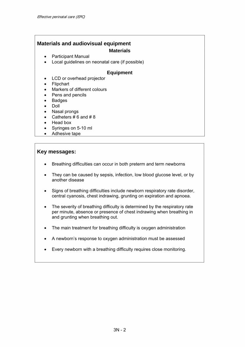
Effective perinatal care (EPC)
Materials and audiovisual equipment Materials
• Participant Manual • Local guidelines on neonatal care (if possible)
Equipment
• LCD or overhead projector • Flipchart • Markers of different colours • Pens and pencils • Badges • Doll • Nasal prongs • Catheters # 6 and # 8 • Head box • Syringes on 5-10 ml • Adhesive tape
Key messages:
• Breathing difficulties can occur in both preterm and term newborns • They can be caused by sepsis, infection, low blood glucose level, or by
another disease
• Signs of breathing difficulties include newborn respiratory rate disorder, central cyanosis, chest indrawing, grunting on expiration and apnoea.
• The severity of breathing difficulty is determined by the respiratory rate
per minute, absence or presence of chest indrawing when breathing in and grunting when breathing out.
• The main treatment for breathing difficulty is oxygen administration • A newborn’s response to oxygen administration must be assessed • Every newborn with a breathing difficulty requires close monitoring.
3N - 2

Module 3N
Part I – Classroom work – 120 min
Activity 1 – Introduction (10 min) • Slide 3N-1 Present the main objectives of this Module. • Inform participants that this module will be taught in 2 parts, the first part is the
theoretical part studied in the classroom; the second part will be clinical during the second week of training.
• Highlight the following points: o Breathing difficulty is the most frequent reason for a baby being
referred to NICU; o If not recognised and treated on time , breathing difficulty may lead to a
rapid deterioration of the baby’s condition and even to death; o A good understanding of the signs of breathing difficulty leads to quick
diagnosis and the appropriate treatment when time in critical.
Activity 2 – “Brain-storming” (10 min) • Before showing Slide 3N-2 ask participants the following question:
o What will you do if a baby who was breathing well at birth, an hour later is gasping or breathing less than 20 breaths per minute?
• Write the answers on a flipchart and come back to them after the presentation
is finished
Activity 3 – Interactive presentation (50 min) • Slide 3N-3 and to 3N-4 explain to participants how to resuscitate a baby who
was breathing at birth and then experienced breathing difficulty. • Slide 3N-5. Explain participants that a small baby with a birth weight <2.5 kg
or preterm baby born before 37 week of gestation could have mild chest indrawing and irregular breathing with many pauses.
• Remind them than central cyanosis (baby’s tongue and lips are blue)) is a late
signs of breathing difficulty. • Slide 3N-6. Show the general management of breathing difficulty:
o Give oxygen o Resuscitate if needed o Measure blood glucose o Establish IV line o Look for possible cause of breathing difficulty (small baby, asphyxia,
sepsis or congenital syphilis). o Classify the breathing difficulty as severe, moderate or mild and
manage accordingly
• Slide 3N-7 shows a simple and convenient classification of breathing difficulty which will lead to correctly managing the problem.
3N - 3

Effective perinatal care (EPC)
• Slide 3N-8 shows the basic principles of apnoea management. o Be clear that the mother must be taught to observe her small or full
term baby with breathing difficulty. She will be the first one to provide help in a case of apnoea (tactile stimulation, changing the position).
o Impress that apnoea in a term baby is always a sign of pathology. • Slide 3N-9. Central cyanosis associated with other signs of breathing difficulty
could suggested a congenital heart anomaly • Before showing the following slide the facilitator will ask participants
what signs they monitor in case of breathing difficulty. How frequently are they monitoring and who is in charge?
• Write all the answers on a flipchart, then show Slide 3N-10 and compare the
information on the slide with participant’s answers. Tell the participants, that monitoring the baby’s condition is one of the basic principles for managing breathing difficulty.
• Slide 3N-11. Emphasize the importance of implementing Essential newborn
care in all circumstances. • Slides 3N-12 – 3N-14. Management of Severe Breathing Difficulty.
Emphasize that these cases need careful monitoring and need to be treated by experienced staff. If the local staff cannot provide appropriate care transfer the baby after stabilisation.
• Slides 3N-15 and 3N-16. Feeding a baby with severe breathing difficulty is
challenging. Emphasize that feeding must start as soon as the baby shows some improvement. It is important that the family/mother play a role in feeding the sick baby. Expressed breast milk is the best food for these severely sick newborns. Feeding needs to be carefully monitored to be sure that breathing difficulties do not increase during feeding
• Slides 3N-17 and 18. Emphasize strict monitoring in cases of moderate
breathing difficulty so staff will know whether the situation is improving or deteriorating and they can take appropriate action. Explain that there is no significant difference between the management of severe and moderate breathing difficulties. However, feeding of babies with moderate breathing difficulty can be initiated earlier as well as “skin-to-skin” contact.
• Slides 3N–19: Discuss managing mild breathing difficulty. Emphasize the
necessity of strict monitoring. • Slide 3N-20. Discuss the main points of providing oxygen therapy. • Slides 3N-21, and 3N-22 review the different techniques of oxygen
administration. • Slide 3N-23 this slide is self explanatory. It shows the advantages and
disadvantages of some methods of oxygen administration. • Slide 3N-24. Pay special attention to the fact that oxygen is expensive, and
should be used only if needed.
3N - 4
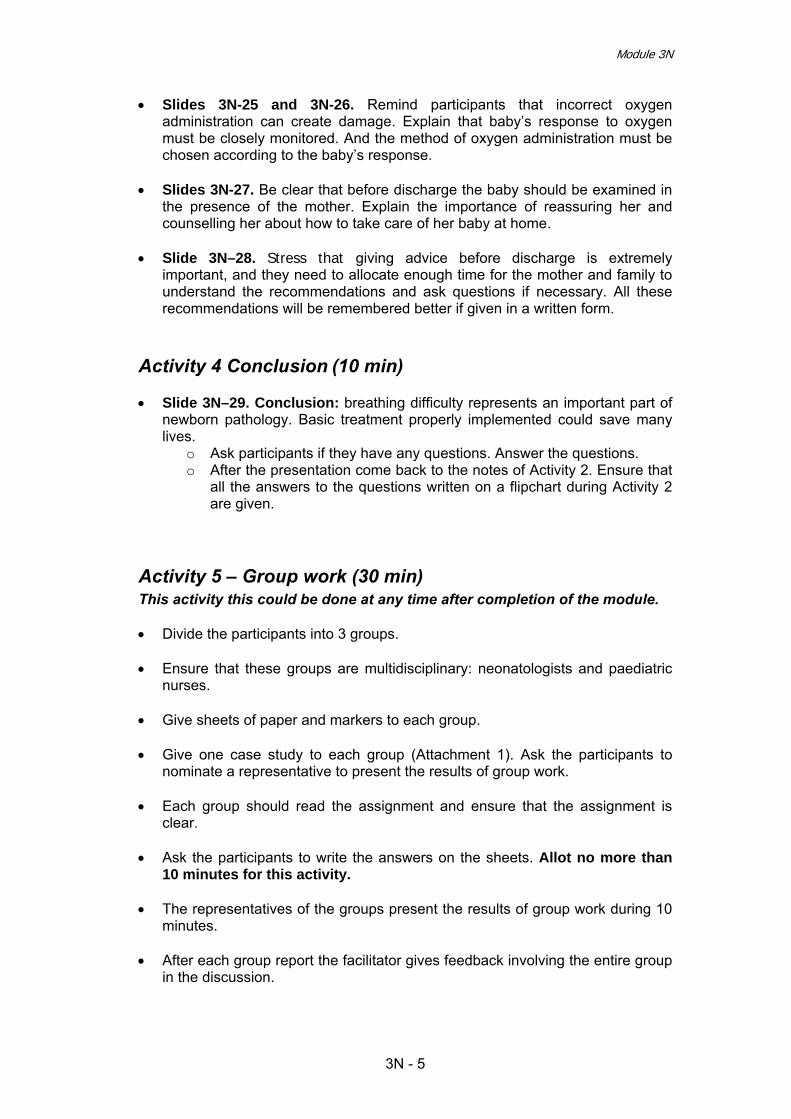
Module 3N
• Slides 3N-25 and 3N-26. Remind participants that incorrect oxygen administration can create damage. Explain that baby’s response to oxygen must be closely monitored. And the method of oxygen administration must be chosen according to the baby’s response.
• Slides 3N-27. Be clear that before discharge the baby should be examined in
the presence of the mother. Explain the importance of reassuring her and counselling her about how to take care of her baby at home.
• Slide 3N–28. Stress that giving advice before discharge is extremely
important, and they need to allocate enough time for the mother and family to understand the recommendations and ask questions if necessary. All these recommendations will be remembered better if given in a written form.
Activity 4 Conclusion (10 min) • Slide 3N–29. Conclusion: breathing difficulty represents an important part of
newborn pathology. Basic treatment properly implemented could save many lives.
o Ask participants if they have any questions. Answer the questions. o After the presentation come back to the notes of Activity 2. Ensure that
all the answers to the questions written on a flipchart during Activity 2 are given.
Activity 5 – Group work (30 min) This activity this could be done at any time after completion of the module. • Divide the participants into 3 groups. • Ensure that these groups are multidisciplinary: neonatologists and paediatric
nurses. • Give sheets of paper and markers to each group. • Give one case study to each group (Attachment 1). Ask the participants to
nominate a representative to present the results of group work. • Each group should read the assignment and ensure that the assignment is
clear. • Ask the participants to write the answers on the sheets. Allot no more than
10 minutes for this activity. • The representatives of the groups present the results of group work during 10
minutes. • After each group report the facilitator gives feedback involving the entire group
in the discussion.
3N - 5

Effective perinatal care (EPC)
Case Study 1 Peter is a second baby born after a normal pregnancy of 41 weeks. At birth Peter’s breathing and heart were good and he was not resuscitated. He did not have any birth defects and was placed skin to skin on his mother. Mother and baby were left alone in the birth room as the birth was normal .The mother tried to breastfeed 45 min after birth but Peter seems uninterested in feeding. One hour only after birth the baby was weighed (his weight was 2500 g) and transferred to postpartum ward together with his mother. Just after arriving in postpartum room the mother tried again to breastfeed Peter but he refused again. She noticed that Peter was breathing more quickly than before, but since she didn’t know if it was not normal, she didn’t call for help. Four hours after the delivery Peter was examined by a pediatrician who found a respiratory rate of 80 breaths per minute; a severe chest indrawing and a central cyanosis. The boy was transferred to the nursery department. When he was admitted to this department his body temperature was 36.4°С. The neonatologist installed Peter in incubator and started IV infusion with glucose 10%. Questions:
1. What is Peter’s problem? Please classify Peter’s problem.
o Suggested answer: o Peter has moderate breathing difficulty (RR 80 per minute; severe
chest indrawing) and mild hypothermia. o Peter was breathing well and didn’t need resuscitation at birth
2. Which treatment do you recommend for Peter? How will you monitor Peter’s health status? How would you feed Peter?
o Peter needs to be rewarmed immediately. o Oxygen should be administrated immediately. o Continue 10% glucose infusion 60 ml/kg (150 ml daily) until the
condition is stable (probably within the first 12 hours). o Monitor: respiratory rate; heart beat rate; skin colour and colour of
mucous membranes, temperature, urine; and oxygen saturation every 3 hours until the condition is stable;
o Monitor glucose blood levels every 6 hours at minimum Start feeding when respiratory rate is less than 90 breaths per minute and the following indicators are exist
o No grunting on expiration, nor severe chest indrawing o Heart rate 100-160 beats per minute o Baby doesn’t vomit o Baby has no convulsions
Feeding: train the mother to express her milk (since Peter was admitted to neonatal department) and initiate an alternative way of feeding (gavage tube or cup) with small amounts of breast milk, increasing the volume of milk and simultaneously decreasing the volume of infusion.
3N - 6

Module 3N
3. Consider what could have been done in the birth room and in the post partum area and compare with what was done for Peter.
Ideal situation immediate drying, head and feet covered, skin to skin with mother and assessment on mother chest, first temperature after 30 min, breastfeeding when signs of readiness. The health care worker carefully monitors the respiratory rate and signs of breathing difficulty, colour and warmth of the baby’s feet every 15 minutes during the 1st hour, then every 30 minutes during the 2nd hour. An axillary temperature is taken any time the baby’s feet feel cold and again at 2 hours. A complete a assessment and body measurements 2 hours after birth.
The mother needs to be counselled about how to observe her baby for how the baby is breathing and feeding, as well as baby’s skin colour and activity
Peter and his mother were not monitored in the birth room; he was left alone with his mother without proper counselling and active support, thus the mother was not able to attach the baby correctly to the breast. The baby was not properly examined 2 hours after birth and the mother not supported and counselled.
In case it is not possible to demonstrate a case of breathing difficulty during the 11 days of the Effective Perinatal Care course, use the following additional cases study to replace the clinical work Activity 7 Case Study 2 Maria was born 4 hours ago after 37 weeks gestation. At birth she didn’t need any resuscitation. Immediately after birth she was examined by a physician, and had a breathing rate of 40 per min. She rapidly became pink and weighted 2550 g. Maria was put skin to skin on her mother’s chest where she stayed for two hours and then she was transferred with her mother to the postpartum room. When Maria was assessed for the second time (4 hours after birth) she was fast breathing (70 breaths per minute), but she has no chest indrawing and no grunting on expiration. Maria was pale and her feet were cold. Her temperature was taken for the first time and was 35.9°C. Maria was breastfed for only few minutes and after that she refused the breast.
1. What is Maria’s problem? Please classify Maria’s problem.
Suggested answer: o Maria is a full term baby with mild breathing difficulty (RR 70 per
minute; no chest indrawing and grunting), moderate hypothermia and feeding difficulty.
2. Which treatment do you recommend for Maria? How would you
monitor Maria’s health status? How would you feed Maria?
Suggested answer: o Start to rewarm Maria immediately by radiant warmer rather than skin
to skin since oxygen is being given. o Administer oxygen immediately o Monitor temperature every 30 minutes until temperature is more than
36.5°C, and after that monitor every 3 hours o Monitor: respiratory rate; heart beat rate, colour of skin and mucous
membranes, urine, and oxygen saturation every 3 hours until the condition is improved;
3N - 7

Effective perinatal care (EPC)
o Monitor: Measure the glucose blood level. If < 45 mg/dl treat for low blood glucose per protocol (F-92).
o Feeding: train the mother to express her milk and initiate an alternative way of feeding (gavage tube or cup), observe Maria’s condition during feeding. Ensure daily required volume of milk.
3. What could have been done in the birth room and in the post partum
area and compare that with was done for Maria?
Ideal situation: immediate drying, head and feet covered, skin to skin with mother and assessment on mother chest, first axillary temperature after 30 min, breastfeeding when signs of readiness. The health care worker carefully monitors the respiratory rate and signs of breathing difficulty, colour and warmth of the baby’s feet every 15 minutes during the 1st hour, then every 30 minutes during the 2nd hour. An axillary temperature is taken any time the baby’s feet feel cold and again at 2 hours. A complete a assessment and body measurements 2 hours after birth .
o The mother needs to be counselled to observe her baby, how baby is breathing and feeding and the baby’s skin colour and activity. o Maria was first examined by a paediatrician and weighed and was not
put immediately on her mother’s chest. o There was no systematic monitoring by the medical staff of breathing
difficulty or warmth in the birth room. o The temperature was not taken 30 min after birth and again after 2
hours. The health care worker did not check the baby’s feet for warmth.
o No one showed to the mother how to express her milk or taught her how to feed her baby with a cup.
Case Study 3 Andrei was born 6 hours ago after 39 weeks gestation. At birth he was breathing well and was active with a respiratory rate 50 per minute, but he had central cyanosis with blue tongue and lips. The physician administered a free flow of oxygen. After 10 minutes the respiratory rate was 70 breaths per minute, with no chest indrawing and no grunting on expiration. Andrei was transferred to NICU under oxygen at 30 minutes after birth. In NICU the physician inserted a nasal catheter to give 100 % oxygen to Andrei. His temperature was 36. 9°C, his respiratory rate was 76 breaths per minute with no chest indrawing and no grunting. The baby was still cyanotic with blue tongue and lips despite the fact he was receiving 100% oxygen.
1. What is Andrei’s problem? Please classify Andrei’s problem
Suggested answer: o Andrei is a full term baby with mild breathing difficulty (RR 70-75 per
minute; no chest indrawing and no grunting) AND persistent central cyanosis while on 100% oxygen. In this case we can suspect a congenital heart abnormality.
2. What management do you recommend for Andrei’s problem?
Suggested answer:
3N - 8
Module 3N
o Continue to give oxygen at a high flow rate o Give of expressed breast milk by gastric tube o If the baby can not tolerate feeding, establish an IV line and give only
IV fluid at maintenance volume according to Andrei’s age o Organize transfer and refer Andrei to a tertiary hospital or specialized
centre for the future evaluation o Continue to monitor Andrei till the transfer: respiratory rate; heart beat
rate; colour of skin and mucous membranes; temperature; urine output.
3. How would you feed Andrei?
Suggested answer: o Help the mother to express her breast milk o Give of expressed milk by gastric tube the required volume according
to the baby’s age and weight o If the baby cannot tolerate feeding, establish an IV line and give IV fluid
at maintenance volume according to the baby’s weight and age.
Part II – Clinical Work
Activity 6 – Practical Work: Demonstration of Oxygen Therapy for Breathing Difficulty (30 min) • Prepare in classroom: doll, head box, nasal prongs (size 1 and 2)/catheters,
bag and mask for resuscitation. Head Box: Ask participants to list the indications for using a head box for oxygen administration. • Spontaneous breathing • Central cyanosis (blue tongue and lips) • O2 saturation <90%. Ask participants to list the advantages and disadvantages of this method: Head box Warms the oxygen
Can give a high concentration
High flow of oxygen required to achieve desired concentration
Methodology of oxygen therapy using a head box: Ask one participant to demonstrate with a doll how to use a head box, ask him/her to comments on what he/she is doing. • Place the head box above the head of the doll • Ensure that doll’s head is positioned inside the box within the head box without
neck compression. The facilitator should say that the oxygen needs to be warmed and humidified
Use of Nasal Prongs: Ask the participants to list the indications for use • Presence of spontaneous breathing • Central cyanosis (blue lip and tongue) • O2 (saturation) < 90%. • Increasing breathing difficulty • Prolonged administration of low flow of oxygen.
3N - 9

Effective perinatal care (EPC)
Аsk the participants to name advantages and disadvantages of the method Nasal prongs
Low flow of oxygenrequired
Constant concentration of oxygen if applied correctly
Requires special prongs for newborn babies
Requires flow control device that allows to control low flow
Cold oxygen flows directly into baby’s lungs
Methodology of oxygen therapy using nasal prongs Ask one participant to demonstrate with a doll how to use nasal prongs, ask him/her to comments on what he/she is doing. • Wash hands thoroughly. • Open the package of prongs. • Insert nasal prongs into nostrils. • Fix the prongs with adhesive tape. The facilitator should say that the oxygen needs to be warmed and humidified
• Replace nasal prongs twice a day. • During the prongs replacement or cleaning the prongs administer oxygen
using a mask to continue oxygen therapy.
Nasal catheter use Ask participants to list the indications for using a nasal catheter for oxygen administration. • Presence of spontaneous breathing • Central cyanosis (blue lip and tongue) • O2 (saturation) < 90%. • Increasing breathing difficulty • Prolonged administration of low flow of oxygen
Advantages and disadvantages: ask the participants list the disadvantages of the method. Nasal catheter
Low flow of oxygen required
Constant concentration of oxygen if applied correctly
Requires flow control device that allows to low flow control
Cold oxygen flows directly into baby’s lungs
Methodology of oxygen therapy by nasal catheter use: the facilitator will demonstrate with a doll how to use nasal catheter, ask him/her to comments on what he/she is doing. • Measure the distance from the nostril to the inner margin of the eyebrow • Wash hands thoroughly. • Open the pack with the catheter. Explain that the catheter will be inserted gently into the nostril without pushing. If it is not possible in one nostril try the second one. Never push.
3N - 10

Module 3N
Ensure that the catheter is in the correct position; look into the baby mouth cavity. If the catheter is visible at the back of the mouth, pull the catheter out slowly until it is no longer visible. Fix the catheter with adhesive tape. The facilitator should say that the oxygen needs to be warmed and humidified.
3N - 11

Effective perinatal care (EPC)
Activity 7 – Demonstration of the Assessment a Newborn with Breathing Difficulty must only be done by a Facilitator • Ask the Course Director and/or the head of maternity to help the facilitators
identify baby with breathing difficulty for demonstration. Meet the mothers and get their consent for the newborn examination.
• It is important that during the total duration of the course signs of breathing
difficulty such as fast breathing, severe chest indrawing and grunting will be demonstrated if possible to participants on any baby
• Before demonstration on a baby with breathing difficulty the following
information should be collected: o Gestational age, birth weight, gender o Characteristics of pregnancy and birth o Medical history of breathing difficulty o Clinical status on the day of assessment o Treatment o Feeding
All the information obtained from the baby’s record, and from an interview with the neonatologist needs to be communicated to participants verbally before the demonstration/examination.
• One facilitator will demonstrate in front of the entire group the correct
technique of assessing a baby with breathing difficulty following the steps listed in Table 1 (The facilitator needs to assess the newborn quickly and gently, and after that discuss with participants).
• If the mother is presents in the room:
o Greet her and ask the name of the baby o Ask her permission to assess her baby in front of participants
3N - 12

Module 3N
Table 1. Assessment of a Baby with Breathing Difficulty
Step
Action Details
1 Wash your hands Soap , towel, water 2 Prepare environment for
assessment - Warm room (no less than 25°С) - Warm surface (table or incubator) with source of radiant heat - Good light - Warm blanket/cloths
3 General information from neonatologist or mother if she is present in the room about baby health
Ask the neonatologist or mother: - Date of baby’s birth, birth weight and weight on assessment day if available - Baby’s last temperature - Was pregnancy and birth normal? - Was the baby resuscitated? - Did the baby have any problems after birth ? - When did breathing difficulty appear? - What treatment has baby received? - What investigations were done? - How is baby fed? - What are changes in baby’s status?
4 Record received information on note pad 5 Ask participants to count
for themselves respiratory rate, and ask after one minute what are the results? Then the facilitator will count before undressing the baby
- Count respiratory rate during 1 minute - Listen for grunting - Look for central cyanosis - If baby receives an oxygen therapy, look for
method of oxygen administration
6 Undress the baby gently - Assess if the baby is cyanotic - Assess for chest indrawing - Count the heart beat rate
8 Assess presence/absence of the signs of infection
- Red umbilicus, or draining pus
9 Assess feeding - Ask for methods of feeding: gavage, cap or
breastfeeding and how baby is tolerating the feedings.
Thank the neonatologist and the mother. The clinical examination is finished and the baby needs to be dressed again.
10
Ask neonatologist to explain how this baby is monitored or ask to show special monitoring format
- Check for monitoring of following indicators: • Respiratory rate and pattern signs • Heart rate • Colour of skin and mucosa • Body temperature • Feeding (sucking) • Blood glucose level • Urine (quantity) • O2 saturation (if possible) • Blood pressure (if possible)
Leave the room and come back to the classroom.
3N - 13

Effective perinatal care (EPC)
Step
Action Details
Discuss with participants following questions:
11 Assess the severity of breathing difficulty
- Assess the severity according to table ( Slide 3N– 7)
12 Develop plan of management of this baby
- Oxygen therapy: method, duration - Feeding: methods, frequency, quality and daily
volume - Additional treatment: antibioticotherapy for
severe breathing difficulty or if infection - Investigations needed - Monitoring needed - Requirements for discharge
13 Compare with the observed case
- Compare management of the observed newborn with developed (point 12th ) plan
14 Make final conclusion
3N - 14

Module 3N
References
1. Basic Newborn Resuscitation: a practical guide. World Health Organisation, Geneva, 1997.
2. Essential Newborn Care Course Training Manual (Draft). World Health
Organization, Geneva, 2006.
3. Managing newborn problems. A guide for doctors, nurses, and midwives. WHO, 2003.
4. Pregnancy, childbirth, postpartum and newborn care. A guide for essential
practice. WHO, 2006. Cochrane reviews: 5. Air versus oxygen for resuscitataion of infants at birth. 6. Caffeine versus teophylline for apnea in preterm infants.
7. Early vs.late discontinuation of oxygen in preterm infants.
8. Gradual vs.abrupt discontinuation of oxygen in preterm or low birthweight
infant’s.
9. Kinesthetic stimulation for treating apnea in preterm infants.
10. Restricted vs. liberal ox. Exposure to preventing morb. and mort. in preterm infants.
3N - 15

Effective perinatal care (EPC)
Attachment 1
Activity 5 Case Study 1 Peter is a second baby born after a normal pregnancy of 41 weeks. At birth Peter’s breathing and heart were good and he was not resuscitated. He did not have any birth defects and was placed skin to skin on his mother. Mother and baby were left alone in the birth room as the birth was normal .The mother tried to breastfeed 45 min after birth but Peter seems uninterested in feeding. One hour only after birth the baby was weighed (his weight was 2500 g) and transferred to postpartum ward together with his mother. Just after arriving in postpartum room the mother tried again to breastfeed Peter but he refused again. She noticed that Peter was breathing more quickly than before, but since she didn’t know if it was not normal, she didn’t call for help. Four hours after the delivery Peter was examined by a paediatrician who found a respiratory rate of 80 breaths per minute; a severe chest indrawing and a central cyanosis. The boy was transferred to the nursery department. When he was admitted to this department his body temperature was 36.4°С. The neonatologist installed Peter in incubator and started IV infusion with glucose 10%. Questions: 1. What is Peter’s problem? Please classify Peter’s problem. 2. Which treatment do you recommend for Peter? How will you
monitor Peter’ health status? How will you feed Peter? 3. Consider what could have been done in the birth room and in
the post partum area and compare with what was done for Peter.
3N - 16

Module 3N
Case Study 2 Maria was born 4 hours ago after 37 weeks gestation. At birth she didn’t need any resuscitation. Immediately after birth she was examined by a physician, and had a breathing rate of 40 per min. She rapidly became pink and weighted 2550 g. Maria was put skin to skin on her mother’s chest where she stayed for two hours and then she was transferred with her mother to the postpartum room. When Maria was assessed for the second time (4 hours after birth) she was fast breathing (70 breaths per minute), but she has no chest indrawing and no grunting on expiration. Maria was pale and her feet were cold. Her temperature was taken for the first time and was 35. 9°C. Maria was breastfed for only few minutes and after that she refused the breast. Questions 1. What is Maria’s problem? Please classify Maria’s problem. 2. Which treatment do you recommend for Maria? How will you
monitor Maria’ health status? How will you feed Maria? 3. Consider what could have been done in the birth room and in
the post partum area and compare with what was done for Maria.
3N - 17

Effective perinatal care (EPC)
Case Study 3 Andrei was born 6 hours ago after 39 weeks gestation. At birth he was breathing well and was active with a respiratory rate 50 per minute, but he had central cyanosis with blue tongue and lips. The physician administered a free flow of oxygen. After 10 minutes the respiratory rate was 70 breaths per minute, with no chest indrawing and no grunting on expiration. Andrei was transferred to NICU under oxygen at 30 minutes after birth. In NICU the physician inserted a nasal catheter to give 100 % oxygen to Andrei. His temperature was 36. 9°C, his respiratory rate was 76 breaths per minute with no chest indrawing and no grunting. The baby was still cyanotic with blue tongue and lips despite the fact he was receiving 100% oxygen. Questions: 1. What is Andrei’s problem? Please classify Andrei’s
problem. 2. Which treatment do you recommend for Andrei? How will
you monitor Andrei‘s health status? How will you feed Andrei?
3. Consider what could have been done in the birth room and in the post partum area and compare with what was done for Andrei.
3N - 18

Effective Perinatal Care (EPC) Facilitator Guide
4N - 1
Module 4N
Neonatal jaundice
Learning objectives: At the end of the module participants will:
• Be able to perform a newborn examination to identify the early signs of jaundice
• Be able to monitor a jaundiced newborn and recognize when the jaundice is severe
• Be able to identify “physiological” and “pathological” jaundice • Know the main steps for managing newborns with jaundice • Know that phototherapy is the key treatment for neonatal jaundice • Be able to monitor a baby with jaundice during the treatment • Know the criteria for discharging a jaundiced baby from the hospital • Be able to counsel the mother and family to monitor a baby with
jaundice after discharge
Module outline and duration: Part I – Classroom work - 140 min
Activity 1 – Introduction 5 min Activity 2 – “Brainstorming” 10 min Activity 3 – Interactive presentation 75 min Activity 4 – Conclusion 5 min Activity 5 – Small group work 45 min
Part II – Clinical work – 120 min Activity 6 – Examination of 2 jaundiced newborns and discussion on these cases
120 min
Preparation for the module • Review current publications, evidence materials and public health strategies
recommended for jaundiced newborn care. • Ensure that all participants have Participant Manual. • Ensure that all co-facilitators know their functions working with this module.

Effective Perinatal Care (EPC)
4N - 2
Materials and Audiovisual Equipment Materials
• Participant Manual • 4 copies of case studies for small group work • Local guidelines and orders relevant to neonatal jaundice
Equipment
• Video projector or projector overhead • Presentation 4N-EPC ENG • Flipchart • Markers • Pens and pencils • Name badges
Key Messages of the Module
• Up to 50 % of term newborns and up to 80% of pre-term newborns have jaundice in the first days of life.
• Every jaundiced newborn must be monitored by the medical staff at least
every 8 – 12 hours. They must check for a worsening of the baby’s clinical condition and watch for signs of severe jaundice.
• The risk factors must be assessed in every jaundiced newborn so that
timely decision could be made about starting phototherapy. • Jaundice that appears within the first 24 hours or after day 7, or jaundice
accompanied by an elevation of direct bilirubin or darkening of the urine is always pathological and requires immediate intervention.
• Phototherapy is the key method for the effective treatment of neonatal
jaundices.
• Every baby receiving phototherapy needs careful monitoring • Exclusive breastfeeding is the important part of the treatment for neonatal
jaundice. • Every family must be trained in the basic principles of observation and
care of a newborn with jaundice.

Module 4N
4N - 3
Part I – Classroom work – 140 min
Activity 1 – Introduction (5 min) • Show Slide 4N-1. Explain that participants will learn modern strategies of
effective neonatal care for jaundiced newborns, recommended by World Health Organization (WHO).
• Explain that this Module has two parts: Part 1 covers classroom work which is
the theoretical part and Part 2 covers clinical activities which will be done in the second part of the course during the second week.
• Ask one participant to find the notes to slide 4-N 1 in his/her Participant Manual
and read learning objectives of this module.
Activity 2 – “Brainstorming” exercise (10 min) • Ask the participants if neonatal jaundice is an important problem for them? How
do they treat it? • Write down or ask your co-facilitator or a participant to do so, all
questions/concerns on the flipchart. • Ensure that all participants have a chance to ask their questions. • Explain that you will refer to the list of concerns/questions during and after the
presentation and share answers to all questions.
Activity 3 – Interactive presentation (75 min) • Slide 4N-2: This slide shows the global prevalence of neonatal jaundice. • After showing this slide ask participants to answer the following questions:
o Have you observed the same prevalence of neonatal jaundice in your region?
o Do you have local statistical data about the prevalence of neonatal jaundice?
• Slides 4N-3 and 4N-4. These slides provide information about the physiology
of neonatal jaundice. • Reasons for serum bilirubin elevation in the neonatal period:
o Massive red cell destruction. The daily average of bilirubin production for a neonate is 6-10 mg/kg compared to 3-4 mg/kg for an adult.
o Poor liver’ bilirubin conjugation. o Poor intestinal bilirubin transformation and important reabsorption by the
intestinal cells. • Unconjugated bilirubin is the main cause of neonatal jaundice. Unconjugated
bilirubin has a neurotoxic effect if it reaches high levels of concentration.

Effective Perinatal Care (EPC)
4N - 4
• Slide 4N-5. Stress that there is no clear line between physiological and pathological jaundice. So called physiological jaundice may become “severe” /pathological at any time. Therefore, all jaundiced babies need careful observation and assessment so signs of severity can be identified early and appropriate therapies can be started.
• Slide 4N-6 and 4N-7 Key clinical and biological characteristics of pathological
jaundice. • Slide 4N-8. Clinical estimation of severity of jaundice using body zones and the
age when jaundice appears It is a very simple and effective decision-making tool. If severe jaundice is identified, start phototherapy immediately and then identify bilirubin levels.
Before showing the following slice ask participants to list the main complications of neonatal jaundice. • Slide 4N-9. List the complications of bilirubin neuro toxicity: acute bilirubin
encephalopathy and the irreversible kernicterus. Stress the importance of careful monitoring of each jaundiced newborn in order
to quickly recognize and treat “severe” jaundice and prevent the complications of neonatal jaundice.
• Slide 4N-10. Jaundice is a clinical sign, thus the clinical examination is crucial
for recognising and monitoring the jaundiced newborn. Insist on the presence of a parent during the examination and stress that the baby must be examined under a good light , in a warm place, and be completely naked.
• The Kramer scale is an easiest way to estimate bilirubin levels. Stress that visual assessment of approximate bilirubin levels, based on zones of dermal (skin) jaundice is subjective and could be mistaken. Note that these levels are approximate and this scale is just one help in making a decision regarding the severity of jaundice, or the need for phototherapy.
• Nevertheless if jaundice appears anywhere on day 1 or appears on day 2
and 3 in zones different from those where physiological jaundice appears or reaches zones 3 – 5 according to the Kramer scale, it is recommended to identify trans cutaneous bilirubin (TCB) levels and/or total serum bilirubin [С] and to start phototherapy immediately.
• Slide 4N-11 shows a list of steps for a complete newborn clinical examination.
It is important to carefully assess for the early signs of central nervous system damage (bilirubin encephalopathy).
• Slides 4N-12 and 4N-13: Checking medical history to identify possible risk
factors. Some factors could increase the level of haemolysis such as bruises or haemolytic diseases; others could prevent bilirubin conjugation or proper excretion. Risk factors by themselves are not indications to start therapy
• Slide 4N-14. It is necessary to test the mother’s blood group and Rhesus factor
during the pregnancy. o At birth o if Mother is Rhesus negative, a Combs test needs to be done using the
blood from umbilical cord [B]
Module 4N
4N - 5
o If the Mother is group O(I) and Rhesus + , ABO and Combs test also needs to be done using the blood from umbilical cord [C]
• Slide 4N-15. Regular clinical assessments (8-12 hours) are important to
recognise early signs of severe jaundice. • Baby needs to be kept warm [A] and exclusively breastfed at least 8 times/24 h
[B]. Emphasize that exclusive breastfeeding is one of the most important parts of the management/treatment of neonatal jaundice.
• Tell participants that giving water or glucose to a jaundiced newborns
does not prevent the development of hyperbilirubinemia and does not reduce total serum bilirubin level. [B]
British Columbia Reproductive Care Program.
Jaundice in the Healthy Term Newborn. Newborn Guideline 4. April 2002 (Revised)
• Slide 4N-16 “So called” physiological jaundice, good clinical assessment,
biological tests and monitoring. • Slide 4N-17. Specifics of Breast milk related jaundice. This jaundice is not
considered as pathological and does not require any specific treatment. • Slide 4N-18 Pathological jaundice clinical assessment, biological tests,
phototherapy and monitoring. Ask participants how they treat neonatal jaundice in their facility? Write their answers on the flip chart. • Slide 4N-19. The slide shows that only 2 treatments are effective for treating
neonatal jaundice: phototherapy and exchange transfusion.
Phototherapy is the most effective method to decrease the bilirubin levels in cases of neonatal jaundice. [A]
• Stress that to date there is no evidence supporting the use of any drug for
routine treatment of neonatal jaundice .Discuss the example of Phenobarbital. • Slide 4N-20: Indications for phototherapy and exchange transfusion for full
term newborns without any signs of haemolytic disease. • Stress that there are no randomized trials that indicate the appropriate level of
TSB necessary to start phototherapy. Indications to start phototherapy are only based on medical observations. [C]
• Remind participants about risk factors. In case of risk factor the decision to
begin phototherapy should be taken using a lower TSB level. • Slide 4N-21 Indications for phototherapy and exchange transfusion in pre-term
newborn or in newborns with signs of haemolytic disease. • Stress that currently, for full term babies as well as for preterm babies or babies
with signs of haemolytic diseases there are no randomized trial that indicate the appropriate level of TSB necessary to start phototherapy or to perform an

Effective Perinatal Care (EPC)
4N - 6
exchange transfusion. This issue is critical for babies with high risk of bilirubin encephalopathy and kernicterus.
• Indications to start phototherapy and to start exchange transfusion for
preterm babies are estimated to be identical to the one used for newborns with haemolytic disease, these indications are only based on medical observation.
• Slide 4N-22: How to implement phototherapy? Explain that phototherapy could
be done without an incubator using the baby’s cradle • Slide 4N-23. Baby needs to be carefully monitored during phototherapy. Stress
that phototherapy needs to be continuous with only short breaks for breastfeeding.
• Slide 4N-24. Indications for finishing phototherapy. The decision to finish
phototherapy depends on the levels of TBS, age of the baby and risk factors They are the same for both full term and pre term babies. Make this decision using Graphs I and 2, and if the baby’s urine is dark and stools are discolored which shows that she/he has conjugated hyperbilirubinemia.
• Slide 4N-25. Key requirements to exchange transfusion. • Strongly state that exchange transfusion is a dangerous procedure that needs
to be done because of strict indications and must be performed only by experienced staff.
Before showing the following slide conduct a brief discussion: Ask participants how they decide when a jaundiced baby is ready to be discharges from the hospital
o Note all participants’ answers on a flip-chart and continue the presentation.
• Show Slide 4N-26. The criteria for discharge a jaundiced baby. Facilitator must
insist that newborns have a complete clinical examination before discharge. The decision to discharge should be decided on an individual basis .
• Slide 4N-27. Bhutani nomograph is a “support” tool to estimate the risk of
developing a severe jaundice after discharge from the hospital. The Bhutani scale helps medical staff make the appropriate decision to discharge a jaundiced baby home or to keep him in the hospital.
• Remind participants that this chart was developed for newborns over 35 weeks. • Click and the green point will appear. Tell participants that a term baby
without risk factors has bilirubin level of 250 µmol/L at 100 hours of life. This baby can be discharged home where the mother should continue to observe the baby’s status.
• Click again and the red point will appear. Tell to participants that a term
baby without risk factors has a bilirubin level of 300 µmol/L at 100 hours of life This baby has a high risk for developing severe jaundice after discharge from the hospital (high risk zone). Discharging this baby needs to be postponed by at least 24 hours.

Module 4N
4N - 7
• Slide 4N-28 shows a list of criteria to refer a jaundiced baby to the 3rd level of care or to re – hospitalize a jaundiced infant after discharge from the maternity.
Activity 4 - Conclusion (5 min) • Show Slide 4N-29. • Come back to the questions/concerns listed during Activity 2 “Brainstorming”
and answer questions, which were not covered during the presentation. (If you are not able to answer to some question tell the group that you will discuss it with the course director and provide answers as soon as possible).
Activity 5 – Small group work (45 min) This activity could be conducted at any convenient time during the first or the second week if module 4N is completed. • Divide the participants into 4 groups. Ensure that each group includes
physicians and nurses. Give one piece of flip chart paper and markers to each group.
• Ask participants to carefully read the case study and questions in the
Participant’s Manual at the end of this module. Explain that Group 1 needs to answer to question 1, Group 2 needs to answer to question 2 etc.
• Ensure participants understand the task. • Tell each group that they have 10 min to answer one question. This answer
needs to be written on a flip chart and presented to the entire group by one small group member.
• After 10 min inform the groups that they have to give their presentation. • When each presentation is done ask for comments and questions. After the last
presentation, quickly summarize the important points. Case study It is Maria’s first pregnancy. The pregnancy was normal. Maria’s blood group is А (II), Rh-positive. Her daughter Sofia was born after 40 weeks and 2 days, she was not resuscitated. Sofia was examined by the neonatologist 2 hours after birth. She weighted 3,200 g. Her medical assessment was normal. Sofia was pink, active, she didn’t have bruises nor cephalohematoma, her liver and spleen were normal size. Maria and Sofia went together to “room-in” with recommendation to breastfeed on demand. Sofia was assessed the next morning (26 hours after birth), her face was yellow, she was suckling well and her mother said Sofia breastfed 7 times since birth.

Effective Perinatal Care (EPC)
4N - 8
At 8.00 PM (38 hours after birth) the mother informed the nurse that Sofia’s chest was yellow. Neonatologist assessed Sofia under artificial and weak light and, because it was cold in the room the baby was only partially naked with her diaper, socks and shirt still on. Sofia was active and the clinical examination did not change the birth assessment except that baby’ face and chest were yellow. By morning 8 AM, Sofia fed only 4 times because she didn’t requested more. The neonatologist prescribed blood tests and SB dosages. The results showed erythrocytes: 5, 4 х 1012, hemoglobin: 180 g/L, leucocytes: 8, 5 х 109; TSB level: 160 µmol/L According to these results the neonatologist prescribed phototherapy: 4 hours phototherapy followed by a 2 hour break, and recommended to continue breastfeeding on demand. The next day (50 hours after birth) Sofia was assessed again. No change in the clinical status except that jaundice had reached abdomen and arms. The neonatologist recommended to continuing phototherapy, and start an intravenous infusion and activated charcoal. Phototherapy was stopped during the infusion and Sofia received activated charcoal per os diluted in water. As soon as the IV was finished, phototherapy restarted, and trans Cutaneous bilirubin was evaluated to 180 µmol/L The following morning (74 hours after birth) the icteric coloration was in the same zones but less intense. Sofia was active, she breastfed 8 times during the past day, liver and spleen were normal; stools were dark, and urines pale yellow. Questions for group discussion:
1. How do you evaluate the severity of jaundice? Explain your answer and compare with what happened in Sofia’s case.
2. What are the basic principles of jaundice management? Explain your
answer and compare with what was done in Sofia’s case.
3. Which investigations were necessary in Sofia’s case? Explain your answer and compare with what was done in Sofia’s case.
4. Can Sofia be discharged from the hospital? Please explain your answer.
Q1: How to evaluate the severity of jaundice? Explain your answer and compare with what happened in Sofia’s case
The severity of jaundice is evaluated based on the time and localisation ( The Kramer zones )when the jaundice appears and on the clinical status of the baby (Clinical status: activity, signs of infection, breathing difficulty, feeding, urine and stools coloration and size of spleen and liver) According to recommendations jaundice needs to be assessed every 8-12 hours for intensity and localization. Sofia jaundice can’t be considered as “severe” because: 1. Jaundice appears at hour 26 2. Jaundice never reaches Sofia’s legs and hands . 3. Sofia’s clinical is good Nevertheless Sofia was not assessed according to recommendation as the second assessment was conducted only at hour 26 . In addition Sofia was not

Module 4N
4N - 9
completely naked during the assessment and likely the artificial light was not appropriate to assess properly the yellow colour of the skin ..
Q2 : What are the basic principles of jaundice management ? Explain your answer and compare with what was done in Sofia’s case.
1. Conduct an assessment of medical history and risk factors. 2. Check for the appearance of jaundice according to the Kramer scale. This
assessment needs to be done every 8-12 hours with a good light 3. Clinical examination daily 4. Insure adequate breastfeeding not less than 8 times a day and no night
break 5. Keep baby warm 6. If jaundice appears anywhere on day 1 or reaches the extremities on day
2 and 3 TBS o In the case study
1. Sofia was not examined by the medical staff on the 1st day and the
examination with a poor light) 2. The neonatologist prescribed blood tests and TSB to Sofia despite the
fact that on day 2, jaundice was only visible on the face and on the chest. 3. Moreover the doctor advice to phototherapy without proper indication 4. The phototherapy was not continuous . 5. Sofia was not breastfed enough and the doctor did not recommend
increasing breastfeeding . 6. On day 3 an infusion was prescribed as well as a useless drug, and
phototherapy and breastfeeding were stopped during this procedure.
In conclusion: Sofia’s jaundice didn’t need phototherapy, an IV or drug. Only breastfeeding should have been strongly encouraged and reinforced
Q 3. Which investigations were necessary in Sofia’s case? Explain your answer and compare with what was done in Sofia’s case.
No laboratory investigation was necessary in Sofia‘s case. 1. According to the Kramer scale Sofia’s approximated bilirubin level at hour
26 was probably 100 µmol/L; at hour 38 it reached 150 µmol/L ; at hours 50 the bilirubin level reached 200 µmol/L and went up to 250 µmol/L at hour 74 . Thus the probable TBS levels were never in the danger zone .
2. The blood group of Maria is A(II) Rhesus positive . She can not have
developed during the pregnancy any ABO incompatibility or Rhesus imcompatibity thus is not necessary to ask Sofia’s Rhesus test and Coombs test.
Q 4. Could Sofia be discharged from the hospital? Explain your answer.
By day 3 after birth (at hour 74) Sofia is in good clinical condition. She is breastfed 8 times a day and her jaundice is decreasing According to the Bhutani nomograph the result of transcutaneous bilirubin test 180 µmol/L is in the ”low - intermediate risk zone”, thus there is no risk that Sofia will develop a severe jaundice at home.

Effective Perinatal Care (EPC)
4N - 10
Sofia can be safely discharged from the hospital. o Maria must be counselled to continue exclusive breastfeeding at least 8
time a day, to keep Sofia warm and to observe where the jaundice is on Sofia ‘s body and her activity level. If Sofia is not feeding well or there is any change in her conditions, Maria must immediately call for medical support.
o It is recommended that Sofia will have a complete medical assessment for her jaundice by 120 hours after birth (by day 5)
• Ask participants if they have any questions. Answer all questions if possible

Module 4N
4N - 11
Part II – Clinical work
Activity 6 – Examination of 2 jaundiced newborns and discussion of these cases (120 min) • Ensure that during the theoretical week participants understand key points of
the management of jaundiced newborns. • Ask the Course Director and/or the head of maternity to help the facilitators to
select three jaundiced babies for examination. Meet the mothers to get their agreements to a newborn examination.
• Before examining selected newborns, check the babies’ history to collect all the
following information: o Gestational age, birth weight o Characteristics of pregnancy and delivery o Risk factors o Mother blood group and Rhesus o Jaundice story.. Results of the different clinical examinations since the
baby’s birth (frequency, assessment of skin colour, baby’s general condition, urine, stools, neurological status, size of spleen and liver)
o Feeding o Lab test and results o Treatment.
All these information need to be communicated to participants verbally before examination or if you have the possibility printed and given to each participant.
• One facilitator will demonstrate in front of the entire group good technique for
assessing a jaundiced baby following the steps listed in Table 1 (30 min in the room and 30 minutes discussion).
Table 1. Demonstration of assessing a jaundiced baby
Step
Action Details
1 Wash your hands Soap , towel, water 2 Prepare environment for
assessment • Warm room (no less than 25°С) • Warm surface (table) • Good light • Source of radiant heat • Warm blanket/cloths
3 - Greet the mother and - Ask her permission to assess her baby in front of participants
• Welcome the mother • Introduce yourself • Congratulate her on the birth of her baby
4 General information from mother
Ask the mother: • Name of the baby and gender • Date of baby’s birth, birth weight and weight on
assessment day if available - Baby’s last temperature

Effective Perinatal Care (EPC)
4N - 12
Step
Action Details
5 Ask mother about baby;s health.
Ask the mother : • How was your pregnancy; any complications? • How was your labour and birth? • Was the baby resuscitated? • Did the baby faced any problems during the first
days of life?. • Did the baby convulse? • Do you know if your baby received any treatment? • Do you know if your baby received water or glucose? • How are you feeding your baby now? • How many times are you breastfeeding in 24 hours? • How is your baby nursing e.g. without difficulty, some
problems 6 Record received information on note pad 7 Look before undressing the
baby • count respiratory rate during 1 minute • listen for grunting • look for nasal flaring
8 Undress the baby gently • Assess if the jaundiced is in good condition (warm room no less than 25°С;warm surface ,good light)
o The yellow skin coloration has to be check first on the face and then following the Kramer scales zones.
o Press gently a finger • Assess jaundice commenting it for participants
9 Check for baby’s activity
• Look for infant ‘s movements • Does the infant move only when stimulated? • Does the infant not move event when stimulated?
10 Assess colour and quantity of urine and stools • Ask the mother what she observed
11 Assess size of spleen and liver
• Liver could be palpated + 2cm from costal grid • Spleen could be palpated under costal grid
12 Assess presence/absence of signs of infection
• Red umbilicus, or draining pus • More than 10 pustules on the skin
13 Assess breastfeeding • Is the attachment correct? • Is the position correct? • Does the baby suckle effectively Observe it carefully, take notes and ask participants to take note on all these issues because you will ask them to answer these questions later.
Thank the mother and ask her if she has any question.
The clinical examination is finished , the baby needs to be dressed again and given back
to his /her mother .Ask her kindly if she agrees to breastfeed her baby in front of participants
14 Assess the phototherapy lamp if present if not ask, participants to leave the room.

Module 4N
4N - 13
Step
Action Details
15 Once out of the mother room lead the discussion with the participants: 1. Does the baby have a good general status?? 2. Analyse the results of the clinical assessment
o localisation of jaundice o baby activity o signs of local infections o liver and spleen o urine and stools
3. Ask participants to evaluate the severity of jaundice 4. Ask participants to evaluate quality of feeding (ask all question on attachment ,
position, frequency and suckling ) 5. Ask participants to evaluate the quality of treatment (including timeliness)
o Phototherapy (duration, technical specificity, eyes protection, baby naked? genitals protected?)
o infusion o drugs o exchange transfusion
6. Ask when these babies could be discharged home:
• Then divide the participants into sub-groups: 1 facilitator – 1 sub-group. • Select one participant in each group to perform an examination. • Ask the other participants not to make any comments during the examination.
Participants should make their notes and comments on note pads. • Each group “received” a jaundiced newborn to assess; the facilitator will provide
all information on the case. • The responsible for assessment will follow Table 1 in participant module. • Ensure that the participant washes her/his hands. • Ensure that participant follows the main requirements of keeping the baby warm
during assessment. • Ask the sub group to come back to the class room and discuss the cases. • Develop a plan of management for each examined baby according to
recommendations of module 4N. Coordinate conclusion with the entire group 10 min
1. Ask each group to give short presentations of their case ( not more than 5 min )
2. Then lead a discussion to highlight the different points of these cases

Effective Perinatal Care (EPC)
4N - 14
References 1. American Academy of pediatrics & American College of Obstetricians and
Gynecologists. Guidelines for Perinatal Care. 1997 2. American Academy of Pediatrics Subcommittee of Hyperbilirubinemia.
Management of Hyperbilirubinemia in the newborn infants 35 or more weeks of gestation. Pediatrics 2004; 114:297-316
3. American academy of pediatrics, clinical practice guideline, subcommittee on
hyperbilirubinemia management of hyperbilirubinemia in the Newborn Infant 35 or More Weeks of Gestation, Pediatrics 2004; 114:297–316;
4. Bhutani V., Johnson L., Siviery E. Predictive ability of a predischarge hour-
specific serum bilirubin for subsequent significant hyperbilirubinemia in healthy term and near-term newborns. 1999; Pediatrics, 103 (1), 6-14
5. Bhutaniy V, Gourley G, Adler S, Kreamer B, Dalin c, Johnson L. Noninvasive
Measurement of Total Bilirubin in a Multiracial Predischarge Newborn Population to Assess the Risk of Severe Hyperbilirubinemia. J Pediatrics (106) 2000, NO 2.
6. British Columbia Reproductive Care Program. Jaundice in the Healthy Term
Newborn. Newborn Guideline 4. April 2002 (Revised) 7. Brown AK, Kim MH, Wu PYK, Brylaa DA. Efficacy of Phototherapy in
Prevention and Management of Neonatal Hyperbilirubinemia. Pediatrics 1985; 75 (Suppl): 393-400
8. Bryla DA, Nelson KB et al. Phototherapy for neonatal hyperbilirubinemia: six-
year follow-up of the National Institute of child Health and Human Development clinical trial. Pediatrics 1990;85:455-463
9. Chalmers I, Enkin M, Keirse MJNC, eds. Effective care in pregnancy and
childbirth. Oxford, Oxford University Press, 1989. 10. Cochrane review relevamt for jaundice: Early intravenous nutrition for the
prevention of neonatal jaundice 11. Cochrane review relevamt for jaundice: Fibreoptic phototherapy for neonatal
jaundice.htm 12. Cochrane review relevamt for jaundice: Metalloporphyrins for treatment of
unconjugated hyperbilirubinemia in neonates.htm 13. Cochrane review relevamt for jaundice: Single versus double volume exchange
transfusion in jaundiced newborn infants.htm 14. Dennery PA, Seidman DS, Stevenson DK. Neonatal hyperbilirubinemia. N Engl
J Med 2001; 344: 581-590 15. Gale R, Seldman DS, Stevenson DK. Hyperbilirubinemia and early discharge. J
Perinatol 2001; 21:40-43

Module 4N
4N - 15
16. Garcia FJ, Nager AL. Jaundice as an early diagnostic sign of urinary tract infection in infancy. Pediatrics. 2002;109:846-851
17. Gartner LM, Herrarias CT, Sebring RH. Practice patterns in neonatal
hyperbilirubinemia. Pediatrics 1998; 101( 1 pt 1): 25-31 18. Jackson JC. Adverse events associated with exchange transfusion in healthy
and ill newborns. Pediatrics 1997; 99:E7) 19. Johnson LH, Bhutani VK, Brown AK. System based approach to management
of neonatal jaundice and prevention of kernicterus. J Pediatr 2002; 140: 396-403
20. Knudsen A, Ebbesen F. Cefalocaudal progression of jaundice in newborn
admitted to neonatal intensive care units. Biol Neonate 1997;71:357-361 21. Kramer LI. Advancement of dermal icterus in the jaundiced newborn. Amer J
Dis Child. 1969; 118:454-458 22. Kumar RK. Neonatal jaundice. An update for family physicians. Aust Fam
Physician 1999: 28: 679-682 23. Meredith L. Porter, Beth L.Dennis. Hyperbilirubinemia in the Term Newborns.
American Family Physician, 2002, Volume 65, Number 4 24. Mills JF, Tudehope D. Fibreoptic phototherapy for neonatal jaundice (Cochrane
review). The Cochrane library. Issue 2, 2003 25. Newborn Services Clinical Guideline. Protocol Management of Neonatal
jaundice. New Zeeland. Reviewed by Peter Nobbs, May 2001 26. Royal Prince Alfred Hospital. RPA newborn Care Protocol Book. Jaundice,
2003 27. Shelly C Springer.Kernicterus.eMedicine.2006. 28. Tan KL. Efficacy of Bidirectional Fiber-optic Phototherapy for Neonatal
Hyperbilirubinemia. Pediatrics electronic pages 1997; 99: May, e 13) 29. The challenge of preventing neonatal bilirubin encephalopathy: a new nursing
protocol in the well newborn nursery. Monica A.Cabra, RNC, NNP, and Jonathan V. Whitfield, MBChB.

Effective Perinatal Care (EPC)
4N - 16

Effective Perinatal Care (EPC) Facilitator Guide
Module 5N
Management of Neonatal Infections
Learning objectives: Upon completion of the module the participants will know:
• That infectious diseases represent the leading cause of neonatal mortality in the world
• What are the periods and ways of transmission of neonatal infection • What are the main causes of these infections • How to diagnose neonatal infections taking medical history, assessing for
risk factors, and for clinical signs and requesting specific biological tests • That the clinical signs of infection in newborn are usually non specific • That neonatal infections can be localized, focalized or disseminated • How to treat neonatal infections • How to prevent neonatal nosocomial infections.
Module structure and duration: Total duration – 210 min
Part I – Classroom work Activity 1 – Introduction 5 min Activity 2 – Interactive presentation 100 min Activity 3 – Conclusion 10 min Activity 4 – Small group work 90 min
Part II – Clinical practice Activity 5 – Examination of a newborn with infection (or a newborn receiving antibiotic therapy)
150 min
Module preparation • Review the current publications, medical evidence and recommended public
health strategies on prevention and treatment of neonatal infections • If possible, review the local data on the incidence of neonatal infections, as
well as the local practices to manage neonatal infections • Ensure that each participants have a copy of the participant Manual
5N - 1

Effective Perinatal Care (EPC)
Module preparation • Ensure that all facilitators know their respective duties while working on this
module
Training Materials and Audiovisual Equipment Training Materials
• Participant Manual • 4 printed case studies for small group work • Local guidelines and orders related to the management of bacterial
neonatal infections (if possible)
Equipment • LCD or slide projector • PowerPoint presentation 5N – EPC ENG • Flipchart • Markers • Pens or pencils • Name badges
Key messages • Infections are the leading cause of neonatal mortality in the world (38%),
mainly due to sepsis and pneumonia • From a practical point of view, it is important to differentiate localized
neonatal infection usually responding well to local treatment
• The diagnosis of any neonatal infection is the result of a synthesis done after the analysis of the documented medical history, clinical signs and results of biological tests/ instrumental investigations.
• Prolonged membrane ruptures > 18 hours, maternal fever and intrauterine
infection are important risk factors frequently in relation with severe infection
• The clinical signs of neonatal infections are not specific
• The knowledge of local epidemiological is important to prevent and treat
neonatal infections
5N - 2

Module 5N
Key messages • First line antibiotic for neonatal infections should be effective on Gram-
positive and Gram-negative bacteria
• Poor implementation of hand washing policy is the first cause of nosocomial neonatal infections
Part I – Classroom work - 210 min
Activity 1 – Introduction (5 min) • Show Slide 5N-1 and explain to participants that the objective of this module is
to remind them important information about neonatal bacterial infections. Present the learning objectives and ask if the participants have any questions which will be written down on the flip-chart.
• This module will be taught in two parts: part 1 will focus on work in the classroom and part 2 is the clinical week and will take place in the maternity.
• Inform participants that some neonatal infection such as congenital syphilis, listeriosis, and TB were taught in module 11C.
Activity 2 – Interactive presentation (100 min) • Slide 5N-2. Discuss with the participants the data presented on the slide. Ask:
“What is the % of neonatal mortality due to neonatal infections in your facility?”; “Did you see any change during the past 10 years?”
• Show Slide 5N-3 and discuss with the participants briefly the transmission routes and periods of possible contamination.
• Slide 5N-4 show the main causes of neonatal infections. Stress that Group B streptococcus, E. coli, Enterococcus and Listeria are the most frequent causes of early neonatal infections in high-resource countries. In low-resource countries neonatal infections are more often due to Staphylococcus, Klebsiella, Pseudomonas, and Salmonella.
• Slide 5N-5. Stress the fact that newborns get easily infected due to their immature immune system and fragile skin and intestinal mucosa.
• Show Slide 5N-6 and discuss with the participants the 3 key components to diagnose neonatal infections. Tell the participants that each component will be analysed in details.
• Show Slide 5N-7 and tell the participants that an important part of the diagnosis is to take detailed and complete medical history. Ask participants if they are using all questions listed on the slide, and if no, why.
• Show Slides 5N-8 and 5N-9. Stress that assessing maternal and newborn risk factors supports the diagnosis and the selection of the initial treatment.
5N - 3

Effective Perinatal Care (EPC)
• Show Slides 5N-10 and 5N-11 and discuss briefly how to assess the specific signs of localized infection and the general signs of infection. Stress that general signs of infection are not specific and can be found in many other neonatal pathologies.
• Show Slide 5N-12 and discuss with the participants which biological tests
support the diagnosis of neonatal infection.
• Inform participants that in 25% of cases of neonatal sepsis the blood culture is negative. Remind participants that ideally blood culture should be performed before the administration of antibiotic and that at least 0.5 ml of blood is necessary to obtain reliable results. Insufficient quantity of blood and “technical” mistakes during sampling, storing, transportation and bacteriological processing can affect the result.
• Stress that CRP is one of most specific but “late” markers of bacterial infection. The first result is often not informative; therefore, CRP levels should be repeated. A series of negative CRP results allows the provider to exclude, with a high reliability, neonatal bacterial infection and to stop the antibiotic treatment.
• Discuss other screening tests: leukocyte count, leukocyte index and erythrocyte sedimentation rate and remind participants that biological tests need to be repeated to follow the disease evolution.
• Slide 5N-13. Discuss with the participants other biological tests.
• Urine tests are usually a part of a complete biological evaluation of neonatal infection. Insist that urine culture should be done if germs are found in the urine.
• Surface bacterial culture should be done in case of skin or mucosa lesions (pustules, omphalitis etc.).
• Lumbar puncture should be done if meningitis is suspected or if baby’s weight <1500 g with suspicion on generalized infection. Inform the participants that the methodology of LP is in Annex 1.
• Slide 5N-14. Briefly discuss the necessity to perform X-ray in special cases of neonatal infection.
• Slide 5N-15 Discuss with the participants the possible clinical forms of neonatal infections. Stress that list is not a real classification but a simplification to present this module
• Before showing next slides inform the participants that the following slides are focusing on localized infections. Ask the participants if localised infections such as skin, umbilical or eyes infection are frequent in their practices and How do they treat them.
• Show Slides 5N-16 and 5N-17 and discuss with the participants the clinical signs of skin and umbilicus infection. Stress that it is mandatory to take a full medical history and to assess for general signs of infection in each case (Slides 5N-7 to 5N-9). Repeat that any local infection can become severe.
• Describe local treatment of skin and umbilicus infection and compare with the treatment used by the participants. Pay special attention to follow-up after 2 days to decide to continue or to change the treatment.
5N - 4

Module 5N
• Show Slides 5N-18 and 5N-19. Discuss with the participants the clinical signs of eyes infection and the appropriate treatment. Underline the special treatment for Chlamydia and Gonorrhoea conjunctivitis.
• Before showing the next slides ask participants “How do they treat focalised /systemic infections” and record their answers on the flip chart .
• Show Slides 5N-20 to 5N-22 and discuss the signs, diagnosis and treatment of meningitis. Some clinical signs are more specific in case of meningitis such as full or bulging fontanelle which is a late and rare sign. All biological tests (blood culture, screening tests) and lumbar puncture need to be performed to confirm the diagnosis. Meningitis is often associated with sepsis
• Stress that the dose of Ampicillin given for meningitis doubles the dose given for sepsis.
• Inform the participants that monitoring and supportive care for sick baby will be presented at the end of the module.
• Show Slides 5N-23 to 5N-25 and discuss how to diagnose and treat necrotizing enterocolitis.
• Slides 5N-26 to 5N-28. Discuss how to diagnose and treat pneumonia.
• Slides 5N-29 to 5N-30. Discuss how to diagnose and treat the rare case of osteomylistis and osteoarthritis. Stress that osteomylistis and osteoarthritis are usually late infections and often due to nosocomial infections.
• Before showing next slides ask the participants how they treat sepsis, and record their answers on the flip chart .
• Show Slide 5N-31 and discuss the definition of neonatal sepsis. Repeat that very often the clinical signs of neonatal infection are not specific.
• Before showing Slide 5N-32 Stress that from a practical point of view it is important to differentiate between early and late sepsis to select appropriate antibiotics. Ask the participants how they differentiate between early and late sepsis. Some sources defined early sepsis until 6 days after birth. Currently it is a consensus to define early sepsis if it occurs within the first 72 hours of life.
• Show Slide 5N-32 and ask the participants to help you to characterize first early neonatal sepsis by the items of the first column in the table, then show second column and discuss late sepsis. Summarize the characteristics of early and late neonatal sepsis.
• Slide 5N-33 shows the different risk factors for early neonatal sepsis and emphasize the three first one.
• Show Slide 5N-34 and discuss the criteria to diagnose neonatal sepsis. Remind participants the necessity of careful assessment of the medical history, to identify maternal and newborn’s risk factors to be associated with some clinical signs. The diagnosis is confirmed by a positive blood culture and/or 2 abnormal screening tests (Slides 5N-7 to 5N-12).
• Show Slides 5N-35 and 5N-36 and discuss with the participants how to manage neonatal sepsis.
• Before showing the next slide ask the participants when they start antibiotic in case of neonatal sepsis in their facilities.
5N - 5

Effective Perinatal Care (EPC)
• Write down all answers and show Slide 5N-37. The main message on Slide 5N-37 is that clinical suspicion of neonatal sepsis is a sufficient indication to immediately start the antibiotic treatment.
• Show Slide 5N-38 and present the recommended antibiotic used for sepsis with and without meningitis. Inform participants that they can find all details about dosages and dilution of these antibiotics in Attachment 2.
• Show Slide 5N-39 and explain that if the mother has risk factors and the baby is ≤34 weeks with not any sign of infection it is recommended to treat the baby for suspected sepsis. The treatment will be adapted after 48-72 hours according to the clinical evolution and to the results of the different biological tests (blood culture and screening tests). In case of good clinical conditions and negative biological tests, the treatment with antibiotics should be stopped.
• Show Slide 5N-40 which describes shortly how to manage infected newborn after the end of the antibiotic treatment.
• It will depend on the facilitator to show and pass Slides 41 to 45.
• Before showing next slides inform the participants that the following slides are probably known as they show the general principles of management of sick newborns).
• Show Slide 5N-46.
• Go to a brief discussion on the extreme importance of neonatal nosocomial infection issue.
• Ask the participants, how neonatal nosocomial infection can be prevented, write the answer on the flip chart.
• Show Slide 5N-47 and 48. Compare the two lists with the participants’ answers. Stress that the prevention of nosocomial infection is one of the main responsibility of the maternity staff.
• Emphasize the necessity of strict implementation of hand washing policy and rooming-in.
Activity 3 – Conclusion (10 minutes) • Read the questions/notes written on the flip chart if any and summarize the
module with the participants or ask a participant to do it for the group.
o The diagnosis of infection is done as a synthesis between the medical history (including risk factors), the assessment of clinical signs which are often asymptomatic and the results of different biological tests and other investigations.
o In the majority of cases, local infection can be treated by local treatment.
o In case of severe infection antibiotic treatment needs to be added to supportive care.
o The selection of antibiotic should be effective on Gram-negative and Gram-positive bacteria.
o Nosocomial neonatal infection can be improved by a better organisation of care, a strict hand washing policy, and a decreasing of invasive procedures.
• Ask the participants if they have any questions, and answer these questions if
any.
5N - 6

Module 5N
Activity 4 – Small Group Work (90 minutes) • Divide the participants into 4 groups. Ensure that each group includes
physicians and nurses. Give one piece of flip chart paper and markers to each group.
• Ask participants to carefully read the case study the received and the
questions. • Ensure that participants understand what they have to do. • Tell each group that they have 15 min to perform the task and to answer to
each questions of their case study. Ask them to write the answers on the flip chart paper .Inform each group to select a member to present the results of the group’s work.
• Each group will have 5 min to present the group’s results • When each presentation is done ask for comments and questions. After the
last presentation, quickly summarize the important points. Case Study 1: Svetlana and Andrey Svetlana gave birth to Andrey after 40 weeks of a normal pregnancy. The birth was normal; Andrey was not resuscitated at birth and was immediately put on his mother’s chest for skin to skin for 10 min. He was breastfed 30 min after birth. A neonatologist assessed Andrey immediately. The newborn had good clinical condition; he weighed 3, 400 g. The baby was weighed , swaddled and put in a cradle close to Svetlana. Mother and baby were transferred together to the postpartum department to rooming in. 10 hours after birth Svetlana called the nurse because she felt that Andrey was breathing fast. The baby was assessed, he was fast breathing with 70 breaths per min, but he did not have chest indrawing neither grunting. His temperature was 35.6°С and he refuses to breastfeed. 1st question: What are the possible diagnoses for Andrey? 2nd question: What are the biological tests you will ask if any and why? 3rd question: How do you take care of Andrey immediately? Possible answers: 1st question: What are the possible diagnoses for Andrey? • According to the clinical signs, the absence of risk factors and the medical
history (full term baby, normal birth and weight , swaddled and separated from the mother after only 10 min) the following diagnoses can be suggested likely moderate hypothermia and unlikely neonatal infection (pneumonia or sepsis).
2nd question: What are the biological tests you will ask if any? • Andrey does not need any routine tests for infection (no blood culture, no
screening tests).
5N - 7

Effective Perinatal Care (EPC)
• As Andrey was swaddled and not breastfed since birth, he now has mild breathing difficulty and moderate hypothermia, Andrey’s blood glucose should be measured.
3rd question: How do you take care of Andrey immediately? • Andrey needs to be put naked in skin-to-skin contact with the mother, his head
and feet have to be covered • Svetlana and Andrey must be covered with a warm blanket in a warm room (if
possible 26°C) • Andrey’s temperature needs to be assessed every hour until it becomes
normal (> 36.5°C) during two consecutive measurements • Svetlana should to be helped to breastfeed her son, but if the baby is not to
able to suck, he needs to be fed with expressed milk with a cup or a spoon • Andrey should be monitored for every 3 hours for breathing difficulty
(respiratory rate, severe chest indrawing and grunting, apnea, and cyanosis) and for other general signs of infection (lethargy or convulsions, regurgitation after feeding etc)
• Svetlana should be trained to observe her baby and to control his temperature Case Study 2: Maria and Stephan Stephan was born after 38 weeks of the second normal pregnancy of Maria. During the labour Maria had temperature 38.2ºС but the birth was normal and Stephan was put on her mother’s chest in skin to skin contact immediately and was breastfed 25 minutes after birth. The neonatologist assessed Stephan after 2 hours and he found that this 3,200 g baby was in good condition. Maria and Stephan were transferred together to the postpartum department for rooming in. When neonatologist assessed Stephan for the second time 16 hours after birth he discovered that the baby was fast breathing: 80 breaths per min with a severe chest indrawing. Stephan temperature was 37.8°С and he did not show any interest in breastfeeding. The neonatologist decided to immediately transfer Stephan to NICU. An umbilical venous catheter was immediately placed, Stephan received IV fluids, Ampicillin, Gentamicin IV and immunoglobulin. Results of blood screening tests: Leucocytes: 26х109/L Leukocytes index: 0.44 CRP: 11 mg/L Erythrocytes sedimentation rate: 10 mm 1st question: What are the possible diagnoses for Stephan? 2nd question: How do you interpret the results of the biological test? Do you want more tests? If yes, which tests and why? 3rd question: Do you agree on what was done for Stephan? What could have been done differently? Possible answers 1st question: What are the possible diagnoses for Stephan?
5N - 8
Module 5N
• The mother had fever during delivery (risk factor). The clinical signs appeared 16 hours after birth (fast breathing with 80 breaths per min with severe chest indrawing, hyperthermia with 37.8°С and feeding difficulties). Stephan who is a full term baby can have an early neonatal infection (pneumonia or sepsis).
2nd question: How do you interpret the results of the biological test? Do you want more tests? If yes, which tests and why? • Stephan has 2 positive screening tests (Leukocytes index: 0.44 (normal is < 0,
2) and CRP: 11 mg/L (normal is < 8 mg/L) which is considered as sign of a possible bacterial infection.
• In Stephan case additional laboratory and instrumental tests should be done: o blood culture to identify germ and sensitivity o Chest X-Ray to confirm or exclude pneumonia as the baby has
breathing difficulty 3rd question: Do you agree on what was done for Stephan? What could have been done differently? • Maria should have been evaluated for amnionitis and received antibiotics
during labour. • Since Stephan’s birth skin to skin contact after birth, early breastfeeding,
delayed first medical assessment and rooming in were correctly done. • As Stephan has severe breathing difficulty (fast breathing and severe chest
indrawing ) it was correct: o To immediate transfer Stephan to NICU o To establish IV line o To give immediately IV Ampicillin and Gentamicin IV to cover Gram
(+) and Gram (-) germs • What was not done correctly since Stephan’s birth:
o Stephan was not monitored by medical staff during 16 hours after birth, in consequence the baby’s problems were not discovered in time and the treatment was delayed
o The mother was not trained to observe her baby o Routine administration of IV immunoglobulin for full term newborns
is not recommended o The insertion of umbilical catheter could be discussed as Stephan
is a large full term baby, the antibiotics should have been given by a peripheral perfusion.
• What should be have been done differently: o Stephan has clear maternal risk factor of infection thus it was
recommended to carefully monitor the baby’s condition o The mother should have been trained to observe her baby and to
call for help if necessary o As Stephan has severe breathing difficulty he’s required immediate
oxygen therapy. Case Study 3: Olena and Anna Olena gave birth to Anna with weight of 2,100 g after 33 weeks of gestation. The pregnancy and birth were normal. Immediately after birth Anna was assessed by a neonatologist under a radiant heater. Her clinical condition was good but the neonatologist decides to transfer Anna to NICU because she was born preterm and small.
5N - 9

Effective Perinatal Care (EPC)
Anna’s clinical condition was monitored in NICU for the 1st day where she was fed by gavage with infant formula. Then she was transferred to post-partum department to be roomed-in with her mother. She was not suckling very well and was fed with expressed breast milk by a cup. The 4th day after birth Anna starts to regurgitate after each feeding. Her temperature was unstable from 36.2°С to 37.8°С. She was fast breathing: 64 breaths per minute but she did not have chest indrawing nor grunting. Her abdomen was distended. She was immediately readmitted to NICU, where she got several blood tests and a lumbar puncture. A venous umbilical catheter was inserted and Ampicillin + Gentamicin + Cefotaxime + immunoglobulin were given IV. Results of blood screening tests: Leucocytes: 18х109/L Leukocytes index: 0.28 CRP: 12 mg/L Erythrocytes sedimentation rate: 18 mm The results of the lumbar puncture were normal. 1st question: What are the possible diagnoses for Anna? 2nd question: How do you interpret the results of the biological test? Do you want more tests? If yes, which tests and why? 3rd question: Do you agree on what was done for Anna? What could have been done differently? Possible answers 1st question: What are the possible diagnoses for Anna? • Anna has several risk factors:
o Preterm delivery (33 weeks of gestation) o No skin-to-skin contact after birth o Anna was transferred to NICU for monitoring despite on Anna’s
good clinical condition, thus separate from her mother o During her first day of life she was fed with infant formula through
gavage • Anna got clinical signs of infection on day 4
o She regurgitated after each feeding o Her temperature was unstable from 36.2°С to 37.8°С o She was fast breathing with 64 breaths per minute without chest
indrawing and grunting o Her abdomen was distended
• Taking into consideration all listed above, Anna is probably infected. Different
diagnosis could be evocated: sepsis, pneumonia or NEC. 2nd question: How do you interpret the results of the biological test? Do you want more tests? If yes, which tests and why? • Anna has 3 positive screening tests which indicate a sign of infection
(Leukocytes index: 0.28 (normal is < 0, 2), CRP: 12 mg/L (normal is < 10 mg/L), erythrocytes sedimentation rate: 18 mm (normal is < 15 mm)
• Anna condition needs to be more thoroughly assessed. Additional laboratory and instrumental tests should be done:
o Blood culture to identify germ and sensitivity o Platelets count
5N - 10
Module 5N
o Stool for occult blood and culture o Chest X rays as the baby has breathing difficulty and abdominal X
rays because of possible NEC.
3rd question: Do you agree on what was done for Anna? What could have been done differently? • Incorrect actions since Anna’s birth were done:
o Immediately after birth Anna was assessed by a neonatologist. This immediate complete assessment was not necessary as the baby was in good condition
o Skin to skin contact and early breastfeeding were not done o Neonatologist transferred Anna to NICU soon after birth without
any medical reason. She was in good condition and could stay with her mother to benefit of skin to skin contact and to be breastfed.
o She was fed by gavage with infant formula instead of expressed breast milk through cup or spoon.
o Lumbar puncture was done without any indications o 3 antibiotics were given without any medical indications o Routine administration of immunoglobulin is not recommended
• Correct actions were done since problems were discovered (after 4th day after birth):
o Immediate readmission to NICU o Insertion of umbilical venous catheter o Performing several blood tests
• What should be done for Anna differently: o Immediately after birth Anna had to be put in skin to skin contact to
maintain her temperature o Anna should have stayed with her mother, to be closely monitored
by the medical staff. If Anna and the mother would have been together gavage feeding with infant formula could have been prevented.
o Administration of Ampicillin + Gentamicin IV. In case of NEC Metronidasole IV should be administered in addition
o No any food per mouth – give only IV fluid for 5 days if NEC is diagnosed.
Case study 4: Tatiana and Denis Denis was born by C-section after 41 weeks of a normal pregnancy. The C-section was done because the previous baby was delivered by C-section. Denis did not require resuscitation and was placed under a radiant heater then went to the nursery for 2 days. During the day time he was brought to his mother Tatiana for breastfeeding. During the night he was fed with infant formula with a baby bottle. On day 4 the mother notices that the umbilicus was red and smelling foul. The neonatologist assessed Denis completely, the assessment was normal. He was requested a complete blood screenings, a Gram-stained smear from the umbilicus and he was given Ampicillin IV. Denis was sent to a special isolation department where all the care was provided by a nurse in charge of 5 newborns.
5N - 11

Effective Perinatal Care (EPC)
1st question: What are the possible diagnoses for Denis? 2ndquestion: Do you agree on what was done for Denis since his birth? What could have been done differently? 3rd question: When Denis could be discharged from the maternity? What recommendation are you giving to the mother? Possible answers 1st question: What are the possible diagnoses for Denis? • Denis has only a red and smelling foul umbilicus without other signs of
infection. It is a localized umbilical infection. 2ndquestion: Do you agree on what was done for Denis since his birth? What could have been done differently? • Incorrect actions since Denis’ birth:
o Denis and his mother were separated after birth during 2 days o Denis was fed in the nursery department during the night with infant
formula with a baby bottle during the first 2 days o As the signs of umbilical infection were found Denis was separated
again from his mother and sent to a special department where the nurse was in charge of too many newborns; ideal ratio 1-3
o Due to this transfer breastfeeding will probably have been interrupted
o The treatment with IV antibiotic was not necessary o Denis didn’t receive any local treatment for his infected umbilicus.
• What could have been done differently:
o Denis could have been be put in skin to skin contact with his father (or with another relative) after birth
o Denis and Tatiana should not have been separated during 2 days and a relative could have helped Tatiana to take care of her baby, thus the baby would have been breastfed
o Denis did not require any IV antibiotic as he had a localized umbilical infection without other sign of infection
o Denis required local treatment with 0,5% Gentian Violet solution and strict monitoring
o Train the mother to treat her baby whenever it is possible.
3rd question: When Denis could be discharged from the maternity? What recommendation are you giving to the mother? • After 2 days of local treatment with signs of improvement Denis can be
discharged telling the mother to continue the local treatment until the infection is over
• Denis can be discharged from maternity if in addition: o His temperature is stable at 36.5-37.5°C o The baby is alert and feeding well o The baby is breathing well and his heartbeat rate is >100 o The baby has no convulsions and any signs of disease o Immunizations are done according to the national guidelines
• The mother should be trained to take care for her baby at home: o Train to recognize “Danger Signs” and to know when to seek for
urgent care.
5N - 12

Module 5N
Part II – Clinical Practice - 150 min
Activity 5 – Demonstration of assessment of an infected newborn or a newborn receiving antibiotic (150 minutes) • Ensure that during the theoretical week participants understood the key points
of the management of newborns with bacterial infection • Ask the Course Director and/or the head of maternity to help the facilitators to
select 3 neonates with infection or receiving antibiotics
Newborn with severe breathing difficulties, history of convulsions need to be excluded from this selection
• Meet the mothers to get their agreements to their newborns’ examination. • Assess in details the 3 medical files of the selected newborns to collect all
necessary information:
o Gestational age and birth weight o Date of birth and newborn age (in days or hours)
o Risk factors (intrapartum fever, prolonged membrane rupture, infections in pregnancy, sepsis history in previous children, neonatal resuscitation)
o Birth scenario, complication during labour and birth
o Newborn’s condition at birth (breathing, heartbeat rate, skin, floppiness);
o Ask the mother:
how skin-to-skin was conducted
How and when the baby was fed: breastfeeding or other
Feeding quantity
Was the baby roomed-in with the mother or transferred to NICU or nursery
o Is the newborn’s temperature monitored?
o What investigations (biological tests and instrumental investigations) were done and what are the results?
o What treatment was given (when, doses and frequency)?
o How is the newborn monitored?
o Does the mother take care of her baby (this question is especially important if the baby is in the nursery or in NICU)?
o Medical history of the newborn from birth until the assessment.
• All these information need to be communicated to participants verbally before the examination or if you have the possibility print and give the information to them
• One facilitator will demonstrate in front of the entire group good technique for assessing an infected baby or a baby treated with antibiotics following the steps listed in Table 1 (30 min in the room and 30 minutes discussion).
5N - 13
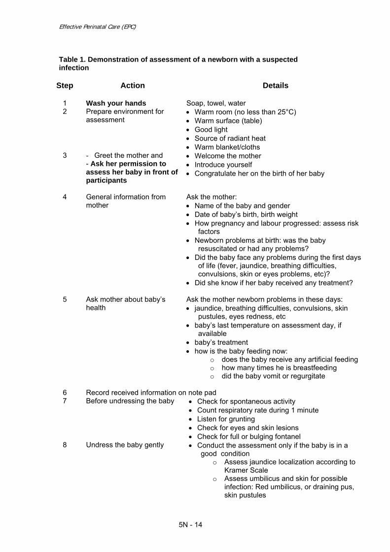
Effective Perinatal Care (EPC)
Table 1. Demonstration of assessment of a newborn with a suspected infection
Step
Action Details
1 Wash your hands Soap, towel, water 2 Prepare environment for
assessment • Warm room (no less than 25°С) • Warm surface (table) • Good light • Source of radiant heat • Warm blanket/cloths
3 - Greet the mother and - Ask her permission to assess her baby in front of participants
• Welcome the mother • Introduce yourself • Congratulate her on the birth of her baby
4 General information from mother
Ask the mother: • Name of the baby and gender • Date of baby’s birth, birth weight • How pregnancy and labour progressed: assess risk
factors • Newborn problems at birth: was the baby
resuscitated or had any problems? • Did the baby face any problems during the first days
of life (fever, jaundice, breathing difficulties, convulsions, skin or eyes problems, etc)?
• Did she know if her baby received any treatment?
5 Ask mother about baby’s health
Ask the mother newborn problems in these days: • jaundice, breathing difficulties, convulsions, skin
pustules, eyes redness, etc • baby’s last temperature on assessment day, if
available • baby’s treatment • how is the baby feeding now:
o does the baby receive any artificial feeding o how many times he is breastfeeding o did the baby vomit or regurgitate
6 Record received information on note pad 7 Before undressing the baby • Check for spontaneous activity
• Count respiratory rate during 1 minute • Listen for grunting • Check for eyes and skin lesions • Check for full or bulging fontanel
8 Undress the baby gently • Conduct the assessment only if the baby is in a good condition
o Assess jaundice localization according to Kramer Scale
o Assess umbilicus and skin for possible infection: Red umbilicus, or draining pus, skin pustules
5N - 14

Module 5N
Step
Action Details
o Check for abdomen distension 9
Check for baby’s activity
• Check for infant ‘s movements: o Does the infant move only when
stimulated? o Is the infant motionless even when
stimulated?
Thank the mother and ask her if she has any question. The clinical examination is finished; the baby needs to be dressed again and given back to
his /her mother. Ask her kindly if she agrees to breastfeed her baby in front of the participants
12
Assess breastfeeding or other feeding method
• Is the attachment correct? • Is the position correct? • Does the baby suckle effectively? Observe it carefully, take notes and ask the participants to take note on all these issues because you will ask them to answer these questions later. If other feeding method: - Assess appropriateness of the method - quantity - frequency - any incident
13 Once out of the mother room lead the discussion with the participants: 1. Does the baby have a good general status?? 2. Analyse the results of the clinical assessment
o localisation of jaundice o baby activity o signs of infections o liver and spleen o urine and stools
3. Ask the participants to evaluate the quality of feeding 4. Ask the participants to interpret the results of biological tests and other investigations 5. Ask the participants to evaluate the quality of treatment (including timeliness)
o drugs o infusion o supportive care
6. Ask when the baby can be discharged home
• After the demonstration split the participants into 2 groups: 1 group with 1
facilitator.
• Remind briefly the methodology of newborn examination, according to Table 1.
• Nominate one participant in each group to perform the examination for 20 min maximum
• Ask the other participants not to make any comments during the examination.
Participants should make their notes and comments in note pads.
5N - 15

Effective Perinatal Care (EPC)
• Each group “receives” a baby with infection or suspected infection to be assessed; the facilitator will provide all information on each case.
• Ensure that the participants wash their hands. • Ensure that the participants follow the main requirements of keeping the baby
warm during assessment. • Ask the sub group to come back to the class room to list during 10 min :
- What was correctly done - What could have been done differently according to module 5N (medical
history, biological and other investigations, treatment, monitoring feeding, and supportive care)
Coordinate the conclusion of the clinical activities with the entire group for approximately 10 min
5N - 16

Module 5N
References 1. A. Durandy, Development of specific immunity during prenatal life, 2001 2. Ann L Anderson-Berry Neonatal Sepsis University of Nebraska School of
Medicine Updated August 18,2006 3. Barbara J. Stoll et al., Late-Onset Sepsis in Very Low Birth Weight Neonates:
The Experience of the NICHD Neonatal Research Network. PEDIATRICS Vol. 110 No. 2 August 2002, pp. 285-291
4. Candice E. Johnson, Judy K. Whitwell, Kalpana Pethe, Kapil Saxena, and
Dennis M. Super. Term Newborns Who Are at Risk for Sepsis: Are Lumbar Punctures Necessary? PEDIATRICS Vol. 99 No. 4 April 1997, p. e10
5. Chalmers I, Enkin M, Keirse MJNC, eds. Effective care in pregnancy and
childbirth. Oxford, Oxford University Press, 1989 6. Claudio Chiesa, Alessandra Panero, John F. Osborn, Antonella F. Simonetti
and Lucia Pacifico. Diagnosis of Neonatal Sepsis: A Clinical and Laboratory Challenge. Clinical Chemistry. 2004;50:279-287
7. Cochrane Database Systematic Rev 2003; (3) Webster J, Pritchard M.A.
Gowning by attendants and visitors in newborn nurseries for prevention of neonatal morbidity and mortality
8. Cochrane review relevant references: Antibiotic regimens for suspected early
neonatal sepsis 9. Cochrane review relevant references: Antibiotic regimens for suspected late
onset sepsis in newborn infants 10. Cochrane review relevant references: Gowning by attendants and visitors in
newborn nurseries for prevention of neonatal morbidity and mortality 11. Cochrane review relevant references: Intraventricular antibiotics for bacterial
meningitis in neonates 12. Cochrane review relevant references: One dose per day compared to multiple
doses per day of gentamicin for treatment of suspected or proven sepsis in neonates
13. Cochrane review relevant references: Patient isolation measures for infants
with candida colonization or infection for preventing or reducing transmission of candida in neonatal units
14. Essential new born care and Breastfeeding PEPC WHO 2002 15. Essential Newborn care and Breastfeeding WHO Euro 2002 16. Handbook IMCI Integrated Management of Childhood Illnesses.
WHO/FCH/CAH/00.12 Geneva: WHO, 2000.
5N - 17

Effective Perinatal Care (EPC)
17. JA Garcia-Prats, TR Cooper. The critically ill neonate with infection: management considerations in the term and preterm infant. Seminars in pediatric infectious diseases, Saunders Company. 2000.
18. Kenyon S., Boulvain M. Antibiotics for preterm premature rupture of
membranes. In: The Cochrane Library, Issue 2, 2002. Oxford: Update Software
19. Kenyon SL., Taylor DJ., Tarnow-Mordi W, for the ORACLE Collaborative
Group. Broad-spectrum antibiotics for preterm, prelabour rupture of fetal membranes: the ORACLE I randomised trial. Lancet 2001;357:979-88
20. Klein J, Marcy S. Bacterial sepsis and meningitis. Mosby, 1995 21. Lawn J.E. et al., Statistical annex – World Health Report 2005. Geneva: World
Health Organization, 2005 22. Lawn, J, Simon Cousens, Jelka Zupan. Neonatal Survival 1: 4 million
neonatal deaths: When? Where? Why? Lancet, Vol. 365 March 5, 2005 23. Managing Newborn Problems: A guide for doctors, nurses, and midwives.
WHO, Geneva, 2003 24. Martin Weber and all. Predictors of neonatal sepsis in developing countries.
Pediatr Infect Dis J, 2003; 22:711–16 25. Pocket book of Hospital care of children. Guidelines for the management of
common illnesses with limited resources. WHO, 2005 26. Pong A, Bradley JS. Bacterial meningitis and the newborn infant. Infect Dis
Clin North Am. 1999 Sep;13(3):711-33 27. Pregnancy, Childbirth, Postpartum and newborn Care: A guide for essential
practice. WHO, Geneva, 2006 28. Prevention of perinatal Group B Streptococcus Disease. Revised guideline
from CDC. August 16, 2002 29. Randall W King, David Johnson. Osteomyelitis. eMedicime from WebMD. July
13, 2006 30. S Vergnano, M Sharland, P Kazembe, C Mwansambo. P T Heath. Neonatal
sepsis: an international perspective Archives of Disease in Childhood Fetal and Neonatal Edition 2005;90:F220-FF224
31. Shelley C Springer and David J Annibale Necrotizing Enterocolitis, eMedicine,
2007 32. The Merk Merck Manuals online medical library. Skin Infections, November
2005 33. The WHO multicentre study group. Clinical prediction of serious bacterial
infections in young infants in developing countries. Pediatria Infection Disease 1999; 18:23–31
5N - 18

Module 5N
34. Tricia Lacy Gomella et al. Neonatology: Management, Procedures, On-Call Problems, Diseases, and Drugs. McGraw-Hill, 2004. 724 p
35. William E. Benitz, Jeffrey B. Gould, and Maurice L. Druzin Risk Factors for
Early-onset Group B Streptococcal Sepsis: Estimation of Odds Ratios by Critical Literature Review in Pediatrics,1999
36. William E. Benitz, Michael Y. Han, Ashima Madan, Pramela Ramachandra.
Serial Serum C-Reactive Protein Levels in the Diagnosis of Neonatal Infection. PEDIATRICS Vol. 102 No. 4 October 1998, p. e41
Case Study 1: Svetlana and Andrey Svetlana gave birth to Andrey after 40 weeks of a normal pregnancy. The birth was normal; Andrey was not resuscitated at birth and was immediately put on his mother’s chest for skin-to-skin contact for 10 min. He was breastfed 30 min after birth. A neonatologist assessed Andrey immediately. The newborn had good clinical condition; he weighted 3, 400 g. The baby was swaddle and put in a cradle close to Svetlana. The Mother and the baby were transferred together to the postpartum department for rooming-in. In 10 hours after birth Svetlana called the nurse because she felt that Andrey was breathing fast. The baby was assessed, he was fast breathing with 70 breaths per min, but he did not have chest indrawing neither grunting. His temperature was 35.6°С and he refused to breastfeed. 1st question: What are the possible diagnoses for Andrey? 2nd question: What are the biological test you will ask, if any? 3rd question: How do you take care of Andrey immediately? Case Study 2: Maria and Stephan Stephan was born after 38 weeks of the second normal pregnancy of Maria. During the labour Maria had temperature 38.2 ºС but the birth was normal and Stephan was put on her mother’s chest in skin-to-skin contact immediately and was breastfed 25 minutes after birth. The neonatologist assessed Stephan after 2 hours and he found that this 3,200 g baby was in a good condition. Maria and Stephan were transferred together to the postpartum department for rooming-in. When neonatologist assessed Stephan for the second time 16 hours after birth he discovered that the baby was fast breathing 80 per min with a severe chest indrawing. Stephan temperature was 37.8°С and did not show interest in breastfeeding. The neonatologist decided to immediately transfer Stephan to NICU. An umbilical venous catheter was immediately placed. Stephan received IV fluids, including Ampicillin, gentamicin IV and immunoglobulin. Results of blood screening tests: Leucocytes: 26х109/L
5N - 19

Effective Perinatal Care (EPC)
Leukocytes index: 0.44 CRP: 11 mg/L Erythrocytes sedimentation rate: 10 mm 1st question: What are the possible diagnoses for Stephan? 2nd question: How do you interpret the results of the biological tests? Do you want more tests? If yes, which tests and why? 3rd question: Do you agree on what was done for Stephan? What could have been done differently? Case Study 3: Olena and Anna Olena gave birth to Anna 2,100 g after 33 weeks of gestation. The pregnancy and birth were normal. Immediately after birth Anna was assessed by a neonatologist under a radiant heater. Her clinical condition was good but the neonatologist decides to transfer Anna to NICU because she was born preterm and was only 2,100 g. Anna’s clinical condition was monitored in NICU for 1 day where she was fed by gavage with infant formula. Then she was transferred to post-partum department to be roomed in with her mother. She was not suckling very well and was fed with expressed breast milk with a cup. On the 4th day after birth Anna started to regurgitate after each feeding. Her temperature was instable from 36.2°С to 37.8°С. She was fast breathing: 64 breaths per minute but she did not have chest indrawing nor grunting. Her abdomen was distended. She was immediately readmitted to NICU, where she got several blood tests and a lumbar puncture. A venous umbilical catheter was inserted and Ampicillin + Gentamicin + Cefotaxime + immunoglobulin were given IV. Results of blood screening tests: Leucocytes – 18х109/L Leukocytes index – 0.28 CRP – 12 mg/L Erythrocytes sedimentation rate - 18 mm The results of the lumbar puncture were normal. 1st question: What are the possible diagnoses for Anna? 2nd question: How do you interpret the results of the biological test? Do you want more tests? If yes, which tests and why? 3rd question: Do you agree on what was done for Anna? What could have been done differently? Case study 4: Tatiana and Denis Denis was born by C-section after 41 weeks of a normal pregnancy. The C-section was done because the previous baby was delivered by C-section. Denis did not require resuscitation and was placed under a radiant heater then to the nursery for 2 days.
5N - 20

Module 5N
During the day time he was brought to his mother Tatiana for breastfeeding but during the night he was fed by infant formula with a baby bottle. On day 4 the mother notices that the umbilicus was red and smelling foul. The neonatologist assessed Denis completely, the assessment was normal. He was requested a complete blood screenings and a Gram-stained smear from the umbilicus and he was given Ampicillin IV. Denis was sent to special isolation department where all the care was provided by a nurse in charge of 5 newborns. 1st question: What are the possible diagnoses for Denis? 2ndquestion: Do you agree on what was done for Denis since his birth? What could have been done differently? 3rd question: When Denis could be discharged from the maternity? What recommendation are you giving to the mother?
5N - 21

Module 6N
Care of a Newborn with Birth Defects/Congenital Malformations or Birth Trauma

Effective Perinatal Care (EPC)
6N - 2

Module 6N
Slide 6N-1 Care of a Newborn with Birth Defects/Congenital Malformations or Birth Trauma At the end of this module, the participants will:
• Know how to Assess, Classify and Treat newborns with birth defects/ congenital malformations or birth trauma
• Understand how to take care of a newborn with congenital birth defects /congenital malformations or birth trauma
• Learn effective and safe skills to care for newborns with birth defects /malformations or birth trauma.
Slide 6N-2 Causes of Neonatal Deaths Worldwide (2000) Congenital malformations or birth defects are responsible for 7% of neonatal deaths in the world. In developed countries, many of congenital malformation could be diagnosed during the antenatal period. Nevertheless, in these countries the neonatal mortality due to congenital malformations is high, as compared with lower neonatal mortality caused by infection and asphyxia.
Joy E lawn, Simon Cousens, Jelka Zupan, for the Lancet Neonatal Survival Streeting Team. 4 million neonatal deaths: When? Where? Why?
The Lancet, March 2005, 9-18.
Slide 6N-3 Birth Defects/Congenital Malformations A birth defect or congenital malformation is an abnormality of structure, function or metabolism (body chemistry) present at birth that results in a physical or mental disability, or is fatal. Several thousand different birth defects have been identified. Birth defects are the leading cause of death in the first year of life.
March of Dimes Perinatal Data Center. Maternal, Infant, and Child Health
in the United States, 2001.
6N - 3

Effective Perinatal Care (EPC) Both genetic and environmental factors can cause birth defects. However, the causes of about 60 to 70 percent of birth defects are currently unknown. Malformations often come in clusters; if you find one malformation, check for others. Frequency of minor and severe congenital malformations comprise 3-4% of all births.
Robinson A. and Linden MG. Clinical Genetic Handbook, Boston, Blackwell Scientific Publications, 1993.
Care for newborns with minor birth defects can be provided in any maternity and the baby doesn’t need to be separated from his/her mother. These newborns need to be kept warm, breast-fed and receive care from their mother. Newborns with severe abnormalities need to be transferred to a third-level medical facility for special care and treatment.
Managing Newborn Problems: A guide for doctors nurses and midwives.
WHO, Geneva, 2003.
Slide 6N-4 Assessing and Classifying Newborns with Birth Defects or Birth Traumas Immediately after birth, as the baby is dried, conduct an immediate assessment to see if there is a need for immediate care such as neonatal resuscitation. If the newborn is breathing well, has heartbeat rate over 100 per minute and become rapidly pink, he/she do not need any resuscitation measure. This rapid assessment allows recognising important malformation which need
immediate treatment such as, Spina bifida or Gastroschisis. The majority of threatening life malformations need to be treated in a third-level medical facility, in this case the newborn with a severe birth defect must be prepared for transportation.
Essential Newborn Care and Breastfeeding: Training Module. WHO Euro, Copenhagen, 2002.
6N - 4

Module 6N
Slide 6N-5 Examples of Minor Birth Defects Newborns with minor birth defects should stay with their mother in the facility and if necessary, eventually and timely referred to a specialised institution/department. These newborns require essential care: to be kept warm, to be breastfed, to receive dry cord care management and to be immunised. They can be discharged from hospital on a standard basis. The mother needs to be reassured and trained to observe the newborn and to
provide appropriate care if any.
Managing Newborn Problems: A guide for doctors nurses and midwives. WHO, Geneva, 2003.
Slide 6N-6 Minor Birth Defect: Cleft Palate Cleft palate is a frequent congenital malformation. The malformation may be limited to the lip or could involve the hard and soft palate. The malformation can be unilateral or bilateral. The major problem for newborns with cleft palate during the first months of life is feeding. If the malformation is minor, the newborn can breastfeed. If the malformation is more complex alternative feeding
methods are recommended, such as spoon or syringe to feed with expressed breast milk; in some cases an obturator should be applied. Newborns with cleft palate are at risk for milk aspiration and may not gain weight well. The date for surgical correction depends on the type of malformation: 3-6 months for cleft lip and 9-12 months for cleft palate. .
Managing Newborn Problems: A guide for doctors nurses and midwives. WHO, Geneva, 2003.
Essential Newborn Care and Breastfeeding: Training Module.
WHO Euro, Copenhagen, 2002.
6N - 5

Effective Perinatal Care (EPC)
Slide 6N-7 Cleft Lip with Cleft Palate Photograph of a newborn with cleft lip involving also the palate.
“Mother and Infant Health Project”, JSI, Ukraine.2004
Slide 6N-8 Minor Birth Defect: Club Foot (Talipes Equinovarus) Provide emotional support and reassurance to the mother. Refer to a specialized department within one month, if necessary, to treat the club foot. Managing Newborn Problems: A guide for
doctors nurses and midwives. WHO, Geneva, 2003
David E. Hertz. Care of the Newborn: A Handbook for Primary Care. Lippincott
Williams & Wilkins, 2005, p 234.
Slide 6N-9 Major Birth Defect: Congenital Diaphragmatic Hernia (CDH) (1) Congenital diaphragmatic hernia is a severe abnormality defined by:
• Lung hypoplasia (most severe on the affected side)
• Structural and functional lung immaturity
• Reduction of pulmonary arteriolar cross
• Muscular hyperplasia of remaining pulmonary arterioles.
In almost 20 % of cases CDH is associated with other major anomalies.
After birth the following clinical signs could be observed:
• Chest movements are not well coordinated with baby's breathing rhythm • Breath sounds are absent on the affected side
6N - 6

Module 6N
• Bowel sounds are heard in the chest • Concave abdomen that feels less full when touched.
March of Dimes Perinatal Data Center. Maternal, Infant, and Child Health in the United States, 2001.
Langham MR Jr et al. Congenital diaphragmatic hernia. Epidemiology
and outcome. Clin Perinatol. 1996 Dec, 23(4), 671-88.
Slide 6N-10 Congenital Diaphragmatic Hernia (CDH) (2) During the antenatal period:
• If ultrasonography confirms the diagnosis, refer the pregnant woman to the 3rd level health facility
Delivery Strategy: In a full term pregnancy spontaneous vaginal delivery is recommended. . Principles of postnatal care:
• If unexpected delivery occurs in a low level of care facility, resuscitation must be undertaken and supervised by the most experienced clinician available
• Never ventilate with bag and mask, intubate immediately • Establish venous access • Insert a large gastric tube to decompress the stomach and small bowel • Transfer to specialized surgery department • Provide emotional support and reassurance to the mother.
Krisa Van Meurs, Billie Lou Short. Congenital Diaphragmatic Hernia: The
Neonatologist's Perspective. Paediatrics in Review. 1999, 20, 79-87.
Slide 6N-11 Major Birth Defect: Oesophageal Atresia Oesophageal atresia is a severe malformation requiring urgent treatment. Ultrasonography supports antenatal diagnostic. Polyhydramnios is associated in 60% of cases or difficulty detecting the foetal stomach. If the diagnosis is suggested during the antenatal period the pregnant woman needs to be referred to a special multidisciplinary fetal diagnostic/management team.
At birth if clinical signs are observed such as excessive salivation, breathing difficulties, cough or apnoea, a trial should be made to gently insert a suction catheter or feeding tube (size 8-10F
6N - 7

Effective Perinatal Care (EPC) is adequate) into the stomach before the first feeding, then inject 2 or 3 cm3 of air while listening to the stomach with a stethoscope. If the tube is in the stomach, the sound of air will be heard. The diagnosis of oesophageal atresia is confirmed if it is impossible to insert the tube into the stomach. Most often only half of the tube can be inserted; when air is pushed in it comes back into the mouth. A smaller, softer tube may curl up in the upper oesophageal pouch and give a false negative result. Establish an IV line, and give only IV fluids at maintenance volume according to the baby’s age Insure that the baby is in supine position with the head up (approximately 30 to 60 degrees). Insure free drainage. The baby should be transported to a level III surgical neonatal unit as soon as possible. Provide emotional support and reassurance to the mother.
Dwayne C. Clark. Oesophageal Atresia and Tracheoesophageal Fistula. American family Physician, February 15, 1999, Vol. 59, № 4.
Slide 6N-12 Major Birth Defect: Imperforate Anus Imperforate anus is a severe congenital malformation, which may be complicated by intestinal obstruction and requires surgical correction. Imperforate anus is identified during the first complete newborn examination within 2 hours after birth. In case of imperforate anus, the abdomen can swell; there is an absence of meconium discharge, and vomiting. Some newborns have imperforate anus with fistula, the meconium is discharged in
uncommon places: through vagina, perineum, or with urine. Do not let the baby receive anything by mouth: establish an IV line and give only IV fluids at maintenance volume according to the baby’s age. Insert a gastric tube and ensure free drainage. Transfer the baby to a tertiary level hospital or to a specialised ward. Provide emotional support and reassurance to the mother.
Managing Newborn Problems: A guide for doctors nurses and midwives. WHO, Geneva, 2003.
6N - 8

Module 6N
Slide 6N-13 Imperforate Anus Photograph of a newborn with imperforate anus.
Slide 6N-14 X-ray of a Baby with Imperforate Anus This x-ray (invertogram) shows there is no air in the rectal canal.
Slide 6N-15 Major Birth Defects: Gastroschisis and Omphalocoele:
• Is a defect of the anterior abdominal wall just lateral to the umbilicus which is associated with an evisceration of the gastro intestinal tract
• The surface of the bowel is usually oedematous and matted and it is not covered by a membrane
• Associated anomalies are reported in up to 15% of cases (mainly gastrointestinal)
• Preterm delivery and growth restriction are frequent
• Necrotising enterocolitis and malabsorption may occur • Survival rates are about 90%.
Omphalocoele (Exomphalos): • Is a malformation of the abdominal ring of the umbilicus • Covered by a thin membrane of amnion and peritoneum
6N - 9
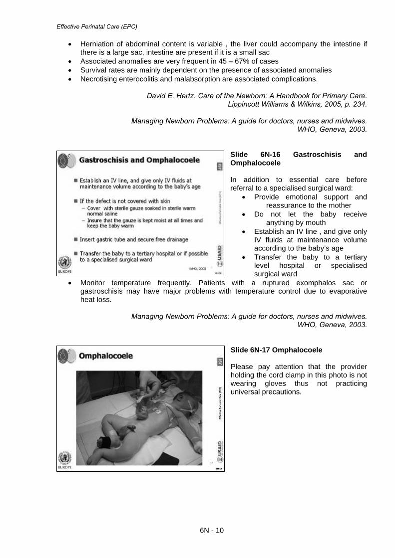
Effective Perinatal Care (EPC)
• Herniation of abdominal content is variable , the liver could accompany the intestine if there is a large sac, intestine are present if it is a small sac
• Associated anomalies are very frequent in 45 – 67% of cases • Survival rates are mainly dependent on the presence of associated anomalies • Necrotising enterocolitis and malabsorption are associated complications.
David E. Hertz. Care of the Newborn: A Handbook for Primary Care.
Lippincott Williams & Wilkins, 2005, p. 234.
Managing Newborn Problems: A guide for doctors, nurses and midwives. WHO, Geneva, 2003.
Slide 6N-16 Gastroschisis and Omphalocoele In addition to essential care before referral to a specialised surgical ward:
• Provide emotional support and reassurance to the mother
• Do not let the baby receive anything by mouth
• Establish an IV line , and give only IV fluids at maintenance volume according to the baby’s age
• Transfer the baby to a tertiary level hospital or specialised surgical ward
• Monitor temperature frequently. Patients with a ruptured exomphalos sac or gastroschisis may have major problems with temperature control due to evaporative heat loss.
Managing Newborn Problems: A guide for doctors, nurses and midwives.
WHO, Geneva, 2003.
Slide 6N-17 Omphalocoele Please pay attention that the provider holding the cord clamp in this photo is not wearing gloves thus not practicing universal precautions.
6N - 10

Module 6N
Slide 6N-18 Birth Traumas/Birth Injuries Birth injuries are mechanical injuries avoidable or inevitable. Predisposing factors include macrosomia, prematurity, cephalopelvic disproportion, dystocia, prolonged labour and breech presentation as well as instrumental deliveries ( vacuum, forceps) (ENC, 2002, page 108)
Joint Commission on Accreditation of Healthcare Organizations. Sentinel Event Alert, issue 30: Preventing infant death and injury
during delivery. July 21, 2004.
Essential Newborn Care and Breastfeeding: Training Module. WHO Euro, Copenhagen, 2002.
Slide 6N-19 Cephalohaematoma One of the most frequent birth injuries is the cephalohematoma – subperiosteal haemorrhage. A cephalohaematoma occurs when friction during the birth causes blood vessels to rupture between the periosteum and the skull. The blood accumulates under the periosteum. Unless there is a history of prolonged head engagement, a cephalohematoma is not usually present at birth. Its develops slowly during the first 24 hours of life. A cephalohaematoma is a palpable mass more often in the parietal
region; it can be unilateral or bilateral and is limited by the suture lines. Cephalohaematoma does not need any treatment such as ice, or puncture The cephalohaematoma will disappear spontaneously and completely by 3 months of age. Be alert for hyperbilirubinemia (late onset) and if there has been significant blood loss, look for, mild anaemia.
Essential Newborn Care and Breastfeeding. Training Module. WHO Euro, 2002
Parker, Leslie A. Advances in Neonatal Care: Part 1: Early recognition and
treatment of birth trauma, injuries of the head and face [foundations in newborn care]. December 2005, Volume 6(5), p 288-297.
6N - 11

Effective Perinatal Care (EPC)
Slide 6N-20 Subaponeutrotic (Subgaleal) Haemorrhage A subgaleal haemorrhage is a potentially severe haemorrhage into a large potential space between the skull periosteum and the scalp galea aponeurosis. The epicranial aponeurosis is a fibrous tissue covering the entire cranial arch. It is a large space (large enough to carry an infant's entire blood volume). Extensive blood loss is possible, and mortality rate is high (22%). Subgaleal haemorrhage can occur spontaneously but it is more often associated with vacuum or forceps assisted deliveries.
The initial signs of subgaleal haemorrhage are not specific such as generalized scalp oedema and ecchymosis. Important periorbital and periauricular oedema could appear as the haemorrhage progresses. Other signs such as an irritable cry and or cry of pain can be observed, especially when the head is touched. Most references state there is no definitive treatment . The subgaleal haemorrhage will disappear spontaneously. In rare cases bleeding may be serious and a blood transfusion may be necessary thus these infants need to be carefully monitored so that clinical signs of anaemic shock can be recognised in time.
Essential Newborn Care and Breastfeeding. Training Module.
WHO Euro, Copenhagen, 2002.
Slide 6N-21 Fractured Clavicle Fractured clavicle is the most common bone trauma in newborns. Occurs frequently during shoulder dystocia or in breech presentation. The fracture is easy to diagnose by clavicle palpation which finds typical crepitation, and displacement of fragments. The infant may have restricted active movements on the affected side, with absent Moro reflex but normal biceps reflex. This fracture can be accompanied by limited arm mobility. In some cases the
fracture is discovered a few days after birth. No special treatment; however, if arm movements are painful, the arm could be fixed with a bandage on the newborn’s chest. Sometimes the fracture is found later after a bone callus is discovered.
Managing Newborn Problems: A guide for doctors nurses and midwives.
WHO, Geneva, 2003.
6N - 12

Module 6N
Kaplan B, Rabinerson D, Avrech OM, Carmi N, Steinberg DM, Merlob P.
Fracture of the clavicle in the newborn following normal labor and delivery. Int J Gynaecol Obstet. 1998 Oct, 63(1), 15-20.
Slide 6N-22 Fractured Humerus Clinical signs are variable - the baby could seem normal, or could have pain, or may seem paralyzed. The diagnosis is confirmed by radiography. The treatment is immobilisation of the arm. It is important to check the fingers twice daily: • If the fingers become blue or swollen -
remove the bandage and rewrap it more loosely. • If the bandage is rewrapped – observe the fingers for blueness or swelling for an additional
three days. This fracture usually heals very well.
Managing Newborn Problems: A guide for doctors nurses and midwives.
WHO, Geneva, 2003.
Slide 6N-23 Fractured Femur Clinical signs are variable - legs are not moving symmetrically; leg swelling, leg in abnormal position, baby cries when leg is touched. Fractured femur requires leg immobilization. It is important to check the toes twice daily: • If the toes become blue or swollen -
remove the bandage and rewrap it more loosely.
• If the bandage is rewrapped – observe the toes for blueness or swelling for an additional three days.
Train the mother to care for the baby.
Managing Newborn Problems: A guide for doctors nurses and midwives. WHO, Geneva, 2003.
6N - 13

Effective Perinatal Care (EPC)
Slide 6N-24 Splinting Fractured Humerus and Fractured Femur Pictures of splinting fractured humerus and fractured femur. Managing Newborn Problems: A guide for
doctors nurses and midwives. WHO, Geneva, 2003.
Slide 6N-25 Brachial Plexus Injury/Arm Palsy Approximately 45% of brachial nerve injuries are associated with shoulder dystocia. They are two categories of arm palsy: 1. Erb's palsy (also called Erb-Duchenne paralysis).The arm is in adduction with an extended forearm, internally rotated and pronated. It is the classical "porter's tip" or "waiter's tip" appearance. Bicepital and Moro reflexes are absent on the arm. The sensory function is usually preserved. 2. Klumpke's palsy.The distal part of the
arm is paralyzed, hand sensitivity and mobility are affected, palmar grab reflex is absent. If mobility and sensitivity are not restored within 3 months, newborns need to be referred for specialised treatment. Selected Differential Diagnosis of Abnormal Arm Posture in a Newborn
Condition Description Klumpke's paralysis/palsy Hand paralysis with possible ptosis (drooping upper eyelid), and
myosis (constricted pupil), anhidrosis (decreased perspiration) (Horner syndrome).
Fractured clavicle Crepitation and bone callus ; occasional bruising; possibly restricted active movements with absent Moro reflex on affected side; biceps reflex present
Erb's palsy
Restricted active movements and absent Moro and biceps reflexes on affected side; "porter's tip" or "waiter's tip" appearance of upper extremity
Fractured humerus
Restricted active movements and absent Moro reflex on affected side, biceps reflex present; crepitus may be felt.
6N - 14

Module 6N
Nik Hemady, Colleen Noble. Newborn with Abnormal Arm Posture. American Family Physician, June 1, 2006, Vol. 73, No. 11.
Managing Newborn Problems: A guide for doctors nurses and midwives.
WHO, Geneva, 2003.
Slide 6N-26 Brachial Plexus Injury/Arm Palsy Photograph of a brachial plexus injury on the right side. The injured arm lies limply by the baby’s side. The arm is in adduction with an extended forearm internally rotated and pronated.
“Mother and Infant Health Project”, JSI,
Ukraine.2004
Slide 6N-27 Facial Palsy Facial nerve palsy is caused by the compression of the facial nerve during the delivery or due to traumatic forceps delivery. Facial palsy usually becomes visible on the first or second day after birth. On the paralyzed side, the naso-labial fold is evened–out, the corner of the mouth droops and when crying, the mouth is drawn to the normal side. The baby is unable to wrinkle forehead or close eye on affected side Apply ointment to this eye, 4 times daily and as long as this eye does not close.
Teach the mother how to do it. Newborns with facial palsy can have difficulty attaching to the breast. Most facial nerve palsies resolve spontaneously within days, although full recovery may require weeks to months. Referral to a specialised department may be necessary if there is no improvement within 10 days.
Managing Newborn Problems: A guide for doctors nurses and midwives. WHO, Geneva, 2003.
Mamta Fulora, Shelley Kreiter. The Newborn Examination: Part I. Emergencies and Common Abnormalities Involving the Skin, Head, Neck, Chest, and Respiratory and
Cardiovascular Systems. American Family Physician, 2002, 65, 61-8.
6N - 15
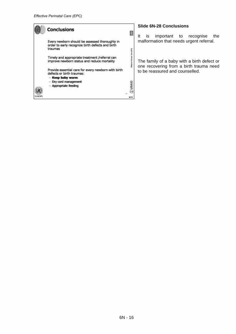
Effective Perinatal Care (EPC)
Slide 6N-28 Conclusions It is important to recognise the malformation that needs urgent referral. The family of a baby with a birth defect or one recovering from a birth trauma need to be reassured and counselled.
6N - 16

Effective perinatal care (EPC) Facilitator Guide
7N - 1
Module 7N
Low- Birth Weight Baby (LBW)/Small Baby
Care and Feeding
Learning objectives
By the end of the training the participants should know:
• The definitions for Small Baby, Very Small Baby, Low Birth Weight(LBW) and Very Low-Weight Baby (VLBW)
• The special needs of the Small Baby/LBW • How to keep the Small Baby/LBW warm • What additional care is required for the Small Baby/LBW • Feeding methods for the Small Baby /LBW • The Kangaroo Mother Care Method and its advantages • Know the criteria for discharging the Small Baby/LBW from maternity • How to counsel the mother of a Small Baby/LBW to care for her baby at home
Plan and Duration of the Module:
Part I – Classroom - 110 min Activity 1 – Introduction 5 min Activity 2 – Interactive presentation 90 min Activity 3 – Conclusion 15 min
Part II – Clinical Work – 180 min
Activity 4 - Cases study 45 min Activity 5 – Examination of small baby 90 min Activity 6 – Demonstration of Cup-feeding of Small Baby 30 min Activity 7- Demonstration of Insertion of gastric tube 15 min
Preparation to the Module • Review existing publications and materials related to the care and treatment of
the small baby. • Make sure that each of the participants have the Participant Manual • If possible get information about the existing practices of small baby care in
the participant’s health facilities. • Make sure that other facilitators know their duties for this module

Effective perinatal care (EPC)
7N - 2
Materials and Audio-visual materials Materials
• Training Manuals for participants • Case study for group work
Equipment
• Video or slide projector • Flipchart • Colour markers • Badges • Pens and pencils • Baby-doll (at least one, preferably 2) • Clothes for baby-doll • Cups and spoons for feeding • Breast model • Gloves • Gastric tube (gavage) to demonstrate gavage feeding • Syringe • Adhesive tape to fix gastric tube
Key information • It is important to understand the difficulties involved in caring for small
babies, especially in developing countries where 18% of newborn are born “small”. The percentage of small babies in developed countries is 5 – 7 %. Intrauterine growth retardation is the main reason for small babies’ births in developing countries.
• Improving the quality of antenatal care as well as the mother’s health status
play an important role in reducing the percentage of LBW births. • Official y data in NIS countries for small babies Georgia - 6, 6%, Russia - 6,
1%, Kazakhstan - 5.3%. − These countries do not follow the WHO’s definition of live birth. They
register a child born under 1,000g at birth as a late aborting if he dies before day 7.
− Baltic countries follow the WHO definition of live birth. The percentage of small babies born in Lithuania is 4.5% and for Estonia 4.3% in 2003.
• It is important to remember that not all small babies are ill babies;
nevertheless small babies are more vulnerable than newborns of a normal weight. They have special needs and thus require special care.
• A small baby stays in the maternity longer than babies of normal weight.
Small babies are frequently referred to another care level and they are more prone to become ill.

Module 7N
7N - 3
• At birth small babies are more likely to develop hypothermia thus health
care providers should pay special attention to prevent hypothermia and infection.
• Small babies need to be carefully monitored specially during the first hour
and days of life. • The mother of a small baby needs help and support in feeding her baby.
The mother should be encouraged to breastfeed her low-weight baby. If the baby is not able to suckle well, alternative feeding methods should be used. The mother needs to be trained to use alternative feeding methods.
• Health care personnel must know and use the Kangaroo Mother Care (KMC) Method to care for small baby.
• It is important to discharge the small baby as soon as possible from the
maternity. • Before discharge, the mother and the family must be counselled on baby
care at home and for danger signs that require the baby to be immediately brought to the hospital.
Part I – Classroom – 120 min
Activity 1 – Introduction (5 min) • Show Slide 7N-1 and explain that during this Module you will discuss the main
principles of effective care for small and very small babies. • This Module will be taught in two parts: part 1 will focus on work in the
classroom and part 2 is the clinical week and will take place in the maternity.
Activity 2 – Interactive presentation (90 min) • Show Slide 7N-2 and define what is meant by the term of small baby. Also
discuss the definition of very small baby. • Slide 7N-3 shows the on the percentage of small babies in EU region and in
NIS countries. • Ask participants: According to WHO statistics, the number of small
babies in NIS countries is decreasing and the number of small babies in EU countries is increasing. How can we compare this data?
• Stress on the fact that EU countries use the WHO definition of live birth.
Explain the WHO definition to participants. A live newborn is the product of conception with at least one sign of life (movement of an extremity, heartbeat, and breathing) despite body weight at birth and gestational age. Therefore in EU countries extremely small babies even < 500g can be declared alive.

Effective perinatal care (EPC)
7N - 4
• Slide 7N-4 shows the gestation chart. Use this chart you can assess the correlation of foetal weight and its gestational age, starting from 26 weeks. Explain that the area between the two black lines represents the normal foetal development; the area under the lower line shows insufficient foetal growth. Infants under that line are called “Small for Gestational Age”, during the pregnancy they suffered from, intrauterine growth retardation. Intrauterine growth retardation is caused by many factors. Children above the upper line are called “Large for Gestational Age” for example infants born to mothers with diabetes.
• Explain that the red dot represents a child born after 34 weeks weighing 2.100
g. This child is classified as “low-weight, preterm baby” since his /her weight corresponds to his/her gestational age.
• The green dot represents another child born after 38 weeks weighing 1,600 g.
This child is not considered as a preterm baby but he/she is “Small for the Gestational Age”.
• Ask the participants to use the gestational chart then ask: “What is the
normal weight for a baby born at 38 weeks of gestation?” The correct answer is - 2,400g - 3,400g. If some participants do not understand the facilitator can ask participants to give more examples of preterm babies and SGA, using the intrauterine growth chart.
• Slide 7N-5 - 7N-6 shows the main causes of LBW’s births and summarizes the
challenges in providing care to small babies Stress the effects of alcohol and tobacco use by pregnant women on the development of the foetus.
• Slide 7N-7 shows the characteristics of LBW. Emphasize that the small baby
doesn’t have storage of fat and glycogen and they are more at risk for hypothermia. Lack of iron store can lead to anaemia, lack of calcium can lead to hypocalcaemia.
• Before showing the next slides, ask participants “How do you prepare
the birth room for the birth of a small baby” and “What are the main principles of care for a small baby during the first 2 hours in the delivery room”? Write all answers on the flipchart.
• Show slides 7N-8 - 7N-12 and focus on those details which were not
mentioned by participants. Then summarize. • Slides 7N-13. Ask participant why the small baby has problems to
maintaining a normal temperature? Stress the fact that the thermoregulation of a small baby is difficult and it could take several days before body temperature is stabilized. Stress that thermal protection for a small baby needs special attention. It is important to keep a small baby warm.
• Slides 7N-14 to 7N-17. Show the main principles of small baby care in
postpartum care. It is important for the baby to room in, to breastfeed early and to receive care provided by the mother. Emphasize that it is mainly the mother who will monitor the baby and provide day to day care, including controlling the baby’s temperature. It is important to teach the mother to immediately provide skin-to-skin contact to warm a cold newborn. The medical staff needs to spend enough time to reassure, counsel and support the mother.
• Slides 7N-18 and 7N-19. The ways to treat hypothermia are important and
valid for any newborn but extremely important for a small baby.

Module 7N
7N - 5
• Underline if the baby is not able to feed at all, begin feeding expressed breast
milk by gastric tube once the baby’s temperature reaches 35 °C. • Slides 7N-20. List the several challenges involved in feeding the small baby.
Stress again on the necessity to feed the baby frequently with small quantities of milk. Ask participants what they think about the quantity and frequency involved in feeding the small baby.
• Slides 7N-21 - 7N-22 illustrate the quantity and the frequency of feeding.
Stress again the necessity to feed the small baby frequently with small quantity of milk. This gives the small baby the necessary calories while also taking into consideration their small stomach capacity.
• Ask the question “What is the best feeding method for a “small baby” and
show Slide 7N-23. • Slides 7N-24 - 7N-26. Selection of a feeding method. Underline that the
baby’s weight is not the best criteria to guess the baby’s ability to suckle. Stress the fact that it is not easy to breastfeed a small baby. Patience and commitments is necessary. And the mother is the best one to do this. She wants what is the best for her baby and is willing to be patient. She is also very committed to her baby’s well being. Health care personnel should avoid if possible proposing “easy” feeding methods such as feeding by gavage. Health care personnel need to actively support and counsel the mother to do the best for her baby.
• Before showing the next slides ask participants how they help the
mothers to express their milk. Then show Slide 7N-27. Teach the mother correct milk-expression techniques.
• Slides 7N-28 - 7N-31. Alternative feeding methods: cup-feeding, preparation
for cup-feeding, cup-feeding techniques. Alternative feeding methods can replace breastfeeding or complement it. Feeding the very small baby and the sick baby via gavage. Emphasize that the most physiological way is slow gavage over 15-20 minutes.
• Slide 7N-32. In some cases, when the baby is not digesting well, feeding
needs to be interrupted or the volume needs to be reduced. • Slide 7N-33. Small babies are at risk for developing anaemia and rickets.
They need to receive iron and vitamin D supplementation beginning the second week after birth. Breastfed children do not require supplementation of Vitamin A as breast milk contains enough vitamin A.
• Before showing the following slides ask participants: What is the kangaroo
Mother Care Method and What are the advantage of this method? Write down all the answers on the flipchart.
• Slide 7N-34 -7N-38. KMC technique is described. Answer questions on this
issue. • Slide 7N-39. Criteria for discharging a small baby from the maternity.
Emphasize that the small baby should be discharged from the maternity as soon as possible. Do not apply an arbitrary weight limit for discharge.

Effective perinatal care (EPC)
7N - 6
• Slides 7N-40. It is important to give all recommendation for home care of the baby to the mother in a written form if possible. - Stress that counselling before discharge is extremely important and the staff
needs to allocate enough time for it and to use words that the mother can understand.
- Stress SIDS prevention as preterm babies have a higher risk of SIDS • A recent survey conducted in the USA shows that mothers who delivered
preterm were almost twice as likely to place their baby on the belly to sleep. The authors think that very preterm infants in intensive care nurseries are frequently managed in the prone position; and infants and their caregivers become used to this position. Mothers are likely to follow the advice/example of health care professionals and advice is more likely to be conveyed during a long hospitalization.
• Emphasize that health care professional responsible for organizing the
hospital discharge of infants from neonatal intensive care units should become more vigilant about endorsing and modelling the SIDS risk-reduction recommendations significantly before the infant’s anticipated discharge.
• Follow up visits needed to be clearly planned during the discharge visit. Activity 3- Conclusions (15 min) • Slides 7N-41. Summarise the chapter, answer any questions and stress the
fact that except for very small babies expensive medical equipment is not needed to provide good care to small babies
Part II – Clinical work (180 min)
Activity 4 -Case Study Work in small groups (45 min) • This activity can be conducted at any convenient time during the first or
second week after the module is completed. • Split participants into 3 groups. Neonatologists and nurses should be in equal
proportion in each group. • Give to each group one case study (Attachment 1). Make sure participants
understand the task. • Give to participantіs flipchart paper and markers. Case Study 1: Denis and His mother Maria Maria gave birth to Denis at 35 weeks of gestation. Denis did not need resuscitation at birth. Immediately after birth he was placed on a table and assessed by the neonatologist. Then a nurse weighted him (he weighed 2,000g) dressed him and give him back to his mother. After 15 minutes of incomplete skin to skin contact, the nurse told Maria that it was time to breastfeed Denis and the nurse tried to attach Denis who was sleeping

Module 7N
7N - 7
calmly. He didn’t show any interest in breastfeeding. After 10 minutes of unsuccessful attempts, the nurse took Denis away from Maria, swaddled him and placed him in the cot beside Maria’s bed. After 30 min Denis’ temperature was 350
C. Questions for discussion:
1. What information do you get from Denis‘s story? 2. Classify Denis using the gestational age graph. 3. What treatment does Denis require? What would you recommend that
Maria do over the next 2 hours? 4. When does Denis’s next assessment to be done and what needs to be
assessed? 5. When Denis can be discharged from the maternity?
1. What information do you get from Denis‘s story? o Gestational age 35 weeks o Denis didn’t need resuscitation o Birth weight 2,000g o Denis didn’t get enough skin-to-skin contact o Denis is hypothermic o Denis doesn’t show any interest in breastfeeding o The nurse was trying to attach Denis to his mother’s breast before he was
ready. o Denis was swaddled and placed in his cot 2. Classify Denis using the gestational age graph o Denis’s weight correlates with his gestational age. Denis is a preterm baby o Denis is a preterm baby suffering from a moderate hypothermia. He has
problems with breastfeeding probably because he is cold.
3. What treatment does Denis needs? o Immediate treatment of hypothermia. Warm the baby, using skin-to-skin
contact, monitoring his temperature every hour for three hours. If the baby’s temperature is increasing at least 0.5 C per hour over the last three hours, rewarming is successful; continue measuring the baby’s temperature every two hours. Once the baby’s temperature is normal, measure the baby’s temperature every three hours for 12 hours. If the baby’s temperature remains within normal range, discontinue measurements.
o Explain to the mother that Denis is hypothermic and needs skin-to-skin contact get warm again. Initiate skin-to-skin contact as soon as possible.
o Denis needs to be fed because during warming process the consumption of calories and oxygen will increase.
o While the child is in skin-to-skin contact help the mother attach the child to the breast.
o Explain to Maria that her milk/colostrum is the best food for Denis. o If Denis is unable to suckle at the breast, express colostrum into a cup and
give it to Denis. o Denis doesn’t need any other food except breast milk.
4. When does Denis‘s next assessment need to be done and what needs to
be assessed? o It’s necessary to check Denis’s temperature every hour for three hours. If the
baby’s temperature is increasing at least 0.5 C per hour over the last three hours, rewarming is successful; continue measuring the baby’s temperature every two hours. Once the baby’s temperature is normal, measure the baby’s

Effective perinatal care (EPC)
7N - 8
temperature every three hours for 12 hours. If the baby’s temperature remains within normal range, discontinue measurements.
o The following day, Denis needs to be completely examined by health care worker.
− Carefully assess breastfeeding, check correct attachment, position and swallowing.
− Control the numbers of feeds and assess the quantity of urine, the quantity and colour of stools.
− Check for jaundice. o Monitor twice daily the temperature and daily Denis’s weight. 5. When can Denis be discharged from the maternity? o When Denis has a good suckling reflex o When Denis gains weight (15 g -20 g minimum) for 3 consecutive days. o When Denis has a stable temperature for 3 consecutive days. o When the mother does not have any concern about Denis’s heath and is able
to care for Denis at home with access to medical care. Case Study 2: Anna and Her mother Svetlana Svetlana gave birth to Anna at gestational age of 34 weeks. Anna’s weight was 1,975 g. Anna was suckling poorly during the first week. The mother began cup-feeding her 8 times a day starting the day she was born according to the neonatologists recommendation. On day 7th Anna’s health state was good, she had a stable temperature (36,7 to 37,1°C), she started breastfeeding and she didn’t have breathing difficulties .She was jaundiced on her chest and abdomen from day 3 until day 6 . Anna was weighted daily and by day 4 her weight was 1,905 g (she lost 70 g in the first 4 days after birth). By day 6 she weighted - 1,920 g, on day 7 -1,950g , on day 9 -1.970g and on day 10 -1990 g. The baby was slowly gaining weight and by the day 11 she weighed 2000g and she was breastfeeding 10 times a day. Anna was immunized against hepatitis В (HB-1) and BCG on day 10. Data on immunization were written in the baby’s medical card. Svetlana does not feel confident about caring for Anna and she is afraid to take her home. Questions for discussion: 1. Classify Anna using the gestational age graph o Anna is a preterm baby, with a normal birth weight for her gestational age 2. Is Anna ready to be discharged from the maternity on 11th day? o Anna can be discharged from the maternity as she began gaining weight from
day 6 for 4 consecutive days. Her body temperature is stable and there are no breathing problems. She has physiological jaundice. She has a good suckling reflex.

Module 7N
7N - 9
3. What recommendations would you give to Svetlana when Anna is be discharged from the maternity?
o Anna needs to be kept warm, bath only in a warm room. o She has to be breastfed 8-10 times during day and through the night, o Anna should sleep on her back in a smoke free room but not over heated. o The mother must know the danger signs for seeking immediate care. if the
baby refuses breastfeeding or feeds poorly or becomes sick. o Follow up visit on the 7th day after discharge. 4. It seems that Anna was gaining weight poorly during her first week of
life. What could have been done differently? o Svetlana needed help with breastfeeding. Medical personnel should help with
attaching the baby to the breast daily from day 1 to day 6. o The feeding method with two cups was not explained to the mother:
expressing fore-milk in one cup and hind-milk in another cup and feeding the baby with the higher calorie milk- the hind-milk first.
5. On day 3 day Anna received 15 ml of milk at each feeding – Was it
enough? o No, this amount of milk was not enough. Anna should have received 20-23 ml
of milk per feed on day 3 according to her age and birth weight. . Case Study 3: Stephan and His Mother Sofia Stephan was born without any problem after 35 weeks of gestation. Stephan was very small, nevertheless immediately after birth he was placed on his mother’s chest for a few minutes “to be colonized by the mother’s flora” as the nurse explained. Then he was assessed and weighed. After 30 minutes his temperature was 36,3°C. Stephan's weight was 1530 g. Stephan was dressed and tightly swaddled “to keep him warm “the nurse said .Mother and the baby were left alone for two hours in the birth room where the temperature was 25°C. The nurse said that the baby didn’t need a blanket as it was warm enough in the room, and she also told the mother to feed Stephan if the baby seemed hungry. The neonatologist examined Stephan after 2 hours under a radiant heater. She counted the breathing rate and found 70 breaths per minute and heard grunting when the baby breathed out. She recounted and found 65 breaths per minute. Stephan was pale and cold. His temperature was 35.4°C. During the 2 hours in the birth room Stephan was not fed as he didn’t show any interest in breastfeeding. The nurse had said “Never force a baby to eat”. Questions for discussions: 1. Classify Stephan using the gestational age graph o Stephan is a preterm baby, small for gestational age; his weight would
normally be between 2,000g to 2,900g 2. List what should have been done in the birth room for Stephan. o Stephan should have been longer with his mother o Since Stephan was breathing well, he could stay with this mother during the 2
hours and be weighted and examined at this time o Stephan was tightly swaddled which made him cold and he was not covered
by a blanket when he was in the delivery room.

Effective perinatal care (EPC)
7N - 10
o Baby and mother were left alone and no one counselled the mother to start breastfeeding. Stephan was probably not interested in for feeding because he was cold and tightly swaddled.
3. Describe what should be done for Stephan and his mother during and
after their transportation to the paediatric department. Before the transportation:
o The baby needs immediate skin to skin contact with his mother and transport to paediatric department with her.
o Give oxygen with a moderate flow rate o Express the mother’s milk and give 10 ml to the baby by cup, spoon or pipette,
if he refuses it, install an IV perfusion of glucose 10%.
In the paediatric unit o Conduct a full examination of the newborn under radiant heater while giving
oxygen. o Continue to warm the baby using skin to skin, or radiant heater or hot
mattress. The room has to be warm at least 25 ºC. o Assess the baby’s temperature every hour for three hours. If the baby’s
temperature is increasing at least 0.5 C per hour over the last three hours, rewarming is successful; continue measuring the baby’s temperature every two hours. Once the baby’s temperature is normal, measure the baby’s temperature every three hours for 12 hours. If the baby’s temperature remains within normal range, discontinue measurements.
o Monitor breathing parameters every 3 hours (breathing rate, chest indrawing, and grunting) until the baby no longer requires oxygen and then for an additional day.
o Give oxygen until breathing difficulties disappear, monitor if possible the response to oxygen with an oximeter, if not possible use clinical signs, and remember that central cyanosis is a very late sign of breathing difficulties.
o If the baby is not able to feed after becoming warm, install a gastric tube and feed him by gavage feeding every 3 hours.
o Check for the quantity of urine.
Activity 5 – Examination of small baby (90 min) • Make sure that participants understand the main principles of small baby
care and feeding. • The trainer must ask the help of the maternity head-physician or chief
neonatologist in selecting three children for examination if it is possible .If only one child is available the clinical session will be limited to a demonstration done by one facilitator.
• The identification of small babies for assessment by participants can be
done at any convenient time after the demonstration. • Ask the mothers’ permission to examine the children. • Prepare a radiant heater, warm baby clothes, wall thermometer,
thermometer for measuring body temperature, scales and a good light.
a) Facilitator ’s demonstration of Small Baby examination (30 min with discussion)

Module 7N
7N - 11
Steps Actions Details 1 Wash your hands! Use soap and towels.
2 Prepare a warm room and a radiant heater if necessary
Good lighting, warm room. Check the ambient temperature in the room. Prepare warm baby clothes
3 Ask the mother’s/family’s permission to examine the child in their presence
4 Ask the mother
Note the child’s birth date, gender, name, birth weight and weight on the day of the examination
5 Ask the mother
• Did you have problems during your pregnancy?
• What problems has the baby had since the birth?
• Temperature, urine, jaundice, breathing difficulties
• Does the mother have concerns about her child’s health?
6 Write down all the information the mother gives you.
7 Assess the child’s breathing before you undress him/her. The child needs to be calm not crying.
• Count the number of breaths per minute and whether the breathing is > 60 or less than < 30 per minute
• Check for grunting when breathing out
8
Ask the mother to gently undress the baby and check for chest indrawing and jaundice (on abdomen, palms and sole on the feet, depending of the baby’s age)
• Check for severe chest indrawing when breathing in
9 Measure the child’s temperature and weight.
• Assess for hypothermia or hyperthermia.
• Weigh the baby or check the weight of the day.
10 Assess for other symptoms and possible signs of infection
• Assess bulging fontanel • Assess umbilical infection • Is the baby alert, or
lethargic? • Is the baby moving
normally?
11 Assess for other possible problems of the small baby
• E.g. Congenital anomaly or other problems.
12
Ask how the baby is fed Is he breastfed? if not which method is used? What is the daily quantity of milk given? What is the frequency of feeding? Is the baby gaining weight?

Effective perinatal care (EPC)
7N - 12
Steps Actions Details
13
Praise the mother for her good child care and ask her to dress the baby. Ask whether she has any questions regarding her baby
14 Comment on everything you did and summarize the examination of the small baby.
15 Ask if the other trainers/facilitators have comments. Ask the participants if they have questions and answer them.
b) Assessment of small baby by participants (if children are identified for this activity). • After that split participants into 2 groups. There will one small baby for each
group with one trainer per group. • One participant is nominated to conduct the examination following the same
plan as the facilitator. The other participants watch the examination, without comment or interruption and write their observations on the baby and on the way the examination is conducted
• Make sure that the participant washes his/her hands before touching the
baby. • Make sure that the small baby is kept warm during the examination. • Discuss what was done correctly and what could be improved • Discuss possible treatment and observation.
Activity 6 – Demonstration of Cup-feeding of a Small Baby (30 min) Every facilitator should demonstrate cup-feeding to his/her sub-group, using the following recommendations. If possible the facilitator should to demonstrate cup feeding on a real small baby (after getting the authorization from the mother and from the staff in charge of the baby) but if it not possible the facilitator has to do a demonstration with a doll. One of the facilitator plays the role of mother. Imagine a baby born 5 days ago; his birth weight was 1,800g. • Wash your hands • Measure the necessary quantity of milk 30 ml. • Hold the baby in semi-vertical position • Touch the baby’s lips so that milk just slightly reaches the lips • Do not pour the milk into the mouth. • The baby laps the milk or may sip it. Allow time for the baby to swallow the
milk

Module 7N
7N - 13
• The trainer should ask the participants to observe the cup-feeding role play or demonstration without making comments or interruption.
• Then ask participants if they practice cup feeding in their facilities. • What the problems do they faced with cup feeding technique? • How do they counsel the mother to use this technique? Activity 7 – Demonstration - Inserting a gastric tube (15 min) Every facilitator should demonstrate how to insert a gastric tube, or a least how to measure it, and how to confirm the proper placement of a gastric tube. SUPPLIES • Clean examination gloves • Clean plastic tube or catheter appropriate for baby’s weight:
o If the baby weighs less than 2 kg, use a 5-F tube o If the baby weighs 2 kg or more, use an 8-F tube
• Writing pen or flexible tape measure • 3- to 5-ml syringe (for aspiration) • Stethoscope • Sterile syringe • Cap for gastric tube (if the tube will be used for feeding) • Adhesive tape PROCEDURE • Gather necessary supplies. • Wash hands, and put on clean examination gloves. • Estimate the required length of tube: • Hold the tube so that it mimics the route that it will follow once inserted (i.e.
from the mouth or the tip of the nostril to the lower tip of the ear lobe and then to the stomach, just below the rib cage. Place a mark on the tube with a pen or a piece of tape)
• Alternatively, estimate the distance using a flexible tape measure, and mark the distance on the tube with a pen or a piece of tape.
• Flex the baby’s neck slightly and gently pass the tube through the mouth or through one nostril to the required distance.
o If the tube does not slide easily into the nostril, try the other nostril. o If the tube still does not slide easily into the nostril, use the oral route.
• Never force the gastric tube into the nostril if you encounter resistance • Secure the tube in position with adhesive tape CONFIRMING PROPER PLACEMENT OF GASTRIC TUBE To confirm proper placement of the tube: • Fill a syringe with 1 to 2 ml of air and connect it to the end of the tube. Use a
stethoscope over the stomach to listen as air is quickly injected into the tube • If a whistling sound is heard through the stethoscope as the air is injected, the
end of the tube is correctly positioned in the stomach • If a whistling sound is not heard, the tube is not properly positioned. Remove
the tube and repeat the procedure • Use a syringe to aspirate some fluid • Replace the tube with another clean gastric tube after three days, or

Effective perinatal care (EPC)
7N - 14
• Earlier if it is pulled out or becomes blocked FEEDING THROUGH GAVAGE • Determine the required volume of the milk according to the baby weight. • Connect the barrel of the syringe to the end of gavage. • Pour the required volume of milk for the feed into the syringe. • Hold the syringe 5-10 cm above the baby or suspend the gavage above baby
and allow the milk to run down the gavage with gravity: do not force milk through the gavage using the plunger.
• Each feeding should last for 10-15 minutes. • During the feeding baby’s condition must be monitored closely: colour of skin
and mucous membranes; respiratory rate and character of breathing; baby’s anxiety.
• After feeding disconnect syringe and gastric tube and close the tube with the plug. The head of doll must be elevated.
• The facilitator should stress that the mother has to be involved in gavage feeding by holding her baby in her arms.

Module 7N
7N - 15
Attachment 1
Activity 4 Case Study 1: Denis and His mother Maria Maria gave birth to Denis at 35 weeks of gestation. Denis did not need resuscitation at birth. Immediately after birth he was placed on a table and assessed by the neonatologist. Then a nurse weighted him (he weighed 2,000g) dressed him and give him back to his mother. After 15 minutes of incomplete skin to skin contact, the nurse told Maria that it was time to breastfeed Denis and the nurse tried to attach Denis who was sleeping calmly. He didn’t show any interest in breastfeeding. After 10 minutes of unsuccessful attempts, the nurse took Denis away from Maria, swaddled him and placed him in the cot beside Maria’s bed. After 30 min Denis’ temperature was 350 C. Questions for discussion:
1. What information do you get from Denis‘s story? 2. Classify Denis using the gestational age graph. 3. What treatment does Denis require? What would you
recommend that Maria do over the next 2 hours? 4. When does Denis’s next assessment to be done and what
needs to be assessed ? 5. When Denis can be discharged from the maternity?

Effective perinatal care (EPC)
7N - 16
Case Study 2: Anna and Her mother Svetlana Svetlana gave birth to Anna at gestational age of 34 weeks. Anna’s weight was 1,975 g. Anna was sucking poorly during the first week. The mother began cup-feeding her 8 times a day starting the day she was born according to the neonatologist’s recommendation. On day 7th Anna’s health state was good, she had a stable temperature (36,7 to 37,1°C), she started breastfeeding and she didn’t have breathing difficulties .She was jaundiced on her chest and abdomen from day 3 until day 6 . Anna was weighed daily and by day 4 her weight was 1,905 g (she lost 70 g in the first 4 days after birth). By day 6 she weighted 1,920 g, on day 7 1,950g , on day 9 1.970g and on day 10 1990 g. The baby was slowly gaining weight and by the day 11 she weighed 2000g and she was breastfeeding 10 times a day. Anna was immunized against hepatitis В (HB-1) and BCG on day10. Data on immunization were written in the baby’s medical card. Svetlana does not feel confident about caring for Anna and she is afraid to take her home. Questions for discussion:
1. Classify Anna using the gestational age graph 2. Is Anna ready to be discharged from the maternity on day
11? 3. What recommendations would you give to Svetlana when
Anna is discharged from the maternity? 4. It seems that Anna was gaining weight poorly the first
week of her life. What could have been done differently? 5. On day 3 Anna received 15 ml of milk for each feed – was
that enough?

Module 7N
7N - 17
Case Study 3: Stephan and His Mother Sofia Stephan was born without any problem after 35 weeks of gestation. Stephan was very small, nevertheless immediately after birth he was placed on his mother’s chest for a few minutes “to be colonised by the mother’s flora” as the nurse explained. Then he was assessed and weighted. After 30 minutes his temperature was 36,3°C. Stephans’ weight was 1530 g. Stephan was dressed and tightly swaddled “to keep him warm “ the nurse said .Mother and the baby were left alone for two hours in the birth room where the temperature was 25°C. The nurse said that the baby didn’t need a blanket as it was warm enough in the room, and she also told the mother to feed Stephan if the baby seemed hungry. The neonatologist examined Stephan after 2 hours under a radiant heater. She counted the breathing rate and found 70 breaths per minute and heard grunting when the baby breathed sout. She recounted and found 65 breaths per minute. Stephan was pale and cold. His temperature was 35.4°C. During the 2 hours in the birth room Stephan was not fed as he didn’t show any interest in breastfeeding. The nurse had said “Never force a baby to eat”. Questions for discussions:
1. Classify Stephan using the gestational age graph. 2. List the what should have been done for Stephan in the
birth room. 3. Describe what should be done for Stephan and his mother
during and after their transportation to the paediatric department.

Effective perinatal care (EPC)
7N - 18
References:
1. Alain Dabadie. Alimentation de l ‘enfant.1999. 2. Bull J, Mulvihill C, Quigley R. Prevention of low birth weight:
assessing the effectiveness of smoking cessation and nutritional interventions: evidence briefing. 2003.
3. Cochrane review relevant to the module: Ad libitum or demand-semi-
demand feeding versus scheduled interval feeding for preterm infants.
4. Cochrane review relevant to the module: Cup feeding versus other
forms of supplemental enteral feeding for newborn infants unable to fully breastfeed.
5. Cochrane review relevant to the module: Developmental care for
promoting development and preventing morbidity in preterm infants.
6. Cochrane review relevant to the module: Early developmental intervention programs post hospital discharge to prevent motor and cognitive impairments in preterm infants.
7. Cochrane review relevant to the module: Formula milk versus donor
breast milk for feeding preterm or low birth weight infants.
8. Cochrane review relevant to the module: Interventions to prevent hypothermia at birth in preterm and-or low birth weight infants.
9. Cochrane review relevant to the module: Kangaroo mother care to
reduce morbidity and mortality in low birthweight infants.
10. Cochrane review relevant to the module: Massage for promoting growth and development of preterm and-or low birth-weight infants.
11. Cochrane review relevant to the module: Non-nutritive sucking for
promoting physiologic stability and nutrition in preterm infants. 12. Edwin Chandraharan, Sabaratnam Arulkumaran. Recent advances in
management of preterm labor. J Obstet Gynecol India Vol. 55, No. 2: March/April 2005 Pg 118- 124.
13. Essential Antenatal, Perinatal and Postpartum Care. WHO Euro,
2002.
14. Essential new born care and Breastfeeding PEPC WHO 2002
15. Essential Newborn Care Course Training Manual (Draft) WHO, Geneva. 2006.
16. European Resuscitation Council Guidelines for Resuscitation.
Section 6: Paediatric life support, Resuscitation of babies at birth, 2005:S115-S133
17. Guideline for hand Hygiene in Health-Care Settings. CDC, MMWR,
October 25, 2002/Vol.51/No. RR-16

Module 7N
7N - 19
18. Guideline for hand Hygiene in Health-Care Settings. CDC, MMWR, October 25, 2002/Vol.51/No. RR-16
19. Health For All, Database, 2006.
20. Karen Edmond, Rajiv Bahl. Optimal feeding of low-birth-weight infant.
WHO Technical review, 2007.
21. Kangaroo Mother Care: A practical guide. WHO, Geneva. 2003
22. Lumley J, Oliver S, Waters E. Smoking cessation programs implemented during pregnancy. Cochrane review. Evidence-Based Nursing 1999; 2:42.
23. Managing Newborn Problems: A guide for doctors, nurses and
midwives. WHO, Geneva, 2003
24. McGuire W, Anthony MY. Donor human milk versus formula for preventing necrotizing enterocolitis in preterm infants: systematic review. Arch Dis Child Fetal Neonatal Ed.
25. Pishva N., Hemmati F. Comparison of Protein, Fat, Sodium and
Calcium in the Milk of Mothers of Preterm and Full term Infants. Shiraz E-Medical Journal Vol. 5, No. 4, October 2004.
26. Pregnancy, Childbirth, Postpartum and Newborn Care: A guide for
essential practice. WHO, Geneva, 2006.
27. Premji S, Chessell L. Continuous nasogastric milk feeding versus intermittent bolus milk feeding for premature infants less than 1500 grams (Cochrane Review), May 2002.
28. The Cochrane library, Issue 1,2004.
29. The Changing Concept of Sudden Infant Death Syndrome:
Diagnostic Coding Shifts, Controversies Regarding the Sleeping Environment, and New Variables to Consider in Reducing Risk. Policy statement AAP. November 2005.
30. Thermal Protection of the Newborn: a practical guide. WHO, Geneva,
1997.Vol. 5, No. 4, October 2004.
31. William McGuire, Ginny Henderson and Peter W Fowlie. Feeding the preterm infant. BMJ 2004;329;1227-1230.

Effective perinatal care (EPC)
7N - 20

PART III. INSTRUCTIONS ON COURSE ORGANIZATION

Facilitator’s Manual
iii - 2

iii - 3
CRITERIA FOR SELECTING FACILITATORS A group of motivated facilitators is needed. Facilitators will work on modules in pairs and will supervise clinical practice in a maternity hospital. Their tasks are described in detail in the Facilitator’s Manual. Note: Each facilitator may have weak and strong areas. If a facilitator is weak in some areas, it is important to assign a second facilitator of the pair, who is strong in these areas. For this course facilitators should be good teachers. They will lecture, guide participants through printed materials, role-play exercises, discussions, clinical practice, etc. It is very important for facilitators to be observant and notice when participants encounter difficulties to provide clear explanations and to secure effective feedback. Facilitators must be organized. They must follow the course plan/schedule and ensure timely arrival to clinical sessions and materials availability. They must be flexible to properly allocate time. For instance, if a baby with a rare symptom indicator enrolled out-patients department, they should hold up their current work and present the patient to the group. Facilitators must complete facilitator’s training and be always present throughout training course delivery. They have to be energetic and motivated to work with participants all day long and then participate in facilitator meetings for discussion of completed work and preparation for the next day of training. Facilitators should currently work at a maternity hospital and, if possible, be employees of the hospital where training is delivered. Facilitators must have approved skills of clinical training. Facilitator must be fluent in effective perinatal care methods and have experience of their use. Completion of an Effective Perinatal Care (EPC) training course as a participant or instructor is a prerequisite. Facilitators must be proficient in their clinical skills to quickly select mothers and newborns at the hospital. They must understand clinical diagnosis and prognosis of parturient woman’s and baby’s condition to avoid tangled cases and detect critically ill children who require urgent treatment. A facilitator should be able to handle ill babies easily and demonstrate positive and solicitous approach to their problems. Facilitators must possess good organizational skills. Their actions must be effective to accomplish all tasks of a clinical session, including clinical case analysis. They have to be able to keep to the point, avoiding unnecessary directions or discussions. Facilitators have to be friendly and sociable towards hospital personnel, mothers and training participants. They must serve as an example while communicating with mothers. It would be useful if at least one facilitator had completed a training course and has an experience of assessing breast-feeding and teaching mothers to improve positioning of babies during feeding and attaching the baby to the breast. An experience of supervising newborns and 1-month babies would also be an asset. While preparing for their role, facilitators, if possible, should work as facilitator’s assistants in a hospital during some other training course in order to see how patients are selected and clinical sessions are organized, and how to communicate with participants. Optionally, an

Facilitator’s Manual
iii - 4
experienced instructor may assist them for a few days during facilitator’s training or training course delivery.

iii - 5
MODEL OF THE TRAINING COURSE’ AGENDA
“Effective Perinatal Care”
Time Topic
DAY 1 – Monday
09:00-10:00
(60 min)
• Opening
- Registration of participants - Greetings - Presentation of participants - Interactive presentation «Training course objectives, information on the course»
10:00-10:45
(45 min)
• MODULE 1C. Safe Motherhood and Effective Perinatal Care: Are Changes
Necessary? - Activity 1 – Presentation (45 min)
10:45-11:00
Coffee break
11:00-13:00
(120 min)
• Module 2C. Introduction to Evidence-Based Medicine
- Activity 1 – Introduction (5 min) - Activity 2 – «Brainstorming» (15 min) - Activity 3 – Presentation (40 min) - Activity 4 – Case study (30 min)
13:00-14:00
Lunch
14:00-15:45
(105 min)
• Module 3С. Counselling Skills in Maternal and Neonatal Care
- Activity 1 – Introduction (10 min) - Activity 2 – Group work (20 min) - Activity 3 – Interactive presentation (50 min) - Activity 4 – Demonstration of effective counselling (15 min) - Activity 5 – Role plays (10 min)
15:45-16:00
Coffee break
16:00-16-45
(45 min)
• Module 3С. Counselling Skills in Maternal and Neonatal Care (continuation)
- Activity 5 – Role plays (continuation) (40 min) - Activity 6 – Conclusions (5 min)
16:45-17:45
(60 min)
• “The Icebreaker”
17:45-18:00 • Day summary by facilitators and group representatives

Facilitator’s Manual
iii - 6
Time Topic
DAY 2 – Tuesday
Midwifery Group
Neonatal Group
09:00-09:30 • Group representatives report on topics of Day 1
09:30-10:45
(75 min)
• Module 1MO. Antenatal Care - Activity 1 – Introduction (5 min) - Activity 2 – «Brainstorming» (10 min) - Activity 3 – Interactive presentation (55 min) - Activity 4 – Conclusions
(5 min)
• Module 1N. Complete Examination of a Newborn - Activity 1 – Introduction
(10 min) - Activity 2 – Group discussion
exercise (10 min) - Activity 3 – Interactive
presentation (45 min) - Activity 4 – Case studies (10 min)
10:45-11:00
Coffee break
11:00-12:00
(60 min)
• Module 4C. Assessment of Foetal Well-Being During Pregnancy and Labour.
Assessment of Small for Gestational Age (SGA) Foetuses - Activity 1 – Introduction (5 min) - Activity 2 – Work in small groups (10 min) - Activity 3 – Interactive presentation (40 min) - Activity 4 – Conclusions (5 min)
12:00-13:00
(60 min)
• Module 5C. Management of Normal Labour and Delivery
- Activity 1 – Introduction (20 min) - Activity 2 – Interactive presentation (40 min)
13:00-14:00
Lunch
14:00-15:45
(105 min)
• Module 5C. Management of Normal Labour and Delivery (continuation)
- Activity 2 – Interactive presentation (continuation) (105 min)
15:45-16:00
Coffee break
16:00-17:45
(105 min)
• Module 5C. Management of Normal Labour and Delivery (continuation)
- Activity 2 – Interactive presentation (continuation) (85 min) - Activity 3 – Conclusions (20 min)
17:45-18:00 • Day summary by facilitators and group representatives
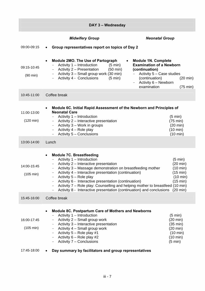
iii - 7
DAY 3 – Wednesday
Midwifery Group
Neonatal Group
09:00-09:15 • Group representatives report on topics of Day 2
09:15-10:45
(90 min)
• Module 2MO. The Use of Partograph
- Activity 1 – Introduction (5 min) - Activity 2 – Presentation (50 min) - Activity 3 – Small group work (30 min) - Activity 4 - Conclusions (5 min)
• Module 1N. Complete
Examination of a Newborn (continuation) - Activity 5 – Case studies
(continuation) (20 min)- Activity 6 – Newborn
examination (75 min) 10:45-11:00
Coffee break
11:00-13:00
(120 min)
• Module 6C. Initial Rapid Assessment of the Newborn and Principles of
Neonatal Care - Activity 1 – Introduction (5 min) - Activity 2 – Interactive presentation (75 min) - Activity 3 – Work in groups (20 min) - Activity 4 – Role play (10 min) - Activity 5 – Conclusions (10 min)
13:00-14:00
Lunch
14:00-15:45
(105 min)
• Module 7C. Breastfeeding
- Activity 1 – Introduction (5 min) - Activity 2 – Interactive presentation (20 min) - Activity 3 – Massage demonstration on breastfeeding mother (10 min) - Activity 4 – Interactive presentation (continuation) (15 min) - Activity 5 – Role play (10 min) - Activity 6 - Interactive presentation (continuation) (15 min) - Activity 7 – Role play: Counselling and helping mother to breastfeed (10 min) - Activity 8 - Interactive presentation (continuation) and conclusions (20 min)
15:45-16:00
Coffee break
16:00-17:45
(105 min)
• Module 8C. Postpartum Care of Mothers and Newborns
- Activity 1 – Introduction (5 min) - Activity 2 – Small group work (20 min) - Activity 3 – Interactive presentation (35 min) - Activity 4 – Small group work (20 min) - Activity 5 – Role play #1 (10 min) - Activity 6 – Role play #2 (10 min) - Activity 7 – Conclusions (5 min)
17:45-18:00 • Day summary by facilitators and group representatives
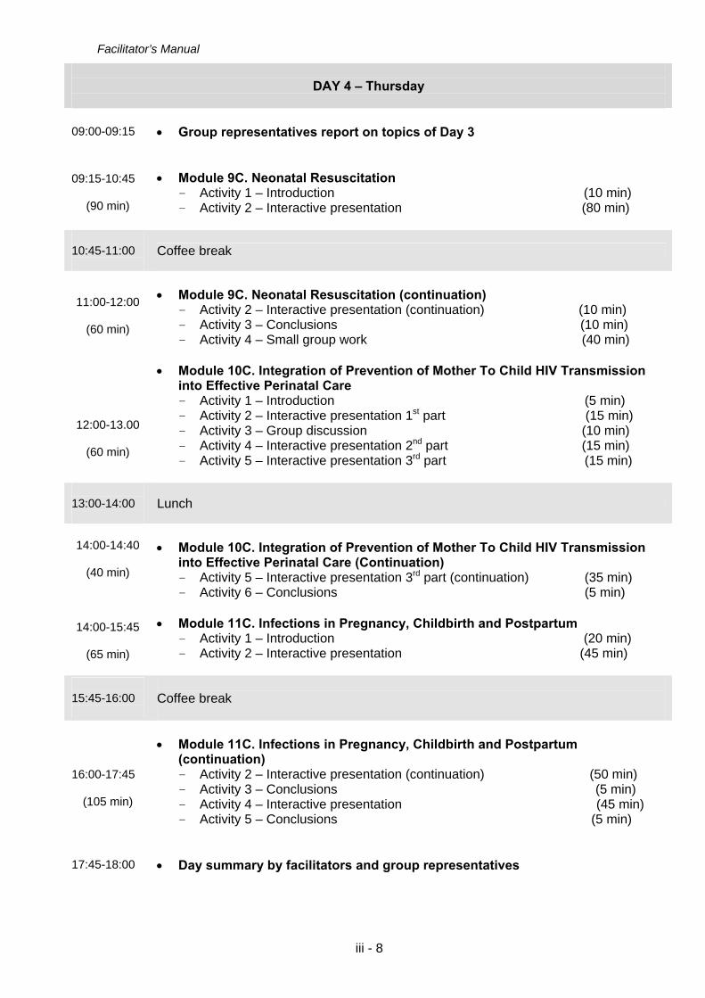
Facilitator’s Manual
iii - 8
DAY 4 – Thursday
09:00-09:15 • Group representatives report on topics of Day 3
09:15-10:45
(90 min)
• Module 9C. Neonatal Resuscitation
- Activity 1 – Introduction (10 min) - Activity 2 – Interactive presentation (80 min)
10:45-11:00
Coffee break
11:00-12:00
(60 min)
12:00-13.00
(60 min)
• Module 9C. Neonatal Resuscitation (continuation)
- Activity 2 – Interactive presentation (continuation) (10 min) - Activity 3 – Conclusions (10 min) - Activity 4 – Small group work (40 min)
• Module 10C. Integration of Prevention of Mother To Child HIV Transmission
into Effective Perinatal Care - Activity 1 – Introduction (5 min) - Activity 2 – Interactive presentation 1st part (15 min) - Activity 3 – Group discussion (10 min) - Activity 4 – Interactive presentation 2nd part (15 min) - Activity 5 – Interactive presentation 3rd part (15 min)
13:00-14:00
Lunch
14:00-14:40
(40 min)
14:00-15:45
(65 min)
• Module 10C. Integration of Prevention of Mother To Child HIV Transmission
into Effective Perinatal Care (Continuation) - Activity 5 – Interactive presentation 3rd part (continuation) (35 min) - Activity 6 – Conclusions (5 min)
• Module 11C. Infections in Pregnancy, Childbirth and Postpartum
- Activity 1 – Introduction (20 min) - Activity 2 – Interactive presentation (45 min)
15:45-16:00
Coffee break
16:00-17:45
(105 min)
• Module 11C. Infections in Pregnancy, Childbirth and Postpartum
(continuation) - Activity 2 – Interactive presentation (continuation) (50 min) - Activity 3 – Conclusions (5 min) - Activity 4 – Interactive presentation (45 min) - Activity 5 – Conclusions (5 min)
17:45-18:00 • Day summary by facilitators and group representatives

iii - 9
DAY 5 – Friday
Midwifery Group
Neonatal Group
09:00-09:15 • Group representatives report on topics of Day 4
09:15-10:45
(90 min)
• Module 3MO. Hypertension in
Pregnancy - Activity 1 – Introduction (5 min) - Activity 2 – Small group work (20 min)- Activity 3 – Interactive presentation
(40 min) - Activity 4 – Discussion of cases (20 min) - Activity 5 – Conclusions (5 min)
• Module 2N. Post- Resuscitation
Neonatal Care - Activity 1 – Introduction (10 min) - Activity 2 – «Brainstorming»
(10 min) - Activity 3 – Interactive
presentation (55 min) - Activity 4 – Work in small
groups (15 min)
10:45-11:00
Coffee break
11:00-13:00
(120 min)
• Module 4MO. Obstetric Haemorrhages
- Activity 1 – Introduction (15 min)- Activity 2 – Small group work
(15 min) - Activity 3 – Interactive presentation
(75 min) - Activity 4 – Conclusions (15 min)
• Module 3N. Breathing Difficulty
- Activity 1 – Introduction (10 min) - Activity 2 – «Brainstorming»
(10 min) - Activity 3 – Interactive
presentation (60 min) - Activity 4 – Conclusions (10 min)- Activity 5 – Group work (30 min)
13:00-14:00
Lunch
14:00-15:45
(105 min)
• Module 12C. Preterm Labour
- Activity 1 – Introduction (10 min) - Activity 2 – Work in small groups (10 min) - Activity 3 – Interactive presentation by obstetrician-gynaecologist
with discussion of small group work results (50 min) - Activity 4 – Interactive presentation by a neonatologist (30 min) - Activity 5 – Conclusions (5 min)
15:45-16:00
Coffee break
Midwifery Group Neonatal Group
16:00-17:45
(105 min)
• Module 5MO. Prelabour Rupture of
Membranes - Activity 1 – Introduction (5 min) - Activity 2 – Small group work (25 min)- Activity 3 – «Brainstorming» (15 min) - Activity 4 – Presentation (40 min) - Activity 5 – Conclusions (20 min)
• Module 4N. Neonatal Jaundice
- Activity 1 – Introduction (5 min) - Activity 2 – «Brainstorming»
(15 min) - Activity 3 – Interactive
presentation (75 min) - Activity 4 – Conclusions (10 min)

Facilitator’s Manual
iii - 10
17:45-18:00 • Day summary by facilitators and group representatives
DAY 6 – Saturday
Midwifery Group
Neonatal Group
09:00-09:15 • Group representatives report on topics of Day 5
09:15-10:15
(60 min)
10:15-10:45
(30 min)
• Module 6MO. Labor Induction: Who?
Why? How? - Activity 1 – Introduction (5 min)- Activity 2 – Work in small groups (15 min) - Activity 3 – Interactive presentation (35 min) - Activity 4 – Conclusions (5 min)
• Module 7MO. Unsatisfactory Progress
of Labour. Intrapartum Oxytocin Administration - Activity 1 – Introduction (5 min) - Activity 2 – Small group work (15 min) - Activity 3 – Interactive presentation
(10 min)
• Module 5N. Neonatal Bacterial
Infections - Activity 1 – Introduction (5 min) - Activity 2 – Interactive
presentation (85 min)
10:45-11:00
Coffee break
11:00-11:30
(30 min)
11:30-13:00
(90 min)
• Module 7MO. Unsatisfactory Progress
of Labour. Intrapartum Oxytocin Administration (continuation).
- Activity 3 – Interactive presentation (continuation) (15 min)
- Activity 4 – Small group work (10 min) - Activity 5 – Conclusions (5 min)
• Module 2MO. The Use of Partograph (continuation). - Activity 5 – Practical use of a
partograph (90 min)
• Module 5N. Neonatal Bacterial
Infections (continuation). - Activity 2 – Interactive
presentation (continuation) (15 min)
- Activity 3 – Conclusion (10 min) - Activity 4 – Small group work (90 min)
13:00-14:00
Lunch
14:00-14:50
(50 min)
14:50-15:45
• Module 13C. Sudden Infant Death Syndrome (SIDS)
- Activity 1 – «Brainstorming» (10 min) - Activity 2 – Interactive presentation (35 min) - Activity 3 – Conclusions (5 min)
• Module 14C. Postpartum Depression, Loss and Tragedies
- Activity 1 – Introduction (5 min) - Activity 2 – Group work (10 min)

iii - 11
(55 min)
- Activity 3 – Presentation (35 min) - Activity 4 – Conclusion (5 min)
15:45-16:00
Coffee break
16:00-17:45
(105 min)
• Module 5C. Management of normal labour and deliveries (continuation) - Activity 4 – Practice with a manikin
“Active Management of the Third Stage of Labour” (60 min)
- Activity 5 – Discussion and preparation of the clinical week
(45 min) Dividing of midwifery group into 2 subgroups for the clinical week.
• Module 6N. Care of a Newborn with Birth Defect/Congenital Malformation or Birth Trauma - Activity 1 –Introduction (5 min) - Activity 2– Work in small groups
(15 min) - Activity 3– Interactive
presentation (50 min) - Activity 4 – Work in small
groups (25 min)- Activity 5 – Conclusions
(10 min)
17:45-18:00 • Summary of theoretical week. Preparation for clinical week.
DAY 7 - Sunday – DAY OFF

Facilitator’s Manual
iii - 12
Clinical Sessions
12 Hour shift Option
Time
Topic
DAY 8 – Monday
Midwifery subgroup 1 – working hours: 09:00-21:00 (visiting labour – depends on the situation at the maternity ward) Midwifery subgroup 2 – working hours: 09:00-14:00 Neonatal group – working hours: 09:00-18:00 Lunch – 13:00 – 14:00 Coffee breaks – 10:45 and 15:45
Midwifery Group
Neonatal Group
9:00-13:00 (225 min)
• Module 5C. Management of
Normal Labour and Delivery (continuation) - Activity 6 – Preparation of the
individual delivery rooms
• Module 7N. Low-Birth Weight
Baby/ ”Small Baby” Care and Feeding - Activity 1 – Introduction (5 min) - Activity 2 – Interactive
presentation (90 min) - Activity 3 – Conclusions
(15 min) • Module 6С. Initial Rapid
Assessment of the Newborn and Principles of Neonatal Care (continuation) - Activity 6 – Assessing the
preparation of a delivery room (warm, clean, light, family-oriented, safe, equipped)
• Clinical work Topics for discussion: • Module 3MO. Hypertension in
Pregnancy (continuation)
• Clinical work Topics for discussion: • Module 7N. Low-Birth Weight
Baby/“Small Baby” Care and Feeding (continuation)

iii - 13
- Activity 6 - Management of severe preeclampsia and postoperative period in women with severe preeclampsia/ eclampsia (60 min)
• Module 1MO. Antenatal Care
(continuation) - Activity 5 - Practice: consulting
pregnant women on the topic: Birth Planning (105 min)
• Module 7C. Breastfeeding
(continuation) - Activity 9 – Assessment of
breastfeeding, breastfeeding counselling session (120 min)
• Demonstration and discussion
of films: “Caesarean section” “Vacuum extraction of foetus”, “Delivering together”, “Postpartum contraception”
- Activity 4 – Cases study (45 min) - Activity 5 – Examination of
small baby (90 min)- Activity 6 – Demonstration of
Cup-feeding of Small Baby (30 min) - Activity 7 – Demonstration of
Insertion of gastric tube (15 min)
• Module 6С. Initial Rapid
Assessment of the Newborn and Principles of Neonatal Care (continuation) - Activity 7 - Assessing newborn
care in the delivery room at birth
• Module 4N. Neonatal Jaundice
(continuation) - Activity 5 – Small group work (45 min)
17:45-18:00 • Day summary by facilitators and group representatives
DAY 9 – Tuesday
Midwifery subgroup 1 – working hours: 09:00-14:00 Midwifery subgroup 2 – working hours: 09:00-21:00 (visiting labour – depends on the situation at the maternity ward) Neonatal group – working hours: 09:00-18:00 Coffee breaks – 10:45 and 15:45
09:00-10:00
(60 min)
• Participation in the morning clinical conference • Reports by group representatives
- Midwifery subgroup 1 – shifting results - Review on clinical cases
10:00-13:00
(165 min)
• Module 9C. Neonatal Resuscitation (continuation)
- Activity 5 – Practical exercise on bag and mask assembling - Activity 6 – Practical exercise on neonatal resuscitation
13:00-14:00
Lunch
Midwifery Group
Neonatal Group
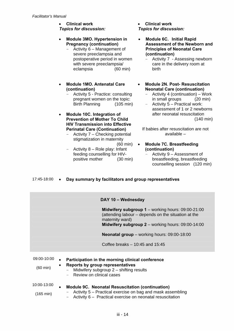
Facilitator’s Manual
iii - 14
• Clinical work Topics for discussion: • Module 3MO. Hypertension in
Pregnancy (continuation) - Activity 6 – Management of
severe preeclampsia and postoperative period in women with severe preeclampsia/ eclampsia (60 min)
• Clinical work Topics for discussion: • Module 6С. Initial Rapid
Assessment of the Newborn and Principles of Neonatal Care (continuation) - Activity 7 - Assessing newborn
care in the delivery room at birth
• Module 1MO. Antenatal Care (continuation) - Activity 5 - Practice: consulting
pregnant women on the topic: Birth Planning (105 min)
• Module 10C. Integration of
Prevention of Mother To Child HIV Transmission into Effective Perinatal Care (Continuation) - Activity 7 – Checking potential
stigmatization in maternity (60 min) - Activity 8 – Role play: Infant
feeding counselling for HIV-positive mother (30 min)
• Module 2N. Post- Resuscitation Neonatal Care (continuation) - Activity 4 (continuation) – Work
in small groups (20 min) - Activity 5 – Practical work:
assessment of 1 or 2 newborns after neonatal resuscitation
(140 min)
If babies after resuscitation are not available –
• Module 7C. Breastfeeding
(continuation) - Activity 9 – Assessment of
breastfeeding, breastfeeding counselling session (120 min)
17:45-18:00 • Day summary by facilitators and group representatives
DAY 10 – Wednesday
Midwifery subgroup 1 – working hours: 09:00-21:00 (attending labour – depends on the situation at the maternity ward) Midwifery subgroup 2 – working hours: 09:00-14:00 Neonatal group – working hours: 09:00-18:00 Coffee breaks – 10:45 and 15:45
09:00-10:00
(60 min)
• Participation in the morning clinical conference • Reports by group representatives
- Midwifery subgroup 2 – shifting results - Review on clinical cases
10:00-13:00
(165 min)
• Module 9C. Neonatal Resuscitation (continuation)
- Activity 5 – Practical exercise on bag and mask assembling - Activity 6 – Practical exercise on neonatal resuscitation

iii - 15
13:00-14:00
Lunch
Midwifery Group
Neonatal Group
• Clinical work Topics for discussion: • Module 8C. Postpartum Care of
Mothers and Newborns (continuation) - Activity 8 – Practical work:
counseling women in postpartum contraception
• Module 4C. Assessment of
Foetal Well-Being During Pregnancy and Labour and Assessment of Small for Gestational Age (SGA) Foetuses (continuation) - Activity 5 – Revision of the
delivery or pregnancy records (60 min)
• Module 11C. Infections in
Pregnancy, Childbirth and Postpartum (continuation) - Activity 6 – Practical work:
hand washing
• Clinical work Topics for discussion: • Module 6С. Initial Rapid
Assessment of the Newborn and Principles of Neonatal Care (continuation) - Activity 7 - Assessing
newborn care in the delivery room at birth
• Module 3N. Breathing Difficulty
(continuation) - Activity 5 – Group work (30 min) - Activity 6 – Practical work:
Oxygen administration for breathing difficulty (30 min)
- Activity 7 – Demonstration of assessment of an infant with breathing difficulty (50 min)
• Module 10C. Integration of
Prevention of Mother To Child HIV Transmission into Effective Perinatal Care (Continuation) - Activity 7 – Checking potential
stigmatization in maternity (60 min) - Activity 8 – Role play: Infant
feeding counselling for HIV-positive mother (30 min)
If babies with breathing difficulty are
not available –
• Module 7C. Breastfeeding (continuation) - Activity 9 – Assessment of
breastfeeding, breastfeeding counselling session (120 min)
• Module 5N. Neonatal Bacterial
Infections (continuation) - Activity 5 – Examination of a
newborn with infection (or a newborn receiving antibiotic therapy) (150 min)
17:45-18:00 • Day summary by facilitators and group representatives

Facilitator’s Manual
iii - 16
DAY 11 – Thursday
Midwifery subgroup 1 – working hours: 09:00-14:00 Midwifery subgroup 2 – working hours: 09:00-21:00 (visiting labour – depends on the situation at the maternity ward) Neonatal group – working hours: 09:00-18:00 Lunch – 13:00 – 14:00 Coffee breaks – 10:45 and 15:45
09:00-10:00
(60 min)
• Participation in the morning clinical conference • Reports by group representatives
- Midwifery subgroup 1 – shifting results - Review on clinical cases
Midwifery Group
Neonatal Group
• Clinical work • Clinical work
Topics for discussion: • Module 7C. Breastfeeding
(continuation) - Activity 9 – Assessment of
breastfeeding, breastfeeding counselling session (120 min)
• Demonstration and discussion
of films: “Caesarean section” “Vacuum extraction of foetus”, “Delivering together”, “Postpartum contraception”
• Module 8C. Postpartum Care of
Mothers and Newborns (continuation) - Activity 8 – Practical work:
counseling women in postpartum contraception
• Module 4C. Assessment of
Foetal Well-Being During Pregnancy and Labour and Assessment of Small for Gestational Age (SGA) Foetuses (continuation) - Activity 5 – Revision of the
delivery or pregnancy records (60 min)
• Module 11C. Infections in
Pregnancy, Childbirth and Postpartum (continuation) - Activity 6 – Practical work:
hand washing
Topics for discussion: • Module 6С. Initial Rapid
Assessment of the Newborn and Principles of Neonatal Care (continuation) - Activity 7 - Assessing
newborn care in the delivery room at birth
• Module 4N. Neonatal Jaundice
(continuation) - Activity 6 – Examination of 2
jaundiced newborns and discussion on these cases
(120 min)

iii - 17
17:45-18:00 • Day summary by facilitators and group representatives
DAY 12 – Friday
09:00-9:45
(45 min)
• Participation in the morning clinical conference • Reports by group representatives
- Midwifery subgroup 2 – shifting results - Review on clinical cases
09:45-10:45
(60 min)
• Module 15C: How to Improve Existing Practices: Strategy of Changes
- Activity 1- Presentation (60 min)
10:45-11:00
Coffee break
10:30-11:30
(60 min)
• Module 15C: How to Improve Existing Practices: Strategy of Changes
(continuation) - Activity 2 - General discussion “Is it necessary to change the existing
system of perinatal care for women and children in maternities?” (60 min)
11:30-13:00
(90 min)
• Module 15C: How to Improve Existing Practices: Strategy of Changes
(continuation) - Activity 3 - Participants develop plan of action (90 min)
13:00-14:00
Lunch
14:00-15:45
• Module 15C: How to Improve Existing Practices: Strategy of Changes
(continuation) - Activity 4 - Presentation of developed plans of action (105 min)
15:45-16:00
Coffee break
16:00-18:00
• Conclusion - Conclusion by facilitators - Feedback of the training participants - Certification - The final word

Facilitator’s Manual
iii - 18
24 Hour Shift Option The 24 hour Shift Option requires flexibility. The priority is actual clinical experience. Every effort is made to review the modules per the outlined schedule. The Course Director will adjust the time and/or order of the modules as needed.
Time
Topic
DAY 8 – Monday TEAM A Midwifery multidisciplinary team 1 – working hours: 09:00 through 08:00am Midwifery multidisciplinary team 2 – working hours: 09:00 through 08:00am TEAM B Midwifery multidisciplinary team 1 – working hours: 09:00 through 13:00am Midwifery multidisciplinary team 2 – working hours: 09:00 through 13:00am Neonatal group – working hours: 09:00-18:00 (The course director will decide if neonatologists and midwifes will be requested to attend birth after 18:00 pm) Lunch – 13:00 – 14:00 Coffee breaks – 10:45 and 15:45
Midwifery Group Neonatal Group 9:00-13:00 (225 min.)
• Module 5C. Management of Normal Labour and Delivery (continuation).
• Module 7N. Low-Birth Weight Baby/ ”Small Baby” Care and Feeding
• Module 6С. Initial Rapid
Assessment of the Newborn and Principles of Neonatal Care (continuation).
• Clinical work Topics for discussion: • Module 3MO. Hypertension in
Pregnancy (continuation). • Module 1MO. Antenatal Care
(continuation).
• Module 7C. Breastfeeding (continuation).
• Demonstration and discussion of films: “Caesarean section”
• Clinical work Topics for discussion: • Module 7N. Low-Birth Weight
Baby/ ”Small Baby” Care and Feeding (continuation).
• Module 6С. Initial Rapid
Assessment of the Newborn and Principles of Neonatal Care (continuation).

iii - 19
• “Vacuum extraction of foetus”, “Delivering together”, “Postpartum contraception”.
• Module 4N. Neonatal Jaundice (continuation).
17:45-18:00
• Day summary by facilitators and group representatives • Review options for night clinical work e.g. continue care for labouring
women, postpartum/breastfeeding rounds, clinical chart reviews.
DAY 9 – Tuesday
TEAM B Midwifery multidisciplinary team 1 – working hours: 08:00 through 08:00am Midwifery multidisciplinary team 2 – working hours: 08:00 through 08:00am Neonatal group – working hours: 09:00-18:00
Lunch – 13:00 – 14:00 Coffee breaks – 10:45 and 15:45
08:00-08:30
Clinical report from Team A to Team B
09:00-10:00
(60 min.)
• Participation in the morning clinical conference • Reports by group representatives
10:00-13:00 (165 min.)
• Module 9C. Neonatal Resuscitation (continuation).
13:00-14:00
Lunch
Midwifery Group
• Clinical work Topics for discussion: • Module 3MO. Hypertension in
Pregnancy (continuation). • Module 1MO. Antenatal Care
(continuation).
Neonatal Group
• Clinical work Topics for discussion: • Module 6С. Initial Rapid
Assessment of the Newborn and Principles of Neonatal Care (continuation).
• Module 2N. Post- Resuscitation
Neonatal Care (continuation).
If babies after resuscitation are not available –

Facilitator’s Manual
iii - 20
• Module 7C. Breastfeeding (continuation).
• Module 3N. Breathing Difficulty
(continuation).
17:45-18:00
• Day summary by facilitators and group representatives • Review options for night clinical work e.g. continue care for labouring
women, postpartum/breastfeeding rounds, clinical chart reviews.
DAY 10 – Wednesday
TEAM A Midwifery multidisciplinary team 1 – working hours: 08:00 through 08:00am Midwifery multidisciplinary team 2 – working hours: 08:00 through 08:00am Neonatal group – working hours: 09:00-18:00
Lunch – 13:00 – 14:00 Coffee breaks – 10:45 and 15:45
08:00-08:30
Clinical report from Team B to Team A
09:00-10:00
(60 min.)
• Participation in the morning clinical conference • Reports by group representatives
10:00-13:00 (165 min.)
• Module 9C. Neonatal Resuscitation (continuation).
13:00-14:00
Lunch
Midwifery Group Neonatal Group • Clinical work
Topics for discussion: • Module 8C. Postpartum Care of
Mothers and Newborns (continuation).
• Module 4C. Assessment of Foetal
Well-Being during Pregnancy and Labour and Assessment of Small for Gestational Age (SGA) Foetuses (continuation).
• Clinical work Topics for discussion: • Module 6С. Initial Rapid
Assessment of the Newborn and Principles of Neonatal Care (continuation).
• Module 5N. Neonatal Bacterial
Infections (continuation).

iii - 21
• Module 11C. Infections in Pregnancy, Childbirth and Postpartum (continuation).
17:45-18:00
• Day summary by facilitators and group representatives • Review options for night clinical work e.g. continue care for labouring
women, postpartum/breastfeeding rounds, clinical chart reviews.
DAY 11 – Thursday
TEAM B Midwifery multidisciplinary team 1 – working hours: 08:00 through 18:00 to 20:00 Midwifery multidisciplinary team 2 – working hours: 08:00 through 18:00 to 20:00 Neonatal group – working hours: 09:00-18:00
Lunch – 13:00 – 14:00 Coffee breaks – 10:45 and 15:45
08:00-08:30
Clinical report from Team A to Team B
09:00-10:00
(60 min.)
• Participation in the morning clinical conference • Reports by group representatives
Midwifery Group
Neonatal Group
• Clinical work
Topics for discussion: • Module 7C. Breastfeeding
(continuation). • Demonstration and discussion of
films: “Caesarean section” “Vacuum extraction of foetus”, “Delivering together”, “Postpartum contraception”.
• Module 8C. Postpartum Care of
Mothers and Newborns (continuation).
• Clinical work Topics for discussion: • Module 4N. Neonatal Jaundice
(continuation). • Module 8C. Postpartum Care of
Mothers and Newborns (continuation).
• Module 6N. Care of a Newborn
with Birth Defect/Congenital Malformation or Birth Trauma (continuation).

Facilitator’s Manual
iii - 22
• Module 4C. Assessment of Foetal Well-Being During Pregnancy and Labour and Assessment of Small for Gestational Age (SGA) Foetuses (continuation).
• Module 11C. Infections in
Pregnancy, Childbirth and Postpartum (continuation).
17:45-18:00
• Day summary by facilitators and group representatives • Assess whether the midwifery multidisciplinary teams will stay to complete
a labour in progress. Report to maternity staff when leaving facility.
DAY 12 – Friday
09:00-9:45 (45 min.)
• Participation in the morning clinical conference • Reports by group representatives
09:45-10:45 (60 min.)
• Module 15C: How to Improve Existing Practices: Strategy of Changes
10:45-11:00
Coffee break
10:30-11:30 (60 min.)
• Module 15C: How to Improve Existing Practices: Strategy of Changes
(continuation).
11:30-13:00 (90 min.)
• Module 15C: How to Improve Existing Practices: Strategy of Changes
(continuation).
13:00-14:00
Lunch
14:00-15:45
• Module 15C: How to Improve Existing Practices: Strategy of Changes
(continuation).
15:45-16:00
Coffee break
16:00-18:00 • Conclusion. Closing of the course.

iii - 23
GENERAL MODULES Модуль 1G
Effective Perinatal Care Opening ceremony

Facilitator’s Manual
iii - 24

iii - 25

Facilitator’s Manual
iii - 26

iii - 27
Модуль 2G
«The Icebreaking»

Facilitator’s Manual
iii - 24

Effective Perinatal Care (EPC)
iii - 25
Module 2G
“The Icebreaker”
Objectives
By the end of this section:
• Participants are expected to know each other closer and get acquainted with the facilitators
• Favourable environment for further training course delivery should be created
Plan and Duration of the Section: Classroom activities – 60 minutes
Activity 1 – “The Icebreaker” 60 min.
Preparations • Make sure that the list of participants and facilitators is available • Make sure that all participants have Participant’s Manuals • Make sure that other facilitators are aware of their responsibilities for the
current section
Materials and Audio-visual Equipment Materials
• Small cards with pair numbers corresponding to the total amount of participants and facilitators
Equipment • Video-projector or OHP • Flipchart • Colour markers

Facilitator’s Manual
iii - 26
Classroom Activities
Activity – “The Icebreaker” • Explain that the goal of this section is to allow all course participants to make a closer
acquaintance with each other and with course facilitators. • Emphasize that this acquaintance is game-like and any initiative is welcome to make
this section more interesting and cheerful. Remember that the main objective of this activity is to establish contact with the participants to create a warm and friendly atmosphere.
• Display slides 2G-1 and 2G -2 which outline the point of this activity. Explain that
several minutes will be allocated to acquaintance, i.e. each participant and facilitator will have to introduce themselves to the group using a lottery method.
• Participants and all free facilitators are offered to draw a “lucky number” which will let
them to find their pair. • When everybody receives a “lucky number”, give each pair 5-7 minutes to talk to each
other and exchange information about themselves. After this time each pair has to introduce themselves to a group. Presenting should be made using a principle “introduce your partner rather than yourself”.
• To shorten presentation time and avoid long stories you may offer to limit presentations
to a certain number of sentences (e.g. ask them to use 4-5 key phrases) or certain number of topics (e.g. topics presented on slide 2G-3).
• Try to make these mutual presentations interesting rather than formal and boring.
Remember that the main objective is to create a favourable environment and atmosphere among course participants.
• After the acquaintance emphasize that many of the present participants have very
valuable experience. Obviously each participant would have something to share or learn.
• Remind the participants to have their nametags on so that everybody can see them and
use in communication. Alternative methods for participants’ acquaintance • If there is no time or at your own will you may try other alternative acquaintance
methods. • For instance, you may offer a short game to “heat up” the participants, help them to
know each other better and feel at ease. • The two suitable games “Acquaintance without words” and “I am…” are described
below.

Effective Perinatal Care (EPC)
iii - 27
Acquaintance without words (Non-verbal acquaintance) Method 1 For a group of 20-30 participants: This game may take up to 30 minutes and requires a flipchart, markers and adhesive tape. Goal: effective communication between people can also occur without use of words. Stages: Break the group in pairs who have to get acquainted without words using any non-verbal communication methods (pictures, signs, poses, signals). One can point to a wedding ring to indicated marital status or simulate running to denote morning runs. Allow 2-3 minutes for a detailed acquaintance so that each participant could guess what his/her partner is trying to inform. At the end ask how accurately have you 1) described yourself, and 2) guessed the meaning of your partner’s signs. Method 2 For a group of 20-30 participants: This game takes 15-20 minutes and requires paper, markers and adhesive tape. Goal: Help the participants to get acquainted with each other Stages: Ask each participant to write his/her name on a sheet of paper ending with “I am…”, followed by six different items. With this sheet attached to their chest participants walk around the room and read the items written below participants’ names. Ask the participants to spend only 30 seconds for each acquaintance. At the end of activity these sheets may be attached to a wall thus creating a Group Gallery (if photographs are available, they can also be attached to these sheets).

Facilitator’s Manual
iii - 28