Embed Size (px)
Citation preview

©FUNPEC-RP www.funpecrp.com.brGenetics and Molecular Research 14 (2): 4102-4112 (2015)
Effects of autologous SCF- and G-CSF-mobilized bone marrow stem cells on hypoxia-inducible factor-1 in rats with ischemia-reperfusion renal injury
L.Y. Bi1, D.A. Zhao1, D.S. Yang1, J.G. Guo1, B. Liang1, R.X. Zhang1,J.L. Zhao1, H.T. Bai2 and S.J. Li1
1Department of Pediatrics, The First Affiliated Hospital of Xinxiang Medical University, Weihui, Henan Province, China2Department of Pediatrics, The First Affiliated Hospital of Xiamen University, Xiamen, Fujian Province, China
Corresponding author: D.S. YangE-mail: [email protected]
Genet. Mol. Res. 14 (2): 4102-4112 (2015)Received January 2, 2014Accepted September 2, 2014Published April 27, 2015DOI http://dx.doi.org/10.4238/2015.April.27.25 ABSTRACT. To explore the mechanism whereby stem cell factor (SCF) and granulocyte colony-stimulating factor (G-CSF) jointly mobilize bone marrow stem cells (BMSCs) and promote kidney repair, male Sprague-Dawley rats were randomly assigned into 4 groups. In the treatment control group, rats were administered SCF (200 μg·kg-1·day-1) and G-CSF (50 μg·kg-1·day-1) for 5 days. In the treatment group, RIRI models were established, and 6 h later, SCF (200 μg·kg-1·day-1) and G-CSF (50 μg·kg-1·day-1) were administered for 5 days. In the model and treatment groups, tubular epithelial cell degeneration and necrosis were noticed, but the extent of repair in the treatment group was significantly better than in the model group. Five days after the operation, renal tissue CD34+ cells significantly increased in the model and treatment groups compared with the control and treatment

4103
©FUNPEC-RP www.funpecrp.com.brGenetics and Molecular Research 14 (2): 4102-4112 (2015)
Hypoxia-inducible factor-1 and renal injury
control groups. HIF-1α, VEGF, and EPO expression in treatment groups increased significantly compared with the other groups. HIF-1α, VEGF, EPO expression in the treatment control group increased significantly compared with the control group. Joint use of SCF and G-CSF increased the number of BMSCs in damaged kidney tissue and reduced the degree of renal tissue damage. BMSCs promote increased HIF-1α expression in renal tissue. Increased kidney tissue HIF-1α and its target gene products VEGF and EPO expression possibly induce SCF and G-CSF to promote acute tubular necrosis repair.
Key words: Bone marrow stem cells; Kidney; Erythropoietin;Ischemia-reperfusion injury; Hypoxia-inducible factor-1α;Vascular endothelial growth factor
INTRODUCTION
Acute tubular necrosis (ATN) is the most common cause of acute renal failure, ac-counting for 75-80% of all acute renal failure cases. Renal ischemia and direct kidney damage caused by renal toxic substances are the major causes of ATN, which results in renal ischemia and hypoxia. Tubular epithelial degeneration and necrosis often appear subsequently, and res-toration of blood flow after ischemia and reperfusion may aggravate this injury (Hutchens et al., 2011). The treatment for clinical ischemia and/or hypoxia depends on the restoration of normal blood flow. Therefore, for renal ischemic-reperfusion injury (RIRI)-associated damage and repair, rational intervention is warranted to shorten treatment course, reduce mortality, and improve prognosis, all of which have important clinical implications. Experiments have confirmed that bone marrow stem cells (BMSCs) can differentiate into renal tubular epithelial cells, mesangial cells, and endothelial cells (Gupta et al., 2006; Masereeuw, 2009). Mobili-zation of autologous BMSCs might be a new therapeutic approach for RIRI, which would involve complex partial factors. During the natural repair process of RIRI, hypoxia-inducible factor-1 (HIF-1) is activated, which can participate in the repair process. HIF-1α is the core factor for initiating the anti-ischemia and hypoxia response. It is the main cytokine to maintain cellular oxygen homeostasis and energy-metabolism balance under hypoxia and reperfusion conditions (Zagorska and Dulak, 2004). Many downstream cytokines such as erythropoietin (EPO), vascular endothelial growth factor (VEGF) and similar gene products can participate in anti-hypoxia injury to promote tissue repair and induce HIF-1α to prevent various forms of renal injury (Tanaka et al., 2005a,b; Bernhardt et al., 2006; Weidemann et al., 2008). In mo-bilizing autologous BMSCs for the treatment of RIRI, whether enhancing HIF-1 expression system could lead to renoprotection and renal tissue repair promotion is not clear. This study was designed to study the influence of BMSC mobilization on HIF-1α system expression and to further explore possible mechanisms of ATN repair by BMSCs.
MATERIAL AND METHODS
Reagents and drugs
Stem cell cytokines (lot No. 20090415) were purchased from Chengdu Di’ao Jiu Hong Pharmaceutical Company, Chengdu, China. Granulocyte colony-stimulating factor (G-

4104
©FUNPEC-RP www.funpecrp.com.brGenetics and Molecular Research 14 (2): 4102-4112 (2015)
L.Y. Bi et al.
CSF) (batch No. 200904Y21) was purchased from North China Pharmaceutical Jintan Bio-technology Co. Ltd., Shijiazhuang, China. Rabbit anti-rat CD34+ monoclonal antibody was purchased from Beijing Biosynthesis Biotechnology Co. Ltd., Beijing, China. Rabbit anti-rat HIF-1α polyclonal antibodies, rabbit anti-rat VEGF polyclonal antibody, rabbit anti-rat EPO polyclonal antibodies, and the SP immunohistochemistry kit were purchased from Wuhan Boster Biological Technology Co. Ltd., Wuhan, China.
Preparation of the animal model
Animal groups and production of the renal ischemia-reperfusion model
One hundred and sixty healthy 13-week-old male Sprague-Dawley rats weighing 250-280 g were obtained from the Experimental Animal Center of Zhengzhou University (experi-mental animal license No. SYXK (Henan) 2009-052). They were kept separately and given free access to food and water. After urine screening to rule out nonhealthy rats, they were randomly divided into 4 groups with 40 rats each. Stem cell factor (SCF) and G-SCF doses were accord-ing to an earlier study (Orlic et al., 2001). Because bone marrow mobilization agents usually cause peripheral blood stem cells to peak 3-5 days after treatment (Martion et al., 2005), the fifth day after intervention was chosen as the starting point. In the control group, normal rat kid-ney tissues were observed. In the model group, a non-invasive arterial clip was used to occlude the left renal artery, and the blood flow was restored 45 min after ischemia-reperfusion. In the treatment group, 6 h after ischemia-reperfusion treatment, SCF (200 μg·kg-1·day-1 and G-CSF (50 μg·kg·day-1) were subcutaneously injected for 5 consecutive days. In the treatment control group, SCF (200 μg·kg-1·day-1) and G-CSF (50 μg·kg-1·day-1) were subcutaneously injected for 5 consecutive days. Rats were preoperatively fasted for 12 h with free access to water. Chloral hydrate (10%, 3.5 mg/kg) intraperitoneal injection was used for anesthesia. The rats were fixed in the prone position, and the left rib skin was prepared and routinely disinfected for exposing the surgical field. A 0.8-1.0-cm incision 1 cm along the left side of the dorsal midline was made from the left rib towards the tail. The renal artery was isolated and occluded with clip to cause renal ischemia for 45 min, after which the clips were released. After the restoration of kidney perfusion, the kidney turned to red color, indicating that the reperfusion was successful. The incision was then sutured, and a normal diet and water were given.
Specimen collection
Eight rats in each group were killed by cervical dislocation 5, 10, 17, 24, and 31 days after the operation. For kidney tissue treatment, the renal capsule of the left kidney was re-moved. After rinsing with saline, 10% paraformaldehyde was used for fixation. Next, dehydra-tion, waxing, embedding, and sectioning were performed for immunohistochemical analysis under light microscopy.
Index detection
Renal tissue morphology
Kidney tissues were fixed, dehydrated, made transparent, waxed, embedded, and sec-

4105
©FUNPEC-RP www.funpecrp.com.brGenetics and Molecular Research 14 (2): 4102-4112 (2015)
Hypoxia-inducible factor-1 and renal injury
tioned for hematoxylin-eosin staining.
Immunohistochemistry
The two-step SP immunohistochemical staining method was used. Paraffin-embed-ded sections (4 μm thick) were subjected to conventional immunohistochemistry after de-waxing and rehydration. Rabbit anti-rat CD34+ monoclonal antibody (1:100), rabbit anti-rat HIF-1α polyclonal antibody (1:100), rabbit anti-rat VEGF polyclonal antibody (1:100), and rabbit anti-rat EPO polyclonal antibody (1:100) were taken as the primary antibodies and incubated at 4°C overnight. After PBS washing, goat anti-rabbit lgG was added as the second antibody and incubated at 37°C for 30 min. The stain appeared after PBS washing. Throughout the experiment, PBS took the place of primary antibody as a control. Brown or brown granular precipitate under light microscopy was the positive stain. The IDA-2000 automatic computer image analysis system was used to quantitatively assess the scope and intensity of antigen expression in each sample: 10 different high-magnification (400X) im-ages were selected, and the integral luminosity was measured. The luminosity mean of 10 different fields of microscope was calculated as the final score.
Statistical analysis
The SPSS18.0 statistical software was used for analysis, and data are reported as means ± standard deviation. ANOVA was used for comparison among multiple groups, and the independent sample t-test was used to compare 2 groups. Correlation analysis was per-formed using linear correlation. P < 0.05 and P < 0.01 were considered to be statistically significant.
RESULTS
Renal pathological changes
Five days after the operation, the brush border epithelial cells sloughed off. The cells were flat, and the luminal cavity was expanded. Epithelial cells exhibited signs of degeneration, swelling, and rupture. The nuclei disappeared, and the lumen was filled with red granular necrosis, and cell casts or protein casts were visible. Interstitial edema and scattered inflammatory cell infiltration appeared. Ten days later, intraluminal necrosis de-creased, and casts were occasionally observed. Proximal tubules began regenerating, and the regenerated epithelial cells were flattened. The nuclei varied in size, arrangement dis-order was common, and interstitial infiltration of inflammatory cells was reduced. Within 17 days, luminal necrotic material decreased, no casts were noticeable, and proximal tubule regeneration was active. Comparison of the treatment group and model group showed no significant difference between them 5 days later (P > 0.05). After 10 and 17 days, the renal tubular injury in the treatment group was significantly lower than that in the model group (P < 0.05), and renal tubules returned to normal in the treatment group after 24 days (Figures 1 and 2).

4106
©FUNPEC-RP www.funpecrp.com.brGenetics and Molecular Research 14 (2): 4102-4112 (2015)
L.Y. Bi et al.
CD34+ expression in renal tissue
CD34+ cells had large, round nuclei with little cytoplasm and deep staining. The cyto-plasm was positively stained brown or brown-yellow. In the model group and treatment group,
Figure 1. Renal patholodical changes after 10 days. HE staining (200X) and CD34+ cells (immunohistochemistry, 400X). A. Renal pathological changes of the model group and the treatment group were that the brush border epithelial cells sloughed off, the cells flatted, the luminal cavity expanded, epithelial cells degenerated, swelled, and ruptured, the nuclei disappeared, the lumen was filled with red granular necrosis, cell casts or protein casts were visible, interstitial edema and scattered inflammatory cell infiltration appeared. Ten days later, intraluminal necrosis decreased, and casts were occasionally observed. Proximal tubules began regenerating, and the regenerated epithelial cells were flattened. The nuclei varied in size, arrangement disorder was common, and interstitial infiltration of inflammatory cells was reduced. Within 17 days, luminal necrotic material decreased, no casts were noticeable, and proximal tubule regeneration was active. B. CD34+ cells had large, round nuclei with little cytoplasm and deep staining. The cytoplasm was positively stained brown or brown-yellow.
Figure 2. HE staining compared with the model group (*P < 0.05). After 10 and 17 days, the renal tubular injury in the treatment group was significantly lower than that in the model group (P < 0.05), and renal tubules returned to normal in the treatment group after 24 days.
A
B

4107
©FUNPEC-RP www.funpecrp.com.brGenetics and Molecular Research 14 (2): 4102-4112 (2015)
Hypoxia-inducible factor-1 and renal injury
the renal tissue CD34+ cells significantly increased after surgery. Compared with the control group and the treatment group, the difference was significant (P < 0.05), CD34+ in the treat-ment group was higher than that in the model group, and the expression levels in both groups gradually decreased over time (Figures 1 and 3).
Figure 3. CD34+ cells, with the control group, the treatment group, compared with the model group (*P < 0.05), with the control group, the treatment group compared to the control group (#P < 0.05). In the model group and treatment group, the renal tissue CD34+ cells significantly increased after surgery. Compared with the control group and the treatment group, the difference was significant (P < 0.05), CD34+ in the treat¬ment group was higher than that in the model group, and the expression levels in both groups gradually decreased over time.
Renal tissue HIF-1α, VEGF, and EPO expression
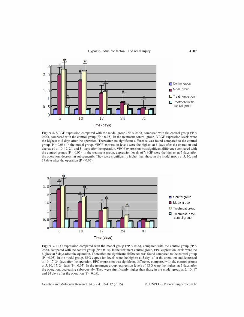
In normal kidneys, HIF-1α expression was mainly in the renal tubular cells and med-ullary collecting duct. Staining occurred mainly in the cytoplasm, and the nucleus has a small expression. VEGF expression was in the renal tubular, renal tubular interstitial, and perivas-cular areas. EPO is mainly expressed in the cytoplasm of renal tubular and tubulointerstitial cells. In the control group, HIF-1α, VEGF, and EPO were weakly and positively expressed, with no significant change. In the treatment control group, HIF-1α, VEGF, and EPO expres-sion levels were the highest 5 days after the operation. Thereafter, no significant differences were found compared to the control group (P > 0.05). In the model group, HIF-1α, VEGF, and EPO expression levels were the highest 5 days after the operation and decreased at 10, 17, 24, and 31 days after the operation. Until 31 days, HIF-1α and VEGF expression still showed sig-nificant differences when compared with the control groups (P < 0.05), while EPO expression showed no significant difference (P > 0.05) when compared with the control group, indicating that the tissue pathological hypoxia state was not yet over. In the treatment group, expression levels of HIF-1α, VEGF, and EPO were the highest 5 days after the operation, decreasing subsequently. No significant difference was found with the control group at 31 days after the operation, but they were significantly higher than those in the model group at 5, 10, and 17 days after the operation (P < 0.05) (Figures 4-7).

4108
©FUNPEC-RP www.funpecrp.com.brGenetics and Molecular Research 14 (2): 4102-4112 (2015)
L.Y. Bi et al.
Figure 5. HIF-1α expression compared with the model group (*P < 0.05), compared with the control group (#P < 0.05), compared with the control group (¥P < 0.05). In the treatment control group, HIF-1α expression levels were the highest at 5 days after the operation. Thereafter, no significant difference was found compared to the control group (P > 0.05). In the model group, HIF-1α expression levels were the highest at 5 days after the operation and decreased at 10, 17, 24, and 31 days after the operation. HIF-1α expression was significant difference compared with the control groups (P < 0.05). In the treatment group, expression levels of HIF-1α were the highest at 5 days after the operation, decreasing subsequently. They were significantly higher than those in the model group at 5, 10, and 17 days after the operation (P < 0.05).
Figure 4. Renal tissue HIF-1α, VEGF, and EPO-1 expression after 10 days (immunohistochemistry, arrows, 400X). In normal kidneys, HIF-1α expression was mainly in the renal tubular cells and med-ullary collecting duct. Staining occurred mainly in the cytoplasm, and the nucleus has a small expression. VEGF expression was in the renal tubular, renal tubular interstitial, and perivascular areas. EPO was mainly expressed in the cytoplasm of renal tubular and tubulointerstitial cells. In the control group, HIF-1α, VEGF, and EPO were weakly and positively expressed.
4109
©FUNPEC-RP www.funpecrp.com.brGenetics and Molecular Research 14 (2): 4102-4112 (2015)
Hypoxia-inducible factor-1 and renal injury
Figure 7. EPO expression compared with the model group (*P < 0.05), compared with the control group (#P < 0.05), compared with the control group (¥P < 0.05). In the treatment control group, EPO expression levels were the highest at 5 days after the operation. Thereafter, no significant difference was found compared to the control group (P > 0.05). In the model group, EPO expression levels were the highest at 5 days after the operation and decreased at 10, 17, 24 days after the operation. EPO expression was significant difference compared with the control groups at 5, 10, 17, 24 days (P < 0.05). In the treatment group, expression levels of EPO were the highest at 5 days after the operation, decreasing subsequently. They were significantly higher than those in the model group at 5, 10, 17 and 24 days after the operation (P < 0.05).
Figure 6. VEGF expression compared with the model group (*P < 0.05), compared with the control group (#P < 0.05), compared with the control group (¥P < 0.05). In the treatment control group, VEGF expression levels were the highest at 5 days after the operation. Thereafter, no significant difference was found compared to the control group (P > 0.05). In the model group, VEGF expression levels were the highest at 5 days after the operation and decreased at 10, 17, 24, and 31 days after the operation. VEGF expression was significant difference compared with the control groups (P < 0.05). In the treatment group, expression levels of VEGF were the highest at 5 days after the operation, decreasing subsequently. They were significantly higher than those in the model group at 5, 10, and 17 days after the operation (P < 0.05).

4110
©FUNPEC-RP www.funpecrp.com.brGenetics and Molecular Research 14 (2): 4102-4112 (2015)
L.Y. Bi et al.
DISCUSSION
In the RIRI process, renal tubular epithelial cell loss and dysfunction are the most important changes. Therefore, after RIRI, the regeneration or replacement of missing or in-jured tubular epithelial cells is needed to maintain the integrity of the tubular structure and to ensure normal function. After ATN, regeneration cells came from kidney inherent cells (Lin et al., 2005; Duffield et al., 2005). BMSCs can differentiate into renal tubular epithelial cells, which can weakly repair renal tubules. Under normal conditions, BMSCs remain in a relatively quiescent state, and their levels in peripheral blood are below detectable levels (Li et al., 2009). However, under certain stimulation signals, the division and differentiation po-tential can be induced to make them enter the cell division cycle and differentiate into target cells. Studies have shown that ischemia and injury of certain body organs can stimulate and considerably improve peripheral blood stem cell concentrations. Our research showed that af-ter acute tubular injury, the number of bone marrow cells in renal tissue increased, suggesting that kidney injury had autologous BMSC mobilization effects, but this mobilization effect was weak. Application of stem cell mobilization agents can further improve peripheral blood stem cell concentration. The BMSC mobilization agent SCF acts on early hematopoietic stem cells and primitive hematopoietic progenitor cells, while G-CSF can act on the late progenitor cells. Orlic et al. (2001) reported that when SCF and G-CSF were used together for the mobilization of stem cells as treatment, they increased the number of peripheral blood stem cells, attaining 250 times the normal levels. In another experiment (Zhang et al., 2007), the combined effects of G-CSF and SCF mobilization were found to be stronger than the effects of either factor used singly. Therefore, in this experiment, we used G-CSF and SCF in conjunction for stem cell mobilization. Our previous study showed that joint use of G-CSF and SCF enabled the peripheral blood mononuclear cell percentage of CD34+ cells to increase up to 10 times. Most researchers treated CD34+ cells as BMSCs and took CD34+ cells as the cell surface markers of BMSC biological activity and clinical application. Our study showed that the renal tissue CD34+ cell count in the model group increased compared with that in the control group, sug-gesting that injury was the necessary condition for stem cell homing to the damaged tissues. The degree of renal injury in the treatment group was lower than that in the model group. However, the number of CD34+ cells in the treatment group increased compared with those in the model group, indicating that stem cell mobilization agents can promote stem cell prolifera-tion and accelerate renal tubular repair.
In addition to the fact that BMSCs can differentiate into kidney cells, they can also secrete growth factors to stimulate renal tubular epithelial cell proliferation. BMSCs can se-crete epidermal growth factor (EGF), insulin-like growth factor-1 (IGF-1), hepatocyte growth factor, and other growth factors, which can promote cell proliferation and can still promote HIF-1α system expression. In the natural state, HIF-1α and its downstream gene products play a renoprotective role by increasing the oxygen supply, improving energy metabolism, promot-ing angiogenesis and remodeling, repair of free radical damage and inflammatory injury, and anti-apoptotic effects. Enhanced expression of HIF-1α and its downstream gene products can increase renal tissue repair effects. HIF-1α siRNA has been used to reduce hypoxia-reperfu-sion renal injury caused by HIF-1α expression aggravation. HIF-1α RNAi has been used to reduce hypoxia-reperfusion renal injury caused by enhanced expression of HIF-1α. Several cytokines such as EGF and IGF-1 activate the PI3K or MAPK signaling pathway after bind-ing with the receptor protein tyrosine kinase under normal circumstances to increase HIF-1α
4111
©FUNPEC-RP www.funpecrp.com.brGenetics and Molecular Research 14 (2): 4102-4112 (2015)
Hypoxia-inducible factor-1 and renal injury
protein synthesis (Semenza, 2003). It was found that the degree of tissue repair had some positive correlation with HIF-1α expression intensity. HIF-1α cytokines VEGF and EPO have an obvious synchronization trend. They also have a synchronization trend with the degree of renal pathological injury, suggesting that HIF-1α plays the central role in anti-injury and repair promotion under hypoxia and reperfusion conditions. In the treatment group, increased expression of HIF-1α was associated with quicker damage repair and increased metabolism. The enhanced expression of HIF-1α can improve the mobilization and recruitment effects of BMSCs by enhancing the activity of the SDF-1/CXCR4 axis (Karshovska et al., 2007), further accelerating tissue repair and enhancing local metabolism. This would increase local oxygen supply and demand and enhance the expression of HIF-1α.
Our experimental results showed that the control group had a weak positive HIF-1α expression compared to the treatment group and the control group. After 5 days, HIF-1α expression significantly increased and was associated with the local effects of the BMSC mobilization agents.
Recent experimental studies (Liu et al., 2010) have confirmed that the bone mar-row inducer G-CSF can increase the expression of HIF-1α under normal circumstances. The increased expression of HIF-1α in the treatment group and the treatment control group was associated with stimulating effects of endocrine secretion of BMSCs and strong effects of lo-cal metabolism. The elevated expression was also associated with renal local direct action of G-CSF. However, the effectiveness of the local effect may be limited.
In summary, the combination of SCF and G-CSF to mobilize BMSCs and treat ATN enhanced the expression of renal tissue HIF-1α and its downstream cytokines VEGF and EPO to play a role in injury resistance and recovery promotion. This experiment revealed the mechanism of BMSC partial mobilization to promote chemical repair of renal tubular tissue and provided a new theoretical basis of stem cell therapy for ATN. However, for this experi-ment, we used stem cell inducer, SCF and G-CSF, and the inducer itself can affect renal tis-sue. Therefore, we cannot enhance the extent of HIF-1α system through the BMSC pathway alone. Further studies are needed to establish the specific mechanism and the extent of action of the two mechanisms through joint use of SCF and G-CSF in the treatment of bone marrow dysfunction and kidney injury.
ACKNOWLEDGMENTS
Research supported by the Public Relations Project of Medical Technology, Henan Province (Project #210-89).
REFERENCES
Bernhardt WM, Campean V, Kany S, Jürgensen JS, et al. (2006). Preconditional activation of hypoxia-inducible factors ameliorates ischemic acute renal failure. J. Am. Soc. Nephrol. 17: 1970-1978.
Duffield JS, Park KM, Hsiao LL, Kelley VR, et al. (2005). Restoration of tubular epithelial cells during repair of the postischemic kidney occurs independently of bone marrow-derived stem cells. J. Clin. Invest. 115: 1743-1755.
Gupta D, Verfaillie C, Chmielewski D, Kren S, et al. (2006). Isolation and characterization of kidney derived stem cell. J. Am. Soc. Nephrol. 17: 3028-3040.
Hutchens MP, Nakano T, Kosaka Y, Dunlap J, et al. (2011). Estrogen is renoprotective via a nonreceptor-dependent mechanism after cardiac arrest in vivo. Anesthesiology 112: 395-405.
Karshovska E, Zernecke A, Sevilmis G, Millet A, et al. (2007). Expression of HIF-1α in injured arteries controls SDF-1α-mediated neointima formation in apolipoprotein E-deficient mice. Arterioscler. Thromb. Vasc. Biol. 27: 2540-2547.

4112
©FUNPEC-RP www.funpecrp.com.brGenetics and Molecular Research 14 (2): 4102-4112 (2015)
L.Y. Bi et al.
Li N, Li XR and Yuan JQ (2009). Effects of bone-marrow mesenchymal stem cells transplanted into vitreous cavity of rat injury by ischemia/reperfusion. Graefes Arch. Clin. Exp. Ophthalmol. 274: 503-14.
Lin F, Moran A and Igarashi P (2005). Intrarenal cells, not bone marrow-derived cells, are the major source for regeneration in post ischemic kidney. J. Clin. Invest. 115: 1756-1764.
Liu SP, Lee SD, Lee HT, Liu DD, et al. (2010). Granulocyte colony-stimulating factor activating HIF-1a acts synergistically with erythropoietin to promote tissue plasticity. PLoS One 5: e10093.
Martion M, Console G, Irrera G, Callea I, et al. (2005). Harvesting peripheral blood progenitor cells from healthy donors: retrospective comparison of filgrastim and lenograstim. J. Clin. Apher. 20: 129-136.
Masereeuw R (2009). Contribution of bone marrow derived cells in renal repair after acute kidney injury. Minerva Urol. Nefrol. 61: 373-384.
Orlic D, Kajstura J, Chimenti S, Limana F, et al. (2001). Mobilized bone marrow cells repair the infracted heart, improving function and survival. Proc. Natl. Acad. Sci. U.S.A. 98: 10344-10349.
Semenza GL (2003). Targeting HIF-1 for cancer therapy. Nat. Rev. Cancer 3: 721-732. Tanaka T, Kojima I, Ohse T, Inagi R, et al. (2005a). Hypoxia-inducible factor modulates tubular cell survival in cisplatin
nephrotoxicity. Am. J. Physiol. Renal Physiol. 289: F1123-F1133.Tanaka T, Matsumoto M, Inagi R, Miyata T, et al. (2005b). Induction of protective genes by cobalt ameliorates
tubulointerstitial injury in the progressive Thy1 nephritis. Kidney Int. 68: 2714-2725.Weidemann A, Bernhardt WM, Klanke B, Daniel C, et al. (2008). HIF activation protects from acute kidney injury. J. Am.
Soc. Nephrol. 19: 486-494.Zagorska A and Dulak J (2004). HIF-1: the knows an unknows of hypoxia sensing. Acta Biochim. Pol. 51: 563-585.Zhang JJ, Yi ZW, Dang XQ, He XJ, et al. (2007). Stem cell factor joint granulocyte colony stimulating factor of unilateral
ureteral obstruction rat bone marrow stem cells and endothelial progenitor cell mobilization. Zhongguo Dang Dai Er Ke Za Zhi 9: 144-148.