Embed Size (px)
Citation preview
Biochemical Pharmacology, Vol. 28, pp, 1885-1891. 0006-2952/79/0615-1885 $02.00/0 © Pergamon Press Ltd., 1979. Print~l in Great Britain.
EFFECTS OF METHOTREXATE ON THYMIDINE TRIPHOSPHATE LEVELS IN CHINESE HAMSTER CELL
CULTURES
SUSANNE KYBURZ*, JEAN-CLAUDE SCHAER and RICHARD SCHINDLERt
Department of Pathology, University of Bern, 3010 Bern, Switzerland
(Received 7 August 1978)
Abstract--Chinese hamster ovary cells were incubated in medium containing hypoxanthine and glycine and supplied with 10 -5 M methotrexate to inhibit the endogenous synthesis of thymidine nucleotides. Within 1- 5 min after addition of the inhibitor, incorporation of [6-3H]deoxyuridine into DNA was as low as 1 to 1.5 per cent of control values, indicating that endogenous synthesis of thymidine nucleotides was blocked rapidly and almost completely. The cellular dTTP content was determined under various culture conditions as a function of time after addition of methotrexate. If the medium used contained undialyzed fetal calf serum, dTTP levels decreased relatively slowly in asynchronous as well as in synchronous S-phase cell populations. Similar results were obtained with asynchronous cultures incubated in medium with dialyzed serum. In contrast, if synchronous cultures consisting predominantly of cells in S phase were incubated in medium containing dialyzed serum and supplied with methotrexate, dTTP levels decreased within 15 min from control values of 15-20 pmoles/106 cells to levels of 1.5 to 3 pmoles/106 cells or lower. The previously reported failure of methotrexate to cause a rapid depletion of cellular dTTP may reflect, therefore, maintenance of thymidine nucleotide pools by cells that are not in the S phase and/or uptake by cells of thymidine present in undialyzed serum used for preparation of culture media.
Methotrexate is a specific inhibitor ofdihydrofolic acid reductase. The depletion of tetrahydrofolic acid deriva- tives induced by this antimetabolite results in impair- ment of 1-carbon transfer reactions which are required for the synthesis of various metabolites. In mammalian cell cultures, the inhibition of cell proliferation by methotrexate was shown to be prevented by the addi- tion of glycine, a purine, and thymidine to the culture medium [ 1, 2]. The addition of methotrexate in combi- nation with glycine and a purine may be expected, therefore, to result in a specific deficiency of thymidine nucleotides mediated by inhibition of the conversion of deoxyuridylate to thymidylate.
Contrary to this expectation, it has been reported that, after addition of methotrexate to HeLa cell cul- tures, the cellular dTTP content remained essentially constant for as long as 16 hr [ 3 ]. In conjunction with observations on the kinetics of[3H]thymidine incorpo- ration into DNA in the presence and absence of metho- trexate, this finding was interpreted as reflecting the existence of two separate cellular pools of thymidine nucleotides synthesized de novo and from exogenous thymidine respectively [ 41. In mouse lymphoma ceils, a 50 per cent reduction of the dTTP pool was observed 1 hr after addition of methotrexate, but almost no further decrease occurred for up to 24 hr [5]. Similarly, after addition of methotrexate to cultures of human lymphocytic cells, the cellular dTTP content decreased within 1 hr by approximately 50 per cent; during fur- ther incubation in the presence of the inhibitor, how- ever, cellular dTTP levels decreased at a rather slow
* Present address: Schweizerisches Serum- and Impfinsti- tut, Rehhagstrasse 79, 3018 Bern, Switzerland.
t To whom reprint requests should be addressed.
rate, and 7 hr was required before 10 per cent of the control value was attained [6].
Amounts of dTTP within a cell are relatively small. For Chinese hamster ovary (CHO) cell cultures, it was shown that the cellular dTTP pool is sufficient to support DNA synthesis for approximately 5 rain [7]. This supports the assumption of a rapid turnover of dTTP and is in contrast to the observations of a slow and/or limited decrease of cellular dTTP after inhibi- tion of endogenous thymidine nucleotide synthesis by methotrexate.
The present work was undertaken to study various factors which might affect the extent and time course of changes in cellular dTTP content after addition of methotrexate to CHO cell cultures. It will be shown that, under appropriate experimental conditions, a rapid and nearly complete depletion of intracellular dTTP after addition of methotrexate may be observed.
MATERIALS AND METHODS
Cell line and culture techniques. Chinese hamster ovary cells [8] were grown as monolayer cultures in Eagle's minimal essential medium {9[ supplemented with L-alanine, L-asparagine, L-aspartic acid, L-glu- tamic acid, L-proline, L-serine, glycine (all at 10 -4 M ) ,
penicillin (50 units/ml), streptomycin (50#g/ml), aureomycin (50#g/ml), and 15% fetal calf serum (FCS). In one series of experiments, medium contain- ing 15% dialyzed fetal calf serum was used. The serum was dialyzed at 4 ° for at least 24 hr against 10-15 vol. saline, and the saline was changed four times during dialysis. Cultures were tested frequently and found to be free of mycoplasma contamination (tests were kindly performed by G. Kronauer).
1885

1886 S. KYBURZ, J.-C. SCHAER and R. SCH1NDLER
For experiments with asynchronous cultures, ap- proximately 3 × 10⁵ cells in 2 ml medium were incu- bated in Petri dishes (3 cm diameter, Nunc, Denmark) overnight at 37 ° in an atmosphere of 5% CO 2 and 95% air. For subsequent studies of the effects of methotrex- ate on deoxyuridine incorporation or on cellular dTTP content, the dishes were placed on the surface of a 37 ° water bath, and the medium was replaced by pre- warmed fresh medium supplemented with 3 × 10 -5 M hypoxanthine and 20 mM 4-(2-hydroxyethyl)-1-pipera- zinc-ethane sulfonic acid (HEPES) buffer (pH 7.2) and containing 15% dialyzed or undialyzed FCS. The water bath was covered with a plastic foil, and the atmosphere above the water surface was saturated with a mixture of 5% CO 2 and 95% air to maintain the pH in the culture medium. At the time of the experiments, two Petri dishes were used for determination of cell num- bers. Cells were removed from the dishes by trypsiniza- tion and counted with a hemocytometer.
Synchronous cell populations were obtained by se- lectively detaching mitotic cells from non-confluent monolayer cultures [ 10, 11 ]. Mitotic ceils were kept at 0 ° for up to 3 hr, and during this time the procedure of detaching mitotic cells was repeated until sufficient cells had been collected [ 12]. After centrifugation, the mitotic cells were suspended, at a density of approxi- mately 1.8 × l0 s cells/ml, in conditioned medium con- taining 3 × 10 -5 M hypoxanthine and 10 mM HEPES buffer (pH 7.2), and 2 ml of this suspension was pipet- ted into a series of Petri dishes and incubated at 37 ° in an atmosphere of 5% CO 2 and 95% air.
Determination of relative numbers of DNA-synthes- izing cells. Cells were incubated for 15 min with [3H]thymidine (1/tCi/ml of medium), removed from the Petri dishes, fixed with a mixture of ethanol and acetic acid (3: 1), and the fixed cells were brought to microscope slides. For autoradiography, the slides were covered with Kodak NTB-2 emulsion. After the appropriate exposure time, the preparations were devel- oped and stained with Giemsa. For determination of the percentage of labeled cells, 500 cells were scored per preparation.
Chemicals. [~4C]Thymidine (57 mCi/m-mole), [3H]thymidine (20Ci/m-mole), [6-3H]deoxyuridine ( 14 Ci/m-mole), [ 2-1"C ]deoxyuridine (58 mCi/m- mole), and [3H]dATP (15-29 Ci/m-mole) were pur- chased from the Radiochemical Centre, Amersham, Bucks, England while [ 5- ~H ]deoxyuridine ( 12 C i/m- mole) was obtained from ICN Pharmaceuticals Inc., Irvine, CA. Methotrexate was kindly supplied by Cy- anamid GmbH, Miinchen, West Germany. Unlabeled deoxyribonucleoside triphosphates were obtained from Serva, Heidelberg, West Germany. These nucleotides were dissolved in 0.02 M Tris-HCl buffer (pH 7.5), and their concentrations were determined by optical density measurements (at 267 nm for dTTP and at 259nm for dATP). DNA polymerase (EC2.7.7.7) from Micrococcus luteus was obtained from Miles Lab- oratories, Slough, England, with specific activity of approximately 600 units/mg of protein, and diluted to 200 units/ml with 0.05 M potassium phosphate buffer (pH 6.9) containing 0.01 M 2-mercaptoethanol and 30% ethylene glycol. Poly [d(A-T)] • [d(A-T)] was purchased from Boehringer, Mannheim, West Ger- many and dissolved in 0.01 M Tris-HC1 (pH 7.5).
Determination of[ 6-~H ldeoxyuridine incorporation
into DNA. These incorporation studies were performed as double labeling experiments. After incubation of cells in Petri dishes overnight with [~Clthymidine (10 -3 #Ci/ml), the medium was replaced by fresh me- dium containing hypoxanthine and HEPES buffer, and 2 hr later the cultures were transferred to the 37 ° water bath and supplied with 10 -5 M methotrexate. At var- ious times after addition of the inhibitor, 16- 3H]deoxyuridine (final concn: 1 #Ci/ml, 4 × 10 7M) was added, and incubation with the labeled precursor was continued for 15 min. The cells were lysed with 0.5 ml of 1 N NaOH. and cellular RNA was hydro- lyzed by incubation during 60 min at 37 °. After addi- tion of 200/ag of calf thymus DNA, the acid-insoluble material was precipitated with 1 ml of 1.5 N HCI con taining 6% NaaP20 7 and brought to Whatman GF/C filters. The filters were washed with a solution of 0.1 N HCl containing 0.5% Na4P:O 7 and dehydrated with ethanol and acetone. Radioactivities attributable to t4C and 3H were measured in a Packard scintillation spectrometer.
Determination of relative incorporation of deoxyuri dine into dTTP and dUTP. Replicate cultures were incubated with medium containing hypoxanthine, HEPES buffer and 15% dialyzed FCS. After 45 min, one group of cultures was supplied with 10 -5 M metho- trexate and 15 min later a mixture of[5- 3H ]deoxyuridine and [ 2-14C ]deoxyuridine (final concn: 6#Ci/ml and 0.1pCi/ml respectively) was added. After further incubation for 15 or 60 min, the cultures were washed with an ice-cold solution of 0. 15 M NaC1 and 0.015 M sodium citrate, 2 ml of cold (approximately - 2 0 ° ) 66% ethanol was added, and the Petri dishes were maintained at - 2 0 ° for 5-20 hr. Subsequently, the extracts were centrifuged (3200 rev/ min, 0°), and the clear supernatant fraction was de canted, lyophilized, and stored at - 2 0 °. For separation of nucleotides by thin-layer chromatography I 13], the lyophilized extracts were dissolved in 0.1 ml of 60% methanol, and aliquots were applied to PEI-cellulose plates in addition to a marker solution containing unla- beled deoxyuridine, thymidine, dTMP, dTDP and dTTP. The plates were developed with 50% methanol. followed by 0.38 M LiCI in 1 N formic acid-sodium formate buffer (pH 2.0) without intermediate drying. The spot containing dUTP and dTTP was scraped off, and after elution with 2 ml of 0.02 M Tris-HCl buffer (pH 6.5) containing 0.7 M MgCI 2, 18 ml ofa xylene- Triton X-100 mixture was added, and radioactivities attributable to 3H and ~4C were determined. While 12- ~4C ]deoxyuridine is a precursor for both dUTP and dTTP, [5-3H]deoxyuridine may be assumed to be in- corporated into dUTP only, since the tritium atom is removed during dTMP synthesis.
Extraction and assay of thymidine triphosphate. Cultures containing 6-9 × 10⁵ cells were washed with an ice-cold solution of 0 .15MNaCI and 0.015M sodium citrate, extracted with cold 66% ethanol, and the extracts were lyophilized as described above. For determination of dTTP, the lyophilized extracts were dissolved in 0.1 ml of 0.02M Tris-HCl buffer (pH 7.5), and 0.01 ml was used for the assay. More than 90 per cent of cellular dTTP was recovered by this extraction procedure, and the extent of hydrolysis of dTTP during extraction and preparation of extracts for the assay was below 10 per cent.

[ 8
4 o x
(D O < PC
THYMIDINE TRIPHOSPHATE, pm0tes/01 ml
Effects of methotrexate on cellular dTTP 1887
Fig. 1. Incorporation of[3H]dATP into acid-insoluble mate- rial as a function of dTI'P concentration in a reaction mixture containing DNA polymerase and poly [d(A-T) • d(A-T)] un- der standard assay conditions. Bars indicate ranges of dupli- cate measurements, unless these were smaller than diameter of
circles.
Table 1. Effect of methotrexate on incorporation of [6- 3H]deoxyuridine (1 ~Ci/ml of medium, 4 x 10 -7 M) into
DNA *
Time after addition of methotrexate Incorporationt
(min) (% of control)
1 3.3 + 1.2 5 1.1+0.2
10 1.4 _+ 0.2 30 1.0 + 0.2
* At various times after addition of methotrexate ( 10 -5 M) to asynchronous CHO cultures, the cells were incubated with the labeled precursor for 15 min.
1 Means of results obtained in two experiments each, with ranges.
RESULTS
Effect of methotrexate on incorporation of [6- ~H ]deoxyuridine into DNA. To asynchronous cultures containing hypoxanthine, methotrexate was added to obtain a concentration of 10 -5 M. At different times after the addition of the inhibitor, cultures were sup- plied with [6-3Hldeoxyuridine, and incubation was stopped 15 min later to determine incorporation of the precursor into DNA. The results are presented in Table 1. If deoxyuridine was added 1 min after methotrexate, incorporation during the subsequent 15-min period was less than 5 per cent of the control value, and addition of deoxyuridine from 5 to 30 min after methotrexate re- suited in an incorporation which was approximately 1
To determine dTTP, the method of Lindberg and Skoog [ 14] was used. The standard reaction mixture (usually 0.1 ml) had the following composition: 60 mM Tris-HC1 buffer (pH 7.5); 2 mM MgCI2; 0.3 mM 2-mercaptoethanol; [3H]dATP, 1-5 #Ci/mi, 10 -6 M; poly [d(A-T) • d(A-T)], 4.5 to 23 × 10 -3 E260 units; D N A polymerase, 7 units/ml; and 1 to 50 × 10 -9 M dTTP (for obtaining reference curve) or cell extract respectively. This mixture was incubated at 37 ° for 60 min. To stop the reaction, the tubes were placed in an ice-water bath, and 2 ml of ice-cold 0.2 N NaOH containing 200 #g/ml of calf thymus D N A was added. Subsequently, the acid-insoluble ma- terials were precipitated with 1 ml of 1.5 N HCI con- taining 6% Na4P2OT, quantitatively transferred to Whatman G F / C filters, and processed for liquid scintil- lation counting as described above for cellular DNA. For calibration, a standard curve was established with known amounts of dTTP in parallel with each assay.
As shown in Fig. 1, incorporation of radioactivity increased in a linear fashion with dTTP concentrations up to 5 pmoles/0.1 ml of reaction mixture, and amounts of dTTP as small as 0.1 pmole, which corre- sponds to approximately 1.3 pmoles/106 cells, were detectable.
If dUTP was added to the reaction mixture in place of dTTP, essentially the same concentration-dependent incorporation of PH]dATP into acid-insoluble mate- rial was observed. This indicates that the assay had a limited specificity and did not permit us to distinguish between dTTP and dUTP.
!
100'
80
60
40
. . J tlA c) 20 C3
N kU m
TIME AFTER HOURS
l # T T
"7 ; 9 1'0 1'1 17 1'3 INCUBATION OF MITOTIC CELLS,
Fig. 2. Synchrony of cell populations obtained by collection of mitotic ceils, as evaluated by time-dependent variations of the labeling index. At 7.25 hr (solid arrow), the medium was replaced by fresh medium containing 15% undialyzed FCS (O O) or 15% dialyzed FCS ([] [~). The broken line arrow indicates the time of addition of methotrexate ( 10 -5 M) to cultures used for determination of dTTP content. Symbols represent means of values obtained in three inde- pendent experiments each, with standard errors, unless these
were smaller than the diameter of the symbols.

1888 S. KYBURZ, J.-C. SCHAER and R. SCHINDLER
per cent of that observed in the absence of the inhibitor. These results support the conclusion that inhibition of endogenous synthesis of thymidine nucleotides was nearly complete at time intervals as short as I -5 rain after addition of methotrexate at the concentration of 10 -5 M.
Characterization of asynchronous and synchronous cell populations. In asynchronous cultures, relative numbers of DNA-synthesizing cells, as determined by autoradiography, were 63-75 per cent.
To characterize the synchrony of cell populations obtained by mechanical detachment of mitotic cells, cultures were labeled for 15 min with [3H]thymidine at different times after reincubation of mitotic cells. If the labeled precursor was added 30 rain after reineubation, less than 3 per cent of the total cell population was found to be labeled. Variations of the labeling index from 7 to 13 hr after reincubation of mitotic cells are illustrated in Fig. 2. It is seen that at 8-9 hr, labeling indices were 94-97 per cent. In order to study the effects of methotrexate on as pure S-phase cell popula- tions as possible, the following experimental design was chosen. At 7.25 hr after incubation of mitotic cells (solid arrow in Fig. 2), the medium was replaced by fresh medium containing hypoxanthine and HEPES buffer. In one series of experiments, this medium con- tained 15% undialyzed FCS, while in a second series of experiments, the medium contained 15% dialyzed FCS. At 8 hr, as indicated by the broken line arrow, methotrexate was added to all cultures with the excep- tion of those used for subsequent determinations of labeling indices. Changes with time of cellular dTTP content were studied during the 60-rain period after addition of the inhibitor. As seen in Fig. 2, at times exceeding 9 hr after incubation of mitotic cells, a de- crease of relative numbers of labeled cells was observed.
40
$
5 30
E o ° 20
o
I - -
T
0 15 30 45 60 Time after addition of methofrexafe, min
Fig. 3. Effect of methotrexate on cellular dTTP content of asynchronous cultures (-~ O) and S-phase cell popula tions ( © ~ ) incubated in medium containing 15% undi- alyzed FCS. Symbols represent means of values obtained in three independent experiments each, with standard errors.
In medium with 15% dialyzed FCS, this decrease occurred more slowly than in medium containing 15% undialyzed FCS. This supports the assumption that the duration of the S phase was longer in cell populations that had been transferred to medium containing di- alyzed serum.
Effects of methotrexate on cellular dTTP content, In a first series of experiments, medium containing 15% undialyzed FCS was used. At the time of transfer to the water bath, asynchronous or synchronous cultures were supplied with prewarmed fresh medium contain- ing hypoxanthine and HEPES buffer. Methotrexate was added 45rain later to obtain a concentration of 10 -5 M, and at various times, two cultures each were used for determination of cellular dTTP content. The results of three experiments with asynchronous and three experiments with synchronous cultures are pre- sented in Fig. 3. In asynchronous cell populations, the dTTP content decreased by a factor of two within 15 rain after addition of the inhibitor. During further incubation, dTTP levels continued to decrease, al- though at a slower rate, and at 60 rain, cellular dTTP content was, on the average, still 6-7 pmoles/106 cells.
Similar results were obtained if methotrexate was added to synchronous cell populations consisting pre- dominantly of S-phase cells, i.e. at 8 hr after incubation of mitotic cells (broken line arrow in Fig. 2). The initial dTTP content of S-phase cells was rather variable. This may be atributed to small differences in cell cycle progression between individual experiments and/or to different serum batches used in the three experiments. On the average, somewhat smaller initial dTTP levels were observed in S-phase cells, as compared to those in asynchronous cell populations. Within the first 15 rain
30
§ o~ o
8 10
l o
E
F-
0 15 30 ~,5 60 Time after addition of methotrexafe, rnin
Fig. 4. Effect of methotrexate on cellular dTTP content of asynchronous cultures ( O - - O ) and S-phase cell popula tions (open symbols) incubated in medium containing 15% dialyzed FCS. Filled circles represent means of values ob mined in three independent experiments, with standard errors; open symbols represent values obtained in three individual
exper iments.
Effects of methotrexate on cellular dTTP 1889
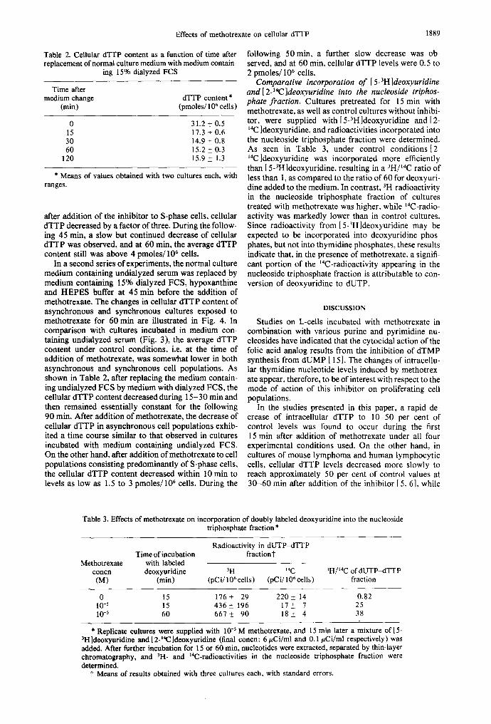
Table 2. Cellular dTTP content as a function of time after replacement of normal culture medium with medium contain-
ing 15% dialyzed FCS
Time after medium change dTTP content *
(min) (pmoles/106 cells)
0 31.2 + 0.5 15 17.3 +_ 0.6 30 14.9 + 0.8 60 15.2 + 0.3
120 15.9 _+ 1.3
* Means of values obtained with two cultures each, with ranges.
after addition of the inhibitor to S-phase cells, cellular dTTP decreased by a factor of three. During the follow- ing 45 rain, a slow but continued decrease of cellular dTTP was observed, and at 60 min, the average dTTP content still was above 4 pmoles/106 cells.
In a second series of experiments, the normal culture medium containing undialyzed serum was replaced by medium containing 15% dialyzed FCS, hypoxanthine and HEPES buffer at 45 min before the addition of methotrexate. The changes in cellular dTTP content of asynchronous and synchronous cultures exposed to methotrexate for 60 min are illustrated in Fig. 4. In comparison with cultures incubated in medium con- mining undialyzed serum (Fig. 3), the average dTTP content under control conditions, i.e. at the time of addition of methotrexate, was somewhat lower in both asynchronous and synchronous cell populations. As shown in Table 2, after replacing the medium contain- ing undialyzed FCS by medium with dialyzed FCS, the cellular dTTP content decreased during 15-30 min and then remained essentially constant for the following 90 min. After addition of methotrexate, the decrease of cellular dTTP in asynchronous cell populations exhib- ited a time course similar to that observed in cultures incubated with medium containing undialyzed FCS. On the other hand, after addition of methotrexate to cell populations consisting predominantly of S-phase cells, the cellular dTTP content decreased within I0 min to levels as low as 1.5 to 3 pmoles/106 cells. During the
following 50 min, a further slow decrease was ob- served, and at 60 min, cellular dTTP levels were 0.5 to 2 pmoles/106 cells.
Comparative incorporation of [ 5-3H ]deoxyuridine and [ 2-14C ]deoxyuridine into the nucleoside triphos- phate fraction. Cultures pretreated for 15 min with methotrexate, as well as control cultures without inhibi- tor, were supplied with [5-3H]deoxyuridine and [2- 14C ]deoxyuridine, and radioactivities incorporated into the nucleoside triphosphate fraction were determined. As seen in Table 3, under control conditions [2- 14C]deoxyuridine was incorporated more efficiently than [5-3H ]deoxyuridine, resulting in a 3H/~4C ratio of less than 1, as compared to the ratio of 60 for deoxyuri- dine added to the medium. In contrast, 3H-radioactivity in the nucleoside triphosphate fraction of cultures treated with methotrexate was higher, while ~4C-radio- activity was markedly lower than in control cultures. Since radioactivity from [5-3H]deoxyuridine may be expected to be incorporated into deoxyuridine phos- phates, but not into thymidine phosphates, these results indicate that, in the presence of methotrexate, a signifi- cant portion of the ~4C-radioactivity appearing in the nucleoside triphosphate fraction is attributable to con- version of deoxyuridine to dUTP.
DISCUSSION
Studies on L-cells incubated with methotrexate in combination with various purine and pyrimidine nu- cleosides have indicated that the cytocidal action of the folic acid analog results from the inhibition of dTMP synthesis from dUMP [ 15 ]. The changes of intracellu- lar thymidine nucleotide levels induced by methotrex- ate appear, therefore, to be of interest with respect to the mode of action of this inhibitor on proliferating cell populations.
In the studies presented in this paper, a rapid de- crease of intracellular dTTP to 10-50 per cent of control levels was found to occur during the first 15 min after addition of methotrexate under all four experimental conditions used. On the other hand, in cultures of mouse lymphoma and human lymphocytic cells, cellular dTTP levels decreased more slowly to reach approximately 50 per cent of control values at 30-60 min after addition of the inhibitor [ 5, 6], while
Table 3. Effects of methotrexate on incorporation of doubly labeled deoxyuridine into the nucleoside triphosphate fraction *
Radioactivity in dUTP~ITTP Time of incubation fractiont
Methotrexate with labeled concn deoxyuridine 3H 14C 3H/tac of dUTP-dTTP (M) (rain) (pCi/106 cells) (pCi/106 cells) fraction
0 15 176+ 29 220+ 14 0.82 10 -5 15 436 + 196 17 +_ 7 25 10 -5 60 667+_ 90 18+ 4 38
* Replicate cultures were supplied with 10 -5 M methotrexate, and 15 rain later a mixture of l 5- 3H]deoxyuridine and [2-14C]deoxyuridine (final concn: 6 #Ci/ml and 0.1 #Ci/ml respectively) was added. After further incubation for 15 or 60 rain, nucleotides were extracted, separated by thin-layer chromatography, and 3H- and ~4C-radioactivities in the nucleoside triphosphate fraction were determined.
t Means of results obtained with three cultures each, with standard errors.

1890 S. KYBURZ, J.-C. SCHAER and R. SCHINDLER
the dTTP content of HeLa cells treated with methotrex- ate was reported to remain essentially constant [ 3 ]. The comparatively high initial rate of depletion of cellular dTTP observed for CHO cells may be attributable to the cell cycle characteristics of the cell line. In asyn- chronous cultures, 63-75 per cent of the cell popula- tion was found to be in S phase. Alternatively, the rapid initial decrease of cellular dTTP described in the pres- ent report may be due to a more rapid onset of action of methotrexate which was used at a concentration of 10 -5 M, as compared to 10 -6 M [3, 5] or 5 × 10 -7 M 16]. Even at 10 -5 M, the effect of metho- trexate on endogenous synthesis of thymidine nucleo- tides was specific. This was demonstrated by compar- ing cell cycle characteristics, including the duration of the S phase, of CHO cells in the presence of 10 -~ M methotrexate, hypoxanthine, thymidine and glycine with those in the absence of the inhibitor 116], as shown previously for P-815 murine mastoeytoma cul- tures [ 17 ].
As seen in Table 1, within 5 min after addition of methotrexate at a concentration of 10 -5 M, incorpora- tion of [6-3H]deoxyuridine into DNA decreased to ap- proximately 1 per cent control values, indicating that endogenous synthesis of thymidine nucleotides was blocked rapidly and almost completely. Furthermore, it was shown previously that, in CHO cells, the size of the dTTP pool is sufficient to support DNA synthesis for a time period of less than 6 min[ 7]. On the other hand, in three of the four series of experiments that are summa- rized in Figs. 3 and 4, the depletion of cellular dTTP observed during the first 15 min after the addition of methotrexate was rather incomplete, and substantial amounts of dTTP were detected after incubation with the inhibitor for as long as 30-60 min. A rapid decrease of cellular dTTP content to very low levels was ob- served, however, when synchronous S-phase cell popu- lations in medium containing dialyzed serum were supplied with methotrexate.
These findings may be interpreted as follows. In asynchronous cell populations, cells not engaged in DNA synthesis also contain dTTP. As reported for CHO cells [7, 18] and HeLa cells [ 19], dTTP levels during the G2 phase of the cell cycle were even higher than those in DNA-synthesizing cells. The results pre- sented in Fig. 4 favor the assumption that rates of turnover of dTTP in cells not engaged in DNA synthe- sis were considerably lower than those of cells in S phase.
The results also demonstrate that, if methotrexate was added to S-phase cell populations cultured in me- dium with undialyzed serum (Fig. 3), cellular dTTP levels decreased more slowly than in S-phase cells incubated in medium with dialyzed serum (Fig. 4). This suggests that, in the presence of undialyzed serum, the cells had the possibility of synthesizing small amounts of dTTP from exogenous thymidine. In fact, it was shown that FCS may contain thymidine at concentra- tions of 8 to 20 x 10 -7 M, depending on the serum batch [20]. In medium containing undialyzed FCS, sufficient thymidine may, therefore, be present to sus- tain cellular synthesis of thymidine nucleotides at sub- stantial rates. Despite the essentially complete inhibi- tion of endogenous synthesis, appreciable intracellular dTTP levels may thus be observed. It should be noted that, in previous studies of the effects of methotrexate
on cellular dTTP content, culture media containing undialyzed bovine serum [3] or fetal calf serum [5, 61 were used. On the other hand, methotrexate was shown to inhibit the proliferation of chicken fibroblasts in medium containing chicken plasma, but not in medium containing serum with a thymidine content more than ten times higher than that of plasma [21 ].
As seen in Fig. 4, although the addition of metho- trexate to S-phase cells in medium with dialyzed serum resulted in a rapid decrease of cellular dTTP, low levels of up to 3 pmoles/106 cells were observed for at least 30--60 min. This residual dTTP content may be attrib- uted, at least in part, to the presence of 3-6 per cent of cells not engaged in DNA synthesis in the synchronous cell populations used. Furthermore, possibilities such as incomplete inhibition of the endogenous synthesis of thymidine nucleotides by methotrexate, and/or incom- plete removal of thymidine from the serum by dialysis, should be considered. In fact, it was reported that incorporation of labeled deoxycytidine into DNA oc- curred at a reduced rate for several hours after addition of methotrexate to human lymphoblasts cultured in medium with dialyzed serum [22]. On the other hand, the results obtained on the incorporation of l5- 3H]deoxyuridine and [2-~4C ]deoxyuridine into the nu- cleoside triphosphate fraction suggest that cells treated with methotrexate may contain significant amounts of dUTP. Due to the limited specificity of the dTTP assay, the residual dTTP levels observed following the first 15 rain after the addition of methotrexate to S-phase cells (Fig. 4) thus may represent, at least in part, cellular dUTP.
In conclusion, failure of methotrexate to cause a rapid depletion of cellular dTTP may reflect mainte- nance of thymidine nucleotide pools by cells that are not in S phase and/or uptake by cells of thymidine present in the culture medium. Under appropriate ex- perimental conditions, a decrease of cellular dTTP to near-zero levels was observed within time periods as short as 10-15 min after addition of the inhibitor. Thus, the results presented do not support the assump- tion of two separate dTTP pools, and are compatible with the hypothesis that the cytocidal action of metho- trexate is attributable to depletion of cellular dTTP as substrate for DNA replication.
Acknowledgements--This work was supported by the Swiss National Science Foundation and by the "Stipendienfond zur Unterstfitzung von Doktoranden auf dem Gebiete der Chemie."
REFERENCES
1. M. T. Hakala and E. Taylor, J. bioL Chem. 234, 126 (1959).
2. R. Schindler, N. Odartchenko, L. Ramseier and A. Grieder, Eur. J. Cancer 3, 349 (1967).
3. C. N. Baumunk and D. L. Friedman. Cancer Res. 31, 1930 (1971).
4. D. Kuebbing and R. Werner, Proc. natn. Acad. Sci. U.S.A. 72, 3333 (1975).
5. M. H. N. Tattersall and K. R. Harrap, Cancer Res. 33, 3086 (1973).
6. A. Fridland, Cancer Res. 34, 1883 (1974). 7. R. A. Waiters, R. A. Tobey and R. L. Ratliff, Biochim.
biophys. Acta 319, 336 (1973). 8. T. T. Puck, S. J. Cieciura and A. Robinson, J. exp. Med.
108, 945 (1958).

Effects of methotrexate on cellular dTTP 1891
9. H. Eagle, Science 130, 432 (1959). 10. T. Terasima and L. J. Tolmach, Expl. Cell Res. 30, 344
(1963). 11. R. A. Tobey, E. C. Anderson and D. F. Petersen, J. cell.
Physiol, 70, 63 (1967). 12. H. Nagasawa and W. C. Dewey, J. cell. Physiol. 80, 89
(1972). 13. K. Randerath and E, Randerath, J. Chromat. 16, 111
(1964). 14. U. Lindberg and L. Skoog, Analyt. Biochem. 34, 152
(1970). 15. J. Borsa and G. F. Whitmore, Cancer Res. 29, 737
(1969).
16. G. G. Miller, Ph.D. Thesis, University of Bern (1975). 17. M. P. Siegers, J. C. Schaer, H. Hirsiger and R. Schindler,
J. Cell Biol. 62, 305 (1974). 18. K. L. Skoog, B. A. Nordenskj/51d and K. G. Bjursell, Eur.
J. Biochem. 33, 428 (1973). 19. G. Bray and T. P. Brent, Biochim. biophys. Acta 269, 184
(1972). 20. J. C. Schaer, U. Maurer and R. Schindler, Expl. CellBiol.
46~ 1 (1978). 21. R. S. Mitchell, S. D. Balk, O. Frank, H. Baker and M. J.
Christine, Cancer Res. 35, 2613 (1975). 22. A. Fridland and T. P. Brent, Eur. J. Biochem. 57, 379
(1975).